Downregulation of lymphocyte mitogenesis by breast cancer-associated p43
7
CANCER ELSEVIER Cancer Letters 82 (1994) 105-l I I LETTERS Downregulation of lymphocyte mitogenesis by breast cancer-associated p43 Harald FL Rosen*a, Christoph Auscha, Georg Reinera, Marta Reinerovab, Jurai Svecb, Heinz Tiichler’, Rudolf Schiessel”, Chaya Morozd “Department of Surgery, Danube Hospital, SMZ-OST, Langobardenstrasse 122, A-1220 Vienna, Austria bSlovak Academy of Sciences, University Bratislava, Bratislava. Slovak Republic cLudwig Boll:mann Research Institute for Oncology, Vienna, Austria dDepartment of Immunology. Beilinson Medical Center, Petah Tikva, Israel Received 22 April 1994: accepted 12 May 1994 Abstract Placental isoferritin-associated p43 has proven to induce immune suppression during pregnancy in order to avoid rejection of the fetus’ alloantigens by maternal lymphocytes. It has been demonstrated previously that p43 is also syn- thesized by breast cancer cells and can also be found on the surface of a subpopulation of T cells in women with this disease. Therefore, it was the aim of the present study to investigate if breast cancer-associated p43 has immunosup- pressive properties. In 40 women undergoing surgical excision of a suspicious lump in their breast, blood was withdrawn and lymphocytes were isolated. Lymphocyte cultures were incubated with p43 antigen and anti-p43 anti- body (CM-H-9). In a second series, lymphocyte mitogenesis was activated by addition of concanavalin A (Con A), Con A + p43 and Con A + anti-p43, respectively. While lymphocytes of breast cancer patients (n = 21) and women with benign breast disease (n = 19) incubated with p43 as well as with anti-p43 antibody did not show any difference in terms of incorporation of [3H]thymidine, activation of lymphocytes by addition of Con A was significantly inhibited after addition of p43 antigen in breast cancer patients compared to women with benign breast disease (P = 0.0178). Analysis of prognostic factors for breast cancer showed that inhibition of lymphocyte mitogenesis was dependent on the degree of tumor differentiation and was significantly higher in well differentiated tumors (GI) compared with more dedifferen- tiated tumors (GUI). The present study shows that breast cancer-associated antigen p43 is able to induce immune sup- pression in breast cancer patients but not in women with benign breast disease. Keywords: Breast cancer; Immune suppression; Lymphocyte cultures; p43 1. Introduction Immunosuppres,sive mechanisms have been con- sidered to play a role during the development and * Corresponding au!hor. progression of cancer [ 1,2]. A decreased lympho- cyte count has been repeatedly described in dif- ferent stages of breast cancer and has been advocated to be of certain importance for the pro- gression of the disease [3,4]. 0304-3835194/$07.00 D 1994 Elsevier Science Ireland Ltd. All rights reserved SSDI 0304-3835(94)03385-V
Transcript of Downregulation of lymphocyte mitogenesis by breast cancer-associated p43

CANCER
ELSEVIER Cancer Letters 82 (1994) 105-l I I LETTERS
Downregulation of lymphocyte mitogenesis by breast cancer-associated p43
Harald FL Rosen*a, Christoph Auscha, Georg Reinera, Marta Reinerovab, Jurai Svecb, Heinz Tiichler’, Rudolf Schiessel”, Chaya Morozd
“Department of Surgery, Danube Hospital, SMZ-OST, Langobardenstrasse 122, A-1220 Vienna, Austria bSlovak Academy of Sciences, University Bratislava, Bratislava. Slovak Republic
cLudwig Boll:mann Research Institute for Oncology, Vienna, Austria dDepartment of Immunology. Beilinson Medical Center, Petah Tikva, Israel
Received 22 April 1994: accepted 12 May 1994
Abstract
Placental isoferritin-associated p43 has proven to induce immune suppression during pregnancy in order to avoid rejection of the fetus’ alloantigens by maternal lymphocytes. It has been demonstrated previously that p43 is also syn- thesized by breast cancer cells and can also be found on the surface of a subpopulation of T cells in women with this disease. Therefore, it was the aim of the present study to investigate if breast cancer-associated p43 has immunosup- pressive properties. In 40 women undergoing surgical excision of a suspicious lump in their breast, blood was withdrawn and lymphocytes were isolated. Lymphocyte cultures were incubated with p43 antigen and anti-p43 anti- body (CM-H-9). In a second series, lymphocyte mitogenesis was activated by addition of concanavalin A (Con A), Con A + p43 and Con A + anti-p43, respectively. While lymphocytes of breast cancer patients (n = 21) and women with benign breast disease (n = 19) incubated with p43 as well as with anti-p43 antibody did not show any difference in terms of incorporation of [3H]thymidine, activation of lymphocytes by addition of Con A was significantly inhibited after addition of p43 antigen in breast cancer patients compared to women with benign breast disease (P = 0.0178). Analysis of prognostic factors for breast cancer showed that inhibition of lymphocyte mitogenesis was dependent on the degree
of tumor differentiation and was significantly higher in well differentiated tumors (GI) compared with more dedifferen- tiated tumors (GUI). The present study shows that breast cancer-associated antigen p43 is able to induce immune sup- pression in breast cancer patients but not in women with benign breast disease.
Keywords: Breast cancer; Immune suppression; Lymphocyte cultures; p43
1. Introduction
Immunosuppres,sive mechanisms have been con- sidered to play a role during the development and
* Corresponding au! hor.
progression of cancer [ 1,2]. A decreased lympho- cyte count has been repeatedly described in dif- ferent stages of breast cancer and has been advocated to be of certain importance for the pro- gression of the disease [3,4].
0304-3835194/$07.00 D 1994 Elsevier Science Ireland Ltd. All rights reserved SSDI 0304-3835(94)03385-V

106 H.R. Rosen et al. /Cancer Lett. 82 (1994) 105-111
In this context, Moroz and co-workers describ- ed a breast cancer-associated protein which was specifically synthesized by the tumor and which
was able to bind to a subset of peripheral blood lymphocytes [4,5]. This protein had the same mo- lecular features as an isoferritin found in the human placenta (PLF) and could be distinguished
from ‘normal’ ferritin (in liver or spleen) by the presence of a 43 kDa ‘superheavy’ chain (~43). While it had been demonstrated that p43 syn- thesized by the placenta serves as a powerful
downregulator of maternal lymphocyte function during pregnancy, thus preventing rejection of fetal allo-antigens by the mother, the immunosup- pressive properties of p43 in breast cancer have
not yet been completely elucidated [6,7].
However, recent studies have provided some evidence for a possible interaction between p43 and lymphocytes of women with breast cancer
[8,9]. It was therefore the goal of the present study to investigate the influence of p43 on lymphocytes of women with breast cancer as well as with benign breast disease.
2. Patients and methods
Blood samples were taken from 40 women who were admitted to the Department of Surgery of the
Danube Hospital for surgical excision of a suspicious lump in their breast. All patients had been seen for the first time at the OPD for Breast Disease at the Danube Hospital and showed nor-
mal peripheral white blood cell and lymphocyte counts.
Five millilitres of heparinized blood were taken on the day of admission (i.e. the day before sur-
gery). Histologic diagnosis was confirmed by intra-operative frozen sections as well as by post- operative paraffin-embedded, hematoxylin-eosin stained sections. In the case of cancer, surgical
removal of axillary lymph node compartments I and II was mandatory. The median number of lymph nodes removed was 12 [9-161. Histologic grading of malignant tumors was performed ac-
cording to Bloom and Richardson [lo], steroid receptor content was determined by an im- munohistochemical (ICA) method previously published using monoclonal antibodies against es-
trogen receptors (ER-ICA, Abbot, Chicago, IL) and progesterone receptors (PgR-ICA, Abbot, Chicago, IL) [1 11.
2.1. Lymphocyte stimulation Peripheral blood lymphocytes (PBL) were
separated on a Ficoll-Hypaque gradient cen- trifugation and subsequently resuspended in
RPM1 1640 and counted [ 121. The cells were resuspended in a final concentration of 2 x 10⁶/ml in RPMI-1640 containing 15% FCS, peni- cillin and streptomycin. Lymphocyte cultures were
performed in microtiter plates by adding 10’ cells in 100 ~1 of RPMI-1640 medium.
The lymphocyte proliferation assay was per- formed for every patient in quadruplicates. To
study the effect of p43 antigen (Ag) and anti-p43 antibody (Ab) on lymphocyte proliferation, the assay was carried out in each of the following cul- ture media: (1) 100 ~1 of medium alone (LYM), (2)
100 ~1 medium + 0.1 pg of p43 (LYMAG) and (3) 100 ~1 medium + 0.2 Fg of anti-p43 CM-H-9 monoclonal antibody (McAb) (LYM Ab).
In a parallel series Concanavalin (Con A) (1
pg/well) was added to each of the above culture media, i.e. (1) medium + Con A (CONLYM), (2) medium + Con A + p43 (CONAg) and (3)
medium + Con A + CM-H-9 anti-p43 McAb
(CON Ab). The microplates were incubated for 72 h at 37°C
in a humidified atmosphere with 5% COZ. After 3
days, 3H-labelled thymidine was added (1 &i/well) and the plates were further incubated for 16 h. Cells were harvested on paper-glass filters by
Automash 2000 harvester (Dynatech). Radioac- tivity was measured in a Tri-Carb Scintillation Counter (Tri-Carb 4530; Packard Instruments Comp., Downers Grove, IL, USA).
The amount of 3H-thymidine incorporated into the LYM-preparation was used as baseline level and was subtracted from all other preparations (LYMAg, LYMAb, CONLYM, CONAg and
CONAb).
2.2. Statistical methods The results of the quadruplicates were averaged
by computing the geometric mean of the four re- sults. Ratios of mean values obtained in different

H.R. Rosen et al. /Cancer Lerr. 82 (1994) 105-111 107
culture conditions were built for each patient to measure the proportional change in activation be- tween different conditions.
Differences in patient means and culture ratios between patients with benign lumps and cancer pa- tients were tested for significance using the Mann- Whitney U-test. This method was chosen not only because it makes non-parametric assumptions regarding the distribution, but also because it is in- variant with respect to the definition of culture ratio (the latter can be defined for example as condition-A/condition-B or in the reciprocal form, condition-B/condition-A). A logistic regression with stepwise elimination of variables was per- formed with the logarithms of the patient means and the histology of the excised tumors as the de- pendent variable.
Correlations between culture ratios and ordered prognostic factors for breast cancer (tumor size and histologic grade) were performed by Kendall’s tau-b-test. The impact of dichotomized variables (nodal status, estrogen and progesterone receptor status) on culture ratios was also analysed by the Mann-Whitney U-test.
All computations were done by use of the soft- ware SPSS for Windows (SPSS Inc., Chicago, IL, USA).
A P-value < 0.05 was chosen as the level of sig- nificance.
3. Results
The patient’s characteristics are depicted in Table 1.
Thymidine incorporation into non-activated lymphocyte cultures with and without the addition of p43 and anti-p43 MoAb did not reveal any statistically significant differences between cancer patients and women with benign lumps. No differ- ence’s were observed in direct comparison of the mean values (counis/min) as well as in comparison of their culture ratios (LYMAg-LYM, LYMAb- LYM and LyMAEI-LYMAg) (Table 2).
Following the addition of Con A to the cultures, lymphocytes of cancer patients showed a signiti- cant proportional decrease in lymphocytes mito- genesis after addition of p43 (CONAg-CONLYM)
Table I Patient characteristics (n = 40)
n
Benign tumors 19
Median age (years) 51.3 (43-65)
Fibrocystic disease 10
Fibroadenoma 5
Papilloma 3
Sclerosing adenosis I
Cancer
Median age (years)
Tumor size
<2 cm
>2 cm
Lymph node status Negative
Positive
Estrogen receptor Negative
Positive
Progesterone receptor
Negative Positive
Histologic grading
I II
111
21
62.5 (50-76)
I2
9
13
8
9
12
II
10
4
10
7
compared to women with benign lumps (Tables 2 and 3, Fig. 1).
Contrary to this finding, the addition of anti- p43 did not affect the Con A T cell proliferation compared to culture medium alone, either in the cancer group or in the benign breast disease group (CONAWCONLYM). As a result, the ratio of lymphocyte activation in the presence of anti-p43 compared to activated cultures with p43 was in- creased in cancer patients (CoNAb/CONAg) but not in benign diseases (Tables 2 and 3).
These results were re-examined by a logistic regression analysis which reconfirmed the above- mentioned observations. Using the diagnosis of the excised tumors as the dependent variable (presence versus absence of cancer), it was found that lymphocyte activation (i.e. patient means) as well as inhibition of activated lymphocytes after addition of ~43, was significantly influenced by the
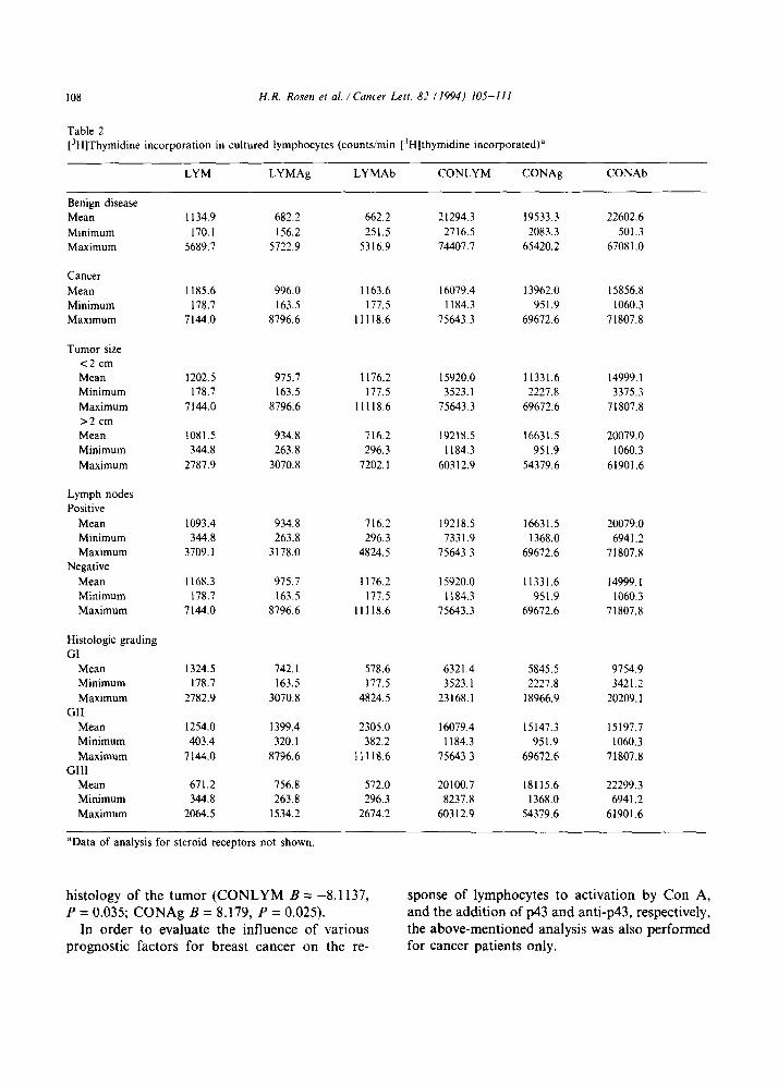
108
Table 2
H. R. Rosen et al. ! Cancer Lett. 82 (1994) 105-111
[3H]Thymidine incorporation in cultured lymphocytes (counts/min [3H]thymidine incorporated)”
LYM LYMAg LYMAb CONLYM CONAg CONAb
Benign disease
Mean
Minimum
Maximum
Cancer
Mean
Minimum
Maximum
Tumor size
<2 cm
Mean Minimum
Maximum
>2 cm
Mean
Minimum
Maximum
Lymph nodes
Positive
Mean Minimum
Maximum
Negative
Mean Minimum
Maximum
Histologic grading
GI
Mean
Minimum
Maximum
GII
Mean
Minimum
Maximum
GIlI
Mean
Minimum
Maximum
1134.9 682.2 662.2 21294.3 19533.3 22602.6
170.1 156.2 251.5 2716.5 2083.3 501.3
5689.7 5722.9 5316.9 74407.7 65420.2 6708 I .O
1185.6 996.0 1163.6 16079.4 13962.0 15856.8
178.7 163.5 177.5 1184.3 951.9 1060.3
7144.0 8796.6 11118.6 75643.3 69672.6 71807.8
1202.5 975.7 1176.2 15920.0 11331.6 14999. I 178.7 163.5 177.5 3523.1 2227.8 3375.3
7144.0 8796.6 11118.6 75643.3 69672.6 71807.8
1081.5 934.8 716.2 19218.5 16631.5 20079.0 344.8 263.8 296.3 1184.3 951.9 1060.3
2787.9 3070.8 7202. I 60312.9 54379.6 61901.6
1093.4 934.8 716.2 19218.5 16631.5 20079.0
344.8 263.8 296.3 7331.9 1368.0 6941.2 3709.1 3178.0 4824.5 75643.3 69672.6 71807.8
1168.3 975.7 1176.2 15920.0
178.7 163.5 177.5 1184.3 7144.0 8796.6 1 I1 18.6 75643.3
11331.6 14999. I
951.9 1060.3
69672.6 71807.8
1324.5 742.1 578.6 6321.4 178.7 163.5 177.5 3523. I
2782.9 3070.8 4824.5 23168.1
5845.5 9754.9
2227.8 3421.2
18966.9 20209. I
1254.0 1399.4 2305.0 16079.4 15147.3 15197.7 403.4 320.1 382.2 1184.3 951.9 1060.3
7144.0 8796.6 11118.6 75643.3 69672.6 71807.8
671.2 756.8 572.0 20100.7 18115.6 22299.3 344.8 263.8 296.3 8237.8 1368.0 6941.2
2064.5 1534.2 2674.2 60312.9 54379.6 61901.6
“Data of analysis for steroid receptors not shown.
histology of the tumor (CONLYM B = -8.1137, P = 0.035; CONAg B = 8.179, P = 0.025).
In order to evaluate the influence of various prognostic factors for breast cancer on the re-
sponse of lymphocytes to activation by Con A, and the addition of p43 and anti-p43, respectively, the above-mentioned analysis was also performed for cancer patients only.
H.R. Rosen et al. /Cancer Letr. 82 (1994) 1O.FIll 109
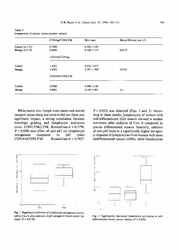
Table 3
Comparison of culture ratios (median values)
CONAgICONLYM Min-max Mann-Whitney test (P)
Cancer (n = 21) 0.7990 0.166-1.185
Benign (n = 19) 0.8809 0.564-1.718 0.0178
CONAbXONAg
Cancer
Benign
1.1952 0.678-5.077
I .0302 0.205-I .994 0.0165
CONAbKONLYM
Cancer
Benign
0.9500 0.440-2.160
0.9601 0.140-1.801 n.s.
While tumor size, lymph node status and steroid receptor status (data not shown) did not show any significant impact, a strong correlation between histologic grading and lymphocyte activation (ratio CONLYMI’LYM, Kendall-tau-6 = 0.3798, P = 0.038) and effect of anti-p43 on lymphocyte mitogenesis compared CONAWCONLYIM, Kentdoall-tat? = 0 56”::” . 3
0” N II ,b
“mhyl bY
Fig. 1. Significant inhibition of lymphocyte mitogenesis activa-
ted by Con A after addition of p43 antigen in breast cancer pa-
tients (P = 0.0178).
P = 0.032) was observed (Figs. 2 and 3). Accor- ding to these results, lymphocytes of women with well-differentiated (GI) tumors showed a weaker activation after addition of Con A compared to poorer differentiated tumors. Similarly, addition of anti-p43 leads to a significantly higher mitogen- ic response of lymphocytes from women with more dedifferentiated tumors (GIII), while lymphocytes
0
Fig. 2. Significantly decreased lymphocyte activation in well differentiated breast cancer tumors (P = 0.038).

110 H. R. Rosen et al. /Cancer Leit. 82 (1994) 105-I I I
Fig. 3. Significantly higher inhibition of lymphocyte mitogene-
sis by addition of p43 in well differentiated breast cancer
tumors (P = 0.032).
of patients with well-differentiated cancer did not respond to addition of anti-p43 MoAb (ratio CONAWCONLYM, Kendall-tau-6 = 0.3782,
P = 0.033).
4. Discussion
The current study revealed that in breast cancer patients, the addition of p43 was able to inhibit
lymphocyte activation by Con A significantly compared to lymphocytes from patients with
benign tumors. The lymphocytes of women with benign disease did not react at all to the addition
of p43 or anti-p43 McAb (ratio = 1.0). P43 isoferritin has been shown to have an im-
munosuppressive activity in vitro [4,6]. It was found that p43 synthesized by the placenta plays a
role in suppressing the maternal lymphocyte re- sponse to the embryo’s alloantigens. Thus, women with low or deficient blood levels of p43 were at high risk of abortion or preterm delivery [13]. To
perform this regulatory activity, p43 isoferritin reacts with receptors on T cells [14].
Recently, it was demonstrated that proliferating
T lymphocytes express binding sites for a heavy (H)-subunit (21 kDa) of ferritin which acts as a downregulator of proliferation [ 151. P43 isofer- ritin, the 43 kDa subunit of PLF is structurally re-
lated to H-ferritin [6] and is also immuno-
suppressive [ 14). P43 bound to a subpopulation of peripheral blood T lymphocytes (CD8+) was detected with anti-p43 CM-H-9 MCAb in women
with early stages of breast cancer but not in pa- tients with benign breast disease [5,8]. This is com- patible with the finding of the current study that p43 inhibited the lymphocyte proliferation from
cancer patients but not from benign breast disease. The above observations may suggest that only in breast cancer is there activation of T cells with receptors for p43 isoferritin as opposed to benign
breast disease. Binding of isoferritin to T cells in cancer pa-
tients results in the masking of cell surface recep-
tors [ 16,171. The degree of in vivo generated immunosuppression may depend on the degree of receptor saturation by p43 isoferritin which is
secreted from the breast cancer tissue [ 181.
In the current study, it was observed that lym- phocytes from patients with more differentiated tumors responded less in Con A activation and were more suppressed by p43 and also by anti-p43
than those with the poorly differentiated tumors. These results seem paradoxical since patients
with more differentiated tumors who have a favourable prognosis seem to be immunosuppress-
ed more than those with non-differentiated tumors who have an unfavorable prognosis. This phenom- enon was previously observed by Wanebo and co- workers who reported that certain lymphocyte
responses showed maximum depression in patients with (early) stage I and an increased lymphocyte response in patients with (more advanced) stages III and IV who had a worse prognosis [ 191.
Furthermore, Whitehead et al. demonstrated a significant depression of T cell levels in breast cancer stage I and II, compared to almost normal
levels in stage III (2OW). These findings parallel the appearance of circulating T cells masked by surface bound p43 iso-ferritin which appear at stage I and are low or undetected in more ad-
vanced stages of breast cancer [8]. Previous observations also showed a strong cor-
relation between the levels of p43 in the cytosols of primary breast cancer and the degree of differ-
entiation [9,18]. Thus, it may be suggested that the more differentiated tumors produce high levels of p43 and induce T cells to express receptors for p43 isoferritin unlike dedifferentiated tumors [9,18].

H. R. Rosen et al. / Cancer Lert. 8-7 ( 1994) 105-I I I III
The inhibitory effect of anti-p43 on Con-A prolif-
eration of lymphocytes from patients with more differentiated malignant tumors could be explain- ed by its binding to p43 on the surface of circula- ting T cells from these patients. Further support for the interaction of breast cancer associated p43 with anti-cancer immune mechanisms can be ob-
tained from previous findings of a negative corre- lation between the level of cytosolic p43 and the ‘inflammatory’ (lymphocytic) reaction found in breast cancer [9].
In conclusion, the results of this study suggest that breast cancer, as opposed to benign tumors, induce in the host the appearance of T cells, which
can be suppressed in vitro by p43 possibly via its binding to receptors on their surface. However, the induction of these T cells may depend on the degree of tumor dil‘ferentiation which determines
the amount of ~43 isoferritin produced by the
tumor. Further studies are needed to elucidate the
mechanism of the immunosuppression exerted by
breast cancer associated ~43, as well as therapeutic applications to overcome its immunosuppressive effects.
References
Bast, R.C. (1982) Effects of cancers and their treatment
on host immunity. In: Cancer Medicine, pp. 1134-l 140.
Editors: J.F. Holland and E. Frey. 111. Lea and Febiger,
Philadelphia, PA.
Penn, I. (1991) Principles of tumor immunity: im-
munocompetence and cancer. In: Biologic Therapy of
Cancer, pp. 53-66. Editors: V.T. DeVita, S. Hellman and
S.A. Rosenberg. Lippincott. Philadelphia, PA.
Keller, E.K., Ioachim, H.L., Pearse. T. and Siletti, D.M.
(1976) Decreased ‘T-lymphocytes in patients with mam-
mary cancer. Am. J. Clin. Pathol.. 65, 445-449.
Moros, C. and Bessler, H. (1989) Ferritin as a marker in
malignancy. In: Iron in Immunity, Cancer and Inflamma-
tion, pp. 283-301. Editors: M. De Sousa and J. Brock.
Wiley, New York. Moroz. C., Kahn, h4. and Ron, E. (1989) The use of on-
cofetal ferritin-bear-ng lymphocytes as a marker for the
screening, diagnosis, and follow-up of patients with early
breast malignancies Screening of 3400 women. Cancer,
64. 691-697.
Moroz, C.. Shterm~l, N. and Kupfer, B. (1989) T-cell mi-
togenesis stimulates the appearance of a new mRNA spe-
cies coding for a 43 kD peptide reactive with CM-H-9
monoclonal antibody specific for placental isoferritin. Proc. Nat]. Acad. Sci (Washington). 86. 3282-3285
7
8
9
10
II
I2
I3
14
15
I6
I7
I8
I9
20
Maymon. R.. Bahari, C. and Moros. C. (1989) Placental
isoferritin measured by a specific monoclonal antibody as a new predictive marker for premature contraction out-
come. Obstet. Gynecol.. 74. 597-599.
Rosen. H.R., Stierer, M.. Giittlicher. J., Wolf, H., Weber.
R., Vogl, E. and Eibl. M. (1992) Determination of placen-
tal ferritin (PLF)-positive lymphocytes in women in early
stages of breast cancer. Int. J. Cancer. 52, 229-233.
Rosen. H.R., Moroz. C.. Reiner. A.. Reinerova. M..
Stierer. M.. Svec, J.. Schemper. M. and Jakesz. R. (1992)
Placental isoferritin associated p43 antigen correlates with
features of high differentiation in breast cancer. Breast
Cancer Res. Tr., 24. 17-26.
Bloom. H.J.G. and Richardson. W.W. (1957) Histological
grading and prognosis in breast cancer. Br. J. Cancer, I I,
359-364.
Remmele. W. and Stegner, H.E. (1987) Vorschlag zur
einheitlichen Definition eines immunreaktiven Score
(IRS) fiir den immunhistochemischen Rezeptornachweis
(ER-ICA) in Mammakarzinomgewebe. Pathologe. 8.
138-140.
Boyum. A. (1968) Isolation of mononuclear cells and
granulocytes from human blood. Stand. J. Lab. Invest.,
21 (Suppl. 97). 77-86.
Moroz. C., Bessler. H. and Sirota. L. (1987) Difference in
the placenta ferritin levels measured by a specific mono-
clonal antibody enzymoassay in preterm and term deliv-
ery. Clin. Exp. Immunol.. 69. 702-706.
Sirota. L.. Kupfer. B. and Moroz, C. (1989) Placental
isoferritin as a physiological downregulator of cellular im-
munoreactivity during pregnancy. Clin. Exp Immunol.,
77, 257-262.
Fargion. S., Fracanzani. A.L. and Brando. B.( 1991) Spe-
cific binding sites for H-ferritin on human lymphocytes
modulation during cellular proliferation and potential im-
plication in cell growth control. Blood, 78. 1056-1061.
Moroz. C., Giler. S.H, Kupfer, B. and Urea, I. (1977)
Lymphocytes bearing surface ferritin in patients with
Hodgkin’s disease and breast cancer. N. Engl. J. Med.,
298. 1175-1176.
Hahn, H.W.L.. Stahlhut, M.W. and Chung, L. (1984) In-
hibitory effects of isoferritins from tumour and non-
tumour tissue on E-rosettes formation. Lancet. i. 43-44.
Rosen, H.R.. Moroz, C., Reiner, A., Stierer, M., Svec, J.,
Reinerova, M.. Schemper, M. and Jakesz. R. (1992) Ex-
pression of ~43 in breast cancer tissue, correlation with
prognostic parameters. Cancer Lett., 67, 35-45.
Wanebo, H.J.. Rosen, P.P.. Thalert, T., Urban. J.A. and
Oetgen, H.F. (1976) Immunobiology of operable breast
cancer. An assessment of biologic risk by im-
munoparameters. Ann. Surg., 184, 258-262.
Whitehead. R.H.. Roberts, G.P.. Thatcher. J.. Teasdale.
C. and Hughs. L.E. (1977) Masking of receptors for sheep
erythrocytes on human T-lymphocytes by sera from
breast cancer patients. J. Nat]. Cancer Inst.. 58,
l573- 1577.