Dorsal luxation of the scapula in a cat
3
618 http://journals.tubitak.gov.tr/veterinary/ Turkish Journal of Veterinary and Animal Sciences Turk J Vet Anim Sci (2013) 37: 618-620 © TÜBİTAK doi:10.3906/vet-1209-34 Dorsal luxation of the scapula in a cat Serhat ÖZSOY, Özlem GÜZEL* Department of Surgery, Faculty of Veterinary Medicine, İstanbul University, Avcılar, İstanbul * Correspondence: [email protected] 1. Introduction Luxation or dislocation of the scapula occurs as a result of traumatic rupture of the muscles supporting the scapula to the thoracic wall, allowing an upward displacement of the bone (1,2). Most commonly the serratus ventralis and the rhomboid and trapezius muscles, as well as possibly the teres major and latissimus dorsi muscles, rupture close to their insertions on the scapula. Costal fractures and pneumothorax and lung contusions may also be seen combined with the luxation. erefore, it is important that the patient be given a full physical examination and that these problems be addressed without delay (1–3). Luxation of the scapula is a rare condition, and it is encountered more frequently in cats compared to dogs. Neither age nor sex has any significance in the occurrence of scapular dislocation (1). Upon orthopedic examination, a dorsal displacement of the scapula is evident when the animal puts weight on the affected limb. On adduction of the limb, the scapula displaces laterally (1,3). e most important differential diagnosis is fracture of the scapula. erefore, radiographic examination is indicated. Sometimes dorsal displacement of the scapula is evident on the radiographs. Fractures are infrequently observed. Radiographs are also used to evaluate the presence of any additional complication such as costal fractures, pulmonary contusion, or pneumothorax (1). e treatment of choice is surgery. However, in cats with acute luxation, a Velpeau bandage is also recommended following the reintroduction of the normal position of the scapula (1). In the surgical treatment of scapular luxation, the ruptured muscle ends are exposed and reattached to their insertions on the scapula. For this, absorbable or nonabsorbable suture materials may be used. Additional support is achieved by placing stainless steel sutures through holes drilled in the caudal edge of the scapula and passing around the adjacent costa. Movement of the leg is restricted by applying a Velpeau bandage for a period of 2–3 weeks. e patient’s activity is restricted for 4–6 weeks by cage rest (1–4). In the postoperative period, complications such as insufficient fixation, infection, and wire migration may be encountered. However, the likelihood of these complications is low and most of the animals regain full function (1,3). In the present study, we present a cat with dorsal luxation of the scapula and its surgical treatment. 2. Case history An adult mixed breed male cat of undetermined age with a bodyweight of 4.5 kg was referred to the Istanbul University’s Veterinary Faculty Surgery Department Clinics with a complaint of right foreleg lameness for approximately 1 week. Case history revealed that the cat was a stray cat and had been lame for approximately 1 week. However, it was not known how the injury had occurred. No medical treatment was given during this time. Clinical examination revealed a dorsal dislocation of the scapula (Figure 1). Upon palpation, the scapula was Abstract: Dorsal luxation of the scapula is a rare orthopedic disorder caused by traumatic rupture of the muscles supporting the scapula. e presented case consisted of an adult cat in which luxation of the scapula had occurred due to trauma. During clinical examination, dorsal displacement of the scapula was evident upon manipulation of the limb. Radiological examination did not reveal any fractures or other concurrent injuries. In the surgical treatment of the patient, the muscles were sutured in place. e patient regained its normal gait without any complications within 4 weeks. Key words: Scapula, dorsal luxation, cat Received: 25.09.2012 Accepted: 30.05.2013 Published Online: 26.08.2013 Printed: 20.09.2013 Case Report
-
Upload
truongkhue -
Category
Documents
-
view
219 -
download
0
Transcript of Dorsal luxation of the scapula in a cat
618
http://journals.tubitak.gov.tr/veterinary/
Turkish Journal of Veterinary and Animal Sciences Turk J Vet Anim Sci(2013) 37: 618-620© TÜBİTAKdoi:10.3906/vet-1209-34
Dorsal luxation of the scapula in a cat
Serhat ÖZSOY, Özlem GÜZEL*Department of Surgery, Faculty of Veterinary Medicine, İstanbul University, Avcılar, İstanbul
* Correspondence: [email protected]
1. IntroductionLuxation or dislocation of the scapula occurs as a result of traumatic rupture of the muscles supporting the scapula to the thoracic wall, allowing an upward displacement of the bone (1,2). Most commonly the serratus ventralis and the rhomboid and trapezius muscles, as well as possibly the teres major and latissimus dorsi muscles, rupture close to their insertions on the scapula. Costal fractures and pneumothorax and lung contusions may also be seen combined with the luxation. Therefore, it is important that the patient be given a full physical examination and that these problems be addressed without delay (1–3).
Luxation of the scapula is a rare condition, and it is encountered more frequently in cats compared to dogs. Neither age nor sex has any significance in the occurrence of scapular dislocation (1).
Upon orthopedic examination, a dorsal displacement of the scapula is evident when the animal puts weight on the affected limb. On adduction of the limb, the scapula displaces laterally (1,3). The most important differential diagnosis is fracture of the scapula. Therefore, radiographic examination is indicated. Sometimes dorsal displacement of the scapula is evident on the radiographs. Fractures are infrequently observed. Radiographs are also used to evaluate the presence of any additional complication such as costal fractures, pulmonary contusion, or pneumothorax (1).
The treatment of choice is surgery. However, in cats with acute luxation, a Velpeau bandage is also recommended following the reintroduction of the normal position of the scapula (1).
In the surgical treatment of scapular luxation, the ruptured muscle ends are exposed and reattached to their insertions on the scapula. For this, absorbable or nonabsorbable suture materials may be used. Additional support is achieved by placing stainless steel sutures through holes drilled in the caudal edge of the scapula and passing around the adjacent costa. Movement of the leg is restricted by applying a Velpeau bandage for a period of 2–3 weeks. The patient’s activity is restricted for 4–6 weeks by cage rest (1–4).
In the postoperative period, complications such as insufficient fixation, infection, and wire migration may be encountered. However, the likelihood of these complications is low and most of the animals regain full function (1,3).
In the present study, we present a cat with dorsal luxation of the scapula and its surgical treatment.
2. Case history An adult mixed breed male cat of undetermined age with a bodyweight of 4.5 kg was referred to the Istanbul University’s Veterinary Faculty Surgery Department Clinics with a complaint of right foreleg lameness for approximately 1 week.
Case history revealed that the cat was a stray cat and had been lame for approximately 1 week. However, it was not known how the injury had occurred. No medical treatment was given during this time.
Clinical examination revealed a dorsal dislocation of the scapula (Figure 1). Upon palpation, the scapula was
Abstract: Dorsal luxation of the scapula is a rare orthopedic disorder caused by traumatic rupture of the muscles supporting the scapula. The presented case consisted of an adult cat in which luxation of the scapula had occurred due to trauma. During clinical examination, dorsal displacement of the scapula was evident upon manipulation of the limb. Radiological examination did not reveal any fractures or other concurrent injuries. In the surgical treatment of the patient, the muscles were sutured in place. The patient regained its normal gait without any complications within 4 weeks.
Key words: Scapula, dorsal luxation, cat
Received: 25.09.2012 Accepted: 30.05.2013 Published Online: 26.08.2013 Printed: 20.09.2013
Case Report
619
ÖZSOY and GÜZEL / Turk J Vet Anim Sci
easily detached from the thoracic wall and was dislocated laterally on adduction of the limb. Palpation revealed no findings to suggest the presence of a fracture.
On radiographs taken in ventrodorsal, mediolateral, and oblique positions, there were no fractures visible either in the scapula or in the costae. The scapula, however, was dislocated dorsally. There were no findings regarding trauma-related pneumothorax, hemothorax, or pulmonary contusion.
Routine blood analysis and some biochemical blood components (aspartate aminotransferase, alanine transaminase, urea, creatinine, glucose, and total protein) measured prior to anesthesia were within normal limits.
General anesthesia was induced with administration of 1 mg/kg xylazine (Rompun 2%, Bayer, Turkey) combined with 5 mg/kg ketamine HCl (Ketamin 10%, Bremerpharma, Germany) given intravenously. Following induction of anesthesia, endotracheal intubation was performed, and general anesthesia was maintained with isoflurane (Forane liquid, Abbott, UK) in oxygen started at 4% and maintained at 2%.
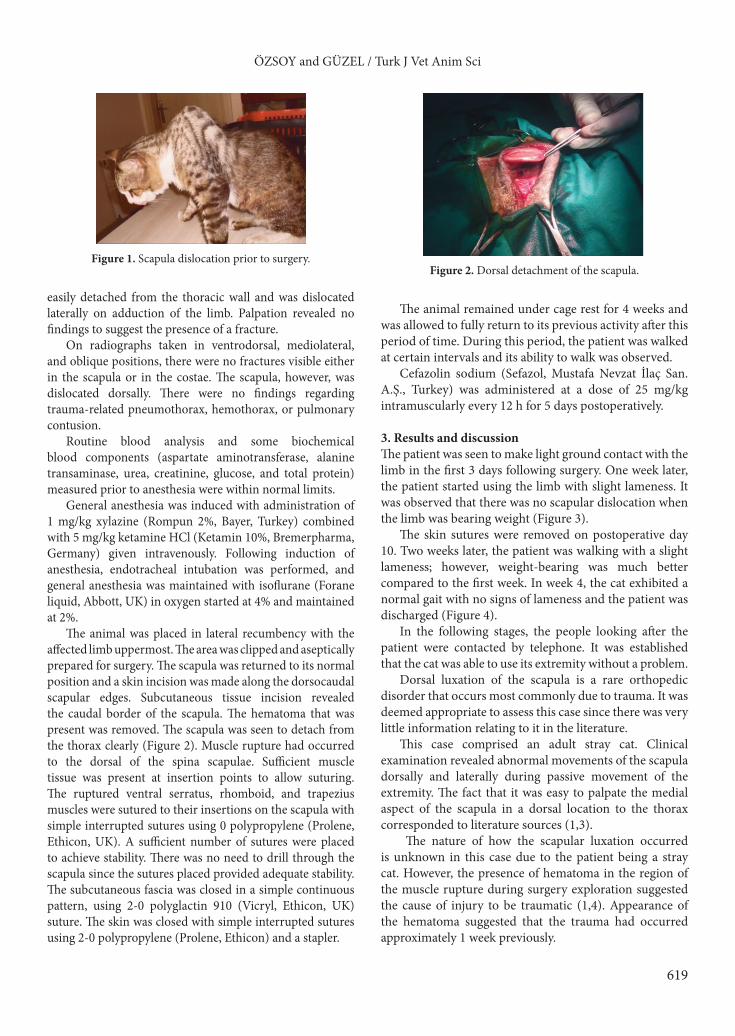
The animal was placed in lateral recumbency with the affected limb uppermost. The area was clipped and aseptically prepared for surgery. The scapula was returned to its normal position and a skin incision was made along the dorsocaudal scapular edges. Subcutaneous tissue incision revealed the caudal border of the scapula. The hematoma that was present was removed. The scapula was seen to detach from the thorax clearly (Figure 2). Muscle rupture had occurred to the dorsal of the spina scapulae. Sufficient muscle tissue was present at insertion points to allow suturing. The ruptured ventral serratus, rhomboid, and trapezius muscles were sutured to their insertions on the scapula with simple interrupted sutures using 0 polypropylene (Prolene, Ethicon, UK). A sufficient number of sutures were placed to achieve stability. There was no need to drill through the scapula since the sutures placed provided adequate stability. The subcutaneous fascia was closed in a simple continuous pattern, using 2-0 polyglactin 910 (Vicryl, Ethicon, UK) suture. The skin was closed with simple interrupted sutures using 2-0 polypropylene (Prolene, Ethicon) and a stapler.
The animal remained under cage rest for 4 weeks and was allowed to fully return to its previous activity after this period of time. During this period, the patient was walked at certain intervals and its ability to walk was observed.
Cefazolin sodium (Sefazol, Mustafa Nevzat İlaç San. A.Ş., Turkey) was administered at a dose of 25 mg/kg intramuscularly every 12 h for 5 days postoperatively.
3. Results and discussionThe patient was seen to make light ground contact with the limb in the first 3 days following surgery. One week later, the patient started using the limb with slight lameness. It was observed that there was no scapular dislocation when the limb was bearing weight (Figure 3).
The skin sutures were removed on postoperative day 10. Two weeks later, the patient was walking with a slight lameness; however, weight-bearing was much better compared to the first week. In week 4, the cat exhibited a normal gait with no signs of lameness and the patient was discharged (Figure 4).
In the following stages, the people looking after the patient were contacted by telephone. It was established that the cat was able to use its extremity without a problem.
Dorsal luxation of the scapula is a rare orthopedic disorder that occurs most commonly due to trauma. It was deemed appropriate to assess this case since there was very little information relating to it in the literature.
This case comprised an adult stray cat. Clinical examination revealed abnormal movements of the scapula dorsally and laterally during passive movement of the extremity. The fact that it was easy to palpate the medial aspect of the scapula in a dorsal location to the thorax corresponded to literature sources (1,3).
The nature of how the scapular luxation occurred is unknown in this case due to the patient being a stray cat. However, the presence of hematoma in the region of the muscle rupture during surgery exploration suggested the cause of injury to be traumatic (1,4). Appearance of the hematoma suggested that the trauma had occurred approximately 1 week previously.
Figure 1. Scapula dislocation prior to surgery.Figure 2. Dorsal detachment of the scapula.

620
ÖZSOY and GÜZEL / Turk J Vet Anim Sci
On radiographs taken of the patient, no fractures were identified in the scapula. Trauma-related complications (1), such as costal fractures or pneumothorax, were not encountered.
Surgical intervention was preferred in order to achieve a better functional result from the treatment. The muscles providing the anatomical position of the scapula, the ventral serratus, rhomboid, and trapezius muscles, were seen to have detached from their insertions (1–3). Since the ruptured muscle ends were sufficient enough to allow suturing, these were sutured using nonabsorbable suture materials. Therefore, anatomical unity was achieved without the need to resort to extra procedures such as drilling a tunnel through the scapula.
Contrary to literature (1,4) advising bandage application to the limb after surgical treatment, the patient was kept under cage rest for 4 weeks.
Complications, such as infection formation, reported to be a possibility in the postoperative period (1,3) were not encountered. Four weeks later, the patient had regained its normal gait without any complications.
In the study presented here, dorsal luxation of the scapula, rarely seen in small animals such as cats and dogs, has been assessed. This was the first time an orthopedic disorder of this kind was seen in a cat at the İstanbul University Veterinary Faculty Surgery Department. The case was treated surgically and the procedure was performed successfully.
Figure 3. Five days after surgery.
Figure 4. Four weeks after surgery.
References
1. Schulz, K.: Disease of the joints. In: Fossum, T.W., Ed. Small Animal Surgery. 3rd edn., Mosby Elsevier, St Louis, MO, USA. 2007; 1143–1316.
2. Scott, H.W., McLaughlin, R.: Fractures and disorders of the forelimb. In: Scott, H.W, McLaughlin, R., Eds. Feline Orthopedics. Manson Publishing Ltd., London. 2007; 107–167.
3. Denny, H.R., Butterworth, S.: The shoulder. In: A Guide to Canine and Feline Orthopaedic Surgery. 4th edn., Blackwell Science, London. 2000; 303–341.
4. Tselepidis, T.S., Desiris, A.: Scapular dislocation in a dog. J. Hellenic Vet. Med. Soc.,1998; 49: 148–150.