Dorsal Approach Rhinoplasty
23
Otorhinolaryngology Clinics: An International Journal, January-April 2013;5(1):1-23 1 AIJOC Dorsal Approach Rhinoplasty Kenneth R Dubeta ABSTRACT Direct dorsal excision of skin and subcutaneous tissue is employed in rhinoplasty cases characterized by thick rigid skin to achieve satisfactory esthetic results, in which attempted repair by more conventional means would most likely frustrate both surgeon and patient. This historical review reminds us of the lesson: ‘History repeats itself.’ Built on a foundation of reconstructive rhinoplasty, modern cosmetic and corrective rhinoplasty have seen the parallel development of both open and closed techniques as ‘new’ methods are introduced and reintroduced again. It is from the perspective of constant evolution in the art of rhinoplasty surgery that the author presents, in Part II, his unique ‘eagle wing’ chevron incision technique of dorsal approach rhinoplasty, to overcome the problems posed by the rigid skin nose. Keywords: Dorsal approach rhinoplasty, Eagle wing incision, Rigid skin nose, External approach rhinoplasty, Historical milestones. How to cite this article: Dubeta KR. Dorsal Approach Rhinoplasty. Int J Otorhinolaryngol Clin 2013;5(1):1-23. Source of support: Nil Conflict of interest: None declared INTRODUCTION Throughout the ages, numerous techniques of altering, correcting and more recently, improving the appearance and function of the nose have been described. Methods of altering the appearance of the nose have been limited only by man’s imagination. These have varied from nasal amputation, as a form of punishment, to forms of embellishment such as tattooing, or adornment with precious gems, metals and/or pieces of bone. With the additional burdens imposed by disease and accidental trauma, the nose has been forced to endure an almost equal number of corrective solutions and ‘cures’. Thus, even in ancient times, bandages were being applied to support or straighten the broken nose 1 (Fig. 1: Perike- phalea) and the missing nose was being rebuilt in India with forehead skin flaps. 2 It has been the portal of entry for innumerable inhalations, regardless of the affliction. Occasionally, it has fallen off because of medications taken by mouth. 3 It is only within the past one hundred years or so, however, that modern surgical techniques have evolved, permitting actual enhancement of both structural esthetics and functions of the nose. Refinement of these techniques seemingly had to await three antecedent developments; topical vasoconstriction; topical, systemic and local anesthesia; and safe, reliable sources of illumination. The last half of the 20th century has seen the dissemination of two of the most important developments in the history of nasal surgery: 1. Recognition of the key role of the ethmoid sinuses in sinus disease, followed shortly by concomitant development of the sinus endoscope. 2. The open or external approach to rhinoplasty. With regard to this latter development, it is ironic that rhinoplastic surgeons were themselves responsible for retarding progress in the evolution of esthetic and reconstructive nasal surgery, by restricting visualization of the complicated skeletal infrastructure of the nose through adherence to the conventional closed techniques popularized by Joseph in the first half of this century. Many will still argue that the infrastructure of the nose can be exposed just as well via closed techniques, without ‘risking’ an external (transcolumellar) scar. But none who are at least familiar with the external approach can deny that the superior exposure provided by this technique facilitates surgical correction of the more challenging nasal asymmetries and deformities. There remains a group of ‘problem noses’, however, in which even the transcolumellar external approach will prove ORIGINAL ARTICLE 10.5005/jp-journals-10003-1105 Part I: Historical Milestones in Rhinoplasty Fig. 1: Ancient Greek ‘perikephalea’ to support the straightened nose 1
Transcript of Dorsal Approach Rhinoplasty

Otorhinolaryngology Clinics: An International Journal, January-April 2013;5(1):1-23 1
Dorsal Approach Rhinoplasty
AIJOC
Dorsal Approach RhinoplastyKenneth R Dubeta
ABSTRACT
Direct dorsal excision of skin and subcutaneous tissue isemployed in rhinoplasty cases characterized by thick rigid skinto achieve satisfactory esthetic results, in which attempted repairby more conventional means would most likely frustrate bothsurgeon and patient.
This historical review reminds us of the lesson: ‘Historyrepeats itself.’ Built on a foundation of reconstructive rhinoplasty,modern cosmetic and corrective rhinoplasty have seen theparallel development of both open and closed techniques as‘new’ methods are introduced and reintroduced again. It is fromthe perspective of constant evolution in the art of rhinoplastysurgery that the author presents, in Part II, his unique ‘eaglewing’ chevron incision technique of dorsal approach rhinoplasty,to overcome the problems posed by the rigid skin nose.
Keywords: Dorsal approach rhinoplasty, Eagle wing incision,Rigid skin nose, External approach rhinoplasty, Historicalmilestones.
How to cite this article: Dubeta KR. Dorsal ApproachRhinoplasty. Int J Otorhinolaryngol Clin 2013;5(1):1-23.
Source of support: Nil
Conflict of interest: None declared
INTRODUCTION
Throughout the ages, numerous techniques of altering,correcting and more recently, improving the appearance andfunction of the nose have been described. Methods ofaltering the appearance of the nose have been limited onlyby man’s imagination. These have varied from nasalamputation, as a form of punishment, to forms ofembellishment such as tattooing, or adornment with preciousgems, metals and/or pieces of bone.
With the additional burdens imposed by disease andaccidental trauma, the nose has been forced to endure analmost equal number of corrective solutions and ‘cures’.Thus, even in ancient times, bandages were being appliedto support or straighten the broken nose1 (Fig. 1: Perike-phalea) and the missing nose was being rebuilt in India withforehead skin flaps.2 It has been the portal of entry forinnumerable inhalations, regardless of the affliction.Occasionally, it has fallen off because of medications takenby mouth.3
It is only within the past one hundred years or so,however, that modern surgical techniques have evolved,permitting actual enhancement of both structural esthetics
and functions of the nose. Refinement of these techniquesseemingly had to await three antecedent developments;topical vasoconstriction; topical, systemic and localanesthesia; and safe, reliable sources of illumination. Thelast half of the 20th century has seen the dissemination oftwo of the most important developments in the history ofnasal surgery:1. Recognition of the key role of the ethmoid sinuses in
sinus disease, followed shortly by concomitantdevelopment of the sinus endoscope.
2. The open or external approach to rhinoplasty.With regard to this latter development, it is ironic that
rhinoplastic surgeons were themselves responsible forretarding progress in the evolution of esthetic andreconstructive nasal surgery, by restricting visualization ofthe complicated skeletal infrastructure of the nose throughadherence to the conventional closed techniques popularizedby Joseph in the first half of this century. Many will stillargue that the infrastructure of the nose can be exposed justas well via closed techniques, without ‘risking’ an external(transcolumellar) scar. But none who are at least familiarwith the external approach can deny that the superiorexposure provided by this technique facilitates surgicalcorrection of the more challenging nasal asymmetries anddeformities.
There remains a group of ‘problem noses’, however, inwhich even the transcolumellar external approach will prove
ORIGINAL ARTICLE10.5005/jp-journals-10003-1105
Part I: Historical Milestones in Rhinoplasty
Fig. 1: Ancient Greek ‘perikephalea’ to support thestraightened nose1

2
Kenneth R Dubeta
inadequate. These noses are characterized by bulky and/orrigid skin, due to heredity, scarring or disease of thecutaneous and subcutaneous tissues themselves.
HISTORICAL MILESTONES
Reconstructive rhinoplasty is an ancient art. Long before800 BC, when Susruta4 wrote of the procedure, a low-castesect of potters in ancient India were using the forehead flapto reconstruct the absent nose.2 Ancient Egyptians werepracticing the art at least seven centuries earlier,2 but thetechnique came to be known as the Indian rhinoplasty(Fig. 2).5,14
Twenty-three hundred years after Susruta, the Indianrhinoplasty arrived in Europe. In the 15th and 16th centuries,the Brancas and Tagliacozzi utilized the forehead flap andthen invented the delayed arm flap, developing what is todayknown as the Italian method (Fig. 3).2,14 Condemned bythe Church for ‘interfering with the handiwork of God’,Tagliacozzi’s teachings died with him.
During the 17th and 18th centuries, flap reconstructionof the nose fell out of favor, but was brought back to Europefrom India by the British in the late 18th century.2 The firstEuropean case of Indian rhinoplasty in over 200 years wascarried out in 1814 by Mr John Carpue, an English surgeon(Fig. 4).2-6 He was familiar with the writings of Tagliacozzi,and had read an account of an Indian rhinoplasty reportedin the Gentlemen’s Magazine, October, 1794 (taken fromthe Madras Gazette, 1793).5 Numerous cases were recordedbetween 1814 and 1830, in England and on the continent.6,7
Up to this time, all of these surgeries were, of course, carriedout without anesthesia.
By the early 1800’s, the Indian rhinoplasty had reachedNorth America. In 1832, Dr Gurdon Buck of New York
reported a case on which he carried out five operations on amale patient to reconstruct his missing right maxilla andsidewall of the nose, utilizing a forehead flap (Figs 5A toD).3 Mercifully, it was now possible to carry out this typeof surgery under ether anesthesia. (Dr Buck, incidentally,was credited in 1846 with the first interosseous wiring of afractured mandible).1
As can be expected, numerous refinements on the basicIndian and Italian flaps have taken place in the past centuryand a half, paralleling the development of modernanesthesia. These are too numerous to detail here. Notableamong these refinements, however, are the scalping flap8
of Dr John Converse of New York, in 1942; and theretroauricular-temporal flap9 developed by Dr HiroshiWashio of Tokyo, first published in 1969.
The history of cosmetic and corrective rhinoplasty, asopposed to reconstructive rhinoplasty, dates back only tothe mid-1800’s. It was Dieffenbach,10 in 1845, who isgenerally credited with having carried out the first reductionrhinoplasty. He treated a rhinophyma by means of a stellateexternal incision (Fig. 6).10
Fig. 2: Circa 800 BC: Susruta recorded Indian foreheadflap rhinoplasty5,14
Fig. 3: Italian delayed arm flap of Tagliacozzi, 15972,14
Fig. 4: Mr John Carpue reintroduced Indian forehead flap inEngland, 18146

Otorhinolaryngology Clinics: An International Journal, January-April 2013;5(1):1-23 3
Dorsal Approach Rhinoplasty
AIJOC
beformities of the nose by a subcutaneous operation’.Besides describing strict antiseptic precautions and the useof iodoform powder blown over the wound to preventinfection, this second paper is remarkable for describing
Figs 5A to D: Dr Gurdon Buck reported Indian forehead flap in USA, 18323
Fig. 6: Dr JF Dieffenbach, first reduction rhinoplasty, 184510
The first rhinoplasty by means of intranasal incisionswas carried out in 1887 by Dr John Roe,11 an Otolaryngo-logist in Rochester, New York. In February of that year, heread a paper before the Medical Society of New York,describing correction of a ‘Pug-Nose’ deformity via anendonasal approach.12 He presented a second paper to thesame society in 1891, entitled ‘Correction of angular
Fig. 7: Dr Jacques Joseph, pioneer of cosmetic and correctivenasal surgery, 18985

4
Kenneth R Dubeta
the use of cocaine for topical anesthesia, and for the injectionof cocaine ‘... under the skin with a hypodermic syringe...’.11
Though Dr Jacques Joseph reported his first case ofrhinoplasty in 1898, 11 years after Roe’s first publication,Joseph is generally acknowledged as the father of moderncorrective rhinoplasty because of his extensive writings5,13,14
on this subject in the early 20th century, in Berlin (Fig. 7).5
Interestingly, his first case was carried out by means ofexternal nasal incisions through tip and dorsum. His classicendonasal techniques were evolved later, and were probablybased on the earlier writings of John Roe. To his credit,Joseph mentions these earlier reports in his first article onrhinoplasty,15 stating ‘...we found the mention of a fewendonasal hump removals by Dr Row...’ Dr GustaveAufricht16 and Dr J Safian17 brought Joseph’s techniquesto the United States in the early 1900’s, and themselvesmade significant contributions to the art of endonasalrhinoplasty surgery.
In 1899, Dr Friedrich von Mangoldt18 was the firstsurgeon to use autogenous rib cartilage, implanted viaglabellar and nasolabial fold incisions to repair a saddlenose deformity. Among Joseph’s noteworthy contributions,he reported use of part of one ala to repair the other19 in
1912, in effect the first use of a composite graft in nasalreconstruction. In 1914, Dr F Koenig reported his series ofcomposite grafts to the nose, from the upper ear,20 while DrA Limberg reported a similar and more successful series ofcases21 in 1935 (Fig. 8).6
Meanwhile, Dr Aurel Rethi, of Budapest, reported histranscolumellar incision and external approach to the nasaltip, in 1921.22 This innovative technique was the beginning
Fig. 8: Dr F Koenig, composite grafts from ear to nose, 19146
Fig. 9: Dr Aurel Rethi created the external approach open rhinoplasty technique, 19212

Otorhinolaryngology Clinics: An International Journal, January-April 2013;5(1):1-23 5
Dorsal Approach Rhinoplasty
AIJOC
of what is today known as the external approach or ‘open’rhinoplasty (Fig. 9).2 Like many new and ‘radical’ ideas,his technique was initially not readily accepted, even withfurther writings in 1934,22 perhaps because he initiallyconfined this approach to the nasal tip. It was in 1956 thatDr A Sercer, in Zagreb, reported the use of Rethi’s approachto expose the entire nasal infrastructure,23 the same yearthat Rethi himself reported using his approach in the repairof saddle nose deformity.24 The external approach torhinoplasty gained wider acceptance in Europe during thenext decade, but was slower to catch on in North America.Dr Samuel Fomon, of New York, mentioned and illustratedRethi’s technique in his influential text2 in 1939 describingit as the best of the external incisions, to which he remainedgenerally opposed. (Ironically, a study of the text describingthe illustration reveals an incomplete understanding of thetechnique). He mentions it again in his second text11
published in 1960, but mainly to state that he abandonedthe approach for use in dorsal augmentation.
Several other ‘new’ developments took place in the1940’ and 1950’s. Among these was the appearance, in theEnglish literature, of the use of the composite ear graft in
nasal reconstruction. Following the leads of Koenig andLimberg, Dr Harold Gillies, in England, described the useof a composite conchal graft in reconstruction of the ala25
in 1943; and Dr’s James Brown and B Cannon reported useof similar grafts26 in the United States in 1946. In 1953, DrIrving Goldman published his techniques for narrowing thenasal tip and increasing tip projection,27 a major advancein the management of the thick-skinned nose.
The pace of introduction of new ideas in cosmetic andcorrective rhinoplasty quickened during the 1960’s and1970’s. Dr Jack Anderson described his cartilage-splittingincision28 in 1966, minimizing the number of intranasalincisions. He also emphasized the importance of shorteningthe lateral crura, rather than the septum, in effectingshortening of the nasal tip and nose overall. Shortlythereafter, in 1966, Dr L Padovan (also of Zagreb) publishedfurther on the external approach technique.29 Like Sercer,his cases numbered in the hundreds. Padovan reintroducedthe concept to North America in 1970, at the 1st InternationalSymposium on Plastic and Reconstructive Surgery, in NewYork; and Dr Wilfred Goodman of Toronto, Canada, helpedpopularize it through papers30,31 published in 1973 and 1974.
Figs 10A to F: Dr Bromley Freeman—direct cutaneous incisions and skin grafting for rhinophyma rhinoplasty, 19706,32

6
Kenneth R Dubeta
In 1970, another ‘new’ concept was reintroduced by DrBromley Freeman, in Texas: like Dieffenbach before him,he reported on management of rhinophyma by means ofdirect excision of involved skin and subcutaneous tissues,combined with surgical planing (Figs 10A to F).6,32
Meanwhile, it was Dr Reed Dingman from Michigan andDr Claus Walter from Germany who in 1968 again presentedthe use of composite ear grafts, this time to reconstruct theskeletal infrastructure of the nose.33 During the 1970’s, theuse of free grafts in nasal reconstruction was furtheramplified by Walter34 in both the English and Germanliterature.
On the cosmetic side of the scale, Dr Jack Sheen of LosAngeles reintroduced the concept of inserting a septalcartilage autograft35 into the nasal tip region in 1975, toachieve more nasal tip projection. His placement of theautograft was anterior to the domes of the tip cartilages,rather than overlying the domes, as described by Fomon11
in 1960.During the past two decades, many minor but significant
advances have been described, pertaining to refinements inconventional rhinoplasty technique. Many of theserefinements have dealt with methods of achieving ormaintaining tip projection, with the use of allograftmaterials, and with the management and prevention ofsecondary deformities.36-56 During the same period, thevalue of the external approach in rhinoplasty has gainedwider acceptance, as exemplified in 1986 by the publicationof Anderson’s and Ries’ monograph on rhinoplasty for theAmerican Academy of Facial Plastic and ReconstructiveSurgery, emphasizing the external approach.41
Through a review of the history of rhinoplasty, it canthus be seen that the development of the external approachor ‘open’ rhinoplasty has paralleled the modern developmentof conventional or ‘closed’ methods, since the latter half of19th century. Neither method, however, is suited to dealingwith the problems posed by the rigid skin nose.
Part II of this paper58 will hopefully draw attention tothe role that dorsal cutaneous incisions and excisions canplay in the management of the spectrum of nasal deformities,be they cosmetic, hereditary, traumatic or iatrogenic inorigin.
CONCLUSION
Through this brief review of key historical milestones inthe history of rhinoplasty, it can be seen that thedevelopment of the external approach or ‘open’ rhinoplastyhas paralleled the modern development of conventional or‘closed’ methods since the latter half of the 19th century.Neither method, however, is suited to dealing with theproblems posed by the rigid skin nose.
The second part of this paper is entitled ‘DorsalApproach Rhinoplasty—Part II: A Radical Approach to theRigid Skin Nose.’58 In Part II, the author introduces his novel‘eagle wing’ incision technique of dorsal approachrhinoplasty, which he designed to overcome the challengesof the rigid skin nose. Part II will also draw attention to therole that dorsal cutaneous incisions and excisions can playin the management of the entire spectrum of nasaldeformities.
Part II: A Radical Approach to the Rigid Skin Nose
ABSTRACT
Direct dorsal excision of skin and subcutaneous tissue isemployed in rhinoplasty cases characterized by thick rigid skinto achieve satisfactory esthetic results, in which attempted repairby more conventional means would most likely frustrate bothsurgeon and patient.
The dorsal approach facilitates debulking of the nose bymeans of excision of nasal subcutaneous musculoaponeuroticsystem (SMAS) and thickened subcutaneous tissues. Moreimportantly, direct dorsal excision permits direct reduction inskin surface area and volume without which repair attemptswould most likely fail in spite of alteration of the nasal skeletalframework, because of the rigidity of the skin itself. Exposurevia the dorsal ‘eagle wing’ incisions is equal or superior to thetranscolumellar approach. Intraoperative dermabrasion andproper positioning of the incisions are important in minimizingthe resultant dorsal cutaneous scar.
Two cases are described: Secondary repair of a scarred nosein an older male previously treated elsewhere for rhinophyma;and esthetic correction in a young male, of a broad nose affected
by early acne rosacea. Preoperative, intraoperative andpostoperative photographs and diagrams are presented, to helpillustrate the surgical techniques and results achieved. Thesubject of dorsal nasal incisions is reviewed in the literature.The author feels the technique presented provides a valuablesurgical alternative for those cases who might otherwise beadvised ‘nothing further can be done’, because of their scarredor rigid nasal skin.
Keywords: Dorsal approach rhinoplasty, Eagle wing incision, Rigidskin nose, External approach rhinoplasty, Historical milestones.
INTRODUCTION
Review of the history of rhinoplasty, as presented in ‘DorsalApproach Rhinoplasty—Part I: Historical Milestones inRhinoplasty,’57 reveals that the development of the externalapproach or ‘open’ rhinoplasty has paralleled the moderndevelopment of conventional or ‘closed’ methods, since thelatter half of the 19th century.

Otorhinolaryngology Clinics: An International Journal, January-April 2013;5(1):1-23 7
Dorsal Approach Rhinoplasty
AIJOC
Table 1: Goodman classification30 of indications for theexternal approach rhinoplasty
1. Congenital deformities of the nose.2. Major nasal septal deformities.3. Large hump nose with cartilaginous deformities.4. Major augmentation procedures accompanied by
cartilaginous deformities.5. Excision of dermoid cysts or other pathology.6. Correction of the bifid tip.7. Excision of subcutaneous scar tissue.8. Reoperations with major deformities.
Table 2: Dubeta classification of indications for the dorsalapproach rhinoplasty
A. Reduction rhinoplasty—in noses with:1. Scarred or rigid skin
• Surgical or traumatic causes• Acne vulgaris scarring
2. Thick skin and subcutaneous tissues• Subcutaneous fibrosis• Acne rosacea, cystic
3 Large or broad nose with bulky tip• Hereditary
B. Rhinophyma repair:1. Primary2. Secondary
C. Nasal deformity repair:1. Severe short nose deformity2. Severe nasal deformity
• Primary trauma repair• Secondary/delayed repair
There remains a group of ‘problem noses’, however, inwhich even the transcolumellar external approach will proveinadequate, as neither method is capable of dealing withthe challenges posed by the rigid skin nose. These nosesare characterized by bulky and/or rigid skin, due to heredity,scarring or disease of the cutaneous and subcutaneoustissues themselves.
This paper, Part II, will draw attention to the role thatdorsal cutaneous incisions and excisions can play in themanagement of the entire spectrum of nasal deformities, bethey cosmetic, traumatic or iatrogenic in origin. To thisend, the author presents his novel ‘Eagle Wing’ incisiontechnique, and the procedure he has named the ‘DorsalApproach’ (as opposed to ‘external approach’) rhinoplasty.
INDICATIONS
The indications for external approach rhinoplasty have beenelegantly summarized by Dr Wilfred Goodman,30 as listedin Table 1. To these, some surgeons would add: ‘Anycosmetic nasal asymmetries.’ I personally prefer to reservethis approach for the more challenging nasal deformities inwhich, however, the skin is more-or-less normal.
The indications for dorsal approach rhinoplasty aresomewhat different, having first of all to do with thecondition of the skin and subcutaneous tissues overlyingthe skeletal infrastructure, as shown in Table 2. If rigidity,thickness or scarring of these tissues is likely to preventthem from conforming to the surgically-altered skeletalframework, consideration should be given to direct excisionof the skin, subcutaneous tissues and nasal subcutaneousmusculoaponeurotic system (SMAS), to reduce skin volumeand surface area directly.
In rhinophyma, there is often an associated hypertrophyand/or drooping of the nasal tip cartilages and skeletalelements. Dorsal approach rhinoplasty technique permitssimultaneous correction of both the soft tissue and skeletalelements, in managing this difficult disfiguring problem.
The same applies equally well to the more purely-cosmetic problems posed by reduction rhinoplasty of the
large or broad nose with rigid, thick skin of the tip andsupratip regions. Skin thickening in these individuals maybe purely hereditary, or due to the sebaceous glandhypertrophy of acne rosacea.
Finally, the severe short-nose deformity and severe nasaldeformities in general can be approached by a variety ofdorsal nasal incisions, not only the ‘eagle wing’ or ‘chevron’incision used to manage the two cases described herein.
PREOPERATIVE MANAGEMENT
Patients who are candidates for dorsal approach rhinoplastyshould have bacterial culture and sensitivity studies of swabstaken from the nasal vestibules, and in the case of acnescarring, from any active pustular acne lesions. They shouldbe treated with appropriate prophylactic antibiotics, andshould ideally have preoperative dermatologic assessmentand management as well.
Preoperative planning and photographic analysis of theface and nose are essential elements in ensuring success ofthe procedure. This can be done by computer-assistedimaging and analysis, though I personally prefer the ‘hands-on’ experience gained by drawing the corrected facial andnasal profile on tracing paper or on the back of standardizedtwo-thirds life-size 5 × 7 inch black and white or colorphotographs, working on a back-illuminated drawing table.The image so produced should realistically represent theresults to be expected from this type of surgery and notmerely reflect the surgeon’s and the patient’s esthetic ideal.Pre- and postoperative photographs are shown, of otherpatients with similar problems. Whenever possible, andparticularly if requested, arrangements are madepreoperatively to have the rhinoplasty candidate meet withan individual who has already had a similar procedure, toassess his or her experiences, preoperative findings andpostoperative results.

8
Kenneth R Dubeta
Because the surgery involves the creation of a dorsalnasal scar, albeit a well-camouflaged or hopefully ‘invisible’one, these patients require more than the usual amount ofpreoperative counseling and reassurance. Informed consentmust be given, and the risks of the procedure weighedcarefully against the potential benefits. Patient selection istherefore extremely important, and this type of surgerywould best be avoided if a surgical procedure with less-conspiciously located scars can be anticipated to give asatisfactory result, in the particular individual underconsideration.
Technique of Dorsal Approach Rhinoplasty
The technique described pertains to cosmetic or reductionrhinoplasty in the thick skin nose.
The key to the success of this surgery, from the estheticpoint of view, is the design and placement of the dorsalcutaneous incisions. Second in importance is the use ofintraoperative dermabrasion to maximize epidermalblending, and minimize the resultant dorsal cutaneous scar.
Caution should be exercised, however, in the use ofdermabrasion in darker-skinned individuals (Fitzpatrick typeIV or higher) because of the risk of post-inflammatoryhyperpigmentation. Similarly, they should be advised thatANY dorsal nasal incision scar in of itself may heal as avisible brownish line in the nasal supratip region, with orwithout dermabrasion. In this sense, the visibility or lackthereof of the post-surgical scar is always a 'trade-off', to beweighed against the visibility of the pre-existing deformity.
The surgery is carried out under general anesthesia, orlocal anesthesia with sedation. In either case, the
subcutaneous tissues are infiltrated with lidocaine 1 or 2%with 1:100,000 or 1:200,000 adrenaline, to assist inhemostasis.
To achieve debulking and narrowing of the tip andsupratip, direct excision of skin, subcutaneous tissues andnasal SMAS will be carried out. As is appropriate for theparticular features which have to be corrected in the nosein question, an ‘eagle wing’ incision is marked out on thetip dorsum and supratip skin, encompassing the skin stripto be excised (Fig. 1A). The center of the incision forms ashallow curved ‘V’ at the cephalic margins of the reshapedlateral crura. The inferior and superior incision margins aresimilarly marked out, but the ‘V’ of the superior margin issomewhat shallower. The vertical (sagittal) width of the skinstrip to be excised corresponds to the amount of tip elevationrequired and/or the amount of dorsal and tip narrowingdesired.
The lateral ‘wings’ of the eagle wing incision curvecephalically as well as dorsally, i.e. ‘up and away’ from themidline (Fig. 1B). Curving the incision away from the alarlobules avoids segregating the lobules from the supratipsidewalls (as would occur with a standard gullwing incisioncurving down into the alar grooves), and extends the amountof dorsal exposure possible. The lateral wings of the incisioncan be extended a considerable distance up the nasalsidewalls, if necessary, while still maintaining excellentlength to width ratios of the resultant superior hinge flap ofdorsal nasal skin.
At this point, the shape and position of the incision isconceptualized, or a template can be made of paper or cloth.The skin of the nose is then dermabraded, removing a layer
Fig. 1A: Skin marking for dorsal ‘Eagle Wing’ incisions: Planned skin and sub-Q excision (refer to text)
Otorhinolaryngology Clinics: An International Journal, January-April 2013;5(1):1-23 9
Dorsal Approach Rhinoplasty
AIJOC
of epidermis to the depth of erythema and slight vascularoozing. This erases the markings for the eagle wing incision,as dermabrasion is carried out for at least 5 mm cephalicand caudal to the superior and inferior incision marginspreviously delineated.
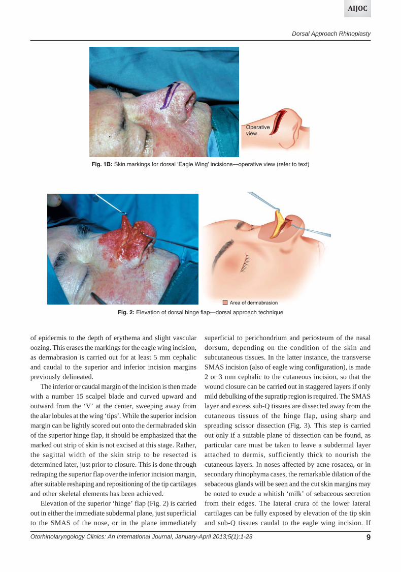
The inferior or caudal margin of the incision is then madewith a number 15 scalpel blade and curved upward andoutward from the ‘V’ at the center, sweeping away fromthe alar lobules at the wing ‘tips’. While the superior incisionmargin can be lightly scored out onto the dermabraded skinof the superior hinge flap, it should be emphasized that themarked out strip of skin is not excised at this stage. Rather,the sagittal width of the skin strip to be resected isdetermined later, just prior to closure. This is done throughredraping the superior flap over the inferior incision margin,after suitable reshaping and repositioning of the tip cartilagesand other skeletal elements has been achieved.
Elevation of the superior ‘hinge’ flap (Fig. 2) is carriedout in either the immediate subdermal plane, just superficialto the SMAS of the nose, or in the plane immediately
superficial to perichondrium and periosteum of the nasaldorsum, depending on the condition of the skin andsubcutaneous tissues. In the latter instance, the transverseSMAS incision (also of eagle wing configuration), is made2 or 3 mm cephalic to the cutaneous incision, so that thewound closure can be carried out in staggered layers if onlymild debulking of the supratip region is required. The SMASlayer and excess sub-Q tissues are dissected away from thecutaneous tissues of the hinge flap, using sharp andspreading scissor dissection (Fig. 3). This step is carriedout only if a suitable plane of dissection can be found, asparticular care must be taken to leave a subdermal layerattached to dermis, sufficiently thick to nourish thecutaneous layers. In noses affected by acne rosacea, or insecondary rhinophyma cases, the remarkable dilation of thesebaceous glands will be seen and the cut skin margins maybe noted to exude a whitish ‘milk’ of sebaceous secretionfrom their edges. The lateral crura of the lower lateralcartilages can be fully exposed by elevation of the tip skinand sub-Q tissues caudal to the eagle wing incision. If
Fig. 1B: Skin markings for dorsal ‘Eagle Wing’ incisions—operative view (refer to text)
Fig. 2: Elevation of dorsal hinge flap—dorsal approach technique

10
Kenneth R Dubeta
necessary, dissection can be carried well down into thecolumella, caudal to or between the medial crura. As withexternal approach rhinoplasty, the membranous septum canbe bivalved to expose the caudal margin of the quadrangularcartilage, down to at least the level of the nasal spine (Fig. 4).
Such adjustments to the bony and cartilaginous skeletonas are required can now be carried out under direct vision.If the nasal dorsum is opened, as with hump removal orstraightening procedures, the nasal septum can be accessedsubmucosally and subperiosteally, in its entirety.
The SMAS layer is handled in one of two fashions,depending on the amount of debulking required. If theproblem is a rigid, drooped tip with relatively—normalpliability of the supratip skin, the SMAS flap can be trimmedalong its caudal margin at a level near to the caudal marginof the skin incision, once the tip has been adjusted to thedesired operative position. Alternately, if greater debulkingof tip, supratip and dorsum is required, the thickened SMASand subcutaneous tissues may have to be resected entirely.In either case, defatting of the tip is usually indicated,excising fibrotic or hypertrophied sub-Q tissues and SMAS
of the tip itself to permit better skin redraping over thereshaped tip cartilages.
Trimming of the superior hinge flap can now be carriedout. This is achieved by overlapping the lower, caudalmargin of the eagle wing incision with the hinge flap, usingskin hooks for traction. With the nasal tip in the desiredposition and the nasolabial angle satisfactorily adjusted, theamount of skin overlap determines the width of the skinstrip to be excised, and the position of the upper margin ofthe eagle wing incision. This process can be assisted byincising sagittal ‘darts’ along the overlapped superior skinflap margin, as far cephalically as the underlying (caudal)eagle wing incision. Then, the tips of the darts are joinedtogether to create an upper incision margin and excise theintervening skin.
Or, if the previously-created template seems appropriate,the template can be applied along the free margin of thehinge flap once the skeletal elements have been adjustedand the SMAS layer and excess sub-Q tissues have beenexcised. The superior margin of the eagle wing incision isthen made along the superior template margin, and theintervening strip of skin is excised.
Fig. 4: Full dorsal, septal and tip exposure is provided with dorsal approach technique
Fig. 3: Elevation of skin and SMAS flaps. Note: Swollen sebaceous glands in skin flap

Otorhinolaryngology Clinics: An International Journal, January-April 2013;5(1):1-23 11
Dorsal Approach Rhinoplasty
AIJOC
The wound is then closed in layers. If dorsal SMAS and/or sub-Q has been preserved, it can be reattached to thesub-Q or perichondrial tissues anterior and lateral to theanterior septal angle using a few tacking sutures of 5-0chromic gut. Fine interrupted sutures of 6-0 nylon are thenused to reapproximate the skin wound margins (Fig. 5).Slight further trimming of the superior and inferior skinmargins may be carried out, as appropriate, at the time ofclosure. Correction of any columellar overhang can becarried out at this time, via separate columellar vestibularincisions.
Considerable judgment must be exercised regarding theuse of alar base wedge resections, to narrow the base of thenose. If there is evidence of previous columellar surgery,this procedure should probably be deferred to another day,for fear of jeopardizing blood supply to the skin of the nasaltip. However, it is felt that the up-sweep of the eagle wingincision, away from the alar lobule and groove, helps tomaintain the vascular supply of the inferior skin flapoverlying the tip structures. This conversely reduces therisk of avascular necrosis of the tip skin if wedge resectionsof the alar lobules are carried out at the time of reductionrhinoplasty, but the condition of the regional skin must stillbe taken into careful consideration.
It should be mentioned that in primary rhinophymasurgery, subepidermal skin flaps are created by planing ofthe hypertrophic skin.32 Following this, reductionrhinoplasty can then proceed in the same manner describedabove. No further dermabrasion of the raw-surfacedsubepidermal skin is necessary, except as required tosmoothen out the planing margins circumferentially.
At the conclusion of the procedure, light packing of thenasal vestibules and fossae may be carried out if necessaryusing folded Telfa strips and Gelfoam coated with antibioticointment. A thin layer of the same ointment is spread overthe dorsal incision and dermabraded skin, followed by
application of a routine tape and plaster splint which isremoved on the second to sixth postoperative day. Routinepostoperative care is implemented. Nasal packing, if any,is removed 1 to 2 days following surgery. Prophylactic anti-staphylococcal antibiotics, topically and orally, aremaintained until re-epithelialization of the dermabraded skinis complete. Sutures are removed on the fifth to seventhpostoperative day, depending of the condition of thedermabrasion eschar. The patient is then seen at regularintervals for routine follow-up care, not the least of whichis reassurance that the initially reddish or violaceousdermabraded skin and dorsal scar will eventually fade tonormal color. Caution should again be advised inrecommending dermabrasion to darker skinned individuals,because of the risk of post-inflammatory hyperpigmentation.
CASE REPORTS
Case 1
A 21-year-old man presented with concerns about his large,broad nose and facial acne scarring as well as skin ‘bumps’near the nasal tip. He was in the care of a dermatologistwhen first seen. The facial skin showed moderategeneralized acne scarring. Preoperative assessment revealedhim to have extremely thick nasal skin with largeintracutaneous sebaceous cysts projecting above thesurrounding skin surface (Fig. 6). It was judged impossibleto achieve the nasal reduction and narrowing he desired,without direct excision of nasal skin. Furthermore, theunsightly supratip cysts could not have otherwise beenremoved. Facial dermabrasion and dorsal approachrhinoplasty were recommended.
Full face dermabrasion was carried out by the author,with satisfactory results, following which the patientreturned for cosmetic dorsal approach rhinoplasty 5 monthslater. The nasal surgery was carried out under general
Fig. 5: Closure following tip repositioning, followed by skin and SMAS excisions. Note: Dermabraded skin to minimize scar visibility

12
Kenneth R Dubeta
Fig. 7: Dorsal approach rhinoplasty, cosmetic. 21-year-old male,9 months postoperative. Note: Development of new sebaceous cyst,just cephalic to minimally-visible chevron-shaped ‘Eagle Wing’dorsal incision scar
Fig. 8: Dorsal approach rhinoplasty, cosmetic. 21-year-old male,9 months postoperative. Note: Minimally-visible chevron-shapedsupratip ‘Eagle Wing’ dorsal incision scar
Fig. 9: Dorsal approach rhinoplasty, cosmetic. 21-year-old male,9 months postoperative. Symmetrical narrowing of nose with thickskin and subcutaneous tissues
Fig. 6: Dorsal approach rhinoplasty, cosmetic. 21-year-old malewith extremely thick nasal skin, broad nasal tip and largeintracutaneous sebaceous cysts projecting above skin surface(cystic acne rosacea)
anesthetic. It included nasal dermabrasion, full exposure ofnasal tip and dorsum via supratip ‘eagle wing’ incision,profile reduction of bony and cartilaginous dorsum (withperiosteal preservation), osteotomies with in-fracturing ofthe bony nasal sidewalls, modified Goldman tip procedure(to gain tip projection), resection of thick SMAS andsubcutaneous tissues, graduated supratip skin excision, andbilateral alar base wedge resection, 3 mm per side. Carewas taken to not increase tip rotation or open the satisfactorynasolabial angle.
The supratip skin strip excised was 5 mm wide at itscenter, and incorporated the two largest intracutaneoussebaceous cysts. The skin exuded whitish ‘milk’ fromdensely-packed dilated sebaceous glands at the incisionmargins. The skin incision curved cephalically, away fromthe alar lobules at the incision tips, to lie in a naturaldepression of each lateral supratip sidewall and ‘hide’slightly behind the superior aspect of each alar lobule flare.
One of the most striking features of this case was theextreme thickness of the subcutaneous tissues. A sheet ofsubcutaneous tissue was excised in the immediatesubdermal plane using fine curved scissors. It measured3.5 cm in width by 5 cm in length, and varied in thicknessfrom 2.5 mm at the nasion to 5 mm in the region of theanterior septal angle.
Postoperative recovery was uneventful. Splint andsutures were removed on the sixth postoperative day. Bythe third postoperative week, he was very pleased withthe appearance of his narrowed nose, despite theviolaceous appearance of the dorsal supratip scar. In fact,he did not return for further follow-up until 9 months later,stating that he was so happy with the appearance of his‘new’ nose he did not see any point in coming back sooner,until requested to do so for postoperative photography.The nose remained nicely narrowed and the dorsum wasseen to be symmetrical and straight in both frontal andprofile views, except for having developed a new acnecyst in the supratip region (Figs 6 to 9). He was referredfor continuing dermatologic care.

Otorhinolaryngology Clinics: An International Journal, January-April 2013;5(1):1-23 13
Dorsal Approach Rhinoplasty
AIJOC
Case 2
A 66-year-old man complained of chronic left nasalobstruction and was concerned about the appearance of hisnose. He had had two septal submucous resections, at ages19 and 30 years, with a history of subsequent nasal reinjury.Rhinophyma surgery, consisting of surgical planing, hadbeen carried out by a general plastic surgeon approximately5 years prior to first consultation with the author.58 He hadbeen advised nothing further could be done regarding theappearance of his nose. There was some recent history ofrecurrent anterior epistaxis.
He was noted to have a large, bulbous nasal tip withmarked tip droop, columella retraction and consequentnostril ‘hooding’ (Fig. 10). Manual elevation of the nasaltip by upward traction on the dorsal nasal skin gavesignificant relief of partial right nasal breathing obstruction,previously unrecognized by the patient because of theseverity of the obstruction on the left. Highly visible flat,shiny scars of each inferolateral nasal sidewall contrastedwith large-pored, pebbled nasal skin. The entire nasal tip,particularly the medial crura, had a rather ‘skeletonized’appearance. There was notching of the superolateral nostrilmargins. Intranasally, he was found to have severe deviationof the anterior septum to the left, and a 1 cm in diameterchronic septal perforation located at the junction of anteriorand mid-septum. He was referred for a dermatologicassessment and diagnosed as having sebaceous hyperplasiaof nasal and facial skin.
For all of the above reasons, dorsal approach rhino-septoplasty was recommended. Aside from improving theappearance of the nose, it was felt that debulking of the
Fig. 10: Dorsal approach rhinoseptoplasty, cosmetic and functional.66-year-old male, 9 months postoperative. Previous rhinophymasurgery, with scarring and rigid-skin nose. Very acceptable dorsalnasal ‘Eagle-Wing’ incision scar
nasal tip cartilages could provide sufficient crural cartilageto reinforce the septal mucosal flaps during repair of thechronic perforation. Narrowing, debulking and elevatingthe nasal tip were considered possible only if direct excisionof the scarred lateral supratip skin was carried outsimultaneously along with correction of the skeletalinfrastructure deformities.
Dorsal approach rhinoseptoplasty and chronicperforation repair were carried out simultaneously. Nasaldermabrasion was carried out to minimize the planned dorsalincision scar, ‘eagle wing’ incisions were made to exciseskin of the supratip and lateral nasal sidewalls, allowingupward tip rotation, tip debulking and excision of theunsightly nasal sidewall scars. The initial incision was madedown to the perichondrial level. The dorsal cutaneous hingeflap was thinned, excising a thick layer of sub-Q and SMAS3 mm thick, 3.5 cm long and 3 cm wide. This was followedby excision of the subcutaneous tissues overlying the tipdomes and lateral crura. Subcutaneous elevation via thedorsal approach was carried down well into the columella,at the caudal margins of the medial crura, to permitrepositioning of the ptotic left tip cartilage medial crus.Despite the previous skin planing of the primary rhinophymasurgery, the skin itself was still noted to be very thick andpacked with hypertropic sebaceous glands.
The caudal margin of the septal cartilage was exposedby dividing the intercrural ligaments and bivalving themembranous septum, revealing severe buckling to the leftand weakness of the caudal and dorsal cartilage struts.Insufficient septal cartilage remained to permit both septalreconstruction and repair of the chronic septal perforation.This problem was overcome by resecting a 1.5 mm thicksegment of dorsal nasal hump and grafting it to the rightside of the weak caudal strut, trephining the bony portionto enable it to be sutured to the right side of the nasal spine.The cartilage portion was mattress sutured to the weak,bowed cartilage strut and anterior septal angle. A thin, strongstrip of septal cartilage, salvaged from above the chronicperforation, was grafted between the transected margin ofthe right upper lateral cartilage and quadrangular cartilage,to repair the weak dorsal strut and correct medial depressionof the right upper lateral nasal cartilage (right spacer graft).Resected cephalic segments of the lateral crura were usedto replace absent septal cartilage at the site of chronicperforation repair, carried out using ‘flip-flop’ flaps of septalmucoperichondrium.
Tip reduction was carried out using a modified Goldmantip technique, with vertical dome division and trimming ofthe medial ends of the lateral crura, as the dome angles werenoted to be very wide and obtuse. All tip work was donewithout intranasal incisions.

14
Kenneth R Dubeta
Using the technique of overlapping the caudal eaglewing incision with the dorsal hinge flap (with thereconstructed tip rotated to the desired position), graduatedskin strip incision was carried out, starting with a verticaldart at the center ‘V’ of the dorsal flap. The resected skinstrip was 8 mm wide at its center and incorporated all of thelateral rhinophyma planing scar at the left side of the nose,and 3/4 of that on the right. The excision apices ended atthe upper lateral nasal sidewalls, at each junction of upperlateral cartilage with nasal bone. The cutaneous excisiondefect was closed with interrupted 6-0 nylon sutures. Via asmall columella base incision, some of the resected sub-Q/SMAS tissue was repositioned between the feet of the medialcrura to act as a ‘plumping’ graft. At the conclusion of thecase, profile and symmetry of the nose were judged to besatisfactory, though upward tip rotation had revealed thefull extent of notching of the nostril margins from scarringand contracture of the planed, rigid rhinophymatous skin.
Postoperative recovery was unremarkable, and theplaster splint and anterior nasal packing were removed onthe second postoperative day while the dorsal nasal sutureswere removed on the sixth postoperative day along withthe remaining eschar from nasal dermabrasion.
The bilateral nostril notching was corrected at a secondsurgery 3 months later, at which time small composite graftsof the anterior limb of each crus helicis were grafted intointravestibular incisions made cephalic to each nostril rim.Residual columellar overhang was corrected by resectionof 1.5 mm strips of skin and cartilage at the caudal marginsof the medial crura. Minor revision of a slightly broadenedand depressed scar at the ‘V’ apex of the previous eaglewing resection was also carried out along with thinning ofthe right side of the septum at the vestibular ‘valve’ region.A further 3 months later, he reported normal breathingthrough both nostrils and was very pleased with the externalappearance of his nose. The chronic perforation wascompletely healed. Postoperative photographs obtained9 months following the dorsal approach rhinoplasty and 6months after correction of residual nostril deformities, showvery satisfactory correction of the external deformities,minimally-visible dorsal supratip scar, and excellentcorrection of the lateral nasal scarring caused by the originalrhinophyma surgery (Figs 10 to 13).
DISCUSSION
While this paper focuses on a rhinoplasty technique I havenamed the ‘dorsal approach rhinoplasty’, it should beappreciated that I use conventional or closed methods,making endonasal incisions only, in the large majority of
Fig. 11: Dorsal approach rhinoseptoplasty, cosmetic and functional.66-year-old male, 9 months postoperative. Correction of tip ptosisand reduction of scarring from previous rhinophyma surgery in rigid-skin nose
Fig. 12: Dorsal approach rhinoseptoplasty, cosmetic and functional.66-year-old male, 9 months postoperative. Improved nasal profile,correction of tip ptosis and reduction of scarring from previousrhinophyma surgery in rigid-skin nose
Fig. 13: Dorsal approach rhinoseptoplasty, cosmetic and functional.66-year-old male, 9 months postoperative. Debulking of scarred,ptotic nasal tip and dorsum (with correction of left obstructive septaldeviation and chronic septal perforation) in rigid-skin nose fromprevious rhinophyma surgery
both cosmetic (Figs 14A to F) and corrective (Figs 15 and16) noses. The creation of additional scars should be avoidedwhenever possible, whether intranasal or external. However,I readily resort to the external or open rhinoplasty (Figs 17and 18) whenever the benefits of this approach outweigh

Otorhinolaryngology Clinics: An International Journal, January-April 2013;5(1):1-23 15
Dorsal Approach Rhinoplasty
AIJOC
Figs 14A to F: Conventional closed technique cosmetic rhinoplasty—1 year pre- and postoperative views
the presence of what should be a barely-visibletranscolumellar scar (Figs 19 and 20A to C). Since carryingout my first external approach rhinoplasty in 1974, thistechnique has comprised 10 to 15% of my rhinoplasty cases.Of these, less than 10% have been purely cosmetic. For therest, the external approach has provided solutions to manychallenging nasal deformities (Figs 21 to 24). Superiorresults have been achieved in many of these cases, likely
because of the precision with which repairs of the nasalskeleton can be carried out under full exposure and directvisualization (Figs 25A to C and 26 A to C).
As in the conventional rhinoplasty, however, goodresults with the external approach are dependent uponhaving relatively normal skin. Bulky skin defeats cartilageand bone work. The transcolumellar external approachcannot handle the additional problems presented by thick,

16
Kenneth R Dubeta
Figs 15A and B: Conventional closed technique cosmetic and corrective rhinoplasty—1 year pre- and postoperative views
Figs 16A and B: Conventional closed technique cosmetic and corrective rhinoseptoplasty—1 year pre- and postoperative views
Figs 17A and B: External approach cosmetic rhinoplasty—1 year postoperative. Enhancing height of nasal bridgewith nasal implant plus tip narrowing
rigid or scarred skin of the nasal tip and dorsum. It is to thisend, achieving satisfactory esthetic results in the rigid skinnose, that I have designed and adopted the eagle wingincision.
Other direct dorsal incisions have, of course, beenutilized in the past to approach the nasal dorsum whensolving certain difficult nasal problems (see Table 3 andFigs 27 and 28). The skilled rhinoplastic surgeon would do

Otorhinolaryngology Clinics: An International Journal, January-April 2013;5(1):1-23 17
Dorsal Approach Rhinoplasty
AIJOC
Figs 18A and B: External approach rhinoplasty repair of saddle nose deformity—1 year postoperative views
Figs 19A and B: External approach cosmetic rhinoplasty—18 months pre- and postoperative views
Fig. 19C: External approach cosmetic rhinoplasty—18 monthspre- and postoperative views showing barely visible transcolumellarincision scar
Fig. 20A: External approach rhinoplasty: Classic transcolumellarand bilateral alar base (Weir) incisions
well to become familiar with all of these external excisionpossibilities. Figures 29 to 31 show where I would use thetransverse subnasion incision and the gull wing incision.With each of these incisions, intraoperative dermabrasionis useful in minimizing the resultant external cutaneous scar(Figs 31 to 33).
With regard to the new ‘eagle wing’ incision described,several points have to be emphasized. As opposed to whatis commonly known as the ‘gull wing’ or ‘seagull wing’incision, the tips of which turn downward (see Fig. 31), the
eagle wing incision curves dorsocephalically away from thealar groove (Fig. 34). This avoids a rotund, demarcatedlobule, isolated from the rest of the nasal sidewall by scar.Forces of contraction pulling superiorly blend and smoothenthe scar instead of deepening the groove or dip betweenalar lobule and the rest of the nose.

18
Kenneth R Dubeta
Fig. 20B: External approach rhinoplasty repair of severe previousnasal surgery deformities with rib and composite ear grafts to nasaldorsum and tip
Fig. 20C: External approach rhinoplasty repair of severe previousnasal surgery deformities—18 months postoperative views with riband composite ear grafts to nose. Note: Barely visible trans-columellar incision scar
Figs 21A and B: External approach rhinoplasty repair of saddlenose deformity—6 years postoperative views. Note: No autograftsor allografts were employed in carrying out this procedure
Figs 22A and B: External approach revision cosmetic rhinoplastyplus chin implant—18 months pre- and postoperative views
The eagle wing incision, with upward tilt of the ‘wingtips’, provides increased dorsal nasal exposure up to thenasion and glabella (Table 3). Even with long ‘wings’, goodlength-to-width ratio of the resultant dorsal hinge flap canstill be maintained. In the bulky nose, thick skin, thicksubcutaneous tissues and thick SMAS all have to be assessedand resected according to need. Vascular supply anddrainage of the cephalic hinge and caudal tip flaps shouldbe well maintained, provided a thin subdermal layer is leftattached to the dermis when SMAS and subcutaneous layersare resected.
Graduated excision, determined by overlapping thecaudal tip flap with the cephalic hinge flap, is the preferredmethod for resecting the redundant supratip skin. This avoidspotential errors in tip positioning which might result fromoverzealous excision of dorsal skin at the commencementof the procedure. Despite the shorter length of the upperexcision margin compared to the lower margin, the eaglewing shape of the resection defect permits coaptationadjustments along the full length of the curved margins thusavoiding dog ear or similar skin closure deformities.

Otorhinolaryngology Clinics: An International Journal, January-April 2013;5(1):1-23 19
Dorsal Approach Rhinoplasty
AIJOC
Figs 23A and B: External approach rhinoseptoplasty repair of severe nasal injury deformities—18 months pre- and postoperative views
Figs 24A and B: External approach rhinoseptoplasty: 7 years later
Figs 25A to C: External approach rhinoplasty repair of saddle nose deformity plus chin implant—One year pre- and postoperative views

20
Kenneth R Dubeta
Figs 26A to C: External approach rhinoplasty repair of nasal deformities from two previousnasal surgeries—2-year pre- and postoperative views
Fig. 28: The images show W-plastied subnasion/SMAS fullthickness skin excision (Dubeta KR)
Fig. 27: Midline, lateral, transverse and inverted-V incisions (after Converse).6 The photo shows ‘gull wing’ tip incision toreduce height of infratip lobule (Dubeta KR)
Table 3: Dubeta classification of dorsal nasal incisions(Dubeta KR, 1991)
1. Through scars2. Midline3. Lateral, along frontal processes of maxillae4. Transverse (subnasion)5. Inverted V into glabella6. Combinations of above7. Gull-wing incision8. Eagle-wing incision

Otorhinolaryngology Clinics: An International Journal, January-April 2013;5(1):1-23 21
Dorsal Approach Rhinoplasty
AIJOC
Fig. 31: Thick, rigid skin and excessive height of tip and infratiplobule, reduced with ‘gull-wing’ incision. Note: Caudallydown-turned curvature of tips of incision ‘wings’
Fig. 32: Minimizing dorsal incision scar with dermabrasion: Dorsalapproach cosmetic and corrective rhinoplasty with W-plastiedexcision of full thickness 10 mm nasion skin/SMAS strip—One yearpre- and postoperative views
Fig. 29: Rigid-skin nose with nasal obstruction due to tip ptosis. Photos show 10 mm W-plastied subnasion/SMAS skin excision toallow upward mobilization of entire soft tissue envelope of nose
Fig. 30: Minimizing dorsal incision scars with dermabrasion: Dorsalsubnasion approach rhinoplasty for extremely thick skin nose—one week postoperative views
Fig. 33: Correction of aging nose skin laxity and structural changescausing nasal obstruction. Full thickness excision of dermabraded W-plastied 10 mm skin/SMAS strip—One year pre- and postoperativeviews

22
Kenneth R Dubeta
Fig. 34: ‘Eagle Wing’ incision detail: Note: Dorsocephalic curvatureof tips of ‘eagle wings’, curving up and away from alar lobules (asopposed to downward curvature of gull-wing incision)
CONCLUSION
The dorsal approach rhinoplasty and eagle wing incisionsare designed to resolve the problems of the rigid skin nose.Once again, the importance of patient selection, preoperativecounseling and close postoperative follow-up cannot beemphasized enough. Patients with normal nasal skin willlikely receive greater benefit from an alternative approachto the nasal dorsum, avoiding the possibility of a highly-conspicuous scar from unfavorable outcome. If properpsychological and selection criteria are met, however, theresults of this approach can be most gratifying and will likelysucceed, where conventional closed or open methods aredoomed to fail.
The first part of this paper was entitled ‘Dorsal ApproachRhinoplasty—Part 1: Historical Milestones in Rhinoplasty’,and this historical view reminds us of the lesson: ‘Historyrepeats itself’. Built on a foundation of reconstructiverhinoplasty, modern cosmetic and corrective rhinoplasty haveseen the parallel development of both open and closedtechniques as 'new' methods are constantly introduced andreintroduced again. It is from the perspective of constantevolution in the art of rhinoplasty surgery that the authorpresents the dorsal approach rhinoplasty, as a hopefullyvaluable addition to the surgical armamentarium of themodern rhinoplastic surgeon.
BIBLIOGRAPHICAL NOTE
Plastic surgery of the nose has been contributed to by manysurgeons, and all deserve corresponding credit. Thismonograph is intended to be a personal record of proceduresI have found useful, rather than an encyclopedic compila-tion. There are long lists of references available in regularreference volumes and in many other published works. Iendeavored to compile a list that would contain what articlespresent-day surgeons wished listed, but did not findcomplete interest or response.
In lieu of a complete comparative discussion of all thevarious recommended procedures, I wish to express ourappreciation of, and dependency on, many procedurescarried out by others, as have been made known to me bypublication, personal communication and observation.
REFERENCES
1. Dingman R, Natvig P. Surgery of facial fractures, Philadelphia,WB Saunders Co., Chapter 2, The Men of the Elder Days1964;88:11-42.
2. Fomon S. The Surgery of Injury and Plastic Repair. Baltimore:Williams and Wilkins Co. Chap XI, ‘The Nose’ 1939;18:742-54.
3. Conway H, Stark R. Plastic surgery at the New York Hospitalone hundred years ago (Chap V). New York, Paul B Hoeber1953; pp 55-69.
4. Bishagratva, Kunja Lal. Susruta Samhita. Calcutta, JN Bose1907. In: Seltzer AP. Plastic Surgery of the Nose (Chap 1).Philadelphia: JB Lippincott 1949;1-6.
5. Brown JB, McDowell F. Plastic Surgery of the Nose (Chap 1).St Louis: CV Mosby Co. 1951:pp 17-26.
6. Converse JM (Ed). Reconstructive Plastic Surgery (Chap 29)(2nd ed). Philadelphia: WB Saunders Co 1977;2:1209-12.
7. Rogers BC. Carl Ferdinand von Graefe (1787-1840). Plast andReconstr Surg 1970;46(6):554-63.
8. Converse JM. New forehead flap for nasal reconstruction. ProcR Soc Med 1942;35:811.
9. Washio H. Retroauricular-temporal flap. Plast Reconstr Surg1969;43:162-66.
10. Dieffenbach JF. Die nasenbehandlung. In: Operative Chirurgie,Leipzig FA Brockhaus 1845.
11. Fomon S. Cosmetic surgery, principles and practice (Chap 7).Philadelphia: JB Lippincott Co. 1960;258-59.
12. Roe JD. The deformity termed ‘pug-nose’ and its correction bya simple operation. Med Rec 1887;31:621.
13. Joseph J. Beitraege zur Rhinoplastik. Berl Klin Wahnschr1907;46:470.
14. Joseph J. Nasenplastik und sonstige Gesichtplastik nebstMammaplastik, Leipzig 1931 Curt Kabitzsch, First edition 1928.
15. Joseph J. Operative reduction of the size of a nose (rhinomiosis).Berlin Klinische Wochen schrift 1898;40:882. In: Aufricht (Ed).Plast Reconstr Surg 1970;46:178-83.
16. Aufricht G. ‘Combined nasal plastic and chin plastic: correctionof microgenia by osteocartilaginous transplant from large humpnose. Am J Surg 1934;25:292.
17. Safian J. Corrective Rhinoplastic Surgery. New York: Paul BHoeber 1935.
18. von Mangoldt F. Reconstruction of saddle nose by cartilageoverlay. Deutsche Gesell F Chir 29:460,1900; translater Dr FMcDowell. In: Plast Reconstr Surg 1970;46:498-501.
19. Joseph J. Handbuch der spezielle chir. Kats, Preysing,Blumenfeld 1912.
20. Koenig F. ‘Ueber Nasenplastik’. Brunsbeitr Z Klin Chir 1914;94:515.21. Limberg A. Rhinoplasty with free transplantation from the
auricle. Sovet Khir 1935;9:70-90.22. Rethi A. Raccourcissement du nez trop long.’ (Operation to
shorten an excessively long nose). Revue de Chirurgie Plastique1934;2:85.
23. Sercer A, Mundnich K. Plastiche operationen an derOhrmuschel, Stuttgart G Rhiemeverlag, 1962.

Otorhinolaryngology Clinics: An International Journal, January-April 2013;5(1):1-23 23
Dorsal Approach Rhinoplasty
AIJOC
24. Rethi A. Operationen weyen entstellender sattelnase. Chirurg1956;27:356-60.
25. Gillies HD. A new free graft applied to reconstruction of thenostril. Br J Surg 1943;30:305.
26. Brown JB, Cannon B. Composite free grafts of skin and cartilagefrom ear. Surg Gynec Obst 1946;82:253.
27. Goldman IB. New technique for corrective surgery of the nasaltip. AMA Arch Otolaryng 1953;58:183-87.
28. Anderson JR. A new approach to rhinoplasty. Trans Am AcadOphth and Otol 1966;70:183-92.
29. Padovan L. External approach in rhinoplasty (decortication).Symp Orl Iug 1966;3-4:354-60.
30. Goodman WS. External approach to rhinoplasty. Can J Otol1973;2:207-10.
31. Goodman WS, Charbonneau PA. External approach torhinoplasty. Laryngoscope 1974;84:2195.
32. Freeman BS. Reconstructive rhinoplasty for rhinophyma. PlastReconstr Surg 1970;46:265-70.
33. Dingman R, Walter C. Use of composite ear grafts in correctionof the short nose. Plast Reconstr Surg 1969;43:117-24.
34. Walter C. The use of composite grafts in the head and neckregion: In: English G (Ed). Otolaryngology, Philadelphia, Harperand Row 1981.
35. Sheen JH. Achieving more nasal tip projection by the use of asmall autogenous vomer or septal cartilage graft. Reconstr Surg1975;56:35-40.
36. Meyer R. Nasal septal perforation and nostril stenosis. In:Goldwyn RM (Ed). The Unfavorable Result in Plastic Surgery.Boston: Little Brown and Co 1972.
37. Bull TR, Mackay IS. Augmentation rhinoplasty. Facial PlastSurg 1984;1(2):125.
38. Farrior RT. ‘Special rhinoplasty techniques (Chap 40). In:Cummings C (Ed). Otolaryngology-Head and Neck Surgery.St Louis: CV Mosby Co 1986;1:751-84.
39. Stucker FJ. The auto-alloplast: An alternative in facialimplantation. Otolaryngol Clin North Am 1982;15:161.
40. Stucker FJ Jr, Bryarly RC Jr, Shockley WW. Reconstructiverhinoplasty. Chap 41. In: Cummings C (Ed). OtolaryngologyHead and Neck Surgery. Krause CJ (Ed). St Louis: CV MosbyCo, 1986l;1:785-810.
41. Anderson JR, Ries WR. Rhinoplasty: Emphasizing the externalapproach. New York: Thieme Inc 1986.
42. Goldman IB. The importance of medial crura in the nasal tipreconstruction. Arch Otolaryng 1957;65:143.
43. Kridel RWH, Konior RJ, Shumrick KA, Wright WK. Advancesin nasal tip surgery: The lateral crural steal. Arch OtolaryngolHead Neck Surgery 1989;115:1206-12.
44. Tardy ME Jr. Graduated sculpture refinement of the nasal tip.Facial Plastic Surg Clin North Am 2004;12:51-80.
45. Guyuron B, DeLuca L, Lash R. Supratip deformity: A CloserLook. Plast Reconstr Surg 2000;105:1140-51.
46. Johnson CMJ, Godin MS. The tension nose: Open structurerhinoplasty approach. Plast Reconstr Surg 1995;95:43-45.
47. Walter C. Aspects of facial correction and reconstruction byusing transplants (composite grafts and implants) with specialreference to surgical membrane implants. Otolaryngol HeadNeck Surg 1994;110(6):524-29.
48. Tellioglu AT, Cimen K. Turn-in folding of the cephalic portionof the lateral crus to support the alar rim in rhinoplasty. AestheticPlast Surg 2007;31(3):306-10.
49. Spielmann PM, White PS, Hussain SS. Surgical techniques forthe treatment of nasal valve collapse: A systematic review.Laryngoscope 2009;119(7):1281-90.
50. Toriumi DM, Josen J, Weinberger M, Tardy ME Jr. Use of alarbatten grafts for correction of nasal valve collapse. ArchOtolaryngol Head Neck Surg 1997;123(8):802-08.
51. Gruber RP, Nahai F, Bogdan MA, Friedman GD. Changing theconvexity and concavity of nasal cartilages and cartilage graftswith horizontal mattress sutures, Part II: clinical results. PlastReconstr Surg 2005;115(2):595-608.
52. Adamson PA, Litner JA, Dahiya R. The M-Arch model: A newconcept of Nasal Tip Dynamics. Arch Facial Plast Surg2006;8(1):16-25.
53. Toriumi DM. New concepts in nasal tip contouring. Arch FacialPlast Surg 2006;8(3):156-85.
54. Conrad K, Torgerson CS, Gillman GS. Applications of GORE-TEX Implants in Rhinoplasty Re-examined After 17 years. ArchFacial Plast Surg 2008;10(04):224-31.
55. Sazgar AA. Lateral crural setback with cephalic turn-in flap: Amethod to treat the drooping nose. Arch Facial Plast Surg2010;12(6):427-30.
56. Apaydin F. Lateral Crural Turn-in Flap in FunctionalRhinoplasty. Arch Facial Plast Surg 2012:14(2):93-96.
57. Dubeta KR. Dorsal Approach Rhinoplasty. Int JOtorhinolaryngol Clin 2013;5(1):1-23.
58. Dubeta KR. (Unpublished) Dorsal Approach Rhinoplasty: ARadical Approach to the Rigid Nose. First presented at AAFPRS(COSM) Spring Meeting, Palm Desert, California, April 13,1992.
ABOUT THE AUTHOR
Kenneth R Dubeta
Clinical Assistant Professor, Department of Otolaryngology and Headand Neck Surgery, Division of Facial Plastic Surgery, University of BritishColumbia, Canada; Founding Director, Pan Asia Academy of Facial Plasticand Reconstructive Surgery (PAAFPRS)
Correspondence Address: Kenneth R Dubeta MD, Clinical AssistantProfessor, Department of Otolaryngology and Head and Neck SurgeryDivision of Facial Plastic Surgery, Suite 300-1144 Burrard StreetVancouver, BC, Canada, V6Z 2A5, Phone: +1(604)689-1585e-mail: [email protected]