Dorothee Perloff, MD; Carlene Grim, MSN, SpDN; John … · Dorothee Perloff, MD; Carlene Grim, MSN,...
40
Transcript of Dorothee Perloff, MD; Carlene Grim, MSN, SpDN; John … · Dorothee Perloff, MD; Carlene Grim, MSN,...


Dorothee Perloff, MD; Carlene Grim, MSN, SpDN; John Flack, MD;Edward D. Frohlich, MD; Martha Hill, PhD, RN;
Mary McDonald, MSPH, RN; and Bruce Z. Morgenstern, MD, Writing Group
©1967, 1980, 1987, 1994, 2001, American Heart Association
Table of Contents
1 Introduction
3 Epidemiology of Hypertension
5 Indirect Blood Pressure Measurement
8 Procedure for Measuring Blood Pressure
18 Blood Pressure Recording in Special Situations
23 Conclusion
24 AppendixesA: Common Problems in Measuring Blood PressureB: Recommendations for Classifying Blood Pressure LevelsC: Precautions Against Contamination With MercuryD: Acceptable Dimensions for Sphygmomanometer Bladders
30 References34 Reprint of Hypertension Editorial

Fall, 2001
Dear Reader:
A subcommittee of the Professional Education Committee of the American HeartAssociation’s Council for High Blood Pressure Research has recently reviewed thisbooklet. They find that the techniques described herein are still current.
There are some newer published references that you should be referred to,however.
A paper entitled “Blood pressure measuring devices: recommendations of theEuropean Society of Hypertension” by E. O’Brien, B. Waeber, G. Parati, et al waspublished in the British Medical Journal, vol 322, pages 531-536, March 3, 2001.
The National High Blood Pressure Education Program’s Working Group report onHigh Blood Pressure in Pregnancy was revised in July, 2000 and is located onlineat http://www.nhlbi.nih.gov/health/prof/heart/hbp/hbp_preg.pdf.
The National High Blood Pressure Education Program’s Task Force report on HighBlood Pressure in Children and Adolescents was updated in September, 1996 andis online at http://www.nhlbi.nih.gov/health/prof/heart/hbp/hbp_ped.pdf
Also, the Sixth Report of the Joint National Committee on Prevention, Detection,Evaluation, and Treatment of High Blood Pressure (JNC VI) was issued by theNational High Blood Pressure Education Program in November, 1997 and can belocated online at http://www.nhlbi.nih.gov/guidelines/hypertension/jnc6.pdf.
Finally, the Council for High Blood Pressure Research published an editorial inHypertension in February, 2001 entitled “Mercury Sphygmomanometers ShouldNot be Abandoned: An Advisory Statement from The Council for High BloodPressure Research, American Heart Association”. This editorial is reprinted at theback of this booklet.

Introduction
This is the sixth edition of the American Heart Association’srecommendations for indirect measurement of arterial blood pressure. The purpose is to facilitate the AHA’s mission to reducedisability and death from cardiovascular diseases and stroke byproviding practical guidelines for measuring an important physiolog-ical variable that contributes to and is associated with these majordiseases. The recommendations are intended to provide a uniformstandard and methodology for day-to-day use by all health careworkers and for use in epidemiological, educational, and clinicalstudies. This edition clarifies and updates information provided inprevious versions. The writing group is indebted to the previousgroups and their chairmen for their extensive work and expressesspecial appreciation to the late Dr. Walter Kirkendall for his lifelongwork in the field of hypertension.
Arterial blood pressure, one of the “vital signs,” is an importantindicator of a person’s state of health; therefore, its measurement isa part of every complete physical examination. Blood pressuremeasurement is done to screen for hypertension, to assess aperson’s suitability for certain occupations and activities, to estimatelong-term cardiovascular risk, to determine eligibility for insurance,and as a part of the management of patients with many types ofmedical problems. Inappropriately low blood pressure, or clinicalshock, is a medical emergency, and inappropriately high blood pres-sure is a marker for the chronic condition hypertension, which is amajor risk factor for premature cardiovascular, cerebrovascular,renovascular, and other vascular diseases.
The gold standard for measurement of arterial pressure is directintra-arterial measurement with a catheter. However, this techniqueis neither practical nor appropriate for repeated measurements innonhospitalized patients or asymptomatic individuals, nor for large-scale public health screenings. Instead, the indirect method ofmeasurement is commonly used. With this technique the pressurerequired to collapse the artery in the upper arm or leg is determinedby use of a sphygmomanometer (an occluding cuff, stethoscope,and manometer). The cuff is inflated to a level above arterial pres-sure (as indicated by obliteration of the pulse). As the cuff is gradually
1

deflated, the pressure is noted at which sounds produced by thearterial pulse waves (Korotkoff sounds) appear and disappear againas flow through the artery resumes. The direct and indirect methodsyield similar measurements, but these are rarely identical becausethe direct method measures pressure and the indirect method ismore indicative of flow. The indirect method is generally less accu-rate and less reproducible. Nevertheless, it is sufficiently accuratefor many diagnostic and therapeutic studies and will continue to beused because it is practical, simple, low in cost, and noninvasive.
Because the level of arterial pressure is the basis for major diag-nostic and therapeutic decisions in medicine, the measurementmust be correct and maximally reproducible. Although the occludingcuff technique appears simple and easy to learn, there are manypossible causes of error and inaccuracy. Therefore, the techniquefor measurement should be standardized so that information fromdifferent observers is comparable and can be readily used in serialevaluations of an individual or for epidemiological and researchstudies. This report outlines a simple, standardized, step-by-stepprotocol for the indirect measurement of blood pressure. Routine,careful adherence to this protocol will facilitate accurate and repro-ducible (and reliable) quantification of this important physiologicalvariable. This report is intended for everyone who measures bloodpressure: health care workers, researchers, and people whomeasure their own blood pressure.
The scientific bases for these recommendations are providedwhen available; otherwise, consensus recommendations are made,in the interest of standardizing the procedure. The bibliographyprovides the sources of our recommendations and is a basis forfurther reading. The special considerations of measuring blood pres-sure in infants, children, the elderly, pregnant women, and the obeseare reviewed. Because of the growing interest in self-measurementand ambulatory blood pressure measurement, these techniquesand their applicability are also discussed. Appendix A is a list ofpotential errors in measuring blood pressure and ways to correct oravoid them. We emphasize the need for systematic training andrecertification of observers and the desirability of regular recalibra-tion of equipment. The importance of making accurate bloodpressure measurements for the evaluation and treatment of hyper-tension are stressed in light of the epidemiological importance of this diagnosis.
2

Epidemiology of Hypertension
Hypertension is one of the major modifiable risk factors for stroke,coronary heart disease, congestive heart failure, renal failure, andperipheral vascular disease.1 It is estimated that 50 millionAmericans have a blood pressure level consistently at or above140/90 mm Hg, and are therefore at increased risk for developingcomplications.2 The risk from hypertension is present whether onlysystolic pressure, only diastolic pressure, or both are elevated.3
Other potentially modifiable risk factors for cardiovascular, cere-brovascular, renovascular, and other vascular diseases includedyslipidemias, tobacco use, lack of exercise, diabetes mellitus, andexcessive alcohol intake; the concurrent presence of multiple riskfactors further increases the risk of developing cardiovasculardisease.2,4,5 The diagnosis of hypertension consequently hasserious implications for longevity and also affects insurability,employability, and suitability for certain occupations and activities. Itis therefore imperative that the diagnosis of hypertension be basedon accurate, representative, and reproducible measurements.
Hypertension is defined as a persistently elevated blood pres-sure: that is, a pressure that exceeds an arbitrarily set level ofnormalcy.6 Pressures above this level are associated with an esca-lating risk for the development of arteriosclerosis, left ventricularhypertrophy, nephrosclerosis, and cerebrovascular disease.Involvement of these target organs in turn leads to risk of decom-pensation of cardiac and renal function and of thrombotic, embolic,aneurysmal, or hemorrhagic vascular disease.5 The risk increasesprogressively: the higher the blood pressure and number of concur-rent risk factors, the more advanced the degree of target organinvolvement.5,7 Systolic and diastolic blood pressure are closelycorrelated and are correlated with risk of cardiovascular diseaseindependently and in combination. However, high systolic pressuremay contribute more to the risk of complications than high diastolicpressure.3 There is no blood pressure level below which there is norisk or above which cardiovascular complications are inevitable.
3

The arbitrary division between “normal” and “abnormal,” despite thefact that risk increases in a continuum, is useful for classification ofindividual patients and for facilitation of both diagnostic and thera-peutic decisions. Because the risk that an elevated pressure willlead to clinical sequelae is long term rather than short term, thediagnosis of hypertension should be based on multiple determina-tions over several visits. The Joint National Committee on theDiagnosis, Evaluation, and Treatment of Hypertension has recom-mended that at least two measurements be made in thenonhospitalized adult on two or more separate occasions, and onlyif the average of two readings is at or above 140/90 mm Hg shouldthe individual be labeled as having hypertension (see Appendix B).2
A staged approach is, of course, unnecessary in the patient whohas very high or rapidly rising blood pressure and is therefore at riskfor developing acute cardiovascular complications.
Blood pressure levels normally fluctuate considerably from day tonight or over longer periods of time.8 Blood pressure is influenced bylevel of activity, exercise or rest, degree of wakefulness or sleep,environmental factors such as temperature, mood (friendly orhostile), and a multitude of other emotional or psychological factorsthat reflect a person’s response to the internal and externalmilieu.9,10 No single blood pressure measurement reflects the entireday’s fluctuations. Yet blood pressure is frequently treated as adefinitive, constant, physiological characteristic, and the individualnumerical values are used as the basis for major diagnostic andtherapeutic decisions, the most important of which is the diagnosisof hypertension. Therefore, to minimize the influence of extraneousfactors on blood pressure variability, the use of a standardizedprotocol for measuring blood pressure is critical.
4

Indirect Blood Pressure Measurement
In 1896 Riva Rocci introduced a method for indirect measure-ment of blood pressure, based on measuring the external pressurerequired to compress the brachial artery so that arterial pulsationscould no longer be transmitted through the artery. The artery isoccluded by wrapping an inflatable bladder, which is encased in anondistensible cuff, around an extremity, and inflating the bladderuntil the pressure in the cuff exceeds that in the artery. When theartery is occluded, transmitted pulse waves can no longer bepalpated or heard distal to the point of occlusion. As the pressure inthe bladder is reduced by opening a valve on the inflation bulb,pulsatile blood flow reappears through the partially compressedartery, producing repetitive sounds generated by the pulsatile flow.These “Korotkoff sounds” are named for the Russian physician whofirst described the auscultatory method in 1905.11 The level of thepressure in the inflatable bladder (reflected by the level on themanometer to which it is connected) at the appearance of the firstKorotkoff sound is the maximum pressure generated during eachcardiac cycle: the systolic pressure. The level of pressure at whichthe sounds disappear permanently, when the artery is no longercompressed and blood flow is completely restored, is the restingpressure between cardiac contractions: the diastolic pressure.11
As the pressure is reduced during deflation of the occluding cuff,the Korotkoff sounds change in quality and intensity. The fivephases of this change are characterized as follows:
Phase I: First appearance of clear, repetitive, tapping sounds.This coincides approximately with the reappearance ofa palpable pulse.
Phase II: Sounds are softer and longer, with the quality of anintermittent murmur.
Phase III: Sounds again become crisper and louder.
Phase IV: Sounds are muffled, less distinct, and softer.
Phase V: Sounds disappear completely.
5
The pressure at which the sounds first appear (onset of Phase I)corresponds to the systolic pressure. There has been some debate,however, over whether the pressure at the onset of muffling (PhaseIV) or disappearance of sound (Phase V) best corresponds withdiastolic blood pressure.12 Generally, disappearance of sound corre-lates better with intra-arterial pressure than does muffling, and thepressures at which muffling and disappearance occur often differ byonly a few millimeters of mercury. Therefore, the onset of Phase V isused to define the diastolic pressure in adults. The onset of Phase Vis defined by the level at which the last sound is heard or by theonset of silence, the level 2 mm Hg below that. Both definitions havebeen used in epidemiological studies, but the onset of silence, orabsence of sound, is difficult to define or describe accurately, andthe level of the last audible sound heard is easier to teach and tostandardize. The consensus of the committee is that, in the interestof uniformity, Phase V should be operationally defined by the lastsound heard. In children less than 13 years old, pregnant women,and patients with high cardiac output or peripheral vasodilatation,sounds are often heard at levels far below those at which mufflingoccurs, sometimes to levels approaching 0 mm Hg. In these situa-tions, for practical purposes, muffling should be used to indicatediastolic pressure, but both muffling (Phase IV) and disappearance(Phase V) should be recorded.
Occasionally the Korotkoff sounds become inaudible duringPhase II or III, only to reappear as the pressure in the cuff isreduced further. This period of silence is called the auscultatory gapand is especially common in older and hypertensive patients.13
Pulsus alternans, or beat-to-beat variation in the intensity of theKorotkoff sounds, occurs in people with fluctuating systolic pressure.When the Korotkoff sounds first appear, while the arterial lumen isstill partially compressed, only alternate ventricular contractionsgenerate a systolic pressure sufficiently high to produce a pulsewave that results in audible sounds. As the pressure in theoccluding cuff is reduced and the pressure in the inflatable bladderfalls below the level generated by even the weaker systolic contrac-tions, these too produce a pulse wave and the frequency of theKorotkoff sounds suddenly doubles.
Pulsus paradoxus is an exaggeration of the normal fall in systolicpressure that occurs during inspiration. During inspiration, venousreturn to the right atrium and, therefore, right ventricular stroke
6

volume increase, but venous return to the left atrium and left ventric-ular stroke volume decrease. This reduction of pressure is normallyon the order of 2 to 4 mm Hg, and is considered abnormal when itexceeds 10 mm Hg. Pulsus paradoxus is commonly seen inpatients with constrictive pericarditis or pericardial tamponade, butmay also occur in patients with severe pulmonary disease and deeplabored breathing or restrictive cardiomyopathy.
7

Procedure for Measuring Blood Pressure
The systematic step-by-step procedure for measuring blood pres-sure is described in this section. The errors that can occur at eachstage, and suggestions for avoiding them, are summarized inAppendix A.
Equipment
The equipment for indirect measurement of blood pressureconsists of a stethoscope, or sensing microphone for auscultation ordetection of the Korotkoff sounds, and a sphygmomanometer. Thesphygmomanometer comprises a manometer (mercury or aneroid),with a calibrated scale for measuring pressure, and an inflationsystem. The latter consists of an inflatable bladder encased in anondistensible cuff that can be securely wrapped around the limb,an inflation bulb for manual inflation of the bladder in the cuff, andtubing connecting both the manometer and the inflation bulb to the bladder.
Stethoscope. The stethoscope, which consists of a microphoneor head, tubing, and ear pieces, is placed over the occluded arteryto amplify the Korotkoff sounds. The bell, or low-frequency filter ofthe microphone, permits more accurate auscultation of the Korotkoffsounds than the diaphragm, especially at diastolic pressures; itsroutine use is therefore recommended.14 However, the diaphragm iswidely used because it is convenient, easily placed, and moregenerally available. Some of the sphygmomanometers designed forself-measurement of blood pressure are equipped with a micro-phone or sensor built into the occluding cuff. This obviates theinconvenience of holding the bell over the brachial pulse with onehand while compressing the bulb with the other hand, but may intro-duce extraneous sounds. The length of the tubing from the earpieces to the microphone should be long enough that the seatedobserver can conveniently place the bell over the artery and listenfor the sounds while observing the manometer at eye level (12 to 15in, 30 to 38 cm).
8

Manometer. The calibrated manometer reflects the pressure inthe occluding cuff by the height of a column of mercury or by thelocation of a rotating needle on a dial scale. The mercurymanometer is preferred and recommended over the aneroidmanometer because it is more accurate, easier to maintain, andless likely to become decalibrated.15 However, because of the risk ofthe toxic effects of mercury spills, mercury manometers must behandled carefully and their use has been discouraged in someareas of high traffic where accidental spills are more prone to occur.The column of mercury should have a height and calibration scalefrom 0 to 300 mm Hg, marked at intervals of 2 and 10 mm Hg.Smaller scales from 0 to 260 mm Hg are also available. Themercury reservoir should be full so that the upper curve of themeniscus is at the zero level before the cuff is inflated. The mercuryshould rise and fall freely in the column; bouncing or hesitation of
9
0
60
120
180
240
40
100
160
220
20
80
140
200
50
110
170
230
30
90
150
210
10
70
130
190
300
280
260
290
270
250
Fig 1. Calibrating the aneroid manometer with the mercury manometer using a Y tube.

the mercury during deflation reflects clogging of the air vent or dirt orair bubbles in the column. The column should be kept verticalexcept on the specially designed floor models, which have a slantedscale. See Appendix C for safety precautions for the use of mercury.
The aneroid manometer is also widely used and can provideaccurate measurements if properly calibrated.16 However, becauseof its construction, it is prone to mechanical alterations that canaffect its accuracy. The aneroid manometer consists of a metalbellows, which expands as the pressure in the cuff increases, and amechanical amplifier that transmits this expansion through a lever tothe indicator needle, which rotates around a circular, calibratedscale. The needle should rest at the zero point before the cuff isinflated and return to that point after the cycle of inflation and defla-tion. Aneroid manometers require maintenance every 6 months andshould be handled gently to avoid decalibration. The accuracy of thecalibration should be checked regularly (Fig 1). Recalibration isrequired when the readings differ from the standard mercurymanometer by more than 4 mm Hg. When decalibrated, aneroidmanometers tend to underread the pressure, especially at higherlevels, but may be inconsistent in their variation from the mercurystandard at any blood pressure level.
Inflation system. The inflation system consists of an expand-able, rectangular rubber bladder encased in a nondistensible cuff,an inflation bulb, and connecting tubing. The cuffs used for oscillo-metric determination of the blood pressure do not have a removablebladder separate from the cuff. The cuff is applied by wrapping itcompletely around the limb so that the noninflatable portion overlapsthat containing the bladder, and is secured with a self-adhesivematerial such as Velcro. The bladder is connected by rubber tubingto the manometer, which reflects the pressure inside the bladder.The tubing must be free of leaks and long enough that the subjectcan be comfortably positioned next to the manometer without thetube’s becoming kinked or compressed, which could interfere withsmooth inflation or deflation. The bladder is also connected by ashorter tube to a valved rubber bulb, which is used to inflate anddeflate the bladder at a rate controlled by the release valve.
Considerable thought has been given to the size and dimensionsof the occluding bladder and its encasing cuff.17-19 The variablesconsidered are the length and width of the bladder and the ratio ofone to the other, the relation of the width of the bladder to the length
10
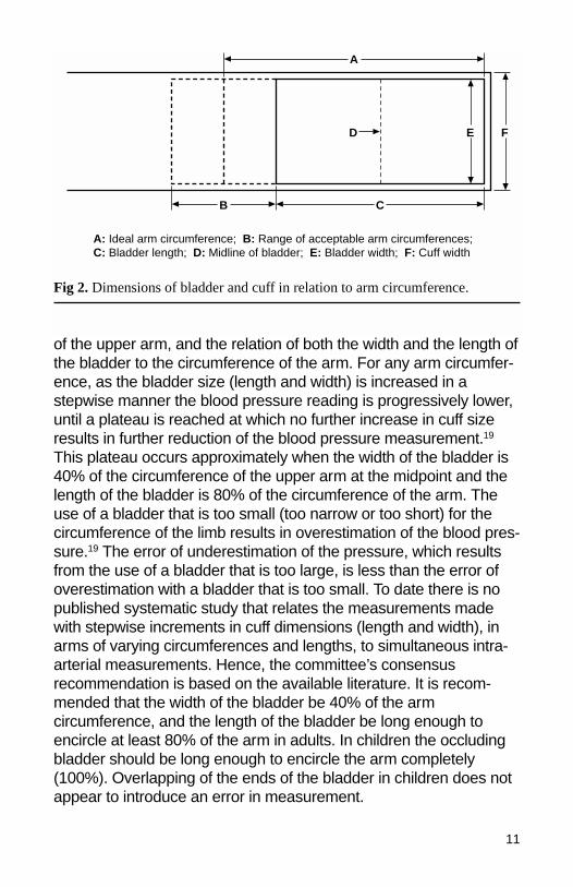
of the upper arm, and the relation of both the width and the length ofthe bladder to the circumference of the arm. For any arm circumfer-ence, as the bladder size (length and width) is increased in astepwise manner the blood pressure reading is progressively lower,until a plateau is reached at which no further increase in cuff sizeresults in further reduction of the blood pressure measurement.19
This plateau occurs approximately when the width of the bladder is40% of the circumference of the upper arm at the midpoint and thelength of the bladder is 80% of the circumference of the arm. Theuse of a bladder that is too small (too narrow or too short) for thecircumference of the limb results in overestimation of the blood pres-sure.19 The error of underestimation of the pressure, which resultsfrom the use of a bladder that is too large, is less than the error ofoverestimation with a bladder that is too small. To date there is nopublished systematic study that relates the measurements madewith stepwise increments in cuff dimensions (length and width), inarms of varying circumferences and lengths, to simultaneous intra-arterial measurements. Hence, the committee’s consensusrecommendation is based on the available literature. It is recom-mended that the width of the bladder be 40% of the armcircumference, and the length of the bladder be long enough toencircle at least 80% of the arm in adults. In children the occludingbladder should be long enough to encircle the arm completely(100%). Overlapping of the ends of the bladder in children does notappear to introduce an error in measurement.
11
A
B
E FD
C
A: Ideal arm circumference; B: Range of acceptable arm circumferences; C: Bladder length; D: Midline of bladder; E: Bladder width; F: Cuff width
Fig 2. Dimensions of bladder and cuff in relation to arm circumference.

The cuffs generally available have been classified by the width ofthe bladder rather than by the length and are labeled “newborn,”“infant,” “child,” “small adult,” “adult,” “large adult,” and “thigh,” butdifferent manufacturers have produced cuffs of varying dimensions(both length and width) under these names because no universalstandards have been established. A large number of cuffs withvarying sizes of bladders are commercially available, but not all ofthese are of the recommended dimensions. Ideally, every cuffshould be labeled with the dimensions of the enclosed bladder; aline should mark the center of the bladder, and two lines should indi-cate the range of arm circumferences for which the bladder issuitable, i.e., encircling 80% to 100% of the circumference (Fig 2).Unlabeled cuffs should be so marked by the user. For cuffs withlonger bladders, a length:width ratio of more than 2:1, the corre-sponding appropriate arm circumference is greater; however, theratio of bladder width to arm circumference should be as close aspossible to 0.40. (See Appendix D for a list of some acceptablebladder sizes and the arm circumferences for which they are suit-able. A number of cuffs with intermediate bladder dimensions areavailable, but to simplify the selection only some of them are listedhere.) Although it is not feasible for every examiner to have all cuffsizes available, except under research conditions, it is stronglyrecommended that the practitioner have several sizes available tomeet the needs of the population served. However, in individualswith very wide but short arms, the appropriately sized bladder maybe difficult to apply. Likewise, in individuals with very large ormuscular arms, even the largest cuff may be inadequate. TheBritish Hypertension Society has recommended the use of one largecuff (12.5 × 35 cm) for all adults with an arm circumference up to 42cm, to avoid the need for multiple cuff sizes.18,20 The use of such acuff could lead to systematic underestimation or overestimation ofthe pressure when the ratio of bladder width to arm circumference isdifferent from 0.40.
Automated devices. Many automated devices are available tomeasure blood pressure by both auscultatory and oscillometrictechniques. The oscillometric method is based on detecting theoscillations on the lateral walls of the occluded artery as the cuff isdeflated. The oscillations begin at approximately the level of systolicpressure and reach their greatest amplitude at the level of meanarterial pressure. Diastolic pressure is a derived value. Systolic
12
blood pressure measurement by these devices is accurate, but dias-tolic pressure, which is derived from the systolic and meanpressures, may not be. The cuffs used for oscillometric measure-ment are constructed without a removable bladder. Althoughabsolute measurements made with these cuffs may vary slightlyfrom those made with standard cuffs, overall trends in blood pres-sure level can be readily tracked. However, serious doubt has beenraised about the accuracy of devices applied to the finger or wristbecause their extreme sensitivity to position results in wide fluctua-tions in blood pressure readings except when they are used understrictly standardized and constant conditions; hence, their use is notrecommended. Doppler devices, which amplify the Doppler signalfrom flowing blood, are also used with standard sphygmomanome-ters and obviate the need for a stethoscope. They are especiallyuseful in taking infants’ blood pressure or in situations where theauscultatory signal is difficult to hear.
Observer
Every person who makes indirect blood pressure measurementsmust be carefully trained and made aware of the potential pitfalls.Several excellent programs, some using videocassettes, providestandardized instruction, training, and testing of observers.21,22
Unfortunately, many health care professionals do not participate inregular retraining programs to improve and reassess their skills inblood pressure measurement, despite considerable variability intheir knowledge, skill, and technique. The observer must be able toconcentrate on the task and have reasonably good eyesight, hearing,and manual dexterity as well as hand-eye-ear coordination. Theobserver must be comfortably positioned to be able to (1) inflate anddeflate the bladder in the cuff gradually, (2) see the manometer andthe meniscus of the column of mercury or the indicator needle onthe aneroid scale, (3) hear the Korotkoff sounds, differentiating themfrom extraneous noises, (4) make note of and remember the level ofthe pressure at the first appearance, at muffling, and at the disap-pearance of the Korotkoff sounds, while continuing to deflate theoccluding cuff, and (5) remember and record the systolic and dias-tolic pressure (Phases IV and V) accurately to the nearest 2 mm Hg.
Observer errors can also result from subconscious biases.Terminal digit preference is caused by the tendency to round pres-sure readings off to numbers ending in zero instead of recording
13

more accurately to the nearest 2 mm Hg. A cut-off or direction biasresults in falsely recording pressures as being above or below apredetermined level or dividing line between “normal” and “hyper-tensive.” An observer may also be influenced by knowledge ofearlier readings.
Subject
For screening and monitoring purposes, the blood pressure ismeasured in the upper arm, with the subject seated (Fig 3). Serialmeasurements should be performed on the same arm for consis-tency. Under clinical circumstances, measurements are oftenperformed in other positions as well. In the initial evaluation ofhypertensive patients, the blood pressure should be measured inboth arms and occasionally in the legs. The subject must becomfortably seated with the midpoint of the upper arm at the level ofthe heart (approximately the fourth intercostal space, when the indi-vidual is seated).23 When the subject is standing, care must betaken to support the raised arm at the level of the heart. When the
14
Fig 3. Blood pressure measurement with the subject seated.

subject is lying down, the arm should be at the side of the body,slightly raised from the bed or examination table, at the level of themiddle of the chest. Ideally the measurements should be made aftera period of rest in a quiet, relaxed setting, not immediately afterexertion or ingestion of coffee or during conversation; the legsshould be uncrossed, with the feet resting firmly on the floor, notdangling, and the back supported, because any form of isometricexercise during the measurement will transiently raise the bloodpressure level.24 Blood pressure levels are affected by environ-mental, emotional, and physical stimuli, so every effort should bemade to standardize the conditions of the measurement, keepingextraneous influences to a minimum. Anticipation of pain or anxietyabout the procedure and its outcome can raise the blood pressurelevel and potentially lead to overestimation of the usual blood pres-sure levels.
Technique
The intent and purpose of the measurement should be explainedto the subject in a reassuring manner and every effort made to putthe subject at ease.
The sequential steps for measuring the blood pressure in theupper extremity, as for routine screening and monitoring purposes,should include the following:
1. Have paper and pen at hand for immediate recording of thepressure.
2. Seat the subject in a quiet, calm environment with his or herbared arm resting on a standard table or other support so themidpoint of the upper arm is at the level of the heart.
3. Estimate by inspection or measure with a tape the circumfer-ence of the bare upper arm at the midpoint between theacromium and olecranon process (between the shoulder andelbow) and select an appropriately sized cuff. The bladderinside the cuff should encircle 80% of the arm in adults and100% of the arm in children less than 13 years old. If in doubt,use a larger cuff. If the available cuff is too small, this shouldbe noted.
4. Palpate the brachial artery and place the cuff so that themidline of the bladder is over the arterial pulsation, then wrap
15

5. and secure the cuff snugly around the subject’s bare upperarm. Avoid rolling up the sleeve in such a manner that it formsa tight tourniquet around the upper arm. Loose application ofthe cuff results in overestimation of the pressure. The loweredge of the cuff should be 1 in (2 cm) above the antecubitalfossa (bend of the elbow), where the head of the stethoscopeis to be placed.
5. Place the manometer so the center of the mercury column oraneroid dial is at eye level and easily visible to the observerand the tubing from the cuff is unobstructed.
6. Inflate the cuff rapidly to 70 mm Hg, and increase by incre-ments of 10 mm Hg while palpating the radial pulse. Note thelevel of pressure at which the pulse disappears and subse-quently reappears during deflation.25 This procedure, thepalpatory method, provides a necessary preliminary approxi-mation of the systolic blood pressure to ensure an adequatelevel of inflation when the actual, auscultatory measurement ismade. The palpatory method is particularly useful to avoidunderinflation of the cuff in patients with an auscultatory gapand overinflation in those with very low blood pressure.
7. Place the earpieces of the stethoscope into the ear canals,angled forward to fit snugly. Switch the stethoscope head tothe low-frequency position (bell). The setting can be confirmedby listening as the stethoscope head is tapped gently.
8. Place the head of the stethoscope over the brachial arterypulsation, just above and medial to the antecubital fossa butbelow the lower edge of the cuff, and hold it firmly in place,making sure that the head makes contact with the skin aroundits entire circumference. Wedging the head of the stethoscopeunder the edge of the cuff may free up one hand but results inconsiderable extraneous noise.
9. Inflate the bladder rapidly and steadily to a pressure 20 to30 mm Hg above the level previously determined by palpation,then partially unscrew (open) the valve and deflate the bladderat 2 mm/s while listening for the appearance of the Korotkoffsounds.26
10. As the pressure in the bladder falls, note the level of the pres-sure on the manometer at the first appearance of repetitive
5. sounds (Phase I) and at the muffling of these sounds (Phase
16

IV) and when they disappear (Phase V). During the period theKorotkoff sounds are audible, the rate of deflation should beno more than 2 mm per pulse beat, thereby compensating forboth rapid and slow heart rates.
11. After the last Korotkoff sound is heard, the cuff should bedeflated slowly for at least another 10 mm Hg, to ensure thatno further sounds are audible, and then rapidly and completelydeflated, and the subject should be allowed to rest for at least30 seconds.
12. The systolic (Phase I) and diastolic (Phase V) pressuresshould be immediately recorded, rounded off (upwards) to thenearest 2 mm Hg. In children, and when sounds are heardnearly to a level of 0 mm Hg, the Phase IV pressure shouldalso be recorded. All values should be recorded together withthe name of the subject, the date and time of the measure-ment, the arm on which the measurement was made, thesubject’s position, and the cuff size (when a nonstandard sizeis used).
13. The measurement should be repeated after at least 30seconds, and the two readings averaged. In clinical situationsadditional measurements can be made in the same or oppo-site arm, in the same or an alternative position.25
Systematic errors that observers often make are listed inAppendix A, as are suggestions for improving the technique.
17
Blood Pressure Recording in Special Situations
Infants and Children
Measuring blood pressure in infants and children presents specialproblems because of their frequent lack of cooperation, although thesame techniques are used as in adults.27-30 Several pediatric cuffsizes are available and should be selected as indicated (seeAppendix D) to ensure that the bladder completely encircles thelimb. Because the Korotkoff sounds are often heard through theentire period of deflation, determining diastolic pressure as Phase IVis recommended in children less than 13 years old. (See “Report ofthe Second Task Force on Blood Pressure Control in Children.”27) Insmall children and infants, the palpatory method is often used forapproximating systolic pressure, even though this may be 5 to 10 mm Hg lower than the level measured by auscultation. In verysmall infants, the blood pressure is often determined by the flushmethod, which involves placing a suitable cuff on the arm or leg,raising the limb, and wrapping the extremity distal to the cuff firmlywith an elastic bandage until the extremity is drained of blood andblanches. The limb is then lowered to heart level, the cuff is rapidlyinflated, and the bandage is removed. As the pressure in the cuff isgradually reduced, flushing of the limb indicates the level at whichflow returns.28 This level corresponds to mean blood pressure but isinaccurate in infants with anemia, hypothermia, or edema. The tech-nique is rarely used now; newer automated oscillometric or Dopplerequipment can be used instead.31
Elderly Patients
In elderly patients who have sclerotic, calcified vessels, it is likelythat the systolic pressure is overestimated by the indirect method ofmeasurement. A readily palpable brachial artery that can be felteven when the cuff has been inflated and the blood flow is inter-rupted (positive Osler’s sign) provides a clue that the measurementmay be inaccurate.32 Under such circumstances, an erroneous diag-
18

nosis of hypertension, or “pseudohypertension,” may be made,although this can only be confirmed by direct measurement.Because postural hypotension is often observed in elderly patients,blood pressure measurement should routinely be made in both thesitting and standing positions, especially in patients who are labeledhypertensive or who are receiving antihypertensive therapy.Because of the tendency for blood pressure to be more labile inelderly patients, it is particularly important to obtain several baselinedeterminations before making diagnostic or therapeutic decisions.
Pregnant Patients
In pregnant women, a rise in blood pressure or the diagnosis ofhypertension has major significance for the outcome of the preg-nancy for both the mother and the fetus. Measuring the bloodpressure of pregnant women is more complicated because of thewide pulse pressure and the need to record both Phase IV andPhase V because sounds can often be heard to 0 mm Hg. In thethird trimester, especially, the mother’s position can affect bloodpressure levels; measurements made with the woman in the leftlateral decubitus position, with the arm above the level of the heart,are often lower than those made in the sitting position with the armat heart level.33
Obese Patients
A longer and wider cuff is needed for adequate compression ofthe brachial artery in the obese patient with a very large upperarm.18,19,34,35 A large cuff may also be required for a big, musculararm with a prominent biceps over which a regular, nontapered cuffmight not fit smoothly. In both situations it is particularly important toplace the center of the bladder over the brachial artery pulse. If theupper arm is relatively short despite the large circumference, it maybe difficult to fit a standard large adult cuff over the arm. The BritishHypertension Society’s recommendation to use a very long cuff(12.5 × 35 cm) could obviate this problem. In the rare patient with anarm circumference greater than 50 cm, when even a thigh cuffcannot be fitted over the arm, it is recommended that the healthcare practitioner wrap the cuff around the patient’s forearm and feelfor the appearance of the radial pulse at the wrist. The accuracy ofthis method has not been validated, but it provides at least a general
19
estimate of the systolic blood pressure. The error of overestimating thepressure when measuring with a cuff that is too small for an obesearm can be considerable and can lead to misclassification of an indi-vidual as hypertensive and to unnecessary concern and therapy.
Miscellaneous Conditions
In patients who are clinically in shock it may be difficult to hear theKorotkoff sounds or palpate a peripheral pulse because of general-ized vasoconstriction. Indirect blood pressure measurements can bevery unreliable in this situation. An approximate estimate of thesystolic pressure can be made using the palpatory method. Thedirect method, using an intra-arterial line, is preferable under thesecircumstances.
In patients who have a high cardiac output (e.g., thyrotoxic orfebrile patients or patients with aortic valve insufficiency, an arterio-venous fistula, or peripheral vasodilatation) and in children, theKorotkoff sounds can often be heard to a level close to 0 mm Hg. Insuch patients the pressure at which Phase IV (muffling) occursshould be used as an approximate index of diastolic pressure, butboth Phase IV and Phase V pressures should be recorded.
In patients who have recently undergone a mastectomy withextensive axillary node dissection or other surgical procedureinvolving the arm or shoulder, it is recommended that blood pres-sure be measured in the opposite arm. Likewise, in dialysis patients,measuring blood pressure in the arm with the arteriovenous fistulashould be avoided.
In patients with cardiac dysrhythmias such as atrial fibrillation orfrequent premature beats, the systolic pressure may vary widelyfrom beat to beat, so the pressures at which the first and lastKorotkoff sounds are heard may differ from measurement tomeasurement. In such situations the rate of deflation should bemore gradual, and multiple blood pressure determinations should bemade and averaged. In patients with a slow heart rate, it is particu-larly important to reduce the pressure in the cuff gradually (2 mm Hgper heart beat) to avoid underestimation of systolic pressure andoverestimation of diastolic pressure.
Disparity in pressure between the two arms may occur in patientswith congenital heart disease, peripheral vascular disease, unilateralneurological and musculoskeletal abnormalities, and aortic dissec-tion. Although the blood pressure should routinely be measured in
20

both arms on the initial examination of a hypertensive patient, underthe above circumstances this recommendation is of particular impor-tance. A consistent difference should be noted, and in hypertensivepatients the arm with the higher pressure should be used for subse-quent measurements. In patients with stenosis of the subclavian,axillary, or brachial artery, the presence of a bruit may interfere withinterpretation of the Korotkoff sounds.
An auscultatory gap, often detected in patients with high systolicpressure, is not abnormal.13,36 However, if the systolic pressure isnot first estimated by palpation, insufficient inflation of the cuff canlead to erroneous designation of the lower end of the gap as thesystolic pressure.
Measurements made during exercise, as during a treadmill test,are difficult to make and often inaccurate when made with currentlyavailable equipment. The use of oscillometric or Doppler equipmentmay provide a more accurate measurement in these situations.
Measuring blood pressure in the lower extremity is indicated insubjects who are suspected of having coarctation of the aorta orother types of obstructive aortic disease. The thigh cuff is used inadults and a large cuff in infants and children. With the subject lyingface down, the cuff is applied with the center of the inflatable bladderover the posterior aspect of the midthigh. The head of the stethoscopeis placed over the artery in the popliteal fossa (behind the knee),and the Korotkoff sounds are monitored as the pressure in thebladder is lowered, in the same manner as in the arms. If the subjectis unable to lie face down, thigh pressures can be taken with thesubject lying on the side or back, with the knee slightly flexed so thestethoscope can be placed over the popliteal pulse easily. The dias-tolic pressure in the legs is usually similar to that in the arms, whilethe systolic pressure may be 20 to 30 mm Hg higher. Systolic pres-sure can be approximated in the lower leg by palpating the posteriortibial or dorsalis pedis pulses while inflating and deflating the cuffapplied to the calf, with the lower end of the cuff above the malleolus.
Self-Measurement or Home Measurement of Blood Pressure
Self-measurement of blood pressure, usually at home, has becomepopular, especially among hypertensive individuals.37 Self-measure-ment facilitates patients’ participation in the health care process andcan simplify titration of antihypertensive drug treatment without the
21

need for frequent visits to a health care facility. It informs both patientand physician of trends or sudden changes in the level of pressure,eliminates the “white-coat” effect of visiting the doctor’s office (seebelow), and permits more frequent sampling of blood pressure.
Patients, like physicians and nurses, need to be carefully trainedand periodically retrained in the techniques of measurement andrecording of blood pressure. The equipment must be portable,simple to use, reliable, and easily calibrated. Manual inflation of thebulb is a form of isometric exercise that can raise the blood pressurewith each inflation, especially in elderly or debilitated individuals.Several automatic and semiautomatic machines have been devel-oped to facilitate self-measurement.38,39 Some give a digital printoutof the pressure and pulse and are completely automatic and self-inflating. Many are based on detection of Korotkoff sounds, andothers use the oscillometric technique. The newer machines areeasier to use than the mercury manometer and stethoscope butrequire periodic recalibration.
Ambulatory Blood Pressure Measurements
Ambulatory blood pressure measurements refers to repeatedmeasurements made away from the medical environment with aportable, automatic (self-inflating) recorder in patients engaging intheir usual activities.39 These measurements are particularly usefulin patients in whom there is a wide disparity between physician- andself-determined pressures, making it difficult to know which bloodpressure level to use for diagnostic and therapeutic decisions.Ambulatory measurements are also useful for relating fluctuations inblood pressure level to symptoms reported by the patient and forassessing the effect of antihypertensive therapy. A number of minia-ture, lightweight, silent, fully automatic machines are now available,but continued validation of their accuracy, calibration, and conformitywith standards is urged.40,41
In most, but not all, hypertensive patients the blood pressureobtained in a physician’s office is higher than the pressuresmeasured during the remainder of the day. If the office pressuresare in the range defined as hypertensive, and the pressures duringthe remainder of the day are in the “normal” range, the patient issaid to have “white-coat hypertension.”42 Patients who respond tothe physician’s examination with an exaggerated rise in blood pres-sure tend to have a lesser pressor response to an encounter with a
22
nurse and may have lower or even normal blood pressures at othertimes, as determined by automatic ambulatory blood pressure moni-toring.43 Patients who have white-coat hypertension may be at riskfor being diagnosed as having persistent hypertension and for beingovertreated. However, because all existing epidemiological studiesof hypertension are based on office blood pressure measurements,further longitudinal research is needed to clarify the role of ambula-tory blood pressure monitoring in the management of patients withraised arterial pressure.
Conclusion
These recommendations for the technique of measuring bloodpressure represent the consensus of the committee and are basedon currently available information and the work of previous commit-tees. The recommendations are subject to further revision as bettervalidation data become available. The intent at this time is toprovide, in the interest of uniformity and consistency, a standardizedtechnique for measuring blood pressure.
23
Appendix A
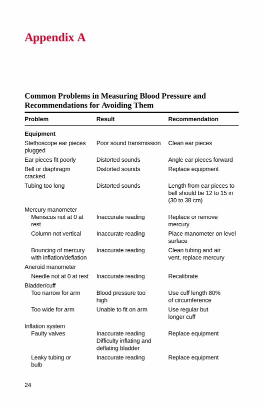
Common Problems in Measuring Blood Pressure andRecommendations for Avoiding Them
Problem Result Recommendation
Equipment
Stethoscope ear pieces Poor sound transmission Clean ear piecesplugged
Ear pieces fit poorly Distorted sounds Angle ear pieces forward
Bell or diaphragm Distorted sounds Replace equipmentcracked
Tubing too long Distorted sounds Length from ear pieces tobell should be 12 to 15 in(30 to 38 cm)
Mercury manometerMeniscus not at 0 at Inaccurate reading Replace or remove rest mercury
Column not vertical Inaccurate reading Place manometer on levelsurface
Bouncing of mercury Inaccurate reading Clean tubing and airwith inflation/deflation vent, replace mercury
Aneroid manometer
Needle not at 0 at rest Inaccurate reading Recalibrate
Bladder/cuffToo narrow for arm Blood pressure too Use cuff length 80%
high of circumference
Too wide for arm Unable to fit on arm Use regular butlonger cuff
Inflation systemFaulty valves Inaccurate reading Replace equipment
Difficulty inflating anddeflating bladder
Leaky tubing or Inaccurate reading Replace equipmentbulb
24

Problem Result Recommendation
Observer
Digit preference Inaccurate reading Be aware of tendency;record blood pressure tonearest 2 mm Hg
Cut-off bias Inaccurate reading Record to nearest 2 mm Hg
Direction bias Inaccurate reading Record to nearest 2 mm Hg
Fatigue or poor Inaccurate reading Write down readingmemory immediately
Subject
Arm below heart level Reading too high Place patient with midpointof upper arm at heart level
Arm above heart level Reading too low Place patient with midpointof upper arm at heart level
Back unsupported Blood pressure Avoid isometric exercise too high during measurement
Legs dangling Blood pressure Avoid isometric exercise too high during measurement
Arrhythmia Blood pressure level Make multiple measure-variable ments and average
Large or muscular arm Blood pressure Use appropriate cuffreading high size
Calcified arteries Blood pressure Note presence of reading high positive Osler sign in
recordTechnique
CuffWrapped too Blood pressure Rewrap more snuglyloosely reading too high
Applied over Inaccurate reading Remove arm from clothing sleeve
ManometerBelow eye level Blood pressure Place manometer at
reading too low eye level
Above eye level Blood pressure Place manometer atreading too high eye level
25
Common Problems (continued)

Problem Result Recommendation
Stethoscope headNot in contact Extraneous noise Place head correctlywith skin
Applied too firmly Diastolic reading Place head correctlytoo low
Not over artery Sounds not well Place head overheard palpated artery
Touching tubing Extraneous noise Place below edge ofor cuff cuff
Palpatory pressure Danger of missing Routinely checkomitted auscultatory gap systolic pressure by
Underestimation of palpation firstsystolic pressure
Inflation level Patient discomfort Inflate to 30 mm Hgtoo high above palpatory blood
pressure
Inflation level Underestimation of Inflate to 30 mm Hgtoo low systolic pressure above palpatory blood
pressure
Inflation rate Patient discomfort Inflate at even ratetoo slow Diastolic pressure
too high
Deflation rate Systolic pressure Deflate at 2 mm Hg/stoo fast too low or 2 mm Hg per beat
Diastolic pressure Deflate at 2 mm Hg/stoo high or 2 mm Hg per beat
Deflation rate Forearm congestion Deflate at 2 mm Hg/stoo slow or 2 mm Hg per beat
Diastolic pressure Completely deflate cuff attoo high end of measurement
In patients in whom the Korotkoff sounds are faint and difficult to hear, thefollowing technique may help: Have the subject raise the arm over the head andmake a fist several times. Inflate the cuff, while the arm is still overhead but thehand relaxed, to a level 50 mm Hg above expected systolic level, have the patientlower the arm rapidly, and measure the blood pressure in the usual manner.Draining the venous blood in this fashion often amplifies the Korotkoff sounds andmakes weak sounds, particularly diastolic sounds, more audible.
26

Appendix B
Recommendations of the Joint National Committee on theDiagnosis, Evaluation, and Treatment of Hypertension forClassifying Blood Pressure Levels for Adults (Aged 18Years and Older)*
Category Systolic (mm Hg) Diastolic (mm Hg)
Normal† <130 and <85
High normal 130-139 or 85-89
Hypertension††
Stage 1 140-159 or 90-99
Stage 2 160-179 or 100-109
Stage 3 ≥180 or ≥110
*Not taking antihypertensive drugs and not acutely ill. When systolic and dias-tolic pressures fall into different categories, the higher category should be selectedto classify the individual’s blood pressure status. For example, 160/92 mm Hgshould be classified as Stage 2, and 174/120 mm Hg should be classified as Stage3. Isolated systolic hypertension is defined as systolic blood pressure ≥140 mm Hgand diastolic blood pressure <90 mm Hg and staged appropriately (e.g., 170/82mm Hg is defined as Stage 2 isolated systolic hypertension).
†Optimal blood pressure with respect to cardiovascular risk is systolic bloodpressure <120 mm Hg and diastolic blood pressure <80 mm Hg. However, unusu-ally low readings should be evaluated for clinical significance.
††Based on the average of two or more readings taken at each of two or morevisits following an initial screening.
In addition to classifying stages of hypertension based on average blood pres-sure levels, the clinician should specify presence or absence of target-organdisease and additional risk factors.
From JNC VI (see reference inside front cover).
27

Appendix C
Precautions Against Contamination With Mercury
Metallic mercury is an element that is liquid at room temperatureand tends to break into tiny, highly mobile droplets when spilled.These droplets vaporize and can contaminate the atmosphere. Inlaboratories, offices, and clinics where mercury manometers areused regularly, precautions must be taken to limit the inhalation,ingestion, or absorption of mercury by personnel in case of a spill orbreakage. It is recommended that health care personnel refer defec-tive mercury manometers for servicing by professionals. In the eventof a mercury spill, the room should be well ventilated and the spilledmercury carefully swept up, not vacuumed, and taken to the labora-tory for disposal. Personnel involved with the regular use of mercurymanometers should be familiar with the available institutional facili-ties for handling mercury spills.
28

Appendix D
Acceptable Bladder Dimensions (in cm) for Arms of Different Sizes*
Arm Bladder Bladder CircumferenceWidth Length Range
Cuff (cm) (cm) at Midpoint (cm)
Newborn 3 6 ≤6
Infant 5 15 6-15†
Child 8 21 16-21†
Small adult 10 24 22-26
Adult 13 30 27-34
Large adult 16 38 35-44
Adult thigh 20 42 45-52
*There is some overlapping of the recommended range for arm circumferencesin order to limit the number of cuffs; it is recommended that the larger cuff be usedwhen available.
†To approximate the bladder width:arm circumference ratio of 0.40 more closelyin infants and children, additional cuffs are available.
29
References
1. An epidemiological approach to describing risk associated withblood pressure levels: final report of the Working Group onRisk and High Blood Pressure. Hypertension. 1985;7:641-651.
2. The fifth report of the Joint National Committee on Detection,Evaluation, and Treatment of High Blood Pressure (JNCV).Arch Intern Med. 1993;153:154-183.
3. Stamler J, Neaton JD, Wentworth DN. Blood pressure (systolicand diastolic) and risk of fatal coronary heart disease.Hypertension. 1989; 13(suppl):I-2-I-12.
4. Assmann G, Schulte H. The Prospective CardiovascularMünster Study: prevalence and prognostic significance ofhyperlipidemia in men with systemic hypertension. Am JCardiol. 1987;59:9G-17G.
5. MacMahon S, Peto R, Cutler J, Collins R, Sorlie P, Neaton J,Abbott R, Godwin J, Dyer A, Stamler J. Blood pressure, stroke,and coronary heart disease: Part I. Prolonged differences inblood pressure: prospective observational studies corrected forthe regression dilution bias. Lancet. 1990;335:765-774.
6. Pickering G. Normotension and hypertension: the mysteriousviability of the false. Am J Med. 1978;65:561-563. Editorial.
7. Sokolow M, Perloff D. The prognosis of essential hypertensiontreated conservatively. Circulation. 1961;23:697-713.
8. Armitage P, Fox W, Rose GA, Tinker CM. The variability ofmeasurements of casual blood pressure: II. survey experience.Clin Sci. 1966;30:337-344.
9. Fredrikson M, Matthews KA. Cardiovascular responses tobehavioral stress and hypertension: a meta-analytic review.Ann Behav Med. 1990;12:30-39.
10. Rosner B, Polk BF. The implications of blood pressure vari-ability for clinical and screening purposes. J Chronic Dis.1979;32:451-461.
30

11. McCutcheon EP, Rushmer RF. Korotkoff sounds: an experi-mental critique. Circ Res. 1967;20:149-161.
12. Frohlich ED, Grim C, Labarthe DR, Maxwell MH, Perloff D,Weidman WH. Recommendations for human blood pressuredetermination by sphygmomanometers. Circulation. 1988;77:501A-514A.
13. Askey JM. The auscultatory gap in sphygmomanometry. AnnIntern Med. 1974; 80:94-97.
14. Prineas RJ, Jacobs D. Quality of Korotkoff sounds: bell vsdiaphragm, cubital fossa vs brachial artery. Prev Med.1983;12:715-719.
15. Sloan PJM, Zezulka A, Davies P, Sangal A, Beevers M,Beevers DG. Standardized methods for comparison of sphyg-momanometers. J Hypertens. 1984; 2:547-551.
16. Bailey RH, Knaus VL, Bauer JH. Aneroid sphygmomanome-ters: an assessment of accuracy at a university hospital andclinics. Arch Intern Med. 1991;151:1409-1412.
17. Karvonen MJ, Telivuo LJ, Järvinen EJK. Sphygmomanometercuff size and the accuracy of indirect measurement of bloodpressure. Am J Cardiol. 1964;13:688-693.
18. van Montfrans GA, van der Hoeven GMA, Karemaker JM,Wieling W, Dunning AJ. Accuracy of auscultatory blood pres-sure measurement with a long cuff. Br Med J (Clin Res Ed).1987;295:354-355.
19. Rastam L, Prineas RJ, Gomez-Marin O. Ratio of cuff width/armcircumference as a determinant of arterial blood pressuremeasurements in adults. J Intern Med. 1990;227:225-232.
20. Petrie JC, O’Brien ET, Littler WA, de Swiet M.Recommendations on blood pressure measurement. Br Med J(Clin Res Ed). 1986;293:611-615.
21. Curb JD, Labarthe DR, Cooper SP, Cutter GR, Hawkins CM.Training and certification of blood pressure observers.Hypertension. 1983;5:610-614.
22. Blood Pressure Measurement: Current Training Programs,Manuals and Instructional Materials. Bethesda, Md: NationalHeart, Lung, and Blood Institute, Education-ProgramInformation Center; 1991.
31
23. Webster J, Newnham D, Petrie JC, Lovell HG. Influence of armposition on measurement of blood pressure. Br Med J (ClinRes Ed). 1984;288:1574-1575.
24. Cushman WC, Cooper KM, Horne RA, Meydrech EF. Effect ofback support and stethoscope head on seated blood pressuredeterminations. Am J Hypertens. 1990;3:240-241.
25. Hill MN, Grim CM. How to take a precise blood pressure. Am JNurs. 1991;91:38-42.
26. King GE. Influence of rate of cuff inflation and deflation onobserved blood pressure by sphygmomanometry. Am Heart J.1963;65:303-306.
27. Task Force on Blood Pressure Control in Children. Report ofthe Second Task Force on Blood Pressure Control inChildren—1987. Pediatrics. 1987;79:1-25.
28. O’Brien ET, O’Malley K. ABC of blood pressure measurement:infancy and childhood. Br Med J. 1979;2:1048-1049.
29. de Swiet M, Dillon MJ, Littler W, O’Brien E, Padfield PL,Petrie JC. Measurement of blood pressure in children: recom-mendations of a working party of the British HypertensionSociety. Br Med J (Clin Res Ed). 1989;299:497.
30. Voors AW. Cuff bladder size in a blood pressure survey of chil-dren. Am J Epidemiol. 1975;101:489-494.
31. Colan SD, Fujii A, Borow KM, MacPherson D, Sanders SP.Noninvasive determination of systolic, diastolic and end-systolic blood pressure in neonates, infants and youngchildren: comparison with central aortic pressure measure-ments. Am J Cardiol. 1983;52:867-870.
32. Messerli FH. Osler’s maneuver, pseudohypertension, and truehypertension in the elderly. Am J Med. 1986;80:906-910.
33. National High Blood Pressure Education Program WorkingGroup report on high blood pressure in pregnancy. Am JObstet Gynecol. 1990;163:1691-1712.
34. Prineas RJ. Measurement of blood pressure in the obese. AnnEpidemiol. 1991;1:321-336.
35. King GE. Errors in clinical measurement of blood pressure inobesity. Clin Sci. 1967;32:223-237.
36. Blank SG, West JE, Müller FB, Pecker MS, Laragh JH,Pickering TG. Characterization of auscultatory gaps with wide-band external pulse recording. Hypertension. 1991;17:225-233.
37. Mejia AD, Julius S, Jones KA, Schork NJ, Kneisley J. TheTecumseh Blood Pressure Study: normative data on bloodpressure self-determination. Arch Intern Med. 1990;150:1209-1213.
38. Hunt JC, Frohlich ED, Moser M, Roccella EJ, Keighley EA.Devices used for self-measurement of blood pressure: revisedstatement of the National High Blood Pressure EducationProgram. Arch Intern Med. 1985;145:2231-2234.
39. Blood pressure monitors. Consumer Reports. 1992;5:295-299.
40. National High Blood Pressure Education Program WorkingGroup report on ambulatory blood pressure monitoring. ArchIntern Med. 1990;150:2270-2280.
41. O’Brien E, Petrie J, Littler W, de Swiet M, Padfield PL,O’Malley K, Jamieson M, Altman D, Bland M, Atkins N. TheBritish Hypertension Society protocol for the evaluation of auto-mated and semi-automated blood pressure measuring deviceswith special reference to ambulatory systems. J Hypertens.1990;8:607-619.
42. Pickering TG, James GD, Boddie C, Harshfield GA, Blank S,Laragh JH. How common is white coat hypertension? JAMA.1988;259:225-228.
43. Mancia G, Parati G, Pomidossi G, Grassi G, Casadei R,Zanchetti A. Alerting reaction and rise in blood pressure duringmeasurement by physician and nurse. Hypertension. 1987;9:209-215.

Editorial
Mercury Sphygmomanometers Should Not be Abandoned:An Advisory Statement From the Council for High BloodPressure Research, American Heart Association
Daniel W. Jones, Edward D. Frohlich, Carlene M. Grim, Clarence E. Grim,Kathryn A. Taubert, for the Professional Education Committee, Council for HighBlood Pressure Research
In healthcare institutions around this country and around the world, mercury sphygmo-manometers are being removed.1 In many situations, the decision to replace the instruments isbeing made without significant input from involved clinicians or consideration of the health risksthat will follow if they are replaced by less accurate devices.
Blood pressure measurement is an important indicator of the current clinical condition ofpatients and a powerful predictor of future cardiovascular and overall health.2 Blood pressuremeasurement is often considered “routine” and is often performed by those with the leasttraining. In many institutions, blood pressure measurement is a low priority, with less than idealquality control related to equipment selection, equipment calibration and repair, and personneltraining and performance.
For more than a century, the mercury gravity sphygmomanometer has been the goldstandard for indirect measurement of blood pressure. Indeed, the world primary standard forpressure measurement is a mercury manometer. It is a simple, gravity-based unit with easy cali-bration, infrequent need for repair, and it has been validated in many clinical circumstancesagainst direct intra-arterial blood pressure measurement.3
In recent years, these mercury units have been replaced with aneroid instruments inmany institutions and more recently with electronic manometers. Justifications for the replace-ment of mercury manometers have included concerns about the safety of mercury, concernsabout regulations regarding the use of mercury in the workplace, and attempts to eliminatehuman error involved in the reading of measurements.4 An examination of the evidence aroundthese concerns is necessary before clinicians contribute actively or passively to the replace-ment of these instruments.
Are mercury manometers dangerous to use in hospital and other clinical settings?Accidental exposure to mercury from sphygmomanometers used in healthcare settings isextremely rare. It is true that there have been a few isolated instances of illness in children frommercury toxicity related to broken elemental mercury-containing instruments used in homes.Most of these occurrences have been related to broken glass thermometers.5 One detailedreport has provided data suggesting that volatilized mercury, after spillage of mercury, producedreversible neurological symptoms.6 Nevertheless, modern mercury sphygmomanometers areavailable in models that prevent accidental spillage of mercury, which essentially eliminates theconcern for this rare occurrence.
Is the use of mercury manometers forbidden by regulation in the United States? No.Most regulations related to mercury deal with mercury compounds, such as those used in themanufacture of some automobile batteries. The use of mercury manometers is presentlyrestricted in only a handful of countries.1 None of the healthcare regulatory agencies in theUnited States, governmental or voluntary, forbid the use of mercury manometers. However,reports of loss of hospital accreditation in the United States (for whatever reasons) haveprompted widespread concern. Notwithstanding, mercury instruments are approved and arelegal devices in this country, and we believe they should remain so.
Are aneroid or electronic instruments a reliable substitute for mercury manometers?There are 2 crucial issues to consider here: validation and calibration. Although both aneroidand electronic instruments have some advantages of portability and ease of use, few of theseinstruments have had adequate validation. Still fewer of these instruments are calibrated regu-larly. To be sure, these instruments have a place in patient management, particularly withrespect to their use as home monitoring devices. However, most of these instruments have notbeen adequately validated over a wide range of blood pressures, ages, and clinical conditionsto warrant routine use in hospitals and outpatient settings.7 What is of critical importance is thatmost manufacturers of aneroid and electronic instruments recommend calibration against amercury manometer every 6 months. However, few hospitals and clinics have a regularprogram of evaluation and calibration. Most of these instruments cannot be calibrated withoutreturn to the manufacturer.8
Can the use of electronic instruments eliminate human error in blood pressure determi-nation? Certainly, a challenging aspect of human blood pressure determination with a mercury
34

sphygmomanometer is the human error introduced with the hearing and recording of theKorotkoff sounds. Hearing impairment and digit preference are 2 major concerns. Electronicinstruments do offer an advantage in this area but still leave ample room for other causes ofhuman error in cuff size determination, placement of the cuff, etc. It is apparent that all humanerror cannot be eliminated with electronic devices.
Is accurate blood pressure measurement really important? Obviously the answer is yes.Consistent overestimation of blood pressure in the population can be associated with costlyovertreatment of hypertension. Consistent underestimation of blood pressure can cost manylives in failing to prevent cardiovascular disease through effective and safe therapy of hyperten-sion.2
There is a constant need for caution in the selection of blood pressure measuringdevices. New is not always better. Just as in the selection of medication for elevated blood pres-sure, evidence should guide our decisions. Sometimes, evidence presents us with unexpectedoutcomes. We encourage consideration of the same level of evidence in the selection of bloodpressure measuring devices as in the selection of drugs and other medical instruments.Clinicians involved in the management of patients with blood pressure problems must acceptthe responsibility for ensuring adequate instruments are available. If we are passive muchlonger, the time to act effectively will be past.
Specifically, we recommend that clinicians educate themselves on the instruments avail-able for use in their clinics and hospitals; engage in the process of selection of instrumentsthrough dialogue with administrators and through hospital committees; encourage the generaluse of mercury manometers as the instrument of choice until other instruments are better vali-dated; where aneroid or electronic instruments are used, ensure that instruments are validatedthrough the Association for the Advancement of Medical Instrumentation or a similar organiza-tion; ensure a program of regular maintenance and calibration of all instruments. In sites whereaneroid or electronic instruments are used exclusively, if mercury instruments cannot be reintro-duced for regular use, insist on the use of mercury instruments for calibration of aneroid andelectronic instruments; ensure a regular training program for those who measure blood pres-sure; join the American Heart Association in encouraging studies to validate the safety andreliability of all instruments used for blood pressure determination; and use evidence in deter-mining both safety and reliability of any instrument.
To these ends, the American Heart Association stands ready to relate with any and allorganizations wishing to further explore this issue. Throughout 6 published editions on humansphygmomanometry for patients, the American Heart Association has presented updated infor-mation on this vital subject. We stand ready to consider any new views and data that aregermane to the publication of our next report. References
1. Markandu ND, Whitcher F, Arnold A, Carney C. The mercury sphygmomanometershould be abandoned before it is proscribed. J Hum Hypertens. 2000;14:31–36.
2. The sixth report of the joint national committee on prevention, detection, evaluation, andtreatment of high blood pressure. Arch Intern Med. 1997;157:2413–2446.
3. Perloff D, Grim C, Flack J, et al, for the Writing Group. Human blood pressure determi-nation by sphygmomanometry. Circulation. 1993;88:2460–2467.
4. Obrien E. Will mercury manometers soon be obsolete? J Hum Hypertens.1995;9:933–934.
5. Langford N, Ferner R. Toxicity of mercury. J Hum Hypertens. 1999;13:651–656.6. Rennie AC, McGregor-Schuerman M, Dale IM, Robinson C, McWilliam R. Mercury
poisoning after spillage at home from a sphygmomanometer on loan from hospital. BMJ.1999;319:366–367.
7. Yarrows SA, Brook RD. Measurement variation among twelve electronic home bloodpressure monitors. Am J Hypertens. 2000;13:276–282.
8. Carney SL, Gillie AH, Green SL, Patterson O, Taylor MS, Smith AJ. Hospital blood pres-sure measurement: staff and device assessment. J Qual Clin Pract. 1999;19:95–98.
35
National Center
7272 Greenville Avenue
Dallas, Texas 75231-4596
americanheart.org
70-1207 12/01
For heart- or risk-related
information, call 1-800-AHA-USA1
(1-800-242-8721) or contact your
nearest office. You can also visit us
online at americanheart.org
For stroke information, call our
American Stroke Association at
1-888-4-STROKE (1-888-478-7653),
or visit StrokeAssociation.org. For
information on life after stroke, call
and ask for the Stroke Family
Support Network.
Your contributions will support
research and educational programs
that help reduce disability and
death from America’s No. 1 killer.
printed on recycled paper