
Domoic Acid Toxicity in California Sea Lions (Zalophus ...
48
Domoic Acid Toxicity in California Sea Lions (Zalophus californianus) Stranded Along the Central California Coast, May-October 1998 Report to the National Marine Fisheries Service Working Group on Unusual Marine Mammal Mortality Events Frances Gulland U.S. Department of Commerce National Oceanic and Atmospheric Administration National Marine Fisheries Service NOAA Technical Memorandum NMFS-OPR- 17 December 2000
Transcript of Domoic Acid Toxicity in California Sea Lions (Zalophus ...

Domoic Acid Toxicity in California Sea Lions (Zalophus californianus) Stranded Along the Central California Coast, May-October 1998
Report to the National Marine Fisheries Service Working Group on Unusual Marine Mammal Mortality Events
Frances Gulland
U.S. Department of Commerce National Oceanic and Atmospheric Administration National Marine Fisheries Service
NOAA Technical Memorandum NMFS-OPR- 17 December 2000

'This technical memorandum series is used for documentation and timely communication of preliminary results, interim reports, or similar special-purpose information. Although the memoranda are not subject to complete formal review, editorial control, or detailed editing, they are expected to reflect sound professional work. The views and conclusions expressed by the authors are not necessarily those of the National Marine Fisheries Service. In addi- tion, the mention of trade names or commercial firms is for information only and does not imply endorsement by the National Marine Fisheries Service.
In response to large numbers of California sea lions washing ashore dead and in obvious physical distress, individuals from the Marine Mammal Center, other stranding network par- ticipants, National Marine Fisheries Service, as well as numerous other State and Federal partners, were called into action to give aid to the animals and determine the cause of the event. In the course of this mortality event, many individuals with expertise in various disciplines contributed their efforts and insight into the initial response, sample collection, and final analyses. The results of their investigations are in this report. This report was prepared by the Marine Mammal Center under contract 40AAND801390.
Suggested citation:
Gulland, F. 2000. Domoic acid toxicity in California sea lions (Znlophus cnliforninnus) stranded along the central California coast, May-October 1998. Report to the National Marine Fisheries Service Working Group on Unusual Marine Mammal Mortality Events. U.S. Dep. Cornmer., NOAA Tech. Memo. NMFS-OPR-17, 45 p.

Domoic Acid Toxicity in California Sea Lions (Zalophus californianus) Stranded Along the Central California Coast, May-October 1998
Report to the National Marine Fisheries Service Working Group on Unusual Marine Mammal Mortality Events
Frances Gulland The Marine Mammal Center 1065Ft Cronkhite Sausalito, CA 94965
NOAA TechnicalMemorandum NMFS-OPR-17 December2000
U.S. Department of Commerce Norman Y. Mineta, Secretary
National Oceanic and Atmospheric Administration D. James Baker, Under Secretary for Oceans and Atmosphere
National Marine Fisheries Service Penelope D. Dalton, Assistant Administrator for Fisheries
TABLE OF CONTENTS
Expert Team of Investigators
Abstract
Introduction
Epidemiology
Clinical Signs
Clinical Pathology
Treatment
Gross Post Mortem Findings
Microbiology
Histopathology
Toxicology
Plankton Bloom
Diagnosis
Post Release Monitoring
Discussion
Acknowledgments
References
List of terms
APPENDICES
Appendix 1. Accession numbers, names, stranding site and date, age and disposition of animals stranding during the unusual mortality event. l a Animals that died during the event 37 l b Animals that survived and were released 39
Appendix 2. Clinical hematology and chemistries of stranded animals 2a. Hematology 40 2b Hematology 41 2c Blood chemistries 42
Appendix 3. Hematology and chemistries, pre-release 43 Appendix 4. Neuroclinical examination sheet 45

EXPERT TEAM OF INVESTIGATORS
A multi-disciplinary team of experts participated in response, data collection, and analyses of this mortality event. They are listed by their area of expertise.
Veterinary Medicine and Pathology Frances Gulland The Marine Mammal Center Marin Headlands Sausalito, CA 94965
Marty Haulena The Marine Mammal Center Marin Headlands Sausalito, CA 94965
Tom Lipscomb Armed Forces Institute of Pathology Veterinary Pathology Washington DC 20306-6000
Linda J. Lowenstine Department of Veterinary Pathology, Microbiology and Immunology University of California at Davis Davis, CA 95616
Teri Rowles National Marine Fisheries Service 13 15 East West Highway Silver Spring, MD 20910
Terry Spraker Colorado State University College of Veterinary Medicine Fort Collins, CO 80523
Toxicology Mark Busman Marine Biotoxins Program NOAADJational Oceanic Service Center for Coastal Environmental Health and Biomolecular Research 2 19 Fort Johnson Road Charleston, SC 294 12
Greg Doucette Marine Biotoxins Program NOAMational Oceanic Service Center for Coastal Environmental Health and Biomolecular Research 2 19 Ft Johnson Road Charleston, SC 294 12
Greg Langlois California Department of Health Services 2 15 1 Berkeley Way Berkeley, CA 94704- 10 1 1
Christine Powell Marine Biotoxins Program1 NOAMational Oceanic Service Center for Coastal Environmental Health and Biomolecular Research 2 19 Fort Johnson Road Charleston, SC 294 12
Chris Scholin Monterey Bay Aquarium Research Institute P.O. Box 628 Moss Landing Monterey, CA 95039
Vera Trainer Marine Biotoxin Group NOAADJMFSECD 2725 Montlake Blvd. Seattle, WA 98 1 12

Fran Van Dolah Marine Biotoxins Program NOAA/National Ocenaic Service Center for Coastal Environmental Health and Biomolecular Research 2 19 Fort Johnson Road Charleston, SC 294 12
Kathy Lefebvre Institute of Marine Science University of Santa Cruz Santa Cruz, CA 95064
Pinniped Biology and Management
Joe Cordaro National Marine Fisheries Service Southwest Region 501 West Ocean Boulevard, Suite 4200 Long Beach, CA 90802-42 13
Robert DeLong National Marine Mammal Laboratory 7600 Sand Point Way Seattle, WA 98 1 15-0070
Jim Harvey Moss Landing Marine Laboratories P.O. Box 450 Moss Landing, CA 95039
William McLellan Biological Sciences University of North Caroline, Wilmington Wilmington, NC 28403
Teri Rowles National Marine Fisheries Service 13 15 East West Highway Silver Spring, MD 20910
Epidemiology
Scott Benson Moss Landing Marine Laboratories P. 0. Box 450 Moss Landing, CA 95039
Andrew DeVogelaere Monterey Bay National Marine Sanctuary 299 Foam St. Monterey, CA 93940
L. Grella Gulf of the Farallones National Marine Sanctuary San Francisco, CA 94 123
Jan Mortenson Farallones Marine Sanctuary Association P.O. Box 29386 San Francisco, CA 94129
Jan Roletto Farallon Islands National Marine Sanctuary Ft Mason Bldg. 201 San Francisco, CA 94 123

ABSTRACT
Between May 15 and June 19, 1998, 70 California sea lions (Zalophus calijornianus) and one northern fur seal (Callorhinus ursinus) stranded along the central California coast from San Luis Obispo to San Mateo County. Of these 70 animals, 54 were adult females, with 27 (50 %) pregnant; three were subadult females; two were juvenile females; one was a yearling female; six were subadult males and four were juvenile males (Appendix 1). All animals were in good nutritional condition and displayed similar clinical signs that were predominantly neurological. The animals had'ievere seizures that either became increasingly frequent, resulting in opisthotonus, then death, or became less frequent with the animals showing ataxia and decreased responsiveness to stimuli between seizures and eventually becoming clinically normal. Forty-eight of the 70 animals (69 %) died despite treatment.
Treatment consisted of supportive care with oral and subcutaneous fluids, antibiotic cover with penicillin, control of seizures using diazepam, lorazepam, and phenobarbitone, and reduct ion o f ce rebra l e d e m a us ing dexamethasone. Induction of parturition using dexamethasone and prostaglandin F2a was attempted in adult females with open cervices and dead intrauterine fetuses.
Hematological parameters in these stranded sea lions were within normal limits, as were serum biochemical profiles other than creatine kinase levels. The creatine kinase levels were elevated in most animals, presumably as a consequence of muscular damage during seizures. Levels of blood lead were normal in the eight affected animals tested, and brain cholinesterase levels were normal in the five affected animals tested.
Virus neutralization (VN) tests for phocine distemper virus (PDV) detected low levels of antibody in 10 of 34 animals (29 %). After two weeks, four of these 10 animals showed low positive, but rising, titers on VN. No signs of respiratory disease or novel neurologic signs were detected. After a further
month, these four animals were seronegative to PDV. Three animals that had been in contact with se ropos i t ive an imals r emained seronegative. A retrospective survey of banked sera from 100 adult California sea lions that had stranded previously revealed that 20 % had low titers to PDV.
A variety of non-specific lesions were observed on gross post mortem examination. These included gastric ulceration and erosion with associated lymphadenopathy, bile stasis, pulmonary congestion and occasional subcutaneous hemorrhages. Sea lions that died within the first two days of stranding had diffusely pale myocardium with occasional focal areas of severe pallor. All pregnant females had necrotic placentae and dead fetuses.
Bacteria isolated from tissues at post mortern were typical of those isolated from stranded California sea lions in recent years. A novel calicivirus was isolated from myocardium of one animal (#3709).
The predominant histologic lesion in affected animals was neuronal necrosis, that was most severe in zones CA3 and CA4 of the hippocampi and the dentate gyri. There were also intramyelinic and neuropil edema and occasional foci of gliosis. In some animals that died within 48 hours of stranding, there was m u l t i f o c a l myocard ia l n e c r o s i s and inflammation.
Domoic acid, a biotoxin produced by a diatom, was detected in serum of 3 of 7 animals, urine 7 of 14 animals, and feces of 3 of 9 animals. Two of the positive fecal samples were examined by electron microscopy, and frustules of Pseudo-nitzschia australis were observed. No domoic acid was found in kidney, stomach washings, cerebrospinal fluid, or brain samples from affected animals. Analyses were carried out using a receptor binding assay (RBA) and High Performance Liquid Chromatography with Ultraviolet detection (HPLCIUV). HPLC 1 mass spectrometry (HPLCMS and HPLCMSMS) carried out on a subset of samples from each tissue type provided independent confirmation of the chemical identity of the biotoxin.
A bloom of Pseudo-nitzschia australis

(P.australis) occurred in Monterey Bay during the latter half of May 1998, reaching its peak on or about May 22. The greatest concentration of P. australis recorded was -200,000 cells per liter. Cells were concentrated near shore, possibly in response to nutrients of terrestrial origin brought to coastal waters by enhanced river outflow. Plankton samples were also analyzed for domoic acid using a receptor binding assay. The rise and fall of domoic acid in these samples corresponded to the rise and fall of P. australis observed in the plankton. Anchovies collected from the bay on May 22, 1998 had levels of domoic acid of 105.6 pg domoic acidtg tissue, whereas fish collected on June 10, 1998 had no detectable levels of domoic acid. Anchovies collected during the peak of the P. australis bloom that contained high amounts of domoic acid had P. australis frustules within their stomachs. In Monterey Bay, the bloom of P. australis was followed by a bloom of P. pseudodelicatissima. Anchovies collected from Monterey Bay reflected this change in species abundance in their stomach contents. No P. australis frustules or domoic acid were detected in stomach contents of these fish, and no domoic acid was detected in plankton during the P. pseudodelicatissima bloom.
Three of the 23 sea lions (#38 15, #3822 and #3815) that survived treatment during the event in May and June 1998 were equipped with satellite and radio-transmitters prior to their release on November 15, 1998. Battery life of the satellite transmitters was three months. Satellite and re-sight data indicated that sea lions survived for at least 48, 64 and 94 days, respectively. Two sea lions traveled as far south as the Channel Islands, whereas the third sea lion remained in the vicinity of Aiio Nuevo Island. All three sea lions that were sightedduring the three-month telemetry period appeared healthy and displayed normal behavior.
During routine surveys around the Monterey Bay during the months of May and June 1998, a 3.5 fold increase in numbers of
dead beach-cast birds and p i ~ i p e d s was detected compare to the same months of the previous year. The majority of the dead birds were Common Murres, Surf Scoters and Sooty Shearwaters. Large numbers of stranded sea lions were also detected along other central California beaches at the time. The extent to which these mortalities was due to domoic acid toxicity rather than as a consequence of starvation due to El Niiio conditions is unclear, since most animals were not examined.
From July 2 to October 17, 1998, an additional 11 California sea lions stranded displaying similar clinical signs to those that stranded during late May and early June. Nine of these animals stranded between October 3 and October 17. Four animals survived and seven died. The dead animals had histologic lesions similar to those in the earlier cases. Domoic acid was detected in urine of two of these animals by the microplate assay. At the same time (early October), cells of P. australis were observed in Monterey Bay around the Santa Cruz and Capitola piers, but were at relatively low concentrations (<10,000 cells per liter). However, further offshore (outside of Monterey Bay), concentrations of P. australis were in excess of 100,000 cells per liter. It is likely that the source of domoic acid affecting sea lions during October was a bloom outside of Monterey Bay.
In summary, the combination of clinical signs, histopathological, toxicological, epidemiological and oceanographic changes led to the conclusion that domoic acid toxicity was the cause of this Marine Mammal Unusual Mortality Event. Domoic acid was first reported as a cause of toxicity in humans in 1987, when four people died and approximately one hundred were clinically ill following ingestion ofcontaminated mussels on Prince Edward Island, Canada. This event is the first documented occurrence of domoic acid toxicity in marine mammals.
-

5
INTRODUCTION
Within the California Marine Mammal Stranding Network, response to stranded marine mammals is shared between a number of organizations under the direction of the NMFS Southwest Regional Stranding Coordinator, Joe Cordaro. The Marine Mammal Center (TMMC) responds to calls on live animals from San Luis Obispo County in the south to Mendocino County in the north. Calls on dead marine mammals in the same range are responded to by a number of organizations, usually a different one in each county. The type of response and extent of sampling beyond the collection of level A data varies between agencies within the California Marine Mammal Stranding Network, due to different interests of volunteer members. In El Niiio years, there is typically an increase in the number of pinnipeds, especially California sea lion (Zalophus californianus) pups and yearlings, stranding along the central California coast (Cordaro, 1997). In late 1997 and early 1998, strandings of emaciated animals increased as predicted in association with changes in sea surface temperature and decreasing food availability. The majority of these animals were emaciated, heavily parasitized California sea lions. In May and June 1998, 334 yearling sea lions were admitted to TMMC, compared to 92 in the same months of 1997. Strandings of adults are rare, and usually a consequence of human interactions or neoplasia (Gulland el al., 1996). Thus, although TMMC admitted 334 yearling California sea lions in May and June 1998, the stranding of 70 adults and subadults in good nutritional condition showing severe neurologic signs was an unusual occurrence.
EPIDEMIOLOGY
Live stranded sea lions From May 18 to June 19, 1998, 70
California sea lions (Zalophus californianus) and one northern fur seal (Callorhinus ursinus) stranded live along the central California coast between Oceano Dunes, San Luis Obispo County in the south and Half Moon Bay, San
Mateo County, in the north. This unusual mortality event has been defined by these dates. A second event of 11 sea lions showing similar signs stranded between July 12 and October 17, with nine of them stranding in the two weeks from October 3, 1998. This second mortality event will be described in the discussion section of this document.
Criteria for inclusion in this unusual mortality event were as follows. All live animals included in this study displayed severe neurologic signs, were in good nutritional condition, and showed no other clinical signs of primary traumatic diseases or chronic infectious diseases. Dead animals were included if they showed good nutritional condition and severe neurologic signs prior to death or consistent pathologic findings of neurologic lesions like those described in the following pathology section. Evidence of tumors or "normal" parasitic diseases did not exclude animals in this study if the above criteria were met. Dead animals for which there was minimal diagnostic information were not included, therefore the 70 animals identified are likely to be an underestimate of the actual numbers of animals involved.
Figure 1 shows the spatial and temporal assessment of the strandings of the animals included in this event. The majority of the animals that stranded in the first week of the mortality event stranded along Oceano Dunes. In the second week of the mortality event, animals stranded in both Monterey Bay and Oceano Dunes, and on June 14 one animal stranded north of Monterey Bay, in San Mateo County. All live animals admitted were transported to TMMC by volunteers following calls from thepublic about animals observed in distress. No surveys for affected animals were undertaken. The distribution of stranding sites was therefore affected by the intensity of public visitation to beaches along the coast. The area between the south end of Monterey Bay and Oceano Dunes, Big Sur, is inaccessible to the public due to lack of roads along the beach, so it is unclear whether or not clinically affected sea lions stranded along this stretch of coastline.

The majority of clinically affected animals were adult females, 50% of which were pregnant (Table 1). Although juvenile animalswere of both sexes, there were no adult male sea lions affected. This may be due to sex differences in dispersal along the California coast. The majority of the other 334 California sea lions that were admitted to TMMC in May and June 1998 were emaciated yearlings of both sexes.
Sea lions (other than yearlings) were aged by counting the number of dentine annuli in longitudinal sections of the upper left canine by Dr. Sue Chivers at the South West Fisheries Science Center, La Jolla California and Dr. R. DeLong, at the National Marine Mammal Laboratory (NMML) in Seattle, Washington. Ages of tooth-aged animals are listed in Appendix 1, and ranged from 3 to 13 years.
Table 1. Age stnicture of total affected and number of California sea lions which stranded or survived from May 16 to June 14, 1998.
Age Class Females, affected Females, survived Males, Males, survived affected
Adult 54 12
Subadult 1 6 4
Juvenile 2 4 2
Weanling 1
Figure 1. Stranding sites for sea lions stranding during the Unusual Mortality event in successive time periods in 1998. Numbers of animals stranding in each time period at each site are given within the circles.

Dead stranded marine mammals and mortality of other species Data on stranded marine birds and dead marine mammals in the Monterey Bay were collected by the Monterey Bay National Marine Sanctuary BeachCOMBER program. The monitoring plan examines the sandy beaches within Monterey Bay and Carmel Bay, divided into 10 roughly equal-length segments for sampling. Data on beach-cast birds and mammals along the sandy beaches north of Aiio Nuevo Island to Bodega Head, Sonoma County, were collected by the Gulf of the Farallones National Marine Sanctuary (GFNMS) Beach Watch program. Pairs of volunteers survey these pre-defined segments on a monthly basis. Surveys are conducted during the first week of each month at low tide; therefore, encountered beach-cast animals represent those deposited during the previous month. For each encountered carcass, the following information is recorded: species, stage of decomposition, age, sex, evidence of scavenging, evidence for the cause of death, the presence of oil, and whether or not a photograph was obtained. A comments section is provided for documentation of any tags present on the carcass, length measurements, photograph roll and frame numbers, or any notes that would aid in post-identification of the encountered carcass. For seabirds, a toe is clipped each month in which the carcass is encountered, allowing determination of residence times and of the number of newly deposited birds. Prior to clipping a toe, the volunteer documents the
number of toes previously removed. Beach-cast marine mammals are not marked.
The results of surveys in May and June 1997 and 1998 are shown in Table 2. They refer only to newly deposited birds, defined as birds with no previously clipped toes. Total beach- cast deposition of marine birds during May and June 1998 was greater at all beaches throughout the study area compared to the same period in 1997. In GFNMS, numbers of encountered carcasses were higher than the mean for these months over the previous three years (1994-1997). The causes of this elevated deposition are not clear. Many foraging guilds seem to have been affected, and overall species diversity was greater in 1998. In particular, overwintering seabirds were more frequently found beach-cast during the May and June 1998 surveys compared to the same period in 1997 (even though May 1997 was the first survey and therefore would be expected to have higher counts because it includes deposition for >1 month). Although no comparable data exist for April 1997, deposition rates of overwintering birds for April 1998 were also high. This indicates that resource limitations (perhaps caused by El Niiio) during the winter months may have been a factor contributing to elevated seabird mortality from March through early June.
Table 2. Number of Dead Marine Birds and Mammals Reported by BeachCOMBER in Monterey Bay
1 Marine Birds
Common Murre + May 1997
Total
83
12
%
51.2
7.4
June 1997
Total
46
4 1
%
32.2
28.7
April 1998
Total
83
15
%
13.3
2.4
May 1998
Total
149
69
%
26.2
12.1
June 1998
Total
239
110
%
44.2
20.3
10 6.2 10 7.0 2 0.3 5 0.9 7 1.3
9 5.6 1 1 7.7 46 7.4 12 2.1 1 0.2
I Pacific Loon 9 5.6 5 3.5 45 7.2 46 8.1 29 5.4


9
Clark'sIWestern
1 0.2Ashy Storm-Petrel
1 0.2Laysan Albatross
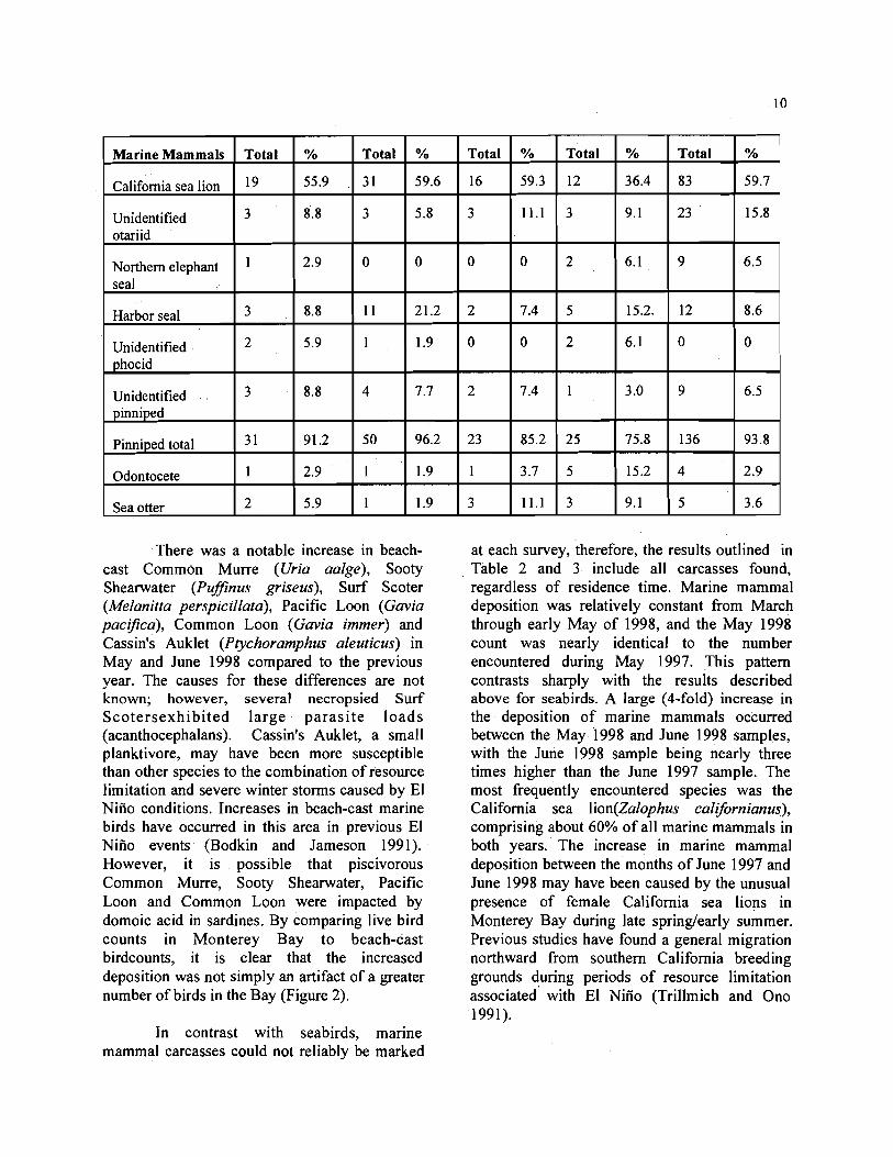
Marine Mammals Total % Total % Total % Total % Total %
California sea lion 55.9 31 59.6 16 59.3 12 36.4 83 59.7
Unidentified otariid
3 8.8 3 5.8 3 11.1 3 9.1 23 15.8
Northern elephant seal
2.9 0 0 0 0 2 6.1 9 6.5
Harbor seal 3 8.8 11 21.2 2 7.4 5 15.2. 12 8.6
Unidentified phocid
2 5.9 1 1.9 0 0 2 6.1 0 0
Unidentified 3 8.8 4 7.7 2 7.4 1 3.0 9 6.5
There was a notable increase in beach- cast Common Murre (Uria aalge), Sooty Shearwater (PufJinus griseus), Surf Scoter (Melanitta perspicillata), Pacific Loon (Gavia pacifica), Common Loon (Gavia immer) and Cassin's Auklet (Ptychoramphus aleuticus) in May and June 1998 compared to the previous year. The causes for these differences are not known; however, several necropsied Surf Scotersexhibited large parasi te loads (acanthocephalans). Cassin's Auklet, a small planktivore, may have been more susceptible than other species to the combination of resource limitation and severe winter storms caused by El Niiio conditions. Increases in beach-cast marine birds have occurred in this area in previous El Niiio events (Bodkin and Jameson 1991). However, it is possible that piscivorous Common Murre, Sooty Shearwater, Pacific Loon and Common Loon were impacted by domoic acid in sardines. By comparing live bird counts in Monterey Bay to beach-cast birdcounts, it is clear that the increased deposition was not simply an artifact of a greater number of birds in the Bay (Figure 2).
In contrast with seabirds, marine mammal carcasses could not reliably be marked
at each survey, therefore, the results outlined in Table 2 and 3 include all carcasses found, regardless of residence time. Marine mammal deposition was relatively constant from March through early May of 1998, and the May 1998 count was nearly identical to the number encountered during May 1997. This pattern contrasts sharply with the results described above for seabirds. A large (4-fold) increase in the deposition of marine mammals occurred between the May 1998 and June 1998 samples, with the June 1998 sample being nearly three times higher than the June 1997 sample. The most frequently encountered species was the California sea lion(Za1ophus calijornianus), comprising about 60% of all marine mammals in both years. The increase in marine mammal deposition between the months of June 1997 and June 1998 may have been caused by the unusual presence of female California sea lions in Monterey Bay during late spring/early summer. Previous studies have found a general migration northward from southern California breeding grounds during periods of resource limitation associated with El Niiio (Trillmich and Ono 1991).

t \
Sooty Shearwater
!"_ 1000
S 500 0 z
Ppril May June July
Month \ /
f \ Common Murre
5 300 250 200
O 150 t l o o n
50 0z
April May June July
Month
\ /
Figure 2. Counts of live and beachcast Common Murres and Sooty Shearwaters in Monterey Bay during 1998.

I
Table 3. Number of dead California sea lions reported to the California Marine Mammal Stranding Network in May and June 1998.
County
San Luis Obispo
Monterey
Santa Cruz
San Mateo
County
Monterey
Santa Cruz
San Mateo
Total
MAY 1998 1
Condition Pup Yearling Subadult Adult Unknown Total
Dead 0 0 0 2 6 8
Alive 11 25 24 30 0 79
Dead I 3 2 1 2 9
Alive 9 3 1 7 17 0 64
Dead 0 0 0 0 0 0
Alive 0 13 4 8 0 25
Dead 3 6 1 3 2 15
Alive 3 10 1 0 0 14
27 8 8 39 6 1 10 214 JUNE 1998 1
Condition Pup Yearling Subadult Adult Unknown Total
0 0 0-----2 13
Alive 4 20
Dead 12 8
Alive 6 36
Dead
Alive 0 31
Dead 3 24
Alive 3 7
28 126
CLINICAL SIGNS
The characteristic clinical signs in affected sea lions were ataxia, head weaving, muscle tremors, tetanic convulsions, rubbing and lethargy. Forty-eight (69 %) of the initial 70 affected California sea lions died or were euthanized. In animals that died, seizures became increasingly frequent, often progressing to status epilepticus before the animal died. If animals were repeatedly in status epilepticus for over an hour, and showed no improvement in clinical signs over subsequent days, or were comatose, they were euthanized (n=22). The seizures varied from a few minutes in duration, to over 30 minutes. The seizure duration was often affected by treatment, as seizuring sea lions were treated with diazepam, lorazepam and phenobarbitone symptomatically to alleviate tetany. Frequency of seizures varied from one to
8 1 0 33
12 11 3 47
14 0 56
4 2 0 37
5 4 4 40
3 0 0 13
46 20 20 24 1
30 in 24 hours. In animals that survived, the frequency of seizures gradually decreased over a one month period. Seizures were usually bilateral, but two animals repeatedly had unilateral seizures. Between seizures, sea lions showed varylng degrees of lethargy and occasionally a characteristic rubbing behavior, in which the animals would rub the back of their necks and dorsum by rolling repeatedly on the pen floor. As assessment of the lethargy was subjective, a neurologic examination sheet (Appendix 4) was developed that could be used to give animals a numeric score. This could be evaluated daily by different personnel, and scores compared between days to determine an animal's clinical progress. A higher Lscore would equate with a worse neurologic condition. In the first days after stranding, most animals had scores above 20. Apparently healthy animals prior to release had scores of between 7

and 10. Other clinical signs observed included
retching blood-tinged mucus, diarrhea, moist cough, and blepharospasm. Although animals retched mucus, no vomitus was observed, and on post mortem examination of animals that died, stomachs were empty.
Electrocardiograms were performed on three clinically affected animals both during seizures and between seizures. No abnormalities were detected.
Pathologic examination of animals that died or were euthanized during the event suggested that fetuses had died in utero sometimes days prior to the female's death. Ultrasonography was used to examine five animals that had distended abdomens. Of the five animals examined, all were diagnosed as pregnant, with no detectable fetal heart beat.
Magnetic resonance imagery (MRI) was used to examine the brain of one affected animal under anesthesia (# 3768) and one control animal. Although examination of histologic sections of the brain(#3768) after death revealed characteristic lesions of neuronal necrosis in the hippocampus, no lesions were detected by MRI when compared to the control animal.
CLINICAL PATHOLOGY
Blood analyses Hematological results from affected sea
lions at admission are shown in Appendix 2a. Hematocrits were elevated in many individuals, presumably as a consequence of dehydration. Three individuals (#4209, #4024 and #4220) had elevated leukocyte counts due to neutrophilia. These three animals stranded after the initial event, and all three had chronic abrasions.
The most consistent serum biochemical abnormality was an increase in creatine kinase ranging from 169 to 6,288 KT/L in animals tested (Appendix 2b). This elevation was believed to be a consequence of muscle damage during seizures and transport. Thirteen of 41 animals tested (32%) on admission had gamma- glutamyl transferase levels over 100 mg/dl. This elevation may have been due to bile stasis, as many animals on necropsy had distended gall
bladders. Both hypo- (#3734) andhypernatremia (# 390 1, #42 17, #37 19, #38 15) were observed. Hyperphosphaternia was also observed in three animals (# 3734, #3806, and #3812). Two of the sea lions with hyperphosphatemia, # 3734 and # 3806, also had increased blood urea nitrogen, suggesting some of these biochemical changes could have resulted fiom impaired renal function. Both these animals were female and #3806 was euthanized; whereas #3734 died. Histologic examination of kidney tissue fiom # 3806 revealed interstitial nephritis, indicating that this clinical pathologic change was indeed a consequence of renal damage. However, no abnormalities were reported in kidney tissue from #3734.
Neurologic analyses To rule out chemical pollutant exposure
as a cause of the seizures, brain cholinesterase levels were assayed in five affected animals and control animals at California Veterinary Diagnostic Laboratory Systems (CVDLS) and were found to be normal (> 1.5 M/g/min) as compared to normal mammalian ranges. In addition, blood lead levels were examined in eight affected and control animals and were found to be below detectable limits.
Cerebrospinal fluid samples from four animals (#3837, #3794, # 3746, #3796) were spun and smears were examined after staining with Wright's stain. No inflammatory cells were observed.
Fecal analyses Fecal samples from 18 animals were
examined by Dr. R. DeLong at the National Marine Mammal Laboratory ( NMML) for presence of prey species, but fish bones were only detected in samples from three sea lions. Vertebrae from anchovies (Engraulis spp.) were identified in 2 samples (#3801 and #3796) and probable sardine (Sardinops sagax) otoliths in one sample (#3760). These are common prey species of California sea lions in spring and summer (Antonelis et al., 1984).

TREATMENT
Seizures were controlled symptomatically with diazepam (0.1 - 0.2 mgkg intramuscularly (IM)' or intravknously (IV) up .to four times daily), or lorazepam (0.03 - 0.04 mgkg LM up to twice a day). The latter was found to be more effective at controlling seizures. Long term control was achieved with phenobarbitone at 2 mgkg IM initially, then orally once the sea lions- recovered sufficiently to eat. Dexamethasone at 0.5 mgkg IM was also given to reduce cerebral edema following seizures. Supportive care was achieved with subcutaneous fluids (either Ringer's or 0.9% NaCl at 60 ml/kg subcutaneously) until the animals were eating consistently. Parturition was induced in the five' pregnant animals in which no fetal heart beat could be detected.' Induction was performed in four animals by intramuscular injection of 500 g prostaglandin F2 (Estrumate), and in one animal by intravaginal administration of prostaglandin E (Misoprostol). The four given prostaglandin F2 gave birth to dead fetuses after 36 hours, while the one that received the drug intravaginally died within 12 hours. Post mortem examination revealed that this animal had a uterine rupture; an intra-abdominal fetus, and peritonitis of several days duration.
Two of the seizuring animals gave birth to live pups. One of the pups died within 48 hours of birth, the other did not nurse and the female showed no interest in it, so it was removed for hand-rearing. This pup' did not survive.
GROSS POST MORTEM FINDINGS
All animals that died or were euthanized were in good nutritional condition with blubber layers over the sternum between 20 and 50 mm. The most common gross lesions observed that
were considered features of the toxicity eventwere pallor of the myocardium (1 6/48), fibrinous plaques on the epicardium (5/48), and, more rarely, fibrinous fluid in the pericardial sac (2148) (Table 4). These lesions were not observed in all cases, but were more frequent in sea lions that died within 48 hrs of stranding. Four animals with pallor of the myocardium had friable livers with a nutmeg pattern on the surface. Congestion of the meningeal blood vessels was observed in two cases, cerebral edema in one. Twenty three of the forty five females that died or were euthanized (51%) were peri-parturient. Placental necrosis was observed in five peri-parturient female sea lions that had not delivered a fetus prior. to death. Uterine pathologes were observed in five sea . lions. Two animals had uterine ruptures with intra-abdominal delivery of the fetuses, two had uterine torsions and one had a uterine prolapse that had become necrotic. Other gross lesions were those associated with parasitism - gastric
ulceration and erosion and enlargement of the gastric lymph nodes (associated with Anisakid nematodes), cholecystitis (associated with Zalophatrema hepaticum) and pulmonary congestion (associated with Parafilaroides spp.) (Table 4). These lesions are common in animals normally stranded in this region. Enlarged tonsils were observed in 10 cases, and pus within the laryngeal pouches in two cases. Abscesses within the skeletal muscle and fascia were observed in three cases, one of which was gun-shot.
Five female sea lions had neoplasms. Two had leiomyomas of the uterus, and three had rough plaques of the vaginal mucosa that were found to be early epithelial carcinoma when examined histologically.

Table 4. Frequency of gross post mortem findings in 48 California sea lions.
Lesion Pallor of the myocardium Fibrinous fluid in the pericardial sac Epicardial fibrinous plaques Left atrial endocardia1 fibrosis Friable liver, nutmeg pattern Thickening of gall bladder wall Atresia of common bile duct Swelling of the kidneys Placental necrosis Uterine rupture Uterine torsion Prolapsed uterus Uterinelvaginal neoplasia Congestion of meningeal blood vessels Cerebral edema Keratitisluveitis Oral ulcers Gastric erosions and ulcers Enlarged gastric lymph nodes Swollen gastric rugae Blood tinged mucus in oropharynx Pulmonary congestion Pulmonary interstitial edema Blood tinged froth in trachea and bronchi Pus in laryngeal pouches Abscess within muscle fascia
MICROBIOLOGY
Bacteriology A variety of bacteria were isolated from
tissues of the sea lions after routine culture of swabs taken at post mortem examination (summarized in Table 5.). These bacteria species are not atypical from bacteria found in sea lions that strand along the California coast at other times (Thornton et al., 1998). Uterine mucosa samples from 25 animals were submitted to National Veterinary Services Laboratory in Ames, Iowa, for Brucella isolation. All cultures were negative.
Virology Swabs from pericardium, lung, liver and
whole blood were submitted to Dr. Carol House, USDA, Plum Island, for viral isolation. A new calicivirus, designated San Miguel Sea Lion
# animals 16 2 5 1 4 6 1 13 5 2 2 1 5 2 1 5 6 22 5 9 7 14 6 3 2 3 (gunshot 1)
virus 18, was isolated from a pericardial swab from sea lion "Dean", #3709. This virus was identified using electron microscopy.
Virus neutralization (VN) tests for phocine distemper virus (PDV) detected low levels of antibody in sera from 10 of 34 animals (29 %). After two weeks, four of these 10 animals showed low positive, but rising, titers on VN. No signs of respiratory disease or novel neurologic signs were detected. After a further month, these four animals were seronegative to PDV. Three animals that they had been in contact with were also seronegative. A retrospective survey of banked sera from 100 adult California sea lions that had stranded previously revealed a seroprevalence of 20 % of low titers to PDV. The significance of these findings is under investigation by Drs. Carol House, USDA, Plum Island and Don King, University of California at Davis.

HISTOPATHOLOGY
Tissues from 48 California sea lions, 4 California sea lion fetuses and one northern fur seal were examined microscopically with special emphasis on the central nervous system. Histopathology was performed at the University of California, Davis, Veterinary Medical Teaching Hospital (Dr. Linda Lowenstine and Dr. Paul Silvagni, pathology resident) (n =20 + 3 fetuses ), the Armed Forces Institute of Pathology (under the direction of Dr. Tom Lipscomb) (n = 9 + one fetus ), and at Colorado
State University (Dr. Terry Spraker) (n = 19).
Brain The most significant lesions were in the
brain, and these varied with duration post stranding. Acute: All animals that died within the first 2 days (acute cases, n = 27) had vacuolation (presumed edema) in both the gray and white matter in many morphologc areas of the brain. There was always a strilung laminar pattern of micro-vesicular vacuolation in the stratum radiatum of the anterior ventral hippocampus along with marked edema in the pyramidal cell layer of the dentate gyms and the

hippocampus. In addition, 15 of these animals had severe acute neuronal necrosis involving most of the neurons of the dentate gyms and many pyramidal cells in sectors CA3, CA4, CA 1 and CA2 (in descending order of frequency). In another six animals there was minimal to mild necrosis in these same areas. All the hippocampal changes were segmental, involving almost exclusively the most ventral and anterior portions of "Ammond's horn". Sometimes the necrosis extended into the laminar cortex of theadjacent pynform lobe and amygdala, or rostrally to involve the ventral rhinencephalon. In seven animals, no acute neuronal necrosis was observed in sections examined. In addition to the hippocampal and rhinencephalic lesions, which appeared to be contiguous, there was also necrosis of thalamic nuclei in two animals, and in the cuneate nucleus of the medulla in a further two animals.
Subacute In animals that survived more than four
days (short-term survivors, n = 21), edema and acute neuronal necrosis were much less apparent in the hippocampus, but there was obvious loss of neurons in dentate gyms and hippocampal sectors as evidence of past necrosis. As survival time increased, reactive lesions such as capillary proliferation, mild diffuse gliosis, and mild perivascular lymphocyte accumulation became more apparent although ongoing acute necrosis of individual neurons could sometimes be appreciated. In some animals there was grossly apparent post necrotic hippocampal atrophy. Overt malacia (necrosis of large areas) with phagocytic glial cell ("gtter" cell) response was noted in the amygdala and pynform lobes in nine animals and in midbrain (inferior colliculus/ lateral geniculate region) in one animal. The lesions in the amygdala and pynform lobes were often grossly evident in fixed brain sections. In one animal that survived for six days, there was severe sub-acute asymmetrical edema causing a mass-like lesion and midline shift.
An additional finding in both groups of
animals (rapid death and short-term survivors) was non-suppurative meningoencephalitis and choroiditis that varied from minimal to florid. In some of the short-term survivors, this was clearlya response to neuronal damage, but in others and in the acute death group, the lesion may have been preexisting. Non-suppurative encephalitis is a common finding in both California sea lions and southern sea otters stranding on the California coast. Chronic viral or protozoal infections are suspected, but not proven. One of the animals from this event did have protozoal tissue cysts in the brain and another one had possible endothelial tachyzooites, butinflammation did not appear to be directed toward them. Two additional animals had neuronal inclusions suggestive of a viral etiology, but electron microscopy, immunohistochemical stains and Reverse Transcriptase- Polymerase Chain Reaction (RT- PCR) were negative for morbillivirus (AFIP).
Cardiac The heart was the second most
frequently affected organ in terms of lesions that could not be attributed to known etiologic agents. Areas of myocardial pallor thought to represent necrosis and edema were grossly evident in several animals. Acute myofiber necrosis and edema were confirmed histologically in 21 of the animals that died early in the event. These varied from scattered areas of individual fiber necrosis to regonally extensive myocardial damage in two animals. In the short-term survivors, small areas of myocardial fibrosis, nuclear hyperplasia and hypertrophy, and mild non-suppurative myocarditis were noted. In one animal, small tachyzooite-like structures were associated with inflammation and in another animal Sarcocystis-like tissue cysts were seen. Rhabdomyolysis (muscle necrosis) was also seen in skeletal muscle of diaphragm and elsewhere in many of the animals and was thought to be secondary to the abnormal exertion of seizure activity.

Other Other lesions identified histologically
confirmed the gross diagnoses. These included cholecystitis and dochitis secondary to Zalophatrema, parasite-induced ulcers and stress-type acute gastric erosions and hemorrhages, pulmonary nematodiasis and verminous pneumonia. Additional common histologic lesions included: enteritis associated with trematodes or acanthocephlans, parasitic granulomas in liver and regional lymph nodes,lymphocytic interstitial nephritis, microfilaremia, vagnitis and cervicitis, cystitis, pharyngitis associated with acariasis, larval cestodiasis in colon, superficial colitis and acute enteritis.
Neoplasias Interesting incidental findings were
several neoplasms including: three cases of very early genital epithelial carcinoma; two uterine leiomyomas (one of which was external and pedunculated); one epithelium-predominant teratoma associated with the surface of the right kidney; one pharyngeal cleft cyst associated with the pineal gland; and one islet cell tumor of the pancreas. The prevalence of genital epithelial neoplasia in California sea lions in this group was 6.3% and the over-all prevalence of tumors (pharyngeal cyst excluded) was 14.6%. Another interesting. case was a very old female with systemic amyloidosis.
Ocular lesions The eyes from 12 animals (including
one fetus) were examined. No lesions were noted in three animals and mild' to moderate lesions noted in nine animals. The most common lesion (n = 7, including the fetus) was vacuolation (presumed edema) in the ganglion cell layer of the retina. Two animals had retinal hemorrhage, one animal had ophthalmitis, onehad bilateral corneal ulceration, one had degeneration of the outer molecular layer, and one had degeneration of the internal molecular layer. Four animals had more than one lesion. In none of the animals did the pattern of lesions
mimic those described in rats with experimental domoic acid intoxication.
Reproductive lesions Most of the females (51%) were either
pregnant or recently post partum at the time of necropsy. Placentitis was apparent is five animals. Tissues from only four fetuses were examined histologically, due to the advanced autolysis of most of the fetuses recovered at gross examination. Of these four fetuses, one was severely autolyzed. One had a bacterial infection acquired in utero that had causedomphalophlebitis and pneumonia. The other two had large numbers of amniotic squames filling the bronchioles of the lungs. This is suggestive of fetal distress. In none of the fetuses were severe brain lesions noted (although domoic acid has been demonstrated to cross the placenta in rats).
In summary, the principal lesions that were unique to this unusual stranding event were in the brain and heart. The acute cerebral edema and neuronal necrosis were most severe in the dentate gyms and pyramidal layers of ' the hippocampus in the anterior ventral regon, with other areas of the brain less severely or consistently affected. Extent of lesions in these areas, especially pynform lobe and amygdala, rhinencephalon, thallamic nuclei and lateral geniculate body and inferior colliculus of the midbrain may have been underestimated due to sectioning. In about 20% of the animals, only edema without necrosis was detected in sections of brain examined including hippocampus. Short term survivors had evidence of neuronal loss and gliosis (scarring) in the same regions as the acute cases with more evidence of extension into the pynform lobe and amygdala in which overt malacia were often seen. The acute lesions in the hippocampus were compatible with exposure to an excitotoxin such as domoic acid as described in human cases and in experimental rats and macaques. Myocardial edema and necrosis were also features in animals that died rapidly during this event, and there was evidence of repair of
previous lesions in the short-term survivors. Myocardial necrosis has been reported in rats exposed to excitotoxins (kanic acid and N-methyl-D-aspartic acid), although it has not been previously described in domoic acid toxicosis (Rockhold et al., 1989). Cardiac arrhythmia have been noted in humans exposed to domoic acid (Per1 et al., 1990).
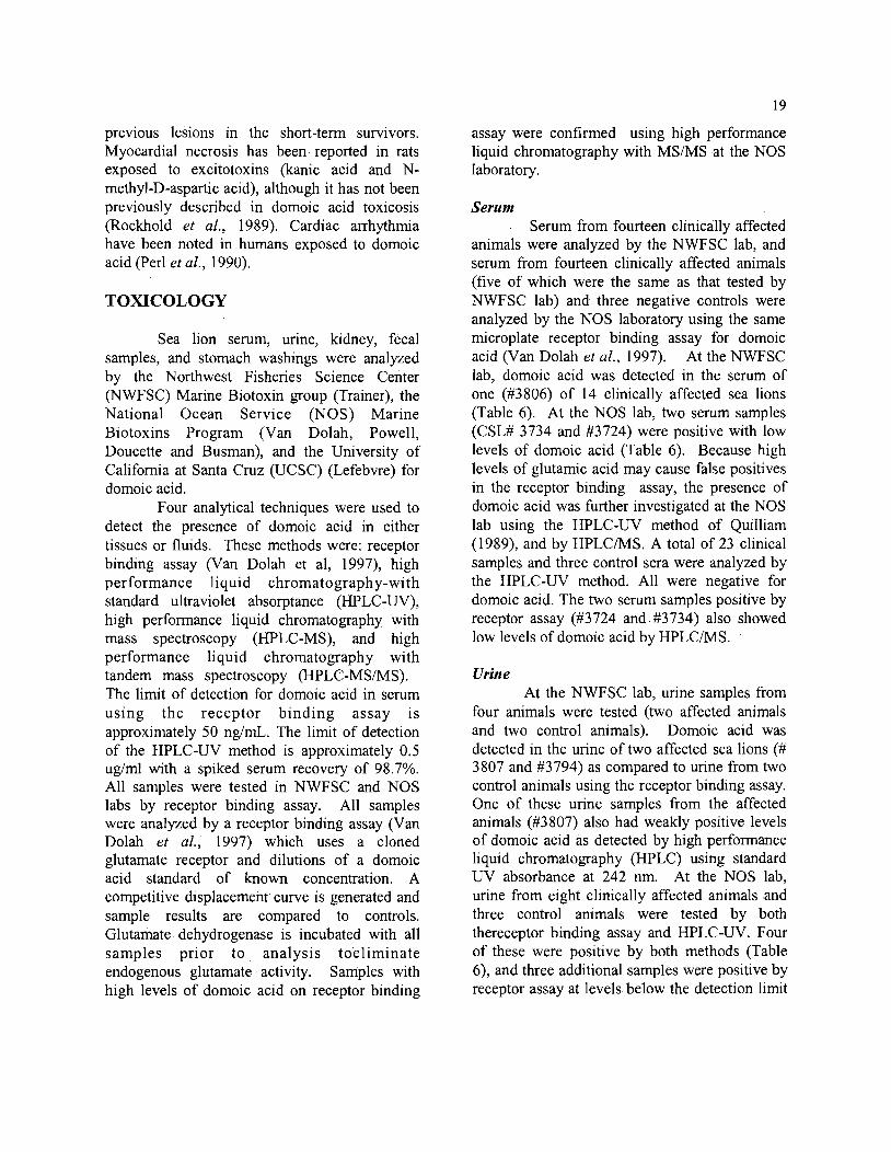
TOXICOLOGY
Sea lion serum, urine, kidney, fecal samples, and stomach washings were analyzed by the Northwest Fisheries Science Center (NWFSC) Marine Biotoxin group (Trainer), the National Ocean Service (NOS) Marine Biotoxins Program (Van Dolah, Powell, Doucette and Busman), and the University of California at Santa Cruz (UCSC) (Lefebvre) for domoic acid.
Four analytical techniques were used to detect the presence of domoic acid in either tissues or fluids. These methods were: receptor binding assay (Van Dolah et al, 1997), high performance liquid chromatography-with standard ultraviolet absorptance (HPLC-UV), high performance liquid chromatography with mass spectroscopy (HPLC-MS), and high performance liquid chromatography with tandem mass spectroscopy (HPLC-MSMS). The limit of detection for domoic acid in serum using the receptor binding assay is approximately 50 ng/rnL. The limit of detection of the HPLC-UV method is approximately 0.5 ug/ml with a spiked serum recovery of 98.7%. All samples were tested in NWFSC and NOS labs by receptor binding assay. All samples were analyzed by a receptor binding assay (Van Dolah et al., 1997) which uses a cloned glutamate receptor and dilutions of a domoic acid standard of known concentration. A competitive displacement curve is generated and sample results are compared to controls. Glutamate dehydrogenase is incubated with all samples prior to analysis toeliminate endogenous glutamate activity. Samples with high levels of domoic acid on receptor binding
assay were confirmed using high performance liquid chromatography with MS/MS .at the NOS laboratory.
Serum Serum from fourteen clinically affected
animals were analyzed by the NWFSC lab, and serum from fourteen clinically affected animals (five of which were the same as that tested by NWFSC lab) and three negative controls were analyzed by the NOS laboratory using the same microplate receptor binding assay for domoic acid (Van Dolah et al., 1997). At the NWFSC lab, domoic acid was detected in the serum of one (#3806) of 14 clinically affected sea lions (Table 6). At the NOS lab, two serum samples (CSL# 3734 and #3724) were positive with low levels of domoic acid (Table 6). Because high levels of glutamic acidmay cause false positives in the receptor binding assay, the presence of domoic acid was further investigated at the NOS lab using the HPLC-UV method of Quilliam (1989), and by HPLCIMS. A total of 23 clinical samples and three control sera were analyzed by the HPLC-UV method. All were negative for domoic acid. The two serum samples positive by receptor assay (#3724 and.#3734) also showed low levels of domoic acid by HPLCMS.
Urine At the NWFSC lab, urine samples from
four animals were tested (two affected animals and two control animals). Domoic acid was detected in the urine of two affected sea lions (# 3807 and #3794) as compared to urine from two control animals using the receptor binding assay. One of these urine samples from the affected animals (#3807) also had weakly positive levels of domoic acid as detected by high performance liquid chromatography (HPLC) using standard UV absorbance at 242 nm. At the NOS lab, urine from eight clinically affected animals and three control animals were tested by both thereceptor binding assay and HPLC-UV. Four of these were positive by both methods (Table 6), and three additional samples were positive by receptor assay at levels below the detection limit

of the HPLC method. Urine from one animal (#3707) was also analyzed by HPLC/MS and confirmed positive for domoic acid.
Feces Feces from seven clinically affected
animals and two control samples were analyzed at the NOS lab by receptor assay and HPLC-W. Fecal samples were extracted using a mussel extraction and SAX clean-up method of Quilliam et al., (1 995) before analysis by HPLC with spiked extraction recovery of 88.4 + 8.4%, n = 3. Three samples were positive for domoic acid (Table7) and good quantitative agreement was seen between the receptor binding assay and HPLC analyses (Table 7).
Feces from eleven clinically affected sea lions were also analyzed at UCSC for the presence of domoic acid by HPLC using the extraction method of Quilliam et al., (1989). Domoic acid was detected in samples from the same three animals as at the NOS lab (Table 7). In addition, fecal samples from' seven control sea lions were analyzed. These animals were three captive sea lions from Long Marine Laboratory (LML # 1, 2, 3), three captive sea lions from Marineworld in Vallejo, CA, and one sea lion from Maverick's Beach, CA (Clutch) that was euthanized at TMMC due to severe gunshot injury two months after the event (August 1998). Domoic acid was not detected in any of these samples.
Kidney, Liver, Muscle, Brain Five kidney samples were analyzed at
the NOS lab by HPLC-UV and were negative for domoic acid (Table 7). Samples were extracted and cleaned using the same procedure as described for feces, with a spiked ludney extraction recovery of 24.6%, n = 1. Liver, muscle, brain, intestinal mucosa and kidney samples from two animals (#3765 and # 3824)analyzed by HPLC at UCSC were also negative for domoic acid.
Stomach washings A single stomach washing sample from
a clinically affected animal (CSL 3708) analyzed by receptor ,assay at the NOS lab was negative for domoic acid.
Confirmation of domoic acid in fluids by mass spectrometry
Serum and urine samples with the highest concentrations of domoic acid, as determined by HPLC-UV, were subjected to HPLC/MS and HPLC-MS/MS analysis at the NOS lab for confirmation of the identity of the HPLC peak in question. For both methods, sample extracts were fractionated on a C18 column eluted with a gradient of 1-95% methanol in 0.1% TFA. A PE SCIEX API-111 triple quadrupole mass spectrometer was used. The ionspray source was operated in positive ion mode utilizing nitrogen for the nebulization gas. The first quadrupole was used to pass only ions of nominal 312 m/z. The conditions in the second quadrupole were adjusted to allow a substantial amount of collisionally-induced dissociation. The third quadrupole was operated in multiple ion monitoring mode, where fragments ions of 161 and 266 m/z, as well as the residual parent ions at 3 12 m/z were allowed to pass to the mass spectrometer ion detector. The presence of domoic acid was confirmed in urine from # 3707 and #3758 and feces from # 3783 (Table 7).
Pseudo-nizchtia frustules Ten of the eleven fecal samples
analyzed at UCSC were also examined for the presence of diatom frustules (glass skeletons) using a compound microscope, (there was insufficient sample from CSL #3734 after HPLC analyses). Diatom frustules were only found in fecal samples that had tested positive for domoic acid (#3758 and #3783). Using scanning electron microscopy, frustules were identified as Pseudo-nitzschia australis.
Anchovies Domoic acid was detected in anchovies
(Engraulis mordax) collected from Monterey Bay and Morro Bay by HPLC-W at the NOS

lab. Two samples of anchovies 'collected fiom Monterey Bay on May 22, 1998 had detectable levels of domoic acid, while fish collected on June 10, 1998 had no detectable levels (Table 8). Two samples of anchovies from Morro Bay on June 4 also had detectable levels of domoic acid (Table 8). Domoic acid was confirmed in two anchovy samples by HPLCMS and one by HPLCMSMS (Table 8)
Summary of Toxicology These results indicate that there is
consistency between domoic acid assays performed at NOS, NWFSC and UCSC labs. The NOS and NWFSC labs run the same microplate receptor binding assay as a rapid screening method for domoic acid. Although highly effective as a screening method, positive results should be confirmed by analytical methods when testing novel tissue matrices. The HPLC-UV method is suitable only when tissue concentrations are sufficiently high, because the detection limit is poorer than that of the receptor assay. HPLCIMS or HPLCMSMS provides the most rigorous analytical confirmation, but are expensive and time-consuming to run. In the
current study HPLCMSIMS provided the necessary independent confirmation of the chemical identity of the toxin to unambiguously conclude the involvement of domoic acid in this event. The greater detection rate in feces and urine than serum (Table 9) suggests feces and urine should be routinely collected for detection of domoic acid in marine mammal die-offs. The low detection rate in serum is probably due to the rapid clearance of this water soluble toxin fiom blood. Clearance in rodents and primates following intravenous inoculation is under four hours (Truelove and Iverson 1994). Although stomach contents might be valuable for analysis,the stomachs of clinically affected sea lions in this event were empty, presumably due to vomiting and the time elapsed between ingestion and post mortem examination. Feces, thus, proved to be the most valuable sample fortoxicological analyses, as well as for determining diet.
Table 6. Presence of Dornoic Acid in Serum from California Sea Lions
NWFSC NOS NWFSC NOS NOS NOS
Receptor Receptor HPLCIUV HPLCNV HPLCMS HPLCMSMS Animal ID assay assay
CSL 3299 C N/D negative N/D
CSL 345 1 C Negative
CSL 3489 C negative N/D N/D N/D
CSL 3510 C negative negative N/D N/D
CSL 3512 C negative negative negative N/D
CSL 3670 Negative

22
CSL 3724
CSL 3727
CSL 373 1
CSL 3732
CSL 3733
CSL 3734
Negative
0.17 pg eq./mL
negative
negative
negative
0.2 Pg eq ./mL
negative
negative
negative
negative
negative
positive
N/D
N/D
N/D
positive
N/D
N/D
N/D
N/D
N/D
Table 7 Presence of Domoic Acid in Urine, Feces and Tissues from California Sea Lions
NWFSC NOS NWFS NOS NOS NOS UC Santa C Cruz
Animal ID Receptor Receptor HPLCI HPLCNV HPLCI HPLCMSMS HPLC/W assay assay UV MS
URINE g eq./mL g/rnL
CSL 345 1 C negative
CSL 3707 3.72 14.05 posltlve pos~tive
CSL 3726 0.12 2 68 N/D N/D
CSL 3741 0.03 negative N/D N/D
CSL 3742 N/D negative N/D N/D
CSL 3749 0.72 2.38 N/D pos~tive

Table 8. Domoic acid levels in Anchovies from the Central California Coast Sample site & Date Receptor Assay HPLC/UV
NOS
5122198a Monterey Bay 7 1.30 100.35 5122198b Monterey Bay 69.67 123.04 614198a Morro Bay 2.53 4.05 614198b Morro Bay 0.27 0.82 6110198a Monterey Bay N/D Negative 6110198b Monterey Bay NID Negative
HPLCMS
N/D Positive Positive N/D N/D N/D
HPLCMSMS
Positive NID N/D N/D N/D N/D

Table 9. Samples positive for Domoid Acid by different laboratory analyses
PLANKTON BLOOM
Within Monterey Bay, plankton samples are routinely collected several times per week fiom the Santa Cruz wharf and Monterey Coast Guard pier. Samples are also collected along several sites running due west of Moss Landing as part of the Monterey Bar Aquarium Research Institute (MBARI) upper water column time series project. Samples are subjected to DNA probe-based tests as a means of identifyng and quantifyng a variety of toxic and non-toxic Pseudo-nitzschia species as well as the toxigenic dinoflagellate Alexandrium tamarense (e.g., Scholin et al. 1996, 1997, submitted;). An example of the time series data collected from the Santa Cruz wharf is shown in Figure 3. During thespring bloom of diatoms, toxigenic Pseudo-nitzschia species were largely absent and no domoic acid was detected in plankton samples, (Fig. 3, arrow #I). As the spring bloom progressed, A. tamarense was detected, and shellfish samples collected from the same area tested positive for paralytic shellfish poisons (Fig. 3, arrow #2). As the bloom ,of Alexandrium declined, the number of Pseudo-nitzschia australis increased sharply (arrow #3). The amount of domoic acid (= bars) in water samples rose in concert with increasing numbers of P. australis (arrow #4). As the bloom of P. australis reached its maximum, toxigenic cells were concentrated in a narrow band near shore. It is possible, although in no way proven, that the cells responded to nutrient enrichment of near shore waters as a result of enhanced river flows following late spring rains.
Anchovies collected from Monterey Bay
during the height of the P. australis bloom contained high levels of domoic acid and high numbers of P. australis in their stomachs (Fig. 4). The P. australis bloom was replaced by a large P. pseudodelicatissima bloom (not shown on Fig. 3). Some P. pungens and P. multiseries were also present, but at relatively low levels. During this transition in species abundance, the concentration of domoic acid declined rapidly and precipitously in the upper water column (Fig. 3, arrow #5). Gut contents of anchovies collected at this time reflected this change in species abundance as well (Fig. 5), as they did not contain detectable levels of domoic acid. Reports of sickened sea lions frornblonterey Bay decreased in concert with the loss of P. australis fiom the upper water column and the rise in dominance of P. pseudodelicatissima. Notably, cultures of P. australis isolated fiom Monterey Bay produce domoic acid whereas isolates of P. pseudodelicatissima from the same area do not.
During the P. pseudodelicatissima bloom inside Monterey Bay, high concentrations of toxigenic P. multiseries were found south and especially north of that region. Thus, species composition of Pseudo-nitzschia populations found within Monterey Bay and contiguous waters of the central California coast can differ substantially. Throughout the remainder of the year P. australis was not abundant in Monterey Bay.

a.*po?pp?s,,,-...,.:a I... "-I*'E,m-r.
Figure 3. 1998 time series data collected at the Santa Cruz wharf station showing results of sandwich hybridization assays (optical density, O.D.) for Pseudonizchtia australis, P. pungens, P. multiseries and North American strains of Alexandrium tamarense/catanella plotted as a function of time (Julian day). Increasing O.D. indicates a rise in species abundance; the dashed line indicates lower limit of detection. Corresponding amounts of domoic acid associated with a particulate fraction of plankton are given as pmol toxin per mL water filtered (top panel, bars). Arrows refer to particular events described in the text.

Anchovy gut contents containing P s e r r d o - ~ t d caustraiis,Nlag 29,1998.
Figure 4. Scanning electron micrographs of anchovy stomach contents at the height of the P. auslralis bloom. Anchovies caught in the Monterey Bay area had stomachs packed with P. australis frustules (A = overview, B and C = close-up showing frustules are P. australis).

Anchovy gut contents 6/ 10198
Figure 5. Scanning electron micrographs of anchovy stomach contents after the P. australis bloom. Overview micrograph shows that anchovies caught in the Monterey Bay area had stomachs packed primarily with P. delicatissima (C), the dominant Pseudonizchtia in the water column at the time. Fragments of P. australis were also found (B), as well as frustules of P. multiseries (a),but the latter two species were not abundant.

DIAGNOSIS
The combination of clinical signs (scratching, seizuring, anchovies in fecal matter), histopathological (hippocampal necrosis), toxicologcal (domoic acid in serum, urine, feces, anchovies) and epidemiological findings (simultaneous mortality in sea lions and the presence of toxin-producing blooms) led to the diagnosis of domoic acid toxicity in the sea lions. This intoxication was due to blooms of Pseudonizchtia australis that were ingested by anchovies that in turn were eaten by California sea lions. Although a variety of incidental histopathologic and serologic changes were detected in the sea lions examined, they were not considered to be the cause of the mortality event. However, virus neutralization titers to phocine distemper virus, presence of protozoa in the brain and early neoplastic lesions warrant further investigation to determine their role in health of the California sea lion population.
POST RELEASE MONITORING
Three sea lions that survived the event were released with telemetry devices to monitor movements and survival after release. Adult females were selected, as animals of this sex and age are currently being tagged and monitored off San Miguel Island by Dr. Robert De Long and Sharon Melin from the National Marine Mammal Laboratory, and could be used as normal controls. The three animals selected had all shown typical clinical signs during the "event". One had seizures for only 48 hrs, one had repeated seizures for up to a week, and the third had seizures for up to one month post stranding. They were thus classed as mild, moderate and severe cases. None of the animals had been pregnant when stranded. As the tags are fixed to the animals using glue on the hair, the animals were not released until after the annual molt in September.
The' three sea lions (CSL #3812, #3815 and #3822) were sedated with midazolam intramuscularly at 0.02 mgkg (mixed with atropine at 0.02 mgkg), then masked with isoflurane until relaxed but with intact gag reflex. Satellite-linked transmitters or platform transmitter terminals (ST-10 Argos PTT model; Telonics, Mesa, AZ) were glued to the dorsum between the scapulae using 5-minute epoxy. These tags were approximately 14 x 5 x 1 cm and weighed 0.2 kg. To conserve battery power and prolong the life of the PTTs, the duty cycle was set to transmit for 24 hrs every other day. Location data were collected by Service Argos. Radio transmitters (Advanced Telemetry Systems, Isanti, MN) were attached to the heads of each sea lion. These transmitters weighed 0.05 kg each and had 30 cm long whip antennae.
The sea lions were tagged on November 4 and released on November 6 at Weston Beach, Point Lobos State Reserve, Camel, California (Fig. 6). This site was chosen as it was within the Monterey Bay, the area in which the sea lions stranded during the event, and is a protected area frequented by this species.
Two of these three sea lions were resighted. One animal was observed on December 10 1998 with a satellite transmitter and a VHF transmitter on Afio Nuevo Island (Morris, pers. comm.), although its ID number was not determined. #3 8 12 and #3 8 15 were sighted on December 23 1998, 48 days after release. On this day, #3815 had a radio transmitter, but had lost the satellite transmitter. Satellite data indicate that the transmitter detached from the sea lion while on shore. Transmissions were received from the transmitter until February 7 1999. Sea lion #38 12 was also sighted on January 8 1999 at Afio Nuevo Island, with both transmitters attached.

Sea lion #3822 swam as far south as Point Benett, San Miguel Island (-325 km), where it remained for five days, before traveling north again to areas off the Big Sur Coast. Sea lion #3822 also made a second trip down to San Miguel Island, and returned north again (Figure 6a). A total of 170 positions was received for 64 days, until transmissions ceased on January 8 1999 off Point Lobos (36.519N, 121.954W). It is unclear whether signal cessation was a result of mortality, tag malfunction or tag loss. A total of 333 positions was received for sea lion #3812, which traveled as far north as the Farallon Islands, and as far south as the Channel Islands. Sea lion #38 12 remained near the Channel lslands for one week, but returned to areas around Aiio Nuevo Island (Figure 6b). The last transmission from #38 12 was 94 days post release on February 7 1999 off Pescadero Point, California (3 7.24 1 N, 122.488W). Sea lion #3815 primarily remained within and around areas adjacent
to Monterey Bay while the satellite tag was attached. Although the date of tag detachment from this animal is unclear, transmissions ceased on February 7, 1999. The failure of transmissions from two tags within a day of each other suggests that this was due to battery expiration rather than death of the sea lions.
At least two of the three satellite- tagged sea lions survived up to three months post release and data indicated they were using areas where wild sea lions could be found. Melin et al. (1993) found that female California sea lions from San Miguel Island, which were tagged with satellite-linked time depth recorders, foraged northwest of San Miguel lsland during winter and spring months. Furthermore, tagged sea lions from San Miguel Island traveled as far as 460 km north along the mainland coast of California (Melin et al. 1993). Thus, movements of the three rehabilitated sea lions may have been typical.

DISCUSSION
This is the first report of domoic acid toxicity 'in marine mammals. However, as algal blooms producing domoic acid have been observed previously along the California coast (Walz et aL, 1994), and domoic acid toxicity was reported in brown pelicans off Monterey in 1991 (Work et al., 1993), it is likely that this toxicosis has occurred previously in marine mammals. Clusters of stranded adult sea lions that showed similar neurologc signs to those in the animals in this 1998 event have been observed at other times along the California coast, although no cause was determined. In July 1978, 40 animals stranded in Ventura County displaying neurologic signs (Gilmartin et aL, 1979); in 1986, 11 sea lions showing opisthotonus and convulsions were admitted to TMMC (Vandenbroek et al., 1987); in 1988, 38 sea lions and ten northern fur seals (Callorhinus ursinus) with similar signs were admitted to TMMC (Gage et aL, 1989) and in 1992 18 sea lions stranded in San Luis Obispo County displaying similar signs (Beckrnen et aL, 1995). Similar histologcal changes in the hippocampus to those in sea lions from this 1998 event were observed by Dr. Linda Lowenstine in animals that died in the 1992 event. Two stomachs from seizuring sea lions during the 1992 seizure event were submitted for domoic acid analysis with negative results. However, the stomachs were devoid of contents. No suitable samples,i.e. serum, urine or feces, are available from earlier animals for retrospective analysis. As there was a bloom of P. australis in Monterey Bay in 1992 (Walz et aL, 1994), it is likely that domoic acid toxicity affected sea lions off California earlier than 1998. In addition, since this event there has been another similar occurrence in October 1998.
From July 12 to October 17, 1998 an additional eleven California sea lions stranded with similar clinical signs to the animals descnbed in this report. Nine of the animals stranded from October 3 to October 17. Six animals were euthanized or died,
and four animals recovered and were released. Four of six urine samples from sea lions in the October event analyzed for domoic acid by HPLC-UV were positive (Table 6), while one CSF sample was negative. Serum, urine, and cerebrospinal fluid (CSF) were tested by receptor binding assay only. Domoic acid was detected by RBA in three out of six urine samples from clinically affected animals. All serum and CSF samples tested were negative for domoic acid. During this time period, cells of P. australis did reach -10,000 cells per liter at the Santa Cruz pier, however, much higher cell concentrations were observed outside of Monterey Bay, again highlighting the difference between water masses within and outside of the Bay. In this case, it is likely that the bloom of P. australis was fueled by upwelling and that cells and their associated toxin did not penetrate Monterey Bay to any great extent. This recent episode illustrates the quick response that stranding rates and pimipeds may have to toxic blooms of P. australis.
T h e c o m b i n a t i o n o f oceanographic data, evidence of domoic acid in the prey eaten by the -sea lions, clinical signs and histopathology are important in making a diagnosis of domoic acid toxicity, as detection of domoic acid in sea lion tissues is limited. The detection of domoic acid in only three serum samples by receptor binding assay is not surprising, as the plasma half-life is short in other species (21.6 minutes in rats, 114 minutes in monkeys; (Truelove and Iverson, 1994). Blood samples were not collected from sea lions until they arrived at TMMC after initial stranding, detection of the animal by the public and transport by road up to 200 miles. It is therefore likely that the domoic acid had been cleared from plasma by the time blood was collected in most cases. As domoic acid is water soluble and excreted in urine, it is also not surprising that it was detected in urine rather than plasma in some animals.

The even higher detection rate in feces may reflect poor absorption from the gastro-intestinal tract due to poor lipid solubility. After oral administration of - domoic acid to rats, recovery from feces was complete (Iverson et al., 1989).
In contrast to the toxicologic results, both clinical signs and histopathologic changes were characteristic of domoic acid toxicity as reported in other species, although a variety of incidental signs and lesions were detected. The seizures were dramatic, and presumably a consequence of domoic acid binding in the brain. Domoic acid is an excitatory neurotoxin that binds to neurones via high-affinity AMPA- and kainate-sensitive glutamate receptors to produce excitotoxic cell death (Iverson and Truelove, 1994; L a m et al., 1997). In between seizures, the scratching behavior was reminiscent of the hallmark scratching syndrome documented in mice that distinguishes paralytic shellfish poisoning from domoic acid toxicity (Todd, 1993). Similar behavior was also described in pelicans intoxicated with domoic acid (Work et al., 1993). The ability of animals to survive exposure, as demonstrated by the satellite-tracking data, is also consistent with the survival of exposed humans in the Canadian outbreak in people who ate contaminated mussels (Per1 et al. 1990). The significance of the virus neutralization titers to phocine distemper virus (PDV) in a few animals is unclear. However, the lack of accompanying histopathological changes consistent with morbillivirus infection, negative RT-PCR for morbillivirus on brain tissue, and the similar seroprevalence of PDV antibodies in banked sea lion sera suggest the presence of antibodies in sea lions dylng during the event did not play a role in this event. In contrast, in the recent
Mediterranean monk seal die-off, a controversy as to the cause of the die-off occurred, due to the isolation of a morbillivirus from tissue of affected animals, in addition to detection of saxitoxin (Osterhaus et al., 1997; Costas and Lopez-Rodas, 1998).
Over the time frame of the "seizuring sea lion" event, large numbers of dead pinnipeds and sea birds were observed on the beaches around Monterey Bay. The causes of death of these animals were not determined. However, many were severely emaciated. Based on gross appearance and predictions of species likely to be affected by changes in food availability during El Niiio events (Bodlun and Jameson 199 1, Trillmich and Ono 199 l), it is likely that many of these mortalities were a consequence of starvation due to food shortage. It is thus unclear how many sea lions in total were affected by domoic acid. The overall mortality due to domoic acid is likely to be more than 57 individuals, but is unknown. As the overall California sea lion population is estimated at 167,000 individuals, an event causing mortality in 0.00004 % does not constitute a threat to the population. However, the rapid and dramatic nature of the event suggests the potential for similar harmful algal blooms to impact endangered species, such as the Steller's sea lion or Hawaiian monk seal, before control measures can be implemented. In add i t ion , the susceptibility of humans to domoic acid suggests that outbreaks of toxicity in marine mammals should be fully investigated and measures taken to prevent human morbidity and mortality.
Acknowledgments. This investigation would not have been possible without the willing collaboration and enthusiasm of each of the organizations involved. Special thanks are due to Diane Paschal1 for assistance in putting this report together, the Monterey Bay Aquarium Research Institute for funding a workshop in the wake of the event to bring investigators together and facilitate exchange of data, Wanda Dietrich for editing and formatting this document, and the staff and volunteers of TMMC for their hard work caring for the sea lions during the event.

REFERENCES Antonelis, G. A., Fiscus, C. H. and R. L. DeLong. 1984. Spring and summer prey of California sea lions, Zalophus californianus, at San Miguel Island, California, 1978-1979. Fishery Bulletin 82: 67-76.
Beckmen, K., Lowenstine, L. J. and F. Galey. 1995. Epizootic seizures in California sea lions (Zalophus californianus). 11th Biennial Conference of the Marine Mammal Society, Orlando, Florida. (Abstract) p. 1 0.
Bodlun, J. L. and R. J. Jameson. 199 1. Patterns of seabird and marine mammal carcass deposition along the central California coast, 1980 - 1986. Canadian Journal of Zoology 69: 1149-1 155.
Cordaro, J. 1997. Pinniped strandings in California: Analysis and Trends. In "Pinnipeds Populations, Eastern North Pacific: Status, Trends and Issues." Eds. G. Stone, J. Goebel and S. Webster. 127th Annual Meeting of the American Fisheries Society, Monterey, California, August 28, 1997. pp. 40-45.
Costas, E, and. Lopez-Rodas, V. 1998. Paralytic phycotoxins in monk seal mass mortality. Veterinary Record 142: 643-644.
Gage, L. J. and D. Smith. 1989. Convulsing sea lions at the California Marine Mammal Center, 20th Annual International Association for Aquatic Animal Medicine Conference, 1989 May 14-18, San Antonio, Texas (Abstract).
Gilmartin, W. G. 1979. Fatal hepatoencephalopathy in a group of California sea lions. 10th ,
Annual International Association for Aquatic Animal Medicine Conference, 1 979 April 22- 26, St. Augustine, Florida (Abstract)
Gulland, F. M. D., Trupkiewicz, J. G., Spraker, T. R. and L. J. Lowenstine. 1996. Metastatic carcinoma of probable transitional cell origin in 66 free-living California sea lions (Zalophus californianus), 1979-1994. Journal of Wildlife Diseases 32: 250-258.
Iverson F. and J. Truelove. 1994. Toxicology and seafood toxins: domoic acid. Natural Toxins2: 334-339.
Iverson, F., ~ruelovk, J., Nera, E., Tryphonas L., Campbell, J. and E. Lok. 1989. Domoic acid poisoning and mussel-associated intoxication: Preliminary investigations into the response of mice and rats to toxic mussel extract. Food and Chemistry Toxicology 27: 377-384.
Lam, J. A., Beart, P. M. and N. S. Cheung. 1997. Neurotoxin domoic acid produces cytotoxicity via kainate and AMPA- sensitive receptors in cultured cortical neurones. Neurochemistry International 3 1 : 677-682.
Melin, S.R., R.L. DeLong, J.R. Thomason, and D.E. Valesquez. 1993. Foraging behavior of female California sea lions at San Miguel Island, California: winter 1992 and 1993. Tenth Biennial Conference on the Biology of Marine Mammals. 11-15 November. Galveston, Texas.
Osterhaus, A. D. M. E., Groen, J., Niesters, H., van de Bildt, M., Martina, B., Vedder, L., Vos, J., van Egrnond, H., Abou Sidi, B. and M. E. 0 . Barham. 1997. Morbillivirus in monk seal mass mortality. Nature 388: 838-839.
Perl, T. M., Bedard, L., Kosatsky, T., Hockin, J. C., Todd, E. C. and R. C. Remis. 1990. An outbreak of toxic encephalopathy caused by eating mussels contaminated with domoic acid. New England Journal of Medicine 322: 1775-1780.
Quilliam, M. A., Sim, P. G., McCulloch, A, W. and A. G. McInnes. 1989. High performance liquid chromatography of domoic acid, a marine neurotoxin, with application to shellfish and plankton. International Journal of Environmental and Analytical Chemistry 36: 139-1 54.
Quilliam, M. A., Xie, M. And W. R. Hardstuff. 1995. Rapid extraction and cleanup for liquid chromatographic determination of domoic acid in unsalted seafood Journal AOAC International 78: 543-554.
Rockhold, R. W., Acuff, C. G. and B. R. Clower. 1989. Excitotoxin-induced myocardial necrosis. European Journal of Pharmacology 166: 571-576.
Schmued, L. C., Scallet, A. C, and W. Slikker. 1995. Domoic acid induced neuronal degeneration in the primate forebrain revealed by degeneration specific histochemistry. Brain Research 695: 64-70.
Scholin, C. A., Buck, K.R., Britschgi, T., Cangelosi, G. & Chavez, F.P. 1996. Identification of Pseudo-nitzschia australis using rRNA-targeted probes in whole cell and sandwich hybridization formats. Plrycologia 35: 190-1 97.
Scholin, C. A., Miller, P.E., Buck, K.R. & Chavez F.P. 1997. Detection and quantification of Pseudo-nitzschia australis in cultured and natural populations using LSUrRNA-targeted probes. Lirnnology and Oceanography 42(5, part 2): 1265-72.
Scholin, C.A., Marin, R. 111,Miller, P.E., Doucette, G.J., Powell, C.L., Howard, J., Haydock, P., Ray, J. Application of DNA Probes and a Receptor Binding Assay for Detection of pseudo-nitzschia (Bacillariophyceae) Species and Domoic Acid in Cultured and Natural Samples. Journal of Phycology (in review).
Teitelbaum, J. S., Zatorre, R. J., Carpenter, S., Gendron, D., Evans, A. C., Gjedde, A. and N. R. Cashman. 1990. Neurologic sequelae of domoic acid intoxication due to the ingestion of contaminated mussels. New England Journal of Medicine 322: 178 1- 1787.
Thornton, S., Nolan, S. and F. M. D. Gulland. 1998. Bacterial isolates from California sea lions (Zalophus californianus), harbor seals (Phoca vitulina), and northern elephant seals (Mirounga angustirostris) admitted to a rehabilitation center along the central California coast, 1994-1995. Journal of Zoo and Wildlife Medicine 29: 171-176.
Todd, E. 1993. Domoic acid and amnesic shellfish poisoning: a review. Journal of Food Protection 56: 69-83.
Trillmich, F. and K. A. Ono (eds.). 1991. Pinnipeds and El Niiio: responses to environmental stress. Springer-Verlag, Berlin.

Truelove J. and F. Iverson. 1994. Serum domoic acid clearance and clinical observations in the cynomolgus monkey and Sprague-Dawley rat following a single i.v. dose. Bulletin of Environmental Contamination and Toxicology 52: 479-486.
Tryphonas, L. and F. Iverson. 1990. Neuropathology of excitatory neurotoxins; the domoic acid model. Toxicologic Pathology 18 (part 2) :165-1 69.
Tryphonas L., Truelove, J. and F. Iverson F. 1990. Acute parenteral neurotoxicity of domoic acid in Cynomolgus monkeys (M.fasicularis). Toxicologic Pathology 1 8 :297-303.
Vandenbroek, D. J., Keeler, D., McBain R., Gage, L. J., Amaya-Sherman, L. and M. Koski. 1987. Tonic-clonic convulsions in California sea lions. 18th Annual International Association for Aquatic Animal Medicine Conference, 1987 May 10- 14, Monterey, California (Abstract).
Van Dolah, F. M., Leighfield, T. A. and J. S. Ramsdell. 1997. A microplate receptor assay for the amnesic shellfish poisoning, domoic acid, utilizing a cloned glutamate receptor. Analytical Biochemistry. 245: 102-103.
Work, T. M., Barr, B., Beale, A. M., Fritz, L., Quilliam, M. A, and J. L. C. Wright. 1993. Epidemiology of domoic acid poisoning in brown pelicans (Pelecanus occidentalis) and Brandt's cormorants (Phalacrocoraxpenicillatus) in California.. Journal of Zoo and Wildlife Medicine 24: 54-62.
Walz, P. M., Garrison, D. L., Graham, W. M., Cattey, M. A., Tjeerdema, R. S. and M. W. Silva. 1994. Domoic-acid producing diatom blooms in Monterey Bay, California: 199 1-1 993. Natural Toxins 2: 271-279.

dl g or gm HPLC HPLCIUV
HPLCMS
HPLCMSMS
IM IV kg mg ml mm M mlz NOS
NWFSC
RBA SAX SSP TFA TMMC u g UCSC
List of Terms
deciliter; one-tenth of a liter gram High Performance Liquid Chromotography High Performance Liquid Chromotography with ultraviolet detection High Performance Liquid Chromotography with mass spectrometry High Performance Liquid Chromotography with tandem mass spectrometry intramuscular intravascular kilogram; one thousand grams milligram; one-thousandth of a gram milliliter; one-thousandth of a liter millimeter; one-thousandth of a meter mass masslcharge National Ocean Service, National Oceanic and Atmospheric Administration, Charleston, South Carolina Northwest Fisheries Science Center, National Marine Fisheries Service, Seattle, Washington Receptor binding assay strong anion exchange species Trichloroacetic acid The Marine Mammal Center, Sausalito, California microgram; one-millionth of a gram University of California, Santa Cruz

~ V ] ~ b ~ ~ b ~ o V ) ~ o o ~ ~ C ' )
~ ~ ~ u ~ u ~ ~ u u ~ u u ~ ~
------------------------- ---- -- ---
V) V) -+ -+ -m 3 3 2 .a-- -.a-u0 u u u u - ~ ~ c c c c c c ~ ~ c u u u ~ - c c - - - - c ~ - + + + - s 7 5 5 5 5 a ~ Y Y Y 3 ~ 3 a ~ 7 5 5 ~ ~ , , , , , , 3 , ~ ~ ~ Y Y ~ u u u u u = u u u u u u u 3 3 u u u u , u u u u u u u u ~ U U U U U 2 a a a a a m ~ 4 4 4 a a a m - , a a a a ~ a a a ~ a a a a ~ a a a a a
v, LLLLLLLLLLZLLLLLLLLLLLLLZLLLLLLLLZLLLLLLLLLLZLLLLLL
m m m m m - m m V) 7
i3 c u u u -0 u u -0.-Y 0 a a a a a a
.-z .N .N .N.-N N .. a-
a C C C C m c d m C C C C C C0 a a cd m a a m a an ~ u u ~ ~ ~ ~ u u ~ ~ ~ u u u u ~ u u ,.a, .iLJ ,.s!.a,.- .a, .a, .g,,,.G2 ,,.a, .2,.a, .a, .a, ,,.a, .a, .a, .cu.a, .a, .a, ,,.g .gn ~ ~ ~ w ~ w n n n n w w n n n a ~ n n n ~ w n n n a ~ ~ n ~ n a ~ ~
2 4 I a m x5
;a ~ u 0 0w U a 3
x.- O ~ ~ ~ o b ~ - ~ ~ ~ ~ ~ ~ ~ ~ ~ ~ u m m m m o o O a ~ N N a m m m e ~ * e e e w * e m m ( D C D C D b a a 0 ) m
C D C D C D C D b b b b b b b b b b b b b b b b b b b b b b b b b b b b b b m m m m m m m m m m m m m m m m m m m m m m m m m m m m m m m m m m A J J J J A J A A J A J J J A A J A A A J A J J A A A A J J J A A J
n o m m m m m m m / , m / , m w m m m / , m m m m m m m m m m m m m m m m m m4 0 0 0 0 0 0 0 0 0 0 0 0 0 0 0 0 0 0 0 0 0 0 0 0 0 0 0 0 0 0 0 0 0 0

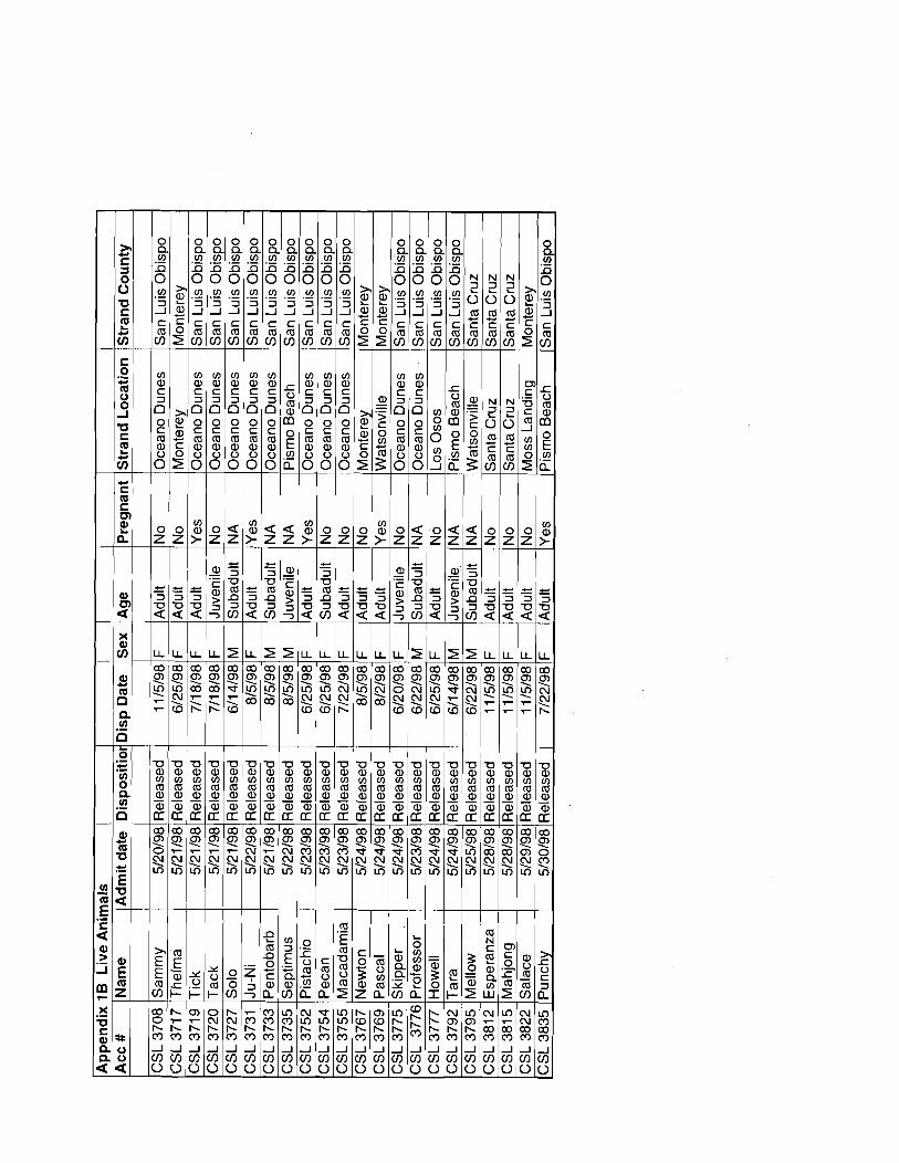


Appendix 2C, Admit Blood Chemistries i Acc. # Name Na K CI Glu BUN Creat Prot Alb Glob Ur. Ac.Ca Phos Tot Bil Dir Bil GGT l ~ l kP ETALT LDH Iron Chol Trigl CK
-
rneqfl rneqll me@ mgldl rngldl rngidl gmldl gm/dl g d d l rngldl mgldl rngldl mgldl rng/dl rngldl lun ull u/l ull rncgldl rngtdl rngldl I I
CSL 3901 (Palm 167 6 ( 112 1 90 66 j 3.1 10.2 3.3 1 6 . 9 1 1 . 2 1 11 1 12 1 0 . 5 10.2 1105 34 1 5 6 1 3 0 1 7 6 0 1 96 2881 62 1 na CSL 3950 t~thena 156 6.1 1110 1 82 1 61 11 .3 8.1 1.9 / 6.2 ( 1.3 1 9 . 6 / 11 ] 0.5 10.2 1188 1 34 1 6 5 1 8 2 1 7 1 8 1 155 1381 24 1 na

Appendix 3A Hematolowv, Pre-release 1 I I I Acc # l ~ a m e IWBC IRBC ~HGB~HCT~ M C V IMCH MCHC lsegs segs bands bands lymph lymph monos monos eos eos bas0 bas0 plat
g / ~ ~1% A ~ S % A ~ S % A ~ S % A ~ S % A ~ S V, A ~ S x lo3
CSL 3731 I J U - ~ i CSL 3733 1 pentobarb
1 19.2 14.191 16 144.8 1106.9 1 12.8 1 4.1 9 1 16 1 46.2 1 11 0.3
. , CSL 3776 Professor 10.7 4.72 18 52.3 11 0.8
p-ppp-
CSL 3792 Howell . 15.3 4.2 16 43.7 103 38.6 37.4

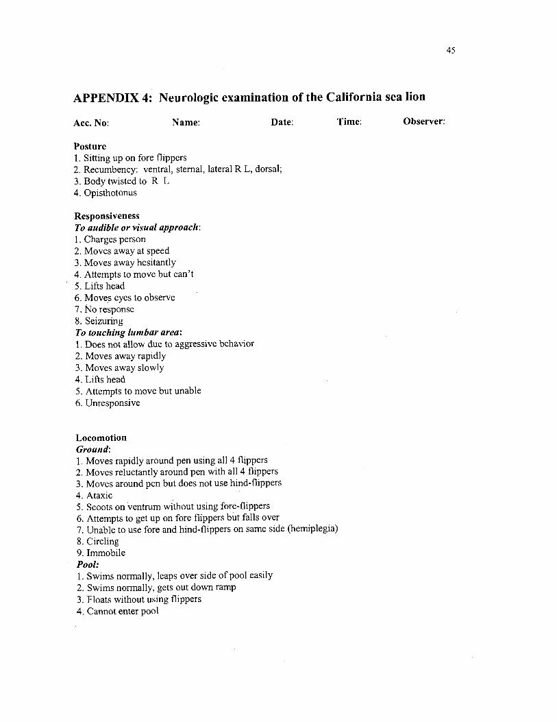
APPENDIX 4: Neurologic examination of the California sea lion
Acc. No: Name: Date: Time: Observer:
Posture 1. Sitting up on fore flippers 2. Recumbency: ventral, sternal, lateral R L, dorsal; 3. Body twisted to R L 4. Opisthotonus
Responsiveness To audible or visual approach: 1. Charges person 2. Moves away at speed 3. Moves away hesitantly 4. Attempts to move but can't 5. Lifts head 6. Moves eyes to observe 7. No response 8. Seizuring To touching lumbar area: I . Does not allow due to aggressive behavior 2. Moves away rapidly 3. Moves away slowly 4. Lifts head 5. Attempts to move but unable 6. Unresponsive
Locomotion Ground: 1. Moves rapidly around pen using all 4 flippers 2. Moves reluctantly around pen with all 4 flippers 3. Moves around pen but does not use hind-flippers 4. Ataxic 5. Scoots on ventrum without using fore-flippers 6. Attempts to get up on fore flippers but falls over 7. Unable to use fore and hind-flippers on same side (hemiplegia) 8. Circling 9. Immobile Pool: 1. Swims normally, leaps over side of pool easily 2. Swims normally, gets out down ramp 3. Floats without using flippers 4. Cannot enter pool