DOI: 10.1212/WNL.0000000000010863 Neurology Publish …
34
Neurology Publish Ahead of Print DOI: 10.1212/WNL.0000000000010863 Association of caffeine and related analytes with resistance to Parkinson’s disease among LRRK2 mutation carriers: A metabolomic study Grace F Crotty MD 1,2 , Romeo Maciuca PhD 3 , Eric A Macklin PhD 2,4 , Junhua Wang PhD 3 , Manuel Montalban BS 3 , Sonnet S. Davis PhD 3 , Jamal I. Alkabsh BS 3 , Rachit Bakshi PhD 1,2 , Xiqun Chen MD PhD 1,2 , Alberto Ascherio MD DrPH 2,5 , Giuseppe Astarita PhD 3 , Sarah Huntwork-Rodriguez PhD 3 , Michael A Schwarzschild MD PhD 1,2 The Article Processing Charge was funded by The Michael J. Fox Foundation for Parkinson s Research. This is an open access article distributed under the terms of the Creative Commons Attribution- NonCommercial-NoDerivatives License 4.0 (CC BY-NC-ND), which permits downloading and sharing the work provided it is properly cited. The work cannot be changed in any way or used commercially without permission from the journal. Neurology® Published Ahead of Print articles have been peer reviewed and accepted for publication. This manuscript will be published in its final form after copyediting, page composition, and review of proofs. Errors that could affect the content may be corrected during these processes. ACCEPTED Copyright © 2020 The Author(s). Published by Wolters Kluwer Health, Inc. on behalf of the American Academy of Neurology. Published Ahead of Print on September 30, 2020 as 10.1212/WNL.0000000000010863
Transcript of DOI: 10.1212/WNL.0000000000010863 Neurology Publish …
Neurology Publish Ahead of PrintDOI: 10.1212/WNL.0000000000010863
Association of caffeine and related analytes with resistance to Parkinson’s disease among LRRK2 mutation carriers: A metabolomic study
Grace F Crotty MD1,2, Romeo Maciuca PhD3, Eric A Macklin PhD2,4, Junhua Wang PhD3, Manuel Montalban BS3, Sonnet S. Davis PhD3, Jamal I. Alkabsh BS3, Rachit Bakshi PhD1,2, Xiqun Chen MD PhD1,2, Alberto Ascherio MD DrPH2,5, Giuseppe Astarita PhD3, Sarah Huntwork-Rodriguez PhD3, Michael A Schwarzschild MD PhD1,2
The Article Processing Charge was funded by The Michael J. Fox Foundation for Parkinson s Research. This is an open access article distributed under the terms of the Creative Commons Attribution-
NonCommercial-NoDerivatives License 4.0 (CC BY-NC-ND), which permits downloading and
sharing the work provided it is properly cited. The work cannot be changed in any way or used
commercially without permission from the journal.
Neurology® Published Ahead of Print articles have been peer reviewed and accepted for
publication. This manuscript will be published in its final form after copyediting, page
composition, and review of proofs. Errors that could affect the content may be corrected during
these processes.
ACCEPTED
Copyright © 2020 The Author(s). Published by Wolters Kluwer Health, Inc. on behalf of the American Academy of Neurology.
Published Ahead of Print on September 30, 2020 as 10.1212/WNL.0000000000010863
1: Department of Neurology, Massachusetts General Hospital, Boston, MA, USA 2: Harvard Medical School, Boston, MA, USA 3: Denali Therapeutics Inc., San Francisco, CA, USA 4: Biostatistics Center, Department of Medicine, Massachusetts General Hospital, Boston, MA, USA 5: Department of Nutrition, Harvard T. H. Chan School of Public Health, Boston, MA, USA Statistical analysis completed by Romeo Maciuca, Denali Therapeutics Inc., San Francisco, CA, USA
Supplementary data present: Data to be deposited into Dryad repository
Corresponding author: Grace Crotty, Email: [email protected] Character and word count: Title character count: 134 Abstract: 265 Body of text: 4317 Number of figures and tables: 5 Number of references: 35 Supplemental Data: doi:10.5061/dryad.nzs7h44pj Search terms: 1. Parkinson's disease/Parkinsonism; 2. LRRK2 gene mutation; 3. Caffeine; 4. Metabolomics; 5. Case control studies
Disclosure:
G.F. Crotty reports no disclosures R. Maciuca is a salaried employee of Denali Therapeutics Inc. E.A. Macklin reports no disclosures J. Wang is a salaried employee of Denali Therapeutics Inc. M. Montalban is a salaried employee of Denali Therapeutics Inc. S. Davis is a salaried employee of Denali Therapeutics Inc. J.I. Alkabsh is a salaried employee of Denali Therapeutics Inc.
ACCEPTED
Copyright © 2020 The Author(s). Published by Wolters Kluwer Health, Inc. on behalf of the American Academy of Neurology.

R. Bakshi reports no disclosures X. Chen reports no disclosures A. Alberto reports no disclosures
G. Astarita is a salaried employee of Denali Therapeutics Inc. S. Huntwork-Rodriguez is a salaried employee of Denali Therapeutics Inc. M.A. Schwarzschild reports no disclosures Study funding: Michael J. Fox Foundation for Parkinson’s Research (to MAS), the Farmer
Family Foundation Initiative for Parkinson’s Disease Research (to MAS), a Jane & Alan Batkin
Research Fellowship (to GC, RB), The Edmond J. Safra Fellowship in Movement Disorders
(GC), and NIH R01NS110879 (to MAS).
ACCEPTED
Copyright © 2020 The Author(s). Published by Wolters Kluwer Health, Inc. on behalf of the American Academy of Neurology.

Abstract:
Objective: To identify markers of resistance to developing Parkinson’s disease (PD) among
LRRK2 mutation (LRRK2+) carriers, we carried out metabolomic profiling in individuals with
PD and unaffected controls (UC), with and without the LRRK2 mutation.
Methods: Plasma from 368 PD and UC subjects in the LRRK2 Cohort Consortium (LCC),
comprising 118 LRRK2+/PD+, 115 LRRK2+/UC, 70 LRRK2-/PD+ and 65 LRRK2-/UC, and CSF
available from 68 of them were analyzed by liquid chromatography with mass spectrometry. For
282 analytes quantified in plasma and CSF, we assessed differences among the four groups and
interactions between LRRK2 and PD status, using ANCOVA models adjusted by age, study site
cohort, and sex, with p-value corrections for multiple comparisons.
Results: Plasma caffeine concentration was lower in PD vs. UC subjects (p<0.001), more so
among LRRK2+ carriers (by 76%) than among LRRK2- subjects (by 31%), with significant
interaction between LRRK2 and PD status (p=0.005). Similar results were found for caffeine
metabolites (paraxanthine, theophylline, 1-methylxanthine) and a non-xanthine marker of coffee
consumption (trigonelline) in plasma, and in the subset of corresponding CSF samples. Dietary
caffeine was also lower in LRRK2+/PD+ compared to LRRK2+/UC with significant interaction
effect with the LRRK2+ mutation (p <0.001).
Conclusions: Metabolomic analyses of the LCC samples identified caffeine, its demethylation
metabolites, and trigonelline as prominent markers of resistance to PD linked to pathogenic
LRRK2 mutations, more so than to idiopathic PD. As these analytes are known both as correlates
of coffee consumption and as neuroprotectants in animal PD models, the findings may reflect
ACCEPTED
Copyright © 2020 The Author(s). Published by Wolters Kluwer Health, Inc. on behalf of the American Academy of Neurology.
their avoidance by those predisposed to develop PD or their protective effects among LRRK2
mutation carriers.
Introduction:
Leucine-rich repeat kinase 2 (LRRK2) gene mutation is considered a major causative influence
on Parkinson disease (PD), and demonstrates variable age-dependent, incomplete penetrance1,2.
This incomplete penetrance suggests that other genetic or environmental factors modulate the
gene’s expression or its effects on PD pathophysiology. The identification of such modulators
could pave the way for future preventative and disease-modifying therapies. Our recent work
identified higher levels of urate in LRRK2 mutation carriers without PD, suggesting that plasma
urate could be a marker of resistance against developing PD in mutation carriers3. Alcalay et al.
studied urinary bis(monoacylglycerol) phosphate isoforms and found slightly higher levels of
2,20-di-18:1-bis(monoacylglycerol)phosphate in LRRK2+/PD+ compared to
LRRK2+/unaffected controls (UC)4.
In this study, we carried out metabolomic profiling in participants enrolled in the LRRK2 Cohort
Consortium (LCC). To our knowledge, there has only been one plasma metabolomic study of
LRRK2 mutation carriers, and its investigation of purine metabolites was limited to 12
LRRK2+/PD+ subjects and 21 LRRK2+/UC subjects5. Our study sought to identify markers of
resistance to developing PD in LRRK2 mutation carriers, and to characterize a metabolomic
signature of pathogenic LRRK2 mutations. Among our pre-specified hypotheses was that
caffeine and its related analytes are reduced in PD subjects compared to unaffected controls, with
reduction similar in those with and without LRRK2 mutations. Our expectation was based on the
link between caffeine intake to reduced risk of PD6–8, as well as on the demonstration of their
lower serum levels in idiopathic PD9,10.
ACCEPTED
Copyright © 2020 The Author(s). Published by Wolters Kluwer Health, Inc. on behalf of the American Academy of Neurology.

Methods:
Subjects: Plasma and CSF were obtained from participants enrolled in the LRRK2 Cohort
Consortium (LCC). The LCC was established in 2009 and has been coordinated and funded by
The Michael J. Fox Foundation for Parkinson’s Research (MJFF). The LCC includes subjects
diagnosed with idiopathic PD (LRRK2-/PD+), pathogenic LRRK2 gene mutation carriers with
PD (LRRK2+/PD+), LRRK2 gene mutation carriers without evidence of PD (LRRK2+/UC), and
unaffected non-carrier controls (LRRK2-/UC). The LCC comprises three distinct studies, all of
which were drawn upon in selecting samples for our research: 23andMe Blood Collection Study;
LRRK2 Longitudinal study; and the LRRK2 Cross-sectional study. Subjects in the LRRK2
Longitudinal study and LRRK2 Cross-sectional study were enrolled in family-, community-, or
clinic-based studies from North America, Europe, Asia, and North Africa. The LRRK2 Cross-
sectional study was subdivided into the Cross-sectional North American site cohort and the
Cross-sectional Europe, Asia, and North African site cohort, with their biological samples
originally stored in separate biorepositories (Coriell and BioRep, respectively) before all LCC
biosamples were transferred for collective management at Indiana University. Therefore, in our
study we adjusted analyses for these four distinctly processed LCC study site cohorts: the
23andMe Blood Collection cohort; the LRRK2 Longitudinal cohort; the LRRK2 Cross-sectional
North American cohort, and the LRRK2 Cross-sectional Europe, Asia, and North African cohort.
Further information on the LCC was published previously11 and is available at
https://www.michaeljfox.org/data-sets and www.michaeljfox.org/lccinvestigators.
From these four LCC study site cohorts, plasma specimens were selected to best match age and
sex across the genotype and disease status groups, similar to our prior study3. Sixteen percent of
requested plasma specimens had corresponding CSF specimens available. MJFF provided codes
ACCEPTED
Copyright © 2020 The Author(s). Published by Wolters Kluwer Health, Inc. on behalf of the American Academy of Neurology.

linking each LCC biospecimen to the associated subject’s genotype, disease status, age, sex, and
clinical data including caffeine intake only after our finalized analyte values were submitted to
the MJFF LCC depository.
Samples: The LRRK2 Longitudinal study and both of the LRRK2 Cross-sectional studies had
standardized protocols for plasma collection, whereas the 23andMe Blood Collection Study did
not (https://www.michaeljfox.org/data-sets and https://www.23andme.com/pd/). CSF was
collected only in the LRRK2 Cross-sectional study (Data available from Dryad (Table e1)). As
per protocol, samples were to be collected between 8 and 10 am with subjects strongly advised to
be in a fasting state, with a minimum of 8 hours since last meal or food intake. Training videos
were provided for collecting, storing and shipping samples. Further information is provided at
https://files.michaeljfox.org/LRRK2_Cohort_Consortium_Biologics_Manual%20_US.pdf.
Dietary caffeine questionnaires were completed by subjects in the LRRK2 Longitudinal study
and the LRRK2 Cross-sectional study. Using data from the Food and Drug Administration
(FDA), we converted the cups of caffeinated coffee, black tea, green tea, and soda into
milligrams of caffeine per day (https://www.fda.gov/consumers/consumer-updates/spilling-
beans-how-much-caffeine-too-much).
Standard Protocol Approvals, Registrations, and Patient Consents: We did not require ethical
approval for this study as it involved anonymized, minimal risk LCC data.
Sample preparation: Plasma and CSF samples received from the LCC were stored at -80oC and
were thawed for assay on wet ice. Plasma samples (10μL) were spun down and then transferred
to polypropylene 96-well V-bottom half deep-well plates. Next, methanol (200μL) containing
internal standards was added into each well using the Velocity 11 Bravo Liquid Handling
Platform (Agilent Technologies, Santa Clara, CA). Plates were shaken for 5 min at room
ACCEPTED
Copyright © 2020 The Author(s). Published by Wolters Kluwer Health, Inc. on behalf of the American Academy of Neurology.

temperature and placed at -20°C for 1 hour to allow further precipitation of proteins. Plates were
then centrifuged at 4,000 x g, 4°C for 20 min. The supernatant was divided into two aliquots: a
100μL aliquot was transferred to 96-well plates with glass inserts for direct liquid
chromatography with mass spectrometry (LC/MS) analyses of lipids and metabolites (Data
available from Dryad (Tables e2-9)); a second 50μL aliquot was transferred to a separate 96-well
plate with glass inserts, dried it down under gentle nitrogen stream, and resuspended in 100 μL
of 92.5 /5/2.5 ACN/IPA/water with 5 mM ammonium formate and 0.5% formic acid for LC/MS
analysis of GlcCer and GalCer species (Data available from Dryad (Tables e2-9)).
CSF samples (20µL) were spun down, transferred into 96-well plates with glass inserts, then
methanol (100µL) containing internal standards was added into each well using the Velocity 11
Bravo Liquid Handling Platform (Agilent Technologies, Santa Clara, CA). Plates were sealed,
shaken for 5 min at room temperature and then centrifuged at 1,000 x g, at 4°C for 2 min. Next,
the samples were divided into two aliquots: a 60µL aliquot was transferred to 96-well plates with
glass inserts for direct LC/MS analyses of lipids and metabolites; a 30µL aliquot was transferred
to a separate 96-well plate with glass inserts, dried it down under gentle nitrogen stream, and
resuspended in 30 µL of 92.5/5/2.5 ACN/IPA/water with 5 mM ammonium formate and 0.5%
formic acid for LC/MS analysis of GlcCer and GalCer species (Data available from Dryad
(Tables e2-9)).
Data reporting: For each analyte, both un-normalized peak area (units=area) and peak area
normalized to an internal standard (units=area ratio) were calculated and reported. For urate,
concentration was calculated (units=mg/dL) as a product of the corresponding area ratio and the
amount of internal standard used. Peak areas with signal-to-noise <3 were not reported. The
ACCEPTED
Copyright © 2020 The Author(s). Published by Wolters Kluwer Health, Inc. on behalf of the American Academy of Neurology.

primary analysis outcome is the internal standard normalized area ratio (“area ratio” or relative
abundance).
282 analytes were quantified in the plasma and CSF. We excluded 5 analytes from our PD and
UC comparisons, and from our volcano-plots (Figure 1A-C) as they correlated closely with
antiparkinsonian medication use (i.e., levodopa and its metabolites dopamine, 3-
methoxytyrosine, dopamine 3-o-sulfate, dopamine 4-sulfate, and 3-hydroxytyrosol, and 3-
hydroxy-N6,N6,N6-trimethyllysine). Plasma urate concentrations measured previously by HPLC
in the same samples3 correlated well with those measured by LC/MS in the current study
(r=0.95).
Results from four of 380 available LCC plasma specimens received for this study did not pass
quality control checks and were excluded from further analysis prior to unblinding. Following
formal unblinding (but while unaware of individual subjects’ analyte values) we identified nine
UC classified subjects as having features indicative of a PD or related diagnosis (e.g., clinician
rating of PD probability of 50-100%, modified Hoehn and Yahr scale (mH&Y) scale score ≥1, or
Unified Parkinson’s Disease Rating Scale (UPDRS) (part 3) score ≥18), suggesting initial
misclassification of their enrollment status. After review of these cases with their site clinicians
and study cohort staff (and while unaware of any analyte values subjects), with concurrence of
MJFF staff, we corrected the classification status of 1 subject and excluded from analysis the
remaining 8 subjects. Of note, these LCC subject classification corrections were made since our
previously published analysis of the dataset3.
Statistical analysis: Sample size (n=380, with n=120 for each of LRRK2+/PD+ and LRRK2+/UC,
the two critical comparison groups) was determined based on estimation of sample size required
to achieve adequate power for replication of a urate difference between these groups in the
ACCEPTED
Copyright © 2020 The Author(s). Published by Wolters Kluwer Health, Inc. on behalf of the American Academy of Neurology.

original test sample analysis (n=64 for each of LRRK2+/PD+ and LRRK2+/UC groups;
p=0.047).
A statistical analysis plan was specified prior to unblinding of investigators to LRRK2 genotype,
PD status, and all other clinical parameters linked to plasma and CSF samples. For plasma and
CSF metabolites we reported the geometric mean of relative abundance. For all plasma analytes,
estimates of mean differences between PD and UC subjects, and between LRRK2+ and LRRK2-
subjects were generated with a robust analysis of covariance (ANCOVA) model with log2(Area
ratio) as the dependent variable, and adjusting by PD status cohort (PD vs UC), LRRK2 gene
status, Sex, Age (modelled as cubic spline, with a knot at 60 years), and substudy (the 4 study
site cohorts). Interaction terms of PD status cohort x LRRK2 status x Sex and PD status cohort x
substudy were included. The ANCOVA model was the basis for pairwise tests for the contrasts
of interest (PD vs UC, overall or within LRRK2+/- subgroups), associated p-values and adjusted
between groups effect sizes. To partially control the false discovery rate, we employed a
Benjamini-Hochberg (BH) correction to the p-values for the PD vs UC (overall, and within each
of the LRRK2+ and LRRK2- groups) comparisons. Statistical significance was judged at a two-
sided 0.05 significance level.
For the smaller subset of subjects with CSF samples, estimates of between group were generated
from a joint CSF plasma linear mixed effects model, considering the CSF/Plasma measurements
as repeated, within subject, measures for each analyte, adjusting for the same set of covariates as
above. A similar, albeit simplified set of interaction terms was included in the model (Cohort x
LRRK2 status x Sample type, Sex x Sample Type, and Age x Sample Type).
Data Availability: Raw data from this study are available following online LCC data request at
https://www.michaeljfox.org/data-sets. Supplemental data (Tables e1-9) are stored on Dryad.
ACCEPTED
Copyright © 2020 The Author(s). Published by Wolters Kluwer Health, Inc. on behalf of the American Academy of Neurology.

Results:
Table 1 presents baseline characteristics of the 368 LCC subjects whose plasma was analyzed for
this study, stratified by PD status (1:1 for diagnosed PD:UC) and LRRK2 gene status (2:1 for
LRRK2+:LRRK2-). Differences between the four groups’ features highlight the value in
adjusting for age, sex, and study site cohort. As expected, Montreal Cognitive Assessment
(MoCA) scores were lower in the PD groups compared to UC (p<0.05). Data on time fasted prior
to blood draw and on race, MoCA, and mH&Y assessments were available for most subjects
except for the 24% from the 23andMe Blood Collection study (which provided plasma only from
LRRK2+ individuals). Regarding specific pathogenic LRRK2 variants present, 91% (n=107) of
the LRRK2+/PD+ and 92% (n=106) LRRK2+/UC groups carried the G2019S LRRK2 mutation.
Other LRRK2 variants in the LRRK2+/PD+ group were R1441G (n=4), R1441C (n=1), N1437H
(n=2), L1795F (n=2), C228S (n=1) and unknown (n=1). Other LRRK2 variants in the
LRRK2+/UC group were R1441G (n=4), R1441C (n=1), N1437H (n=1), L1114L (n=1), and
L1795F (n=2).
Comparing the concentrations of 282 LC/MS-quantified plasma metabolites between people with
and without PD irrespective of LRRK2 gene status revealed a cluster of five analytes showing
the greatest differences, excluding metabolites linked to antiparkinsonian medication. All five
are caffeine-related analytes (Figure 2) and are lower in the PD than UC subjects, as depicted in
Figure 1A in a volcano plot of metabolomics data with effect size (ratio for PD vs UC values,
adjusted for age, sex, study site cohort and LRRK2 status) plotted against BH-adjusted p values.
Compared to UC subjects, those with PD had lower plasma levels not only of caffeine (by 71%)
but also of its partially demethylated xanthine-based metabolites paraxanthine (1,7-
dimethylxanthine), theophylline (1,3-dimethylxanthine), 1-methylxanthine (by 57%, 56% and
ACCEPTED
Copyright © 2020 The Author(s). Published by Wolters Kluwer Health, Inc. on behalf of the American Academy of Neurology.
49%, respectively) and trigonelline, a non-xanthine constituent of coffee, (by 52%) with
associated BH-adjusted p values all ≤0.001 (Table 2). Consistent with their metabolic and dietary
relationships, plasma caffeine correlated well with its demethylation products (paraxanthine,
theophylline and 1-methylxanthine) and its coffee co-constituent, trigonelline, with Spearman
coefficients (r) of approximately 0.8, 0.8, 0.5 and 0.5, respectively in PD subjects.
Stratifying by genotype reveals that the extent to which these caffeine-related analytes are lower
in PD is substantially greater among pathogenic LRRK2 mutation carriers (Figure 1B and 1C;
Table 2). In LRRK2+/ PD+ plasma, caffeine itself was 76% lower than in plasma of LRRK2+/UC
subjects, whereas in idiopathic PD plasma caffeine was 31% lower than in LRRK2-/UC subjects
(p for LRRK2 x PD status interaction =0.005). Similarly, levels in PD were even lower amongst
LRRK2+ vs LRRK2- participants for paraxanthine (66% vs 21%), theophylline (67% vs 21%), 1-
methylxanthine (62% vs 14%) and trigonelline (63% vs 15%) with p for interaction <0.01 for
each. Of note, fasting times prior to blood draw did not differ appreciably across groups (Table
1), suggesting that the lower levels of analytes from caffeinated beverages observed in the
plasma of people with PD are not due to their having fasted longer.
Dietary caffeine consumption questionnaire data were available for 212 of the subjects with
plasma caffeine metabolites (Table 1). In this subset, PD subjects with the LRRK2+ mutation
consumed significantly (41%) less caffeine (in mg/day) when compared to unaffected controls
with the LRRK2+ mutation (p<0.003), and there was a significant interaction effect with the
LRRK2+ mutation (p<0.001). Dietary caffeine consumption positively correlated with plasma
caffeine metabolite concentration (r=0.3). When caffeine metabolite concentration was adjusted
for dietary caffeine intake, the significant difference between PD and controls (p=0.01) and
ACCEPTED
Copyright © 2020 The Author(s). Published by Wolters Kluwer Health, Inc. on behalf of the American Academy of Neurology.

between LRRK2+/PD+ and LRRK2+/UC (p=0.01) persisted, although the interaction effect for
LRRK2+ mutation was not significant (p=0.38).
Similar to plasma caffeine, plasma levels of the endogenous purine urate were found to be a
marker of PD resistance among LRRK2 mutation carriers in our earlier, non-metabolomic
analysis in the nearly identical LCC sample set3. Although urate also inversely associates with
PD among LRRK2+ more than LRRK2- subjects, the associations are weaker than for the
caffeine-related analytes3 and the evidence for an interaction between urate, LRRK2 genotype
and PD status reached only marginal statistical significance in our more recent analysis (p =
0.047) (Figure 1A-C).
Matching CSF samples were available for 68 subjects with analyzed plasma: LRRK2-/UC
(n=14), LRRK2-/PD+ (n=18), LRRK2+/UC (n=18) and LRRK2+/PD+ (n=18), all of whom were
from the LRRK2 Cross-sectional study (Data available from Dryad (Tables e1)). In Figure 3, we
present box-plot illustrations of the overlapping plasma and CSF samples of the four groups by
LRRK2 gene status and PD status. Among the LRRK2 mutation carriers in this subset, both CSF
and plasma caffeine levels were significantly lower in PD than in unaffected controls (after
adjustment for age, sex and study site cohort): by 74% in CSF (p<0.02) and by 76% in plasma
(p=0.01), consistent with a strong positive correlation between the entire sample of plasma and
CSF caffeine concentrations (r=0.90). By contrast among LRRK2- subjects in this subset, CSF
caffeine was 23% lower and plasma caffeine was 24% higher in PD subjects compared to
controls (p=0.65 and 0.71, respectively).
Similar associations were observed for CSF levels of caffeine’s dimethyl metabolites
paraxanthine and theophylline, and of trigonelline, whose plasma and CSF levels also correlated
closely (r=0.94, 0.94 and 0.90, respectively). 1-methylxanthine was not measured in the CSF.
ACCEPTED
Copyright © 2020 The Author(s). Published by Wolters Kluwer Health, Inc. on behalf of the American Academy of Neurology.
Among the LRRK2 mutation carriers in this subset, both CSF and plasma levels of paraxanthine,
theophylline and trigonelline were lower in PD than in unaffected controls after adjustment: by
73%, 74% and 70% for CSF (p=0.003, =0.003, and <0.001) and by 74%, 76% and 69% for
plasma (p=0.002, <0.002 and =0.001), respectively. By contrast among LRRK2- subjects in this
subset, levels of paraxanthine, theophylline, and of trigonelline were largely indistinguishable
after adjustment between those with and without PD; being higher in CSF by 2%, 1% and 9% in
those with vs without PD (p=0.97, 0.99 and 0.82), and in plasma: by 20%, 19% and 37% in those
with vs without PD (p= 0.70, 0.72 and 0.40). Fasting times prior to lumbar puncture, like those
for blood collection, did not differ across the four groups at approximately 12 hours (p=0.29).
Exploratory stratification showed that the low caffeine association with PD among LRRK2
mutation carriers was present irrespective of sex (62% lower with PD among men and 85%
lower with PD among women; p= 0.004 and <0.001, respectively) or age (65% lower with PD
among those less than 60 years old at baseline, and 83% lower among those aged 60 years or
older at baseline; p<0.02 and <0.001, respectively). Similarly, lower caffeine with PD among
LRRK2 mutation carriers was just as strong when PD cases were restricted to the 22% who
appear not to have been taking levodopa (based on its plasma metabolite 3-methoxytyrosine
having a relative abundance levels less than 0.1) being 78% lower with PD (p<0.001); or to those
at an early clinical stage (mH&Y 2.5 or lower being 76% lower with PD (p=0.002), suggesting
that the lower caffeine levels with PD among LRRK2 mutation carriers is not likely due to an
antiparkinsonian medication effect or more advanced disease. In contrast to LRRK2 mutation
carriers, those without mutations (LRRK2-) showed a non-significant, weaker link between low
caffeine and PD, with caffeine 39% lower in men (p=0.25) and 21% in women (p=0.56).
ACCEPTED
Copyright © 2020 The Author(s). Published by Wolters Kluwer Health, Inc. on behalf of the American Academy of Neurology.

Discussion:
In this metabolomics study of LRRK2 mutation carriers, we identified the dietary purine caffeine
and its metabolites as the most affected pathway in plasma with significantly lower levels of
caffeine, its demethylation products, and trigonelline, a non-purine marker of coffee
consumption in subjects with PD compared to unaffected controls, and to a significantly greater
extent among pathogenic LRRK2 mutation carriers than among those without a mutation.
Our observation of lower plasma caffeine concentrations in subjects with PD confirms recent
metabolic9 and metabolomic10 findings, consistent with well-established epidemiological
evidence for increased risk of developing idiopathic PD in individuals who consume less
caffeinated beverages, based on dietary recall12. Interestingly, Fujimaki et al.9 did not find
significantly lower caffeine intake in subjects with PD despite lower serum levels of caffeine and
its metabolites in their cohort, raising the possibility that the lower levels could result from
reduced bioavailability (e.g., due to reduced gastrointestinal absorption). By contrast in the larger
LCC cohort assessed here, a lower intake among PD vs control subjects, and a significantly
lower intake in subjects with LRRK2+/PD+ compared to those with LRRK2+/UC suggest that
lower concentrations of caffeine and its metabolites circulating in people with PD reflects, at
least in part, their lesser consumption of this dietary purine. Of note, the finding of comparably
reduced plasma levels of trigonelline, a non-purine constituent and plasma biomarker of coffee13
(which comprised 84% of the total daily caffeine consumption) further supports a dietary basis
for the lower caffeine concentrations in PD vs controls. However, our results are also consistent
with a role of differential absorption, metabolism or clearance of caffeine in PD given the
persistent, significantly lower level of caffeine and its related analytes in our PD subjects with or
ACCEPTED
Copyright © 2020 The Author(s). Published by Wolters Kluwer Health, Inc. on behalf of the American Academy of Neurology.
without a pathogenic LRRK2 mutation, compared to their control counterparts, following
adjustment for dietary caffeine intake.
The unexpected finding that caffeine’s and related analytes’ associations with resistance to PD
are substantially greater among LRRK2 mutation carriers than non-carriers appears robust and
intriguing. Levels of these analytes were lower in both the plasma and CSF of PD patients versus
unaffected controls among LRRK2 mutation carriers, whereas among non-carriers these analytes
were not significantly reduced in PD versus control subjects. Direct evidence for interaction
between PD and LRRK2 status (p<0.01 for each of the five caffeine-related analytes) suggests a
true gene-environment interaction, rather than sample size bias resulting from the (2:1)
predominance of LRRK2+ over LRRK2- participants in this study’s LCC sampling.
Corroborating these metabolomic data, we obtained complementary epidemiological evidence
that PD patients consumed significantly less caffeine compared to unaffected controls only
among LRRK2 mutation carriers. Interestingly, our findings of LRRK2-caffeine intake
interaction substantiate earlier findings of Kumar et al14. They studied the interplay between
caffeine consumption and a different LRRK2 mutation – the R1628P risk variant in a Chinese
population. They similarly reported a significant LRRK2-caffeine interaction, with a lower odds
ratio (OR) of developing PD among R1628P LRRK2 mutation carriers who consumed caffeine
(OR 3.1) than among mutation carriers who were non-consumers (OR 15.4), when compared to
caffeine consumers without the mutation14, suggesting that the biology underlying the caffeine-
LRRK2 interaction entails the common ability of COR domain-based R1628P and kinase
domain-based G2019S mutations to increase kinase activity15–17.
Similar even if indirect evidence for caffeine’s greater association with resistance to genetic
forms of PD was recently provided by Angelopoulou et al. who explored environmental factors
ACCEPTED
Copyright © 2020 The Author(s). Published by Wolters Kluwer Health, Inc. on behalf of the American Academy of Neurology.
in early-onset (before 50 years of age) compared to later-onset PD in a Greek cohort. They
observed that coffee-drinking was associated with a lower risk of early- but not later-onset PD.
They also detected a linear-dose association between coffee exposure and the risk of developing
familial as well as early-onset PD18. As early-onset PD and familial PD are more likely to be
genetic than later-onset PD, these data together with those of Kumar et al. and ours suggest that
caffeine could be broadly associated with PD gene penetrance.
Of note, no statistically significant analyte-LRRK2 mutation interaction has been reported
previously for PD risk at the metabolome level. Urate – an endogenous antioxidant and end-
product of purine metabolism – was found in our preceding study of the nearly identical LCC
cohort to be associated with PD among LRRK2 mutation carriers to a greater extent than in non-
carriers3. However, in contrast to caffeine’s link to PD resistance, that of urate was relatively
modest and only slightly greater among LRRK2 mutation carriers than non-carriers (Figure 1A-
C).
Our findings for CSF levels of caffeine and related analytes were similar to those for their
plasma counterparts, despite the lower number of CSF (n=68) than plasma (n=368) samples
analyzed. The similarities are in keeping with the close correlation between plasma and CSF
concentrations for these analytes. These findings support the use of peripheral (e.g., plasma)
samples in assessing the caffeine metabolic pathway in relation to PD risk.
Caffeine is the most widely consumed psychoactive substance and a nonselective antagonist of
the adenosine 2A (A2A) receptor. It also possesses neuroprotective properties in animal models of
PD. Both its psychostimulant actions and protective effects on dopaminergic neurons likely rely
on A2A receptors19–21, with A2A receptor blockade by caffeine reducing excitotoxic and
inflammatory processes22–24. The association of caffeine with resistance to PD could reflect the
ACCEPTED
Copyright © 2020 The Author(s). Published by Wolters Kluwer Health, Inc. on behalf of the American Academy of Neurology.

ability of PD determinants to reduce the likelihood of caffeine intake, or conversely it could
reflect a causal relationship driven by a protective effect of caffeine or a related analyte23,25,26.
Interestingly, LRRK2 biology may potentiate either of these caffeine-PD relationships given its
involvement in striatal neuroplasticity and nigrostriatal neurodegeneration, both of which can be
attenuated by caffeine via its actions on adenosine A2A receptors 27,28. For example, the
pathogenic G2019S LRRK2 mutation has been found to alter synaptic plasticity in the A2A
receptor-laden striatum while bolstering resistance to social stress in young animals29.
Alternatively, the well-established neuroprotective properties of caffeine or dimethyl
metabolites, paraxanthine and theophylline, which are themselves A2AR antagonists30,31 in PD
models, could be potentiated in the setting of pathogenic LRRK2 mutations. For example, the
increased kinase activity of pathogenic G2019S LRRK2 may potentiate the dopaminergic
degeneration induced by α-synuclein32, and α-synuclein-induced dopaminergic neuron injury
can be attenuated by caffeine33 and depend upon the A2AR34. Thus, A2AR antagonists like
caffeine and its metabolites may be particularly effective in attenuating LRRK2 kinase-
potentiated α-synuclein pathobiology, raising the possibility that the therapeutic potential of
caffeine or other A2AR antagonists may be greater for slowing or preventing LRRK2 PD than for
idiopathic disease. Similarly, while the non-purine trigonelline may simply be a marker of coffee
and caffeine consumption, it has been shown to have its own protective effects in a PD model35.
There are several strengths to our study. First, our metabolomic analysis of LRRK2 PD was
conducted on the largest cohort to date. Second, the cohort included matched LRRK2 gene status
as well as PD- controls, allowing the opportunity to gauge interactions across genotype and
disease state. Third, the depth of the LCC biorepository allowed us to assess CSF as well as
plasma biomarkers of LRRK2 PD. Finally, we were able to correlate dietary intake with
ACCEPTED
Copyright © 2020 The Author(s). Published by Wolters Kluwer Health, Inc. on behalf of the American Academy of Neurology.
metabolomics, which is relatively novel in allowing us to explore the basis for lower caffeine-
related analytes in LRRK2 PD.
However, several limitations of our study should be noted, and included potential selection bias
as subjects were recruited through the multiple individual study site cohorts that comprise the
LCC. Although we endeavored to match or adjust for relevant covariates to reduce their
influence when differing across groups, unmeasured confounders could have affected our results.
Second, misclassification of PD or control subjects could not be fully excluded in part based on
the lack of a biomarker for definitive diagnosis of PD, although extensive phenotype data were
available for most LCC subjects allowing us to cross-check and confirm classifications, and such
errors would have biased us toward null results. Lastly, the results are cross-sectional precluding
direct assessment of the predictive potential of analytes on PD risk and progression.
The identification of caffeine and adenosine antagonists as potential markers of PD resistance
among LRRK2 mutation carriers supports their potential for development as biomarkers
contributing to phenoconversion risk assessment among carriers, and to progression rates among
LRRK2 PD patients. In addition, identification of caffeine-related analytes as resistance markers
raise the possibility of their development as candidate therapeutics given the relatively low risk
of repurposing these dietary or pharmacological agents -- caffeine, theophylline and trigonelline
– for slowing progression in those with manifest LRRK2 PD, or among at-risk mutation carriers
to reduce the penetrance of the disease. Next steps may include replication of these metabolomic
results in an independent cohort of LRRK2 mutation carriers, and assessing their specificity in
other genetic (e.g., GBA mutation-driven) forms of PD.
ACCEPTED
Copyright © 2020 The Author(s). Published by Wolters Kluwer Health, Inc. on behalf of the American Academy of Neurology.
Acknowledgements:
This study was funded by the Michael J. Fox Foundation for Parkinson’s Research (to MAS), the
Farmer Family Foundation Initiative for Parkinson’s Disease Research (to MAS), a Jane & Alan
Batkin Research Fellowship (to GC, RB), The Edmond J. Safra Fellowship in Movement
Disorders (GC), and NIH R01NS110879 (to MAS). Data and biospecimens used in preparation
of this article were obtained from the MJFF-sponsored LRRK2 Cohort Consortium (LCC) under
LCC project ID #s 108 (plasma) and 122 (CSF). The LRRK2 Cohort Consortium is coordinated
and funded by The Michael J. Fox Foundation for Parkinson’s Research. We gratefully
acknowledge the participants, investigators and staff of the LCC. We thank Ms. Katherine
Callahan for editorial assistance, and Mr. Eoghan Hynes for his technical support.
ACCEPTED
Copyright © 2020 The Author(s). Published by Wolters Kluwer Health, Inc. on behalf of the American Academy of Neurology.

Appendix 1: Authors
Name Location Contribution
Grace F Crotty MD
-Department of Neurology, Massachusetts General Hospital, Boston, MA, USA -Harvard Medical School, Boston, MA, USA
Design and conceptualization of the study; major role in the acquisition of data; analysis and interpretation of the data; drafting and revising the manuscript for intellectual content
Romeo Maciuca PhD
-Denali Therapeutics Inc., San Francisco, CA, USA
Design and conceptualization of the study; major role in the acquisition of data; analysis and interpretation of the data; revising the manuscript for intellectual content
Eric A Macklin PhD
-Harvard Medical School, Boston, MA, USA - Biostatistics Center, Department of Medicine, Massachusetts General Hospital, Boston, MA, USA
Design and conceptualization of the study; major role in the acquisition of data; analysis and interpretation of the data; revising the manuscript for intellectual content
Junhua Wang PhD -Denali Therapeutics Inc., San Francisco, CA, USA
Major role in the acquisition of data; analysis and interpretation of the data; revising the manuscript for intellectual content
Manuel Montalban BS
-Denali Therapeutics Inc., San Francisco, CA, USA
Major role in the acquisition of data; analysis and interpretation of the data; revising the manuscript for intellectual content
Sonnet S. Davis PhD -Denali Therapeutics Inc., San Francisco, CA, USA
Major role in the acquisition of data; analysis and interpretation of the data; revising the manuscript for intellectual content
Jamal I. Alkabsh BS -Denali Therapeutics Inc., San Francisco, CA, USA
Major role in the acquisition of data; analysis and interpretation of the data; revising the manuscript for intellectual content
Rachit Bakshi PhD
-Department of Neurology, Massachusetts General Hospital, Boston, MA, USA -Harvard Medical School, Boston,
Design and conceptualization of the study; major role in the acquisition of data; analysis and interpretation of the data; revising the manuscript for intellectual
ACCEPTED
Copyright © 2020 The Author(s). Published by Wolters Kluwer Health, Inc. on behalf of the American Academy of Neurology.

MA, USA content
Xiqun Chen MD PhD
-Department of Neurology, Massachusetts General Hospital, Boston, MA, USA -Harvard Medical School, Boston, MA, USA
Design and conceptualization of the study; major role in the acquisition of data; analysis and interpretation of the data; revising the manuscript for intellectual content
Alberto Ascherio MD DrPH
- Harvard Medical School, Boston, MA, USA -Department of Nutrition, Harvard T. H. Chan School of Public Health, Boston, MA, USA
Design and conceptualization of the study; major role in the acquisition of data; analysis and interpretation of the data; revising the manuscript for intellectual content
Giuseppe Astarita PhD
-Denali Therapeutics Inc., San Francisco, CA, USA
Design and conceptualization of the study; major role in the acquisition of data; analysis and interpretation of the data; revising the manuscript for intellectual content
Sarah Huntwork-Rodriguez PhD
-Denali Therapeutics Inc., San Francisco, CA, USA
Design and conceptualization of the study; major role in the acquisition of data; analysis and interpretation of the data; revising the manuscript for intellectual content
Michael A Schwarzschild MD PhD
-Department of Neurology, Massachusetts General Hospital, Boston, MA, USA -Harvard Medical School, Boston, MA, USA
Design and conceptualization of the study; major role in the acquisition of data; analysis and interpretation of the data; drafting and revising the manuscript for intellectual content
ACCEPTED
Copyright © 2020 The Author(s). Published by Wolters Kluwer Health, Inc. on behalf of the American Academy of Neurology.
References
1. Healy DG, Falchi M, O’Sullivan SS, et al. Phenotype, genotype, and worldwide genetic
penetrance of LRRK2-associated Parkinson’s disease: a case-control study. Lancet Neurol.
2008;7(7):583–590.
2. Lee AJ, Wang Y, Alcalay RN, et al. Penetrance estimate of LRRK2 p.G2019S mutation in
individuals of non-Ashkenazi Jewish ancestry. Mov. Disord. 2017;32(10):1432–1438.
3. Bakshi R, Macklin EA, Logan R, et al. Higher urate in LRRK2 mutation carriers resistant
to Parkinson disease. Ann. Neurol. 2019;85(4):593–599.
4. Alcalay RN, Hsieh F, Tengstrand E, et al. Higher Urine bis(Monoacylglycerol)Phosphate
Levels in LRRK2 G2019S Mutation Carriers: Implications for Therapeutic Development.
Mov. Disord. 2020;35(1):134–141.
5. Johansen KK, Wang L, Aasly JO, et al. Metabolomic profiling in LRRK2-related
Parkinson’s disease. PloS One 2009;4(10):e7551.
6. Ascherio A, Zhang SM, Hernán MA, et al. Prospective study of caffeine consumption and
risk of Parkinson’s disease in men and women. Ann. Neurol. 2001;50(1):56–63.
7. Ascherio A, Schwarzschild MA. The epidemiology of Parkinson’s disease: risk factors and
prevention. Lancet Neurol. 2016;15(12):1257–1272.
ACCEPTED
Copyright © 2020 The Author(s). Published by Wolters Kluwer Health, Inc. on behalf of the American Academy of Neurology.

8. Ross GW, Abbott RD, Petrovitch H, et al. Association of coffee and caffeine intake with
the risk of Parkinson disease. JAMA 2000;283(20):2674–2679.
9. Fujimaki M, Saiki S, Li Y, et al. Serum caffeine and metabolites are reliable biomarkers of
early Parkinson disease. Neurology 2018;90(5):e404–e411.
10. Hatano T, Saiki S, Okuzumi A, et al. Identification of novel biomarkers for Parkinson’s
disease by metabolomic technologies. J. Neurol. Neurosurg. Psychiatry 2016;87(3):295–
301.
11. Marras C, Alcalay RN, Caspell-Garcia C, et al. Motor and nonmotor heterogeneity of
LRRK2-related and idiopathic Parkinson’s disease. Mov. Disord. 2016;31(8):1192–1202.
12. Costa J, Lunet N, Santos C, et al. Caffeine exposure and the risk of Parkinson’s disease: a
systematic review and meta-analysis of observational studies. J. Alzheimers Dis. JAD
2010;20 Suppl 1:S221-238.
13. Midttun Ø, Ulvik A, Nygård O, Ueland PM. Performance of plasma trigonelline as a
marker of coffee consumption in an epidemiologic setting. Am. J. Clin. Nutr.
2018;107(6):941–947.
14. Kumar PM, Paing SST, Li H, et al. Differential effect of caffeine intake in subjects with
genetic susceptibility to Parkinson’s Disease. Sci. Rep. 2015;5:15492.
15. Berwick DC, Heaton GR, Azeggagh S, Harvey K. LRRK2 Biology from structure to
dysfunction: research progresses, but the themes remain the same. Mol. Neurodegener.
2019;14(1):49.
ACCEPTED
Copyright © 2020 The Author(s). Published by Wolters Kluwer Health, Inc. on behalf of the American Academy of Neurology.
16. Shu Y, Ming J, Zhang P, et al. Parkinson-Related LRRK2 Mutation R1628P Enables Cdk5
Phosphorylation of LRRK2 and Upregulates Its Kinase Activity. PloS One
2016;11(3):e0149739.
17. Tan E-K, Peng R, Teo Y-Y, et al. Multiple LRRK2 variants modulate risk of Parkinson
disease: a Chinese multicenter study. Hum. Mutat. 2010;31(5):561–568.
18. Angelopoulou E, Bozi M, Simitsi A-M, et al. The relationship between environmental
factors and different Parkinson’s disease subtypes in Greece: Data analysis of the Hellenic
Biobank of Parkinson’s disease. Parkinsonism Relat. Disord. 2019;67:105–112.
19. Chen JF, Xu K, Petzer JP, et al. Neuroprotection by caffeine and A(2A) adenosine receptor
inactivation in a model of Parkinson’s disease. J. Neurosci. 2001;21(10):RC143.
20. Lazarus M, Shen H-Y, Cherasse Y, et al. Arousal effect of caffeine depends on adenosine
A2A receptors in the shell of the nucleus accumbens. J. Neurosci. 2011;31(27):10067–
10075.
21. Xu K, Di Luca DG, Orrú M, et al. Neuroprotection by caffeine in the MPTP model of
parkinson’s disease and its dependence on adenosine A2A receptors. Neuroscience
2016;322:129–137.
22. Bagga P, Chugani AN, Patel AB. Neuroprotective effects of caffeine in MPTP model of
Parkinson’s disease: A (13)C NMR study. Neurochem. Int. 2016;92:25–34.
23. Morelli M, Carta AR, Kachroo A, Schwarzschild MA. Pathophysiological roles for purines:
adenosine, caffeine and urate. Prog. Brain Res. 2010;183:183–208.
ACCEPTED
Copyright © 2020 The Author(s). Published by Wolters Kluwer Health, Inc. on behalf of the American Academy of Neurology.

24. Sonsalla PK, Wong L-Y, Harris SL, et al. Delayed caffeine treatment prevents nigral
dopamine neuron loss in a progressive rat model of Parkinson’s disease. Exp. Neurol.
2012;234(2):482–487.
25. Schwarzschild MA, Chen J-F, Ascherio A. Caffeinated clues and the promise of adenosine
A(2A) antagonists in PD. Neurology 2002;58(8):1154–1160.
26. Xu K, Bastia E, Schwarzschild M. Therapeutic potential of adenosine A(2A) receptor
antagonists in Parkinson’s disease. Pharmacol. Ther. 2005;105(3):267–310.
27. Chen J-F, Sonsalla PK, Pedata F, et al. Adenosine A2A receptors and brain injury: broad
spectrum of neuroprotection, multifaceted actions and “fine tuning” modulation. Prog.
Neurobiol. 2007;83(5):310–331.
28. Ferré S, Díaz-Ríos M, Salamone JD, Prediger RD. New Developments on the Adenosine
Mechanisms of the Central Effects of Caffeine and Their Implications for Neuropsychiatric
Disorders. J. Caffeine Adenosine Res. 2018;8(4):121–131.
29. Matikainen-Ankney BA, Kezunovic N, Menard C, et al. Parkinson’s Disease-Linked
LRRK2-G2019S Mutation Alters Synaptic Plasticity and Promotes Resilience to Chronic
Social Stress in Young Adulthood. J. Neurosci. 2018;38(45):9700–9711.
30. Schwarzschild MA, Agnati L, Fuxe K, et al. Targeting adenosine A2A receptors in
Parkinson’s disease. Trends Neurosci. 2006;29(11):647–654.
ACCEPTED
Copyright © 2020 The Author(s). Published by Wolters Kluwer Health, Inc. on behalf of the American Academy of Neurology.

31. Xu K, Xu Y-H, Chen J-F, Schwarzschild MA. Neuroprotection by caffeine: time course
and role of its metabolites in the MPTP model of Parkinson’s disease. Neuroscience
2010;167(2):475–481.
32. Daher JPL, Abdelmotilib HA, Hu X, et al. Leucine-rich Repeat Kinase 2 (LRRK2)
Pharmacological Inhibition Abates α-Synuclein Gene-induced Neurodegeneration. J. Biol.
Chem. 2015;290(32):19433–19444.
33. Yan R, Zhang J, Park H-J, et al. Synergistic neuroprotection by coffee components
eicosanoyl-5-hydroxytryptamide and caffeine in models of Parkinson’s disease and DLB.
Proc. Natl. Acad. Sci. U. S. A. 2018;115(51):E12053–E12062.
34. Kachroo A, Schwarzschild MA. Adenosine A2A receptor gene disruption protects in an α-
synuclein model of Parkinson’s disease. Ann. Neurol. 2012;71(2):278–282.
35. Gaur V, Bodhankar SL, Mohan V, Thakurdesai PA. Neurobehavioral assessment of
hydroalcoholic extract of Trigonella foenum-graecum seeds in rodent models of
Parkinson’s disease. Pharm. Biol. 2013;51(5):550–557.
ACCEPTED
Copyright © 2020 The Author(s). Published by Wolters Kluwer Health, Inc. on behalf of the American Academy of Neurology.
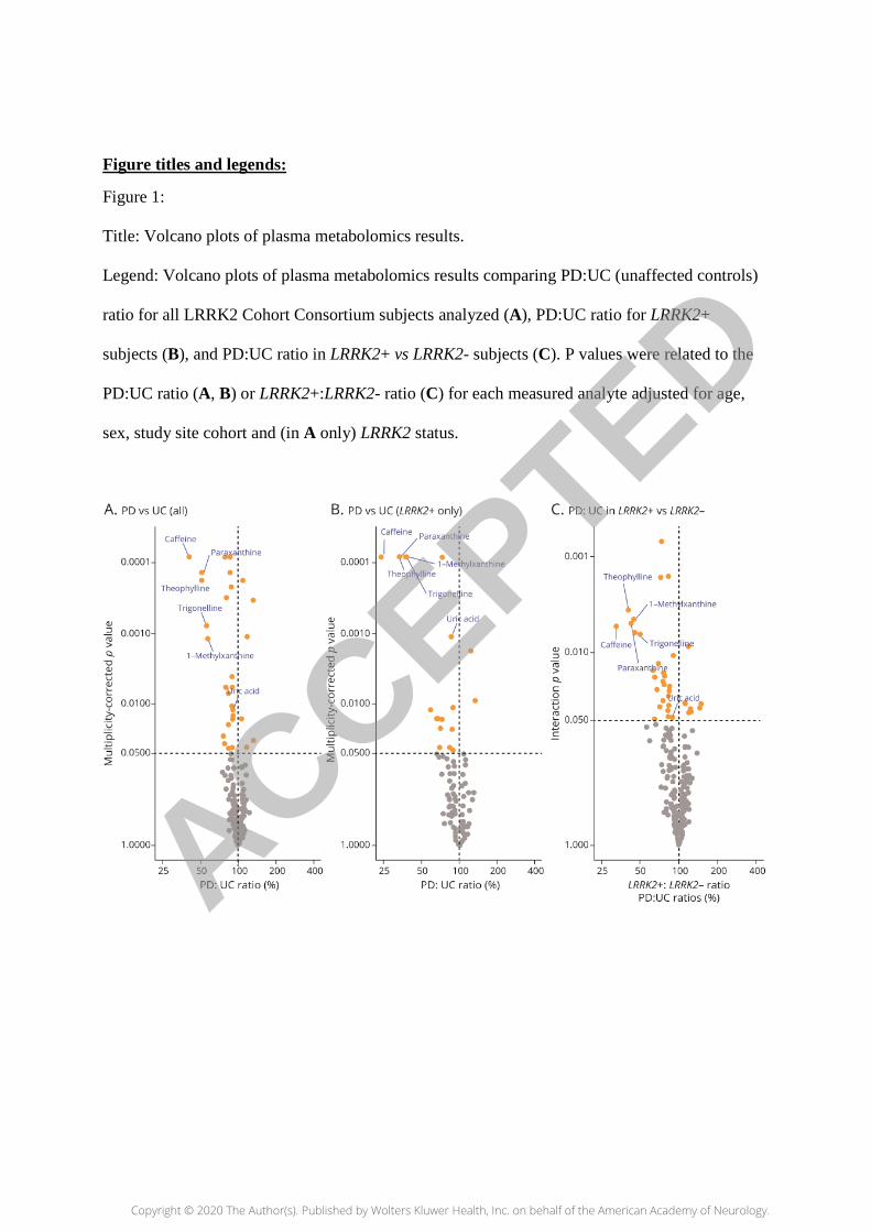
Figure titles and legends:
Figure 1:
Title: Volcano plots of plasma metabolomics results.
Legend: Volcano plots of plasma metabolomics results comparing PD:UC (unaffected controls)
ratio for all LRRK2 Cohort Consortium subjects analyzed (A), PD:UC ratio for LRRK2+
subjects (B), and PD:UC ratio in LRRK2+ vs LRRK2- subjects (C). P values were related to the
PD:UC ratio (A, B) or LRRK2+:LRRK2- ratio (C) for each measured analyte adjusted for age,
sex, study site cohort and (in A only) LRRK2 status.
ACCEPTED
Copyright © 2020 The Author(s). Published by Wolters Kluwer Health, Inc. on behalf of the American Academy of Neurology.

Figure 2:
Title: Chemical structures and metabolic pathways of caffeine and related analytes
(paraxanthine, theophylline, 1-methylxanthine and trigonelline).
ACCEPTED
Copyright © 2020 The Author(s). Published by Wolters Kluwer Health, Inc. on behalf of the American Academy of Neurology.

Figure 3:
Title: Concentrations of caffeine in matched plasma (A) and CSF (B) samples of LCC
participants.
Legend: Concentrations of caffeine in matched plasma (A) and CSF (B) samples of LCC
participants by LRRK2 and PD status, adjusted for age, sex, study site cohort.
ACCEPTED
Copyright © 2020 The Author(s). Published by Wolters Kluwer Health, Inc. on behalf of the American Academy of Neurology.
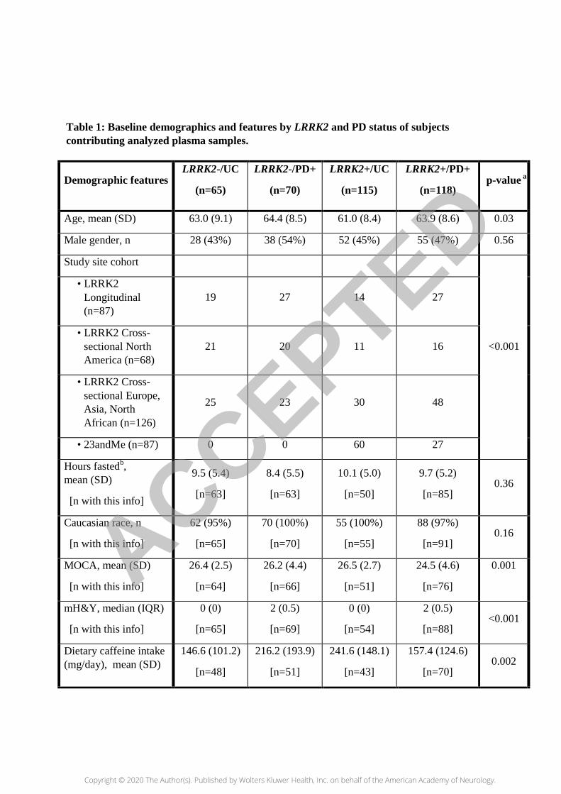
Table 1: Baseline demographics and features by LRRK2 and PD status of subjects contributing analyzed plasma samples.
Demographic features LRRK2-/UC
(n=65)
LRRK2-/PD+
(n=70)
LRRK2+/UC
(n=115)
LRRK2+/PD+
(n=118) p-value a
Age, mean (SD) 63.0 (9.1) 64.4 (8.5) 61.0 (8.4) 63.9 (8.6) 0.03
Male gender, n 28 (43%) 38 (54%) 52 (45%) 55 (47%) 0.56
Study site cohort
• LRRK2 Longitudinal (n=87)
19 27 14 27
• LRRK2 Cross-sectional North America (n=68)
21 20 11 16 <0.001
• LRRK2 Cross-sectional Europe, Asia, North African (n=126)
25 23 30 48
• 23andMe (n=87) 0 0 60 27
Hours fastedb, mean (SD)
[n with this info]
9.5 (5.4)
[n=63]
8.4 (5.5)
[n=63]
10.1 (5.0)
[n=50]
9.7 (5.2)
[n=85] 0.36
Caucasian race, n
[n with this info]
62 (95%)
[n=65]
70 (100%)
[n=70]
55 (100%)
[n=55]
88 (97%)
[n=91] 0.16
MOCA, mean (SD)
[n with this info]
26.4 (2.5)
[n=64]
26.2 (4.4)
[n=66]
26.5 (2.7)
[n=51]
24.5 (4.6)
[n=76]
0.001
mH&Y, median (IQR)
[n with this info]
0 (0)
[n=65]
2 (0.5)
[n=69]
0 (0)
[n=54]
2 (0.5)
[n=88] <0.001
Dietary caffeine intake (mg/day), mean (SD)
146.6 (101.2)
[n=48]
216.2 (193.9)
[n=51]
241.6 (148.1)
[n=43]
157.4 (124.6)
[n=70] 0.002
ACCEPTED
Copyright © 2020 The Author(s). Published by Wolters Kluwer Health, Inc. on behalf of the American Academy of Neurology.

Abbreviations: SD= standard deviation; IQR= interquartile range.
a ANOVA for comparison across groups of age, hours, MoCA and dietary caffeine
intake. Kruskal-Wallis for comparison across groups of mH&Y. Chi-square for
comparison across groups of male gender, study site cohort and Caucasian race.
b A minimum of 8 hours fasting prior to blood draw between 8 and 10 AM was advised
for all subjects (except those in the 23andMe cohort)
[n with this info]
ACCEPTED
Copyright © 2020 The Author(s). Published by Wolters Kluwer Health, Inc. on behalf of the American Academy of Neurology.

Table 2: Caffeine, its metabolites and trigonelline in plasma of LCC participants by LRRK2 and PD status with adjusted geometric mean concentrations (95% CI).
Plasma analyte
LRRK2-/ UC
(n=65)
LRRK2-/ PD+
(n=70)
LRRK2+/UC
(n=115)
LRRK2+/PD+
(n=118)
BH-adj. p value, for PD vs UC
Interaction p value,
PD:UC for LRRK2+ vs -
Caffeine 9.7
(6.1-15.5)
6.7
(4.2-10.9)
12.2
(7.9-18.7)
2.9
(2.0-4.2)
<0.001 0.005
Paraxanthine 2.9
(2.1-4.1)
2.3
(1.6-3.3)
3.7
(2.7-5.1)
1.2
(0.9-1.6)
<0.001 0.005
Theophylline 1.7
(1.2-2.4)
1.3
(0.9-1.9)
2.1
(1.5-3.0)
0.7
(0.5-0.9)
<0.001 0.004
1-methylxanthine
0.0007 (0.0005- 0.001)
0.0006 (0.0004-0.0008)
0.001 (0.0008- 0.001)
0.0004 (0.0003-0.0005)
0.001 0.005
Trigonelline 1.3
(0.9- 1.8)
1.1
(0.8- 1.5)
1.9
(1.4- 2.6)
0.7
(0.5- 0.9)
<0.001 0.006
ACCEPTED
Copyright © 2020 The Author(s). Published by Wolters Kluwer Health, Inc. on behalf of the American Academy of Neurology.
DOI 10.1212/WNL.0000000000010863 published online September 30, 2020Neurology
Grace F Crotty, Romeo Maciuca, Eric A Macklin, et al. mutation carriers: A metabolomic studyLRRK2among
Association of caffeine and related analytes with resistance to Parkinson's disease
This information is current as of September 30, 2020
ServicesUpdated Information &
863.fullhttp://n.neurology.org/content/early/2020/09/30/WNL.0000000000010including high resolution figures, can be found at:
Subspecialty Collections
http://n.neurology.org/cgi/collection/parkinsons_disease_parkinsonismParkinson's disease/Parkinsonism
http://n.neurology.org/cgi/collection/case_control_studiesCase control studiesfollowing collection(s): This article, along with others on similar topics, appears in the
Permissions & Licensing
http://www.neurology.org/about/about_the_journal#permissionsits entirety can be found online at:Information about reproducing this article in parts (figures,tables) or in
Reprints
http://n.neurology.org/subscribers/advertiseInformation about ordering reprints can be found online:
ISSN: 0028-3878. Online ISSN: 1526-632X.Wolters Kluwer Health, Inc. on behalf of the American Academy of Neurology.. All rights reserved. Print1951, it is now a weekly with 48 issues per year. Copyright Copyright © 2020 The Author(s). Published by
® is the official journal of the American Academy of Neurology. Published continuously sinceNeurology