Does inorganic mercury play a role in Alzheimer's disease ... · The brain is the major target...
18
Journal of Alzheimer’s Disease 22 (2010) 357–374 357 DOI 10.3233/JAD-2010-100705 IOS Press Review Does Inorganic Mercury Play a Role in Alzheimer’s Disease? A Systematic Review and an Integrated Molecular Mechanism Joachim Mutter a , Annika Curth b , Johannes Naumann a , Richard Deth c and Harald Walach d,e* a Department of Environmental and Integrative Medicine, Constance, Germany b Ameos Klinikum Dr. Heines, Bremen, Germany c Department of Pharmaceutical Sciences, Northeastern University, Boston, MA, USA d European University Viadrina, Institute of Transcultural Health Studies, Frankfurt/Oder, Germany e Samueli Institute, European Office, Frankfurt (Oder), Germany Accepted 7 July 2010 Abstract. Mercury is one of the most toxic substances known to humans. It has been introduced into the human environment and has also been widely used in medicine. Since circumstantial evidence exists that the pathology of Alzheimer’s disease (AD) might be in part caused or exacerbated by inorganic mercury, we conducted a systematic review using a comprehensive search strategy. Studies were screened according to a pre-defined protocol. Two reviewers extracted relevant data independent of each other. One thousand and forty one references were scrutinized, and 106 studies fulfilled the inclusion criteria. Most studies were case control or comparative cohort studies. Thirty-two studies, out of 40 testing memory in individuals exposed to inorganic mercury, found significant memory deficits. Some autopsy studies found increased mercury levels in brain tissues of AD patients. Measurements of mercury levels in blood, urine, hair, nails, and cerebrospinal fluid were inconsistent. In vitro models showed that inorganic mercury reproduces all pathological changes seen in AD, and in animal models inorganic mercury produced changes that are similar to those seen in AD. Its high affinity for selenium and selenoproteins suggests that inorganic mercury may promote neurodegenerative disorders via disruption of redox regulation. Inorganic mercury may play a role as a co-factor in the development of AD. It may also increase the pathological influence of other metals. Our mechanistic model describes potential causal pathways. As the single most effective public health primary preventive measure, industrial, and medical usage of mercury should be eliminated as soon as possible. Keywords: Alzheimer’s disease, inorganic mercury, neurotoxicity, selenium, systematic review INTRODUCTION Mercury (hydrargyrium = Hg) is well known as the most toxic, non-radioactive element, with a well- ∗ Correspondence to: Prof. Harald Walach, European University Viadrina, Institute of Transcultural Health Studies, Post Box 1786, D-15207 Frankfurt (Oder), Germany. Tel.: +49 335 5534 2380; -2378; Fax: +49 335 5534 2348; E-mail: [email protected]. described neurotoxicology [1–4]. There are various forms of mercury: Organic mercury and inorganic mer- cury (IM), which includes elemental mercury (Hg ◦ ) and mercury ions (Hg + and Hg ++ ). Mercury has been used by humans since ancient times, when the Chinese and Romans used mercury sulfide (cinnabar) for red dye and ink. Widespread use of inorganic mercury started around 1830, when dental amalgams became popular, and calomel (mercury chloride) was used as teething powder in infants [5]. In the early 1900s, the organ- ISSN 1387-2877/10/$27.50 2010 – IOS Press and the authors. All rights reserved
Transcript of Does inorganic mercury play a role in Alzheimer's disease ... · The brain is the major target...

Journal of Alzheimer’s Disease 22 (2010) 357–374 357DOI 10.3233/JAD-2010-100705IOS Press
Review
Does Inorganic Mercury Play a Role inAlzheimer’s Disease? A Systematic Reviewand an Integrated Molecular Mechanism
Joachim Muttera, Annika Curthb, Johannes Naumanna, Richard Dethc and Harald Walachd,e∗
aDepartment of Environmental and Integrative Medicine, Constance, GermanybAmeos Klinikum Dr. Heines, Bremen, GermanycDepartment of Pharmaceutical Sciences, Northeastern University, Boston, MA, USAdEuropean University Viadrina, Institute of TransculturalHealth Studies, Frankfurt/Oder, GermanyeSamueli Institute, European Office, Frankfurt(Oder), Germany
Accepted 7 July 2010
Abstract. Mercury is one of the most toxic substances known to humans.It has been introduced into the human environmentand has also been widely used in medicine. Since circumstantial evidence exists that the pathology of Alzheimer’s disease (AD)might be in part caused or exacerbated by inorganic mercury,we conducted a systematic review using a comprehensive searchstrategy. Studies were screened according to a pre-defined protocol. Two reviewers extracted relevant data independent of eachother. One thousand and forty one references were scrutinized, and 106 studies fulfilled the inclusion criteria. Most studies werecase control or comparative cohort studies. Thirty-two studies, out of 40 testing memory in individuals exposed to inorganicmercury, found significant memory deficits. Some autopsy studies found increased mercury levels in brain tissues of AD patients.Measurements of mercury levels in blood, urine, hair, nails, and cerebrospinal fluid were inconsistent.In vitro models showedthat inorganic mercury reproduces all pathological changes seen in AD, and in animal models inorganic mercury producedchanges that are similar to those seen in AD. Its high affinityfor selenium and selenoproteins suggests that inorganic mercurymay promote neurodegenerative disorders via disruption ofredox regulation. Inorganic mercury may play a role as a co-factorin the development of AD. It may also increase the pathological influence of other metals. Our mechanistic model describespotential causal pathways. As the single most effective public health primary preventive measure, industrial, and medical usageof mercury should be eliminated as soon as possible.
Keywords: Alzheimer’s disease, inorganic mercury, neurotoxicity, selenium, systematic review
INTRODUCTION
Mercury (hydrargyrium= Hg) is well known asthe most toxic, non-radioactive element, with a well-
∗Correspondence to: Prof. Harald Walach, European UniversityViadrina, Institute of Transcultural Health Studies, PostBox 1786,D-15207 Frankfurt (Oder), Germany. Tel.: +49 335 5534 2380;-2378; Fax: +49 335 5534 2348; E-mail: [email protected].
described neurotoxicology [1–4]. There are variousforms of mercury: Organic mercury and inorganic mer-cury (IM), which includes elemental mercury (Hg◦)and mercury ions (Hg+ and Hg++). Mercury has beenused by humans since ancient times, when the Chineseand Romans used mercury sulfide (cinnabar) for red dyeand ink. Widespread use of inorganic mercury startedaround 1830, when dental amalgams became popular,and calomel (mercury chloride) was used as teethingpowder in infants [5]. In the early 1900s, the organ-
ISSN 1387-2877/10/$27.50 2010 – IOS Press and the authors. All rights reserved
358 J. Mutter et al. / Inorganic Mercury and Alzheimer’s Disease
ic mercurial ethyl-mercury was synthesized, and hasbeen used until today as a fungicidal and antimicrobialagent.
Mercury toxicity arises from several strands: Ele-mental or metallic mercury (Hgo) is the only metal thatis liquid at room temperature and can evaporate quick-ly. As mercury vapor, it is taken up via the lungs, and80% of it is absorbed. Due to its uncharged mono-atomic form, it is highly diffusible and lipid soluble.It crosses the blood-brain barrier easily, as well as thelipid bilayers of cells and cell organelles, such as mi-tochondria. Mercury vapor also penetrates the mucosaand connective tissue of the oral or nasal cavities andmay be transported into nerve cells [6–8]. Intracellu-larly, it is oxidized from its comparatively inactive Hg◦
state to its ionic form, Hg++. This mercuric ion reactsimmediately with intracellular molecules or structures(e.g., enzymes, glutathione, tubulin, ion channels, ortransporters), inhibiting their activities and interferingwith normal cellular function.
Very low levels (180 nM) of Hg++ decrease glu-tathione levels (GSH) and increase oxidative and ni-trosative stress, which may lead to cytotoxicity [9]. Theextraordinarily high affinity of Hg++ for selenium, andselenoproteins (dissociation constant= 10−45) [10]can disrupt cellular redox balance [11,12], especially inthe brain, which uniquely depends upon selenoenzymesfor antioxidant protection and hence selenium [13,14].The role of extracellular thiol groups for the transportand absorption of organic mercurials is well describedfor methylmercury [15], but for IM, their role as a vec-tor is still under discussion. When bound to a thi-ol group (e.g., cysteine) methylmercury can cross theblood brain barrier easily and is transported into glialcells and neurons using molecular mimicry [16], whereit is converted to IM. Due to its charge it is less ableto cross cell membranes and can be trapped in cellsand held within the brain. Further, IM has a very highaffinity for thiol groups and forms strong bonds withthem, giving rise to the term “mercaptans” [15,17,18].
The brain is the major target organ for elemen-tal, gaseous Hgo. The half-life of mercury in thebrain is unclear. Modeling mercury deposition in thebrain using autopsy data of traffic victims and intakestreams through food yielded a half-life estimate of 22years [19], and autopsies of proven clinical cases ofHgo poisoning have found high mercury levels in thebrain as long as 17 years after the event [20,21].
In contrast, the half-life of mercury in the body isaround 30 to 60 days [22]. IM binding to seleni-um is almost irreversible and contributes to its long-
term brain retention [23,24]. Mercury from gaseoussources, such as coal burning, and from human activ-ities through waste water, is accumulated in the foodchain, and comes back to humans mainly via fish asmethyl-mercury. Methyl-mercury is also transportedvia the bloodstream to the brain, where it is again con-verted to IM. Administration of oral methyl-mercury tonon-human primates yielded a plasma clearance half-life of 21 days, while the half-life for clearance of IMfrom the brain was too slow to be estimated (> 120days) during a 28 day washout period [25]. IM outsideof the brain is accumulated in the kidneys, and is slowlyexcreted.
The potential role for mercury toxicity in Alzheimer’sdisease (AD) stems from (i) the relevance of the gaseousphase of elemental mercury for the brain with (ii) sub-sequent transformation to ionic mercury, and (iii) theconversion of methyl-mercury to inorganic mercury(Hg++) in the brain. Humans take in about 2.4µgof organic mercury per week, if consuming one fishmeal per week, 2.3µg of which is retained [22]. Themain source for the intake of Hgo are dental amalgamfillings [22]. Such fillings consist of 50% of mercury,which evaporates at a slow rate,but is released at a high-er rate, when the fillings are put in place or removed.From this source, and other, less common ones, 1.2 to27.0µg of Hgo are taken up per day, and 1.0 to 22.0µgof Hgo are retained. Other variable factors of mercuryrelease include the number, age, and size of the fillings,the presence of dental alloys, individual chewing habitsand drinking hot liquids, as well as bruxism.
AD, first described in 1907, is one of the major formsof dementia, with about 15–50% of over 80 year oldelderly being affected [26–34]. Currently about 24 mil-lion people worldwide suffer from dementia, with thenumbers projected to double every 20 years [29], andby 2050 nearly 1 in 45 Americans are predicted to sufferfrom AD [35]. Since the population of most countriesis aging, the problem will continue to increase. As of1998, the lifetime risk of a 55 year old healthy womandeveloping dementia was 33% compared to 16% formen [27].
Clinically, AD reveals itself through increasing cog-nitive decline, impaired attention and short-term mem-ory, and, at later stages, other forms of cognitive in-competence, such as impaired language, face recogni-tion, spatial orientation, and hearing. Pathologically,this is thought to result from a gradual build up of amy-loid plaques that form as a consequence of amyloid-β
(Aβ) being produced at a higher rate than can be re-moved [36]. Amyloid plaques induce inflammation and

J. Mutter et al. / Inorganic Mercury and Alzheimer’s Disease 359
free oxygen radical production,which eventually yieldsa self-reinforcing cycle of neuroinflammation, neu-rodegeneration, and further inflammation. A second,apparently independent process, involves hyperphos-phorylation of the tau-protein, which leads to a break-down of microtubules and the neuronal cytoskeleton.Accumulating neurofibrillary tangles (NFT) promoteneuroinflammation and reinforce the cycle [37]. Boththese processes play a pathological role in the causa-tion of AD [38], potentially exacerbated by deficientmicro-vascularization in the brain [31,39].
The degeneration process starts in the entorhinal cor-tex and the basal ganglia, especially in the nucleusbasalis Meynert, spreads to the hippocampus, and even-tually affects other parts of the cortex as well. Due tothe loss of neurons of the projective cholinergic system,brain cognitive functions such as short term memoryare the first to be noticeably affected.
At present, we do not know what causes AD. Sev-eral genetic factors contributing to AD have been re-vealed [36,40], however, no clear-cut genetic cause hasbeen isolated. Apolipoprotein E (ApoE) genotype is aconsistent risk factor [41–46], and theε4 genotype con-fers up to a 15-fold risk relative to theε3 genotype [47,48], which is the most widely distributed, whereas theε2 genotype is protective. However, it is not entire-ly clear, how this risk can be fitted within a mecha-nistic model. ApoE is a transporter protein that mayalso operate as a free-radical scavenger. It is impor-tant to notice here that all three ApoE forms consistof 299 amino-acids, and the only differences are thatApoEε4 has an arginine in position 112 and 158, whereApoE ε2 has two cysteines, and ApoEε3 one argi-nine and one cysteine [49]. Interestingly, cysteine con-tains a sulfhydryl, which is capable of binding metals,especially bivalent metals such as lead, copper, zinc,and mercury. This has led to the hypothesis that thewell-known differential genetic epidemiology of ApoEmight have to do in part with the differential detoxifi-cation capacity regarding mercury [50], and potentiallyother metals as well. The ApoE lipoprotein complexis taken up into neurons via the ApoER2 receptor. Se-lenoprotein P (SelP), which provides selenium for se-lenoprotein synthesis, is also taken up by ApoER2 [51].Differential competition for uptake between ApoE iso-forms and SelP might therefore affect selenoproteinstatus and vulnerability to oxidative stress. Notably,SelP is physically associated with both Aβ plaques andNFTs in the AD brain [52], further suggesting a rolefor impaired selenoprotein function in AD pathology.
Because of the potential relevance of mercury as acausal factor for initiating AD, we set out to system-
atically review the literature. Because of the apparentspecial relevance of IM, we restricted our review to thisform of mercury. Other forms of mercury toxicity, suchas ethylmercury added as a preservative to vaccines, ormethylmercury from fish, or the presence of other met-als, like aluminum or lead, may synergistically enhanceIM toxicity. This will be reviewed separately.
METHODS
We aimed at capturing all relevant papers that con-tained the semantic fields of “Alzheimer”, “mercury”and “neurotoxic”, limiting them to IM, using the strat-egy most appropriate for each database. We searchedthe following databases: EMBASE (Excerpta Medi-ca); HSDB (Hazardous Substances Data Base); XTOX-LINE; MEDLINE; Biosis; Science Citation Index;Publisher databases of Kluwer, Springer, Thieme fromtheir start date to 2006.
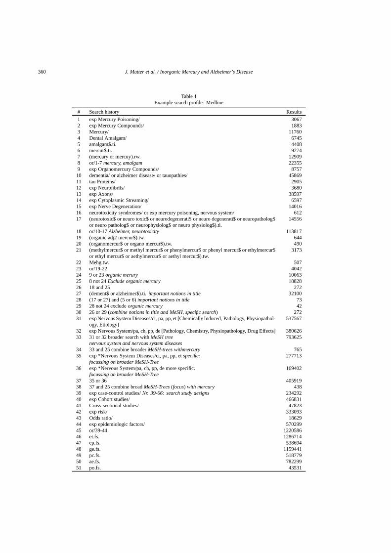
Since each database has a different structure and thethesaurus available differs among them, we deviseda new search strategy for each one. A full report,containing each strategy in detail, can be obtained fromthe authors [53]. An example of the Medline searchstrategy is reproduced in Table 1.
We included studies using any type of research de-sign and any type of work relevant to the topic ofthis review. We excluded studies that were publishedin a language other than German or English and thatwere irrelevant for this topic. All titles and abstractsof the references that were retrieved were scrutinizedby two independent reviewers, and original papers re-trieved. For each paper whose inclusion was not imme-diately clear, two reviewers discussed the inclusion andreached consensus in all cases. Reference lists of allincluded papers were hand searched for more relevantarticles, again by two independent reviewers.
Duplicates were eliminated. References fulfillinginclusion criteria were checked as full papers, for in-clusion by two independent reviewers. All articleswere coded for their potential internal validity follow-ing the procedures adopted by Dettenkofer and col-leagues [54]. Other types of publications were codedas animal experiments orin vitro experiments. Cod-ing was done by two independent reviewers, and incase of differing opinion a third reviewer’s opinion washeard. Controlled studies used, as a rule, unaffectedcontrols that were normally matched for age and gen-der, unless specified otherwise. Trace metal detectionfollowed the state of the art of the time and used mostly
360 J. Mutter et al. / Inorganic Mercury and Alzheimer’s Disease
Table 1Example search profile: Medline
# Search history Results
1 exp Mercury Poisoning/ 30672 exp Mercury Compounds/ 18833 Mercury/ 117604 Dental Amalgam/ 67455 amalgam$.ti. 44086 mercur$.ti. 92747 (mercury or mercuy).rw. 129098 or/1-7mercury, amalgam 223559 exp Organomercury Compounds/ 875710 dementia/ or alzheimer disease/ or tauopathies/ 4586911 tau Proteins/ 290512 exp Neurofibrils/ 368013 exp Axons/ 3859714 exp Cytoplasmic Streaming/ 659715 exp Nerve Degeneration/ 1401616 neurotoxicity syndromes/ or exp mercury poisoning, nervous system/ 61217 (neurotoxic$ or neuro toxic$ or neurodegenerati$ or neuro degenerati$ or neuropatholog$
or neuro patholog$ or neurophysiolog$ or neuro physiolog$).ti.14556
18 or/10-17Alzheimer, neurotoxicity 11381719 (organic adj2 mercur$).tw. 64420 (organomercur$ or organo mercur$).tw. 49021 (methylmercur$ or methyl mercur$ or phenylmercur$ or phenyl mercur$ or ethylmercur$
or ethyl mercur$ or aethylmercur$ or aethyl mercur$).tw.3173
22 Mehg.tw. 50723 or/19-22 404224 9 or 23organic merury 1006325 8 not 24Exclude organic mercury 1882826 18 and 25 27227 (dement$ or alzheimer$).ti.important notions in title 3210028 (17 or 27) and (5 or 6)important notions in title 7329 28 not 24 excludeorganic mercury 4230 26 or 29 (combine notions in title and MeSH, specific search) 27231 exp Nervous System Diseases/ci, pa, pp, et [Chemically Induced, Pathology, Physiopathol-
ogy, Etiology]537567
32 exp Nervous System/pa, ch, pp, de [Pathology, Chemistry,Physiopathology, Drug Effects] 38062633 31 or 32 broader search withMeSH tree
nervous system and nervous system diseases793625
34 33 and 25 combine broaderMeSH-trees withmercury 76535 exp *Nervous System Diseases/ci, pa, pp, etspecific:
focussing on broader MeSH-Tree277713
36 exp *Nervous System/pa, ch, pp, de more specific:
focussing on broader MeSH-Tree169402
37 35 or 36 40591938 37 and 25 combine broadMeSH-Trees(focus) with mercury 43839 exp case-control studies/Nr. 39-66: search study designs 23429240 exp Cohort studies/ 46683141 Cross-sectional studies/ 4782342 exp risk/ 33309343 Odds ratio/ 1862944 exp epidemiologic factors/ 57029945 or/39-44 122058646 et.fs. 128671447 ep.fs. 53869448 ge.fs. 115944149 pc.fs. 51877950 ae.fs. 78229951 po.fs. 43531
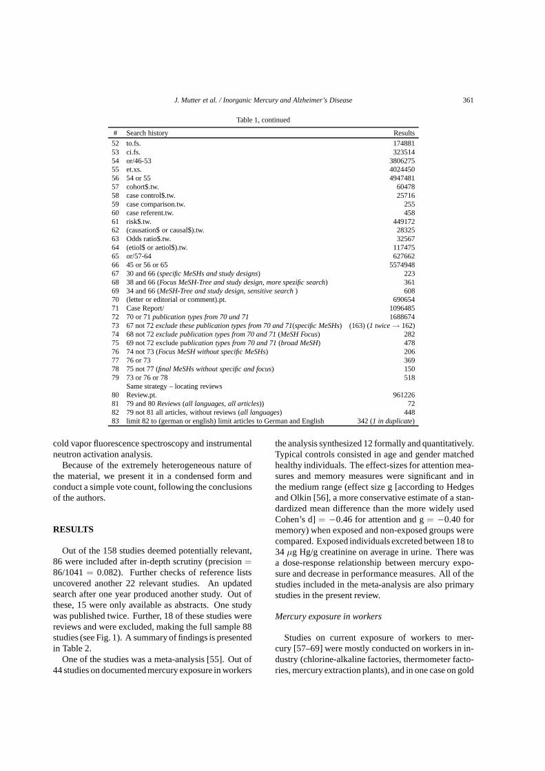
J. Mutter et al. / Inorganic Mercury and Alzheimer’s Disease 361
Table 1, continued
# Search history Results
52 to.fs. 17488153 ci.fs. 32351454 or/46-53 380627555 et.xs. 402445056 54 or 55 494748157 cohort$.tw. 6047858 case control$.tw. 2571659 case comparison.tw. 25560 case referent.tw. 45861 risk$.tw. 44917262 (causation$ or causal$).tw. 2832563 Odds ratio$.tw. 3256764 (etiol$ or aetiol$).tw. 11747565 or/57-64 62766266 45 or 56 or 65 557494867 30 and 66 (specific MeSHs and study designs) 22368 38 and 66 (Focus MeSH-Tree and study design, more spezific search) 36169 34 and 66 (MeSH-Tree and study design, sensitive search) 60870 (letter or editorial or comment).pt. 69065471 Case Report/ 109648572 70 or 71publication types from 70 und 71 168867473 67 not 72exclude these publication types from 70 and 71(specific MeSHs) (163) (1 twice→ 162)74 68 not 72exclude publication types from 70 and 71(MeSH Focus) 28275 69 not 72 exclude publication types from 70 and 71(broad MeSH) 47876 74 not 73 (Focus MeSH without specific MeSHs) 20677 76 or 73 36978 75 not 77 (final MeSHs without specific and focus) 15079 73 or 76 or 78 518
Same strategy – locating reviews80 Review.pt. 96122681 79 and 80Reviews(all languages, all articles)) 7282 79 not 81 all articles, without reviews (all languages) 44883 limit 82 to (german or english) limit articles to German and English 342 (1 in duplicate)
cold vapor fluorescence spectroscopy and instrumentalneutron activation analysis.
Because of the extremely heterogeneous nature ofthe material, we present it in a condensed form andconduct a simple vote count, following the conclusionsof the authors.
RESULTS
Out of the 158 studies deemed potentially relevant,86 were included after in-depth scrutiny (precision=86/1041= 0.082). Further checks of reference listsuncovered another 22 relevant studies. An updatedsearch after one year produced another study. Out ofthese, 15 were only available as abstracts. One studywas published twice. Further, 18 of these studies werereviews and were excluded, making the full sample 88studies (see Fig. 1). A summary of findings is presentedin Table 2.
One of the studies was a meta-analysis [55]. Out of44 studies on documentedmercury exposure in workers
the analysis synthesized 12 formally and quantitatively.Typical controls consisted in age and gender matchedhealthy individuals. The effect-sizes for attention mea-sures and memory measures were significant and inthe medium range (effect size g [according to Hedgesand Olkin [56], a more conservative estimate of a stan-dardized mean difference than the more widely usedCohen’s d]= −0.46 for attention and g= −0.40 formemory) when exposed and non-exposed groups werecompared. Exposed individuals excreted between 18 to34 µg Hg/g creatinine on average in urine. There wasa dose-response relationship between mercury expo-sure and decrease in performance measures. All of thestudies included in the meta-analysis are also primarystudies in the present review.
Mercury exposure in workers
Studies on current exposure of workers to mer-cury [57–69] were mostly conducted on workers in in-dustry (chlorine-alkaline factories, thermometer facto-ries, mercury extraction plants), and in one case on gold

362 J. Mutter et al. / Inorganic Mercury and Alzheimer’s Disease
Tabl
e2
Sum
mar
yof
findi
ngs
Cat
egor
yof
stud
yN
umbe
rN
egat
ive
effe
cts
Stu
dyde
sign
Com
men
tsof
stud
ies
ofm
ercu
ryon
mem
ory
and/
orbr
ain/
brai
nfu
nctio
n–
Indi
catio
nsfo
rne
gativ
eef
fect
ofm
ercu
ryon
neur
onal
func
tioni
ngye
sno
Unc
lear
Stu
die
sin
Hum
ans
Exp
ose
dto
Mer
cury
40
Hig
hD
ose
Exp
osu
res
Pas
tand
curr
ente
xpos
ure
ofw
orke
rs[5
5]1
1M
eta-
anal
ysis
Sum
mar
yof
stud
ies;
sign
ifica
ntco
rrel
atio
nbe
twe
enm
easu
res
ofco
gniti
vefu
nctio
ning
and
Hg
excr
etio
nin
urin
efo
ra
mea
nex
cret
ion
of34
µg;
sig-
nific
ante
ffect
size
sfo
rdi
ffere
nce
inco
gniti
vepe
rfor
mance
mea
sure
sbe
-tw
een
expo
sed
and
non-
expo
sed
fora
ttent
ion
and
mem
ory;
dos
e-re
spon
sere
latio
nshi
pC
urre
ntex
posu
reof
wor
kers
[57–
69]
1310
3C
ross
-sec
tiona
lst
udie
sw
ithco
ntro
ls,1
long
itudi
nal
cont
rolle
dco
hort
stud
y
Cur
rent
expo
sure
docu
men
ted;
Hg
excr
etio
nin
urin
eco
rrel
ate
dw
ithm
ea-
sure
sof
cogn
itive
func
tioni
ng;
not
alw
ays
diffe
renc
eag
ains
tco
ntro
lsin
allm
easu
res
Pas
texp
osur
eof
wor
kers
[70–
74,1
88–1
91]
98
15
retr
ospe
cti
veco
hort
stud
ies,
4ca
sehi
stor
ies
Pas
texp
osur
eto
mer
cury
docu
men
ted;
expo
sure
datin
gba
ck5
to30
year
s;tw
oof
the
case
hist
orie
slik
ely
cove
ring
the
sam
etw
oca
ses;
the
stud
yw
itha
30ye
arre
tros
pect
ive
focu
sfo
und
little
evid
ence
,bu
tsom
ene
urol
ogic
alsi
gns
ofm
ercu
rialis
mw
ere
still
pres
ent
Low
Dose
Exp
osu
res
Den
tists
and
dent
alpe
rson
nel[
75–8
6];
1211
1C
ompa
rativ
e/cr
oss-
sect
iona
lR
elat
ions
hip
betw
een
stre
ngth
ofex
posu
re
(den
tists
vs.
pers
onne
l),m
ark-
ers
ofex
posu
rean
dte
stre
sults
Gen
eral
olde
rpo
pula
tion
[87]
11
Cro
ss-s
ectio
nal
Gen
eral
pop
ulat
ion;
bloo
dH
gle
vela
ndM
MS
Ere
sults
Am
alga
mbe
arer
s[8
8–94
]7
15
1C
ross
-sec
tiona
l,1
coho
rtst
udy
Stu
dies
onol
deri
ndiv
idua
lsof
ten
dono
ttak
epr
evio
usst
atus
into
acco
unt;
the
only
stud
yw
ithtr
ueam
alga
mfr
eein
divi
dual
ssh
ows
effe
cts
Stu
die
sin
Alz
hei
mer
Patie
nts
Liv
ing
Pat
ient
s[9
5–10
1]7
31
3C
ompa
rativ
ecr
oss-
sect
iona
lM
ost
stud
ies
very
smal
l,no
larg
e,lo
ngitu
dina
lst
udie
s;h
ardl
yan
yco
n-vi
ncin
gev
iden
ceA
utop
syst
udie
s[1
02–1
10]
94
41
Cas
eco
ntro
lstu
dies
Stu
die
sas
sess
eddi
ffere
ntar
eas
ofth
ebr
ain,
som
ein
larg
eran
ato
mic
alpo
rtio
ns,
som
ein
mor
esp
ecifi
con
es;t
ime
betw
een
auto
psy
and
mer
cury
anal
ysis
ofte
nve
ryla
rge
with
dang
erof
evap
orat
ion
Anim
alS
tudie
s[111
–119
]9
9E
xper
imen
tals
tudi
esS
ome
stud
ies
only
avai
labl
eas
abst
ract
sIn
vitr
oS
tudi
es[9
,112
,119
,122
–135
]16
16E
xper
imen
tals
tud
ies
All
stud
ies
confi
rmto
xic
effe
cts
ofm
ercu
ryon
neur
ons
orne
uron
altis
sue,
repr
oduc
ing
path
olog
ical
sign
sof
Alz
heim
er’s
dise
ase

J. Mutter et al. / Inorganic Mercury and Alzheimer’s Disease 363
Fig. 1. Flow diagram of study inclusion.
miners in the Philippines who use large amounts of mer-cury without any protection [59]. Correlations betweenthe amount of Hg excreted in urine and measures ofcognitive abilities (memory tests, attention span) werealways significant and negative, i.e., the more mercuryexcreted the worse the test results. Although all studiesexcept one had control groups, the differences betweenexposed and control groups were not always significantand clear-cut. The Mt. Diwata Study [59] might givea hint as to why: although there was a significant cor-relation between mercury excretion and clinical symp-toms, as well as test results, and although the workerswere clearly exposed to large amounts of mercury, thecorrelations were moderate and showed great variationacross individuals. Some individuals showed severeclinical signs of mercurialism, but excreted hardly anymercury, whereas others excreted much more, but hadfewer clinical problems. Also, the control group livingdownstream by the sea showed little difference in ex-cretion rates compared to the mercury exposed group.It is very likely, the authors concluded, that dependingon individual factors mercury might be excreted at dif-ferent rates and captured in body compartments for along time, making urinary excretion of mercury a veryunreliable marker both of mercury load and of clinicalsignificance.
Studies on past exposures to high doses of mercuryspanned times between five and 30 years after the ex-
posure. Five of these studies were on groups of work-ers after their exposure, four were case histories. Fourstudies [70–73] show evidence that workers exposedto mercury 5 to 18 years previously still had signif-icantly worse results in neurological tests and clini-cal symptoms than those without significant exposure,even though one study had excluded all neurologicallyand psychiatrically ill persons. The study that foundno significant differences [74] investigated workers 30years after exposure. Although differences from con-trols were not significant, clinical signs such as tremorand lack of coordination were documented in exposedworkers only.
Dentists and their staff are professionally exposedto low doses of mercury long term. All studies foundsignificant correlations between level of mercury inblood, urine, nails, hairs, or air, and results for the testsused in the respective studies (neurological, psycholog-ical, or both) [75–83]. One study found more physi-cal and psychological symptoms in dentists and theirpersonnel than in controls [84], and one single-groupcross-sectional study found moderate to severe devia-tions from norm results of a standardized neuropsycho-logical test-battery (memory, attention, language tasks,visuo-spatial capacity) in 17% of the tested persons andone standard-deviation from population norms for thegroup as a whole [85]. One study that used sodium-2,3-dimercapto-propane-1-sulfonate (DMPS) found bettercorrelations of symptoms and test results with mercuryburden after the application of this chelating agent,which points to the fact that mercury can be trappedin body compartments [86]. Blood mercury levels andmini-mental state examinations (a standard examina-tion to quickly assess cognitive functioning) do not al-ways correlate, as can be seen in one general populationstudy on low level exposure [87].
Health effects of dental amalgams
Studies on health effects in persons with amalgamshave been largely negative [88–93]. The only studyshowing effects involved a young sample (mean age22.4 and 23.3 years respectively), where the controlgroup had never had any exposure to amalgam [94].There was a positive correlation between number offillings and mercury excretion in urine and hair, as wellas with forgetfulness and symptoms. All other studiesin this section investigated older people. Patients withno teeth left, usually the older ones, often did worsethan those with teeth and amalgams. Clearly, with-out detailed knowledge of the previous history of den-tal treatment regarding actual mercury exposure it isdifficult to draw any conclusions from such studies.

364 J. Mutter et al. / Inorganic Mercury and Alzheimer’s Disease
Mercury exposure, accumulation, and excretion in ADpatients
AD patients are an obvious choice for studies of po-tential long term effects of mercury exposures. In aprospective cohort study there was a negative correla-tion between mercury content in nails and age or pro-gression of dementia, respectively [95]. Since mercurycontent in nails reflects the mercury load over the pastfew weeks and its excretion, this finding means thatmore severely demented people do not excrete as muchmercury as less severely ill patients. This might be dueto the fact that their body is less able to excrete mercury,or mercury has been excreted earlier on, or perhaps areduction in the proportionof mercury distributed to theperiphery versus the brain with AD progression. Alter-natively, this finding could indeed suggest that higherlevels of mercury protect against severe AD, althoughthis possibility is counter-intuitive.
A cross-sectional controlledstudy found differences:significantly more Hg in plasma and non-significantlymore in cerebrospinal fluid of AD patients [96]. In aseries of small studies there was more Hg excretion inurine of AD patients than in age-matched controls, andless Hg in blood of AD patients. These findings were,however, not significant due to the small sample sizeof nine patients only [97]. A retrospective cohort studyfound a probable exposure to mercury in 4.1% of 170patients with AD and 2.4% likely exposure in controls,but the results relied upon retrospective recall by rela-tives [98]. One study found a non-significantly differ-ent higher amount of Hg in hair of ill patients comparedwith controls [99], while another found that the numberof amalgam fillings was not different in 66 AD patientscompared to controls [100]. AD patients had higherblood mercury levels in one study,which was correlatedwith higher Aβ levels in cerebrospinal fluid [101]. Fourof nine autopsy studies document various changes inAD brains that are suggestive of mercury effects: Onestudy treated brains of control persons with an EDTA-mercury complex and found that the interaction of GTPwith β-tubulin was compromised similar to what theysaw in AD brains [102].
Another study found significantly more mercury in81 brain samples of 14 AD patients compared withage-matched controls, and more mercury in grey mat-ter of AD brains compared with white matter. Mer-cury accumulated in the cerebellum, thalamus, puta-men, and in the upper parietal and occipital lobes ofAD patients’ brains [103]. Thompson and colleaguesfound significantly higher mercury levels in the amyg-
dala, the nucleus basalis Meynert and non-significantlyhigher levels in the hippocampus of 14 AD patientscompared with age-matched controls [104], while an-other study found significantly higher mercury levels inmicrosomes from AD brains [105]. One study reportedhigher mercury levels in brains and lower mercury lev-els in nails of 3 AD patients compared to 10 controls butdue to the small patient number cannot be consideredconclusive [106]. Four studies found either no signifi-cant differences or slightly and non-significantly lowerlevels in AD brains compared with controls [107–110].
Experimental animal and in vitro studies
Eight animal studies were included. Five of themshowed that in rats which had been exposed to mer-cury vapors, mercury content of brain tissue was high-er than in controls [111–115]. In one study whererats took up Hg++ with food, GTP-tubulin interac-tions were observed that were similar to those knownfrom AD brains [116]. Two studies found that Purkinjecells of the cerebellum were specifically prone to ac-cumulate mercury after exposure of the animals [117,118], while another one documented the inhibition ofADP-ribosylationin vitro and in vivo [119]. ADP-ribosylation inhibits tubulin polymerization and leadsto depolymerization of microfilaments [120]. The lat-ter finding is interesting in so far as ADP-ribosylationis an important DNA repair mechanism that is activatedunder conditions of oxidative stress which is normallyfound to be enhanced in AD patients [121].
In vitro studies produced the following results:Mercury interferes with polymerization of micro-tubules [122,123], increases secretion of both 1–40and 1–42 forms of Aβ and promotes hyperphospho-rylation of tau protein [9,124–127], changes mito-chondrial structure inducing a stress-response in astro-cytes [128], and interferes with cell-maturation [129]or other aspects of cell functioning, such as DNA re-pair, glutathione level, or linkage and structure of mi-crotubules [119,130,131]. Mercury disturbs the inter-action between tubulin and GTP [132], and the chelatorDMSA can reverse this process [133], while amalgamexposure is toxic for nerve cellsin vitro [134]. Mercuryinterferes with membrane structures, leading to axonaldegeneration and neurofibrillary aggregates [135].
DISCUSSION
This systematic review produced a mixed and para-doxical picture: Experimental studies in animals andin
J. Mutter et al. / Inorganic Mercury and Alzheimer’s Disease 365
vitro systems not only confirmed the well-known toxi-cology of mercury, but also reproduced the pathologi-cal signs of AD quite accurately and without any neg-ative results: hyperphosphorylation of tau protein, thedegeneration of microtubules, and the increased forma-tion of Aβ protein. Animal studies also confirmed thatmercury vapor, inhaled in low doses, accumulates inthe brain.
Human studies, however, do not parallel this clearpicture. Studies of exposed workers demonstrate quiteclearly that continuous contact with mercury as an oc-cupational hazard leads to effects on memory, atten-tion and produces a variety of symptoms. Some ofthem, such as memory and attention deficits are rele-vant to AD, others, like sleep disturbance, mood swingsor pain are rather non-specific. A meta-analysis con-firms significant effect sizes, but they are only mediumsized. Autopsy studies speak a mixed language: whilesome find more mercury content in brain tissues of ADpatients, some do not. Some of the autopsy studiesare fraught with potential problems: gross averagingof mercury content across large brain areas, long lagsbetween autopsy and measurement, not taking into ac-count the volatile nature of Hg. This may decrease Hgvalues in specimens through deposition of Hg in plastictest tubes over several months as described by Hock andcolleagues [101]. In addition, the lack of staging of ADbrains makes it impossible to draw firm conclusionsespecially from the studies reporting no effects.
Quite naturally, there is a lack of good evidence forour study question in human studies. Experimentationis prohibited for obvious ethical reasons, and evidencehas to come from indirect sources. Exposure to highand low doses of mercury through the workplace hasunequivocally led to neuropsychological deficits, bothin workers (high doses) and in dental personnel (lowdose exposure). The question not answered by our da-ta is whether such mild cognitive deficits in attentionand memory will transition into dementias. This ques-tion could only be answered by large longitudinal stud-ies which do not exist. However, we do know fromautopsy studies that brains of deceased persons with-out any clinical signs of dementia show pathologicalsymptoms of degeneration pathognomic for AD at laterstages [136], making it quite plausible that a patholog-ical process might start many years before it manifestsclinically as AD. Hence, it would be crucial to studylarger cohorts of exposed persons longitudinally.
Epidemiological studies that have correlated the in-cidence of dementia with dental status have in gen-eral been unable to find any evidence for such a cor-
relation, and these negative findings are normally cit-ed in support of the lack of harmful effects of amal-gams. Most of these studies have investigated cohortsthat were comparatively old and have used the presentcount of amalgam fillings to estimate the mercury loadacross a lifespan. None of these studies has taken intoaccount the fact that most people who do not have teethany longer at old age or who have dental repairs otherthan amalgam will have had amalgams in their teethat previous times. This might explain the counterintu-itive findings of some studies that many amalgam fill-ings correlate with better cognitive status: those withless fillings at present were likely to have had moreearlier and thus have a higher likelihood of mercuryaccumulation in their lifetime and hence have a worsecognitive status at the time of measurements, when nofillings were present any longer, giving persons with“more amalgam fillings” a spurious benefit over thosewith “no or less amalgam fillings” [137,138]. Strictlyspeaking, such studies should not even be consideredto bring clarity to the debate, since they are of doubt-ful methodological quality. However, since they areamong the most cited ones we thought it is importantto include these studies in the current review and qual-ify their validity. Indeed, the only study in our samplethat had a completely amalgam free control showed ef-fects: there was more excretion of mercury in urine andhair directly related to the number of fillings and moresymptoms, including forgetfulness, in those exposed toamalgam compared to amalgam free controls. Howev-er, since the individuals in that study were rather youngand no longitudinal data exist, this can only be takenas a hint. Longitudinal studies of cohorts completelyfree of amalgams compared with cases with amalgamswould be a way of answering the question conclusively.These studies do not exist.
The findings of this review, thus, are paradoxicaland pose a challenge: experimental data from animalresearch andin vitro studies strongly suggest an in-fluence of inorganic mercury on the nervous system,but epidemiological and other studies suggest a muchweaker relationship. It is likely that two processes playa modifying role here: Humans may be differentiallysusceptible to mercury toxicity, as compared to otherspecies, and some individuals might be better able tochelate and detoxify mercury than others, reducing thestrength of correlations between mercury exposure andAD.
A mechanistic model of mercury toxicity
Genetic risk factors for AD can provide the basis fordifferential susceptibility to the neurotoxic effects of
366 J. Mutter et al. / Inorganic Mercury and Alzheimer’s Disease
mercury, particularly since genetic variation is robustamong humans, as compared to inbred laboratory ani-mals. Thus the influence of any single factor in a mul-tifactorial disorder such as AD is dependent upon thepresence of other factors. For an environmental factorsuch as mercury, the extent of genetic loading, as wellas the presence of other environmental factors, will de-termine the magnitude of its contribution. Indeed, inthe absence of genetic risk factors, exposure to an envi-ronmental factor may not cause disease. In other words,an environmental stressor can reveal genetic limitationswhich otherwise might not be associated with patho-logical consequences. In the case of AD, age-relatedmetabolic changes undoubtedly enhance risk, and mer-cury’s high affinity for selenoproteins and thiols makesredox and methylation metabolism especially promi-nent targets for its toxicity [10–12,24,139].
The ability to maintain a homeostatic balance be-tween reduction and oxidation (i.e., redox equilibrium)is essential for all cells, and the ability of seleniumand sulphur to reversibly transfer electrons makes themideal for redox buffering. This role is particularly im-portant in the brain, since CSF levels of cysteine, thelimiting material for glutathione synthesis, are morethan 100-fold lower than in plasma [140], while oxygenconsumption is disproportionately higher. To meet thishigher demand for antioxidant, selenoproteins, such asthioredoxin reductases and glutathione peroxidases andSelP, play a more prominent role in the brain [13,14],and mechanisms have evolved to assure an adequateselenium supply to the brain, even when other tissuesare depleted [14,141]. Selenoprotein mRNAs containone or more UGA codons, which normally terminatetranslation but in the presence of a selenocysteine inser-tion sequence (SECIS) they effect direct incorporationof a selenocysteine into the nascent peptide chain. Se-lenocysteine tRNA is initially loaded with serine whichis subsequently converted to a selenocysteine in a reac-tion with activated selenide (SeP) [142]. Mercury’s ex-tremely high affinity for selenium can potentially causea functional selenium deficiency in the brain, interfer-ing with its critical role in maintaining redox equilibri-um.
SelP contains ten selenocysteine residues and is con-sidered to be the primary source of selenium for cel-lular synthesis of other selenoproteins, which typical-ly contain only a single selenocysteine in their ac-tive site [143]. SelP forms higher order multimericcomplexes with inorganic mercury and free selenium,and, although it has 10 selenocysteines, and 17 cys-teine residues, it has been estimated that a single SelP
molecule can bind more than 100 molecules of mer-cury [144]. Thus SelP not only serves as a seleniumreservoir to support selenoprotein synthesis, but mayalso function as a high-affinity binding site for mercury,protecting other selenoproteins from its toxic effect.
In the brain, a remarkably high level of SelP is foundin ependymal cells [145], whose asymmetric divisiongives rise to neural stem cells in the subventricularzone and subgranular layer [146,147]. Accordingly,ependymal cells have the highest level of glutathione,more than 10-foldhigher than neurons, and 3-foldhigh-er than astrocytes [148]. Mercury potently interfereswith neural stem cell development [149,150], whichcould contribute to reduced cortical and hippocampalneuronal density in AD. SelP gene expression in hu-man brain increases with age [151], and its expressionlevel is higher in AD patients [152]. Moreover, SelPis preferentially associated with amyloid plaques andNFTs [52], which may limit its utilization for synthesisof other selenoproteins.
Neurons take up SelP via the lipoprotein receptorApoER2 [51], suggesting that the adequacy of seleniumsupply to the cell might be related to the differentialcompetition between variant forms of ApoE and SelP.Indeed, in a Chinese cohort, carriers of the ApoE4 allelehad significantly lower selenium levels, as measured innail samples [153]. ApoER2 also mediates signallingby reelin, which guides neural migration into layersof the cortex and promotes synaptic memory [154].Increased levels of Aβ or low levels of SelP impairsynaptic memory, which can be offset by increasedreelin [155]. Thus ApoER2 is a critical nexus, at whichthe roles of SelP, ApoE, and Aβ are integrated, linkingApoE4 to selenium status.
Elevated plasma levels of homocysteine (HCY) inAD have been reported in numerous studies, as con-firmed by a systematic review [156], and the rate ofcognitive decline is related to the extent of HCY el-evation [157]. Formed during methylation reactions,HCY is converted to methionine by the vitamin B12and methyl-folate-dependent enzyme methionine syn-thase, which is highly sensitive to cellular redox sta-tus and is potently inhibited by mercury in culturedhuman neuronal cells [158] at levels found in post-mortem brain [159]. Plasma levels of B12 and fo-late are lower in AD patients [160–162], and a geneticpolymorphism in methionine synthase (MTR 2756 C> G) has been associated with AD in several [163–165]but not all [166,167] studies. Similarly, genetic vari-ants of methylenetetrahydrofolate reductase (MTHFR),which provides methylfolate for methionine synthase,

J. Mutter et al. / Inorganic Mercury and Alzheimer’s Disease 367
have been linked to AD in some studies [168–172],including a meta-analysis [173]. Lower methioninesynthase activity increases levels of both HCY and S-adenosylhomocysteine (SAH), a general inhibitor ofmethylation, while lowering the level of the methyldonor S-adenosylmethionine (SAM). Lower SAM lev-els in CSF and brain of AD subjects have been reportedby most [174–176], but not all [177] studies. The com-bined influence of lower SAM and higher SAH dramat-ically inhibits methylation reactions and the value ofSAM/SAH is correlated with CSF levels of phospho-rylated tau [178].
We recently found a progressive decrease in methio-nine synthase mRNA levels in human cortex across thelifespan, amounting to more than several hundred-foldMuratore et al., unpublished observotion. Since low-er methionine synthase activity increases diversion ofHCY toward glutathione synthesis [179], this remark-able decrease appears to be an adaptive response toincreased antioxidant demand with age, and impliesthat methylation capacity gradually decreases with age.Taken together, the above findings suggest that geneticvariations affecting methylation metabolism may con-tribute to differential mercury susceptibility, and thatimpaired methylation may account for some of mer-cury’s neurotoxic actions, particularly in aged individ-uals.
The mechanism linking impaired methionine syn-thase activity to the primary pathological features ofAD has been greatly illuminated by recent studies de-tailing the regulation of protein phosphatase 2A (PP2A)by methylation [180–183]. PP2A is responsible forde-phosphorylation of tau and a decrease in its activi-ty leads to tau hyperphosphorylation and formation ofNFTs. Methylation of the catalytic subunit of PP2A,increases its activity and decreases tau phosphoryla-tion, while folate-deficiency, which lowers methioninesynthase activity, has the opposite effect [184]. Re-duced PP2A activity also increases Aβ production, soimpaired methylation can contribute to both NFTs andamyloid plaque formation [182].
An integration of the foregoing metabolic relation-ships is provided in Fig. 2. In summary, mercury’shigh affinity for selenium, and for SelP in particular,disrupts redox regulation, which inactivates methion-ine synthase, increasing HCY and SAH while lower-ing SAM levels. The resultant decrease in methylationof PP2A can promote tau hyperphosphorylation andAβ secretion. Accumulation of Aβ can interfere withApoER2-mediated SelP uptake, further limiting sele-nium availability and creating a self-reinforcing patho-
Fig. 2. Mechanistic summary of mercury actions in relation tothe primary pathological features of AD. Formation of both Abe-ta-containing amyloid plaques and tau-containing neurofibrillary tan-gles is promoted by phosphorylation, which can be decreasedby theprotein phosphatase 2A (PP2A). PP2A activity is enhanced byitsmethylation, which is in turn dependent upon the ratio of SAMtoSAH and the activity of methionine synthase, which is highlyredoxsensitive. During oxidative stress, methylation of PP2A isdecreasedfavoring accumulation of hyperphosphorylate d tau and phosphory-lated amyloid precursor protein-β (APPβ). Selenoproteins, includ-ing selenoprotein P (SelP), thioredoxin reductases (TrxR)and glu-tathione peroxidases (GPx), are critical for maintaining normal redoxstatus in the brain, including adequate levels of reduced glutathione(GSH). SelP, the major source of intracellular selenium forsynthesisof selenoproteins, is taken up via the apolipoprotein E receptor-2(ApoER2), which also traffics ApoE and Reelin. Binding of SelPto amyloid plaques and neurofibrillary tangles may restrictseleniumavailability for selenoprotein synthesis, thereby promoting oxidativestress. The exceptionally high affinity of mercury for selenocys-teine causes an essentially irreversible inhibition of selenoproteins,increasing oxidative stress and inhibiting the activity ofmethioninesynthase, resulting in lower PP2A activity. By virtue of itshighcapacity for binding mercury, SelP, including SelP bound toamyloidplaques and neurofibrillary tangles, may partially protectother se-lenoproteins. Accumulation of mercury in the brain, in excess of theability of SelP to fully buffer its toxicity, can therefore contribute tooxidative stress and apoptosis in AD.
logical cycle. The normal age-related decrease in me-thionine synthase causes this cycle to emerge in laterlife, particularly in the presence of genetic risk factorsaffecting redox buffering or methylation status. More-over, we propose that the contributory role of accumu-lated mercury to AD disease depends upon these samegenetic risk factors.
Our review of the literature has identified seriousknowledge gaps: No solid longitudinal evidence exists,linking mercury toxicity with AD. At the moment, theevidence consists of pieces of the puzzle that are co-herent and suggestive, but not absolutely compelling.Long-term studies are needed that could predict a tran-
368 J. Mutter et al. / Inorganic Mercury and Alzheimer’s Disease
sition from early stages of cognitive impairment to full-blown dementia as a function of mercury load throughamalgams and other sources. However, individual dif-ferences in detoxification capacity and genetic vulnera-bility make this a daunting task. We hope that the mech-anistic relationships outlined above provide a molecu-lar framework which can help to clarify the relationshipbetween mercury and AD.
The situation, it seems to us, is comparable to thestatus of knowledge in the 1970s regarding the rela-tionship between smoking and cancer. There was someexperimental evidence. There was a little epidemiolog-ical data. However, based on methodological dogma,a lot of the epidemiological evidence was dismissed.It was an uphill battle, mainly against strong econom-ic interests, to make the public aware of the dangersand it took more than 20 years to transform knowledgeinto legislation and behavior. We have a very similarsituation nowadays regarding the relationship betweenmercury and AD (and potentially other neurologicaldiseases) [185–189]. The evidence is highly sugges-tive, but some links are missing. Inertia and econom-ic interests due to the potential cost of litigation aredrivers for maintaining the status quo, whereas the dan-ger of inactivity and the huge costs of dementia carefor public health urge us to become active. The datawe have reviewed present a case for caution againstcomplacency. There is a chance of false positives hereand we might be overestimating the role of mercury ondementia, but the danger of doing so is comparativelysmall in the face of the danger of overlooking such arelationship or coming to a wrong negative conclusion.While there are clearly knowledge gaps to be filled, wefeel that the available data are strongly suggestive ofa potential causal link between mercury and AD. Wetherefore suggest the removal of mercury from publicand ecologic circuits and replacing it wherever possi-ble by less toxic alternatives. This would be a sensiblepublic health measure that is supported by current data.
ABBREVIATIONS
SelP – Selenoproteine PTrxR – thioredoxin reductaseGPx – glutathion reductaseGSH – glutathioneHCY – homocysteineSAH – S-adenosylhomocysteineSAM – S-adenosylmethionineMET – methionin
PP2A – phosphatase 2 APhospho – phosphorylationAPP – amyloid precursor proteinAbeta – amyloid betaApoE – apolipoprotein eApoER2 – apolipoprotein e receptor
ACKNOWLEDGMENTS
The authors are grateful to Edith Motschall, Insti-tute of Medical Informatics, University of Freiburgfor conducting the literature searches, to Dr. MarkusDettenkofer, University Hospital Freiburg, Instituteof Environmental Medicine and Hospital Epidemiolo-gy and the Institute’s former director Prof. Dr. FranzDaschner for support and advice. HW was support-ed at the time by the Samueli Institute, VA, US. Thestudy was supported by a grant from the Foundation ofthe Landesbank Baden-Wurttemberg (Bank of Baden-Wurttemberg), Stuttgart, Germany. JM analyzed theinformation, performed literature searches and con-tributed to the drafting of the manuscript. HW as-sisted with the analysis of critical information, draftedthe manuscript, and contributed to the interpretation ofdata. AC performed literature searches, helped withscreening the literature and contributed to the draftingof the manuscript. RD helped with interpretation of thedata and drafting the manuscript. JN helped in draftingthe manuscript and guiding the search process.
Authors’ disclosures available online (http://www.j-alz.com/disclosures/view.php?id=522).
REFERENCES
[1] Clarkson TW, Magos L, Myers GJ (2003) The toxicologyof mercury – current exposures and clinical manifestations.New Engl J Med349, 1731-1737.
[2] Clarkson TW (2002) The three modern faces of mercury.Environm Health Persp110, 11-23.
[3] World Health Organization (2007)Exposure to Mercury: AMajor Public Health Concern – Preventing Disease ThroughHealthy Environment, World Health Organization, Geneva.
[4] World Health Organization (2003)Elemental Mercury andInorganic Mercury Compounds: Human Health Aspects.Concise International Chemical Assessment Document 50,World Health Organization, Geneva.
[5] Warkany J, Hubbard DM (1953) Acrodynia and mercury.JPediatr42, 365-386.
[6] Arvidson B, Arvidsson J, Johansson K (1994) Mercury de-posits in neurons of the trigeminal ganglia after insertionofdental amalgam in rats.Biometals7, 261-263.
[7] Stortebecker P (1989) Mercury poisoning from dental amal-gam through a direct nose-brain transport.Lancet333, 1207.

J. Mutter et al. / Inorganic Mercury and Alzheimer’s Disease 369
[8] Pamphlett R, Coote P (1998) Entry of low doses of mercuryvapor into the nervous system.Neurotoxicology19, 39-48.
[9] Olivieri G, Brack C, Muller-Spahn F, Stahelin HB, Her-rmann M, Renard P, Brockhaus M, Hock C (2000) Mercuryinduces cell cytotoxicity and oxidative stress and increasesbeta-amyloid secretion and tau phosphorylation in SHSY5Yneuroblastoma cells.J Neurochem74, 231-236.
[10] Ganther HE (1980) Interactions of Vitamin E and seleniumwith mercury and silver.Ann NY Acad Sci355, 212-226.
[11] Carvalho CML, Chew E-H, Hashemy SI, Lu J, HolmgrenA (2008) Inhibition of the human thioredoxin system: Amolecular mechanism of mercury toxicity.J Biol Chem283,11913-11923.
[12] Wataha JC, Lewis JB, McCloud VV, Shaw M, OmataY, Lockwood Pe, Messer RLW, Hansen JM (2008) Ef-fect of mercury(II) on Nrf2, thioredoxin reductrase-1 andthoredoxin-1 in human monocytes.Dental Materials24, 765-772.
[13] Whanger PD (2001) Selenium and the brain: A review.NutrNeurosci4, 81-97.
[14] Schweizer U, Brauer AU, Kohrle J, Nitsch R, Savaskan NE(2004) Selenium and brain function: a poorly recognizedliaison.Brain Res Rev45, 164-178.
[15] Rooney JPK (2007) The role of thiols, dithiols, nutritionalfactors and interacting ligands in the toxicology of mercury.Toxicol234, 145-156.
[16] Yokel RA (2006) Blood-brain barrier flux of aluminum, man-ganese, iron and other metals suspected to contribute tometal-induced neurodegeneration.J Alzheimers Dis10, 223-253.
[17] Martin RB (1986) Bioinorganic chemistry of metal ion toxi-city In Metal Ions in Biological Systems. Concepts in MetalIon Toxicity 20, Siegel H, ed. Dekker New York, pp. 21-66.
[18] Henkel G, Krebs B (2004) Metallothioneins: Zinc, cadmi-um, mercury, and copper thiolates and selenolates mimickingprotein active site features – structural aspects and biologicalimplications.Chem Rev104, 801-824.
[19] Sugita M (1978) The biological half-time of heavy metals.The existence of a third, “slowest” component.Int Arch Oc-cup Environ Health41, 25-40.
[20] Hargreaves RJ, Evans JG, Janota I, Magos L, Cavanagh JB(1988) Persistant mercury in nerve cells 16 years after metal-lic mercury poisoning.Neuropathol Appl Neurobiol14, 443-452.
[21] Opitz H, Schweinsberg F, Grossmann T, Wendt-Gallitelli MF,Meyermann R (1996) Demonstration of mercury in the hu-man brain and other organs 17 years after metallic mercuryexposure.Clin Neuropathol15, 139-144.
[22] World Health Organization (2007)Health Risks of HeavyMetals from Long-Range Transboundary Air-Pollution,WHO Regional Office for Europe, Copenhagen.
[23] Friberg L, Mottet NK (1989) Accumulation of methylmer-cury and inorganic mercury in the brain.Biol Trace Elem Res21, 201-206.
[24] Ralston NVC, Ralston CR, Blackwell III JL, Raymond LJ(2008) Dietary and tissue selenium in relation to methylmer-cury toxicity. NeuroToxicology29, 802-811.
[25] Burbacher TM, Shen DD, Liberato N, Grant KS, CernichiariE, Clarkson T (2005) Comparison of blood and brain mercurylevels in infant monkeys exposed to methylmercury or vac-cines containing thimerosal.Environ Health Perspect113,1015-1021.
[26] Breteler MMB, Claus JJ, van Duijn CM, Launer LJ, HofmanA (1992) Epidemiology of Alzheimer’s disease.Epidemiol
Rev14, 59-82.[27] Ott A, Breteler MMB, van Harskamp F, Stijnen T, Hofman
A (1998) Incidence and risk for dementia: The RotterdamStudy.Am J Epidemiol147, 574-580.
[28] Aguero-Torres H, Fratiglioni L, Guo Z, Viitanen M, vonStrauss E, Winblad B (1998) Dementia is the major causeof functional dependence in the elderly: 3-year follow-updata from a population-based study.Am J Public Health88,1452-1456.
[29] Qiu C, De Ronchi D, Fratiglioni L (2007) The epidemiologyof the dementias: an update.Curr Opin Psychiatry20, 380-385.
[30] Fratiglioni L, Winblad B, von Strauss E (2007) Prevention ofAlzheimer’s disease and dementia. Major findings from theKungsholmen Project.Physiol Behav92, 98-104.
[31] Hofman A, de Jong PTVM, van Duijn CM, Breteler MMB(2006) Epidemiology of neurological diseases in elderly peo-ple: what did we learn from the Rotterdam Study?LancetNeurol5, 545-550.
[32] Borjesson-Hanson a, Edin E, Gislason T, Skoog I (2004)The prevalence of dementia in 95 year olds.Neurology63,2436-2438.
[33] Fratiglioni L, Launer LJ, Andersen K, Breteler MMB,Copeland JR, Dartigues J-F, Lobo A, Martinez-Lage J, Soini-nen H, Hofman A, Group NDitER (2000) Incidence of de-mentia and major subtypes in Europe: a collaborative studyof population-based cohorts; .Neurology54, S10-15.
[34] von Strauss E, Vitanen M, De Ronchi D, Winblad B,Fratiglioni L (1999) Aging and the occurrence of dementia:findings from a population based-based cohort with a largesample of nonagenerians.Arch Neurol56, 587-592.
[35] Brookmeyer R, Gray S, Kawas C (1998) Projections ofAlzheimer’s disease in the United States and the public healthimpact of delaying disease onset.Am J Public Health88,1337-1342.
[36] Rogaeva E, Meng Y, Lee JH, Gu Y, Kawarai T, Zou F, Kataya-ma T, Baldwin CT, Cheng R, Hasegawa H, Chen F, Shiba-ta N, Lunetta KL, Pardossi-Piquard R, Bohm C, WakutaniY, Cupples LA, Cuenco KT, Green RC, Pinessi L, RaineroI, Sorbi S, Bruni A, Duara R, Friedland RP, Inzelberg R,Hampe W, Bujo H, Song Y-Q, Andersen OM, Willnow TE,Graff-Radford N, Petersen RC, Dickson D, Der SD, FraserPE, Schmitt-Ulms G, Younkin S, Mayeux R, Farrer LA, StGeorge-Hyslop P (2007) The neuronal sortilin-related recep-tor SORL1 is genetically associated with Alzheimer disease.Nat Gen39, 168-177.
[37] Mattson MP (2004) Pathways towards and away fromAlzheimer’s disease.Lancet430, 631-639.
[38] Goedert M, Spillantini MG (2006) A century of Alzheimer’sdisease.Science314, 777-781.
[39] de la Torre JC (2004) Is Alzheimer’s disease a neurodegen-erative or a vascular disorder? Data, dogma, and dialectics.Lancet Neurol3, 184-190.
[40] Slooter AJ, C., van Duijn M (1997) Genetic epidemiologyofAlzheimer Disease.Epidemiol Rev19, 107-119.
[41] Danik M, Poirier J (2002) Apolipoprotein E and lipid mobi-lizatin in neuronal membrane remodeling and its relevanceto Alzheimer’s disease. InBrain Lipids and Disorders in Bi-ological Psychiatry, Skinner ER, ed. Elsevier Science, Am-sterdam, pp. 53-66.
[42] Corder EH, Saunders AM, Risch NJ, Strittmatter WJ,Schmechel DE, Gaskell PC, Rimmler JB, Locke PA, Con-neally PM, Schmader KE, Small GW, Roses AD, Haines JL,Pericak-Vance MA (1999) Protective effect of apolipoprotein

370 J. Mutter et al. / Inorganic Mercury and Alzheimer’s Disease
E type 2 allele for late onset Alzheimer disease.Nat Gen7,180-184.
[43] Corder EH, Saunders AM, Strittmatter WJ, Schmechel DE,Gaskell PC, Small GW, Roses AD, Haines JL, Pericak-VanceMA (1993) Gene dose of apolipoprotein E type 4 allele andthe risk of Alzheimer’s disease in late onset families.Science261, 921-923.
[44] Schmechel DE, Saunders AM, Strittmatter WJ, Crain BJ,Hulette CM, Joo SH, Pericak-Vance MA, Goldgaber D, Ros-es AD (1993) Increased amyloid beta-peptide deposition incerebral cortex as a consequence of apolipoprotein E geno-type in late-onset Alzheimer disease.Proc Natl Acad Sci US A90, 9649-9653.
[45] Strittmatter WJ, Saunders AM, Goedert M, WeisgraberKH, Dong L-M, Jakes R, Huang DY, Pericak-Vance MA,Schmechel DE, Roses AD (1994) Isoform-specific interac-tions of apolopopretein E with microtubule-associated pro-tein tau: implications for Alzheimer disease.Proc Natl AcadSci U S A91, 11183-11186.
[46] Strittmatter WJ, Saunders AM, Schmechel DE, Pericak-Vance MA, Enghild J, Salvesen GM, Roses AD (1993)Apolipoprotein E: high avidity binding to ß-amyloid andincreased frequency of type 4 allele in late-onset familialAlzheimer disease.Proc Natl Acad Sci U S A90, 1977-1981.
[47] Evans DA, Beckett LA, Field TS, Feng L, Albert MS, BennettDA, Tycko B, Mayeux R (1997) Apolipoprotein Eε4 andincidence of Alzheimer disease in a community populationof older persons.JAMA277, 822-824.
[48] Farrer LA, Cupples LA, Haines JL, Hyman B, Kukull WA,Mayeux R, Myers RH, Pericak-Vance MA, Risch N, vanDuijn CM (1997) Effects of age, sex, and ethnicity on the as-sociation between apolipoprotein E genotype and Alzheimerdisease. A meta-analysis.JAMA278, 1349-1356.
[49] Mahley RW (1988) Apolipoprotein E: cholesterol transportprotein with expanding role in cell biology.Science240,622-630.
[50] Mutter J, Naumann J, Sadaghiani C, Schneider R, Walach H(2004) Alzheimer disease: mercury as pathogenetic factorand apolipoprotein E as a modulator.NeuroendocrinologyLetters25, 275-283.
[51] Burk RF, Hill KE, Olson GE, Weeber EJ, Motley AK, Win-frey VP, Austin LM (2007) Deletion of apolipoprotein Ereceptor-2 in mice lowers brain selenium and causes severeneurological dysfunction and death when a low-selenium dietis fed.J Neurosci27, 6207-6211.
[52] Bellinger FP, He Q-P, Bellinger MT, Lin Y, Raman AV, WhiteLR, Berry MJ (2008) Association of Selenoprotein P withAlzheimer’s pathology in human cortex.J Alzheimers Dis15, 465-472.
[53] Curth A (2008) Der Einfluss von Quecksilber auf dieAlzheimer Erkrankung. Ein systematischer Review [Influ-ence of mercury on Alzheimer’s disease: A systematic re-view] Medizinische Fakultat (Albert-Ludwigs-Universitat,Freiburg).
[54] Dettenkofer M, Merkel H, Mutter J (2003) Evaluation ofdifferent hygiene concepts for controlling MRSA, inHealthTechnology Assessment Reports(DIMDI – German Institutefor Medical Documentation, Koln).
[55] Meyer-Baron M, Schaeper M, Seeber A (2002) A meta-analysis for neurobehavioural results due to occupationalmercury exposure.Arch Toxicol76, 127-136.
[56] Hedges LV, Olkin I (1985)Statistical Methods for Meta-Analysis, Academic Press, Orlando.
[57] Triebig G, Schaller KH (1982) Neurotoxic effects inmercury-exposed workers.Neurobehav Toxicol Teratol4,717-720.
[58] Smith PJ, Langolf GD, Goldberg J (1983) Effects of occupa-tional exposure to elemental mercury on short term memory.Br J Industr Med40, 413-419.
[59] Drasch G, Bose-O’REilly S, Beinhoff C, Roider G, MaydlS (2001) The Mt.Diwata study on the Philippines 1999 -assessing mercury intoxication of the population by smallscale gold mining.Sci Total Environ267, 151-168.
[60] Ellingsen DG, Bast-Pettersen R, Efskind J, Thomassen Y(2001) Neuropsychological effects of low mercury vapor ex-posure in chloralkali workers.Neurotoxicology22, 249-258.
[61] Piikivi L, Hanninen H (1989) Subjective symptoms and psy-chological performance of chlorine- alkali workers.Scand JWor, Environ Health15, 69-74.
[62] Piikivi L, Hanninen H, Martelin T, Mantere P (1984) Psy-chological performance and long-term exposure to mercuryvapors.Scand J Wor, Environ Health10, 35-41.
[63] Roels H, Gennart JP, Lauwerys R, Buchet JP, Malchaire J,Bernard A (1985) Surveillance of workers exposed to mer-cury vapour: validation of a previously proposed biologicalthreshold limit value for mercury concentration in urine.AmJ Ind Med49, 233-240.
[64] Soleo L, Urbano ML, Petrera V, Ambrosi L (1990) Effectsof low exposure to inorganic mercury on psychological per-formance.Br J Ind Med47, 105-109.
[65] Williamson AM, Teo RK, Sanderson J (1982) Occupationalmercury exposure and its consequences for behaviour.IntArch Occup Environ Health50, 273-286.
[66] Camerino D, Cassitto MG, Deisderi E, Angotzi G (1981)Behavior of some psychological parameters in a populationof a Hg extraction plant.Clin Toxicol18, 1299-1309.
[67] Langworth S, Almkvist O, Soderman E, Wikstrom B-O(1992) Effect of occupational exposure to mecury vapour onthe central nervous system.Br J Ind Med49, 545-555.
[68] Ehrenberg RL, Vogt RL, Smith AB, Brondum J, BrightwellWs, Hudson PJ, McManus KP, hannon WH, Phipps FC(1991) Effects of elemental mercury exposure at a thermome-ter plant.Am J Ind Med19, 495-507.
[69] Istoc-Bobis M, Gabor S (1987) Psychological disfunctionsin lead- and mercury-occupational exposure.Rev Roum SciSociales - Serie de Psychologie31, 183-191.
[70] Frumkin H, Letz R, Williams PL, Gerr F, Pierce M, SandersA, Elon L, Manning CC, Woods JS, Hertzberg VS, MuellerP, Taylor BB (2001) Health effects of long-term mercuryexposure among chloralkali plant workers.Am J Ind Med39,1-18.
[71] Mathiesen T, Ellingsen DG, Kjuus H (1999) Neuropsycho-logical effects associated with exposure to mercury vaporamong former chloralkali workers.Scand J Work EnvironHealth25, 342-350.
[72] Kishi R, Doi R, Fukuchi Y, Satoh H, Satoh T, Moriwaka F,Tashiro K, Takahata N, Sasatani H (1994) Residual neurobe-havioural effects associated with chronic exposure to mer-cury vapour.Occup Environ Med51, 35-41.
[73] Kishi R, Doi R, Fukuchi Y, Satoh H, Satoh T, Ono A, Mariwa-ka F, Tashiro K, Takahata N, Group MWS (1993) Subjectivesymptoms and neurobehavioral performances of ex-mercuryminers at an average of 18 years after the cessation of chronicexposure to mercury vapor.Environ Res62, 289-302.
[74] Letz R, Gerr F, Cragle D, Green RC, Watkins J, Fidler AT(2000) Residual neurological deficits 30 years after occupa-
J. Mutter et al. / Inorganic Mercury and Alzheimer’s Disease 371
tional exposure to elemental mercury.Neurotoxicology21,459-474.
[75] Aydin N, Karaoglanoglu S, Yigit A, Keles MS, Kirpinar I,Seven N (2003) Neuropsychological effects of low mercuryexposure in dental staff in Erzurum, Turkey.Int Dent J53,85-91.
[76] Echeverria D, Heyer NJ, Martin MD, Naleway CA, WoodsJS, Bittner ACJ (1995) Behavioral effects of low-level expo-sure to elemental Hg among dentists.Neurotoxicol Teratol17, 161-168.
[77] Echeverria D, Woods JS, Heyer NJ, Rohlman DS, Farin FM,Bittner ACJ, Li T, Garabedian C (2005) Chronic low-levelmercury exposure, BDNF polymorphism, and associationswith cognitive and motor functionNeurotoxicol Teratol27,781-796.
[78] Gonzalez-Ramirez D, Maiorino RM, Zuniga- Charles M,Xu Z, Hurlbut KM, Junco-Munoz P, Aposhian MM, DartRC, Diaz Gama JH, Echeverria D (1995) Sodium 2, 3-dimercaptopropane-1-sulfonate challenge test for mercury inhumans: II. Urinary mercury, porphyrins and neurobehav-ioral changes of dental workers in Monterry, Mexico.J Phar-macol Exp Ther272, 264-274.
[79] Heyer NJ, Echeverria D, Bittner ACJ, Farin FM, GarabedianC, Woods JS (2004) Chronic low-level mercury exposure,BDNF polymorphism, and associations with self-reportedsymptoms and mood.Toxicol Sci81, 354-363.
[80] Ngim CH, Foo SC, Boey KW, Jeyaratnam J (1992) Chronicneurobehavioural effects of elemental mercury in dentists.Br J Ind Med49, 782-790.
[81] Ritchie KA, Gilmour WH, Macdonald EB, Burke FB, McGowan DA, Dale IM, Hammersley R, Hamilton RM, BinnieV, Collington D (2002) Health and neuropsychological func-tioning of dentists exposed to mercury.Occup Environ Med59, 287-293.
[82] Ritchie KA, Macdonald EB, Hammersley R, O’ Neil JM,Mc Gowan DA, Dale IM, Wesnes K (1995) A pilot study ofthe effect of low level exposure to mercury on the health ofdental surgeons.Occup Environ Med52, 813-817.
[83] Uzzell BP, Oler J (1986) Chronic low-level mercury exposureand neuropsychological functioning.J Clin Exp Neuropsy-chol 8, 581-593.
[84] Langworth S, Sallsten G, Barregard L, Cynkier I, Lind ML,Soderman E (1997) Exposure to mercury vapor and impacton health in the dental profession in Sweden.J Dent Res76,1397-1404.
[85] Murry JM, Butler DDS, Butler J (1988) Neuropsychologicaldysfunctioning associated with the dental office environment.Int J Biosoc Res10, 45-68.
[86] Echeverria D, Aposhian HV, Woods JS, Heyer NJ, AposhianMM, Bittner AC, Mahurin RK, Cianciola M (1998) Neurobe-havioral effects from exposure to dental amalgam Hg◦: newdistinctions between recent exposure and Hg body burden.FASEB J12, 971-980.
[87] Johansson N, Basun H, Winblad B, Nordberg M (2002) Re-lationship between mercury concentration in blood, cogni-tive performance, and blood pressure, in an elderly urbanpopulation.Biometals15, 189-195.
[88] Saxe SR, Snowdon DA, Wekstein MW, Henry RG, GrantFT, Donegan SJ, Wekstein DR (1995) Dental amalgam andcognitive function in older women - Findings from the nunstudy.J Am Dent Assoc126, 1495-1501.
[89] Bates MN, Fawcett J, Garrett N, Cutress T, Kjellstrom T(2004) Health effects of dental amalgam exposure: a retro-spective cohort study.Int J Epidemiol33, 1-9.
[90] Bjorkman L, Pedersen NL, Lichtenstein P (1996) Physi-cal and mental health related to dental amalgam fillings inSwedish twins.Community Dent Oral Epidemiol24, 260-267.
[91] Ahlqwist M, Bengtsson C, Furunes B, Hollender L, LapidusL (1988) Number of amalgam tooth fillings in relation tosubjectively experienced symptoms in a study of Swedishwomen.Community Dent Oral Epidemiol16, 227-231.
[92] Factor-Litvak P, Hasselgren G, Jocobs D, Begg M, Kline J,Geier J, Mervish N, Schoenholtz S, Graziano J (2003) Mer-cury derived from dental amalgams and neuropscyhologicfunction.Environ Health Perspect111, 719-723.
[93] Nitschke I, Muller F, Smith J, Hopfenmuller W (2000) Amal-gam fillings and cognitive abilities in a representative sampleof the elderly population.Gerodontol17, 39-44.
[94] Siblerud RL (1989) The relationship between mercury fromdental amalgam and mental health.Am J Psychother43, 575-587.
[95] Vance DE, Ehmann WD, Markesbery WR (1988) Trace el-ements imbalances in hair and nails of Alzheimer’s diseasepatients.NeuroToxicology9, 197-208.
[96] Basun H, Forssell LG, Wetterberg L, Winblad B (1991) Met-als and trace elements in plasma and cerebrospinal fluid innormal aging and Alzheimer’s disease.J Neural Transm –Parkinsons Dis Dement Sect3, 231-258.
[97] Fung YK, Meade A, Rack E, Blotcky A, Claassen JP, BeattyM, Durham T (1995) Determination of blood mercury con-centrations in Alzheimer’s patients.J Toxicol Clin Toxicol33, 243-247.
[98] Gun RT, Korten AE, Jorm AF, Henderson AS, Broe GA,Creasy H, Mc Cusker E, Mylvaganam A (1997) Occupation-al risk factors for Alzheimer disease: a case control study.Alzheimer Dis Assoc Disord11, 21-27.
[99] Mano Y, Takayanagi T, Ishitani A, Hirota T (1989) Mercuryin hair of patients with ALS.Rinsho Shinkeigaku29, 844-848.
[100] Saxe SR, Wekstein MW, Markesbery WR, Kryscio RJ, Wek-stein DR (1996) Assessing dental amalgam history of olderadults with Alzheimers disease.J Dent Res75, 1785.
[101] Hock C, Drasch G, Golombowski S, Muller-Spahn F,Willershausen-Zonnchen B, Schwarz P, Hock U, GrowdonJH, Nitsch RM (1998) Increased blood mercury levels in pa-tients with Alzheimer’s disease.J Neural Transm105, 59-68.
[102] Duhr E, Slevin J, Haley B (1990) Low level mercuric EDTAcomplex specifically blocks phosphorus-32-labeled azido-GTP interaction with human brain tubulin.FASEB J 4,A2151.
[103] Ehmann WD, Markesbery WR, Alauddin M (1986) Braintrace elements in Alzheimer’s disease.Neurotoxicology7,197-206.
[104] Thompson CM, Markesbery WR, Ehmann WD, Mao Y-X,Vance DE (1988) Regional brain trace-element studies inAlzheimer’s disease.Neurotoxicology9, 1-8.
[105] Wenstrup D, Ehmann WD, Markesbery WR (1990) Traceelement imbalances in isolated subcellular fractions ofAlzheimer’s Disease brains.Brain Res533, 125-131.
[106] Chaudhary K, Ehmann WD, Rengan K, Markesbery WR(1992) Trace element correlations between human brain andfingernails.J Trace Microprobe Techn10, 225-237.
[107] Cornett CR, Ehmann WD, Wekstein DR, Markesbery WR(1998) Trace elements in Alzheimer’s disease pituitaryglands.Biol Trace Elem Res62, 107-114.
372 J. Mutter et al. / Inorganic Mercury and Alzheimer’s Disease
[108] Cornett CR, Markesbery WR, Ehmann WD (1998) Im-balances of trace elements related to oxidative damage inAlzheimer’s disease brain.Neurotoxicology19, 339-345.
[109] Saxe SR, Wekstein WM, Kryscio RJ, Henry RG, Cornett CR,Snowdon DA, Grant FT, Schmitt FA, Donegan SJ, WeksteinDR, Ehmann WD, Markesbery WR (1999) Alzheimer’s dis-ease, dental amalgam and mercury.J Am Dent Assoc130,191-199.
[110] Fung YK, Meade AG, Rack EP, Blotcky AJ (1997) Brainmercury in neurodegenerative disorders.J Toxicol Clin Tox-icol 35, 49-54.
[111] Goering PL, Morgan DL, Ali SF (2002) Effects of mercuryvapor inhalation on reactive oxygen species and antioxidantenzymes in rat brain and kidney are minimal.J Appl Toxicol22, 167-172.
[112] Lorscheider FL, Vimy MJ, Pendergrass JC, Haley BE (1994)Toxicity of ionic mercury and elemental mercury vapor onbrain neuronal protein metabolism.Neurotoxicology15, 955.
[113] Lorscheider FL, Vimy MJ, Pendergrass JC, Haley BE (1995)Mercury-Vapor exposure inhibits tubulin binding to GTPin rat-brain – a molecular lesion also present in humanAlzheimer brain.FASEB J9, 663.
[114] Goering PL, Galloway WD, Clarkson TW, LorscheiderFL, Berlin M, Rowland AS (1992) Toxicity assessment ofmercury-vapor from dental amalgams.Fundam Appl Toxicol19, 319-329.
[115] Pendergrass JC, Haley BE, Vimy MJ, Winfield SA, Lorschei-der FL (1997) Mercury vapor inhalation inhibits binding ofGTP to tubulin rat brain: similarity to a molecular lesion inAlzheimer diseases brain.Neurotoxicology18, 315-324.
[116] Duhr E, Pendergrass C, Kasarskis E, Slevin J, Haley B (1991)Mercury induces GTP-tubulin interactions in rat brain similarto those observed in Alzheimer’s disease.FASEB J5, 456.
[117] Hua J, Brun A, Berlin M (1995) Pathological changes in theBrown Norway rat cerebellum after mercury vapour expo-sure.Toxicology104 83-90.
[118] Soerensen FW, Larsen JO, Eide R, Schionning JD (2000)Neuron loss in cerebellar cortex of rats exposed to mercuryvapor: A stereological study.Acta Neuropathol100, 95-100.
[119] Palkiewicz P, Zwiers H, Lorscheider FL (1994) ADP-ribosylation of brain neuronal proteins is altered by in-vitroand in vivo exposure to inorganic mercury.J Neurochem62,2049-2052.
[120] Scaife RM, Wilson L, Purich DL (1992 ) Microtubule proteinADP-ribosylation in vitro leads to assembly inhibition andrapid depolymerization.Biochem31, 310-316.
[121] Love S, Barber R, Wilcock GK (1999) Increased poly(ADP-ribosyl)ation of nuclear proteins in Alzheimer’s disease.Brain 122, 247-253.
[122] Bonacker D, Stoiber T, Wang M, Bohm KJ, Prots I, Unger E,Their R, Bolt HM, Degen GH (2004) Genotoxicity of inor-ganic mercury salts based on disturbed microtubule function.Arch Toxicol78, 575-583.
[123] Stoiber T, Bonacker D, Bohm KJ, Bolt HM, Thier R, DegenGH, Unger E (2004) Disturbed microtubulie function andinduction of micronuclei by chelate complexes of mercury(II). Mutat Res563, 97-106.
[124] Yano K, Hirosawa N, Sakamoto Y, Katayama H, Moriguchi T(2003) Aggregations of amyloid beta-proteins in the presenceof metal ions.Toxicol Lett144, S134.
[125] Olivieri G, Novakovic M, Savaskan E, Meier F, Baysang G,Brockhaus M, Muller-Spahn F (2002) The effects of beta-estradiol on SHSY5Y neuroblastoma cells during heavy met-
al induced oxidative stress, neurotoxicity, and beta-amyloidsecretion.Neurosci113, 849-855.
[126] Mahar S, Miller Barne E (1998) Effects of metals on the invitro biosynthesis of phosphorylated tau.Soc Neurosci Abstr24, 1716.
[127] Zawia NH, Basha MD, Wei W (2002) The influence of leadand mercury on beta-amyloid aggretation and cytotoxicity.Soc Neurosci Abstr688, 10.
[128] Brawer JR, Mc Carthy GF, Gornitsky M, Frankel D, Mehin-date K, Schipper HM (1998) Mercuric chloride induces astress response in cultured astrocytes characterised by mito-chondrial uptake of iron.Neurotoxicology19, 767-776.
[129] Monnet-Tschudi F (1998) Induction of apoptosis by mercurycompounds depends on maturation and is not associated withmicroglial activation.J Neurosci Res53, 361-367.
[130] Stoiber T, Degen GH, Bolt HM, Unger E (2004) Interactionof mercury (II) with the microtubule cytoskeleton in IMR-32neuroblastoma cells.Toxicol Lett151, 99-104.
[131] Young WL, Mi SH, Yong KK (2001) Role of reactive oxygenspecies and glutathione in inorganic mercury-induced injuryin human glioma cells.Neurochem Res26, 1187-1193.
[132] Slevin J, Gunnersen DJ, Duhr E, Haley B (1990) Implica-tion for mercury in the alteration of beta-tubulin observedinAlzheimers-disease.Ann Neurol28, 230.
[133] Pendergrass JC, Duhr E, Slevin J, Haley B (1993) Meso-2, 3-Dimercaptosuccinic (DMSA) acid partially restoresphosphorus-32 8-azido-GTP-beta-tubulin interactions toboth Alzheimer’s diseased (AD) brains and to HgEDTA treat-ed control brains.FASEB J7, 626.
[134] Lobner D, Asrari M (2003) Neurotoxicity of dental amalgamis mediated by zinc.J Dent Res82, 243-246.
[135] Leong CCW, Syed NI, Lorscheider FL (2001) Retrograde de-generation of neurite membrane structural integrity of nervegrowth cones following in vitro exposure to mercury.Neu-roreport 12, 733-737.
[136] Braak H, Griffing K, Braak E (1997) Neuroanatomy ofAlzheimer’s Disease.Alzheimer Res3, 235-247.
[137] Ahlqwist M, Bengtsson C, Lapidus L, Lindstedt G, Lisaner L(1995) Concentrations of blood, serum and urine componentsin relation to number of amalgam tooth fillings in Swedishwomen.Comm Dent Oral Epidemiol23, 217-221.
[138] Bjorkman L, Pedersen NL, Lichtenstein P (1996) Physi-cal and mental health related to dental amalgam fillings inSwedish twins.Comm Dent Oral Epidemiol24, 260-267.
[139] Sausen de Freitas AS, Funck VR, Rotta Mdos S, BohrerD, Morschbacher V, Puntel RL, Nogueira CW, Farina M,Aschner M, Rocha JB (2009) Diphenyl diselenide, a sim-ple organoselenium compount, decreases methylmercury-induced cerebral, hepatic and renal oxidative stress and mer-cury deposition in adult mice.Brain Res Bull79, 77-84.
[140] Castagna A, Le Grazie C, Accordini A, Giulidori P, CavalliG, Bottiglieri T, Lazzarin A (1995) Cerebrospinal fluid S-adenosylmethionine (SAMe) and glutathione concentrationsin HIV infection Effect of parenteral treatment with SAMe.Neurology44, 1678-1683.
[141] Farooqi IS, Bullmore E, Keogh J, Gillard J, O’Rahilly S,Fletcher PC (2007) Leptin regulates striatal regions and hu-man eating behavior.Science317, 1355.
[142] Xu XM, Carlson BA, Mix H, Zhang Y, Saira K, Glass RS,Berry MJ, Gladyshev VN, Hatfield DL (2007) Biosynthesisof selenocysteine on its tRNA in eukaryotes.PLoS Biol5,e4.
J. Mutter et al. / Inorganic Mercury and Alzheimer’s Disease 373
[143] Burk RF, Hill CE (2009) Selenoprotein P – Expression, fun-tions, and roles in mammals.Biochim Biophys Acta1790,1441-1447.
[144] Yoneda S, Suzuko KT (1997) Equimolar Hg-Se complexbinds to selenoprotein P.Biochem Biophys Res Comm231,7-11.
[145] Scharpf M, Schweizer U, Arzberger T, Roggendorf W,Schomburg L, Kohrle J (2007) Neuronal and ependymal ex-pression of selenoprotein P in the human brain.J NeuralTransm114, 877-884.
[146] Gleason D, Fallon JH, Guerra M, Liu J-C, Bryant PJ (2008)Ependymal stem cells divide asymmetrically and transferprogeny into the subventricular zone when activated by in-jury. Neurosci156, 81-88.
[147] Chojnacki AK, Mak GK, Weiss S (2009) Identity crisis foradult periventricular neural stem cells: subventricular zoneastrocytes, ependymal cells or both?Nature Rev Neurosci10, 153-163.
[148] Sun X, Shih AY, Johannssen HC, Erb H, Li P, Murphy TH(2006) Two-photon imaging of glutathione levels in intactbrain indicates enhanced redox buffering in developing neu-rons and cells at the cerebrospinal fluid and blood-brain in-terface.J Biol Chem281, 17420-17431.
[149] Tamm C, Duckworth J, Hermanson O, Ceccatelli S (2006)High susceptibility of neural stem cells to methylmercurytoxicity: effects on cell survival and neuronal differentiation.J Neurochem97, 69-78.
[150] Watanabe J, Nakamachi T, Ogawa T, Naganuma A, Nakamu-ra M, Shioda S, Nakajo S (2009) Characterization of antiox-idant protection of cultured neural progenitor cells (NPC)against methylmercury (MeHg) toxicity.J Toxicol Sci34,315-325.
[151] Lu T, Pan Y, Kao S-Y, Li C, Kohane I, Chan J, YanknerBA (2004) Gene regulation and DNA damage in the ageinghuman brain.Nature429, 883-891.
[152] Corbin BD, Seeley EH, Raab A, Feldmann J, Miller MR,Torres VJ, Anderson KL, Dattilo BM, Dunman PM, GeradsR, Caprioli RM, Nacken W, Chazin WJ, Skaar EP (2008)Metal chelation and inhibition of bacterial growth in tissueabscesses.Science319, 962-968.
[153] Gao S, Jin Y, Hall KS, Liang C, Unverzagt FW, Ma F, ChengY, Shen J, Cao J, Matesan J, Li P, Bian J, Hendrie HC, MurrellJR (2009) Selenium level is associated with apoE episilon4in rural elderly Chinese.Public Health Nutrition12, 2371-2376.
[154] Tremblay LK, Naranjo CA, Graham SJ, Herrmann N, May-berg HS, Hevenor S, Busto UE (2005) Functional neu-roanatomical substrates of altered reward processing in Ma-jor Depressive Disorder revealed by a dopaminergic probe.Arch Gen Psychiatry62, 1228-1236.
[155] Durakoglugil MS, Chen Y, White CL, Kavalali ET, HerzJ (2009) Reelin signaling antagonizes beta-amyloid at thesynapse.Proc Nat Acad Sci U S A106, 15938-15943.
[156] van Dam NT, Earleywine M, Danoff-Burg S (2009) Differ-ential item function across meditators and non-meditatorsonthe Five Facet Mindfulness Questionnaire.Person Ind Diff47, 516-521.
[157] Oulhaj A, Refsum H, Beaumont H, Williams J, King E,Jacoby R, Smith AD (2009) Homocysteine as a predictor ofcognitive decline in Alzheimer’s disease.Int J Ger Psychiatry25, 82-90.
[158] Waly M, Olteanu H, Banerjee R, Choi S-W, Mason JB, ParkerBs, Sukumar S, Shim S, Sharma A, Benzecry JM, Power-Chamitsky V-A, Deth RC (2004) Activation of methionine
synthase by insulin-like growth factor-1 and dopamine: atarget for neurodevelopmental toxins and thimerosal.MolPsychiatry9, 358-370.
[159] Bjorkman L, Lundekvam BF, Laegreid T, Bertelsen BI, Mo-rild I, Lilleng P, Lind B, Palm B, Vahter M (2007) Mer-cury in human brain, blood, muscle and toenail in relation toexposure: an autopsy study.Environ Health6, 30.
[160] McCaddon A, Regland B, Hudson P, Davies G (2002) Func-tional vitamin B12 deficiency and Alzheimer disease.Neu-rology 58, 1395-1399.
[161] Stanger O, Fowler B, Piertzik K, Huemer M, Haschke-BecherE, Semmler A, Lorenzl S, Linnebank M (2009) Homocys-teine, folate and vitamin B12 in neuropsychiatric diseases:review and treatment recommendations.Expert Rev Neu-rothera eutics9, 1393-1412.
[162] Smith AD (2008) The worldwide challenge of the dementias:a role for B vitamins and homocysteine?Food Nutrition Bull29, S143-172.
[163] Beyer K, Lao JI, Latorre P, Riutort N, Matute B, Fernandez-Figueras MT, Mate JL, Ariza A (2003) Methionin synthasepolymorphism is a risk factor for Alzheimer disease.Neu-roreport 14, 1391-1394.
[164] Bosco P, Gueant-Rodriguez RM, Anello G, Romano A,Namour B, Spada Rs, Caraci F, Tringali G, Ferri R, GueantJ-L (2010) Association of IL-1 RN*2 allele and methioninesynthase 2756 AA genotype with dementia severity of spo-radic Alzheimer’s disease.J Neurol Neurosurg Psychiatry75, 1036-1038.
[165] Beyer K, Lao JL, Latorre P, Ariza A (2005) Age at onset:an essential variable for the definition of genetic risk factorsfor sporadic Alzheimer’s disease.Ann N Y Acad Sci1057,260-278.
[166] Linnebank M, Linnebank A, Jeub M, Klockgether T, Wull-ner U, Kolsch H, Heun R, Koch HG, Suormala T, Fowler B(2004) Lack of genetic dispositions to hyperhomocysteine-mia in Alzheimer disease.Am J Med Gen Part A131, 101-102.
[167] Zhao H-L, Li X-Q, Zhang Z-X, Bi X-H, Wang B, Zhang J-W (2008) Association analysis of methionine snythase gene2756 A>G polymorphism and Alzheimer disease i a Chinesepopulation.Brain Res1204, 118-122.
[168] Kageyama M, Hiraoka M, Kagawa Y (2008) Relationshipbetween genetic polymorphism, serum folate and homocys-teine in Alzheimer’s disease.Asia Pac J Public Health20Suppl, 111-117.
[169] Bi X-H, Zhao H-L, Zhang Z-X, Zhang J-W (2009) Associ-ation of RFC1 A80G and MTHFR C677T polymorphismswith Alzheimer’s disease.Neurobiol Aging30, 1601-1607.
[170] Wang B, Jin F, Kan R, Ji S, Zhang C, Lu Z, Zheng C, Yang Z,Wang L (2005) Association of MTHFR gene polymorphismC677T with susceptibility to late-onset Alzheimer’s disease.J Molec Neurosci27, 23-27.
[171] Li K, Liu S, Yao S, Wang B, Dai D, Yao L (2009) Inter-action between interleukin-8 and methylenetetrahydrofolatereductase genes modulates Alzheimer’s disease risk.DementGeriatr Cogn Disord27, 286-291.
[172] Kim JM, Stewart R, Kim SW, Yang SJ, Shin IS, Shin HY,Yoon IS (2008) Methylenetetrahydrofolate reductase geneand risk of Alzheimer’s disease in Koreans.Int J GeriatrPsychiatry23, 454-459.
[173] Bertram L, McQueen MB, Mullin K, Blacker D, Tanzi RE(2007) Systematic meta-analyses of Alzheimer disease ge-netic association studies: the AlzGene database.Nat Gen39,17-23.
374 J. Mutter et al. / Inorganic Mercury and Alzheimer’s Disease
[174] Bottiglieri T, Godfrey P, Flynn T, Carney MW,Toone BK, Reynolds EH (1990) Cerebrospinal fluid S-adenosylmethionine in depression and dementia: effects oftreatment with parenteral and oral S-adenosylmethionine.JNeurol Neurosurg Psychiatry53, 1096-1098.
[175] Bottiglieri T, Hyland K (1994) S-adenosylmethioninelev-els in psychiatric and neurologcal disorders: a review.ActaNeurol ScandSupplement 154, 19-26.
[176] Morrison LD, Smith DD, Kish SJ (1996) Brain S-adenosylmethionine levels are severely decreased inAlzheimer’s disease.J Neurochem67, 1328-1331.
[177] Mulder C, Schoonenboom NSM, Jansen EEW, VerhoevenNM, van Kamp GJ, Jakobs C, Scheltens P (2005) The trans-methylation cycle in the brain of Alzheimer patients.Neu-rosci Lett386, 69-71.
[178] Popp J, Lewczuk P, Linnebank M, Cvetanovska G, SmuldersY, Kolsch H, Frommann I, Kornhuber J, Maier W, Jessen F(2009) Homocysteine Metabolism and Cerebrospinal FluidMarkers for Alzheimer’s Disease.J Alzheimers Dis18, 819-828.
[179] Deth R, Muratore C, Benzecry J, Power-Charnitsky V-A,Waly M (2008) How environment and genetic factors com-bine to cause autism: A redox/methylation hypothesis.Neu-rotoxicology29, 190-201.
[180] Vafai SB, Stock JB (2002) Protein phosphatase 2A methy-lation: a link between elevated plasma homocysteine andAlzheimer’s disease.FEBS Lett518, 1-4.
[181] Sontag E, Hladik C, Montgomery L, Luangpirom A, MudrakI, Ogris E, White CL (2004) Downregulation of protein phos-phatase 2A carboxyl methylation and methyltransferase maycontribute to Alzheimer disease pathogenesis.J NeuropatholExp Neurol63, 1080-1091.
[182] Sontag E, Nunbhakdi-Craig V, Sontag J-M, Diaz-Arrastia R,Ogris E, Dayal S, Lentz Sr, Arning E, Bottiglieri T (2007)
Protein phosphatase 2A mathyltransferase links homocys-teine metabolis with tau and amyloid precursor protein reg-ulation.J Neurosci27, 2751-2759.
[183] Mumby M (2007) The 3D structure of protein phosphatase2A: new insights into a ubiquitous regulator of cell signaling.ACS Chemical Biology2, 99-103.
[184] Sontag J-M, Nunbhakdi-Craig V, Montgomery L, ArningE, Bottiglieri T, Sontag E (2008) Folate deficiency inducesin vitro and mouse brain region-specific downregulationof leucine carboxyl methyltransferase-1 and protein phos-phatase 2A Bα subunit expression that that correlate withenhanced tau phosphorylation.J Neurosci28, 11477-11487.
[185] Grandjean P (2008) Late insights into early origins ofdisease.Basic Clin Pharmacol Toxicol102, 94-99.
[186] Grandjean P, Choi A (2008) The delayed appearance of amercurial warning.Epidemiol19, 10-11.
[187] Austin D (2008) An epidemiological analysis of the ’autismas mercury poisoning’ hypothesis.Int J Risk Safety Med20,135-142.
[188] Yeates KO, Mortensen ME (1994) Acute and chronic neu-ropsychological consequences of mercury vapor poisoning intwo early adolescents.J Clin Exp Neuropsychol16, 209-222.
[189] Miyasaki K, Murao S, Koisumi N (1977) Hemochromatosisassociated with brain lesions – a disorder of trace-metal bind-ing proteins and/or polymers?J Neuropathol Exp Neurol36,964-976.
[190] Davis LE, Wands JR, Weiss SA, Price DL, Girling EF (1974)Central nervous system intoxication from mercurous chlo-ride laxatives. Quantitative histochemical and ultrastructuralstudies.Arch Neurol30, 428-431.
[191] Wands JR, Weiss SW, Yardley JH, Maddrey WC (1974)Chronic inorganic mercury poisoning due to laxative abuse.A clinical and ultrastructural study.Am J Med57, 92-101.