DOCTORAL THESIS SUMMARY - docs.upb.ro
44
POLITEHNICA UNIVERSITY OF BUCHAREST FACULTY OF APPLIED CHEMISTRY AND MATERIALS SCIENCE DOCTORAL THESIS SUMMARY DELIVERY AND VECTORIZATION SYSTEMS OF BIOLOGICAL ACTIVE PRINCIPLES SISTEME CU ELIBERARE ȘI VECTORIZARE DE PRINCIPII BIOLOGIC ACTIVE Author: Eng. Oana Cristina Duță PhD Coordinator: Prof.Dr.Eng. Ecaterina Andronescu Doctoral Committee President Prof.dr.eng. Adelina Ianculescu from Politehnica University of Bucharest PhD Coordinator Prof.dr.eng. Ecaterina Andronescu from Politehnica University of Bucharest Reviewer Prof.dr. Carmen Limban from Carol Davila University of Medicine and Pharmacy Reviewer Prof.dr. Carmen Chifiriuc from University of Bucharest Reviewer Prof.dr.eng. Anton Ficai from Politehnica University of Bucharest
Transcript of DOCTORAL THESIS SUMMARY - docs.upb.ro

POLITEHNICA UNIVERSITY OF BUCHAREST
FACULTY OF APPLIED CHEMISTRY AND MATERIALS SCIENCE
DOCTORAL THESIS SUMMARY
DELIVERY AND VECTORIZATION SYSTEMS OF BIOLOGICAL
ACTIVE PRINCIPLES
SISTEME CU ELIBERARE ȘI VECTORIZARE DE PRINCIPII
BIOLOGIC ACTIVE
Author: Eng. Oana Cristina Duță
PhD Coordinator: Prof.Dr.Eng. Ecaterina Andronescu
Doctoral Committee
President Prof.dr.eng. Adelina
Ianculescu from
Politehnica University of
Bucharest
PhD Coordinator Prof.dr.eng. Ecaterina
Andronescu from
Politehnica University of
Bucharest
Reviewer Prof.dr. Carmen Limban from Carol Davila University of
Medicine and Pharmacy Reviewer Prof.dr. Carmen Chifiriuc from University of Bucharest
Reviewer Prof.dr.eng. Anton Ficai from Politehnica University of
Bucharest
Delivery and vectorization systems of biological active principles
2

Delivery and vectorization systems of biological active principles
3
Acknowledgments
The scientific substantiation and elaboration of this doctoral thesis would not have
been possible without the help, support and guidance of special people who contributed to my
training as a researcher, instilling in me the courage to move forward.
Many thanks and gratitude to the scientific coordinator, Ms. Prof. Dr. Eng. Ecaterina
Andronescu and the guidance committee formed by Mr. Prof. Dr. Eng. Anton Ficai, Mrs.
Conf. Dr. Eng. Denisa Ficai and Mrs. Prof. Dr. Carmen Limban for the trust given, for the
scientific guidance, patience , the professionalism, support and full understanding they
showed during the entire doctoral period, thus contributing to my professional formation, and
also as a person.
Many thanks for the open collaboration and for the effort made throughout the
research activity to those who helped me in characterizing the surfaces of the materials
presented in the doctoral thesis, respectively, Mr Dr. Chem. Dragoș Gudovan, Dna Sci
Ress. Roxana Trușcă, Ms Sci Ress. II Elena Grosu, Ms Assoc. Prof. Lia Mara Dițu, Dna
Prof. Dr. Mariana Carmen Chifiriuc, Mrs PhD St. Eng. Cornelia Ilie.
Thanks to the collaborators in the project “Titanium Oxynitride Coatings for the
Improvement of Biocompatibility and Long-Term Functionality of Cardiovascular Stents:
Development of Novel Deposition Technology” (TiOxTech-Bio) 01DJ15023 in the frame of
Era.Net.Rus.Plus call. for including me in this research activity and for the chance given to
study a type of medical devices that are very useful in the treatment of serious cardiovascular
diseases.
Many thanks to all the colleagues from the Polytechnica University of Bucharest, the
Department of Science and Engineering of Oxide Materials and Nanomaterials, but also to all
the teams with which I had the honor to collaborate.
Deep respect, special gratitude and many thanks to Mr. Eng. Ioan Gârbea and the
company Microsin S.R.L. for the support, trust and understanding provided throughout the
period of scientific research in the doctoral activity.
I especially want to thank my family and my future husband, Eng. Maxim Maximov,
for the unconditional love, moral and financial support, understanding and encouragement
offered permanently throughout these years. Without their support and encouragement I
would not have succeeded.
Thanks to all my friends for their support and encouragement offered during these
years, that have made it easier for me to overcome difficult moments.
At the end, I would like to mention again Mr. Prof. Dr. Eng. Anton Ficai and to
express my full gratitude, deep respect and chosen thanks for the permanent support, help,
constant guidance, advice and ideas generously offered, but also for the open collaboration
and for the effort made during the research activity in order to complete the thesis.
I dedicate this thesis to my grandparents.

Delivery and vectorization systems of biological active principles
4
Table of Contents
I. THE SCIENTIFIC OBJECTIVES OF THE DOCTORAL RESEARCH
ACTIVITY .............................................................................................................. 8
II. INTRODUCTION ............................................................................... 10
II.1. CATHETERS ................................................................................................. 12
II.2. POLYVINYL CHLORIDE……………………………………………18
II.3.1. Surface modification of polyvinyl chloride ......................................... 19
II.3.1.1. Plasma treatment ............................................................................. 21
II.3.1.2. Silver incorporation on the surface .................................................. 24
II.3.1.3. Chemical modification ..................................................................... 26
II.3.1.4. Radiation-induced grafting .............................................................. 29
II.3. CONTROLLED DRUG DELIVERY SYSTEMS .................................. 32
II.3.1. Types of drug delivery systems ............................................................ 34
II.3.1.1. Controlled-release systems at a particular time during treatment or .
over a period of time ...................................................................................... 34
a. Delayed release systems ........................................................................ 34
b. Sustained release systems ...................................................................... 34
II.3.1.2. Loco-regional systems ...................................................................... 36
II.3.2. Controlled released mechanisms ......................................................... 36
II.3.2.1. Difusion [139, 140] .......................................................................... 36
a. Reservoir type systems ......................................................................... 36
b. Matrix systems ....................................................................................... 37
II.3.2.2. Erosion [139, 140] ........................................................................... 38
II.3.2.3. Osmosis [139, 140] ........................................................................... 39
II.3.2.4. Swelling [139, 140] .......................................................................... 39
II.3.2.5. pH dependent [139, 140] .................................................................. 40
II.3.2.6. Ionic exchange ................................................................................. 41
II.4. SPIN COATING METHOD .................................................................... 43
II.4.1. Spin coating process stages ................................................................... 45
II.4.1.1. Dispencer ......................................................................................... 45
II.4.1.2. Spread .............................................................................................. 45
II.4.1.3. EBR .................................................................................................. 45
II.4.1.4. Dry .................................................................................................... 45
II.4.2. Advantages and Disadvantages ............................................................ 46
II.4.3. Studies related to the use of spin coating technique previous
described in the literature .................................................................... 46
II.5. CARDIOVASCULAR STENTS .............................................................. 47
II.5.1. Stents structure ...................................................................................... 48

Delivery and vectorization systems of biological active principles
5
II.5.2. Clasification of stents ............................................................................ 51
II.6.2.1. Self-expanding stents ........................................................................ 51
II.6.2.2. Baloon stents .................................................................................... 51
II.5.3. Stent types .............................................................................................. 52
II.5.3.1. Surface modification of stents .......................................................... 52
a. Organic coatings .................................................................................... 53
b. Biologic coatings ................................................................................... 54
c. Anorganic coatings ................................................................................ 54
II.5.3.2. Bioresorbable stents ......................................................................... 56
a. Bioresorbable metal stents ..................................................................... 56
b. Bioresorbable polymeric stents ............................................................. 58
c. Controlled release stents ........................................................................ 59
II.6.3.3. Polymeric stents or with polymeric coatings ................................... 60
II.6.3.4. Polymer free stents ........................................................................... 62
II.5.4. Drugs used in controlled release stents ............................................... 64
II.5.5. Commercial available stents with controlled drug release ................ 67
III. SCIENTIFIC RESULTS OBTAINED IN THE DOCTORAL RESEARCH
ACTIVITY ........................................................................................ 71
III.1. PHYSICAL MODIFICATION OF THE PVC SURFACE USING
SOLVENTS FOR SURFACE SWELLING ......................................................... 72
III.1.1. Materials and methods ......................................................................... 72
III.1.2. Experimental ......................................................................................... 72
III.1.3. Results and discutions ........................................................................... 73
III.1.4. Conclusions ............................................................................................ 76
III.2. CHEMICAL MODIFICATION OF PVC SURFACE .......................... 77
III.2.1. Materials and methods ......................................................................... 77
III.2.2. Experimental ......................................................................................... 77
III.2.3. Results and discutions ........................................................................... 78
III.2.4. Conclusions ............................................................................................ 85
III.3. DICOUMAROL SYNTHESIS ................................................................ 86
III.3.1. Materials and methods ......................................................................... 86
III.3.2. Experimental ......................................................................................... 86
III.3.3. Results and discutions ........................................................................... 88
III.4. WARFARIN SYNTHESIS ....................................................................... 90
III.4.1. Materials and methods ......................................................................... 91
III.4.2. Experimental ......................................................................................... 92
III.4.3. Results and discutions ........................................................................... 92
III.5. SURFACE MODIFICATION OF PVC USING SPIN COATING
TECHNIQUE .......................................................................................................... 94
III.5.1. Modification performed using PVC/Dicoumarol film deposition .... 94

Delivery and vectorization systems of biological active principles
6
III.5.1.1. Materials and methods ..................................................................... 94
III.5.1.2. PVC/Dicoumarol film deposition ..................................................... 95
III.5.1.3. Results .............................................................................................. 97
a. Dicoumarol release from the film deposited on the surface of PVC ..... 98
b. Protein adsorbtion on the PVC surface ................................................ 106
c. Bacterial adhesion ................................................................................ 108
III.5.1.4. Discutions ....................................................................................... 109
III.5.1.5. Conclusions .................................................................................... 112
III.5.2. Silver nanoparticles deposition on the sutrface of PVC .................. 113
III.5.2.1. Materials and methods ................................................................... 113
III.5.2.2. Experimental .................................................................................. 114
III.5.2.3. Results and discutions .................................................................... 117
a. Surface composition and morphology ................................................. 117
b. Silver ions migration ............................................................................ 122
c. Albumin adsorbtion on the surface ...................................................... 123
d. Biological tests ..................................................................................... 128
III.6. SURFACE EVALUATION OF OXYNITRIDE COATINGS (TIOxNy)
USED FOR OBTAINING LAYERED CARDIOVASCULAR STENTS ....... 131
III.6.1. Materials and methods ....................................................................... 131
III.6.2. Results and discutions ......................................................................... 132
III.6.2.1. In vitro stability .............................................................................. 136
III.6.2.2. Proteins adsorbtion (albumin) ....................................................... 137
III.6.2.3. Biological tests ............................................................................... 149
CONCLUSIONS
C1. GENERAL CONCLUSIONS AND ORIGINAL CONTRIBUTIONS...........150
C2. PERSPECTIVES FOR FURTHER DEVELOPMENT ..................................156
DISSEMINATION OF RESULTS………………….……………………………….……157
REFERENCES ................................................................................................ 158

Delivery and vectorization systems of biological active principles
7
KEYWORDS: polyvinyl chloride, surface modification, catheters, spin coating,
anticoagulant surfaces, antibacterial surfaces, cardiovascular stents, titanium oxynitride.
INTRODUCTION
Catheters are medical devices used on a large scale for medical treatments.
Unfortunatelly these devices present side effects due to bacterial adhesion and proliferation on
the surface. The most important disadvantages of medical devices are related to calcification,
structural flaws, infections, thrombosis. A very important characteristic of medical devices is
biocompatibility, meaning the ability to perform their functions without presenting side
effects. Medical devices behaviour depends on the physical properties of the material
(rigidity, surface roughness), but also the chemical nature [1]. Adherence and proliferation of
bacteria and living cells on the surface that comes in contact with the living tissue are the
main causes of nosocomial contamination, thus a special attention is needed on how to
prevent or minimize the occurrence of infections due to medical devices [2, 3]. Polyvinyl
chloride (PVC) is a biocompatible, easily available material with a low price, thus it was
chosen for this study with the aim of obtaining a biomaterial that reduces the side effects
described earlier.
The objective of this doctoral research study is to modify the surface of polyvinyl
chloride so that the material obtained does not allow the adhesion and proliferation of bacteria
and cells on its surface, does not allow blood clotting or the formation of calcifications. These
aspects are very immportant when the material is intended for use in the medical field for
obtaining catheters. The most common problems encountered in catheters are due to their
blockage or the appearance of an infection at the place of their implantation. Due to these side
effects, the catheters must be removed and replaced frequently, which requires another
surgery, high costs and causes major discomfort to the patient.
In order to achieve the desired objective, in the Doctoral Thesis entitled "Delivery and
vectorization systems of biological active principles", both physical and chemical
modification methods of the PVC surface were studied. Physical modification was performed
in order to eliminate the unevenness caused by the manufacturing process of PVC films and to
obtain a smooth surface. The smoother the surface, the less likely it is for cellular organisms
to adhere to the surface of the material. If the material presents a rough surface, at the site of
these flaws bacteria or cells can anchor and can not be washed when the biological fluids pass
du to the hydrophobic nature of PVC. In order to increase the hidrophilicity, the chemical
modification of the surface was performed by introducing hydrophilic groups. To prevent
blood clotting and thrombosis, a coumarinic compound (dicoumarol and warfarine) with
anticoagulant activity was incorporated on the material surface. This class of compounds has
been used because they are known anticoagulants and also, in addition to anticoagulant
activity, they also have antibacterial, antioxidant, antiallergic, antitumor activities [4, 5]. The
study of the synthesis of some active substances representative of this class of compounds
was another objective of this study.
Another research direction approached in this doctoral research was to study the
behavior of metal stents made of stainless steel, modified on the surface by depositing a
TiOxNy film. This deposition prevents the migration of nickel, molybdenum, chromium or
other metals from the surface of stainless steel, reducing inflammation and toxicity [15]. The
physico-chemical and biological properties of TiOxNy films differ depending on the
deposition method, the N / O ratio present on the surface. Stents are devices used in
percutaneous coronary surgery or coronary angioplasty. These procedures are performed to
determine revascularization in the implanted area [12, 13]. The first embodiment of stents was
developed in 1980, but since then numerous changes in their structure and composition were
performed [12]. At the beginning, most stents were made mostly of metals (stainless steel,
Delivery and vectorization systems of biological active principles
8
cobalt-chromium alloys, nickel-titanium alloy (nitinol), etc.). Over time, other materials have
been also studied, such as biodegradable or non-biodegradable polymers, but also multilayer
materials with different surface coatings, in order to be used in these applications. However,
the complications associated with stent implantation still remain a problem. For a stent to be
considered ideal, it must prevent inflammation, restenosis, prevent the formation of
thrombosis, but at the same time to initiate endothelialization [14].
The thesis was structured in three main chapters, each of which is divided into several
important subchapters. In the first chapter the scientific objectives of doctoral research
activity are presented. Chapter II (INTRODUCTION) consists in a short introduction,
followed by a detailed description of the main topics studied in this paper. This description
was made following a detailed study of the literature in the field of this research study. The
bibliographic research activity was structured on subchapters as follows: II.1 CATHETERS
– this subchapter presents information about the formulation of catheters, the types of
materials used in their manufacture, the types of catheters available on the market, their use,
the properties that these devices must meet depending on the intended application, but also
information about the disadvantages of available catheters on market. The next subchapter,
II.2. POLYVINYL CHLORIDE, focuses on the description of the material chosen to be
studied, namely polyvinyl chloride, its properties, but also methods of surface modification, to
improve its characteristics, in order to be used in the formulation of catheters. To determine
the ways in which the incorporation of active principles on the polymer surface can be
achieved, in order to obtain a suitable release behavior for the intended application, the types
of controlled-drug release systems were studied in detail, and also the delivery mechanisms of
the active substance (II.3. CONTROLLED DRUG DELIVERY SYSTEMS). In
experimental studies, the surface of PVC was physically modified by deposition of thin films
using the spin coating method. Subchapter II.4. SPIN COATING METHOD presents the
detailed description of this coating technique, and also the research direction on this subject,
following the literature journals. II.5. CARDIOVASCULAR STENTS is a subchapter
dedicated to a second type of medical devices studied in this doctoral research activity,
respectively medical stents. This part of the paper contains numerous studies related to the
synthesis, properties, but also on the behaviour of stents when they come in contact with the
biological fluids, the current state of research in this field, but also the trends of further
development.
Chapter III. SCIENTIFIC RESULTS OBTAINED IN THE DOCTORAL
RESEARCH ACTIVITY presents the experimental part of the doctoral thesis, in which the
original contributions and the results obtained by performing laboratory experiments, and also
experimental data interpretations. In the beginning, as described in chapter III.1. PHYSICAL
MODIFICATION OF THE PVC SURFACE USING SOLVENTS FOR SURFACE
SWELLING, physical modification of medical grade PVC surface, that present flaws due to
the production process, was studied with the aim to reduce these surface defects. For this, the
immersion of flat PVC samples in various solvents was carried out, for different periods of
time, so that the polymer swelled to the surface, without changing the properties in bulk. This
treatment led to the correction of defects present on the PVC surface. Although after
performing this modification, the surface roughness is semnificatively reduced, this material
has another immportant disadvantage, PVC being a hydrophobic polymer with reduced
biocompatibility. In order to improve the hidrophilicity, PVC surface was chemically
modified by introducing ester groups on the surface. This modification and the results
obtained are described in detail in Chapter III.2. CHEMICAL MODIFICATION OF THE
PVC SURFACE. In chapters III.3 DICOUMAROL SYNTHESIS and respectively III.4.
WARFARIN SYNTHESIS the experimental procedure and the optimal parameters required
for the synthesis of a pure product with a high yeld, are presented. Further, the incorporation
Delivery and vectorization systems of biological active principles
9
of these compounds was studied using the spin coating method (III.5. SURFACE
MODIFICATION OF PVC USING SPIN COATING TECHNIQUE), by depositing a
film also of PVC that contains the active principle. Following the experiments, it was
determined that warfarin is not a suitable compound for the intended application because it
has a good solubility in water and is quickly released from the inside of the film. Thus, the
study was concentrated on PVC/Dicoumarol film deposition. The experimental procedure,
results and experimental data interpretation were described in chapter III.5.1 Modification
performed using PVC/Dicoumarol film deposition. Here, a detailed description of the
release behaviour of dicoumarol from the surface of the film deposited, protein adsorbtion
behaviour and the ability to prevent bacterial adhesion on the material surface. In order to
obtain a surface with antibacterial properties, silver deposition on the surface of PVC was also
studied. Spin coating technique was used to incorporate silver nanoparticles by performing the
deposition of a thin film of PVC/AgNO3. The experimental procedure, the materials and
methods of analysis, experimental data interpretation and a detailed discution of the surface
composition and morphology, the silver ions release behaviour, albumin adhesion on the
samples surfaces, and also the modified PVC resistance to Gram negative and respectively
Gram positive bacterial adhesion, are described in chapter III.5.2. Silver nanoparticles
deposition on the surface of PVC.
Another direction in the doctoral research activity was related to the characterization
of stainless steel stents surfaces coated with TiOxNy. Chapter III.6. SURFACE
EVALUATION OF OXYNITRIDE COATINGS (TiOxNy) USED FOR OBTAINING
LAYERED CARDIOVASCULAR STENTS presents the materials and methods used, as
well as an extensive discussion of the stability of these stents in vitro, the way that these
surfaces interact with the proteins found in biological fluids and the resistance to the adhesion
of bacteria on the surface.
In the last part of the thesis (CONCLUSIONS) with the title DELIVERY AND
VECTORIZATION SYSTEMS OF BIOLOGICAL ACTIVE PRINCIPLES are presented the
general conclusions and the original contributions that resulted from the doctoral research
activity: C1. GENERAL CONCLUSIONS AND ORIGINAL CONTRIBUTIONS. The
C2. PERSPECTIVES FOR FURTHER DEVELOPMENT are also presented in the final
part of the paper, these perspectives being the objective of a future research study.
The content of this paper also contains the DISSEMINATION OF RESULTS in
scientific articles published in international journals listed ISI, having a cumulative impact
factor .0.41 + 4.184 + 4.421 + 1.205 + 5.88 = 16.1. At the end is listed the studied
bibliography, placed in the order of its appearance in the text.
Below, the content of Chapter III. SCIENTIFIC RESULTS OBTAINED IN THE
DOCTORAL RESEARCH ACTIVITY will be briefly presented, organized in subchapters,
maintaining the numbering used in the thesis, but also a selective bibliography also keeping
the numbering used in the text of the thesis.

Delivery and vectorization systems of biological active principles
10
III. SCIENTIFIC RESULTS OBTAINED IN THE DOCTORAL RESEARCH
ACTIVITY
III.1. PHYSICAL MODIFICATION OF THE PVC SURFACE USING SOLVENTS
FOR SURFACE SWELLING
The aim of this study is to eliminate the flaws (hill-valey appearance) that appears
during the manufacturing process, thus obtaining a smooth, uniform surface. The smoother
the surface, the less likely it is for cellular organisms to adhere to the surface of the material.
If the material presents flaws, bacteria and cells can anchor around them to the surface and
cannot be washed by the biological fluids due to the hydrophobic nature of PVC. Therefore in
these areas the proliferation of bacteria or cells takes place, which leads to infections or, in the
case of blood, to the formation of clots due to coagulation.
III.1.1. Materials and methods
Standard flat sample of polyvinyl chloride Plăci standard de policlorură de vinil
plasticized with dioctyl phthalate, acetone (Silal Trading), heptan (Merck),
dimethilformamide (DMF), demineralized water, were used without prior purification. The
samples were characterised using an infrared microscope, Thermo Scientific Nicolet iN10
Infrared Microscope, in order to analyze the sample surface. Scanning Electron Microscopy
(SEM) was used in order to have a better view on the surface morphology before and after
performing the modification by immersion in solvent.
III.1.2. Experimental
Fig. III-1. Schematical representation of the experimental procedure used for surface modification of PVC by
immersion in solvent
In addition to the experiments performed in acetone, experiments were also performed
in 40% aqueous dimethylformamide solution and in heptane, using the same working method.
III.1.3.Results şi discutions
From the FT-IR analysis it was observed that using heptane and dimethylformamide
no satisfactory results were obtained. The surface morphology of the PVC samples did not
change or the changes were insignificant.
In the case of samples modified by immersion in acetone solution 50% and
respectively 100%, significant changes are observed after 10 minutes of immersion. In Fig.
III-2 are presented the results obtained by FT-IR microscopy in the case of the samples
modified using acetone, before and after surface treatment.
Acetone
PVC samples
1cm x 1cm
Modified
PVC
Characterisation

Delivery and vectorization systems of biological active principles
11
Fig. III-2. FT-IR microscopy images for: a – the sample before acetone immersion, b – the sample after
immersion in acetone for 10 min., c – the sample before immersion in aqueous solution of acetone 50%, d - the
sample after immersion in aqueous solution of acetone 50% for 10 min.
In order to confim the obtained results, the samples were also analyzed by scanning
electron microscopy (SEM)(Fig.III-3).
a. b. c.
Fig. III-3. SEM images performed at 2000x, 5000x and respectively at 10000x, corresponding to samples: a –
unmodified PVC, b – PVC modified by immersion in acetone 50% for 10 min., c – PVC modified by immersion
in acetone for 10 min.
III.1.4. Conclusions
This modification was performed with succes by immersion in acetone and
respectively in aqueous solution of acetone 50%, as it can be seen in the FT-IR microscopy
images. These results were also confirmed by scanning electron microscopy images.
a. b.
c. d.
Delivery and vectorization systems of biological active principles
12
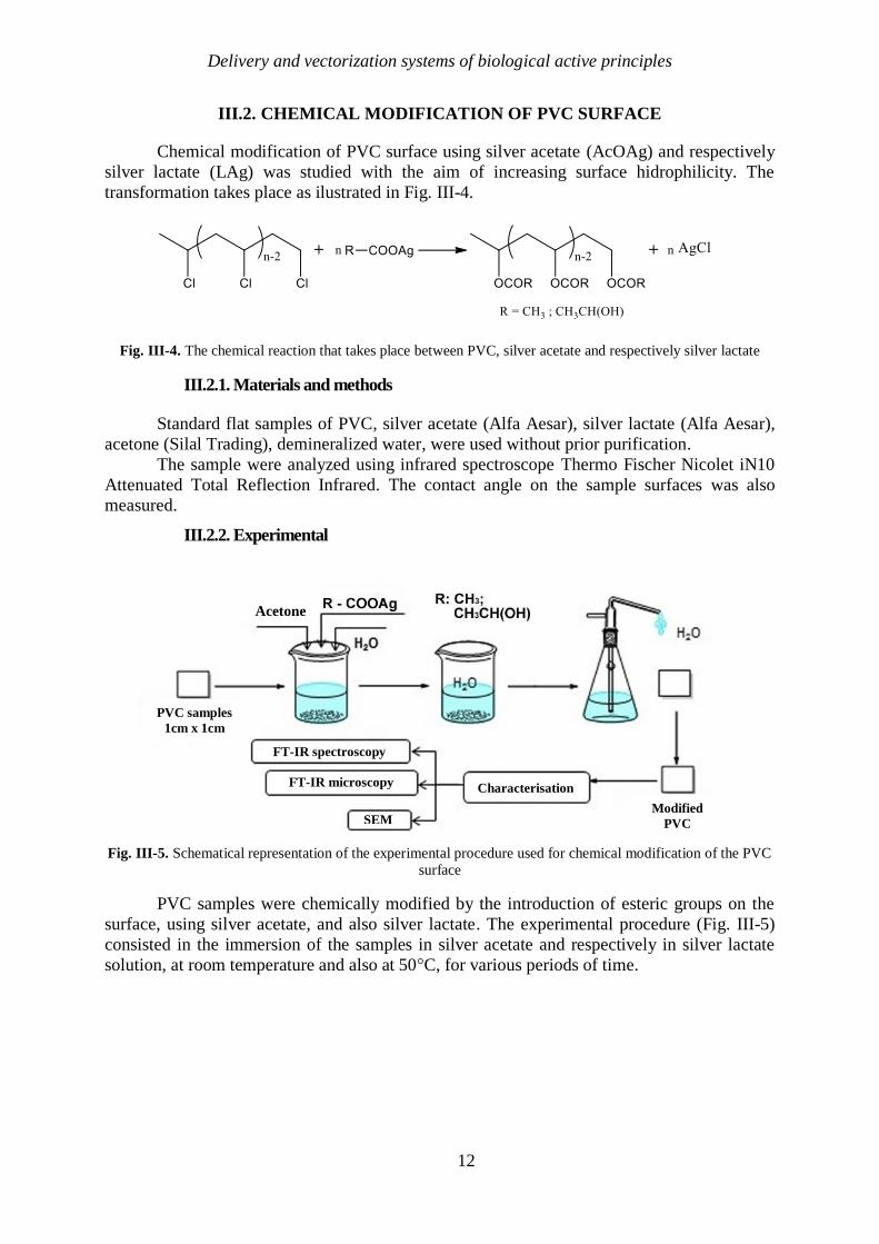
III.2. CHEMICAL MODIFICATION OF PVC SURFACE
Chemical modification of PVC surface using silver acetate (AcOAg) and respectively
silver lactate (LAg) was studied with the aim of increasing surface hidrophilicity. The
transformation takes place as ilustrated in Fig. III-4.
Fig. III-4. The chemical reaction that takes place between PVC, silver acetate and respectively silver lactate
III.2.1. Materials and methods
Standard flat samples of PVC, silver acetate (Alfa Aesar), silver lactate (Alfa Aesar),
acetone (Silal Trading), demineralized water, were used without prior purification.
The sample were analyzed using infrared spectroscope Thermo Fischer Nicolet iN10
Attenuated Total Reflection Infrared. The contact angle on the sample surfaces was also
measured.
III.2.2. Experimental
Fig. III-5. Schematical representation of the experimental procedure used for chemical modification of the PVC
surface
PVC samples were chemically modified by the introduction of esteric groups on the
surface, using silver acetate, and also silver lactate. The experimental procedure (Fig. III-5)
consisted in the immersion of the samples in silver acetate and respectively in silver lactate
solution, at room temperature and also at 50°C, for various periods of time.
Acetone
PVC samples
1cm x 1cm
Modified
PVC
FT-IR spectroscopy
FT-IR microscopy Characterisation
SEM

Delivery and vectorization systems of biological active principles
13
III.2.3. Results and discutions
Fig. III-8. Comparison between the initial PVC spectrum and the spectra obtained in the case of the 2 experiments
performed with AcOAg, after 2 hours of reaction
Fig. III-9. IR spectra for the samples resulted after the reaction with LAg at 50⁰C
When LAg is used, the reaction speed is semnificatively lower even when the reaction
is conducted at 50⁰C (Fig. III-9). The appearance of the ester bond on the surface of PVC
modified using LAg, at room temperature, takes place only after 6h (Fig. III-10). After 8h the
intensity of the peak corresponding to the esteric bound is significantly increased. Increasing
the temperature to 50⁰C, the reaction speed is also increased, the corresponding ester bond
appeared on the surface of PVC after 30 min, even though only in reduced intensity. In order
to obtain a similar yeld with the one obtained after performing the reaction with AcOAg, the
reaction with silver lactate should be performed at least 7-8h.
Ester 1516
C-Cl 746
Ab
sorb
an
ce
0.30
0.25
0.20
0.15
0.10
0.05
0.00
-0.05
-0.10
Wavenumber (cm-1
)
4000 3500 3000 2500 2000 1500 1000 500
P5_ AcOAg_50_2h
P11_ AcOAg_tc_2h
PVC_RB1_Martor
Ester 1516
C-Cl 746
A
bso
rb
an
ce
0.30
0.25
0.20
0.15
0.10
0.05
0.00
-0.05
-0.10
-0.15
-0.20
-0.25
-0.30
-0.35
Wavenumber (cm-1
)
4000 3500 3000 2500 2000 1500 1000 500
P13_ LAg_50_30min
P15_ LAg_50_2h
P16_ LAg_50_4h
P17_ LAg_50_6h
P18_ LAg_50_8h
P19_ LAg_50_16h
P20_ LAg_50_24h
PVC_RB1_Martor

Delivery and vectorization systems of biological active principles
14
Fig. III-10. IR spectra for the samples resulted from the reaction with LAg at room temperature
In order to determine if the hidrophilicity of the surface is modified by introduction of
esteric groups, the contact angle for the samples modified using AcOAg and respectively
LAg, at various reaction times, was determined and compared with the contact angle of the
initial, unmodified PVC (Fig. III-11).
Fig. III-11. Contact angle modification after performing the reaction with AcOAg and respectively with LAg at
various reaction time
III.2.4. Conclusions
Chemical modification of the surface was performed in order to increase the
hydrophilicity of the polymer surface and the wetting properties so as not to allow the
adhesion of bacteria and cells on the material. This modification was successfully performed
by the reaction with silver acetate and also with silver lactate, after introduction of ester
groups on the surface of PVC. The presence of the ester bounds on the samples surfaces was
confirmed by infrared spectrometry. In the spectra corresponding to the modified surfaces, the
appearance of the band corresponding to the ester (1516 cm-1) and the decrease of the band
intensity corresponding to the C-Cl bond (746 cm-1) are observed. In case of chemical
modification of the PVC surface with silver acetate in 50% acetone medium at room
temperature, the time required to complete the reaction is 2 hours, and if the change is made at
0
20
40
60
80
100
120
Co
nta
ct a
ngl
e
Time, [h]
AcOAg t.c.
AcOAg 50 C
LAg t.c.
LAg 50 C
Ester
1516
Ab
sorb
an
ce
0.35
0.30
0.25
0.20
0.15
0.10
0.05
0.00
-0.05
-0.10
-0.15
-0.20
PVC_Martor
P6_PVC_LAg_4h
P7_PVC_LAg_6h
P8_PVC_LAg_8h
P9_PVC_LAg_16h
P10_PVC_LAg_24h

Delivery and vectorization systems of biological active principles
15
50⁰C the time required is 1 hour. If silver lactate is used to introduce ester groups on the PVC
surface, a much longer reaction time of more than 4 hours is required, which can lead to
polymer degradation.
The maximum contact angle achieved using this type of modification has a similar
value in both AcOAg and LAg but the difference between the required reaction times is very
large, the reaction with AcOAg being preferable. The longer the polymer is in contact with
the solvent for a longer period of time, the greater the possibility that changes will occur in
the PVC properties.
III.3. DICOUMAROL SYNTHESIS
Fig. III-12. Dicoumarol synthesis reaction
The synthesis methods used in this research study were prior studied and proposed by
K. M. Khan şi co. [349] and respectively by M. Prabhakar [352]. These synthesis techniques
were improved, the experimetal procedure was optimized and the yeld was increased
significantly. Also, the purity of the compounds obtained was improved.
III.3.1. Materials and methods
4-hidroxycumarine (Merck), fomaldehide (Merck), EDTA (Reanal Budapest), ethanol
(Chemical Company), toluen (Merck), piperydine (Merck), NaOH (Merck), norit charcoal,
were used without prior purification. The product obtained was analyzed using thin layer
chromatography and FT-IR spectroscopy.
III.3.2. Experimental
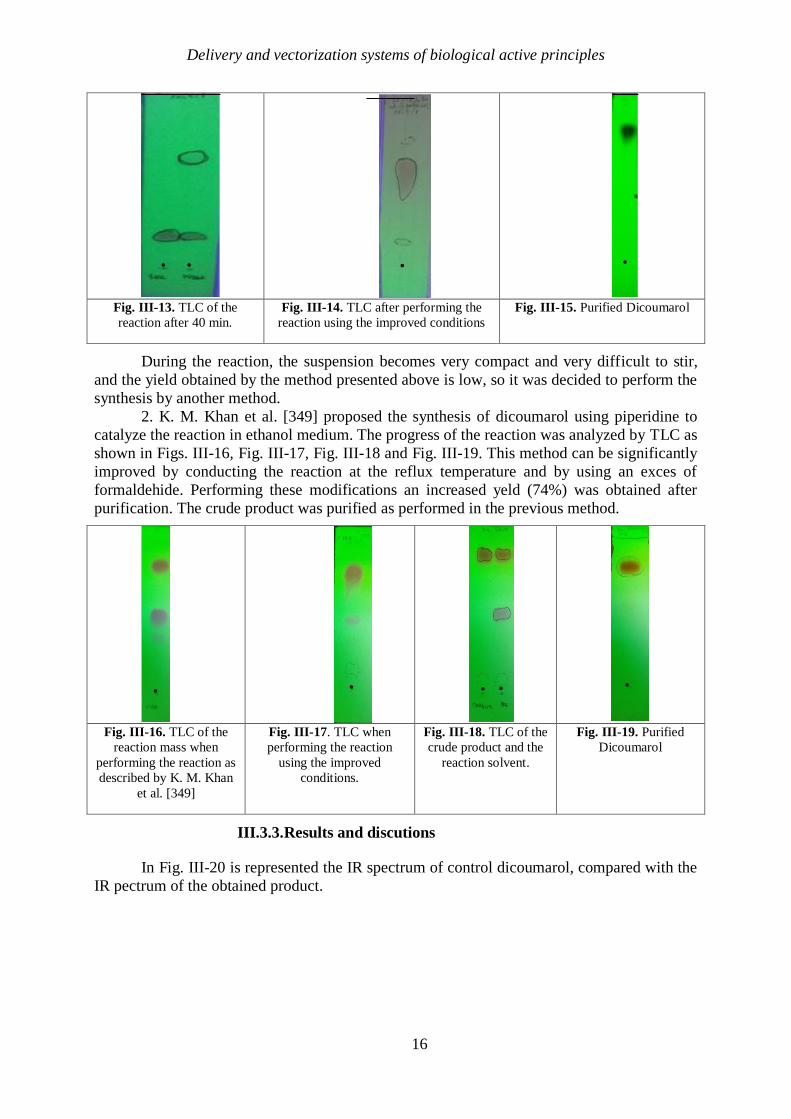
1. Initially the reaction was led in the way described by M. Prabhakar in the work
“EDTA – catalyzed fast and efficient eco-friendly synthesis of dicoumarol derivatives in
water” [352]. The progress of the reaction was verified by thin layer chromatography (plates:
Merck silica gel 60 F254, eluent: toluene: ethyl acetate: acetic acid 20: 5: 1) as shown in Fig.
III-13, Fig. III-14, Fig. III-15. Using the reaction conditions described by the authors, the
yield is low, the ratio between the product formed and the raw material remaining unreacted
being about 1:1. By increasing the reaction time and adding an excess of formaldehyde (1.2
equivalents), the reaction yield was significantly improved. The product obtained cannot be
purified by the method described in the article, so another purification method has been
developed. The crude product was purified by dissolving in dilute sodium hydroxide (NaOH)
(stoichiometric amount), decolorized with charcoal, and the clear solution was extracted with
toluene. The aqueous phase was then acidified with sulfuric acid (H2SO4) to pH = 2. The
resulting suspension was filtered in vacuum and the precipitate was washed with
demineralized water to remove traces of acid. The white solid obtained was dried in an oven
at 105 ° C. Yield obtained: ƞ = 59%.
Delivery and vectorization systems of biological active principles
16
Fig. III-13. TLC of the
reaction after 40 min.
Fig. III-14. TLC after performing the
reaction using the improved conditions
Fig. III-15. Purified Dicoumarol
During the reaction, the suspension becomes very compact and very difficult to stir,
and the yield obtained by the method presented above is low, so it was decided to perform the
synthesis by another method.
2. K. M. Khan et al. [349] proposed the synthesis of dicoumarol using piperidine to
catalyze the reaction in ethanol medium. The progress of the reaction was analyzed by TLC as
shown in Figs. III-16, Fig. III-17, Fig. III-18 and Fig. III-19. This method can be significantly
improved by conducting the reaction at the reflux temperature and by using an exces of
formaldehide. Performing these modifications an increased yeld (74%) was obtained after
purification. The crude product was purified as performed in the previous method.
Fig. III-16. TLC of the
reaction mass when
performing the reaction as
described by K. M. Khan
et al. [349]
Fig. III-17. TLC when
performing the reaction
using the improved
conditions.
Fig. III-18. TLC of the
crude product and the
reaction solvent.
Fig. III-19. Purified
Dicoumarol
III.3.3.Results and discutions
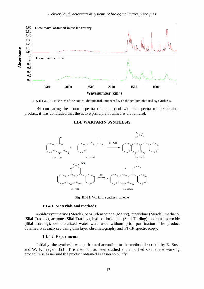
In Fig. III-20 is represented the IR spectrum of control dicoumarol, compared with the
IR pectrum of the obtained product.
. . .
.
.
.
.
.
.
.
.
. .
.
.
.
.
.
.
.
.
.
.
Delivery and vectorization systems of biological active principles
17
Fig. III-20. IR spectrum of the control dicoumarol, compared with the product obtained by synthesis.
By comparing the control spectra of dicoumarol with the spectra of the obtained
product, it was concluded that the active principle obtained is dicoumarol.
III.4. WARFARIN SYNTHESIS
Fig. III-22. Warfarin synthesis scheme
III.4.1. Materials and methods
4-hidroxycumarine (Merck), benzilidenacetone (Merck), piperidine (Merck), methanol
(Silal Trading), acetone (Silal Trading), hydrochloric acid (Silal Trading), sodium hydroxide
(Silal Trading), demineralized water were used without prior purification. The product
obtained was analyzed using thin layer chromatography and FT-IR spectroscopy.
III.4.2. Experimental
Initially, the synthesis was performed according to the method described by E. Bush
and W. F. Trager [353]. This method has been studied and modified so that the working
procedure is easier and the product obtained is easier to purify.
CH3OH
CH3
A
bso
rb
an
ce
0.60
0.50
0.40
0.30
0.20
0.10
0.00
1.2
1.0
0.8
0.6
0.4
0.2
0.0
Wavenumber (cm-1
)
3500 3000 2500 2000 1500 1000
Dicoumarol obtained in the laboratory
Dicoumarol control

Delivery and vectorization systems of biological active principles
18
III.4.3. Results and discutions
As in the case of dicoumarol, in the case of warfarin, the reaction evolution was also
controlled using thin layer chromatography (TLC plates: Silica gel 60 F254 Merck, eluent:
toluen:dioxan:acetic acid 7:0,5:0,15, developing: UV 254 nm, iodine vapour). The product
purity was determined also using TLC, in order to determin the elimination of impurities (Fig.
III-23, Fig. III-24, Fig. III-25, Fig. III-26, Fig. III-27).
Fig. III-23. Reaction in
the first step, compared
with starting materials.
Fig. III -24. The
ester resulted in
the first stage of
synthesis
Fig. III-25. The second
stage of the reaction
compared to the ester.
Fig. III-26. Toluen
used for the
extraction in the
purification step.
Fig. III-27. Purified
warfarin
Fig. III-28. IR spectrum coresponding to warfarin control, compared with the spectrum of the product obtained
by performing the synthesis
By comparing the spectrum of control warfarin with the spectrum of the product
obtained by synthesis, it could be confirmed that the desired active principle was obtained.
After purification, the warfarin obtained was in the form of pure product accordingly to the
thin layer chromatography (Fig. III-27).
A
bso
rb
an
ce
Warfarin obtained by performing the synthesis
Warfarin control
Wavenumber (cm-1
)
4000 3400 2800 2200 1600 1000 600
. . . . . .. . . .
Delivery and vectorization systems of biological active principles
19
III.5. SURFACE MODIFICATION OF PVC USING SPIN COATING TECHNIQUE
In this study surface modification of PVC was performed in order to obtain a material
that inhibits cellular adhesion and bacterial proliferation, and also to prevent blood clotting or
the formation of calcifications. Therefor, the PVC surface was physically modified using spin
coating technique to correct the irregularities arising from the synthesis, and to obtain a
smooth, uniform surface.
III.5.1. Modification performed using PVC/Dicoumarol film deposition
III.5.1.1. Materials and methods
In these experiments dicoumarol (previously synthesized), bovine serum albumin
(Fluka), standard flat samples of PVC and tetrahydrofuran.
The modified samples were analyzed using a infrared microscop Thermo Scientific
Nicolet iN10 in order to analyze the surface of the samples, and also using a infrared
sprectroscope Thermo Fischer Nicolet iN10 Attenuated Total Reflection Infrared to determine
the surface composition. Ultraviolet-visible spectrometry was performed using a Thermo
Scientific Evolution 300 Spectrophotometer with the aim to determine dicoumrol release from
the material surface in SBF (simulated body fluid). Scanning Electron Microscopy (SEM) and
Energy Dispersive Spectroscopy (EDS) were performed in order to have a more clear view of
the salts deposited on the surface of the modified PVC. Protein adsorbtion was studied using
FT-IR microscopy and spectroscopy after several periods of immersion in SBF that contained
albumin, mimicking the human body conditions. The antiaderent properties of the modified
samples were analyzed by determining the values of colony forming units (CFU/mL). The
samples were tested against 2 types of standard strains: Gram-positive (Staphylococcus
aureus ATCC 25923) and Gram-negative (Pseudomonas aeruginosa ATCC 27853). For the
performed tests, fresh cultures of 18-24 h were obtained after inoculation of bacterial strains
on nutrient Agar medium.
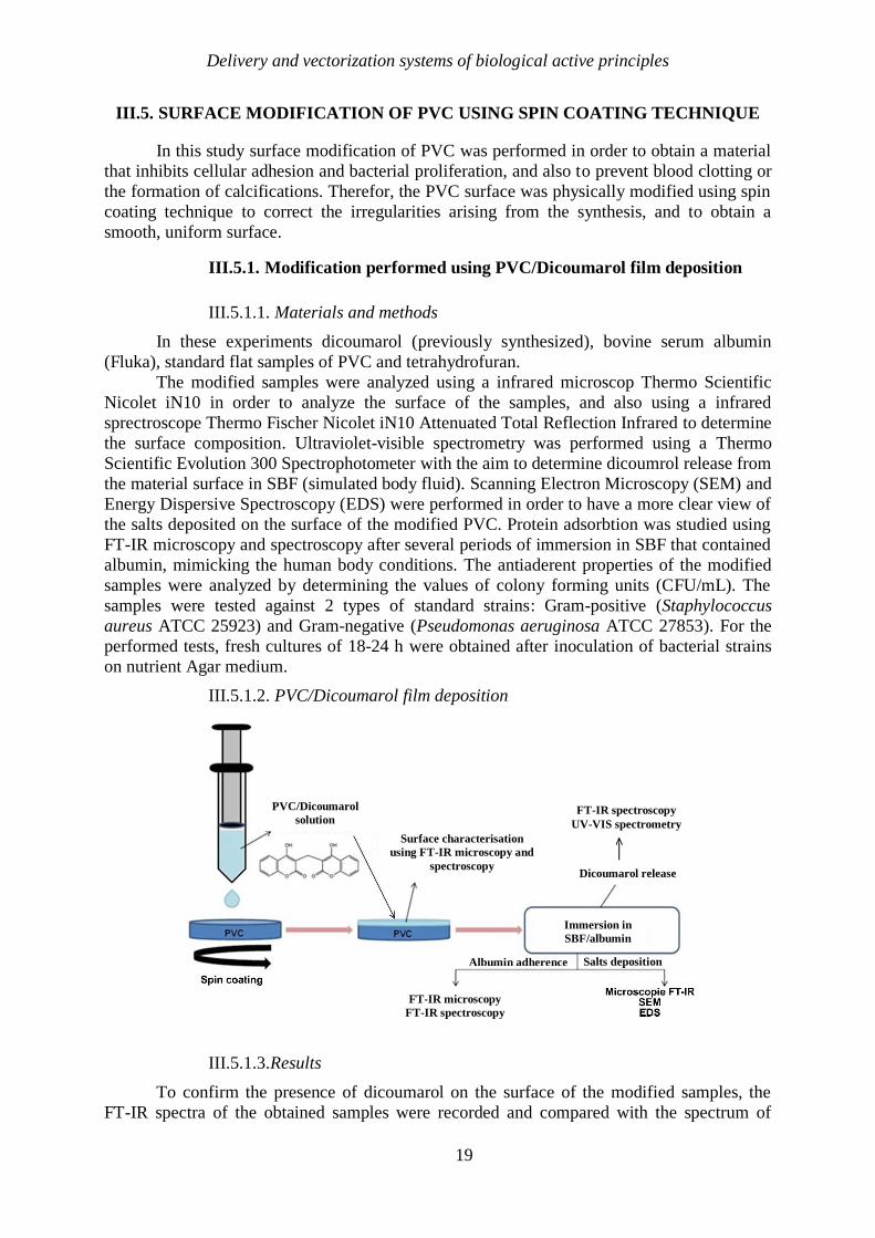
III.5.1.2. PVC/Dicoumarol film deposition
III.5.1.3.Results
To confirm the presence of dicoumarol on the surface of the modified samples, the
FT-IR spectra of the obtained samples were recorded and compared with the spectrum of
PVC/Dicoumarol
solution
Surface characterisation
using FT-IR microscopy and
spectroscopy
FT-IR spectroscopy
UV-VIS spectrometry
Dicoumarol release
FT-IR microscopy
FT-IR spectroscopy
Immersion in
SBF/albumin
Albumin adherence Salts deposition

Delivery and vectorization systems of biological active principles
20
plain PVC and pure dicoumarol, respectively. Fig. III-29 presents a comparison between the
FT-IR spectrum obtained at 4000 rpm, 7000 rpm and 10,000 rpm, respectively, the PVC and
dicoumarol spectrum.
Fig. III-29. Comparison between the FT-IR spectrum obtained at 4000 rpm, 7000 rpm and respectively 10,000
rpm, the spectrum of PVC and of dicoumarol
In the obtained spectra the characteristic peaks of dicoumarol can be observed at 1650
cm−1
and 1350 cm−1, corresponding to the ν (C = O) and ν (C – O) bonds in the rings. Also,
the peaks present at 1110–1130 cm−1
indicate the presence of the ν bond (C – OH) and at
1070 cm−1
the presence of the ν (C – O – C) bond. In Fig. III-30.a-c. are presented the FT-IR
microscopy images of the samples obtained at 4000 rpm, 7000 rpm and respectively 10,000
rpm.
Fig. III-30. FT-IR microscopy images obtained at 1655 cm−1
for the deposition performed at 4000 rpm (a), at
1650 cm−1
for the deposition at 7000 rpm (b) and respectively at 1654 cm−1
for the deposition performed at
10,000 rpm (c).
Ab
sorb
an
ce
-0.8
-0.4
0.0
0.4
2.0
0.8
2.4
1.2
1.6
Wavenumber (cm-1
)
4000 3500 3000 2500 2000 1500 1000 500
2.8

Delivery and vectorization systems of biological active principles
21
a. Dicoumarol release from the film deposited on the surface of PVC
To determine whether the drug is being released from the surface, each sample was
immersed in 100 mL of SBF solution at 36.5°C and thus maintained, with occasional stirring,
for 30 days. The SBF solution was analyzed by UV-VIS at 303 nm (maximum adsorbtion
wavelenght of dicoumarol) after various periods of time (1 h, 2 h, 4 h, 24 h, 2 days, 3 days, 7
days, 10 days, 14 days, 21 days and respectively 30 days) in order to observe the release
behaviour of dicoumarol from the PVC film. Samples were also analyzed by FT-IR
microscopy to determine if the surface morphology had changed in time after the contact with
the SBF solution. FT-IR microscopy was also used to confirm the absence or presence of
dicoumarol on the surface after a certain period of immersion. The results obtained using UV-
VIS analysis are represented in the release graph corresponding to the samples obtained at
4000 rpm, 7000 rpm and respectively 10,000 rpm (Fig. III-31).
Fig. III-31. Dicoumarol release from the films obtained at 4000 rpm, 7000 rpm and respectively 10,000 rpm,
analyzed using UV-VIS spectrometry at a wavelenght of 303 nm; standard deviation: SD < 2% in the case of all
the represented points
Fig. III-32. FT-IR images obtained at 1680 cm−1
for the film deposited at 4000 rpm: (a) after 7 days of
immersion in SBF; (b) after 21 days of immersion in SBF; (c) after 30 days of immersion in SBF
0
5
10
15
20
0 10 20 30 40
Rel
ease
deg
ree,
%
Time, days
Dicoumarol release from the PVC
surface
4000 rpm
7000 rpm
10000 rpm
SD < 2%

Delivery and vectorization systems of biological active principles
22
Fig. III-33. FT-IR images obtained at 1659 cm−1
for the film deposited at 7000 rpm: (a) after 7 days of
immersion in SBF; (b) after 21 days of immersion in SBF; (c) after 30 days of immersion in SBF
Fig. III-34. FT-IR images obtained at 1684 cm−1
for the film deposited at 10,000 rpm: (a) after 7 days of
immersion in SBF; (b) after 21 days of immersion in SBF; (c) after 30 days of immersion in SBF
To determine whether the coating influenced the deposition of salts on the surface, a
sample of unmodified PVC was also immersed in SBF for 30 days and then analyzed by FT-
IR microscopy at 1723 cm-1
(characteristic wavelenght of simple PVC) for the same periods
of time and under the same conditions as in the case of modified samples.

Delivery and vectorization systems of biological active principles
23
Fig. III-35. FT-IR images obtained at 1723 cm−1
for the initial, unmodified PVC: (a) after 7 days of immersion
in SBF; (b) after 21 days of immersion in SBF; (c) after 30 days of immersion in SBF
Scanning Electron Microscopy (SEM) (Fig. III-36) and Energy Dispersive
Spectroscopy (EDS) (Fig. III-37) were performed in order to have a clearer view of the salts
that crystallized on the samples surfaces, and also to determine their composition.
Fig. III-37. SEM image performed on the modified samples
FT-IR microscopy does not provide a very clear information about the presence of
dicoumarol in the polymeric film, therefore the surface of the materials was analyzed using
FT-IR spectroscopy in ATR mode after different periods of time. In order to be able to
estimate the tendency of the active principle to be released from the deposited polymer film,
the relative pea area corresponding to dicoumarol ( 1650 cm−1
) from the FT-IR spectra was
calculated.

Delivery and vectorization systems of biological active principles
24
Fig. III-40. Schematic representation of the changes that occur over time in the case of the relative area of the
peak corresponding to the dicoumarol incorporated on the surface of the sample as the release from the
polymeric film occurs
b. Protein adsorbtion on the PVC surface
To study the behaviour of the material obtained by deposition of a thin film of
PVC/diuoumarol at the interaction with the proteins from the biological fluids, the samples
were immersed in a solution of SBF with a pH value of 7.4 containing 1% albumin (the main
protein found in blood), at 36.5 °C. The samples were maintained in these conditions for 30
days.
To highlight the characteristic peaks of albumin and to observe whether they appear in
the spectrum of samples immersed in the SBF / albumin solution, a PVC sample was coated
with albumin by the drop-cast method. The spectrum corresponding to the sample obtained by
the drop-cast method was therefore compared with the spectrum of samples (uncovered PVC,
4000 rpm, 7000 rpm, 10,000 rpm) after 7 days of immersion in the solution containing protein
and the spectrum of PVC before immersion (Fig. III-41).
Fig. III-41. Comparison between the spectra of PVC/dicoumarol films after 7 days of immersion in the
SBF/albumin solution, the spectrum of the uncovered PVC before immersion and respectively that of the sample
obtained by drop-cast
0
20
40
60
80
100
0 3 14 30
Rel
ativ
e ar
ea
Time [days]
Dicoumarol release from the film deposited on
the surface of PVC
10,000 rpm
7000 rpm
4000 rpm
Wavenumber (cm-1
)
Ab
sorb
an
ce
4000 3500 3000 2500 2000 1500 1000
0.05
0.15
0.25
0.35
0.45
0.55

Delivery and vectorization systems of biological active principles
25
In order to have a clearer picture of how the material reacts when it comes in contact
with the albumin solution, to determine if this protein is adsorbed on the surface of the
material, but also if its deposition leads to changes in morphology, the samples were analyzed.
by FT-IR microscopy (Fig. III-42).
Fig. III-42. Contour maps of the samples obtained at 4000 rpm (a), 7000 rpm (b), 10,000 rpm (c) and
respectively of the unmodified PVC (d), after immersion in SBF/albumin solution for 7 days (1), 21 days (2),
and 30 days (3), obtained by FT-IR microscopy.
c. Bacterial adhesion
This study examined the resistance of the samples to a gram-positive strain (S. aureus)
and a gram-negative strain (P. aeruginosa).
Fig. III-43. Graphical representation of the CFU/mL values for Staphylococcus aureus ATCC 25923 strain
100.000.000
1.000.000.000
10.000.000.000
100.000.000.000
1.000.000.000.000
10.000.000.000.000
100.000.000.000.000
1.000.000.000.000.000
1 2 3 4 5 6 M
Log C
FU
/mL
Tested sample
Staphylococcus aureus ATCC 25923
(1)
(2)
(3)

Delivery and vectorization systems of biological active principles
26
Fig. III-44. . Graphical representation of the CFU/mL values for Pseudomonas aeruginosa ATCC 27853 strain
III.5.1.4.Conclusions
The PVC surface can be successfully modified by the spin coating method. The
obtained samples were analyzed on both the covered and the uncovered surface. According to
FT-IR microscopy it can be concluded that the surface morphology is significantly changed
when this coating method is applied. The initial surface was rough, uneven, with "hill-valley"
defects that were covered with dicoumarol-doped PVC polymer film, resulting in a smooth,
uniform surface.
After comparing the results obtained from the analyzes performed on all three
samples, it can be concluded that the best results related to the surface morphology, the
distribution of dicoumarol on the surface were observed for the sample obtained at 10,000
rpm. The release was also slower and more constant in the case of this sample and showed a
good ability to prevent salts deposition and reduced protein adhesion. The proposed
methodology has led to an improved anti-adherent activity of the PVC surface, especially
against gram-positive strains such as S. aureus.
III.5.2. Silver nanoparticles deposition on the surface of PVC
The aim of this study was to modify the surface of PVC so that the bacterial adhesion
is as low as possible, thus avoiding the discomfort caused by the side effects that occur after
biofilm formation.
III.5.2.1. Materials and methods
Bovine Serum Albumin (Fluka), silver nitrate (Carl Roth), Trisodium Citrate (),
standard flat samples of polyvinyl chloride and cyclohexanone (Silal) were used without prior
purification. SBF (simulated body fluid) was prepared as described by Oyane et al. [359]. The
samples were characterised using a Thermo Scientific Nicolet iN10 Infrared Microscope in
order to analyze the surface morphology. The Thermo Fischer Nicolet iN10 Attenuated Total
Reflection Infrared spectroscope was used to determine the surface composition of the
samples obtained. Inductively Coupled Plasma (ICP) was performed to determine the degree
of migration of silver ions from the surface. Scanning electron microscopy (SEM) was used to
examine the morphology, microstructure and homogeneity of the samples as well as to
visualize the distribution of silver nanoparticles on the surface.
1
1.000
1.000.000
1.000.000.000
1.000.000.000.000
999.999.999.999.996
1 2 3 4 5 6 M
Lo
g C
FU
/mL
Tested sample
Pseudomonas aeruginosa ATCC 27853
Delivery and vectorization systems of biological active principles
27
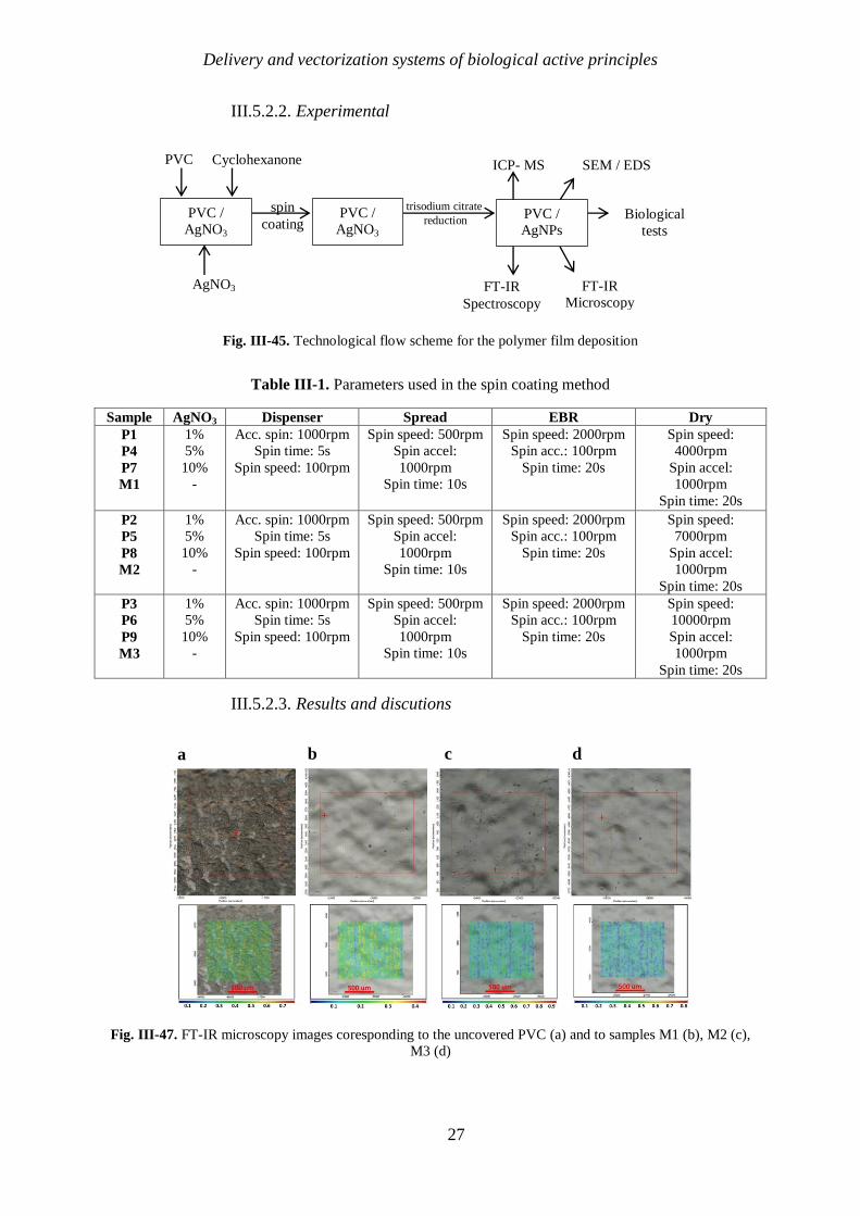
III.5.2.2. Experimental
Fig. III-45. Technological flow scheme for the polymer film deposition
Table III-1. Parameters used in the spin coating method
Sample AgNO3 Dispenser Spread EBR Dry
P1
P4
P7
M1
1%
5%
10%
-
Acc. spin: 1000rpm
Spin time: 5s
Spin speed: 100rpm
Spin speed: 500rpm
Spin accel:
1000rpm
Spin time: 10s
Spin speed: 2000rpm
Spin acc.: 100rpm
Spin time: 20s
Spin speed:
4000rpm
Spin accel:
1000rpm
Spin time: 20s
P2
P5
P8
M2
1%
5%
10%
-
Acc. spin: 1000rpm
Spin time: 5s
Spin speed: 100rpm
Spin speed: 500rpm
Spin accel:
1000rpm
Spin time: 10s
Spin speed: 2000rpm
Spin acc.: 100rpm
Spin time: 20s
Spin speed:
7000rpm
Spin accel:
1000rpm
Spin time: 20s
P3
P6
P9
M3
1%
5%
10%
-
Acc. spin: 1000rpm
Spin time: 5s
Spin speed: 100rpm
Spin speed: 500rpm
Spin accel:
1000rpm
Spin time: 10s
Spin speed: 2000rpm
Spin acc.: 100rpm
Spin time: 20s
Spin speed:
10000rpm
Spin accel:
1000rpm
Spin time: 20s
III.5.2.3. Results and discutions
Fig. III-47. FT-IR microscopy images coresponding to the uncovered PVC (a) and to samples M1 (b), M2 (c),
M3 (d)
PVC /
AgNO3
suspensio
AgNO3
PVC Cyclohexanone
spin
coating PVC /
AgNO3
film
trisodium citrate
reduction
ICP- MS
FT-IR
Microscopy FT-IR
Spectroscopy
SEM / EDS
Biological
tests
PVC /
AgNPs
film
b c d a

Delivery and vectorization systems of biological active principles
28
Fig. III-48. FT-IR microscopy images coresponding to samples P7(a), P8(b) and respectively P9(c)
Fig. III-49. FT-IR microscopy images coresponding to samples P7 (a), P8 (b) and P9 (c) after the reduction was
performed
Silver nanoparticles present on the surface of the thin films deposited by spin coating
was confirmed by SEM (Fig. III-50) and showed a uniform dispersion of silver nanoparticles
on the surface.

Delivery and vectorization systems of biological active principles
29
Fig. III-50. Imaginile SEM images recorded at a magnification of 5000 x (a), 10,000 x (b) and respectively
50,000 x for the samples P7, P8, P9
EDS spectra of the samples characterised after the reduction was performed, also
confirmed the presence of silver and reveals significant differences in the amount deposited.
a. Silver ions migration
A very important aspect to consider when using silver nanoparticle coatings for
medical devices is related to the concentration of ions released in biological fluids because it
can present cytotoxicity at certain concentrations. In order to monitor the migration behavior
of silver ions in the body fluids, the samples were immersed in SBF with pH = 7.4,
maintaining the temperature at 36.5 ⁰C, thus mimic ing the conditions in the body. These
were kept in solution for up to 24 days, periodically analyzing the SBF solution (1h, 2h, 6h, 1
day, 2 days, 3 days, 9 days, 13 days and 23 days, respectively), using ICP-MS analysis (Fig.
III-52).

Delivery and vectorization systems of biological active principles
30
Fig. III-52. Graphical representation of the recovery degree of silver ions fron the SBF solution, in time
b. Albumin adsorbtion on the surface
Adhesion of proteins to the surface of the material can lead to the formation of
thrombosis, calcifications, bacterial adhesion or biofilm formation, which can lead to
blockage of the tubular devices. In order to observe the behavior of the surfaces obtained by
spin coating at the interaction with blood proteins, the samples were immersed in a solution of
SBF pH = 7, at 36.5 ⁰C with albumin content, the main protein found in the blood. The
samples were maintained under these conditions for 24 days and were analyzed by FT-IR
microscopy after 1 day, 6 days, 14 days and 24 days, respectively.
Fig. III-53. FT-IR microscopy images obtained for sample P7 after: a - 1 day, b - 6 days, c – 14 days, d – 24
days of immersion in SBF/albumin solution
0
0,1
0,2
0,3
0,4
0,04 0,08 0,25 1 2 3 9 13 23Deg
ree
of
reco
very
, [%
]
Time, [h]
Silver ions migration from the surface
P7
P8
P9
Delivery and vectorization systems of biological active principles
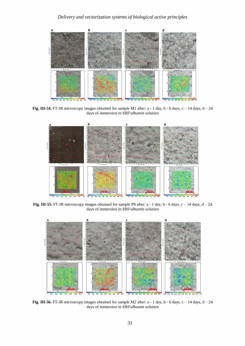
31
Fig. III-54. FT-IR microscopy images obtained for sample M1 after: a - 1 day, b - 6 days, c – 14 days, d – 24
days of immersion in SBF/albumin solution
Fig. III-55. FT-IR microscopy images obtained for sample P8 after: a - 1 day, b - 6 days, c – 14 days, d – 24
days of immersion in SBF/albumin solution
Fig. III-56. FT-IR microscopy images obtained for sample M2 after: a - 1 day, b - 6 days, c – 14 days, d – 24
days of immersion in SBF/albumin solution

Delivery and vectorization systems of biological active principles
32
Fig. III-57. FT-IR microscopy images obtained for sample P9 after: a - 1 day, b - 6 days, c – 14 days, d – 24
days of immersion in SBF/albumin solution
Fig. III-58. FT-IR microscopy images obtained for sample M3 after: a - 1 day, b - 6 days, c – 14 days, d – 24
days of immersion in SBF/albumin solution
Fig. III-59. FT-IR microscopy images obtained for the initial, uncovered PVC after: a - 1 day, b - 6 days, c – 14
days, d – 24 days of immersion in SBF/albumin solution

Delivery and vectorization systems of biological active principles
33
The FT-IR spectrum (Fig. III-60) confirms the presence of albumin on the sample
surface by the appearance of additional peaks after immersion in the albumin solution. These
peaks occur at wavelengths of 1680 cm-1
, the band corresponding to amide I, consisting
mostly of vibrations of the bond (C = O) and at 1580 cm-1
the band corresponding to amide
II, consisting of the most of the vibrations of the (C - N) bond [387-389].
Fig. III-60. Graphical representation of the FT-IR spectra of the surface before and after immersion in the
SBF/albumin solution
c. Biological tests
To test the resistance to colonization of the modified surfaces, but also of the initial,
uncovered surface, the bacterial strain S. aureus ATCC 6538 was used, the final results being
expressed in CFU/ml after different time intervals from the contact of these surfaces with the
bacterial suspension. (Fig. III-61).
Fig. III-61. Quantitative evaluation of the degree of development of the monospecific microbial biofilm
developed by Staphylococcus aureus ATCC 6538 at the initial PVC surface and respectively to the modified
surfaces.
E. coli is the most representative bacterial species among Gram-negative species,
capable of developing biofilms, therefore the ability of the samples to form a monospecific
biofilm on contact with the standard strain Escherichia coli ATCC 25922 was tested. (Fig. III-
62).
1
100
10000
1000000
2 24 48 72
Log
CFU
/mL
Time, [h]
S. aureus ATCC 6538
M1
P4 (PVC - Ag 5%)
P7 (PVC - Ag 10%)
Uncovered PVC
A
bso
rb
an
ce
0.10
0.08
0.06
0.04
0.02
0.00
-0.02
-0.04
Wavenumber (cm-1
)
4000 3500 3000 2500 2000 1500 1000
Before immersion
After immersion

Delivery and vectorization systems of biological active principles
34
Fig. III-62. Quantitative evaluation of the degree of development of the monospecific microbial biofilm
developed by E. coli ATCC 25922 at the initial PVC surface and respectively to the modified surfaces
Nosocomial infections associated with venous catheters are frequently caused by
Candida species. Induction of biofilm formation on the surface of functionalized biomaterials
used in the present study allowed the evaluation of their behavior in contact with planktonic
C. albicans cells ATCC 26790 (Fig. III-63).
Fig. III-63. Quantitative evaluation of the degree of development of the monospecific microbial biofilm
developed by C. albicans ATCC 26790 at the initial PVC surface and respectively to the modified surfaces
III.6. SURFACE EVALUATION OF OXYNITRIDE COATINGS (TIOxNy) USED
FOR OBTAINING LAYERED CARDIOVASCULAR STENTS
In this paper were studied TiOxNy coatings with different N / O ratios deposited by
magnetron sputtering deposition, in order to determine how the properties of this type of
surface are influenced by the concentration of N or O.
III.6.1. Materials and methods
The studied TiOxNy coatings were deposited both on flat samples, in the form of
discs, and also on stainless steel stents. The coatings were performed using various
concentrations of nitrogen (ratio O: N = 1: 2, 1: 5, 1:10) in the gas supply flow. The
depositions were performed using magnetron sputtering technique, using a UVN-200 MI
system at medium frequency.
The surface morphology was analyzed before and after exposure to SBF by Scanning
Electron Microscopy (SEM) using a Philips ESEM-FEG XL 30 microscope, 3.0 kV.
Chemical and elemental analysis was performed using FT-IR spectroscopy and EDS (Energy-
dispersive X-ray spectroscopy). To determine the surface hydrophilicity, the flat samples were
1
100
10000
1000000
100000000
2 24 48 72
Log
CFU
/mL
Time, [h]
E. coli ATCC 25922
M1
P4 (PVC - Ag 5%)
P7 (PVC - Ag 10%)
Uncovered PVC
1
100
10000
2 24 48 72
Log
CFU
/mL
Time, [h]
C. Albicans ATCC 26790
M1
P4 (PVC - Ag 5%)
P7 (PVC - Ag 10%)
Uncovered PVC

Delivery and vectorization systems of biological active principles
35
analyzed using the OCA 20 Contact Angle System. The interaction of the samples with blood
proteins was studied by immersion in an SBF/albumin solution. The samples were analyzed at
different time intervals by FT-IR microscopy. The biocompatibility of the obtained coatings
was analyzed using Human Umbilical Vein Endothelial Cells (HUVEC).
III.6.2. Results and discutions
The samples obtained were analyzed by SEM and as it can be seen from Fig. III-63,
the characteristics of the films obtained differ depending on the O/N ratio used.
Fig. III-64. SEM images corresponding to samples covered with TiOxNy (c).
To analyze the uniformity of the depositions, the flat samples were analyzed by FT-IR
microscopy (Fig.III-65).
a. TiOxNy_1:2 b. TiOxNy_1:5 c. TiOxNy_1:10

Delivery and vectorization systems of biological active principles
36
(i) (ii) (iii)
Fig. III-65. FT-IR microscopy images corresponding to the flat samples covered with TiOxNy (a.i – O2/N2 1/2;
a.ii - O2/N2 1/5; a.iii - O2/N2 1/10), recorded at 840-884 cm-1
(b –FT-IR spectra corresponding to the modified
surfaces, highlighting the peak at which the recording was performed).
The surfaces of the stents coated with TiOxNy by magnetron sputtering were analyzed
both at micro and nano scale. (Fig.III-66).
10.0 20.0 30.0 40.0 50.0 60.0 70.0 80.0
b) TiOxNy 1:10
TiOxNy 1:5
TiOxNy 1:2
a)

Delivery and vectorization systems of biological active principles
37
Fig. III-66. SEM images corresponding to samples covered with TiOxNy , at magnifications of 55x (a), 2000x
(b) and respectively 200,000x (c).
Although the surface of the stent presented only limited areas showing detachments of
the coating, the deposition technology must be improved because these coatings are
performed in order to be used at the manufacture of the stent, and degradation and peeling of
the surface film must be removed.
The measurement of the contact angle was performed on all flat samples. It was
determined that all three coatings made induce a slightly hydrophobic behavior of the
surfaces, the contact angle being greater than 90º. The determined value of the contact angle
corresponding to the stainless steel is approximately 47º and is in accordance with the value in
the literature [398].
III.6.2.1. In vitro stability
To determine the way the modified stents covered with TiOxNy interacts with the body
fluids, the samples were immersed in SBF for several days. SEM images corresponding to the
samples after 7 days of immersion in SBF are presented in Fig. III-67. To verify whether Ti-
OH groups can indeed induce the nucleation and crystallization of apatite, but also to
determine the chemical nature of the precipitate deposited on the surface of the stents after
immersion in SBF [400], elemental mapping using EDS was performed on the flat samples.
The distribution of the elements on the surface is shown in Fig. III-67 and confirms the
presence of phosphates, calcium, magnesium.

Delivery and vectorization systems of biological active principles
38
Fig. III-68. Elemental mapping of the precipitate formed on the surface of TiOxNy coatings after 7 days of
immersion in SBF
III.6.2.2. Protein adsorbtion (albumin)
To determine the behavior of the samples in the presence of proteins in the blood, they
were immersed in a solution of albumin in SBF for 1, 3, 8, 14 and 28 days. They were then
carefully washed with water and analyzed by FT-IR microscopy.
After 14 days, the albumin adsorption started to be present on the surface of the
samples O2 / N2 = 1/5 (Fig. III-73) and O2 / N2 = 1/10 (Fig. III-74). The spectra
corresponding to the areas where the wires meet are different from the other areas of the
stents, because the adsorption of albumin takes place predominantly on the areas where the
TiOxNy deposition was degraded. Defects arising from the deposition detachment favor the
anchoring of proteins on the surface.
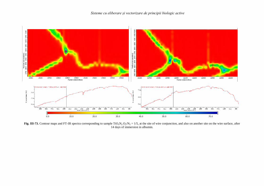
Sisteme cu eliberare și vectorizare de principii biologic active
Fig. III-73. Contour maps and FT-IR spectra corresponding to sample TiOxNy O2/N2 = 1/5, at the site of wire conjunction, and also on another site on the wire surface, after
14 days of immersion in albumin.
0.0 10.0 20.0 30.0 40.0 50.0 60.0 70.0

Delivery and vectorization systems of biological active principles
40
Fig. III-74. Contour maps and FT-IR spectra corresponding to sample TiOxNy O2/N2 = 1/10, at the site of wire conjunction, and also on another site on the wire surface, after
14 days of immersion in albumin.
The analyzes performed revealed that the TiOxNy coating improves the inhibitory properties of albumin deposition on the surface of
stainless steel stents.
0.0 10.0 20.0 30.0 40.0
Sisteme cu eliberare și vectorizare de principii biologic active
41
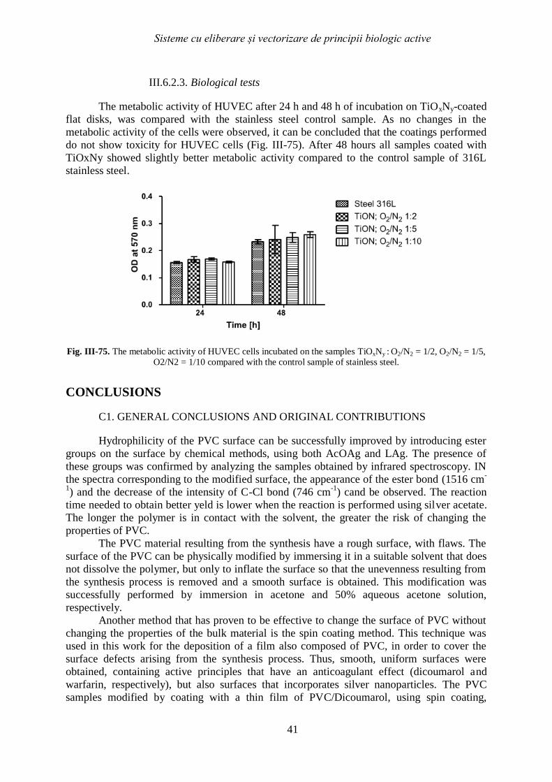
III.6.2.3. Biological tests
The metabolic activity of HUVEC after 24 h and 48 h of incubation on TiOxNy-coated
flat disks, was compared with the stainless steel control sample. As no changes in the
metabolic activity of the cells were observed, it can be concluded that the coatings performed
do not show toxicity for HUVEC cells (Fig. III-75). After 48 hours all samples coated with
TiOxNy showed slightly better metabolic activity compared to the control sample of 316L
stainless steel.
Fig. III-75. The metabolic activity of HUVEC cells incubated on the samples TiOxNy : O2/N2 = 1/2, O2/N2 = 1/5,
O2/N2 = 1/10 compared with the control sample of stainless steel.
CONCLUSIONS
C1. GENERAL CONCLUSIONS AND ORIGINAL CONTRIBUTIONS
Hydrophilicity of the PVC surface can be successfully improved by introducing ester
groups on the surface by chemical methods, using both AcOAg and LAg. The presence of
these groups was confirmed by analyzing the samples obtained by infrared spectroscopy. IN
the spectra corresponding to the modified surface, the appearance of the ester bond (1516 cm-
1) and the decrease of the intensity of C-Cl bond (746 cm
-1) cand be observed. The reaction
time needed to obtain better yeld is lower when the reaction is performed using silver acetate.
The longer the polymer is in contact with the solvent, the greater the risk of changing the
properties of PVC.
The PVC material resulting from the synthesis have a rough surface, with flaws. The
surface of the PVC can be physically modified by immersing it in a suitable solvent that does
not dissolve the polymer, but only to inflate the surface so that the unevenness resulting from
the synthesis process is removed and a smooth surface is obtained. This modification was
successfully performed by immersion in acetone and 50% aqueous acetone solution,
respectively.
Another method that has proven to be effective to change the surface of PVC without
changing the properties of the bulk material is the spin coating method. This technique was
used in this work for the deposition of a film also composed of PVC, in order to cover the
surface defects arising from the synthesis process. Thus, smooth, uniform surfaces were
obtained, containing active principles that have an anticoagulant effect (dicoumarol and
warfarin, respectively), but also surfaces that incorporates silver nanoparticles. The PVC
samples modified by coating with a thin film of PVC/Dicoumarol, using spin coating,

Delivery and vectorization systems of biological active principles
42
presented smooth surfaces with a uniform distribution of the activ principle. Dicoumarol was
released gradually, in time, on a long period of time, thus the material can present
antithrombogenic activity on an extended period of time, sufficient to prevent blood clotting,
thrombosis and thus clogging of the catheter. The morphology of the thin film resulted after
performing coating using the spin coating is influenced by the spin speed value in the drying
step. Albumin adherence takes place on all the studied samples, but the presence of
dicoumarol and the surface smoothness obtained after performing the deposition at 10,000
rpm, resulted in the reduction of protein adhesion. Is important to be mentioned that the
proposed methodology resulted in an improved anti-adherent activity of the modified PVC,
especially against Gram-positive bacteria as for example S. aureus. Bacterial adhesion
resistance has been improved by incorporating dicoumarol on the surface due to its
antibacterial properties and its protein binding capacity.
The PVC surface can be successfully modified by coating with films incorporating
AgNPs. This deposition can be performed by the spin coating method at various working
parameters. The resulting films are compact enough to prevent the migration of silver ions
into biological fluids (the degree of recovery being <0.4%). The nanoparticle size is
between 16 - 27 nm for dispersed nanoparticles and reaches up to 80 nm in agglomerations.
All samples modified by spin coating in this paper showed anti-biofilm activity against E.
coli, S. aureus strains, but not against C. albicans strain.
In the second part of the paper was performed the characterisation of the surfaces of
stainless steel flat samples, but also of stents made from the same material, after the coating
with TiOxNy. was conducted. The coating was made by magnetron sputtering using a mixture
of oxygen and nitrogen as a reactive gas. It was observed that depending on the ratio between
the two gases, the surface properties differ. The best results were observed in the case of an
O2 / N2 = 1/5 ratio in the gas supply flow, followed by O2 / N2 = 1/10. These samples showed
the lowest degree of albumin adsorption and minimal salts depositions due to exposure to
SBF. As the nitrogen content in the surface composition increases, the adsorption of albumin
decreases significantly. The morphology of the surface is also influenced by the amount of
nitrogen, the higher the amount of nitrogen, the smoother the surface is. Although the results
obtained for this type of coating are promising in terms of surface properties, they do not
show a sufficiently good adhesion to the substrate so that the joint areas do not show damage
when a mechanical stress such as stent expansion is applied.
C2. PERSPECTIVES FOR FURTHER DEVELOPMENT
The methods of modifying the PVC surface described in this doctoral thesis can be
used together to obtain a surface with improved properties, which can be used in medical
applications to obtain various medical devices. By using the physical method of modification,
respectively of a solvent that induce surface swelling of the polymer, it favors the contacting
of the C-Cl bonds and can thus be modified by introducing ester groups that increase the
hydrophilicity of the surface. The spin coating method can be used to incorporate in the
polymer film both active principles and silver nanoparticles, thus obtaining smooth, uniform
surfaces with anticoagulant effect (release of the active principle taking place slowly in the
case of dicoumarol due to very low solubility in water), but also antibacterial. The spin
coating method cannot be applied to medical devices such as catheters, but the principles used
can be applied using an alternative method that can mimic the deposition conditions studied in
the spin coating method and that can be used for tubular devices. The study of such a method
is an objective for future studies, and the goal is to develop a method that consists in
circulating a solution (prepared in the same way as the one used in this study) through a
tubular device, using a certain flow, using a peristaltic pump, so that the results obtained can
be reproduced in the case of devices such as catheters.

Delivery and vectorization systems of biological active principles
43
Regarding the study on stents, it can be said that the TiOxNy coating is promising, but
the method of film deposition must be studied in more detail and improved so that the flaws
that appear at the joints due to mechanical stress are eliminated. Establishing the optimal ratio
between oxygen and nitrogen is also very important, because depending on it, the properties
and morphology of the surface changes.
DISSEMINATION OF RESULTS
Published articles
1. O. C. Duţă, A. Ficai, D. Ficai, R. D. Truşcă, E. Grosu, L. M. Ditu, G. Mihăescu, M. C.
Chifiriuc, E. Andronescu, PVC Modification by incorporating silver nanoparticles on the
surface, manuscris acceptat, University Polittehnica of Bucharest Scientific Bulletin, Series
B: Chemistry and Materials Science, 2020, 82 (3), 85-100.
2. O. C. Duta, A. M. Titu, A. Marin, A. Ficai, D. Ficai, E. Andronescu, Surface Modification
of Poly(Vinylchloride) for Manufacturing Advanced Catheters, Current Medicinal Chemistry,
2020, 27(10) ; DOI: 10.2174/0929867327666200227152150
3. N. Beshchasna, M. Saqib, H. Kras iewicz, Ł. Wasylu , O. Kuzmin, O. C. Duta, D. Ficai,
Z. Ghizdavet, A. Marin, A. Ficai, Z. Sun, V. F. Pichugin, J. Opitz, E. Andronescu, Recent
Advances in Manufacturing Innovative Stents, Pharmaceutics, 2020,12, 349;
doi:10.3390/pharmaceutics12040349
4. O. C. Duta, M. Maximov, R. Trusca, A. Ficai, D. Ficai, C.-I. Ilie, L.-M. Ditu, E.
Andronescu, Advanced Drug-Eluting Poly (Vinyl Chloride) Surfaces Deposited by Spin
Coating, Medicina 2019,55, 421; doi:10.3390/medicina55080421
5. N. Beshchasnaa,A. Y. Kwan Hoa,M. Saqib, H. Kraś iewicz,Ł. Wasylu , O. Kuzmin, O. C.
Duta, D. Ficai,R. D. Trusca, A. Ficai,V. F. Pichugin, J. Opitz, E. Andronescu, Surface
evaluation of titanium oxynitride coatings used for developing layered cardiovascular stents,
Materials Science and Engineering: C, 2019, 99, 405-416; doi: 10.1016/j.msec.2019.01.131
International conferences
1. O. C. Duta, D. Ficai, A. Ficai, E. Andronescu, Titanium Oxynitride Coatings Deposited By
Magnetron Sputtering For Improvement Of Cardiovascular Stent Design, Proceedings of the
4th World Congress on New Technologies (NewTech'18), 2018, Paper No. ICNFA 112, DOI:
10.11159/icnfa18.112
2. O. C. Duta, D. Ficai, A. Ficai, L. M. Ditu, M. C. Chifiruc, E. Grosu, E. Andronescu,
Physical and chemical surface modification of PVC based catheters, 19th Romanian
International Conference on Chemistry and Chemical Engineering, 2015.
3. O. C. Duta, E. Andronescu, A. Ficai, D. Ficai, C.-I. Ilie, L.-M. Ditu, Elena. Grosu, N.
Beshchasna, M. Saqib, H. Kraskiewicz, J. Opitz, Ł. Wasylu , O. Kuzmin, Z. Ghizdavet, V. F.
Pichugin, Sisteme cu eliberare și vectorizare de principii biologic active, Aplicații ale Chimiei
în Nanoștiințe și Ingineria Bionanomaterialelor, Academia Oamenilor de Știință din România
Universitatea Politehnica din București, Centrul Național de Micro și Nanomateriale, 2020.

Delivery and vectorization systems of biological active principles
44
SELECTIVE BIBLIOGRAPHY
[1]. P.K. J. Curtis, A comparative assessment of three common catheter materials. Dow Corning
Corporation. (Form no. 52-1116-01).
[2]. A.R. Y.L. Ong, G. Georgiu, M. Sharma, Adhesion forces between E. coli bacteria and
biomaterial surfaces. Langmuir. 15(8), 1999 p. 2719-2725.
[3]. M.P. M. Sowe, I. Kuritka, T. Sedlacek, M. Merchan, Analysis of antibacterial action of
polyvinyl chloride surface modified with gentian violet. Int. J. Polym. Anal. Charact. 14, 2009
p. 678-685.
[4]. F.R. F. Borges, N. Milhazes, L. Santana, E. Uriarte, Simple coumarins and analogues in
medicinal chemistry: occurrence, synthesis and biological activity. Curr. Med. Chem. 12(8),
2005 p. 887-916.
[5]. C.T. M. J. Matos, Y. Perez-Castillo, E. Uriarte, L. Santana, D. Vina, Synthesis and study of a
series of 3-arylcoumarins as potent and selective monoamine oxidase B inhibitors. J. Med.
Chem. 54, 2011 p. 7127–7137.
[12]. J. Iqbal, J. Gunn, and P.W. Serruys, Coronary stents: historical development,current status
and future directions. British Medical Bulletin. 106, 2013 p. 193-211.
[13]. B. Tomberli, A. Mattesini, G.I. Baldereschi, and C.D. Mario, A Brief History of Coronary
Artery Stents Revista Española de Cardiología (English Edition) 71(5), 2018 p. 312-319.
[14]. S. Borhani, S. Hassanajili, S.H.A. Tafti, and S. Rabbani, Cardiovascular stents: overview,
evolution, and next generation. Prog Biomater. 7, 2018 p. 175-205.
[15]. M. Mosseri, H.I. Miller, I. Tamari, M. Plich, Y. Hasin, M. Brizines, A. Frimerman, J. Jefary,
V. Guetta, M. Solomon, and C. Lotan, The Titanium-NO Stent:results of a multicenter
registry. EuroIntervention. 2, 2006 p. 192-196.
[349]. S.I. K. M. Khan, M. A. Lodhi, G. M. Maharvi, Zia-Ullah, M. I. Choudhary, Atta-ur-Rahman, S.
Perveen, Biscoumarin: new class of urease inhibitors; economical synthesis and activity.
Bioorg. & Med. Chem. 12, 2004 p. 1963-1968.
[352]. M. Prabhakar, EDTA-catalyzed fast and efficient eco-friendly synthesis of dicoumarol
derivatives in water. J. Chem. and Pharm. Res. 5(5), 2013 p. 89-93.
[353]. W.F.T. E. Bush, High-yeld synthesis of warfarin and its phenolic metabolites: new
compounds. Journal of Pharmaceutical Sciences. 72(7), 1983.
[387]. H.A. Alhazmi, FT-IR Spectroscopy for the Identification of Binding Sites and Measurements
of the Binding Interactions of Important Metal Ions with Bovine Serum Albumin. Sci. Pharm.
87(5), 2019.
[388]. Y.M.C. JOZE GRDADOLNIK, Bovine Serum Albumin Observed by Infrared Spectrometry. I.
Methodology, Structural Investigation, and Water Uptake. Biopolymers. 62(1), 2000 p. 40-53.
[389]. O.V.S. K V Abrosimova, S V Paston, FTIR study of secondary structure of bovine serum
albumin and ovalbumin in 18th International Conference PhysicA.SPb. 2016, Journal of
Physics: Conference Series.
[398]. H. Yao, J. Li, N. Li, K. Wang, X. Li, and J. Wang, Surface Modification of Cardiovascular
Stent Material 316L SS with Estradiol-Loaded Poly (trimethylene carbonate) Film for Better
Biocompatibility Polymers. 9(11), 2017 p. 598.
[400]. B. Čolović, B. Todorović-Marković, Z. Marković, D. Marković, M. Plavšić, and V. Jokanović,
Nucleation of calcium hydroxyapatite thin films from simulated body fluid. Surface
Engineering 26(7), 2010.