DO NOW … What four characteristics are common to all living things? What are cells? Name...
61
DO NOW … What four characteristics are common to all living things? What are cells? Name different types of cells you know of... What is a difference between living and non-living things?
-
Upload
tracey-henry -
Category
Documents
-
view
218 -
download
3
Transcript of DO NOW … What four characteristics are common to all living things? What are cells? Name...

DO NOW … What four characteristics are common to
all living things?
What are cells?
Name different types of cells you know of...
What is a difference between living and non-living things?
THE CELLChapter 1
Objectives
Describe how cells were discovered and named.
Identify the scientists that discovered and observed cells.
List the 3 parts of the cell theory.

What is a cell?
Basic structural and functional unit of all living organisms!
They come in different shapes + sizes

Unicellular vs. Multicellular
Unicellular – a single cell
Multicellular – made up of many cells

Who discovered the cell?
ROBERT HOOKE
Observed dead cork cells
Said boxes looked like tiny rooms or jail “cells”.
Used a microscope at 30x magnification

How were cells discovered?
The light microscope
helped discover cells!
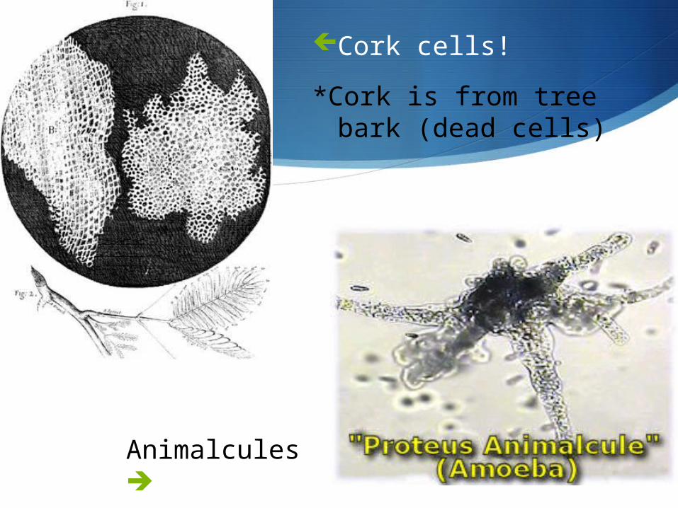
Cork cells!
*Cork is from tree bark (dead cells)
Animalcules

Who else discovered the cell?
ANTON VAN LEEUWENHOEK
Observed pond water
1st to observe “living” cells
Used a microscope at 300x magnification

Do you have these recipes at home?
People thought organisms grew from non-living materials!

Fransisco Redi Experiment
Placed meat in both an open container and a closed container to see what happened.
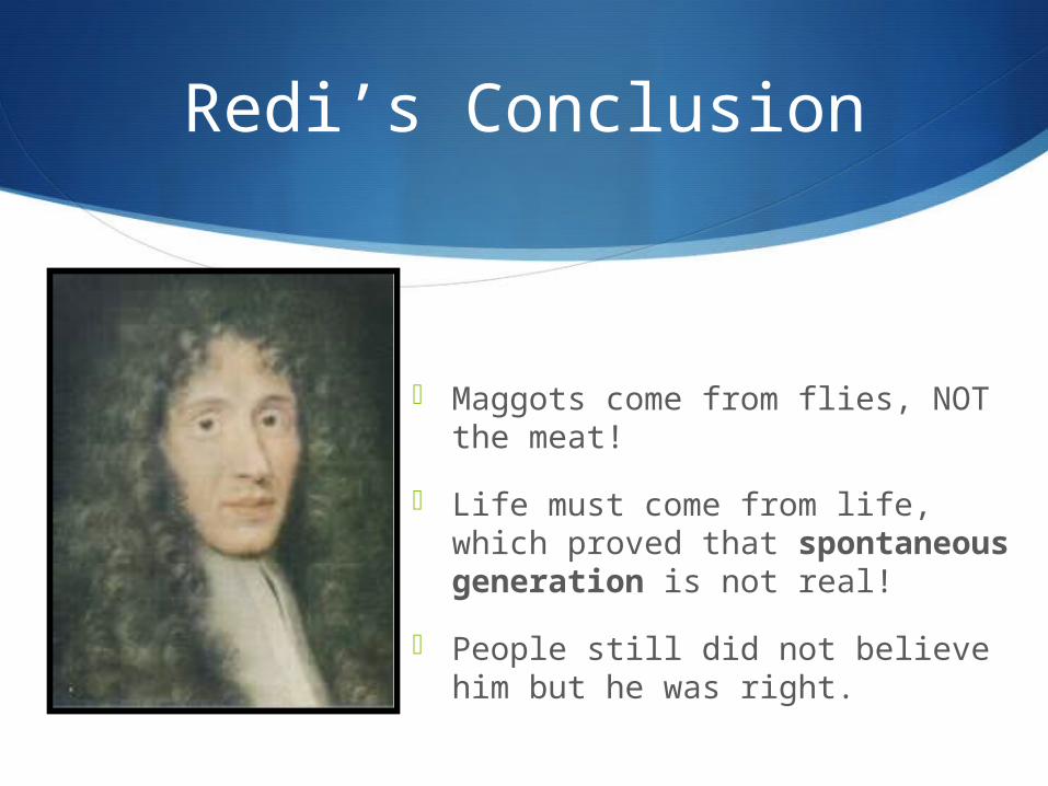
Redi’s Conclusion
Maggots come from flies, NOT the meat!
Life must come from life, which proved that spontaneous generation is not real!
People still did not believe him but he was right.
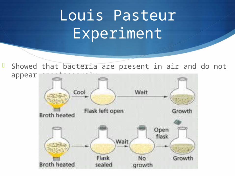
Louis Pasteur Experiment
Showed that bacteria are present in air and do not appear spontaneously.
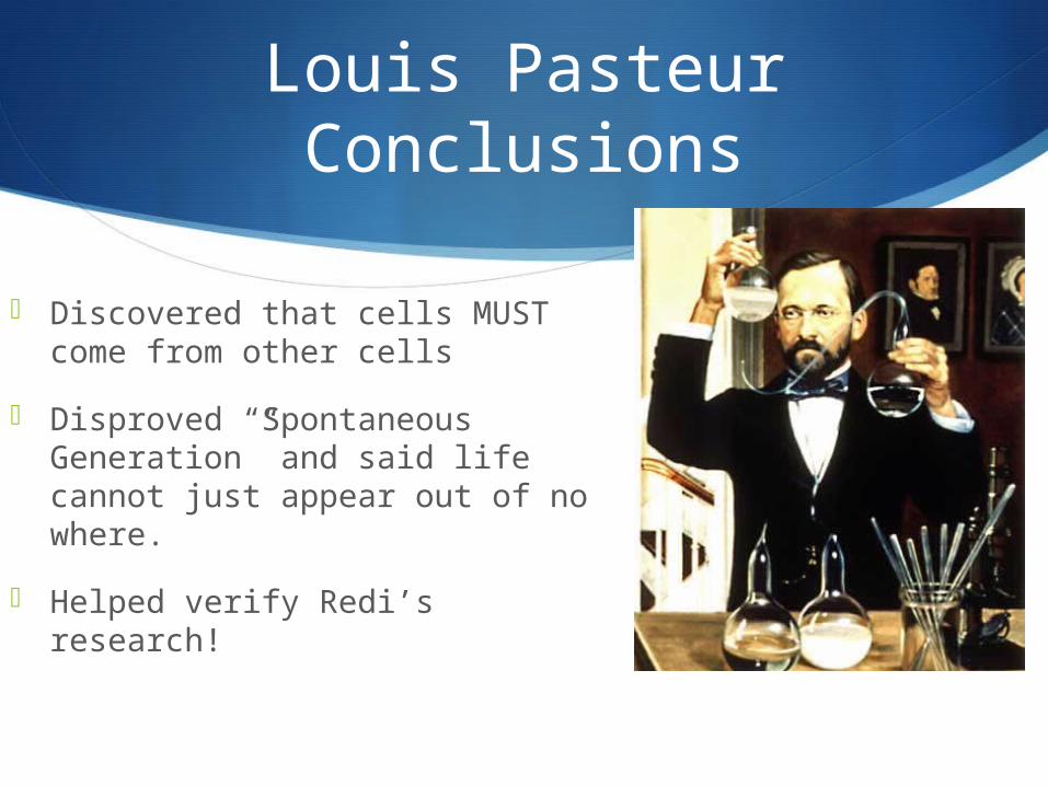
Louis Pasteur Conclusions
Discovered that cells MUST come from other cells
Disproved “Spontaneous Generation” and said life cannot just appear out of no where.
Helped verify Redi’s research!

Pasteurization
Pasteur came up with the idea of Pasteurization after discovering bacteria could contaminate milk from the air.
This process kills the bacteria so that it does not harm us!
Used in milk, cheese, yogurt, etc.
http://bcs.whfreeman.com/thelifewire/content/chp03/0302003.html

Cell Theory = Every living thing is made of one or
more cells Cells carry out the functions needed
to support life Cells come only from other living
cells
Do Now
What are the differences between prokaryotic and eukaryotic cells?
Why do we need so many more organelles than bacteria do? Explain.

Objectives
Compare and contrast a scanning electron microscope vs. a transmission electron microscope.
Describe prokaryotic and eukaryotic cells.
Section 1.2 - Microscopes
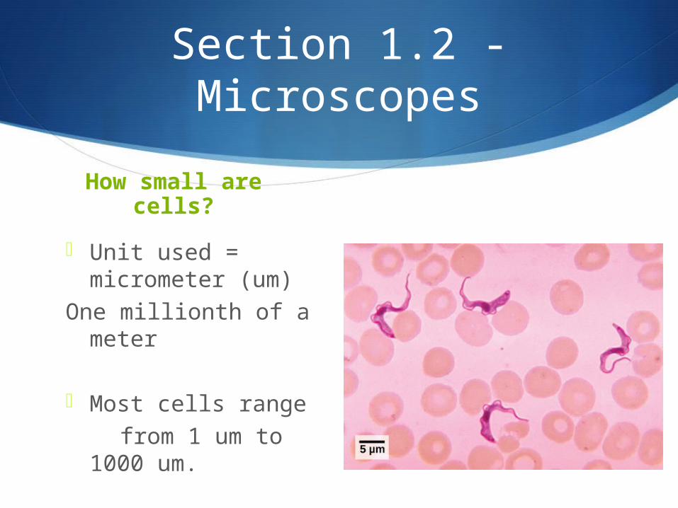
How small are cells?
Unit used = micrometer (um)
One millionth of a meter
Most cells range from 1 um to 1000
um.

Types of Microscopes
1. Light Microscope
2. SEM Microscope
3. TEM Microscope
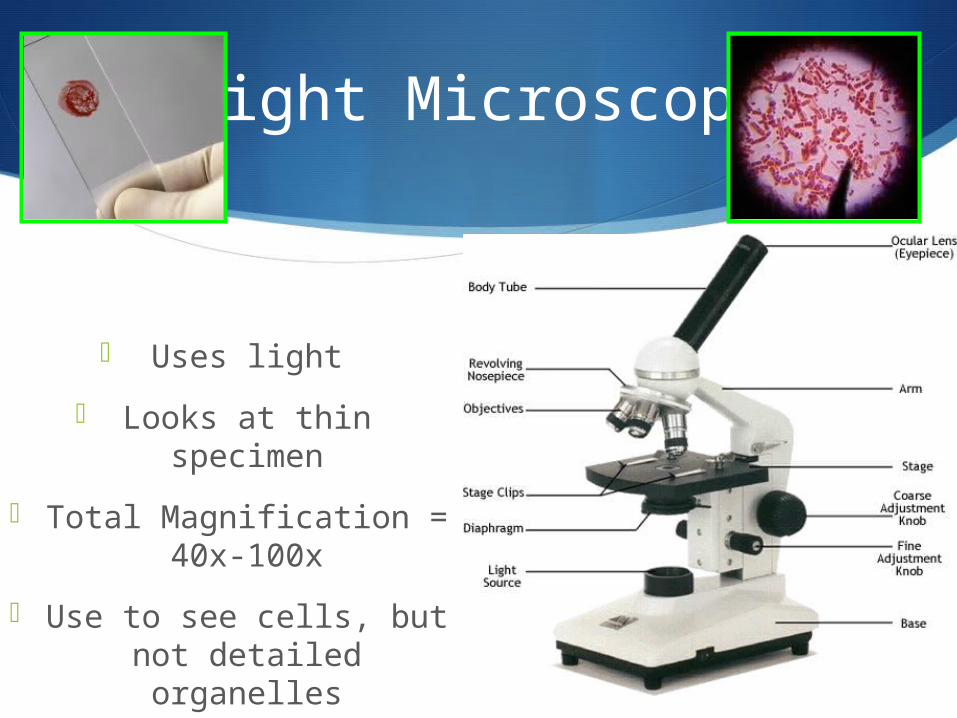
Light Microscope
Uses light
Looks at thin specimen
Total Magnification = 40x-100x
Use to see cells, but not detailed organelles

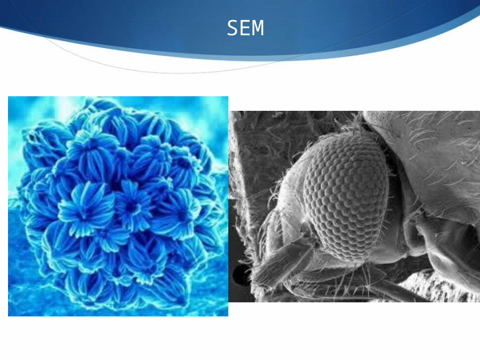
SEM vs. TEM
SEM “Scanning electron microscope”
Beams of electrons bounce of the surface of the coated cell.
Images appear 3D- Outside Specimen
Total Magnification = 100,000x
Must be dead . Specimen coated in metal
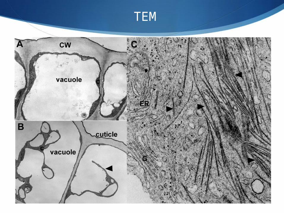
TEM “Transmission electron
microscope”
Electrons pass through the think section.
Images appear 2D
Total Magnification = 300,000x
Allows us to see organelles inside the cell
SEM
TEM

Prokaryotic vs. Eukaryotic cells
What are the differences you can see?
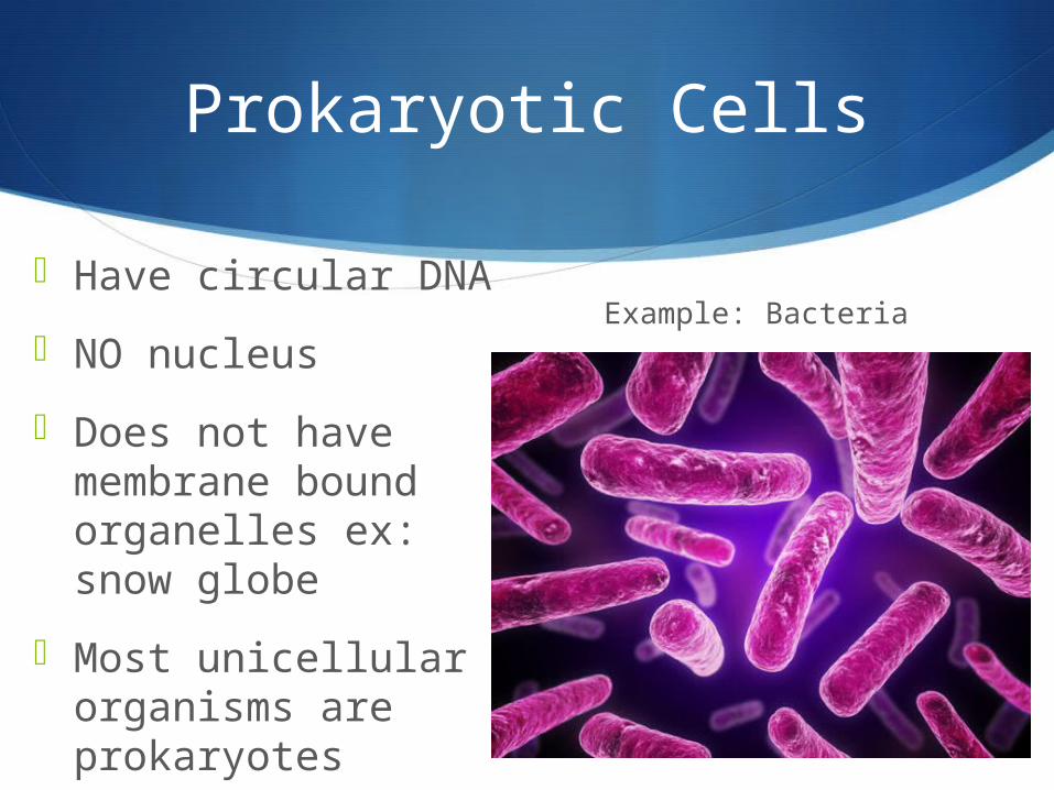
Prokaryotic Cells
Have circular DNA
NO nucleus
Does not have membrane bound organelles ex: snow globe
Most unicellular organisms are prokaryotes
Example: Bacteria

Eukaryotic Cells
Have linear DNA – double helix shape
Has nucleus
Have membrane bound organelles
Most multicellular organisms are eukaryotic cells. Some are unicellular though.
Example: You!
Do Now
What is found INSIDE a cell?
Do plant cells and animal cells have the same stuff inside?
What do plants need to do that animals do not?

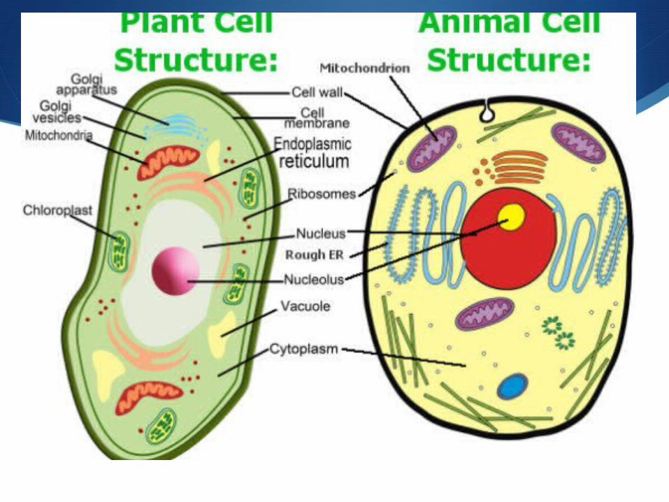
Objective
To compare and contrast animal vs. plant cells.
Identify each organelle in a diagram of a cell.
Explain the function of each organelle
To create flash cards for the next test
CELL ORGANELLES
Nucleus
Endoplasmic reticulum
Vesicle
Golgi apparatus All have specific
Lysosome functions!
Mitochondrion
Chloroplast
Central vacuole

THE WALL – protects the internal structures of the cell.
Selective permeability -Determines what comes in and out of the cell
A.K.A- Cell Membrane!
Plasma Membrane

Cytoplasm/Cytoskeleton
Cytoplasm- Clear FLUID that contains the organelles.
Cytoskeleton- Provides the FRAMEWORK for the cell, holds organelles in place.

Nucleus
Nucleus- CONTROLS the cell
Nucleolus- produces ribosomes
Nuclear Pores- Allows things in and out of the nucleus.
Challenge Question
Where do you find the DNA or a eukaryotic cell?
If prokaryotes do not have a nucleus, where is their DNA?

Ribosomes
Produces Proteins!
Proteins are made up of one or more polypeptide chains of amino acids.
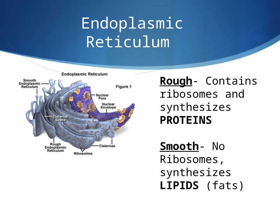
Endoplasmic Reticulum
Rough- Contains ribosomes and synthesizes PROTEINS
Smooth- No Ribosomes, synthesizes LIPIDS (fats)
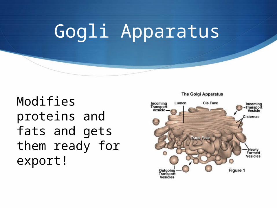
Gogli Apparatus
Modifies proteins and fats and gets them ready for export!

Lyosomes
Contains ENZYMES break down cellular waste product and debris

Centrioles
Involved in cell division! (will talk about this later when we do mitosis!)

Cilia and Flagella
Flagella- Used in cells for movement
Cilia- Used in stationary cells for moving substances around the outside of the cell.

Mitochondria
Convert oxygen into ENERGY (ATP)
(we will talk about this more when we do cellular respiration!!)

Central Vacuole
LARGE WATER “bubble” in the plant cell
Maintains the SHAPE of the cell, without it, the plant cell would shrink and the plant would wilt
Stores water, along with ions, nutrients, and waste.

Chloroplasts
Captures light ENERGY and convert it to chemical energy (sugar)
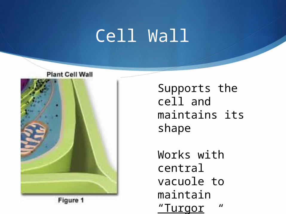
Cell Wall
Supports the cell and maintains its shape
Works with central vacuole to maintain “Turgor Pressure”
Challenge Question!
How can chloroplast, a structure found in plant cells but not in animal cells, provide energy for both plants and animals?
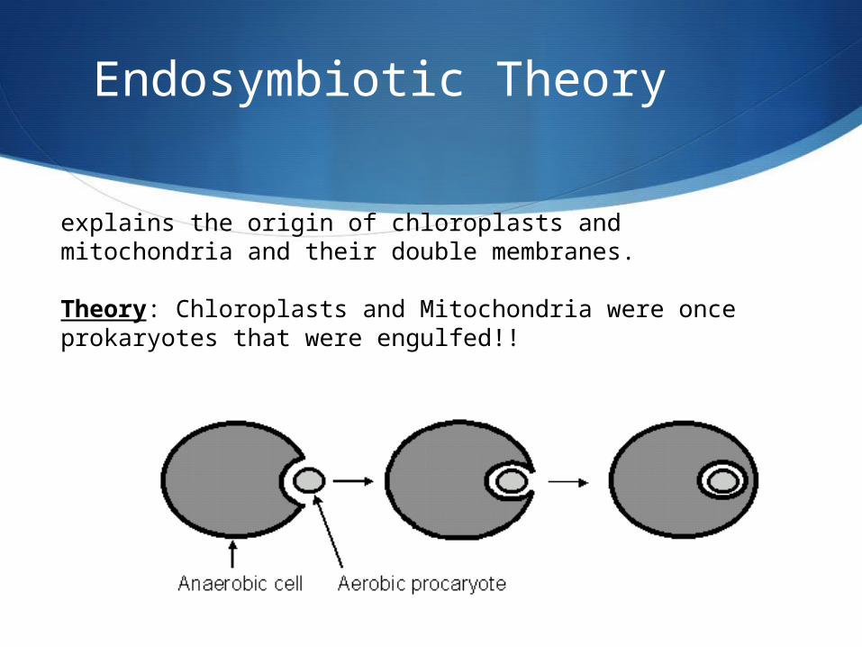
Endosymbiotic Theory
explains the origin of chloroplasts and mitochondria and their double membranes.
Theory: Chloroplasts and Mitochondria were once prokaryotes that were engulfed!!

Theory = Evidence!!
•Have circular DNA like bacteria•Replicates(reproduces) like bacteria separate from the host cell•Make their own proteins•Two membranes (one from the host cell and one from their own cell membrane)

Index Cards!
Front:
Name of Organelle
Drawing
Back:
Location
Function

Plant Both Animal
Do Now: Classify the organelles as plant, animal, or both

Quick Refresher!


Objectives
Compare and contrast the 3 Domains.
Understand multicellular organization.
Explain the significance of models in science.

Section 1.3 - Domains
3 domains of life: Eukarya – Have a
nucleus. Plants, animals, and fungi.
Bacteria – prokaryotics.
Archaea – “ancient”. Genetically different from bacteria.
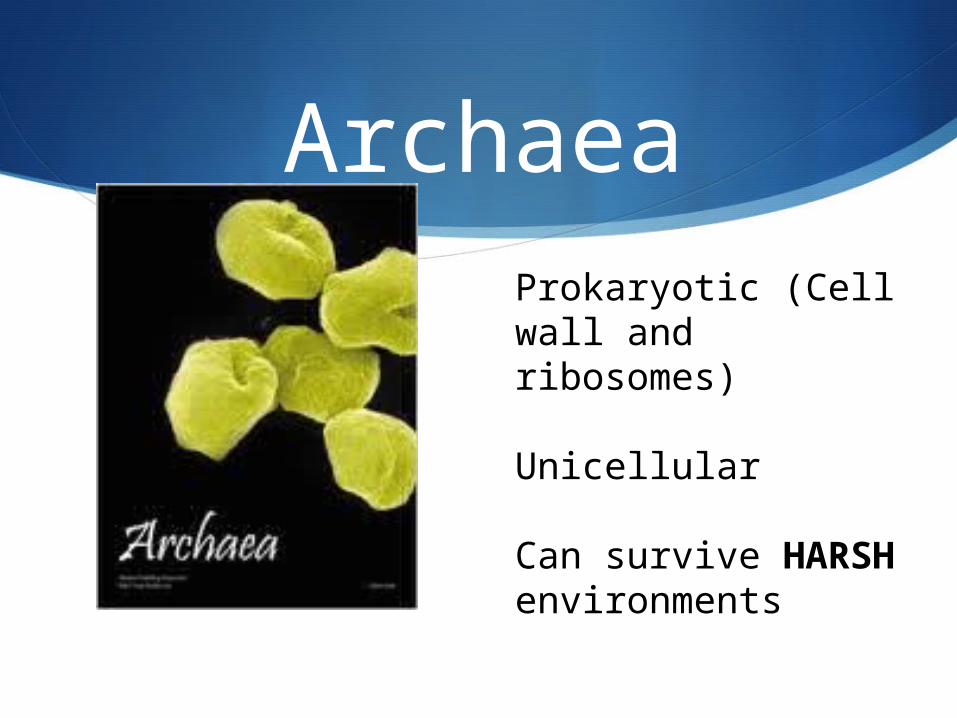
ArchaeaProkaryotic (Cell wall and ribosomes)
Unicellular
Can survive HARSH environments

Bacteria
Prokaryotic
Unicellular
NORMAL living environments
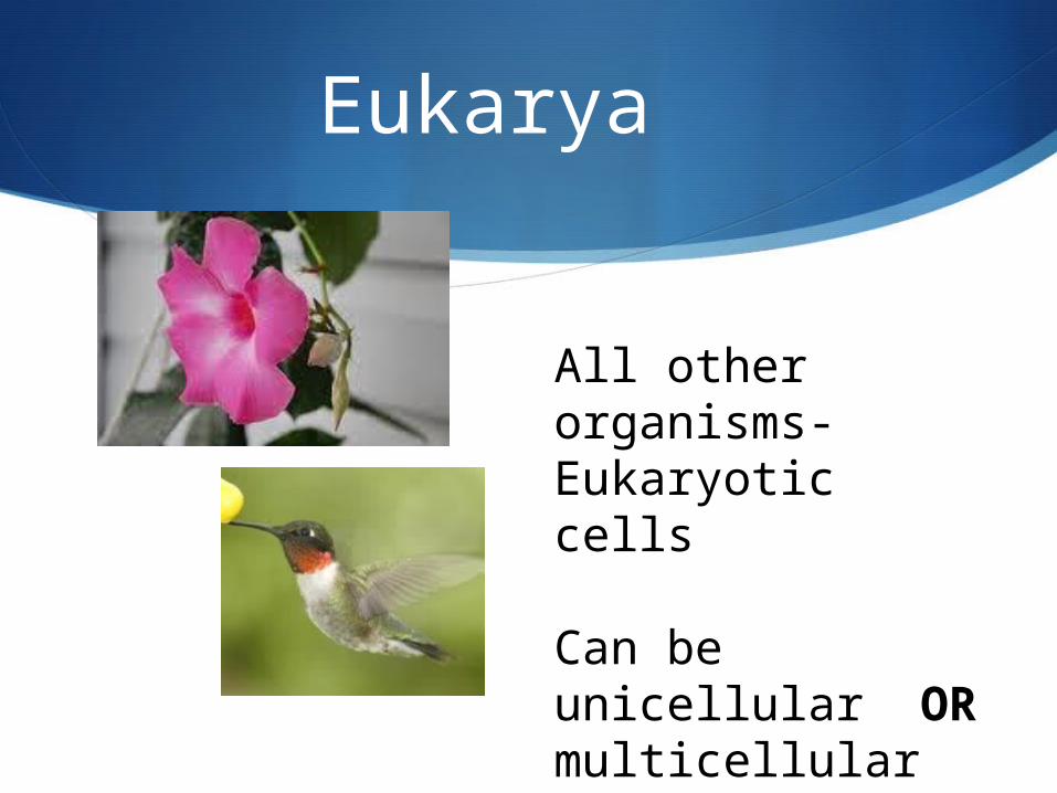
Eukarya
All other organisms- Eukaryotic cells
Can be unicellular OR multicellular
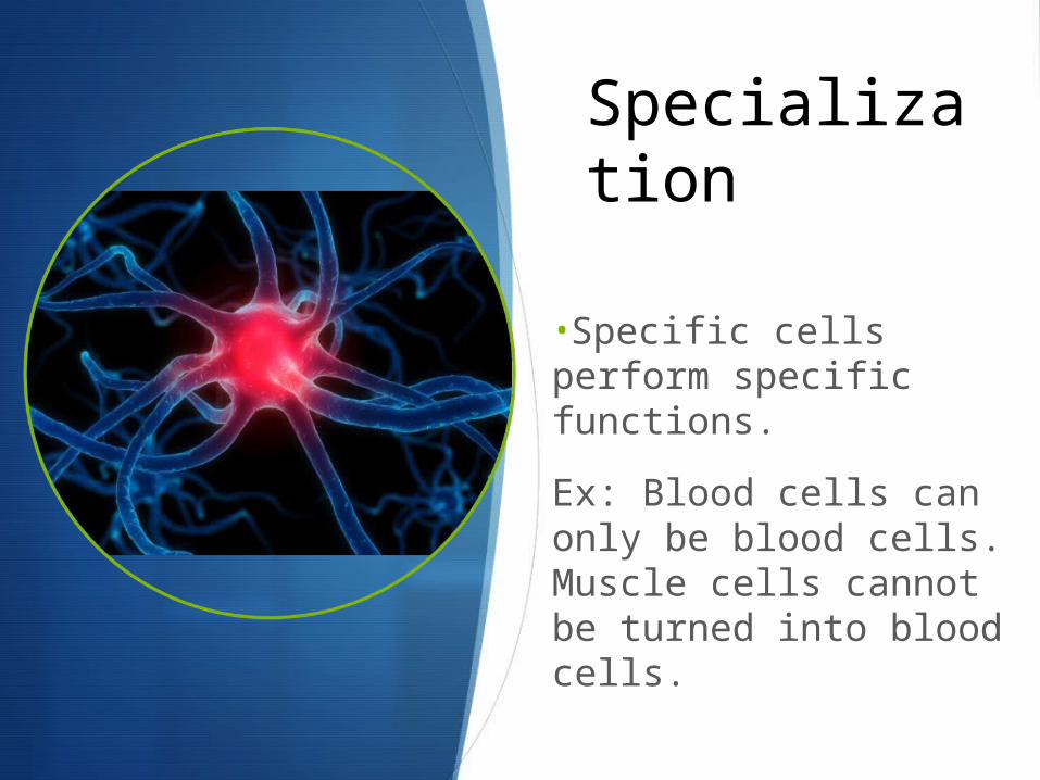
Specialization
•Specific cells perform specific functions.
Ex: Blood cells can only be blood cells. Muscle cells cannot be turned into blood cells.
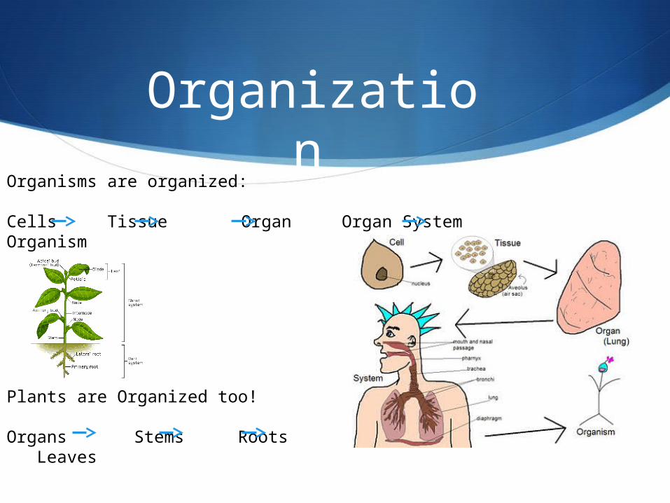
Organization
Organisms are organized:
Cells Tissue Organ Organ System Organism
Plants are Organized too!
Organs Stems Roots Leaves

Tissue and organs
Tissue – group of similar cells that are organized to do a specific job.
Organ – different tissues working together to perform a particular function.
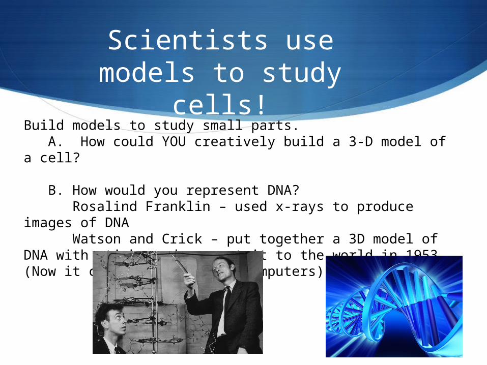
Scientists use models to study cells!
Build models to study small parts. A. How could YOU creatively build a 3-D model of a cell?
B. How would you represent DNA? Rosalind Franklin – used x-rays to produce images of DNAWatson and Crick – put together a 3D model of DNA with
sticks and present it to the world in 1953 (Now it could be done with computers)