DNA, RNA, & Protein Synthesis Discovery of DNA DNA Structure DNA Replication Protein Synthesis.
Upload
ayesha-arshadCategory
view
101download
0
DNA
DNAPresent in all cells and virtually restricted to the nucleusThe amount of DNA in somatic cells (body cells) of any
given species is constant (like the number of chromosomes)The DNA content of gametes (sex cells) is half that of
somatic cells. In cases of polyploidy (multiple sets of chromosomes) the
DNA content increases by a proportional factorThe mutagenic effect of UV light peaks at 253.7nm. The
peak for the absorption of UV light by DNA

Elucidation of DNA Structure: Basic chemical composition of nucleic acids was elucidated in the 1920s through the efforts of P. A. Levene. Levene mistakenly believed that DNA was a very small molecule, probably only four nucleotides long, composed of equal amounts of the four different nucleotides arranged in a fixed sequence.
invention of paper chromatography by Archer Martin and Richard Synge between 1941 and 1944. By 1948 the chemist Erwin Chargaff had begun using paper chromatography to analyze the base composition of DNA from a number of species. He soon found thatthe base composition of DNA from genetic material did indeed vary among species just as he expected. Furthermore, the total amount of purines always equaled the total amount of pyrimidines; and the adenine/thymine and guanine/ cytosine ratios were always 1.
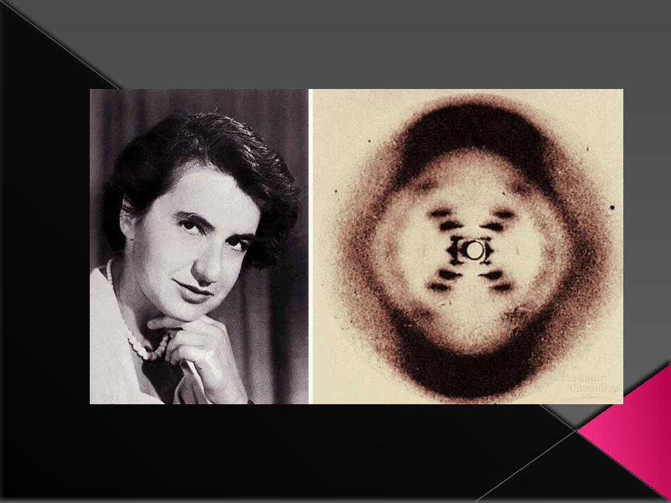
1951 when Rosalind Franklin arrived at King’s College, London, and joined Maurice Wilkins in his efforts to prepare highly oriented DNA fibers and study them by X-ray crystallography.

A diagrammatic representation of the double helix. The backbone consists of deoxyribose sugars (S) joined by phosphates (P) in phosphodiester bridges. The arrows at the top and bottom of the chains point in the 5′ to 3′ direction. The ribbons represent the sugar phosphate backbones.


Organization of prokaryotes DNA:
Figure 11.8 DNA Forms. (a) The DNA double helix of almost all bacteria is in the shape of a closed circle. (b) The circular DNA Figure 11.8 DNA Forms. (a) The DNA double helix of almost all bacteria is in the shape of a closed circle. (b) The circular DNA strands, already coiled in a double helix, are twisted a second time to produce super coils. strands, already coiled in a double helix, are twisted a second time to produce supercoils.

Figure 11.9 An illustration of how a string of nucleosomes, each associated with a histone H1, might be organized to form a highly supercoiled chromatin fiber. The nucleosomes are drawn as cylinders.
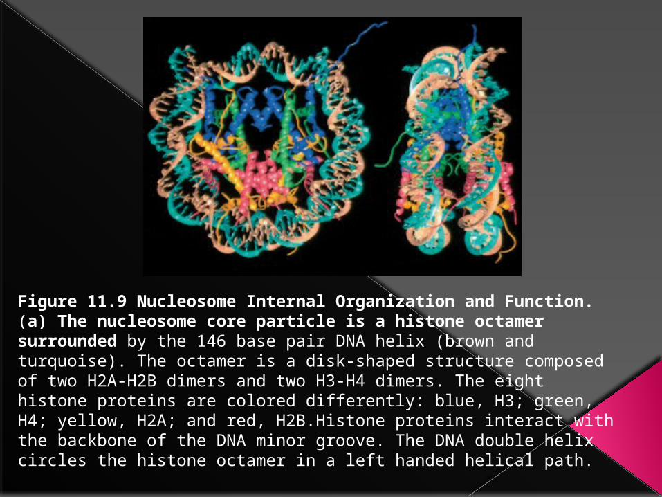
Figure 11.9 Nucleosome Internal Organization and Function.(a) The nucleosome core particle is a histone octamer surrounded by the 146 base pair DNA helix (brown and turquoise). The octamer is a disk-shaped structure composed of two H2A-H2B dimers and two H3-H4 dimers. The eight histone proteins are colored differently: blue, H3; green, H4; yellow, H2A; and red, H2B.Histone proteins interact with the backbone of the DNA minor groove. The DNA double helix circles the histone octamer in a left handed helical path.


DNA Replication:Watson and Crick published their description of DNA structure in April 1953. Almost exactly one month later, a second paper appeared in which they suggested how DNA might be replicated. They hypothesized that the two strands of the double helix unwind from one another and separate. Free nucleotides now line up along the two parental strands through complementary base pairing—A with T, G with C. When these nucleotides are linked together by one or more enzymes, two replicas result, each containing a parental DNA strand and a newly formed strand. Research in subsequent years has proved Watson and Crick’s hypothesis correct.
Replication patterns are somewhat different in procaryotes and eucaryotes. For example, when the circular DNA chromosome of E. coli is copied, replication begins at a single point, the origin. Synthesis occurs at the replication fork, the place at which the DNA helix is unwound and individual strands are replicated. Two replication forks move outward from the origin until they have copied the whole replicon, that portion of the genome that contains an origin and is replicated as a unit. When the replication forks move around the circle, a structure shaped like the Greek letter theta () is formed Finally, since the bacterial chromosome is a single replicon, the forks meet on the other side and two separate chromosomes are released.


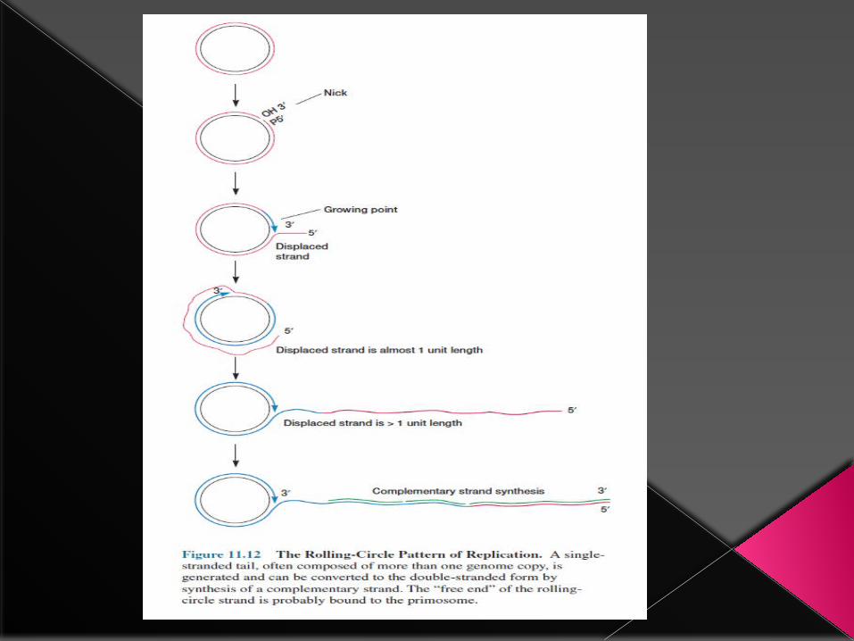
A different pattern of DNA replication occurs during E. coli conjugation and the reproduction of viruses, such as phage lambda).
In the rolling-circle mechanism, one strand is nicked and the free 3′- hydroxyl end is extended by replication enzymes. As the 3′ end is lengthened while the growing point rolls around the circular template, the 5′ end of the strand is displaced and forms an everlengthening tail.
The single-stranded tail may be converted to the double-stranded form by complementary strand synthesis. This mechanism is particularly useful to viruses because it allows the rapid, continuous production of many genome copies from a single initiation event.
Eucaryotic DNA is linear and much longer than procaryotic DNA; E. coli DNA is about 1,300 um in length, whereas the 46 chromosomes in the human nucleus have a total length of 1.8 m (almost 1,400 times longer).
Clearly many replication forks must copy eucaryotic DNA simultaneously so that the molecule can be duplicated in a relatively short period, and so many replicons are present that there is an origin about every 10 to 100 m along the DNA. Replication forks move outward from these sites and eventually meet forks that have been copying the adjacent DNA stretch. In this fashion a large molecule is copied quickly.


DNA replication is initiated at the oriC locus. The DnaA protein binds to oriC while hydrolyzing ATP. This leads to the initial unwinding of double-stranded DNA at the initiation site. Further unwinding occurs through the activity of
the DnaB protein, a helicase .E. coli has three different DNA polymerase enzymes, each of which catalyzes
the synthesis of DNA in the 5′ to 3′ direction while reading the DNA template in the 3′ to 5′ direction.
The polymerases require deoxyribonucleoside triphosphates (dATP, dGTP, dCTP, and dTTP) as substrates and a DNA template to copy. Nucleotides are added to the 3′ end of the growing chain when the free 3′-hydroxyl group on
the deoxyribose attacks the first or alpha phosphate group of the substrate to release pyrophosphate.
Unwinding occurs very quickly; the fork may rotate as rapidly as 75 to 100 revolutions per second. Helicases are responsible for DNA unwinding. These enzymes use energy from ATP to unwind short stretches of helix just ahead
of the replication fork.

The details of dna replication are outlined in a diagram of the replication fork . the replication process takes place in four stages.
1. Helicases unwind the helix with the aid of topoisomerases like the DNA gyrase . it appears that the DNA b protein is the helicase most actively
involved in replication, but the n′ protein also may participate in unwinding. the single strands are kept separate by the DNA binding
proteins (ssbs).
2. DNA is probably replicated continuously by DNA polymeraIII when the leading strand is copied. lagging strand replication is discontinuous,
and the fragments are synthesized in the 5′ to 3′ direction just as in leading strand synthesis. first, a special RNA polymerase called a primase
synthesizes a short RNA primer, usually around 10 nucleotides long, complementary to the DNA . It appears that the primase requires the
assistance of several other proteins, and the complex of the primase with its accessory proteins is called the primosome. DNA polymerase III
holoenzyme then synthesizes complementary DNA beginning at the 3′ end of the RNA primer.

DNA polymerase III holoenzyme then synthesizes complementary DNA beginning at the 3′ end of the RNA primer. Both leading and lagging strand synthesis probably
occur concurrently on a single multiprotein complex with two catalytic sites, the replisome. If this is the case, the lagging strand template must be looped around the complex. The final fragments are around 1,000 to 2,000 nucleotides long in bacteria and approximately 100 nucleotides long in eucaryotic cells. They are called okazaki
fragments after their discoverer, reiji okazaki.
3.After most of the lagging strand has been duplicated by the formation of okazaki fragments, DNA polymerase I or RNase H removes the RNA primer. Polymerase I
synthesizes complementary DNA to fill the gap resulting from RNA deletion. the polymerase appears to remove one primer nucleotide at a time and replace it with the
appropriate complementary deoxyribonucleotide. Polymerase III holoenzyme also may be able to fill in the gap.
4. Finally, the fragments are joined by the enzyme DNA ligase, which forms a phosphodiester bond between the 3′- hydroxyl of the growing strand and the 5′-
phosphate of an okazaki fragment . Bacterial ligases use the pyrophosphate bond of NAD as an energy source; many other ligases employ ATP.