DNA Restriction Analysis. DNA is Tightly Packaged into Chromosome s Which Reside in the Nucleus.
19
DNA Restriction Analysis
-
Upload
lucas-webb -
Category
Documents
-
view
219 -
download
0
Transcript of DNA Restriction Analysis. DNA is Tightly Packaged into Chromosome s Which Reside in the Nucleus.
DNA Restriction Analysis

DNA is Tightly Packaged into Chromosomes Which Reside in the Nucleus
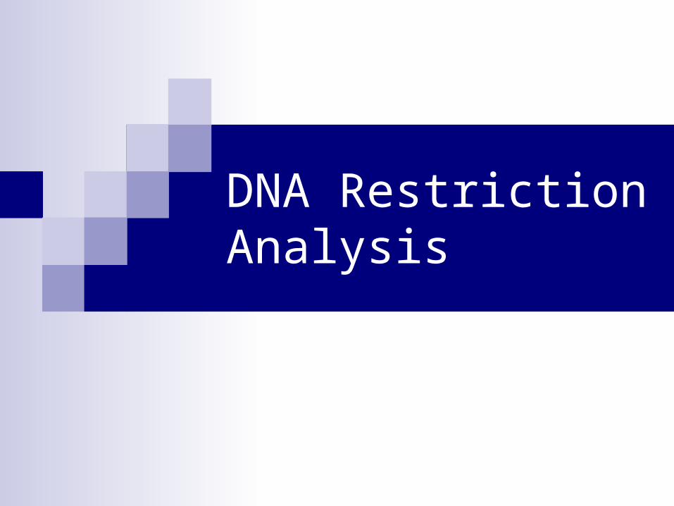
Model of DNA
DNA is Comprised of Four Base Pairs

5’ 3’
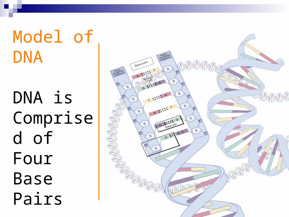
Now, if all the elephants were the same, this would be a regular polymer.
In DNA, one kind of SUPER POLYMER, there are four kind of elephants with the names: A, C, G, and T
A C G T
Note the backbone is the same for each one
5’ 3’

Deoxyribonucleic Acid (DNA)

DNA Schematic
OCH2
O
P O
O
O Base
CH2
O
P
O
O
O
Base
OH
Sugar
Sugar
O
5’ phosphate
3’ hydroxyl

5’ 3’
Now, if all the elephants were the same, this would be a regular polymer.
In DNA, one kind of SUPER POLYMER, there are four kind of elephants with the names: A, C, G, and T
A C G T
Note the backbone is the same for each one
5’ 3’

DNA Restriction Enzymes
• Evolved by bacteria to protect against viral DNA infection
• Endonucleases = cleave within DNA strands
• Over 3,000 known enzymes
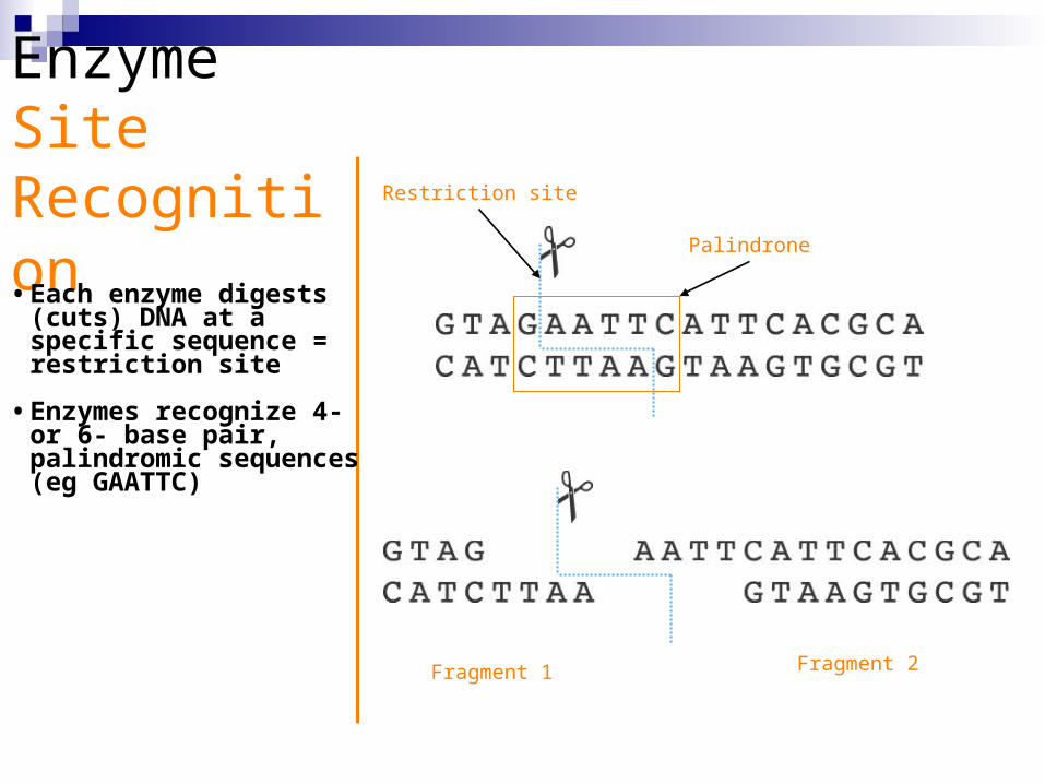
Enzyme Site Recognition • Each enzyme digests
(cuts) DNA at a specific sequence = restriction site
• Enzymes recognize 4- or 6- base pair, palindromic sequences (eg GAATTC)
Palindrone
Restriction site
Fragment 1 Fragment 2
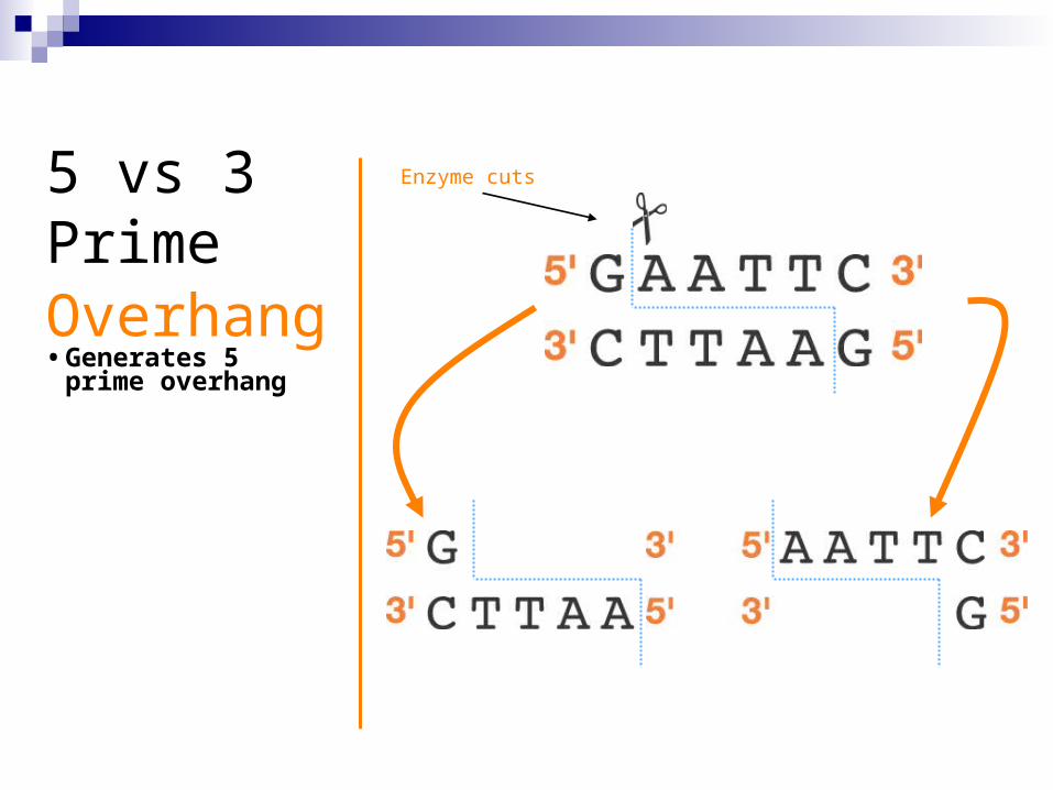
5 vs 3 Prime Overhang • Generates 5 prime
overhang
Enzyme cuts

Common Restriction Enzymes
EcoRI– Eschericha coli– 5 prime overhang
Pstl– Providencia stuartii– 3 prime overhang

The DNA DigestionReaction
Restriction Buffer provides optimal conditions
NaCI provides the correct ionic strength
Tris-HCI provides the proper pH
Mg2+ is an enzyme co-factor

DNA DigestionTemperature
Why incubate at 37°C?
• Body temperature is optimal for these and most other enzymes
What happens if the temperature is too hot or cool?
• Too hot = enzyme may be denatured (killed)
• Too cool = enzyme activity lowered, requiring
longer digestion time

AgaroseElectrophoresisLoading
• Electrical current carries negatively-charged DNA through gel towards positive (red) electrode
Power Supply
Buffer
Dyes
Agarose gel
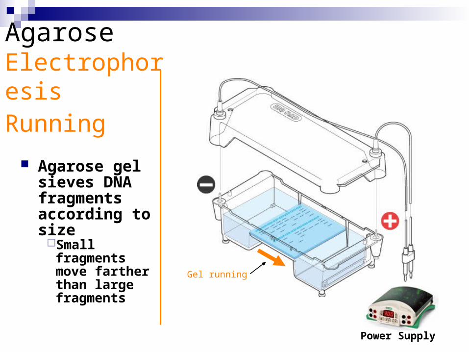
AgaroseElectrophoresisRunning
Agarose gel sieves DNA fragments according to size
Small fragments move farther than large fragments
Power Supply
Gel running
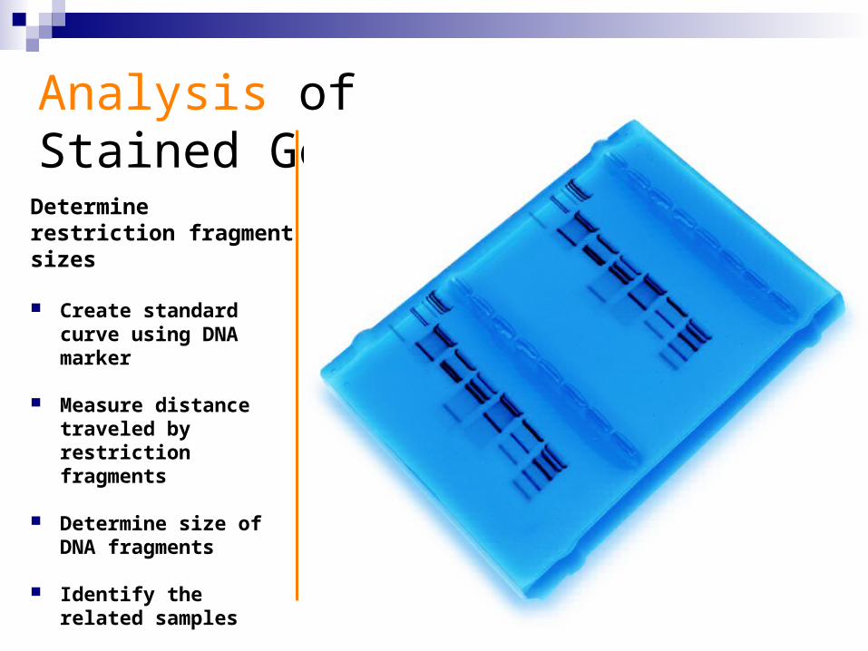
Analysis of Stained Gel
Determinerestriction fragmentsizes
Create standard curve using DNA marker
Measure distance traveled by restriction fragments
Determine size of DNA fragments
Identify the related samples

Molecular Weight Determination
Size (bp) Distance (mm)
23,000 11.0 9,400 13.0
6,500 15.0
4,400 18.0
2,300 23.0
2,000 24.0100
1,000
10,000
100,000
0 5 10 15 20 25 30
Distance, mm
Siz
e, b
ase
pai
rsB
A
Fingerprinting Standard Curve: Semi-log

NEB Cutter Predictions
New England Biolabs Website to paste sequence and predict
restriction enzyme sites and run a virtual gel to see what it would look like
NEB Cutter