DNA-PK Inhibition by NU7441 Enhances Chemosensitivity to ... · 10 M. Yanai et al. radioresistance...
7
9 Yonago Acta Medica 2017;60:09–15 Original Article Corresponding author: Haruhiko Makino, MD, PhD [email protected] Received 2016 December 1 Accepted 2016 December 21 Abbreviations: 53BP1, p53 binding protein 1; ALK, anaplastic lymphoma kinase; AMR, amrubicin; CCK-8, Cell Counting Kit- 8; CI, combination index; CPT-11, irinotecan; DAPI, 4,6-diamid- ino-2-phenylindole; DNA-PK, DNA-dependent protein kinase; DNA-PKcs, DNA-dependent protein kinase catalytic subunit; DSB, double-strand break; EGFR, epidermal growth factor recep- tor; IC50, 50% inhibition of cell growth; NHEJ, non-homologous end-joining; NSCLC, non-small cell lung carcinoma; PTX, pacri- taxel DNA-PK Inhibition by NU7441 Enhances Chemosensitivity to Topoisomerase Inhibitor in Non-Small Cell Lung Carcinoma Cells by Blocking DNA Damage Repair Masaaki Yanai, Haruhiko Makino, Bingqiong Ping, Kenichi Takeda, Natsumi Tanaka, Tomohiro Sakamoto, Kosuke Yamaguchi, Masahiro Kodani, Akira Yamasaki, Tadashi Igishi and Eiji Shimizu Division of Medical Oncology and Molecular Respirology, Department of Multidisciplinary Internal Medicine, School of Medicine, Tot- tori University Faculty of Medicine, Yonago 683-8504, Japan ABSTRACT Background DNA double-strand breaks (DSBs) are the most cytotoxic form of DNA damage and are in- duced by ionizing radiation and specific chemotherapeu- tic agents, such as topoisomerase inhibitors. Cancer cells acquire resistance to such therapies by repairing DNA DSBs. A major pathway for the repair of DNA DSBs is non-homologous end-joining (NHEJ), which requires DNA-dependent protein kinase (DNA-PK) activity. In this study, we investigated the effect of NU7441, a syn- thetic small-molecule compound, as a specific inhibitor of DNA-PK on the chemosensitization of non-small cell lung carcinoma (NSCLC) A549 cells. Methods The combined effects of chemotherapeutic agents and NU7441 were evaluated by isobologram analysis using Cell Counting Kit-8. DNA DSBs were assessed by immunofluorescence assay. Apoptosis was examined by flow cytometry using an Annexin V apop- tosis kit. Activation of DNA-PK was assayed by western blotting. Results The combination of NU7441 and topoisom- erase inhibitors such as amrubicin and irinotecan had a synergistic effect on cell proliferation in A549 cells. NU7441 increased 53BP1 foci and apoptosis induced by topoisomerase inhibitors and decreased phos- pho-DNA-dependent protein kinase, catalytic subunit (pDNA-PKcs) (S2056) protein expression caused by topoisomerase inhibitors. Interestingly, mitotic inhibitors such as pacritaxel did not cause the pDNA-PKcs (S2056) protein expression and the combination of NU7441 and pacritaxel had an only additive effect. Conclusion NU7441 inhibited the growth of NSCLC cells and enhanced the chemosensitization to topoisom- erase inhibitors by blocking DNA repair. A combination of NU7441 and topoisomerase inhibitor may be a prom- ising treatment for NSCLC. Key words DNA-dependent protein kinase; non-ho- mologous end-joining; non-small cell lung carcinoma; NU7441; topoisomerase inhibitor Non-small cell lung carcinoma (NSCLC) is one of the most commonly diagnosed cancers and the leading cause of cancer-related death worldwide. Over half of patients with NSCLC have been advanced or metastatic lesions at the time of diagnosis. 1 Systemic chemotherapy is the standard treatment for patients with advanced NS- CLC. In many countries, including Japan, cytotoxic che- motherapeutic agents are used as the first-line treatment for advanced NSCLC patients. However, these cytotoxic chemotherapies have reached a therapeutic plateau and have limited survival benefits for advanced NSCLC pa- tients. In recent years, treatment with targeted agents has markedly improved progression-free and overall survival in patients with advanced NSCLC harboring EGFR mu- tation or ALK rearrangement. 2, 3 However, only 20–30% of NSCLC patients harbor targets available for molecu- lar targeting agents and most acquire resistance against molecular target agents within 1 year. Therefore, new agents and new combination chemotherapy regimens are required to improve the outcomes of all advanced NS- CLC patients. DNA-dependent protein kinase, catalytic subunit (DNA-PKcs) is an essential component in the non-ho- mologous end-joining (NHEJ) pathway of DNA dou- ble-strand break (DSB) repair. 4 In response to DNA DSBs, DNA-PKcs is rapidly recruited to damage sites by the Ku70/Ku80 heterodimer and phosphorylated at Thr2609 and Ser2056 clusters by ATM and itself, re- spectively. 5, 6 The recruitment and phosphorylation of DNA-PKcs contributes to the processing and direct liga- tion of broken DNA ends. Several studies demonstrated increased protein levels of DNA-PKcs in NSCLC, 7–9 and the elevation of DNA-PKcs has been correlated with
Transcript of DNA-PK Inhibition by NU7441 Enhances Chemosensitivity to ... · 10 M. Yanai et al. radioresistance...

9
Yonago Acta Medica 2017;60:09–15 Original Article
Corresponding author: Haruhiko Makino, MD, [email protected] 2016 December 1Accepted 2016 December 21Abbreviations: 53BP1, p53 binding protein 1; ALK, anaplastic lymphoma kinase; AMR, amrubicin; CCK-8, Cell Counting Kit-8; CI, combination index; CPT-11, irinotecan; DAPI, 4,6-diamid-ino-2-phenylindole; DNA-PK, DNA-dependent protein kinase; DNA-PKcs, DNA-dependent protein kinase catalytic subunit; DSB, double-strand break; EGFR, epidermal growth factor recep-tor; IC50, 50% inhibition of cell growth; NHEJ, non-homologous end-joining; NSCLC, non-small cell lung carcinoma; PTX, pacri-taxel
DNA-PK Inhibition by NU7441 Enhances Chemosensitivity to Topoisomerase Inhibitor in Non-Small Cell Lung Carcinoma Cells by Blocking DNA Damage Repair
Masaaki Yanai, Haruhiko Makino, Bingqiong Ping, Kenichi Takeda, Natsumi Tanaka, Tomohiro Sakamoto, Kosuke Yamaguchi, Masahiro Kodani, Akira Yamasaki, Tadashi Igishi and Eiji ShimizuDivision of Medical Oncology and Molecular Respirology, Department of Multidisciplinary Internal Medicine, School of Medicine, Tot-tori University Faculty of Medicine, Yonago 683-8504, Japan
ABSTRACTBackground DNA double-strand breaks (DSBs) are the most cytotoxic form of DNA damage and are in-duced by ionizing radiation and specific chemotherapeu-tic agents, such as topoisomerase inhibitors. Cancer cells acquire resistance to such therapies by repairing DNA DSBs. A major pathway for the repair of DNA DSBs is non-homologous end-joining (NHEJ), which requires DNA-dependent protein kinase (DNA-PK) activity. In this study, we investigated the effect of NU7441, a syn-thetic small-molecule compound, as a specific inhibitor of DNA-PK on the chemosensitization of non-small cell lung carcinoma (NSCLC) A549 cells.Methods The combined effects of chemotherapeutic agents and NU7441 were evaluated by isobologram analysis using Cell Counting Kit-8. DNA DSBs were assessed by immunofluorescence assay. Apoptosis was examined by flow cytometry using an Annexin V apop-tosis kit. Activation of DNA-PK was assayed by western blotting.Results The combination of NU7441 and topoisom-erase inhibitors such as amrubicin and irinotecan had a synergistic effect on cell proliferation in A549 cells. NU7441 increased 53BP1 foci and apoptosis induced by topoisomerase inhibitors and decreased phos-pho-DNA-dependent protein kinase, catalytic subunit (pDNA-PKcs) (S2056) protein expression caused by topoisomerase inhibitors. Interestingly, mitotic inhibitors such as pacritaxel did not cause the pDNA-PKcs (S2056) protein expression and the combination of NU7441 and pacritaxel had an only additive effect.Conclusion NU7441 inhibited the growth of NSCLC cells and enhanced the chemosensitization to topoisom-erase inhibitors by blocking DNA repair. A combination of NU7441 and topoisomerase inhibitor may be a prom-ising treatment for NSCLC.
Key words DNA-dependent protein kinase; non-ho-mologous end-joining; non-small cell lung carcinoma; NU7441; topoisomerase inhibitor
Non-small cell lung carcinoma (NSCLC) is one of the most commonly diagnosed cancers and the leading
cause of cancer-related death worldwide. Over half of patients with NSCLC have been advanced or metastatic lesions at the time of diagnosis.1 Systemic chemotherapy is the standard treatment for patients with advanced NS-CLC. In many countries, including Japan, cytotoxic che-motherapeutic agents are used as the first-line treatment for advanced NSCLC patients. However, these cytotoxic chemotherapies have reached a therapeutic plateau and have limited survival benefits for advanced NSCLC pa-tients. In recent years, treatment with targeted agents has markedly improved progression-free and overall survival in patients with advanced NSCLC harboring EGFR mu-tation or ALK rearrangement.2, 3 However, only 20–30% of NSCLC patients harbor targets available for molecu-lar targeting agents and most acquire resistance against molecular target agents within 1 year. Therefore, new agents and new combination chemotherapy regimens are required to improve the outcomes of all advanced NS-CLC patients. DNA-dependent protein kinase, catalytic subunit (DNA-PKcs) is an essential component in the non-ho-mologous end-joining (NHEJ) pathway of DNA dou-ble-strand break (DSB) repair.4 In response to DNA DSBs, DNA-PKcs is rapidly recruited to damage sites by the Ku70/Ku80 heterodimer and phosphorylated at Thr2609 and Ser2056 clusters by ATM and itself, re-spectively.5, 6 The recruitment and phosphorylation of DNA-PKcs contributes to the processing and direct liga-tion of broken DNA ends. Several studies demonstrated increased protein levels of DNA-PKcs in NSCLC,7–9 and the elevation of DNA-PKcs has been correlated with
10
M. Yanai et al.
radioresistance in some advanced stage cancers.9 There-fore, targeting of DNA-PKcs is an attractive approach for enhancing radio-sensitivity in NSCLC. Given its im-portant role in the NHEJ repair pathway, different anti-DNA-PKcs strategies have been explored to enhance the sensitivity to radiation or DNA damage-based agents. In a series of small-molecule ATP-competitive inhibitors of DNA-PKcs, NU7441 was found to be the most potent and specific inhibitor of DNA-PKcs at a low concentra-tion relative to other members of the PI3KK family (ATM and ATR).10 Several studies have shown that NU7441 can enhance the sensitivity to radiation in NSCLC.11 However, little is known about the potency of NU7441 in enhancing the sensitivity of cytotoxic chemotherapeu-tic agents. Among cytotoxic anti-cancer drugs, topoisomer-ase inhibitors target DNA topoisomerases, which are essential nuclear enzymes that catalyze the breakage and rejoining of DNA. There are two classes of DNA topoisomerases, type I and type II, which alter the topol-ogy of single- and double-stranded DNA, respectively, and are involved in genetic reactions including DNA replication, transcription, and DNA repair.12 DNA to-poisomerase inhibitors, such as irinotecan (CPT-11; topo I inhibitor) and amrubicin (AMR, topo II inhibitor), are important in lung cancer chemotherapy.13, 14
In the present study, we evaluated the effect of combining topoisomerase inhibitors, which cause DNA damage, with DNA-PKcs inhibitors, which inhibit DNA-DSB repair by blocking the NHEJ pathway, on NSCLC.
MATERIALS AND METHODSChemicals and reagentsAMR (Dainippon Sumitomo Pharma, Tokyo, Japan), CPT-11 (Daiichi Sankyo Pharma, Tokyo, Japan), and pa-clitaxel (PTX) (Bristol-Myers-Squibb, New York, NY) were dissolved in distilled water and stored at –20 °C. NU7441 (AdooQ Bioscience, Irvine, CA) was dissolved in dimethyl sulfoxide and stored at –20 °C.
CellsThe lung adenocarcinoma A549 cell line was purchased from the American Type Culture Collection (ATCC, Manassas, VA). A549 cells were maintained in RPMI-1640 medium supplemented with 10% fetal bovine se-rum and antibiotics. These cells were grown in a humid-ified atmosphere of 5% CO2 and 95% air at 37 °C.
Cell Counting Kit-8 (CCK-8) assayA CCK-8 assay was performed to evaluate cell prolifer-ation inhibition. Cells (1000 per well) were cultured in 96-well flat-bottom multi-plates (Nalge Nunc, Penfield,
NY) for 48 h. Next, the indicated drugs were added to the culture media. After 72 h of drug treatment, 10% CCK-8 solution (Dojindo, Kumamoto, Japan) was added to each well and incubation was continued for an addi-tional 3 h. The absorbance of each well was measured at a wavelength of 450 nm using a Sunrise scanning multi-well spectrometer (Tecan, Männedorf, Switzerland). Each experiment was performed in triplicate for each drug concentration.
Assessment of combination effect To qualitatively assess the combination effect of the indi-cated agents, isobologram analysis was conducted as de-scribed previously.15 The percentage of cell proliferation was calculated as: [(mean absorbance of drug-treated wells – mean absorbance of cell-free wells)/(mean ab-sorbance of vehicle cells – mean absorbance of cellfree wells)] × 100. We used the concentration resulting in 50% inhibition of cell growth (IC50) to evaluate dose-re-sponse interactions.
Apoptosis assayApoptosis of A549 cells was detected by the Annexin V Apoptosis Detection Kit APC (eBioscience, San Diego, CA). A549 cells were treated with or without NU7441 and with anticancer agents for 24 and 48 h. The cells were harvested by trypsinization, collected in centri-fuge tubes, and washed with ice-cold phosphate-buff-ered saline. Next, the cells were suspended in 500 μL of binding buffer and incubated with Annexin V-APC and propidium iodide or 4,6-diamidino-2-phenylindole (DAPI). The apoptotic population was assayed by flow cytometry (LSRFortessa X-20; BD Biosciences, Frank-lin Lakes, NJ) and analyzed using FlowJo software (Treestar, San Carlos, CA).
Immunofluorescence stainingDNA-DSBs were assessed by quantifying the rates of dissolution of p53 binding protein 1 (53BP1) foci as de-scribed. Immunofluorescence staining was performed in cells cultured in glass chamber slides (Lab-Tek II Chamber Slide w/Cover RS Glass Slide Sterile; Nalge Nunc). A549 cells were treated with or without NU7441, and then treated with each anticancer agent for 1 h. At the indicated times, cells were fixed with 4% PFA, per-meabilized with 0.5% Triton X-100, and immunostained with 53BP1 primary antibody (diluted 1:1000; Cell Signaling Technologies, Beverly, MA) and Alexa Fluor 488 goat anti-rabbit secondary antibody (diluted 1:2000; Life Technologies, Carlsbad, CA). The nuclei of cells were stained using blue fluorescent DAPI in Vectashield mounting medium (Vector Laboratories, Burlingame,
11
Synergism of NU7441 and topoisomerase inhibitors in NSCLC
CA). Images were obtained with a fluorescence micro-scope (BZ-9000; Keyence, Osaka, Japan). The 53BP1 foci per nucleus were visually counted in the acquired images.
Western blottingIntracellular proteins from cells treated with AMR, CPT-11, or PTX and with or without NU7441 at the indicated concentrations and time points were prepared using NE-PER Nuclear and Cytoplasmic Extraction Reagents (Thermo Scientific, Waltham, MA). Proteins were mea-sured using the Bio-Rad Protein assay reagent (Bio-Rad Laboratories, Hercules, CA), and protein lysates contain-ing 45 μg of total intracellular protein were subjected to discontinuous SDS-polyacrylamide gel electrophoresis. Proteins were electrotransferred to a polyvinylidene fluoride membrane (GE Healthcare, Little Chalfont, UK) overnight at 4 °C at 45 V. Non-specific binding was blocked by incubation with 5% bovine serum albumin or non-fat milk in Tris-buffered saline containing 0.1% Tween-20 for 2 h at room temperature. The following primary antibodies were used: anti-DNA-PKcs (diluted 1:1000; Thermo Scientific), anti-phospho-DNA-PKcs (Serine2056) (diluted 1:5000; AbCam, Cambridge, MA), and anti-Lamin B (diluted 1:1000; AbCam) overnight and washed twice with Tris-buffered saline containing 0.1% Tween-20. After washing, proteins were detected by incubation with horseradish peroxidase-labeled sec-ondary antibodies (GE Healthcare). Each protein was detected using an enhanced chemiluminescence detec-tion system (ECL prime) (GE Healthcare) and detected using ImageQuant LAS400 (GE Healthcare).
Data analysisThe experimental data was analyzed by using Graphpad Prism (Graphpad Software, San Diego, CA). The results were expressed as the mean ± standard deviation. Two independent samples were analyzed by the t-test.
RESULTSCombination effects of NU7441 and chemothera-peutic agentsFirst, we assessed the IC50 concentrations of chemother-apeutic agents and NU7441 on A549 cells in the CCK-8 assay at 72 h. The IC50 values of AMR, CPT-11, PTX, and NU7441 on A549 cells were 3.1 μM, 27 μM, 7.5 nM, and 0.8 μM, respectively, and these results were similar to those of previous reports.16–18 We evaluated the combination effects of NU7441 and chemotherapeutic agents in A549 cells by isobologram analysis. Represen-tative results of isobologram analysis are shown in Fig. 1. The combination of NU7441 with AMR and CPT-11 synergistically inhibited cell growth, whereas only additive effects were observed for the combination of NU7441 and PTX. We conducted the analyses at least three times and similar trends were obtained.
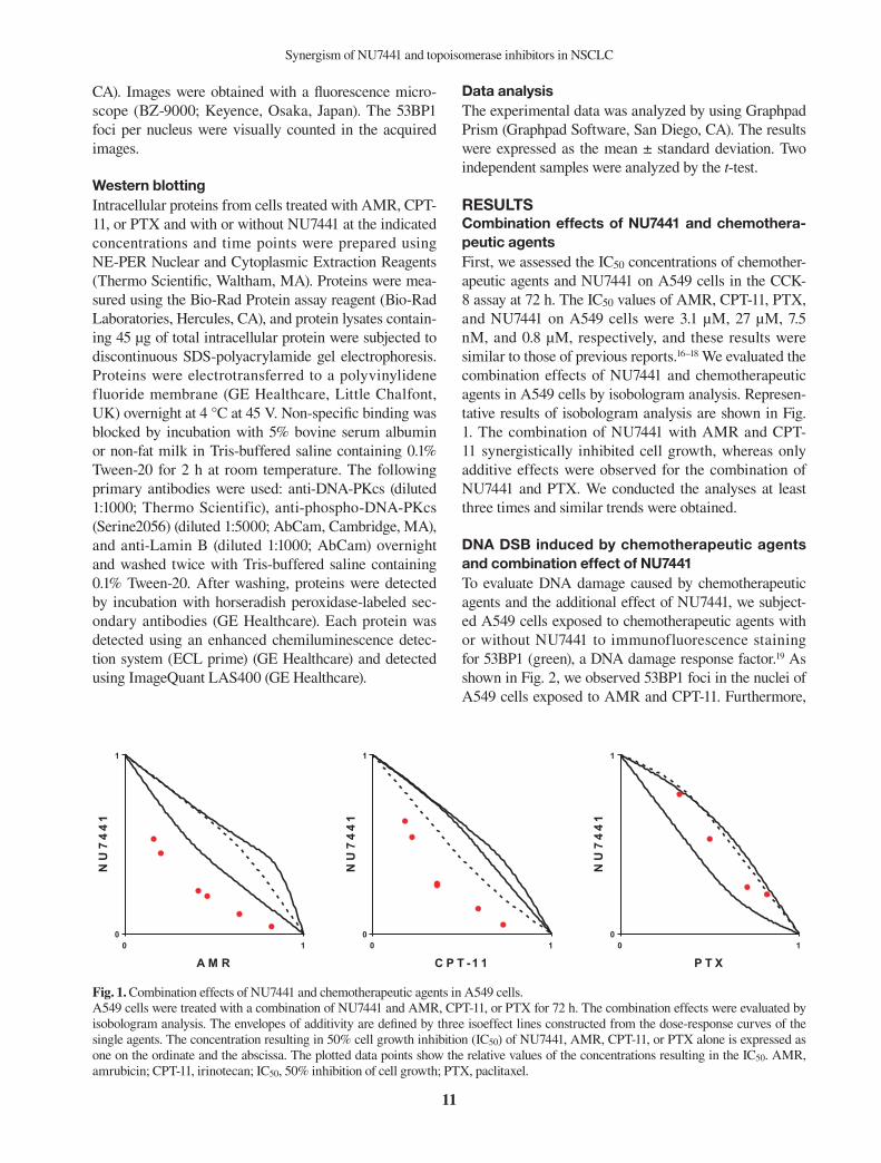
DNA DSB induced by chemotherapeutic agents and combination effect of NU7441To evaluate DNA damage caused by chemotherapeutic agents and the additional effect of NU7441, we subject-ed A549 cells exposed to chemotherapeutic agents with or without NU7441 to immunofluorescence staining for 53BP1 (green), a DNA damage response factor.19 As shown in Fig. 2, we observed 53BP1 foci in the nuclei of A549 cells exposed to AMR and CPT-11. Furthermore,
0 10
1
A M R
NU7441
0 10
1
C P T -1 1
NU7441
0 10
1
P T X
NU7441
Figure 1. Fig. 1. Combination effects of NU7441 and chemotherapeutic agents in A549 cells. A549 cells were treated with a combination of NU7441 and AMR, CPT-11, or PTX for 72 h. The combination effects were evaluated by isobologram analysis. The envelopes of additivity are defined by three isoeffect lines constructed from the dose-response curves of the single agents. The concentration resulting in 50% cell growth inhibition (IC50) of NU7441, AMR, CPT-11, or PTX alone is expressed as one on the ordinate and the abscissa. The plotted data points show the relative values of the concentrations resulting in the IC50. AMR, amrubicin; CPT-11, irinotecan; IC50, 50% inhibition of cell growth; PTX, paclitaxel.

12
M. Yanai et al.
NU–NU+
NU–NU+
NU–NU+
0
1 0
2 0
3 0
4 0
% >
10
foc
i
C o n tro l
C P T -1 1 2 4 h
P T X 2 4 h
n s
n s
NU–NU+
NU–NU+
NU–NU+
NU–NU+
0
2 0
4 0
6 0
8 0
1 0 0
% >
10
foc
i
C o n tro l
A M R 1 h
C P T 1 h
P T X 1 h
n s
n s
n s
NU–NU+
NU–NU+
NU–NU+
0
1 0
2 0
3 0
4 0
% >
10
foc
i
C o n tro l
C P T -1 1 2 4 h
P T X 2 4 h
n s
n s
NU–NU+
NU–NU+
NU–NU+
NU–NU+
0
2 0
4 0
6 0
8 0
1 0 0
% >
10
foc
i
C o n tro l
A M R 1 h
C P T 1 h
P T X 1 h
n s
n s
n s
Fig. 2. DNA DSBs in A549 cells exposed to chemotherapeutic agents in the presence and absence of DNA-PK inhibitor (NU7441). A549 cells were treated with the IC50 dose of AMR (3 μM), CPT-11 (25 μM), or PTX (7.5 nM) for 1 h with or without NU7441 (10 μM, 1 h prior to chemotherapeutic agent exposure) and samples were immunostained with anti-53BP1 antibodies (green) and DAPI (blue) at 1 h (A) and 24 h (B) after chemotherapeutic agent exposure. Representative images for each condition are shown. Percentages of cells with > 10 53BP1 foci treated as in A (C) and B (D). Date are expressed as the mean ± SE (n = 3). *P < 0.05. 53BP1, p53 binding protein 1; AMR, amrubicin; CPT-11, irinotecan; DAPI, 4,6-diamidino-2-phenylindole; DNA-PK, DNA-dependent protein kinase; DSB, dou-ble-strand break; IC50, 50% inhibition of cell growth; ns, not significant; NU, NU7441; PTX, pacritaxel.
NU–NU+
NU–NU+
NU–NU+
0
1 0
2 0
3 0
4 0%
> 1
0 fo
ci
C o n tro l
C P T -1 1 2 4 h
P T X 2 4 h
n s
n s
NU–NU+
NU–NU+
NU–NU+
NU–NU+
0
2 0
4 0
6 0
8 0
1 0 0
% >
10
foc
i
C o n tro l
A M R 1 h
C P T 1 h
P T X 1 h
n s
n s
n s
NU–NU+
NU–NU+
NU–NU+
0
1 0
2 0
3 0
4 0
% >
10
foc
i
C o n tro l
C P T -1 1 2 4 h
P T X 2 4 h
n s
n s
NU–NU+
NU–NU+
NU–NU+
NU–NU+
0
2 0
4 0
6 0
8 0
1 0 0
% >
10
foc
i
C o n tro l
A M R 1 h
C P T 1 h
P T X 1 h
n s
n s
n s
C D
the number of cells with 53BP1 foci increased under combination treatment with NU74441. In contrast, the number of 53BP1 foci in the nuclei of A549 cells ex-posed to PTX was similar to the control condition and did not increase following the addition of NU7441. To quantify DNA DSB, we counted and graphed the num-ber of cells with > 10 53BP1 foci. Immediately after drug exposure, the rate of cells with > 10 53BP1 foci treated with a combination of AMR and NU7441 was higher than that of cells treated with only AMR (P < 0.05, Fig. 2B). At 24 h after drug exposure, the rate of cells with > 10 53BP1 foci treated with a combination of CPT-11 and NU7441 was higher than that treated with only
CPT-11 (P < 0.05, Fig. 2D). In contrast, the rate of cells with > 10 53BP1 foci did not increase following combi-nation treatment with PTX and NU7441. Unfortunately, the cells at 24 h after exposure of AMR were difficult to evaluate because of large changes in cell morphology.
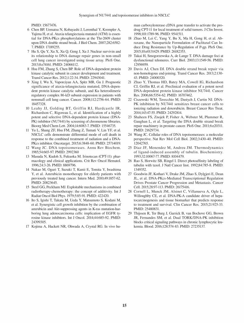
Apoptotic effects of NU7441 and chemotherapeu-tic agentsTo investigate the combination effect of NU7441 and chemotherapeutic agents on cell apoptosis, we assayed the cells treated with or without NU7441 and with AMR, CPT-11, or PTX for 24 or 48 h using an Annexin V apoptosis kit. At 24 h after drug exposure (Fig. 3A),
A B

13
Synergism of NU7441 and topoisomerase inhibitors in NSCLC
NU– NU+NU– NU+
NU– NU+NU– NU+
0
1 0
2 0
3 0
4 0
Ap
op
tos
is r
ate
(%
)
C o n tro l
A M R 2 4 h
C P T -1 1 2 4 h
P T X 2 4 h
ns
ns
ns
*
A 24 h
NU– NU+NU– NU+
NU– NU+0
2 0
4 0
6 0
8 0
1 0 0
Ap
op
tos
is r
ate
(%
)
C o n tro l
C P T -1 1 4 8 h
P T X 4 8 h
ns
ns
*
B 48 h
Fig. 3. Apoptotic effect of combinations of NU7441 and chemotherapeutic agents.A549 cells were treated with IC50 dose of AMR (3 μM), CPT-11 (25 μM), or PTX (7.5 nM) for 24 and 48 h with or without NU7441 (2.5 μM, 1 h prior to chemotherapeutic agent exposure). Samples were assayed with Annexin V. Bar graphs representing the percentage of apoptosis cells at 24 h (A) and 48 h (B). Date are expressed as the mean ± SE (n = 3). *P < 0.05. AMR, amrubicin; CPT-11, irinotecan; IC50, 50% inhibition of cell growth; ns, not significant; NU, NU7441; PTX, pacritaxel.
AMR significantly increased apoptosis following addi-tion of NU7441. At 48 h after drug exposure (Fig. 3B), CPT clearly increased apoptosis when NU7441 was also added. In contrast, PTX induced apoptosis, but there was no combination effect with NU7441. Unfortunately, apoptosis at 48 h after exposure of AMR was difficult to evaluate because of the high rate of cell death.
Activation of DNA-PKcs induced by chemothera-peutic agents and suppression effect by NU7441To assess the activation of DNA-PKcs induced by che-motherapeutic agents in NSCLC cells at different con-centrations and times, we detected pDNA-PKcs (S2056) protein expression in the nuclei of A549 cells exposed
to PTX, CPT-11, and AMR by western blotting. The nuclear proteins were extracted using NE-PER nuclear and cytoplasmic extraction reagents. After 1-h exposure, AMR clearly activated DNA-PKcs, while PTX and CPT-11 only slightly activated DNA-PKcs (Fig. 4A). After long-term incubation (12 and 24 h), following 1-h drug exposure, CPT-11 clearly activated DNA-PKcs over time, while PTX did not show this activation effect (Fig. 4B). These results suggest that the timing and intensity of phosphorylation of DNA-PKcs depends on the che-motherapeutic agent used. In addition, the activation of DNA-PKcs induced by chemotherapeutic agents was suppressed by NU7441.
Figure 4.
A B
Fig. 4. Activation of DNA-PKcs induced by chemotherapeutic agents and effect of DNA-PK inhibitor (NU7441). (A) A549 cells were pre-treated with or without a DNA-PKcs inhibitor (NU7441, 10 μM) and treated with 1/10 IC50 dose or IC50 dose of PTX (0.75 nM, 7.5 nM), CPT-11 (2.5 μM, 25 μM), or AMR (0.3 μM, 3 μM) for 1 h. Activation of DNA-PKcs was assayed by western blotting with antibodies as indicated. (B) A549 cells were pre-treated with or without NU7441 (10 μM) and treated with IC50 dose of PTX (7.5 nM) or CPT-11 (25 μM). Activation of DNA-PKcs was assayed at 1, 12, or 24 h after 1-h treatment with each agent by western blot-ting using the indicated antibodies. AMR, amrubicin; CPT-11, irinotecan; DNA-PKcs, DNA-dependent protein kinase catalytic subunit; IC50, 50% inhibition of cell growth; pDNA-PKcs, phospho-DNA-dependent protein kinase catalytic subunit; PTX, paclitaxel.

14
M. Yanai et al.
DISCUSSIONDNA-PKcs is an essential component of the NHEJ path-way and activation of DNA-PKcs is required for DNA DSB repair.20 Inhibiting phosphorylation of DNA-PKcs blocks DNA damage repair and leads to cell apoptosis. NU7441, a potent and specific DNA-PKcs inhibitor, has been predicted to enhance the therapeutic effect of inducing DNA DSB. Previously, several studies showed that NU7441 has potential for enhancing the radio-sen-sitivity in different tumors, including NSCLC,11 colon cancer,21 breast cancer,22 and prostate cancer,23 and che-mo-sensitivity of etoposide (topoisomerase II inhibitor) in colon cancer.21 The goal of this study was to deter-mine the best combination drug for use with NU7441 to enhance the anti-proliferative effect on NSCLCs. In this study, a DNA-PKcs inhibitor showed promise in combination pharmacological medicine with specific chemotherapeutic agents against lung cancer. The DNA-PKcs inhibitor had a synergistic anti-proliferative effect on the A549 lung cancer cell line when applied with topoisomerase inhibitors. Interestingly, the inhibitor only had an additive effect with the mitotic inhibitor. Topoisomerases have been examined for use in the treatment of certain diseases. Among them, eukaryotic topoisomerases I and II are targets of anti-cancer drugs that act to inhibit these enzymes by blocking the reaction that reseals breaks in the DNA.24 Topoisomerase I inhib-itors, including CPT-11, induce single-strand breaks in DNA by blocking the dissociation of topoisomerase and DNA, leading to replication-mediated DNA damage. Topoisomerase II inhibitors, including AMR, can act by either stabilizing topoisomerase II-DNA complexes that are easily cleaved or interfering with the catalytic activ-ity of the enzyme, both resulting in DSBs. In contrast, mitotic inhibitors, including PTX, bind to tubulin and inhibit its polymerization into microtubules, resulting in tumors and leading to apoptosis without involvement with DSBs.25, 26
Phosphorylation of DNA-PKcs occurs at the S2056 site,5 which provides a marker for assessing the effect of drugs on DNA-PKcs activity. In this study, we found that topoisomerase inhibitor induced DNA-PKcs phos-phorylation of A549. Topoisomerase II inhibitor AMR immediately phosphorylated DNA-PKcs, whereas to-poisomerase I inhibitor CPT-11 only minimally induced DNA-PKcs phosphorylation immediately after exposure to drugs and gradually phosphorylated DNA-PKcs over time. This delay in DNA-PKcs phosphorylation may be related to the time required to shift from single-strand breaks to DSBs. NU7441 clearly inhibited DNA-PKcs phosphorylation caused by both AMR and CPT-11. In this study, we assessed the degree of DNA DSBs
using 53BP1 foci as a marker of DNA DSB.19 AMR and CPT-11 caused DNA DSBs, whereas PTX caused few DNA DSBs. The combined use of NU7441 induced a greater number of DNA DSBs. In addition, NU7441 in combination with topoisomerase inhibitors induced apoptosis by more than the use of topoisomerase inhibi-tors alone. This is the first study to demonstrate that the DNA-PKcs inhibitor Nu7441 has a synergistic effect with topoisomerase inhibitors, but not with a mitotic inhibi-tor, on NSCLC cells. Recently, several clinical studies revealed that DNA-PKcs expression is significantly ele-vated in advanced disease and that an elevation in acti-vated DNA-PKcs predicts the response to treatment and survival.27, 28 Further, DNA-PKcs inhibitors are currently in phase I trials for advanced solid tumors and hemato-logic malignancies (NCT01280487, NCT01421524, and NCT01682473) and the efficacy of DNA-PKcs inhibitors has been demonstrated in clinical trials.29 Based on our results, further studies, including preclinical studies and clinical trials, are necessary to clarify the efficacy of this combination therapy with NU7441 and topoisomerase inhibitors in NSCLC. In conclusion, NU7441 blocks DNA damage repair induced by topoisomerase inhibitors in NCSLC cells and subsequently leads to their apoptosis. Therefore, our re-sults suggest that combination therapy of topoisomerase inhibitors and NU7441 is a potential therapeutic option for NCSLC.
Acknowledgments: This work was supported by JSPS KAKENHI Grant Number 26830075.
The authors declare no conflict of interest.
REFERENCES 1 Siegel RL, Miller KD, Jemal A. Cancer statistics, 2015. CA
Cancer J Clin. 2015;65:5-29. PMID: 25559415. 2 Maemondo M, Inoue A, Kobayashi K, Sugawara S,
Oizumi S, Isobe H, et al. Gefitinib or chemotherapy for non-small-cell lung cancer with mutated EGFR. N Engl J Med. 2010;362:2380-8. PMID: 20573926.
3 Shaw AT, Yeap BY, Solomon BJ, Riely GJ, Gainor J, Engelman JA, et al. Effect of crizotinib on overall survival in patients with advanced non-small-cell lung cancer harbouring ALK gene rearrangement: a retrospective analysis. Lancet Oncol. 2011;12:1004-12. PMID: 21933749.
4 Chan DW, Chen BP, Prithivirajsingh S, Kurimasa A, Story MD, Qin J, et al. Autophosphorylation of the DNA-de-pendent protein kinase catalytic subunit is required for rejoin-ing of DNA double-strand breaks. Genes Dev. 2002;16:2333-8. PMID: 12231622.
5 Chen BP, Chan DW, Kobayashi J, Burma S, Asaithamby A, Morotomi-Yano K, et al. Cell cycle dependence of DNA-de-pendent protein kinase phosphorylation in response to DNA double strand breaks. J Biol Chem. 2005;280:14709-15.
15
Synergism of NU7441 and topoisomerase inhibitors in NSCLC
PMID: 15677476. 6 Chen BP, Uematsu N, Kobayashi J, Lerenthal Y, Krempler A,
Yajima H, et al. Ataxia telangiectasia mutated (ATM) is essen-tial for DNA-PKcs phosphorylations at the Thr-2609 cluster upon DNA double strand break. J Biol Chem. 2007;282:6582-7. PMID: 17189255.
7 Hu S, Qu Y, Xu X, Xu Q, Geng J, Xu J. Nuclear survivin and its relationship to DNA damage repair genes in non-small cell lung cancer investigated using tissue array. PloS One. 2013;8:e74161. PMID: 24066112.
8 Hsu FM, Zhang S, Chen BP. Role of DNA-dependent protein kinase catalytic subunit in cancer development and treatment. Transl Cancer Res. 2012;1:22-34. PMID: 22943041.
9 Xing J, Wu X, Vaporciyan AA, Spitz MR, Gu J. Prognostic significance of ataxia-telangiectasia mutated, DNA-depen-dent protein kinase catalytic subunit, and Ku heterodimeric regulatory complex 86-kD subunit expression in patients with nonsmall cell lung cancer. Cancer. 2008;112:2756-64. PMID: 18457328.
10 Leahy JJ, Golding BT, Griff in RJ, Hardcastle IR, Richardson C, Rigoreau L, et al. Identification of a highly potent and selective DNA-dependent protein kinase (DNA-PK) inhibitor (NU7441) by screening of chromenone libraries. Bioorg Med Chem Lett. 2004;14:6083-7. PMID: 15546735.
11 Yu L, Shang ZF, Hsu FM, Zhang Z, Tumati V, Lin YF, et al. NSCLC cells demonstrate differential mode of cell death in response to the combined treatment of radiation and a DNA-PKcs inhibitor. Oncotarget. 2015;6:3848-60. PMID: 25714019.
12 Wang JC. DNA topoisomerases. Annu Rev Biochem. 1985;54:665-97. PMID: 2992360
13 Masuda N, Kudoh S, Fukuoka M. Irinotecan (CPT-11): phar-macology and clinical applications. Crit Rev Oncol Hematol. 1996;24:3-26. PMID: 8869796.
14 Nakao M, Oguri T, Suzuki T, Kunii E, Tomita Y, Iwashima Y, et al. Amrubicin monotherapy for elderly patients with previously treated lung cancer. Intern Med. 2010;49:1857-62. PMID: 20823645.
15 Steel GG, Peckham MJ. Exploitable mechanisms in combined radiotherapy-chemotherapy: the concept of additivity. Int J Radiat Oncol Biol Phys. 1979;5:85-91. PMID: 422420.
16 Ito S, Igishi T, Takata M, Ueda Y, Matsumoto S, Kodani M, et al. Synergistic cell growth inhibition by the combination of amrubicin and Akt-suppressing agents in K-ras mutation-har-boring lung adenocarcinoma cells: implication of EGFR ty-rosine kinase inhibitors. Int J Oncol. 2014;44:685-92. PMID: 24399305.
17 Kojima A, Hackett NR, Ohwada A, Crystal RG. In vivo hu-
man carboxylesterase cDNA gene transfer to activate the pro-drug CPT-11 for local treatment of solid tumors. J Clin Invest. 1998;101:1789-96. PMID: 9541511.
18 Zhao M, Lei C, Yang Y, Bu X, Ma H, Gong H, et al. Ab-raxane, the Nanoparticle Formulation of Paclitaxel Can In-duce Drug Resistance by Up-Regulation of P-gp. PloS One. 2015;10:e0131429. PMID: 26182353.
19 Takai H, Smogorzewska A, de Lange T. DNA damage foci at dysfunctional telomeres. Curr Biol. 2003;13:1549-56. PMID: 12956959.
20 Davis AJ, Chen DJ. DNA double strand break repair via non-homologous end-joining. Transl Cancer Res. 2013;2:130-43. PMID: 24000320.
21 Zhao Y, Thomas HD, Batey MA, Cowell IG, Richardson CJ, Griffin RJ, et al. Preclinical evaluation of a potent novel DNA-dependent protein kinase inhibitor NU7441. Cancer Res. 2006;66:5354-62. PMID: 16707462.
22 Ciszewski WM, Tavecchio M, Dastych J, Curtin NJ. DNA-PK inhibition by NU7441 sensitizes breast cancer cells to ionizing radiation and doxorubicin. Breast Cancer Res Treat. 2014;143:47-55. PMID: 24292814.
23 Shaheen FS, Znojek P, Fisher A, Webster M, Plummer R, Gaughan L, et al. Targeting the DNA double strand break repair machinery in prostate cancer. PloS One. 2011;6:e20311. PMID: 21629734.
24 Wang JC. Cellular roles of DNA topoisomerases: a molecular perspective. Nat Rev Mol Cell Biol. 2002;3:430-40. PMID: 12042765.
25 Diaz JF, Menendez M, Andreu JM. Thermodynamics of ligand-induced assembly of tubulin. Biochemistry. 1993;32:10067-77. PMID: 8104479.
26 Rao S, Horwitz SB, Ringel I. Direct photoaffinity labeling of tubulin with taxol. J Natl Cancer Inst. 1992;84:785-8. PMID: 1349352.
27 Goodwin JF, Kothari V, Drake JM, Zhao S, Dylgjeri E, Dean JL, et al. DNA-PKcs-Mediated Transcriptional Regulation Drives Prostate Cancer Progression and Metastasis. Cancer Cell. 2015;28:97-113. PMID: 26175416.
28 Cornell L, Munck JM, Alsinet C, Villanueva A, Ogle L, Willoughby CE, et al. DNA-PK-A candidate driver of hepa-tocarcinogenesis and tissue biomarker that predicts response to treatment and survival. Clin Cancer Res. 2015;21:925-33. PMID: 25480831.
29 Thijssen R, Ter Burg J, Garrick B, van Bochove GG, Brown JR, Fernandes SM, et al. Dual TORK/DNA-PK inhibition blocks critical signaling pathways in chronic lymphocytic leu-kemia. Blood. 2016;128:574-83. PMID: 27235137.


![An Integrated Database of Chemosensitivity to 55 ...[CANCER RESEARCH 62, 1139–1147, February 15, 2002] An Integrated Database of Chemosensitivity to 55 Anticancer Drugs and Gene](https://static.fdocuments.us/doc/165x107/5ed99d59801c872007065f4a/an-integrated-database-of-chemosensitivity-to-55-cancer-research-62-1139a1147.jpg)