DNA nanotechnology assisted nanopore-based analysis
16
Nucleic Acids Research, 2020 1 doi: 10.1093/nar/gkaa095 SURVEY AND SUMMARY DNA nanotechnology assisted nanopore-based analysis Taoli Ding 1,2 , Jing Yang 3,† , Victor Pan 4,5,† , Nan Zhao 3 , Zuhong Lu 2,* , Yonggang Ke 4,5,* and Cheng Zhang 1,* 1 Department of Computer Science and Technology, School of Electronics Engineering and Computer Science, Peking University, Beijing 100871, China, 2 Department of Biomedical Engineering, College of engineering, Peking University, Beijing 100871, China, 3 School of Control and Computer Engineering, North China Electric Power University, Beijing 102206, China, 4 Wallace H. Coulter Department of Biomedical Engineering, Georgia Institute of Technology and Emory University, Emory University School of Medicine, Atlanta, GA 30322, USA and 5 Department of Chemistry, Emory University, Atlanta, GA 30322, USA Received July 16, 2019; Revised January 29, 2020; Editorial Decision February 03, 2020; Accepted February 17, 2020 ABSTRACT Nanopore technology is a promising label-free de- tection method. However, challenges exist for its fur- ther application in sequencing, clinical diagnostics and ultra-sensitive single molecule detection. The development of DNA nanotechnology nonetheless provides possible solutions to current obstacles hin- dering nanopore sensing technologies. In this re- view, we summarize recent relevant research con- tributing to efforts for developing nanopore meth- ods associated with DNA nanotechnology. For ex- ample, DNA carriers can capture specific targets at pre-designed sites and escort them from nanopores at suitable speeds, thereby greatly enhancing capa- bility and resolution for the detection of specific tar- get molecules. In addition, DNA origami structures can be constructed to fulfill various design specifica- tions and one-pot assembly reactions, thus serving as functional nanopores. Moreover, based on DNA strand displacement, nanopores can also be utilized to characterize the outputs of DNA computing and to develop programmable smart diagnostic nanode- vices. In summary, DNA assembly-based nanopore research can pave the way for the realization of im- pactful biological detection and diagnostic platforms via single-biomolecule analysis. INTRODUCTION As a new platform, nanopore technology presents vari- ous advantages over other detection modalities including low cost, high throughput and label-free sample analysis. Accordingly, nanopore analysis has been widely applied to fields such as gene sequencing, personalized medicine, biomedicine, food safety, environmental surveillance, and others. In particular, as a milestone for nanopore detec- tion, in 1996, Deamer et al. proposed DNA sequencing us- ing alpha hemolysin (1). In a typical nanopore experiment, two reservoirs are connected via a single nanometer-sized pore. Upon applying an electric field across the nanopore, electrically charged molecules are driven to translocate the nanopore via an electrophoretic force. When this oc- curs, conductive ions in the buffer are excluded from the nanopore, resulting in a measurable drop in current. Ac- cordingly, biomolecules of various sizes and charges can be distinguished based on parameters of translocation such as dwell time and current drop amplitude (2). Inherently, nanopore experiments represent a single molecule detec- tion method with ultra-sensitivity and high resolution, ca- pable of discriminating minute differences among single nucleotide bases A, T, G and C. Compared with previ- ous nanopore applications, which mainly focused on gene sequencing (3–6), recent nanopore technologies have also been extended to analyze the physical and chemical prop- erties of various molecules (7) such as nucleic acids (8–12), proteins (13–18), nanoparticles (19–25) and ions (26–32). * To whom correspondence should be addressed. Tel: +86 01062750359; Fax: +86 01062750359; Email: [email protected] Correspondence may also be addressed to Zuhong Lu. Tel: +86 02583793110; Fax: +86 02583793110; Email: [email protected] Correspondence may also be addressed to Yonggang Ke. Tel: +1 404 712 2712; Fax: +1 404 712 2712; Email: [email protected] † The authors wish it to be known that, in their opinion, the second and third authors should be regarded as Joint Second Authors. C The Author(s) 2020. Published by Oxford University Press on behalf of Nucleic Acids Research. This is an Open Access article distributed under the terms of the Creative Commons Attribution Non-Commercial License (http://creativecommons.org/licenses/by-nc/4.0/), which permits non-commercial re-use, distribution, and reproduction in any medium, provided the original work is properly cited. For commercial re-use, please contact [email protected] Downloaded from https://academic.oup.com/nar/advance-article-abstract/doi/10.1093/nar/gkaa095/5742838 by guest on 26 February 2020
Transcript of DNA nanotechnology assisted nanopore-based analysis
Nucleic Acids Research, 2020 1doi: 10.1093/nar/gkaa095
SURVEY AND SUMMARY
DNA nanotechnology assisted nanopore-basedanalysisTaoli Ding1,2, Jing Yang3,†, Victor Pan4,5,†, Nan Zhao3, Zuhong Lu2,*, Yonggang Ke4,5,* andCheng Zhang 1,*
1Department of Computer Science and Technology, School of Electronics Engineering and Computer Science,Peking University, Beijing 100871, China, 2Department of Biomedical Engineering, College of engineering, PekingUniversity, Beijing 100871, China, 3School of Control and Computer Engineering, North China Electric PowerUniversity, Beijing 102206, China, 4Wallace H. Coulter Department of Biomedical Engineering, Georgia Institute ofTechnology and Emory University, Emory University School of Medicine, Atlanta, GA 30322, USA and 5Departmentof Chemistry, Emory University, Atlanta, GA 30322, USA
Received July 16, 2019; Revised January 29, 2020; Editorial Decision February 03, 2020; Accepted February 17, 2020
ABSTRACT
Nanopore technology is a promising label-free de-tection method. However, challenges exist for its fur-ther application in sequencing, clinical diagnosticsand ultra-sensitive single molecule detection. Thedevelopment of DNA nanotechnology nonethelessprovides possible solutions to current obstacles hin-dering nanopore sensing technologies. In this re-view, we summarize recent relevant research con-tributing to efforts for developing nanopore meth-ods associated with DNA nanotechnology. For ex-ample, DNA carriers can capture specific targets atpre-designed sites and escort them from nanoporesat suitable speeds, thereby greatly enhancing capa-bility and resolution for the detection of specific tar-get molecules. In addition, DNA origami structurescan be constructed to fulfill various design specifica-tions and one-pot assembly reactions, thus servingas functional nanopores. Moreover, based on DNAstrand displacement, nanopores can also be utilizedto characterize the outputs of DNA computing andto develop programmable smart diagnostic nanode-vices. In summary, DNA assembly-based nanoporeresearch can pave the way for the realization of im-pactful biological detection and diagnostic platformsvia single-biomolecule analysis.
INTRODUCTION
As a new platform, nanopore technology presents vari-ous advantages over other detection modalities includinglow cost, high throughput and label-free sample analysis.Accordingly, nanopore analysis has been widely appliedto fields such as gene sequencing, personalized medicine,biomedicine, food safety, environmental surveillance, andothers. In particular, as a milestone for nanopore detec-tion, in 1996, Deamer et al. proposed DNA sequencing us-ing alpha hemolysin (1). In a typical nanopore experiment,two reservoirs are connected via a single nanometer-sizedpore. Upon applying an electric field across the nanopore,electrically charged molecules are driven to translocatethe nanopore via an electrophoretic force. When this oc-curs, conductive ions in the buffer are excluded from thenanopore, resulting in a measurable drop in current. Ac-cordingly, biomolecules of various sizes and charges canbe distinguished based on parameters of translocation suchas dwell time and current drop amplitude (2). Inherently,nanopore experiments represent a single molecule detec-tion method with ultra-sensitivity and high resolution, ca-pable of discriminating minute differences among singlenucleotide bases A, T, G and C. Compared with previ-ous nanopore applications, which mainly focused on genesequencing (3–6), recent nanopore technologies have alsobeen extended to analyze the physical and chemical prop-erties of various molecules (7) such as nucleic acids (8–12),proteins (13–18), nanoparticles (19–25) and ions (26–32).
*To whom correspondence should be addressed. Tel: +86 01062750359; Fax: +86 01062750359; Email: [email protected] may also be addressed to Zuhong Lu. Tel: +86 02583793110; Fax: +86 02583793110; Email: [email protected] may also be addressed to Yonggang Ke. Tel: +1 404 712 2712; Fax: +1 404 712 2712; Email: [email protected]†The authors wish it to be known that, in their opinion, the second and third authors should be regarded as Joint Second Authors.
C© The Author(s) 2020. Published by Oxford University Press on behalf of Nucleic Acids Research.This is an Open Access article distributed under the terms of the Creative Commons Attribution Non-Commercial License(http://creativecommons.org/licenses/by-nc/4.0/), which permits non-commercial re-use, distribution, and reproduction in any medium, provided the original workis properly cited. For commercial re-use, please contact [email protected]
Dow
nloaded from https://academ
ic.oup.com/nar/advance-article-abstract/doi/10.1093/nar/gkaa095/5742838 by guest on 26 February 2020

2 Nucleic Acids Research, 2020
Based on materials, nanopores can be divided intotwo categories: biological nanopore (33) and solid-statenanopore (SS-nanopore) (Figure 1) (34). Biologicalnanopores comprise channel-bearing proteins such asPhi29 (35–37) and �-hemolysin (38–44). With a narrowchannel (general diameter 1 nm–2 nm), protein nanoporescan produce sensitive and accurate electrical signals inDNA sequencing due to the similarities they share with thediameter of single-strand DNA (ssDNA). In addition, ex-perimental repeatability is another advantage of biologicalnanopores, according to which selected portable commer-cial nanopore devices are being developed. However, issuesmust be considered when using biological nanopores. Forexample, it is not easy to readily modify protein nanopores,which limits the scope of their application. Moreover,proteins are more sensitive to external environmentalconditions because small variations in temperature or pHwill significantly affect biological nanopores’ conformationand activity.
SS-nanopores are nanopores are constructed using abi-otic materials (45,46). The straightforward fabrication pro-cedures of SS nanopores render diameter control adjustableand scalable, which enables wider sensing ranges to en-compass not only small molecules such as DNA, but alsolarge protein targets. However, SS nanopore-based detec-tion faces several challenges. First, the translocation veloc-ity of analytes is generally too fast to allow for consistentidentification of smaller biomolecules because of compar-atively large nanopore diameters. To solve this problem,various methods including gel substrates (47), molecularmodifications on the nanopore (48), blocking the pore withnanobeads (49), applying various high-salt buffers (50) areutilized. However, not all of these methods are satisfac-tory because of deficiencies such as increased experimen-tal complexity, random systematic noise, and modificationdifficulties. Additionally, SS nanopore membranes are toothick to acquire high signal-to-noise ratios at a high res-olution, compared with biological nanopores in DNA se-quencing. In order to improve the resolution and sensitiv-ity of SS nanopores, scientists have combined super-thintwo-dimensional materials like molybdenum disulfide (4)and graphene with SS nanopores (51–55). Nevertheless, thepreparation of two-dimensional material-based nanoporesare complicated, inconvenient, and expensive.
DNA nanotechnology focused on using DNA to con-struct various self-assembled structures was recently de-veloped. As a versatile technology, DNA assembly is al-ready applied to various nanopore-based analyses. For ex-ample, DNA self-assembly structures can be used to cap-ture target biomolecules to form complex spatial arrange-ments (56), or to serve as possible channels for potentialdrug delivery (57). Importantly, DNA nanotechnology mayalso provide practical solutions to the aforementioned chal-lenges in nanopore detection (34,58,59). Several efforts havebeen made to combine nanopore analysis and DNA nan-otechnology in the past decade. For example, scientists di-rectly used DNA nanotechnology to construct assemblednanopores performing target molecule nanopore transloca-tion (60). Notably, in these studies, DNA nanotechnologyendowed the nanopore platform with fine-tunable and ad-justable properties.
In this review article, we summarize research associat-ing nanopore techniques with DNA nanotechnology. Sev-eral categories will be discussed to highlight recent stud-ies combining DNA nanotechnology and nanopore de-tection. We will primarily discuss recent progress com-bining DNA nanotechnology and nanopore detection inthe following categories: (i) DNA carrier-mediated escortof targets in nanopore translocation; (ii) DNA-assemblednanopores for single- molecule detection with tunable andadjustable properties; (iii) nanopore detection of targetsbased on the dynamic control of DNA assembly. Com-bined with DNA nanotechnology, nanopore analysis meth-ods have been greatly improved. To date, several studieshave been conducted in this interdisciplinary field. Consid-ering the universal applications of nanopore analysis com-bined with DNA nanotechnology, this review article high-lights related works and inspired ideas to introduce thisstudy area to researchers. Finally, we envision that the asso-ciation of DNA nanotechnology and nanopores will createmany new sensing methods for biodetection, nanoengineer-ing, diagnostics and therapeutics.
DNA NANOTECHNOLOGY
With the recent development of DNA assembly, DNAmolecules can serve not only as carriers of genetic in-formation, but also fulfill significant roles in artificialnanosystems through their diversely modified properties,predictable behavior, nanoscale size and programmable fea-tures (61). In the 1980s, DNA was characterized as be-ing able to form desired nanostructures via computer-assisted molecular designs, i.e. DNA self-assembly (62).Nadrian Seeman, a pioneer in the field of DNA nanotech-nology, constructed various nanostructures based on DNAself-assembly. Utilizing specific DNA molecular recogni-tions, stable DNA supramolecular structures can sponta-neously form via hydrogen bonding, hydrophobic inter-actions, and van der Waals interactions, among others.There are two main strategies for creating DNA struc-tures: (i) short DNA strands employing DNA tile assem-bly and (ii) long DNA strands-associated DNA origamiassembly. DNA nanotechnology is a promising tool withadvantages such as designable nanoscale engineering, highmanufacture efficiency, and a convenient preparation pro-cess. This method has recently been applied to various areassuch as biology, chemistry, medicine, material science, na-noengineering, and molecular computing (63–65). In par-ticular, DNA assembly-based sensing and diagnosis has at-tracted significant interest. It has been shown that engi-neered DNA-assembled constructs can facilitate the controlof various biomolecules (64), and is an excellent material forcomplex molecular information processing (65,66).
Tile based self-assembled DNA nanostructures
As the most commonly used primary DNA component,the typical DNA tile generally comprises several short hy-bridized ssDNA to form a cross-shaped skeleton frame.Then, through cohesive sticky ends, multiple tiles can belinked to assemble more complex structures. As a pioneer-ing DNA assembly method, DNA tiles are generally usedto construct a diverse range of nanostructures (67–69).
Dow
nloaded from https://academ
ic.oup.com/nar/advance-article-abstract/doi/10.1093/nar/gkaa095/5742838 by guest on 26 February 2020

Nucleic Acids Research, 2020 3
Figure 1. Schematics of a biological nanopore (A) (33) and a SS nanopore (B) (34). In (A), the target molecule can pass through the pore of �-hemolysinto produce a significant drop in current. The carrier escorting target molecules through SS nanopores induces specific current signals.
Figure 2. (A) The structure of 2D DNA tile lattices comprising two and four units (62). The images below represent the structures for DAO and DAEunits. (B) The original design and AFM images of nanoribbons (69). (C) Schematic drawings of DNA tiles with multiple DNA bridges (73). (D) Schematicdrawings and transmission electron microscope (TEM) images of DNA tile assembled 3D nanostructures (77).
In 1983, DNA tile-based self-assembly was first proposedby Seeman (62). The original naturally occurring tile (Holl-iday junction) includes four single-stranded DNA. Mean-while, by rational design, 2D lattices can be constructedusing cohesive ends hybridization. Subsequently, to con-struct more stable and rigid tiles, double crossover (DX)tiles were established including double crossover, antiparal-lel, odd spacing (DAO) and double crossover, and antipar-allel and even spacing (DAE) (67) (Figure 2A). Taking ad-vantage of DX tiles, Hao Yan et al. prepared a 2D rigidgrid structure in 1998, where the DNA-assembled struc-ture was imaged by atomic force microscope (AFM), asshown in Figure 2B (69). DNA tiles with high rigidity and
more complexity appeared in succession including triple-crossover (TX) (70), paranemic crossover (PX) (71), DXtriangle (72), nanoarrays (NAs, shown in Figure 2B andC), nanoroads (NTs) (73) and single-stranded tile (74,75).By controlling the concentration ratios and the lengths ofspecific DNA strands, Mao et al. manufactured various 2D(76) and 3D structures (77) including tetrahedron, dodeca-hedron and buckyball arrangements (Figure 2D).
DNA origami-based self-assembly
Compared with minute DNA tiles, DNA origami assem-bly provides a method for assembling larger DNA nanos-
Dow
nloaded from https://academ
ic.oup.com/nar/advance-article-abstract/doi/10.1093/nar/gkaa095/5742838 by guest on 26 February 2020
4 Nucleic Acids Research, 2020
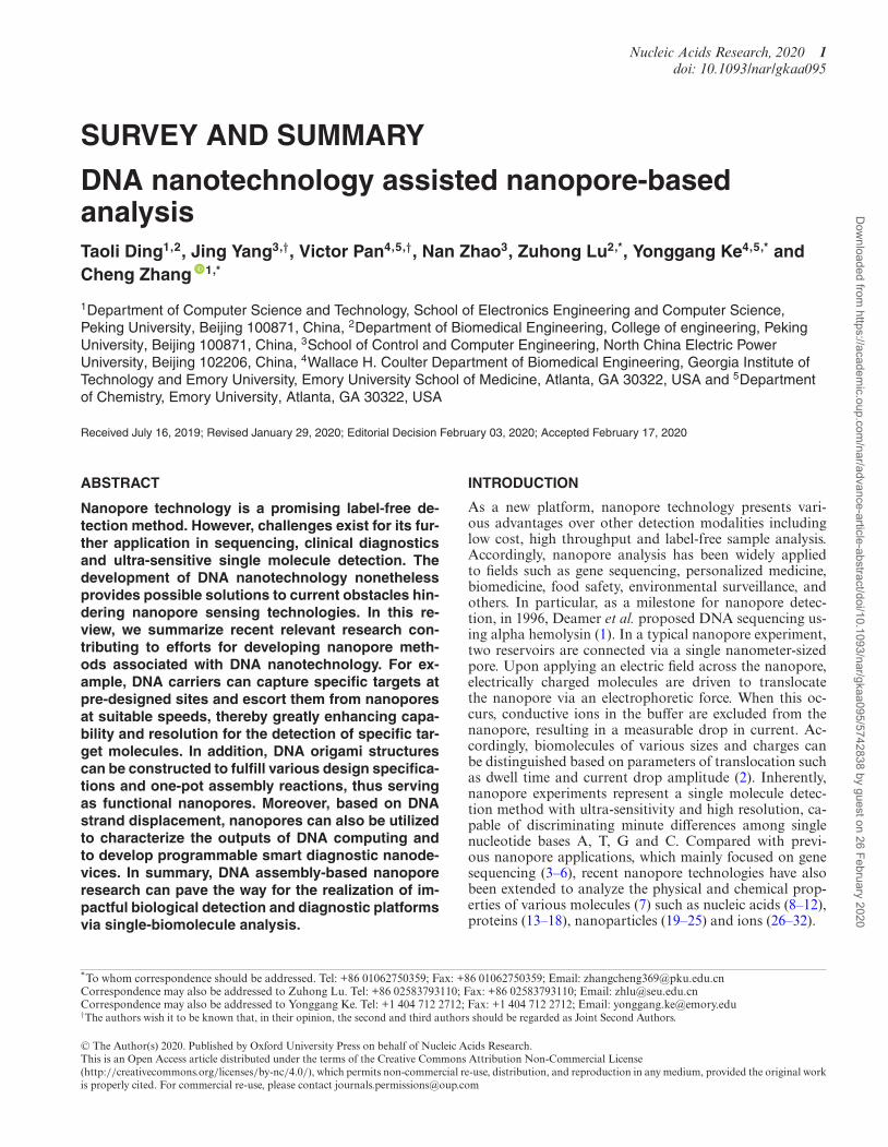
Figure 3. (A) Schematic illustrations of square, rectangle, star, disk with three holes, triangle with rectangular domains, and sharp triangle with trapezoidaldomains (78). (B) Direct self-assembly of DNA into nanoscale three-dimensional shapes and TEM results (86). (C) DNA origami nanostructures withcomplex 3D curvatures (87).
tructures. DNA origami assembly was developed by Rothe-mund in 2006, where hundreds of short synthetic oligonu-cleotides (staple DNA strands) fold a long scaffold strand(typically the M13mp18 genome) into various nanoscaleshapes of unprecedented complexity. As shown in Fig-ure 3A, through complementary base-pairing, various 2DDNA structures were assembled including square, rectan-gle, pentagram, smiling face, triangle and others (78). Con-sequently, asymmetric shapes such as maps (79), dolphin(80) and characters (81) were also constructed based onDNA origami.
To construct 3D structures, additional efforts have beenmade using different design principles: linking 2D self-assembled structures together to obtain 3D aggregationsor directly assembling 3D structures. Hollow cubes andprisms were manufactured (82–84) by linking independent2D structures. Dietz et al. expanded this method by creatingDNA nanostructures with controlled curvature and twist-ing (85). In 2009, Douglas et al. developed a DNA assem-bly method to directly construct 3D-shaped DNA origami(Figure 3B) (86). In 2011, Hao Yan et al. defined the fea-tures of objects via scaffold DNA nanostructures and con-structed a connective frame to modify curvature on the sur-face of 3D DNA origami (Figure 3C) (87).
Dynamic DNA strand displacement
In addition to DNA assembly-based nanostructures, DNAstrand displacement, a dynamic DNA regulation method, isanother recent development in DNA nanotechnology (88–
93). In nature, DNA strand displacement commonly occursat a very slow reaction rate (88). Toehold-mediated DNAstrand displacement reaction (T-SDR) was developed byYurke et al. in 2000 and greatly accelerates reaction rate by∼106-fold (89). In a typical T-SDR process, strand displace-ment initiates from a short single-stranded toehold domain,leading to sequential strand displacement via branch migra-tion (Figure 4A) (90). Importantly, the control of toeholdstrength enables fine-tuning of T-SDR.
In recent years, T-SDR has attracted more attention forits easy design and accurate control abilities, and has be-come a popular area of nanoengineering (91–93). This re-search context includes the following areas. (i) Regulationof switchable nanodevices and nanostructures. As large-scale DNA molecular systems and origami nanomachinesare constructed by DNA molecules, T-SDR- based controlcan be widely used in DNA-assembled nanosystems by tak-ing advantage of dynamic and precise properties (90). (ii)Molecular signal sensing effected via DNA catalysis (Fig-ure 4B) (93). Since the enzyme-free DNA system can am-plify the molecular signal via non-covalent DNA catalysis,catalysis reactions can be used to amplify weak signals (94).(iii) Construction of complex DNA cascading networks andcircuits. Relying on specific base-pair recognitions and pro-grammable sequence designs, T-SDR-based DNA regula-tion was employed to construct various types of molecularmotors (89), cascading networks (92–94), and logic circuitoperations (95,96). This technique can also be used in ar-eas such as smart molecular sensing and DNA computing(94–98).
Dow
nloaded from https://academ
ic.oup.com/nar/advance-article-abstract/doi/10.1093/nar/gkaa095/5742838 by guest on 26 February 2020

Nucleic Acids Research, 2020 5
Figure 4. (A) Schematics of basic DNA strand displacement (90). (B) Catalytic DNA strand displacement circuit where the catalyst DNA can repeatedlyparticipate in multi-cycle reactions (93). (C) Programmable DNA self-assembly pathway based on DNA strand displacement (94).
THE APPLICATION OF DNA NANOTECHNOLOGY INNANOPORE SENSING
Nanopore detection is a new and emerging technique forDNA sequencing, single molecule detection, and clinicaldiagnosis. Although traditional nanopores inherently havefeatures of high sensitivity and label-free detection, sev-eral difficulties persist in their use such as uncontrollabletranslocation and invariable nanopore size or shape. Toaddress these problems, various solutions have been pro-posed, among which the designable and versatile DNA self-assembly method is particularly attractive.
In reality, DNA nanotechnologies have been widely ap-plied to improve the performance of nanopore detection.There are several primary approaches for the applicationof DNA nanotechnology in nanopore sensing. (i) DNA-assembled carriers to assist target molecule translocation,where DNA carriers can serve as a position-controllabletool for assisting in the analysis of target molecules. (ii)DNA-assembled nanopores combining DNA structureswith SS nanopores or lipid membrane. In this instance,DNA origami channels are designable and programmable,and the shapes and sizes of DNA nanopores are flexi-ble to satisfy various nanopore detection demands. (iii)Dynamic regulations of DNA assembly to perform con-trollable nanopore sensing. In this method, elegant DNA
strand displacement may provide new ideas and schemes forthe clinical diagnoses of diseases and single molecule detec-tion. In the past decade, combinations of nanopore analy-ses and DNA nanotechnology have contributed to the rapiddevelopment of new sensing approaches. Herein, we wish tounderscore excellent research studies with respect to the de-velopment of nanopore analysis combined with DNA nan-otechnology.
Nanopore detection of DNA nanostructures
Assembly DNA structures can be created in accuratesizes and shapes, rendering them suitable for analyzinginteractions between well-designed DNA structures andnanopores. On the other hand, studies on nanopore-baseddetections of DNA nanostructures are relatively rare, andmany of the interactions involved remain unknown. There-fore, research on nanopore detection of 2D and 3D DNAstructures is worth conducting.
In 2014, Plesa et al. assembled linear double-stranded(ds)DNA molecules with branches to survey the velocity ofdsDNA during translocation (99). These branches, createdusing the DNA origami technique, divided the entire ds-DNA construct into different segments. As shown in Figure5A, the relative position of secondary peaks related directly
Dow
nloaded from https://academ
ic.oup.com/nar/advance-article-abstract/doi/10.1093/nar/gkaa095/5742838 by guest on 26 February 2020
6 Nucleic Acids Research, 2020
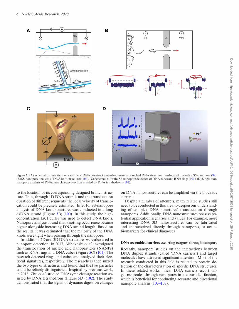
Figure 5. (A) Schematic illustration of a synthetic DNA construct assembled using a branched DNA structure translocated through a SS-nanopore (99).(B) SS-nanopore analysis of DNA knot structures (100). (C) Schematics for the SS-nanopore detection of DNA cubes and RNA rings (101). (D) Single-statenanopore analysis of DNAzyme cleavage reaction assisted by DNA tetrahedrons (102).
to the location of its corresponding designed branch struc-ture. Thus, through 1D DNA strands and the translocationduration of different segments, the local velocity of translo-cation could be precisely estimated. In 2016, SS-nanoporeanalysis of DNA knot structures was conducted in a longdsDNA strand (Figure 5B) (100). In this study, the high-concentration LiCl buffer was used to detect DNA knots.Nanopore analysis found that knotting occurrence becamehigher alongside increasing DNA strand length. Based onthe results, it was estimated that the majority of the DNAknots were tight when passing through the nanopore.
In addition, 2D and 3D DNA structures were also used innanopore detection. In 2017, Alibakhshi et al. investigatedthe translocation of nucleic acid nanoparticles (NANPs)such as RNA rings and DNA cubes (Figure 5C) (101). Theresearch detected rings and cubes and analyzed their elec-trical signatures, respectively. The researchers then mixedthe two types of structures and found that the two particlescould be reliably distinguished. Inspired by previous work,in 2018, Zhu et al. studied DNAzyme cleavage reaction as-sisted by DNA tetrahedrons (Figure 5D) (102). The studydemonstrated that the signal of dynamic digestion changes
on DNA nanostructures can be amplified via the blockadecurrent.
Despite a number of attempts, many related studies stillneed to be conducted in this area to deepen our understand-ing of complex DNA structures’ translocation throughnanopores. Additionally, DNA nanostructures possess po-tential application scenarios and values. For example, moreinteresting DNA 3D nanostructures can be fabricatedand characterized directly through nanopores, or act asbiomarkers for clinical diagnoses.
DNA assembled carriers escorting cargoes through nanopore
Recently, nanopore studies on the interactions betweenDNA duplex strands (called ‘DNA carriers’) and targetmolecules have attracted significant attention. Most of theresearch conducted in this field is related to protein de-tection or the characterization of specific DNA structures.In these related works, linear DNA carriers escort tar-get molecules through nanopores in a controlled fashion,which is beneficial for conducting accurate and directionalnanopore analysis (103–107).
Dow
nloaded from https://academ
ic.oup.com/nar/advance-article-abstract/doi/10.1093/nar/gkaa095/5742838 by guest on 26 February 2020
Nucleic Acids Research, 2020 7
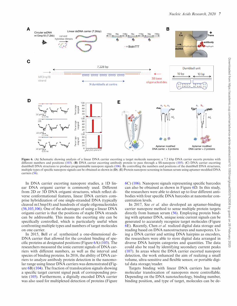
Figure 6. (A) Schematic showing analysis of a linear DNA carrier escorting a target molecule nanopore; a 7.2 kbp DNA carrier escorts proteins withdifferent numbers and positions (103). (B) DNA carrier escorting antibody protein to pass through a SS-nanopore (105). (C) DNA carrier escortingdumbbell DNA structures to produce programmable nanopore signals (106). By controlling the numbers and positions of the dumbbell DNA structures,multiple types of specific nanopore signals can be obtained as shown in (D). (E) Protein nanopore screening in human serum using aptamer-modified DNAcarriers (56).
In DNA carrier escorting nanopore studies, a 1D lin-ear DNA origami carrier is commonly used. Differentfrom 2D or 3D DNA origami structures, which reflect di-verse conformational features, linear DNA carriers com-prise hybridization of one single-stranded DNA (typicallycleaved m13mp18) and hundreds of staple oligonucleotides(56,103,106). One of the advantages of using a linear DNAorigami carrier is that the positions of staple DNA strandscan be addressable. This means the escorting site can bespecifically controlled, which is particularly useful whenconfronting multiple types and numbers of target moleculeson one carrier.
In 2015, Bell et al. synthesized a one-dimensional ds-DNA carrier that allowed for the covalent binding of spe-cific proteins at designated positions (Figure 6A) (103). Theresearchers measured the ionic current signals of DNA car-riers with different numbers, as well as the interval andspecies of binding proteins. In 2016, the ability of DNA car-riers to analyze antibody protein detection in the nanomo-lar range using linear DNA carriers was demonstrated (Fig-ure 6B) (104). The fraction of translocation signals showinga specific target current signal peak of corresponding pro-tein (105). Furthermore, a digitally encoded DNA carrierwas also used for multiplexed detection of proteins (Figure
6C) (106). Nanopore signals representing specific barcodescan also be obtained as shown in Figure 6D. In this study,the researchers were able to detect up to four different anti-bodies with four specific DNA barcodes at nanomolar con-centration levels.
In 2017, Sze et al. also developed an aptamer-bindingcarrier nanopore method to sense multiple protein targetsdirectly from human serum (56). Employing protein bind-ing with aptamer DNA, unique ionic current signals can begenerated to accurately recognize target molecules (Figure6E). Recently, Chen et al. realized digital data storage andreading based on DNA nanostructures and nanopores. Us-ing a DNA carrier and setting DNA hairpins as encoders,the researchers were able to store digital data arranged indiverse DNA hairpin categories and quantities. The datacould also be read by identifying secondary current peaks(107). In areas where the DNA carrier escorted nanoporedetection, the work enhanced the aim of realizing a smallvolume, ultra-sensitive and flexible sensor, or portable digi-tal data storage/reader.
Targets binding with linear DNA carriers has mademolecular translocation of nanopores more controllable.Depending on the DNA nanotechnology, binding number,binding position, and type of target, molecules can be de-
Dow
nloaded from https://academ
ic.oup.com/nar/advance-article-abstract/doi/10.1093/nar/gkaa095/5742838 by guest on 26 February 2020

8 Nucleic Acids Research, 2020
Figure 7. (A) Diagram showing a DNA nanoplate and nanopore. (B) Current trace of a nanoplate-nanopore system. (C) A current–voltage (I–V) char-acteristic curve of a bare pore and the same pore following successful nanoplate docking (109). (D) Schematic of the ionic current simulation system. (E)Theoretically calculated ionic currents for different bases when translocating the origami hybrid nanopore (110).
signed according to specific demands. Using this method,single target molecules can be recognized and analyzedfrom complex mixtures through specific binding. For ex-ample, physicochemical properties such as reaction kineticconstants between carriers and targets can be speculatedbased on signal changes during nanopore translocation.
DNA origami blockage to regulating nanopore
Recently, researchers have made significant attempts at di-rectly using DNA origami nanostructures as a nanoporestructure associated with lipid membrane or SS nanopores.Through DNA origami assembly, studies involving meth-ods such as DNA origami nanoplates and origaminanopore blockages have been developed, where transloca-tion speeds were able to be controlled by increasing interac-tions between analytes and DNA scaffold (108–110).
In 2014, Plesa et al. investigated the mechanical proper-ties of DNA origami nanoplates (Figure 7A) (109). VariousDNA origami nanoplates were prepared and captured onSS nanopores under certain voltages to test their integralionic conductance. Different origami nanoplates were de-signed, with the honeycomb lattice nanoplate providing thebest insulation. All nanoplates exhibited a rectification ef-fect, which the authors attributed to structural deformationof the nanoplates (Figure 7 B and C). The nanoplates couldbe pulled through the pore when using a high electric field
force. Similarly, in 2017, Farimani et al. proposed a sim-ulated DNA origami plate-graphene instrument for DNAdetection (110). Using molecular dynamic simulations, theresearchers computed the ionic conductivity of nanoporeson graphene docked with one or two-layered DNA origami.They demonstrated that even the four types of DNA basescould be distinguished according to the blockade current,due to the specific interactions between the DNA origamiplate layers and the different DNA bases (Figure 7 D andE).
DNA assembled structures directly serving as nanopores
Biological nanopores and SS-nanopores possess their ownstrengths and weaknesses, as noted above. In recent years,a combination of nanopore and DNA origami (so-called‘DNA origami nanopores’) have rapidly been developed.With its strong structural controllability, DNA origamiprovides a versatile method for assembling designablenanopores with precise and accurate shapes and sizes. Italso allows for introducing various addressable modifica-tions at the specific sites of DNA nanopores, thereby en-dowing nanopores with more powerful functions. In par-ticular, DNA origami nanopores may be suitable in con-ditions where variable and dynamic nanopore structuresare required. In this section, we focus primarily on DNA-assembled structures directly serving as nanopores. Based
Dow
nloaded from https://academ
ic.oup.com/nar/advance-article-abstract/doi/10.1093/nar/gkaa095/5742838 by guest on 26 February 2020
Nucleic Acids Research, 2020 9
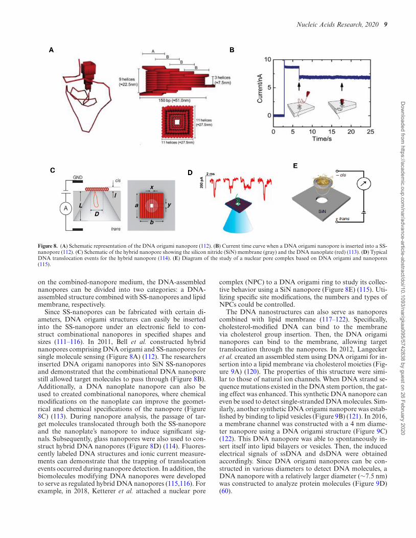
Figure 8. (A) Schematic representation of the DNA origami nanopore (112). (B) Current time curve when a DNA origami nanopore is inserted into a SS-nanopore (112). (C) Schematic of the hybrid nanopore showing the silicon nitride (SiN) membrane (gray) and the DNA nanoplate (red) (113). (D) TypicalDNA translocation events for the hybrid nanopore (114). (E) Diagram of the study of a nuclear pore complex based on DNA origami and nanopores(115).
on the combined-nanopore medium, the DNA-assemblednanopores can be divided into two categories: a DNA-assembled structure combined with SS-nanopores and lipidmembrane, respectively.
Since SS-nanopores can be fabricated with certain di-ameters, DNA origami structures can easily be insertedinto the SS-nanopore under an electronic field to con-struct combinational nanopores in specified shapes andsizes (111–116). In 2011, Bell et al. constructed hybridnanopores comprising DNA origami and SS-nanopores forsingle molecule sensing (Figure 8A) (112). The researchersinserted DNA origami nanopores into SiN SS-nanoporesand demonstrated that the combinational DNA nanoporestill allowed target molecules to pass through (Figure 8B).Additionally, a DNA nanoplate nanopore can also beused to created combinational nanopores, where chemicalmodifications on the nanoplate can improve the geomet-rical and chemical specifications of the nanopore (Figure8C) (113). During nanopore analysis, the passage of tar-get molecules translocated through both the SS-nanoporeand the nanoplate’s nanopore to induce significant sig-nals. Subsequently, glass nanopores were also used to con-struct hybrid DNA nanopores (Figure 8D) (114). Fluores-cently labeled DNA structures and ionic current measure-ments can demonstrate that the trapping of translocationevents occurred during nanopore detection. In addition, thebiomolecules modifying DNA nanopores were developedto serve as regulated hybrid DNA nanopores (115,116). Forexample, in 2018, Ketterer et al. attached a nuclear pore
complex (NPC) to a DNA origami ring to study its collec-tive behavior using a SiN nanopore (Figure 8E) (115). Uti-lizing specific site modifications, the numbers and types ofNPCs could be controlled.
The DNA nanostructures can also serve as nanoporescombined with lipid membrane (117–122). Specifically,cholesterol-modified DNA can bind to the membranevia cholesterol group insertion. Then, the DNA origaminanopores can bind to the membrane, allowing targettranslocation through the nanopores. In 2012, Langeckeret al. created an assembled stem using DNA origami for in-sertion into a lipid membrane via cholesterol moieties (Fig-ure 9A) (120). The properties of this structure were simi-lar to those of natural ion channels. When DNA strand se-quence mutations existed in the DNA stem portion, the gat-ing effect was enhanced. This synthetic DNA nanopore caneven be used to detect single-stranded DNA molecules. Sim-ilarly, another synthetic DNA origami nanopore was estab-lished by binding to lipid vesicles (Figure 9B) (121). In 2016,a membrane channel was constructed with a 4 nm diame-ter nanopore using a DNA origami structure (Figure 9C)(122). This DNA nanopore was able to spontaneously in-sert itself into lipid bilayers or vesicles. Then, the inducedelectrical signals of ssDNA and dsDNA were obtainedaccordingly. Since DNA origami nanopores can be con-structed in various diameters to detect DNA molecules, aDNA nanopore with a relatively larger diameter (∼7.5 nm)was constructed to analyze protein molecules (Figure 9D)(60).
Dow
nloaded from https://academ
ic.oup.com/nar/advance-article-abstract/doi/10.1093/nar/gkaa095/5742838 by guest on 26 February 2020

10 Nucleic Acids Research, 2020
Figure 9. (A) Schematic illustration and TEM images of the transmembrane channel (120). (B) Design and AFM images of DNA origami nanopores(121). (C) Design of the T-shape pore, composed of a double-layered top plate (gray) and a 27 nm-long stem (red) (122). (D) Synthetic protein conductivemembrane DNA nanopore (60).
More recently, smaller DNA channels consisting of sev-eral DNA strands were synthesized to mimic channel pro-teins, allowing for spontaneous transport of lipid molecules(123). For example, in 2016, Burns et al. developed anautomatic molecular valve made of seven DNA strands,which was able to perform the nanopore open or close func-tions through DNA strand displacement (Figure 10A) (57).The valve was also sensitive enough to distinguish smallmolecules that differed by only a single charged group (Fig-ure 10B). Due to its ability to regulate target translocation,the DNA valve can potentially be utilized for drug deliveryand synthetic cell or ionic logic circuits. In 2017, Guo et al.established a functional DNA nanopore for uptake by tu-mor cells (Figure 10C). The DNA nanopore comprised asmall DNA tube and was functionalized with Ramos cellaptamers and cell-penetrating peptides (124). Experimentalresults demonstrated that the DNA nanopores were able torecognize and penetrate Ramos cells with high specificity.
Nanopore logic sensing based on dynamic DNA assembly
The fields of smart molecular sensing and molecular com-puting have rapidly developed in recent years (93–97). Par-ticularly, logic operations based on DNA strand displace-ment reaction is one of the most common ways throughwhich to achieve intelligent detection and biocomputing.Existing methods for characterizing the output of logic op-erations are gel and fluorescence arrays (95–97). The superi-ority of nanopore technology, with features that include sin-
gle molecule sensing, being label free, and having significantrapidity presents potential detection solutions for address-ing particularly smart molecular sensing and biocomputing(125).
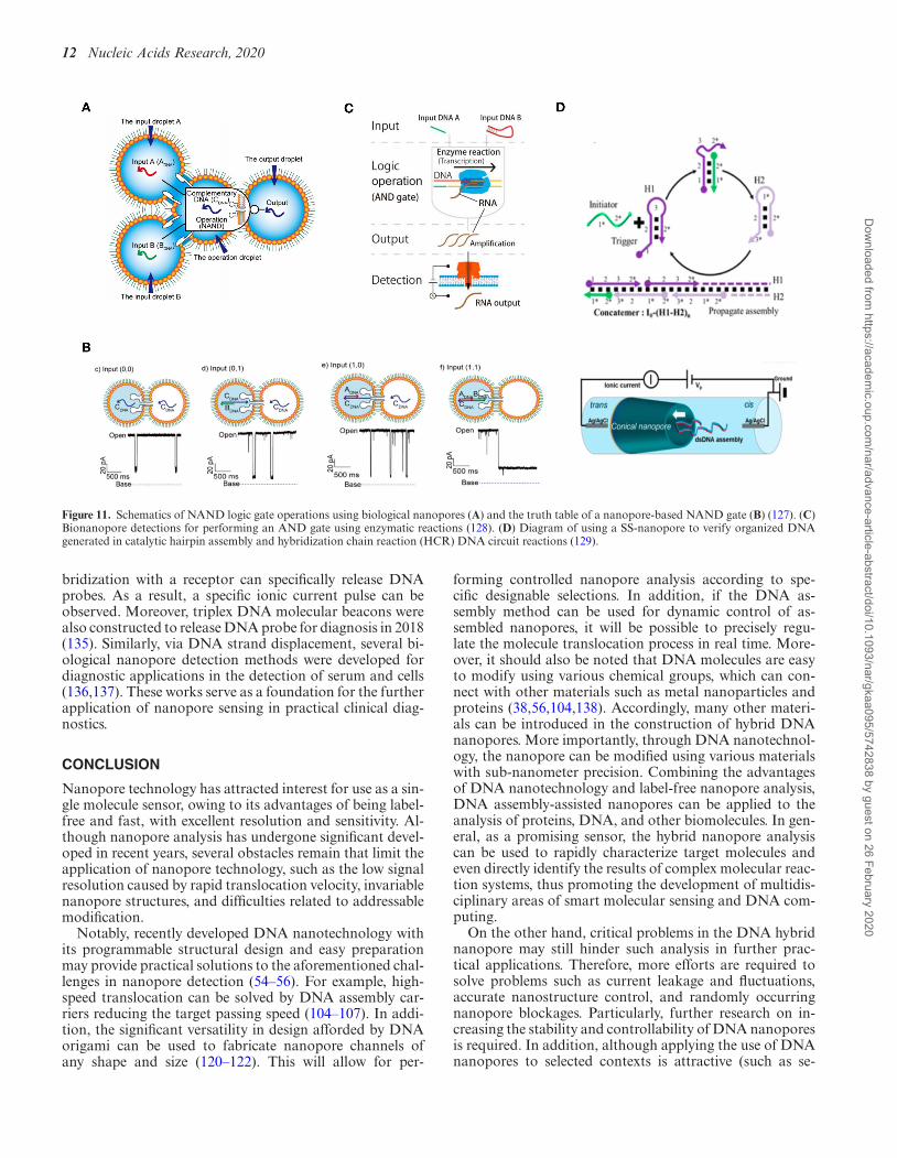
Recently, several research teams used nanopores to char-acterize the output of DNA logic operations. In 2009, Aliet al. demonstrated that conical nanopores functionalizedwith polyprotic acid chains showed three levels of conduc-tance based on pH value (126). The researchers utilizedthe functionalized nanocapillary and different chemical in-puts to realize AND and NOT logic gates. In 2016, Ya-suga et al. proposed a logic operating system with DNAmolecules, droplets, and biological nanopores (127). To re-alize a NAND operation, they set dsDNA that could notpass through the biological nanopore as ‘0’, while ssDNAthat could pass through the pore was set as ‘1’ (Figure 11A and B). In 2017, Ohara et al. described an AND logicoperation using T7 RNA polymerase (Figure 11C) (128).The existence of input DNA A or DNA B represented ‘1’while the non-existence of input DNA A or DNA B rep-resented ‘0’. Only when both DNA A and DNA B werepresent, could the electric signal be detected using a biolog-ical nanopore. Meanwhile, nanopores could also be used toanalyze the complex hairpin DNA structures generated inDNA circuit reactions. For example, Zhu et al. employedthe bionanopore technique to characterize complex DNAstructures at the single molecule level (129). By analyzingthe dwell time and blockade of ionic current signal fromDNA structure translocation, information pertaining to hy-
Dow
nloaded from https://academ
ic.oup.com/nar/advance-article-abstract/doi/10.1093/nar/gkaa095/5742838 by guest on 26 February 2020

Nucleic Acids Research, 2020 11
Figure 10. (A) Schematic illustrations of the DNA nanopore structure with a valve (57). Fluorophore carboxy-fluorescein (CF, red) and sulpho-rhodamineB (SRB, green) are self-quenched molecules. (B) Fluorescence signals of CF and SRB for vesicles with open valves. (C) Schematic illustration of the DNAnanopore’s recognition and endocytosis of a tumor cell (124).
bridization chain reaction could be successfully monitored(Figure 11D).
DNA assembly-based nanopore diagnosis
In recent years, programmable diagnoses based onnanopore methods has attracted significant attention.Combined with DNA assembly, nanopores can alsobe used in the detection of ultra-sensitive moleculesand accurate diagnoses. For example, the DNA stranddisplacement- assisted nanopore method can be used tosense target molecules at low concentrations, and even inimpure clinical samples. Through complex DNA assemblycircuit systems, low target signals can be amplified toproduce significant nanopore translocation results. Thisprogrammable DNA assembly-based nanopore methodwill undoubtedly contribute to more rapid and accuratediagnoses of diseases in future.
The utilization of nanopores in programmable diagnoseswill promote its use in practical clinical applications (130).In 2016, Rauf et al. designed a label-free nanopore biosen-sor for rapid detection of cocaine in human serum using an
aptamer for cocaine (Figure 12A) (131). In the experiment,the aptamer DNA was initially protected by hybridiza-tion with a short complementary DNA strand. Then, thegreater affinity of the cocaine/aptamer induced displace-ment of the short complementary DNA strand, thus allow-ing subsequent biological nanopores’ detection. In 2018,Xi et al. developed a biological nanopore method to de-tect cancer cells through enzymatic amplification reaction(Figure 12B) (132). Similarly, in 2017, Hiratani et al. de-scribed a strategy for cancer diagnoses using microRNA(miRNA) (Figure 12C) (133). The researchers amplifiedand quantified miRNA from cancer cells through stranddisplacement and used a biological nanopore to detect tar-gets. In addition, the ultra-sensitive and label-free nanoporemethod can be used for early diagnoses of various can-cers. In 2015, Li et al. reported a series of works aboutstrand displacement-based molecular sensing with biologi-cal nanopores. They hybridized aptamers with DNA probesbearing specific biomodification (CB[7]) to form a double-stranded structure that could not translocate through a bi-ological nanopore (Figure 12D) (134). Due to the higheraffinity between full complementary DNA strands, hy-
Dow
nloaded from https://academ
ic.oup.com/nar/advance-article-abstract/doi/10.1093/nar/gkaa095/5742838 by guest on 26 February 2020
12 Nucleic Acids Research, 2020
Figure 11. Schematics of NAND logic gate operations using biological nanopores (A) and the truth table of a nanopore-based NAND gate (B) (127). (C)Bionanopore detections for performing an AND gate using enzymatic reactions (128). (D) Diagram of using a SS-nanopore to verify organized DNAgenerated in catalytic hairpin assembly and hybridization chain reaction (HCR) DNA circuit reactions (129).
bridization with a receptor can specifically release DNAprobes. As a result, a specific ionic current pulse can beobserved. Moreover, triplex DNA molecular beacons werealso constructed to release DNA probe for diagnosis in 2018(135). Similarly, via DNA strand displacement, several bi-ological nanopore detection methods were developed fordiagnostic applications in the detection of serum and cells(136,137). These works serve as a foundation for the furtherapplication of nanopore sensing in practical clinical diag-nostics.
CONCLUSION
Nanopore technology has attracted interest for use as a sin-gle molecule sensor, owing to its advantages of being label-free and fast, with excellent resolution and sensitivity. Al-though nanopore analysis has undergone significant devel-oped in recent years, several obstacles remain that limit theapplication of nanopore technology, such as the low signalresolution caused by rapid translocation velocity, invariablenanopore structures, and difficulties related to addressablemodification.
Notably, recently developed DNA nanotechnology withits programmable structural design and easy preparationmay provide practical solutions to the aforementioned chal-lenges in nanopore detection (54–56). For example, high-speed translocation can be solved by DNA assembly car-riers reducing the target passing speed (104–107). In addi-tion, the significant versatility in design afforded by DNAorigami can be used to fabricate nanopore channels ofany shape and size (120–122). This will allow for per-
forming controlled nanopore analysis according to spe-cific designable selections. In addition, if the DNA as-sembly method can be used for dynamic control of as-sembled nanopores, it will be possible to precisely regu-late the molecule translocation process in real time. More-over, it should also be noted that DNA molecules are easyto modify using various chemical groups, which can con-nect with other materials such as metal nanoparticles andproteins (38,56,104,138). Accordingly, many other materi-als can be introduced in the construction of hybrid DNAnanopores. More importantly, through DNA nanotechnol-ogy, the nanopore can be modified using various materialswith sub-nanometer precision. Combining the advantagesof DNA nanotechnology and label-free nanopore analysis,DNA assembly-assisted nanopores can be applied to theanalysis of proteins, DNA, and other biomolecules. In gen-eral, as a promising sensor, the hybrid nanopore analysiscan be used to rapidly characterize target molecules andeven directly identify the results of complex molecular reac-tion systems, thus promoting the development of multidis-ciplinary areas of smart molecular sensing and DNA com-puting.
On the other hand, critical problems in the DNA hybridnanopore may still hinder such analysis in further prac-tical applications. Therefore, more efforts are required tosolve problems such as current leakage and fluctuations,accurate nanostructure control, and randomly occurringnanopore blockages. Particularly, further research on in-creasing the stability and controllability of DNA nanoporesis required. In addition, although applying the use of DNAnanopores to selected contexts is attractive (such as se-
Dow
nloaded from https://academ
ic.oup.com/nar/advance-article-abstract/doi/10.1093/nar/gkaa095/5742838 by guest on 26 February 2020

Nucleic Acids Research, 2020 13
Figure 12. (A) Schematic diagram of an aptamer-based nanopore sensor for cocaine detection (131). (B) Schematic diagram of cancer detection basedon biological nanopores (132). (C) Schematic illustration of the nanopore diagnosis system for small cell lung cancer via detection of miR-20a (133). (D)Schematic illustration of a nanopore sensing strategy based on aptamer binding (134).
quencing and drug delivery), significant developments arerequired in these areas. Though commercial viability is notyet a reality, it is believed that more significant achievementswill be made regarding the combination of nanopores andDNA nanotechnology in the next few years. Taking advan-tage of recent cross-disciplinary studies on DNA nanotech-nology and bioelectronic engineering, the DNA assembly-assisted nanopore method will deliver new opportunities inthe areas of single biomolecular analysis, gene sequencing,and clinical diagnosis.
FUNDING
National Key Research and Development Programof China [2017YFE0130600, 2016YFA0501600,2017YFE0103900]; National Natural Science Founda-
tion of China [61872007, 61320106005, 61772214]; JointFund of the Equipment Pre Research Ministry of Educa-tion [6141A02033607, 6141A02033608]; Beijing NaturalScience Foundation [4182027]; Beijing Municipal KeyR&D Project [Z151100003915081]. Funding for openaccess charge: National Key Research and DevelopmentProgram of China [2017YFE0130600].Conflict of interest statement. None declared.
REFERENCES1. Kasianowicz,J.J., Brandin,E., Branton,D. and Deamer,D.W. (1996)
Characterization of individual polynucleotide molecules using amembrane channel. Proc. Natl. Acad. Sci. U.S.A., 93, 13770–13773.
2. Stein,D. (2015) Nanopore sequencing: forcing improved resolution.Biophys. J., 109, 2001–2002.
Dow
nloaded from https://academ
ic.oup.com/nar/advance-article-abstract/doi/10.1093/nar/gkaa095/5742838 by guest on 26 February 2020

14 Nucleic Acids Research, 2020
3. Ayub,M., Hardwick,S.W., Luisi,B.F. and Bayley,H. (2013)Nanopore-based identification of individual nucleotides for directRNA sequencing. Nano Lett., 13, 6144–6150.
4. Feng,J., Liu,K., Bulushev,R.D., Khlybov,S., Dumcenco,D., Kis,A.and Radenovic,A. (2015) Identification of single nucleotides inMoS2 nanopores. Nat. Nanotech., 10, 1070–1076.
5. Huang,S., Romero-Ruiz,M., Castell,O.K., Bayley,H. andWallace,M.I. (2015) High-throughput optical sensing of nucleicacids in a nanopore array. Nat. Nanotech., 10, 986–991.
6. Fuller,C.W., Kumar,S., Porel,M., Chien,M., Bibillo,A.,Stranges,P.B., Dorwart,M., Tao,C., Li,Z., Guo,W. et al. (2016)Real-time single-molecule electronic DNA sequencing by synthesisusing polymer-tagged nucleotides on a nanopore array. Proc. Natl.Acad. Sci. U.S.A., 113, 5233–5238.
7. Yu,R.J., Ying,Y.L., Gao,R. and Long,Y.T. (2019) Confinednanopipette sensing: from single molecules, single nanoparticles, tosingle cells. Angew. Chem. Int. Ed., 58, 3706–3714.
8. Deamer,D.W. and Branton,D. (2002) Characterization of nucleicacids by nanopore analysis. Acc. Chem. Res., 35, 817–825.
9. Deamer,D. (2010) Nanopore analysis of nucleic acids bound toexonucleases and polymerases. Annu. Rev. Biophys., 39, 79–90.
10. Ying,Y.L., Zhang,J., Gao,R. and Long,Y.T. (2013) Nanopore-basedsequencing and detection of nucleic acids. Angew. Chem. Int. Ed.,52, 13154–13161.
11. Robert,P.J., Aaron,M.F., Rukshan,T.P., Cynthia,J.B. and Henry,S.W.(2017) Dynamics of a DNA mismatch site held in confinementdiscriminate epigenetic modifications of cytosine. J. Am. Chem. Soc.,139, 2750–2756.
12. Wang,L., Chen,X., Zhou,S., Roozbahani,G.M., Zhang,Y., Wang,D.and Guan,X. (2018) Displacement chemistry-based nanoporeanalysis of nucleic acids in complicated matrices. Chem. Commun.,54, 13977–13980.
13. Waduge,P., Hu,R., Bandarkar,P., Yamazaki,H., Cressiot,B.,Zhao,Q., Whitford,P.C. and Wanunu,M. (2017) Nanopore-basedmeasurements of protein size, fluctuations, and conformationalchanges. ACS Nano, 11, 5706–5716.
14. Oukhaled,G., Mathe,J., Biance,A.L., Bacri,L., Betton,J.M.,Lairez,D., Pelta,J. and Auvray,L. (2007) Unfolding of proteins andlong transient conformations detected by single nanopore recording.Phys. Rev. Lett., 98, 158101.
15. Soskine,M., Biesemans,A., Moeyaert,B., Cheley,S., Bayley,H. andMaglia,G. (2012) An engineered ClyA nanopore detects foldedtarget proteins by selective external association and pore entry.Nano Lett., 12, 4895–4900.
16. Wu,D., Bi,S., Zhang,L. and Yang,J. (2014) Single-molecule study ofproteins by biological nanopore sensors. Sensors, 14, 18211–18222.
17. Derrington,I.M., Craig,J.M., Stava,E., Laszlo,A.H., Ross,B.C.,Brinkerhoff,H., Nova,I.C., Doering,K., Tickman,B.I., Ronaghi,M.et al. (2015) Subangstrom single-molecule measurements of motorproteins using a nanopore. Nat. Biotechnol., 33, 1073–1075.
18. Yusko,E.C., Bruhn,B.R., Eggenberger,O.M., Houghtaling,J.,Rollings,R.C., Walsh,N.C., Nandivada,S., Pindrus,M., Hall,A.R.,Sept,D. et al. (2017) Real-time shape approximation andfingerprinting of single proteins using a nanopore. Nat.Nanotechnol., 12, 360–367.
19. Jubery,T.Z., Prabhu,A.S., Kim,M.J. and Dutta,P. (2012) Modelingand simulation of nanoparticle separation through a solid-statenanopore. Electrophoresis, 33, 325–333.
20. Tan,S., Wang,L., Liu,H., Wu,H. and Liu,Q. (2016) Singlenanoparticle translocation through chemically modified solidnanopore. Nanoscale Res Lett., 11, 50.
21. Cao,Y., Lin,Y., Qian,R.C., Ying,Y.L., Si,W., Sha,J., Chen,Y. andLong,Y.T. (2016) Evidence of single-nanoparticle translocationthrough a solid-state nanopore by plasmon resonance energytransfer. Chem. Commun., 52, 5230–5233.
22. Darvish,A., Goyal,G., Aneja,R., Sundaram,R.V., Lee,K.,Ahn,C.W., Kim,K.B., Vlahovska,P. M. and Kim,M.J. (2016)Nanoparticle mechanics: deformation detection via nanoporeresistive pulse sensing. Nanoscale, 8, 14420–14431.
23. Roman,J., Jarroux,N., Patriarche,G., Francais,O., Pelta,J., LePioufle,B. and Bacri,L. (2017) Functionalized solid-state nanoporeintegrated in a reusable microfluidic device for a better stability andnanoparticle detection. ACS Appl. Mater. Interfaces, 9,41634–41640.
24. Fu,K., Han,D., Crouch,G.M., Kwon,S.R. and Bohn,P.W. (2018)Voltage-gated nanoparticle transport and collisions inattoliter-volume nanopore electrode arrays. Small, 14, 1703248.
25. Hulings,Z.K., Melnikov,D.V. and Gracheva,M.E. (2018) Browniandynamics simulations of the ionic current traces for a neutralnanoparticle translocating through a nanopore. Nanotechnology, 24,445204.
26. Stefureac,R.I., Madampage,C.A., Andrievskaia,O. and Lee,J.S.(2010) Nanopore analysis of the interaction of metal ions with prionproteins and peptides. Biochem. Cell. Biol., 88, 347–358.
27. Korman,C.E., Megens,M., Ajo-Franklin,C.M. and Horsley,D.A.(2013) Nanopore-spanning lipid bilayers on silicon nitridemembranes that seal and selectively transport ions. Langmuir, 29,4421–4425.
28. Wang,G., Wang,L., Han,Y., Zhou,S. and Guan,X. (2014) Nanoporedetection of copper ions using a polyhistidine probe. Bioelectronics,53, 453–458.
29. Chen,L., He,H., Xu,X. and Jin,Y. (2015) Single glassnanopore-based regenerable sensing platforms with anon-immobilized polyglutamic acid probe for selective detection ofcupric ions. Anal. Chim. Acta, 889, 98–105.
30. Mayne,L.J., Christie,S.D. and Platt,M. (2016) A tunable nanoporesensor for the detection of metal ions using translocation velocityand biphasic pulses. Nanoscale, 8, 19139–19147.
31. Roozbahani,G.M., Chen,X., Zhang,Y., Xie,R., Ma,R., Li,D., Li,H.and Guan,X. (2017) Peptide-mediated nanopore detection of uranylions in aqueous media. ACS Sensor., 2, 703–709.
32. Roozbahani,G.M., Chen,X., Zhang,Y., Juarez,O., Li,D. andGuan,X. (2018) Computation-assisted nanopore detection ofthorium ions. Anal. Chem., 90, 5938–5944.
33. Kawano,R., Osaki,T., Sasaki,H., Takinoue,M., Yoshizawa,S. andTakeuchi,S. (2011) Rapid detection of a cocaine-binding aptamerusing biological nanopores on a chip. J. Am. Chem. Soc., 133,8474–8477.
34. Keyser,U.F. (2016) Enhancing nanopore sensing with DNAnanotechnology. Nat. Nanotech., 11, 106–108.
35. Lieberman,K.R., Cherf,G.M., Doody,M.J., Olasagasti,F.,Kolodji,Y. and Akeson,M. (2010) Processive replication of singleDNA molecules in a nanopore catalyzed by phi29 DNA polymerase.J. Am. Chem. Soc., 132, 17961–17972.
36. Manrao,E.A., Derrington,I.M., Laszlo,A.H., Langford,K.W.,Hopper,M.K., Gillgren,N., Pavlenok,M., Niederweis,M. andGundlach,J.H. (2012) Reading DNA at single-nucleotide resolutionwith a mutant MspA nanopore and phi29 DNA polymerase. Nat.Biotechnol., 30, 349–353.
37. Wang,S., Haque,F., Rychahou,P.G., Evers,B.M. and Guo,P. (2013)Engineered nanopore of Phi29 DNA-packaging motor for real-timedetection of single colon cancer specific antibody in serum. ACSNano, 7, 9814–9822.
38. Campos,E., McVey,C.E., Carney,R.P., Stellacci,F., Astier,Y. andYates,J. (2013) Sensing single mixed-monolayer protected goldnanoparticles by the alpha-hemolysin nanopore. Anal. Chem., 85,10149–10158.
39. Cabello-Aguilar,S., Balme,S., Chaaya,A.A., Bechelany,M.,Balanzat,E., Janot,J.M., Pochat-Bohatier,C., Miele,P. andDejardin,P. (2013) Slow translocation of polynucleotides and theirdiscrimination by alpha-hemolysin inside a single track-etchednanopore designed by atomic layer deposition. Nanoscale, 5,9582–9586.
40. Zhang,X., Price,N.E., Fang,X., Yang,Z., Gu,L.Q. and Gates,K.S.(2015) Characterization of interstrand DNA-DNA cross-links usingthe alpha-hemolysin protein nanopore. ACS Nano, 9, 11812–11819.
41. Perera,R.T., Fleming,A.M., Peterson,A.M., Heemstra,J.M.,Burrows,C.J. and White,H.S. (2016) Unzipping of A-formDNA-RNA, A-form DNA-PNA, and B-form DNA-DNA in thealpha-hemolysin nanopore. Biophys. J., 110, 306–314.
42. Ding,Y. and Kanavarioti,A. (2016) Single pyrimidine discriminationduring voltage-driven translocation of osmylatedoligodeoxynucleotides via the alpha-hemolysin nanopore. BeilsteinJ. Nanotechnol., 7, 91–101.
43. Ding,Y., Fleming,A.M. and Burrows,C.J. (2016) Alpha-hemolysinnanopore studies reveal strong interactions between biogenicpolyamines and DNA hairpins. Mikrochim Acta, 183, 973–979.
Dow
nloaded from https://academ
ic.oup.com/nar/advance-article-abstract/doi/10.1093/nar/gkaa095/5742838 by guest on 26 February 2020
Nucleic Acids Research, 2020 15
44. Tan,C.S., Riedl,J., Fleming,A.M., Burrows,C.J. and White,H.S.(2016) Kinetics of T3-DNA ligase-catalyzed phosphodiester bondformation measured using the alpha-hemolysin nanopore. ACSNano, 10, 11127–11135.
45. Howorka,S. and Siwy,Z. (2009) Nanopore analytics: sensing ofsingle molecules. Chem. Soc. Rev., 38, 2360–2384.
46. Kidan,L., Kyeong-Beom,P., Hyung-Jun,K., Jae-Seok,Y.,Hongsik,C., Hyun-Mi,K. and Ki-Bum,K. (2018) Recent progress insolid-state nanopores. Adv. Mater, 30, 1704680.
47. Fujii,S. and Takeuchi,S. (2017) Pesticide vapor sensing using anaptamer, nanopore, and agarose gel on a chip. Lab Chip., 17,2421–2425.
48. Wei,R., Gatterdam,V., Wienke,R., Tampe,R. and Rant,U. (2012)Stochastic sensing of proteins with receptor modified solid statenanopores. Nat. Nanotech., 7, 257–263.
49. Goto,Y., Haga,T., Yanagi,I., Yokoi,T. and Takeda,K. (2015)Deceleration of single stranded DNA passing through a nanoporeusing a nanometre sized bead structure. Sci. Rep., 5, 16640.
50. Kowalczyk,S.W., Wells,D.B., Aksimentiev,A. and Dekker,C. (2012)Slowing down DNA translocation through a nanopore in lithiumchloride. Nano Lett., 12, 1038–1044.
51. Deng,Y., Huang,Q., Zhao,Y., Zhou,D., Ying,C. and Wang,D. (2017)Precise fabrication of a 5 nm graphene nanopore with a helium ionmicroscope for biomolecule detection. Nanotechnology, 28, 045302.
52. Bai,Z., Zhang,L., Li,H. and Liu,L. (2016) Nanopore creation ingraphene by ion beam irradiation: geometry, quality, and efficiency.ACS Appl. Mater. Interfaces, 8, 24803–24809.
53. Kundu,S. and Karmakar,S.N. (2016) Detection of base-pairmismatches in DNA using graphene-based nanopore device.Nanotechnology, 27, 135101.
54. Nam,S., Choi,I., Fu,C.C., Kim,K., Hong,S., Choi,Y., Zettl,A. andLee,L.P. (2014) Graphene nanopore with a self-integrated opticalantenna. Nano Lett., 14, 5584–5589.
55. Qiu,W. and Skafidas,E. (2014) Detection of protein conformationalchanges with multilayer graphene nanopore sensors. ACS Appl.Mater. Interfaces, 6, 16777–16781.
56. Sze,J.Y.Y., Ivanov,A.P., Cass,A.E.G. and Edel,J.B. (2017) Singlemolecule multiplexed nanopore protein screening in human serumusing aptamer modified DNA carriers. Nat. Commun., 8, 1552.
57. Burns,J.R., Seifert,A., Fertig,N. and Howorka,S. (2016) Abiomimetic DNA-based channel for the ligand-controlled transportof charged molecular cargo across a biological membrane. Nat.Nanotechnol., 11, 152–156.
58. Bell,N.A.W. and Keyser,U.F. (2014) Nanopores formed by DNAorigami: a review. FEBS Lett., 588, 3564–3570.
59. Yong-An,R., Han,G. and Xiangyuan,O. (2018) Advances in DNAorigami nanopores: fabrication, characterization and applications.Chin. J. Chem., 36, 875–885.
60. Diederichs,T., Pugh,G., Dorey,A., Xing,YZ., Burns,J.R.,Nguyen,Q.H., Tornow,M., Tampe,R. and Howorka,S. (2019)Synthetic protein-conductive membrane nanopores built with DNA.Nat. Commun., 10, 5018.
61. Dong,Y., Dong,C., Wan,F., Yang,J. and Zhang,C. (2015)Development of DNA computing and information processing basedon DNA-strand displacement. Sci. China Chem., 10, 1515–1523.
62. Kallenbach,N.R., Ma,R.I. and Seeman,N.C. (1983) An immobilenucleic acid junction constructed from oligonucleotides. Nature,305, 829–831.
63. Pinheiro,A.V., Han,D., Shih,W.M. and Yan,H. (2011) Challengesand opportunities for structural DNA nanotechnology. Nat.Nanotech., 6, 763–772.
64. Chen,Y.J., Groves,B., Muscat,R.A. and Seelig,G. (2015) DNAnanotechnology from the test tube to the cell. Nat. Nanotech., 10,748–760.
65. Hong,F., Zhang,F., Liu,Y. and Yan,H. (2017) DNA origami:scaffolds for creating higher order structures. Chem. Rev., 117,12584–12640.
66. Fu,D., Shah,S., Song,T. and Reif,J. (2018) DNA-based analogcomputing. Methods Mol. Biol., 1772, 411–417.
67. Fu,T.J. and Seeman,N.C. (1993) DNA double-crossover molecules.Biochemistry, 32, 3211–3220.
68. Winfree,E., Liu,F., Wenzler,L.A. and Seeman,N.C. (1998) Designand self-assembly of two-dimensional DNA crystals. Nature, 394,539–544.
69. Yan,H., LaBean,T.H., Park,S.H., Finkelstein,G. and Reif,J.H.(2003) DNA-templated self-assembly of protein arrays and highlyconductive nanowires. Science, 301, 1882–1884.
70. Labean,H.T., Yan,H., Kopatsch,J., Liu,F., Winfree,E., Reif,J.H. andSeeman,N.C. (2000) Construction, analysis, ligation, andself-assembly of DNA triple crossover complexes. JACS, 122,1848–1860.
71. Shen,Z., Yan,H., Wang,T. and Seeman,N.C. (2004) Paranemiccrossover DNA: A generalized holliday structure with applicationsin nanotechnology. JACS, 126, 2324–2325.
72. Ding,B., Sha,R. and Seeman,N.C. (2004) Pseudohexagonal 2DDNA crystals from double crossover cohesion. J. Am. Chem. Soc.,126, 10230–10231.
73. Park,S.H., Finkelstein,G. and Labean,T.H. (2008) Stepwiseself-assembly of DNA tile lattices using dsDNA bridges. J. Am.Chem. Soc., 130, 40–41.
74. Yin,P., Hariadi,R.F., Sahu,S., Choi,H.M.T., Park,S.H., Labean,T.H.and Reif,J.H. (2008) Programming DNA tube circumferences.Science, 321, 824–826.
75. Wei,B., Dai,M. and Yin,P. (2012) Complex shapes self-assembledfrom single-stranded DNA tiles. Science, 485, 623–626.
76. He,Y., Chen,Y., Liu,H., Ribbe,A.E. and Mao,C. (2006)Self-assembly of hexagonal DNA two-dimensional arrays. J. Am.Chem. Soc., 128, 15978–15979.
77. He,Y., Ye,T., Su,M., Zhang,C., Ribbe,A.E., Jiang,W. and Mao,C.(2008) Hierarchical self-assembly of DNA into symmetricsupramolecular polyhedra. Nature, 452, 198–201.
78. Rothemund,PW. (2006) Folding DNA to create nanoscale shapesand patterns. Nature, 440, 297–302.
79. Qian,L., Wang,Y., Zhang,Z., Zhao,J., Pan,D., Zhang,Y., Liu,Q. andFan,C. (2006) Analogic China map constructed by DNA. Chin. Sci.Bull., 51, 2973–2976.
80. Andersen,E.S., Dong,M., Nielsen,M.M., Jahn,K.,Lind-Thomsen,A., Mamdouh,W., Gothelf,K.V., Besenbacher,F. andKjems,J. (2008) DNA origami design of dolphin-shaped structureswith flexible tails. ACS Nano, 2, 1213–1218.
81. Pound,E., Ashton,J.R., Becerril,H.A. and Woolley,A.T. (2009)Polymerase chain reaction based scaffold preparation for theproduction of thin, branched DNA origami nanostructures ofarbitrary sizes. Nano Lett., 9, 4302–4305.
82. Andersen,E.S., Dong,M., Nielsen,M.M., Jahn,K., Subramani,R.,Mamdouh,W., Golas,M.M., Sander,B., Stark,H., Oliveira,C.L.P.et al. (2009) Self-assembly of a nanoscale DNA box with acontrollable lid. Nature, 459, 73–76.
83. Kuzuya,A. and Komiyama,M. (2009) Design and construction of abox-shaped 3D-DNA origami. Chem. Commun., 28, 4182–4184.
84. Endo,M., Hidaka,K., Kato,T., Namba,K. and Sugiyama,H. (2009)DNA prism structures constructed by folding of multiplerectangular arms. J. Am. Chem. Soc., 131, 15570–15571.
85. Dietz,H., Douglas,S.M. and Shish,W.M., (2009) Folding DNA intotwisted and curved nanoscale shapes. Science, 325, 725–730.
86. Douglas,S.M., Dietz,H., Liedl,T., Hogberg,B., Graf,F. andShih,W.M. (2009) Self-assembly of DNA into nanoscalethree-dimensional shapes. Nature, 459, 414–418.
87. Han,D., Pal,S., Nangreave,J., Deng,Z., Liu,Y. and Yan,H. (2011)DNA origami with complex curvatures in three-dimensional space.Science, 332, 342–346.
88. Li,Q., Luan,G., Guo,Q. and Liang,J. (2002) A new class ofhomogeneous nucleic acid probes based on specific displacementhybridization. Nucleic Acids Res., 30, e5.
89. Yurke,B., Turberfield,A.J., Mills,A.P., Simmel,F.C. andNeumann,J.L. (2000) A DNA-fuelled molecular machine made ofDNA. Nature, 406, 605–608.
90. Zhang,D. and Seelig,G. (2011) Dynamic DNA nanotechnologyusing strand-displacement reactions. Nature Chem., 3, 103–113.
91. Zhang,D.Y. and Winfree,E. (2009) Control of DNA stranddisplacement kinetics using toehold exchange. J. Am. Chem. Soc.,131, 17303–17314.
92. Dirks,R.M. and Pierce,N.A. (2004) Triggered amplification byhybridization chain reaction. Proc. Natl Acad. Sci. U.S.A., 101,15275–15278.
93. Seelig,G., Soloveichik,D., Zhang,D.Y. and Winfree,E. (2006)Enzyme-free nucleic acid logic circuits. Science, 314, 1585–1588.
94. Yin,P., Choi,H., Calvert,C. and Pierce,N.A. (2008) Programmingbiomolecular self-assembly pathways. Nature, 451, 318–322.
Dow
nloaded from https://academ
ic.oup.com/nar/advance-article-abstract/doi/10.1093/nar/gkaa095/5742838 by guest on 26 February 2020

16 Nucleic Acids Research, 2020
95. Qian,L. and Winfree,E. (2011) Scaling up digital circuitcomputation with DNA strand displacement cascades. Science, 332,1196–1201.
96. Lopez,R., Wang,R. and Seelig,G. (2018) A molecular multi-geneclassifier for disease diagnostics. Nature Chem., 10, 746–754.
97. Rothemund,W.P., Papadakis,N. and Winfree,E. (2008) Algorithmicself-assembly of DNA tile lattices using dsDNA bridges. J. Am.Chem. Soc., 130, 40–41.
98. Adleman,L.M. (1994) Molecular computation of solutions tocombinatorial problems. Science, 266, 1021–1024.
99. Plesa,C., Van Loo,N., Ketterer,P., Dietz,H. and Dekker,C. (2015)Velocity of DNA during translocation through a solid-statenanopore. Nano Lett., 15, 732–737.
100. Plesa,C., Verschueren,D., Pud,S., van der Torre,J., Ruitenberg,J.W.,Witteveen,M.J., Jonsson,M.P., Grosberg,A.Y., Rabin,Y. andDekker,C. (2016) Direct observation of DNA knots using asolid-state nanopore. Nat. Nanotech., 11, 1093–1097.
101. Alibakhshi,M.A., Halman,J.R., Wilson,J., Aksimentiev,A.,Afonin,K.A. and Wanunu,M. (2017) Picomolar fingerprinting ofnucleic acid nanoparticles using solid-state nanopores. ACS Nano,11, 9701–9710.
102. Zhu,L., Xu,Y., Ali,I., Liu,L., Wu,H., Lu,Z. and Liu,Q. (2018)Solid-state nanopore single-molecule sensing of DNAzyme cleavagereaction assisted with nucleic acid nanostructure. ACS Appl. Mater.Interfaces, 10, 26555–26565.
103. Bell,N.A.W. and Keyser,U.F. (2015) Specific protein detection usingdesigned DNA carriers and nanopores. J. Am. Chem. Soc., 137,2035–2041.
104. Kong,J., Bell,N.A.W. and Keyser,U.F. (2016) Quantifyingnanomolar protein concentrations using designed DNA carriers andsolid-state nanopores. Nano Lett., 16, 3557–3562.
105. Plesa,C., Ruitenberg,J.W., Witteveen,M.J. and Dekker,C. (2015)Detection of individual proteins bound along DNA using solid-statenanopores. Nano Lett., 15, 3153–3158.
106. Bell,N.A.W. and Keyser,U.F. (2016) Digitally encoded DNAnanostructures for multiplexed, single-molecule protein sensing withnanopores. Nat. Nanotech., 11, 645–651.
107. Chen,KK., Kong,JL., Zhu,JB., Ermann,N., Predki,P. andKeyser,U.F. (2019) Digital data storage using DNA nanostructuresand solid-state nanopores. Nano Lett., 19, 1210–1215.
108. Hernandez-Ainsa,S. and Keyser,U.F. (2014) DNA origaminanopores: developments, challenges and perspectives. Nanoscale, 6,14121.
109. Plesa,C., Ananth,A.N., Linko,V., Gulcher,C., Katan,A.J., Dietz,H.and Dekker,C. (2014) Ionic permeability and mechanical propertiesof DNA origami nanoplates on solid-state nanopores. ACS Nano, 8,35–43.
110. Barati Farimani,A., Dibaeinia,P. and Aluru,N.R. (2017) DNAorigami-graphene hybrid nanopore for DNA detection. ACS Appl.Mater. Interfaces, 9, 92–100.
111. Hernandez-Ainsa,S., Misiunas,K., Thacker,V.V., Hemmig,E.A. andKeyser,U.F. (2014) Voltage-dependent properties of DNA origaminanopores. Nano Lett., 14, 1270–1274.
112. Bell,N.A.W., Engst,C.R., Ablay,M., Divitini,G., Ducati,C., Liedl,T.and Keyser,U.F. (2012) DNA origami nanopores. Nano Lett., 12,512–517.
113. Wei,R., Martin,T.G., Rant,U. and Dietz,H. (2012) DNA origamigatekeepers for solid-state nanopores. Angew. Chem. Int. Ed., 51,4864–4867.
114. Hernandez-Ainsa,S., Bell,N.A.W., Thacker,V.V., Gopfrich,K.,Misiunas,K., Fuentes-Perez,M.E. and Keyser,U.F. (2013) DNAorigami nanopores for controlling DNA translocation. ACS Nano,7, 6024–6030.
115. Ketterer,P., Ananth,A.N., Laman Trip,D.S., Mishra,A., Bertosin,E.,Ganji,M., van der Torre,J., Onck,P., Dietz,H. and Dekker,C. (2018)DNA origami scaffold for studying intrinsically disordered proteinsof the nuclear pore complex. Nat. Commun., 9, 902.
116. Fisher,P.D.E., Shen,Q., Akpinar,B., Davis,L.K., Chung,K.K.H.,Baddeley,D., Saric,A., Melia,T.J., Hoogenboom,B.W., Lin,C. andLusk,C.P. (2018) A programmable DNA origami platform fororganizing intrinsically disordered nucleoporins within nanoporeconfinement. ACS Nano, 12, 1508–1518.
117. Gopfrich,K., Zettl,T., Meijering,A.E.C., Hernandez-Ainsa,S.,Kocabey,S., Liedl,T. and Keyser,U.F. (2015) DNA-tile structures
induce ionic currents through lipid membranes. Nano Lett., 15,3134–3138.
118. Burns,J.R., Stulz,E. and Howorka,S. (2013) Self-assembled DNAnanopores that span lipid bilayers. Nano Lett., 13, 2351–2356.
119. Burns,J.R., Al-Juffali,N., Janes,S.M. and Howorka,S. (2014)Membrane-spanning DNA nanopores with cytotoxic effect. Angew.Chem. Int. Ed., 53, 12466–12470.
120. Langecker,M., Arnaut,V., Martin,T.G., List,J., Renner,S.,Mayer,M., Dietz,H. and Simmel,F.C. (2012) Synthetic lipidmembrane channels formed by designed DNA nanostructures.Science, 338, 932–935.
121. Gopfrich,K., Li,C.Y., Ricci,M., Bhamidimarri,S.P., Yoo,J.,Gyenes,B., Ohmann,A., Winterhalter,M., Aksimentiev,A. andKeyser,U.F. (2016) Large-conductance transmembrane porin madefrom DNA origami. ACS Nano, 10, 8207–8214.
122. Krishnan,S., Ziegler,D., Arnaut,V., Martin,T.G., Kapsner,K.,Henneberg,K., Bausch,A.R., Dietz,H. and Simmel,F.C. (2016)Molecular transport through large-diameter DNA nanopores. Nat.Commun., 7, 12787.
123. Ohmann,A., Li,C.-Y., Maffeo,C., Al Nahas,K., Baumann,K.N.,Gopfrich,K., Yoo,J., Keyser,U.F. and Aksimentiev,A. (2018) Asynthetic enzyme built from DNA flips 107 lipids per second inbiological membranes. Nat. Commun., 9, 2426.
124. Guo,X.L., Yuan,D.D., Song,T. and Li,X.M. (2017) DNA nanoporefunctionalized with aptamer and cell-penetrating peptide for tumorcell recognition. Anal. Bioanal. Chem., 409, 3789–3797.
125. Kawano,R. (2018) Nanopore decoding of oligonucleotides in DNAcomputing. Biotechnol. J., 13, e1800091.
126. Ali,M., Mafe,S., Ramirez,P., Neumann,R. and Ensinger,W. (2009)Logic gates using nanofluidic diodes based on conical nanoporesfunctionalized with polyprotic acid chains. Langmuir, 25,11993–11997.
127. Yasuga,H., Kawano,R., Takinoue,M., Tsuji,Y., Osaki,T.,Kamiya,K., Miki,N. and Takeuchi,S. (2016) Logic gate operation byDNA translocation through biological Nanopores. PLoS One, 11,e0149667.
128. Ohara,M., Takinoue,M. and Kawano,R. (2017) Nanopore logicoperation with DNA to RNA transcription in a droplet system.ACS Synth. Biol., 6, 1427–1432.
129. Zhu,Z.T., Zhou,Y., Xu,X.L., Wu,R.P., Jin,Y.D. and Li,B.L. (2018)Adaption of solid-state nanopore to homogeneous DNAorganization verification and label-free molecular analysis withoutcovalent modification. Anal. Chem., 90, 814–820.
130. Liu,L. and Wu,H.C. (2016) DNA-based nanopore sensing. Angew.Chem. Int. Ed., 55, 15216–15222.
131. Rauf,S., Zhang,L., Ali,A., Liu,Y. and Li,J. (2017) Label-freenanopore biosensor for rapid and highly sensitive cocaine detectionin complex biological fluids. ACS Sensor, 2, 227–234.
132. Xi,D., Li,Z., Liu,L., Ai,S. and Zhang,S. (2018) Ultrasensitivedetection of cancer cells combining enzymatic signal amplificationwith an aerolysin nanopore. Anal. Chem., 90, 1029–1034.
133. Hiratani,M., Ohara,M. and Kawano,R. (2017) Amplification andquantification of an antisense oligonucleotide from targetmicroRNA using programmable DNA and a biological nanopore.Anal. Chem., 89, 2312–2317.
134. Li,T., Liu,L., Li,Y., Xie,J. and Wu,H.C. (2015) A universal strategyfor aptamer-based nanopore sensing through host-guest interactionsinside alpha-hemolysin. Angew. Chem. Int. Ed., 54, 7568–7571.
135. Guo,B., Sheng,Y., Zhou,K., Liu,Q., Liu,L. and Wu,H.C. (2018)Analyte-triggered DNA-probe release from a triplex molecularbeacon for nanopore sensing. Angew. Chem. Int. Ed., 57, 3602–3606.
136. Liu,L., Li,Y., Li,T., Xie,J., Chen,C., Liu,Q., Zhang,S. and Wu,H.C.(2016) Selective detection of 8-Oxo-2’-deoxyguanosine insingle-stranded DNA via nanopore sensing approach. Anal. Chem.,88, 1073–1077.
137. Liu,L., Li,T., Zhang,S., Song,P., Guo,B., Zhao,Y. and Wu,H.C.(2018) Simultaneous quantification of multiple cancer biomarkers inblood samples through DNA-assisted nanopore sensing. Angew.Chem. Int. Ed., 57, 11882–11887.
138. Wang,F., Zahid,O.K., Swain,B.E., Parsonage,D., Hollis,T.,Harvey,S., Perrino,F.W., Kohli,R.M., Taylor,E.W. and Hall,A.R.(2017) Solid-state nanopore analysis of diverse DNA basemodifications using a modular enzymatic labeling process. NanoLett., 17, 7110–7116.
Dow
nloaded from https://academ
ic.oup.com/nar/advance-article-abstract/doi/10.1093/nar/gkaa095/5742838 by guest on 26 February 2020