![TLC and HPTLC fingerprinting of leaf extracts of Wedelia ... · HPTLC also facilitates repeated detection of chromatogram with same or different parameters [7, 8]. Wedelia chinensis](https://static.fdocuments.us/doc/165x107/5b56313b7f8b9adf7d8c3933/tlc-and-hptlc-fingerprinting-of-leaf-extracts-of-wedelia-hptlc-also-facilitates.jpg)
DNA Fingerprinting with a Dispersed Repeated Sequence ... · DNA Fingerprinting with a Dispersed...
9
The Plant Cell, Vol. 3, 95-102, January 1991 O 1991 American Society of Plant Physiologists DNA Fingerprinting with a Dispersed Repeated Sequence Resolves Pathotype Diversity in the Rice Blast Fungus Morris Levy,".' Jose Romao,' Marco A. Marchetti,b and John E. Hamer a a Department of Biological Sciences, Purdue University, West Lafayette, Indiana 47907 United States Department of Agriculture, Agricultura1 Research Service, Route 7, Box 999, Beaumont, Texas 7771 3 The poor definition of pathotype variation in the rice blast fungus has historically handicapped strategies for reducing blast disease damage to the world's rice crop. We have employed a probe for a dispersed repeated DNA sequence called MGR [Hamer et al. (1989). Proc. Natl. Acad. Sci. USA 86, 9981-99851 to construct genotype- specific, EcoRl restriction fragment length profiles (MGR-DNA fingerprints) from United States field isolates of this fungus. By using a blind-test design, we demonstrated that MGR-DNA fingerprints distinguished the major patho- types in the United States, accurately identified the pathotypes of isolates collected over a 30-year period, and defined the organization of clonal lineages within and among pathotype groups. These results resolved a lingering controversy regarding rice blast pathotype stability and illustrated new opportunities for tracking the population dynamics and evolution of this important crop pathogen. INTRODUCTION The blast fungus Magnaporthe grisea (Hebert) Barr is a geographically widespread plant pathogen whose various host-limited forms collectively parasitize more than 50 different grass species (Ou, 1985). The economically most important form of the pathogen is the rice blast fungus (Pyricularia grisea Sacc.), which is composed of clonal isolates that infect rice and cause the devastating rice blast disease (Ou, 1980). This disease occurs worldwide in chronic epidemic cycles despite the frequent introduction of new resistant rice cultivars. The absence of durable blast resistance in the field has been attributed to high levels of virulence form (pathotype)polymorphism in path- ogen populations (Ou, 1980). However, the nature of this polymorphism is the subject of much debate (for reviews see Day, 1974; Ou, 1985), marked by strongly contrasting views on the diversity and stability of pathotypes. The pathotype of a rice blast fungus isolate is deter- mined by assaying its infection spectrum, i.e., pathogenic- ity, on a set of differential rice cultivars. The differential set generally includes seven or more cultivars that differ from one another by one or more resistance genes (Ou, 1985). Using such an assay, Ou (1980) reported finding more than 250 pathotypes (conventionally called races) among field isolates from the Philippines. He and others (Ou and Ayad, 1968; Giatgong and Frederiksen, 1969) further judged that many isolates were pathogenically unstable, with multiple pathotypes being recovered from single plant lesions and from monoconidially derived cultures. Their ' To whom correspondenceshould be addressed. results led to the view that pathotypes may be promiscu- ously polymorphic and continually changing (Ou, 1985). Opposite conclusions were reached in pathogenicity as- says conducted by Latterell and colleagues (Latterell, 1975; Latterell and Rossi, 1986). They identified 50 path- otypes in a worldwide sampling of 2000 isolates. More- over, their extensive testing of clonally derived cultures showed that changes in pathotype were rare, even after prolonged culture storage. Latterell (1 975) and others (Bonman et al., 1987) have suggested that difficulties in standardizing assay procedures and interpreting lesion types lead to inflated estimates of pathotypic diversity and variability. The key to resolving these issues is to provide genetic definition to the phenotypic variations observed in pathogenicity assays. Although a variety of genetic markers can be used to study funga1 phytopathogen populations (for reviews see Michelmore and Hulbert, 1987; Newton, 1987), few have been definitive for the rice blast fungus. For example, Leung and Williams (1 986) found that the isozyme profiles of rice blast isolates from around the world are nearly uniform and differ little from those of M. grisea isolates that parasitize other grass hosts. Leung et al. (1989) also reported that mitochondrial DNA restriction fragment length polymorphisms (RFLPs) are similarly limited in dis- crimination power. In another study, Hunst et al. (1986) found some polymorphism for the lengths of double- stranded RNAs among rice pathogen isolates, but this variation was not correlated with pathotype. Recently, however, Hamer et al. (1 989) identified a family of dispersed repetitive DNA sequences, called MGR, that
-
Upload
hoanghuong -
Category
Documents
-
view
222 -
download
0
Transcript of DNA Fingerprinting with a Dispersed Repeated Sequence ... · DNA Fingerprinting with a Dispersed...

The Plant Cell, Vol. 3, 95-102, January 1991 O 1991 American Society of Plant Physiologists
DNA Fingerprinting with a Dispersed Repeated Sequence Resolves Pathotype Diversity in the Rice Blast Fungus
Morris Levy,".' Jose Romao,' Marco A. Marchetti,b and John E. Hamer a
a Department of Biological Sciences, Purdue University, West Lafayette, Indiana 47907 United States Department of Agriculture, Agricultura1 Research Service, Route 7, Box 999, Beaumont, Texas 7771 3
The poor definition of pathotype variation in the rice blast fungus has historically handicapped strategies for reducing blast disease damage to the world's rice crop. We have employed a probe for a dispersed repeated DNA sequence called MGR [Hamer et al. (1989). Proc. Natl. Acad. Sci. USA 86, 9981-99851 to construct genotype- specific, EcoRl restriction fragment length profiles (MGR-DNA fingerprints) from United States field isolates of this fungus. By using a blind-test design, we demonstrated that MGR-DNA fingerprints distinguished the major patho- types in the United States, accurately identified the pathotypes of isolates collected over a 30-year period, and defined the organization of clonal lineages within and among pathotype groups. These results resolved a lingering controversy regarding rice blast pathotype stability and illustrated new opportunities for tracking the population dynamics and evolution of this important crop pathogen.
INTRODUCTION
The blast fungus Magnaporthe grisea (Hebert) Barr is a geographically widespread plant pathogen whose various host-limited forms collectively parasitize more than 50 different grass species (Ou, 1985). The economically most important form of the pathogen is the rice blast fungus (Pyricularia grisea Sacc.), which is composed of clonal isolates that infect rice and cause the devastating rice blast disease (Ou, 1980). This disease occurs worldwide in chronic epidemic cycles despite the frequent introduction of new resistant rice cultivars. The absence of durable blast resistance in the field has been attributed to high levels of virulence form (pathotype) polymorphism in path- ogen populations (Ou, 1980). However, the nature of this polymorphism is the subject of much debate (for reviews see Day, 1974; Ou, 1985), marked by strongly contrasting views on the diversity and stability of pathotypes.
The pathotype of a rice blast fungus isolate is deter- mined by assaying its infection spectrum, i.e., pathogenic- ity, on a set of differential rice cultivars. The differential set generally includes seven or more cultivars that differ from one another by one or more resistance genes (Ou, 1985). Using such an assay, Ou (1980) reported finding more than 250 pathotypes (conventionally called races) among field isolates from the Philippines. He and others (Ou and Ayad, 1968; Giatgong and Frederiksen, 1969) further judged that many isolates were pathogenically unstable, with multiple pathotypes being recovered from single plant lesions and from monoconidially derived cultures. Their
' To whom correspondence should be addressed.
results led to the view that pathotypes may be promiscu- ously polymorphic and continually changing (Ou, 1985). Opposite conclusions were reached in pathogenicity as- says conducted by Latterell and colleagues (Latterell, 1975; Latterell and Rossi, 1986). They identified 50 path- otypes in a worldwide sampling of 2000 isolates. More- over, their extensive testing of clonally derived cultures showed that changes in pathotype were rare, even after prolonged culture storage. Latterell (1 975) and others (Bonman et al., 1987) have suggested that difficulties in standardizing assay procedures and interpreting lesion types lead to inflated estimates of pathotypic diversity and variability. The key to resolving these issues is to provide genetic definition to the phenotypic variations observed in pathogenicity assays.
Although a variety of genetic markers can be used to study funga1 phytopathogen populations (for reviews see Michelmore and Hulbert, 1987; Newton, 1987), few have been definitive for the rice blast fungus. For example, Leung and Williams (1 986) found that the isozyme profiles of rice blast isolates from around the world are nearly uniform and differ little from those of M. grisea isolates that parasitize other grass hosts. Leung et al. (1989) also reported that mitochondrial DNA restriction fragment length polymorphisms (RFLPs) are similarly limited in dis- crimination power. In another study, Hunst et al. (1986) found some polymorphism for the lengths of double- stranded RNAs among rice pathogen isolates, but this variation was not correlated with pathotype.
Recently, however, Hamer et al. (1 989) identified a family of dispersed repetitive DNA sequences, called MGR, that
96 The Plant Cell
was diagnostically conserved in M. grisea rice pathogen genomes. Severa1 features of MGR sequences suggested that they could be useful for population leve1 genetic analyses. MGR sequences occurred in approximately 50 copies per haploid genome and were dispersed among all chromosomes. DNA gel blot analysis using an MGR se- quence (pCB586) as a hybridization probe yielded mitoti- cally stable and isolate-specific EcoRl RFLP profiles, con- taining 50 or more resolvable fragments 0.7 kb to 20 kb in length. Consequently, MGR RFLP profiles could serve as genotype-specific DNA fingerprints (in the general sense of Jeffreys et al., 1985) that would also identify the distri- bution of clonal lineages within and among pathotypes of the rice blast fungus.
Here we report the results of experiments designed to resolve the issues of pathotype stability and diversity in United States rice blast pathogens by determining the diagnostic value of MGR-DNA fingerprints for pathotype identification. This resolution was based on the following rationale. If pathotypes are stable and represent distinct genotypes, then they should be composed of specific clonal lineages and, therefore, marked by lineage-associ- ated DNA fingerprints. This rationale also assumed that MGR restriction site polymorphisms were sufficiently vari- able to distinguish between clonal lineages but not so hypervariable that they obscured relatedness within lineages.
To maximize the objectivity of our analyses, we used a blind-test experimental design in which MGR-DNA finger- print variation was analyzed in a collection of United States field isolates without prior knowledge of their specific pathotypic identities or collection histories. Our results demonstrated that MGR-DNA fingerprints were diagnostic for seven of the eight United States pathotypes tested, supporting the view that pathotypes are stable and com- posed of discernible clonal lineages. We conclude that MGR-DNA fingerprinting has excellent potential for resolv- ing the population dynamics and the evolution of the rice blast pathogen in the United States.
RESULTS
MGR-Based Pathotype Assignments
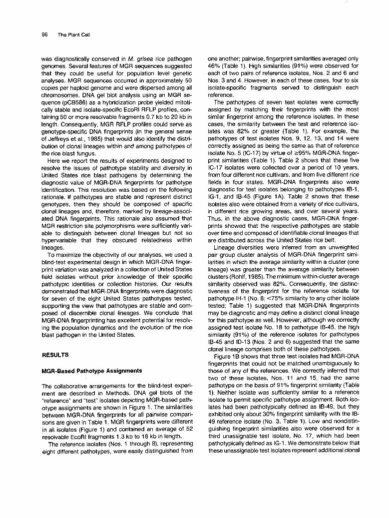
The collaborative arrangements for the blind-test experi- ment are described in Methods. DNA gel blots of the “reference” and “test” isolates depicting MGR-based path- otype assignments are shown in Figure 1. The similarities between MGR-DNA fingerprints for all pairwise compari- sons are given in Table 1. MGR fingerprints were different in all isolates (Figure 1) and contained an average of 52 resolvable EcoRl fragments 1.3 kb to 18 kb in length.
The reference isolates (Nos. 1 through 8), representing eight different pathotypes, were easily distinguished from
one another; pairwise, fingerprint similarities averaged only 46% (Table 1). High similarities (91%) were observed for each of two pairs of reference isolates, Nos. 2 and 6 and Nos. 3 and 4. However, in each of these cases, four to six isolate-specific fragments served to distinguish each reference.
The pathotypes of seven test isolates were correctly assigned by matching their fingerprints with the most similar fingerprint among the reference isolates. In these cases, the similarity between the test and reference iso- lates was 82% or greater (Table 1). For example, the pathotypes of test isolates Nos. 9, 12, 13, and 14 were correctly assigned as being the same as that of reference isolate No. 5 (IC-17) by virtue of r95% MGR-DNA finger- print similarities (Table 1). Table 2 shows that these five IC-17 isolates were collected over a period of 10 years, from four different rice cultivars, and from five different rice fields in four states. MGR-DNA fingerprints also were diagnostic for test isolates belonging to pathotypes IB-1, IG-1, and 18-45 (Figure 1A). Table 2 shows that these isolates also were obtained from a variety of rice cultivars, in different rice growing areas, and over severa1 years. Thus, in the above diagnostic cases, MGR-DNA finger- prints showed that the respective pathotypes are stable over time and composed of identifiable clonal lineages that are distributed across the United States rice belt.
Lineage diversities were inferred from an unweighted pair group cluster analysis of MGR-DNA fingerprint simi- larities in which the average similarity within a cluster (one lineage) was greater than the average similarity between clusters (Rohlf, 1985). The minimum within-cluster average similarity observed was 82%. Consequently, the distinc- tiveness of the fingerprint for the reference isolate for pathotype IH-1 (No. 8; <75% similarity to any other isolate tested; Table 1) suggested that MGR-DNA fingerprints may be diagnostic and may define a distinct clonal lineage for this pathotype as well. However, although we correctly assigned test isolate No. 18 to pathotype 18-45, the high similarity (91 “/O) of the reference isolates for pathotypes 18-45 and ID-13 (Nos. 2 and 6) suggested that the same clonal lineage comprises both of these pathotypes.
Figure 1 B shows that three test isolates had MGR-DNA fingerprints that could not be matched unambiguously to those of any of the references. We correctly inferred that two of these isolates, Nos. 11 and 15, had the same pathotype on the basis of 91 O/O fingerprint similarity (Table 1). Neither isolate was sufficiently similar to a reference isolate to permit specific pathotype assignment. Both iso- lates had been pathotypically defined as 18-49, but they exhibited only about 30% fingerprint similarity with the IB- 49 reference isolate (No. 3, Table 1). Low and nondistin- guishing fingerprint similarities also were observed for a third unassignable test isolate, No. 17, which had been pathotypically defined as IG-1. We demonstrate below that these unassignable test isolates represent additional clonal

DMA Fingerprints of the Rice Blast Fungus 97
IB-1 IC-17 IG-1 IB-45 ID-13 IH-1
1 | 1 6 5 | 9 |12 |13 |14 7 | 10 2 | 1 8 6 8
B IB-49 (IB-49) (IB-54) (IG-1)
9.4 - __ nt*fi5 tin (kb) 1 1 | 1 5 3 4 1 7
19.3 - •
Isf""""*"" "* 5 fi
5!
I
!- - i~_ • • *
ig si "• 2 m „_-" =EiiNlj 1:s ** •! 11 - :
I
Figure 1. MGR-DNA Fingerprints of the Isolates Used in the Blind Test.
Reference and test isolates are numbered 1 to 8 and 9 to 18, respectively. Isolate pathotypes are noted by International Race designations(Ling and Ou, 1969). The collection histories of the isolates are given in Table 2.(A) Isolates for which unambiguous assignments were made by MGR-DNA fingerprinting.(B) Isolates that gave ambiguous results.
lineages within their associated pathotype groups thatwere not apparent among the original references.
Resolving Lineages in the IB-49 and IB-54 PathotypeGroups
The low (30% to 31%) similarity between test and refer-ence isolates of pathotype IB-49 (Nos. 11 and 15 versusNo. 3, Figure 1B) was accompanied by an additionalambiguity, the high similarity (91%) between the referenceisolates of pathotypes IB-49 and IB-54 (No. 3 versus No.4, Figure 1 B). At first, we suspected that culture contami-nation or clerical errors were involved; these explanationswere ruled out by a repeated pathogenicity assay of thesource cultures. We then hypothesized that the MGR-DNAfingerprints accurately reflected the phylogenetic relation-ships among these isolates, i.e., that pathotype IB-49 wascomposed of two distinct clonal lineages, one of which
had recent common ancestry with the lineage representedin pathotype IB-54. To test this hypothesis, we obtainedMGR-DNA fingerprints from an additional set of 11 isolatesthat were characterized before DNA gel blot analysis onlyas having either pathotype IB-49 or IB-54. The MGR-DNAfingerprints of all the IB-49 and IB-54 isolates are shownin Figure 2. As predicted, this broader sampling definedthe presence of two very dissimilar MGR-DNA fingerprintgroups in pathotype IB-49. Groups A and B correspondedto the distinctive lineages represented by the original testand reference isolates, respectively. MGR-DNA fingerprintsimilarities were 91% or greater within each group and31% or less between groups. The broader sampling alsoconfirmed the extremely close relatedness between IB-49group B and IB-54 isolates. The results showed that MGR-DNA fingerprints are capable of distinguishing even com-plex lineage relationships within and among pathotypegroups. The isolates analyzed in Figure 2 were collectedin four different states and Puerto Rico from 1959 to 1988

98 The Plant Cell
Table 1. Percent Similarity (Nei and Li, 1979) of MGR Fingerprints among lsolates (Numbered as in Figure 1)
lsolates 2 3 4 5 6 7 8 9 10 11 12 13 14 15 16 17 18
1 2 3 4 5 6 7 8 9
10 11 12 13 14 15 16 17
35 35 37 64 33 60 62 65 61 46 65 34 36 39 91 40 37 41 39 32 37
91 31 30 35 33 31 30 30 32 35 32 37 37 35 29 30 35
39 80 72 97 80 39 96 39 35 41 37 30 37
70 81 87 33 80 73 68 44 74
38 80 36
a3 36 97
67 40 32 38 95 40 81 75 96 79 38 95
66 40 31 37 96 40 82 74 97 80 37 96 98
40 82 36 43 35 41 29 84 31 31 35 38 28 28 35 39 36 64 40 44 33 39 33 80 30 62 40 44 36 60 40 41 32 67 40 46 36 63 44 46 91 42 39 34
33 67 37 45 32 66 38 45
38 34 32 36 43
40
32 63 38 43
(see Figure 2 legend for details), supporting the conclusion that lineage-pathotype relationships in pathotypes IB-49 and IB-54 are stable. It remains possible that the A and B groups of pathotype IB-49 may be further differentiated pathogenically by as yet untested host genotypes.
Resolving Lineages in the IG-1 Pathotype Group
The preceding results suggested that the two very dissim- ilar MGR-DNA fingerprint types observed among IG-1 iso- lates in the original sample (isolates Nos. 7 and 1 O, Figure lA , versus isolate No. 17, Figure 1B) also reflected the presence of two distinct clonal lineages within the IG-1 pathotype group. A broader sampling of IG-1 isolates confirmed that this was the case. We obtained fingerprints for 13 additional IG-1 isolates, seven of which are shown in Figure 3. The fingerprints defined two lineages corre- sponding to those detected in the original blind test. The histories of these isolates indicated that their lineage- pathotype associations have been maintained from 1967 to 1989 across a three-state distribution.
DISCUSSION
Our experiments demonstrated that MGR sequence poly- morphisms have unprecedented utility as both population genetic and phylogenetic markers for the rice blast fungus in the United States. MGR-DNA fingerprints were diagnos- tic for seven of the eight pathotypes sampled (IB-1, IC-17, IG-1, 18-45, ID-13, IH-1, and IB-49A) and delimited eight distinctive clonal lineages. Six lineages exhibited only a
single pathotype (IB-1, IC-17, IG-1 A, IG-1 B, IH-1, and IB- 49A) and two exhibited multiple pathotypes (IB-49B/lB-54 and IB-45/ID-13). In both pathotypes comprising a pair of lineages, 18-49 and IG-1, the lineages were highly differ- entiated from one another (40% or less fingerprint similar- ity, Table l), suggesting that an episode of convergent evolution has occurred for each of these pathotypes. In lineages exhibiting multiple pathotypes, MGR-DNA finger- prints further indicated which pathotype groups may share recent common ancestry. In total, we analyzed 42 isolates collected over the last 30 years, and the pathotypes that were sampled represent the major blast fungus pathotypes found in the United States (Marchetti et al., 1976, 1987). A preliminary analysis of an additional 79 United States isolates, collected over the same period and representing pathotype groups IG-1, IC-17, IB-45, and IB-1, has not revealed any new MGR-DNA fingerprint lineages (M.A. Marchetti, M. Levy, S. Xu, and J.E. Hamer, unpublished results). Consequently, we anticipate that more extensive MGR-DNA fingerprinting will reveal that the United States blast fungus population contains only a limited number of clonal lineages. This will provide a reference base by which to document the emergence of new blast pathotypes as well as the appearance of new clonal lineages. Future integration of this information with rice cropping patterns will provide valuable insight on the influence of host gen- otype on the population biology of the rice blast fungus.
Our results strongly support the view that M. grisea pathotypes are stable and reliably distinguishable by care- fully standardized pathogenicity assays (Latterell, 1975; Latterell and Rossi, 1986; Bonman et al., 1987). If patho- types (or MGR-DNA fingerprints) were subject to sudden, continuous, or wholesale changes, it would have been impossible to identify pathotypes on the basis of genetic

DMA Fingerprints of the Rice Blast Fungus 99
Table 2. History of Rice Blast Fungus Isolates Used in thePathotype Assignment Blind Test
PathotypeIB-1
IC-17
IG-1 (A)
IG-1 (B)IB-45
ID-13IH-1IB-49 (A)IB-49 (B)
IB-54
Isolate8
11659
1213147
10172
18683
11154
Year ofIsolation198119851975197819811985198519711982198519751985198219741967198819851959
RiceCultivar"MarsMarsLabelleLabelleLebonnetSkybonnetNewbonnetBrazosUnknownBasmatiSaturnTebonnetAkceltikNova 66SaturnUnknownMarsZenith
Location0
Beauregard, LACalcasieau, LAColorado, TXARPanama City, FLHarris, TXAcadia, LAJefferson, TXStuttgart, ARHollandale, MSJeff Davis, LAJefferson, TXJefferson, TXAcadia, LACalcasieau, LAAcadia, LAAcadia, LACalcasieau, LA
" Isolate numbers are as in Figure 1 and were assigned by J.E.H.b Blast resistance breeding history and the resistance genes inthese rice cultivars are described in Marchetti et al. (1987).c Location names denote either state parishes or counties withthe exceptions of the cities of Stuttgart, AR; Hollandale, MS; andPanama City, FL.
genetic lineages. This pattern may have arisen by multipleintroductions of the pathogen from a variety of geographicorigins. In addition, periodic strong host selection for spe-cific pathogen genotypes may have resulted in the selec-tion of one or a few genetic lineages within each pathotype,thereby reducing the number of variant genetic lineages.The strong evidence for a worldwide, monophyletic originof the rice blast fungus (Hamer et al., 1989) rules out thepossibility that rice blast disease arose de novo in NorthAmerica. In the future, it may be possible to decipher thegeographic origins of United States pathotypes through
IB-49 IB-49 IB-54Group A Group B
(kb) 1 ,2 ,3 ,4 [5 |6 , 7,8 |9 ,10 ,11 ,12 ,13,14
19.3 -
9.4 - i
6.6 -•
*"* SB«•• <*•
^^ ^^fe alur ^M '̂ Bfc M1M ^M ̂ ^B <^^» ^HV -—" ^^^ ^^^f "̂̂ ^^w ̂ fff
3.5 -
lineage associations maintained over 30 years. We rec-ognize the possibility that uniquely mutable genetic lociinvolved in determining rice-cultivar specificity may exist insome field isolates. However, our results indicate that thecontribution of such isolates to the pathogen's populationdynamics is likely to be minimal. We are currently evalu-ating the utility of MGR-DNA fingerprinting in rice-growingregions outside the United States. The recent findings thatPhilippine and Korean rice blast isolates may not be aspathogenically variable as previously described (Bonmanet al., 1987) suggest that MGR-DNA fingerprinting mayhave broad application.
Rice cultivation, and by association blast disease, has arather short history in the United States (approximately300 years since introduction with approximately 100 yearsof substantial planting in the Gulf Coast states; Grist, 1986)compared with other rice-growing regions (e.g., more than5000 years in China; Chang, 1976). Consequently, weanticipated that the resolving power of our analysis mightbe limited by the presence of highly related genetic lineagesamong United States pathotypes. However, with the ex-ception of the IB-45/ID-13 and the IB-49B/IB-54 pathotypegroups, our results showed that most major United Statespathotypes are composed of discrete, well-differentiated
.-.•""̂ •••»»«»«w» —
Figure 2. MGR-DNA Fingerprints of IB-49 and IB-54 Isolates.
The original IB-49 and IB-54 reference isolates (Nos. 3 and 4,Figure 1B) are presented in lanes 8 and 9, respectively, and anoriginal test isolate of IB-49 (No. 15, Figure 1B) is presented inlane 1. The state and date of collection of isolates are as follows:IB-49 group A: 1 (LA, 1985), 2 (FL, 1985), 3 (Puerto Rico, 1987),4 (FL, 1982); IB-49 group B: 5 (LA, 1981), 6 (LA, 1967), 7 (TX,1967), 8 (LA, 1967); IB-54: 9 (LA, 1959), 10 (1976 reisolation ofLA, 1959 in lane 9 after numerous serial transfers), 11 (1984reisolation of LA. 1959 in lane 9 after numerous serial transfers),12 (LA, 1959), 13 (LA, 1964), 14 (TX, 1965).

100 The Plant Cell
IG-1 Pathotype
Group GroupA B
1 .2 .3 .4 5 .6 .7
«**•
to* • ^
«H*
MGR-DNA fingerprinting of a international sampling of riceblast fungus populations.
We have demonstrated that a repeated DNA sequenceprobe can be used as both a population and phylogeneticmarker in M. grisea. In addition, this same probe has alsobeen used to map genes in M. grisea (Hamer and Givan,1990). Traditionally, single-copy DNA sequences havebeen the probes of choice for RFLP analysis. However,for population level analysis, a large number of single-copyprobes is often necessary. For example, diversity andphylogenetic relationships among 25 isolates of the lettucedowny mildew fungus required the use of 35 differentsingle-copy DNA probes (Hulbert and Michelmore, 1988).By comparison, MGR restriction site polymorphisms aresufficiently variable to distinguish individual field isolatesbut not so hypervariable as to obscure their genetic relat-edness. This especially informative pattern of variation maybe peculiar to repetitive DNAs like MGRs, which appear tobe components of a transposable genetic element andmay be subject to high rates of base substitution (Hameretal., 1989).
Our demonstration that MGR-DNA fingerprinting reliablyindexes genetic and pathotypic diversity in United Statesrice blast pathogens promises that much needed epide-miologic information on the disease may become availablesoon. MGR-DNA fingerprinting can determine the currentgeographic organization of pathotypic diversity and moni-tor its change over time as well as the dispersal ranges ofspecific genotypes. This will improve the process of se-lecting and deploying more durably resistant rice cultivarsfor particular rice-growing areas (Morrison, 1987). MGRmarkers will also facilitate the study of the rate and direc-tion of pathotype evolution in this important pathogenwhich, like the evolution of resistance in its host, is drivenby the domestication process. Recently, the rice blastpathosystem was proposed as a model system for geneticand molecular biology studies in plant pathology (Valent,1990). The definition of genetic variation provided by MGR-DNA fingerprinting extends the utility of the rice blastpathosystem for clarifying the evolutionary dynamics thatunderlie host-parasite interactions.
***
Figure 3. MGR-DNA Fingerprints of IG-1 Isolates.
The state and date of collection of isolates are as follows: groupA: 1 (TX, 1967), 2 (LA, 1989), 3 (TX, 1976), 4 (LA, 1976); groupB: 5 (TX, 1976), 6 (TX, 1976), 7 (AR, 1975).
METHODS
Strains, Culture Conditions, and Pathogenicity Assays
All Magnaporthe grisea (Hebert) Barr (Pyricularia grisea Sacc.)isolates in this study are currently stored in the laboratories ofJ.E.H. and M.L. at Purdue University (West Lafayette, IN) or inthe laboratory of M.A.M. at Texas A&M University AgriculturalResearch and Extension Center (Beaumont, TX). Isolates werepropagated first on oatmeal agar media and then grown, beforeDNA isolation, in 2YEG liquid culture per Valent et al. (1986).Pathotype assignments were made by M.A.M. using the eightcultivars of the International differential set and defined by thenomenclature of Ling and Ou (1969).

DNA Fingerprints of the Rice Blast Fungus 101
DNA lsolations and MGR-DNA Fingerprint Production
DNA was prepared from liquid grown cultures as previously described (Valent et al., 1986; Hamer and Givan, 1990). M. grisea DNA was digested with EcoRI, fractionated on 0.8% agarose gels, and transferred to Hybond-N hybridization membranes (Amersham International) according to the manufacturer's sug- gestions. DNA blots were hybridized with the MGR subclone pCB586 (Hamer et al., 1989), which was radioactively labeled by the random primer method (Feinberg and Vogelstein, 1983). Fol- lowing hybridization, the DNA blots were washed at high strin- gency at 65% in 0.1% SDS, 0.1% PPi, 0.2 x SSPE (1 x SSPE = 0.1 8 M NaCI, 1 mM EDTA, 1 O mM sodium phosphate, pH 7.4), and then exposed to x-ray film.
Experimental Design
To test whether MGR-DNA fingerprints could reliably identify blast pathotypes, a collaboration was arranged between laboratories at Purdue University (J.E.H., J.R., and M.L.) and Texas A&M University (M.A.M.). M.A.M. provided 18 field isolates whose specific pathotypes and collection origins were not identified. Eight isolates were characterized by M.A.M. as "references" for different pathotypes, the remaining 1 O isolates as "test unknowns" assign- able to the reference pathotypes. The eight reference isolates and 10 test isolates were marked by M.A.M. collection codes when they were received. They were then recoded in arbitrary order as "reference isolates Nos. 1 through 8" and "test isolates Nos. 9 through 18" before DNA extraction and MGR-DNA fingerprinting. DNA fingerprints of the reference sets and test sets were obtained on separate DNA gel blots. Pathotype assignments were made by M.L. from fragment profiles identified solely by isolate number codes. The assignments were then translated into M.A.M. identi- fiers and conveyed to him by telephone for evaluation. Documen- tation of correspondence with regard to the experimental design is available upon request. It should be noted that this design provided an extremely rigorous test of Latterell's (1975) view of pathotype stability, given the small sample sizes of the test and reference isolate sets. Accurate pathotype assignments would have been confounded by the absence of a particular clonal lineage in either set (as was the case for the IB-49A, IB-49B, and IG-lB pathotype groups; Table 2) as well as by instances of pathogenic instability.
MGR-Based Pathotype Assignments
Similarities between MGR-DNA fingerprints were calculated using Nei and Li's (1979) index of genetic similarity for RFLP compari- sons (Sxy), which has the form S,, = 2n,,/(n. + n,), where n,, is the number of shared fragments and n, and n, are the numbers of fragments in the fingerprints x and y, respectively. For the test unknowns (isolates Nos. 9 through 18, Figure l ) , pathotypes were assigned to that of the reference isolates (Nos. 1 through 8, Figure 1) with the greatest fingerprint similarity, except where all S,, values were less than 50%. The correct assignment of test isolate No. 18 to the pathotype of reference isolate No. 2 (18-45) rather than to that of reference isolate No. 6 (ID-13) was based both on comparative overall similarity (84% versus 8O%, respectively) and the relative number of reference-specific fragments shared (6 versus 2, respectively; Figure 1A). Lineage diversities were in-
ferred from a cluster analysis of S,, values employing the un- weighted pair group method using arithmetic averages (UPGMA) option in the NT-SYS statistical package of Rohlf (1985).
ACKNOWLEDGMENTS
We thank Kathy F. Dobinson, Yangkyo Salch, Verel Shull, and Mark Decker for their critical reading of the manuscript. We also appreciate fruitful discussions with Dr. Frances Latterell. J.R. is the recipient of a graduate fellowship from INVOTAN (NATO/ Portugal). This work was supported by an award of an Eli Lilly Young Investigator Grant and a fellowship from the David and Lucille Packard Foundation to J.E.H.
Received September 7, 1990; accepted November 8, 1990.
REFERENCES
Bonman, J.M., Vergel de Dios, T.I., Bandong, J.M., and Lee, E.J. (1 987). Pathogenic variability of rnonoconidial isolates of Pyricularia oryzae in Korea and in the Philippines. Plant Disease
Chang, T.T. (1 976). The origin, evolution, cultivation, dissemina- tion, and diversification of Asian and Africap rices. Euphytica
Day, P.R. (1 974). Genetics of Host-Parasite lnteraction (San Fran- cisco: W.H. Freeman and co.), pp. 82-86.
Feinberg, A.P., and Vogelstein, B. (1983). A technique for ra- diolabelling DNA restriction fragments to high specificity. Anal. Biochem. 132,6-13.
Giatgong, P., and Frederiksen, R.A. (1 969). Pathogenic variabil- ity and cytology of monoconidial subcultures of Pyricularia oryzae. Phytopathology 59, 1152-1 157.
Grist, D.H. (1986). Rice, 6th ed. (New York: Longman, Inc.), pp,
Hamer, J.E., and Givan, S. (1990). Genetic mapping with dis- persed repeated sequences in the rice blast fungus: Mapping the SMO locus. MOI. Gen. Genet. 223, 487-495.
Hamer, J.E., Farrall, L., Orbach, M.J., Valent, B., and Chumley, F. (1989). Host species-specific conservation of a family of repeated DNA sequences in the genome of a funga1 plant pathogen. Proc. Natl. Acad. Sci. USA 86, 9981-9985.
Hulbert, S.H., and Michelmore, R.W. (1 988). DNA restriction fragment length polymorphism and somatic variation in the lettuce downy mildew fungus, Bremia lactucae. MOI. Plant- Microbe Interact. 1, 17-24.
Hunst, P.L., Latterell, F.M., and Rossi, A.E. (1 986). Variation in double-stranded RNA from isolates of Pyricularia oryzae. Phy- topathology 76, 674-678.
Jeffreys, A.J., Wilson, V., and Thien, S.L. (1985). Individual- specific "fingerprints" of human DNA. Nature 316, 76-79.
Latterell, F.M. (1 975). Phenotypic stability of pathogenic races of Pyricularia oryzae, and its implications for breeding of blast
71, 127-130.
25,425-441.
3-1 1.

102 The Plant Cell
resistant rice varieties. In Proceedings of the Seminar on Hori- zontal Resistance to the Blast Disease of Rice, October 1971, Calí, Colombia (Centro Internacional de Agricultura Tropical),
Latterell, F.M., and Rossi, A.E. (1 986). Longevity and pathogenic stability of Pyricularia oryzae. Phytopathology 76, 231 -235.
Leung, H., and Williams, P.H. (1 986). Enzyme polymorphism and genetic differentiation among geographic isolates of the rice blast fungus. Phytopathology 76, 778-783.
Leung, H., Nelson, R., Amante, A., Oliva, N., Borromeo, E., Raymundo, A.K., Ardales, E., Dalmacio, R.D., and Sitch, L.A. (1989). Molecular rice pathology at IRRI. In Abstracts, Third Annual Meeting of the Rockefeller Foundation’s lnternational Program on Rice Biotechnology, March 8-1 O, 1989 (New York: The Rockefeller Foundation).
Ling, K.C., and Ou, S.H. (1969). Standardization of the interna- tional lace numbers of Pyricularia oryzae. Phytopathology 59,
Marchetti, M.A., Rush, M.C., and Hunter, W.E. (1 976). Current status of rice blast in the southern United States. Plant Dis. Rep. 60,721-725.
Marchetti, M.A., Lai, X., and Bollich, C.N. (1987). lnheritance of resistance to Pyricularia oryzae in rice cultivars grown in the United States. Phytopathology 77, 799-804.
Michelmore, R.W., and Hulbert, S.H. (1 987). Molecular markers
pp. 199-234.
339-342.
for genetic analysis of phytopathogenic fungi. Annu. Rev. Phy- topathol. 25, 383-404.
Morcison, D. (1987). Blast disease threatens again. The Rice World & Soybean News, March issue, pp. 8-1 2.
Nei, M., and Li, W.H. (1979). Mathematical model for studying genetic variation in terms of restriction endonucleases. Proc. Natl. Acad. Sci. USA 76, 5269-5273.
Newton, A.C. (1987). Markers in pathogen populations. In Ge- netics and Plant Pathogenesis, P.R. Day and G.J. Jellis, eds (Oxford, UK: Blackwell Scientific Publishers), pp. 187-1 94.
Ou, S.H. (1 980). Pathogen variability and host resistance in rice blast disease. Annu. Rev. Phytopathol. 18, 167-1 87.
Ou, S.H. (1985). Rice Diseases (Slough, UK: Commonwealth Agricultura1 Bureaux), pp. 109-201.
Ou, S.H., and Ayad, M.R. (1 968). Pathogenic races of Pyricularia oryzae originating from single lesions and monoconidial cul- tures. Phytopathology 58, 179-1 82.
Rohlf, F.J. (1 985). Numerical Taxonomy System of Multivariate Statistical Programs. (Stony Brook, NY: The State University of New York at Stony Brook).
Valent, B. (1 990). Rice blast as a model system for plant pathol- ogy. Phytopathology 80, 33-36.
Valent, B., Crawford, M.S., Weaver, C.G., and Chumley, F.G. (1 986). Genetic studies of pathogenicity and fertility of Magna- porthe grisea. lowa State J. Res. 60, 569-594.
DOI 10.1105/tpc.3.1.95 1991;3;95-102Plant Cell
M. Levy, J. Romao, M. A. Marchetti and J. E. HamerRice Blast Fungus.
DNA Fingerprinting with a Dispersed Repeated Sequence Resolves Pathotype Diversity in the
This information is current as of April 6, 2019
Permissions X
https://www.copyright.com/ccc/openurl.do?sid=pd_hw1532298X&issn=1532298X&WT.mc_id=pd_hw1532298
eTOCs http://www.plantcell.org/cgi/alerts/ctmain
Sign up for eTOCs at:
CiteTrack Alerts http://www.plantcell.org/cgi/alerts/ctmain
Sign up for CiteTrack Alerts at:
Subscription Information http://www.aspb.org/publications/subscriptions.cfm
is available at:Plant Physiology and The Plant CellSubscription Information for
ADVANCING THE SCIENCE OF PLANT BIOLOGY © American Society of Plant Biologists