DNA AND RNA Structure of Nucleic Acids · The Trinity of Molecular Genetics: DNA, RNA and protein....
47
DNA AND RNA Structure of Nucleic Acids
Transcript of DNA AND RNA Structure of Nucleic Acids · The Trinity of Molecular Genetics: DNA, RNA and protein....

DNA AND RNAStructure of Nucleic Acids
2
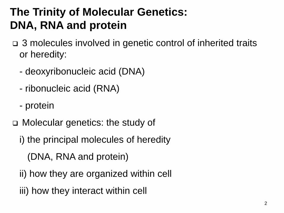
3 molecules involved in genetic control of inherited traits
or heredity:
- deoxyribonucleic acid (DNA)
- ribonucleic acid (RNA)
- protein
Molecular genetics: the study of
i) the principal molecules of heredity
(DNA, RNA and protein)
ii) how they are organized within cell
iii) how they interact within cell
The Trinity of Molecular Genetics:
DNA, RNA and protein

3
4

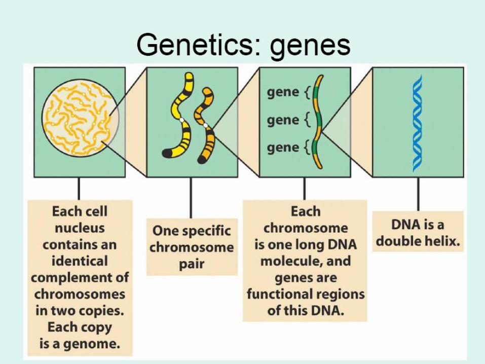
One human cell contains
about 2 meters of DNA,
packed into 46 chromosomes
which fits inside the nucleus
only 0.006mm in diameter
How is this possible???

In this animation we'll see the remarkable way our DNA is tightly packed up so that six feet of this long molecule fits into the microscopic nucleus of every cell. The process starts when DNA is wrapped around
special protein molecules called histones. The combined loop of DNA and protein is called a nucleosome. Next the nucleosomes are packaged into a thread, which is sometimes described as "beads on a string". The end
result is a fiber known as chromatin. Now the chromatin fiber is coiled into a structure called a "solenoid". This fiber is then looped and coiled yet again, leading finally to the familiar shapes known as chromosomes, which can be seen in the nucleus of dividing cells. Chromosomes are not always present. They form around the time
cells divide when the two copies of the cell's DNA need to be separated. At other times, as we can see now after the cell has divided, our DNA is less highly organized. It is still wrapped up around the histones, but not
coiled into chromosomes. 6

7

8
30 nm
(b) 30 nm fiber
(a) Nucleosomes (“beads on a string”)
2 nm
Wrapping of DNA around
a histone octamer
Nucleosome
DNA double helix
11 nm
Formation of a three-dimensional zigzag structure
via histone H1 and other DNA-binding proteins
Anchoring of radial loops to the
nuclear matrix
Histone
octamer
Histone H1
Nucleosome
Brooker, Fig 12.17a and b
Levels of DNA Packaging

9
(c) Radial loop domains
(d) Metaphase chromosome
300 nm
700 nm
1400 nm
Further compaction ofradial loops
Formation of a scaffold from the nuclear matrixand further compaction of all radial loops
Protein scaffold
Brooker, Fig 12.17
Compaction level in euchromatin(interphase)
Compaction level in heterochromatin
Levels of DNA Packaging, cont.

10
Assembly of DNA into
Chromosomes
• In eukaryotes, DNA is normally complexed with histone
proteins to form chromatin. Histones H2A, H2B, H3, and
H4 form a histone octamer, around which DNA wraps, to
form a nucleosome core. The nucleosome core plus 60
bp of linker DNA and one molecule of histone H1 forms a
nucleosome, which is the basic unit of chromatin
structure.
• This chromatin (known as the 10 nm fiber) can be more
highly organized and condensed, into a 30 nm fiber,
which can in turn be folded further to form the highly
condensed metaphase chromosomes. The 10 nm fiber
corresponds to euchromatin, while the 30 nm fiber
corresponds to heterochromatin. 11

• Chromosomes are long linear pieces of DNA
that associate with histones, forming chromatin.
• Some regions of each chromosome exist as
euchromatin during interphase, whereas other
regions remain as heterochromatin all the time.
• Since heterochromatin is genetically inactive,
any genes found in these heterochromatic
region will not function. Example of
heterochromatin that contains inactive genes is
the inactive X chromosome (Barr body) of
female mammals.
• The latter regions include repetitive DNA,
centromeres, and telomeres. 12

• The centromere is the region of each eukaryotic
chromosome that attaches to spindle fibers
during mitosis and meiosis.
• Telomeres are the ends of chromosomes, and
consist of numerous repeats of a six base
sequence
• In humans the telomeric sequence is TTAGGG.
This sequence is repeated over 50 times at the
end of each chromosome. These sequences are
added to the ends of the chromosomes by an
enzyme called telomerase.
• The reason telomeres exist is to provide stability
to the chromosomal ends.
13

14
Pentose sugars
Ribonucleic acid (RNA) contains ribose.
Deoxyribonucleic acid (DNA) contains 2-deoxyribose,
which is often referred to simply as deoxyribose.
• The only difference between the two sugars is that
ribose has a hydroxyl group on carbon 2, whereas
deoxyribose has only hydrogen in that position.
• Both have hydroxyl groups on carbons 3 and 5.
• In the configuration found in nucleic acids, carbons 1
through 4 are part of a ring structure, whereas carbon 5
is on a side chain.
Structural components of nucleic acids

15
Nucleic Acid Structure
Nucleotides
• The building blocks or genetic units of nucleic acid are called nucleotides
• Three (3) components that make up a single unit of nucleotide:
- pentose sugar (deoxyribose or ribose)
- phosphate group
- nitrogenous base: purine and pyrimidine
• purine – adenine and guanine
• pyrimidine – cytosine and thymine
16
Phosphate
• In both RNA and DNA, the phosphate groups formphosphodiester linkages between the 3' hydroxyl groupon one pentose sugar and the 5' hydroxyl group on thenext pentose sugar.
• The polymeric structure consisting of alternating sugarand phosphate residues provides nucleic acids with anextended backbone structure.
• The pentose sugars in the nucleic acid backbones allhave the same 5' to 3' orientation, giving each chain a 5'to 3' directionality.
17
Purine bases
• Purines are flat planar molecules consisting of a sixmember ring fused to a five member ring, with twonitrogen in each of the rings.
• Adenine (A) has amino nitrogen at position 6 of its largerring.
• Guanine (G) has amino nitrogen at position 2 andoxygen at position 6 of the larger ring.
• A and G occur in both DNA and RNA.
• The bond between the purines and the nucleic acidbackbone is from position 1' on the pentose sugar toposition 9 (a nitrogen in the smaller ring) of the purine.
18

19
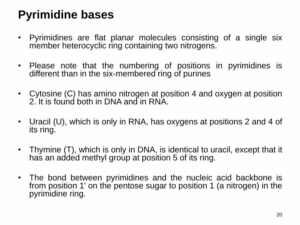
Pyrimidine bases
• Pyrimidines are flat planar molecules consisting of a single sixmember heterocyclic ring containing two nitrogens.
• Please note that the numbering of positions in pyrimidines isdifferent than in the six-membered ring of purines
• Cytosine (C) has amino nitrogen at position 4 and oxygen at position2. It is found both in DNA and in RNA.
• Uracil (U), which is only in RNA, has oxygens at positions 2 and 4 ofits ring.
• Thymine (T), which is only in DNA, is identical to uracil, except that ithas an added methyl group at position 5 of its ring.
• The bond between pyrimidines and the nucleic acid backbone isfrom position 1' on the pentose sugar to position 1 (a nitrogen) in thepyrimidine ring.
20

21
nucleotide comprises of
i) sugar: deoxyribose
ii) phosphate group
iii) nitrogenous base: adenine (A), thymine (T), guanine (G) and
cytosine (C)
at C1, sugar attached to nitrogenous base
at C5, sugar attached to phosphate group
at C2, only H atom is attached instead of OH group, that’s why the
sugar known as deoxyribose
Nucleotide sequences in DNA are always read 5' to 3‘
The 5' to 3' orientations of the two strands in a DNA double helix are
in the opposite directions. The two strands are described as being
antiparallel in their orientation.
DNA (DEOXYRIBONUCLEIC ACID)

22

23

24
Nucleic Acid Structure

25
• Purine and pyrimidine bases form hydrogen bonded base pairs in
double stranded DNA
• The normal pattern of base pairing in DNA is A to T and G to C.
• Abnormal pairing during DNA synthesis can result in mutation
• The base pairs in the DNA double helix are flat planar structures
• In the DNA double helix, the flat base pairs are tightly stacked on
top of one another, with the deoxyribose phosphate backbones
wound around the outside of the stack.
• The base pairs are 3.4 Å (0.34 nm) thick, with ten base pairs per
complete turn of the double helix.
DNA (DEOXYRIBONUCLEIC ACID)

A three dimensional model
of the DNA double helix
26

27
• The two deoxyribose phosphate backbones are unevenly spaced
around the helix, such that there is a wide groove and a narrow
groove between them.
• Separation of DNA strands (melting) occurs at high salt levels
and/or high temperature.
• GC base pairs have 3 hydrogen bonds and are more stable than AT
base pairs, which only have 2.
• Stability of double-helical DNA increases with higher GC content.
• Complex genomes, such as those of mammals, contain a mixture of
repetitive and unique sequence DNA.
DNA (DEOXYRIBONUCLEIC ACID)

Major features of DNA:
• Molecule :
• The DNA molecule is a double helix.
• The molecule is formed by two antiparallel polynucleotide strands which are spirally coiledround each other in a right-handed helix.
• The two strands are held together by hydrogen bonds.
• The double stranded helical molecule has alternate major (or deep) and minor grooves.
28
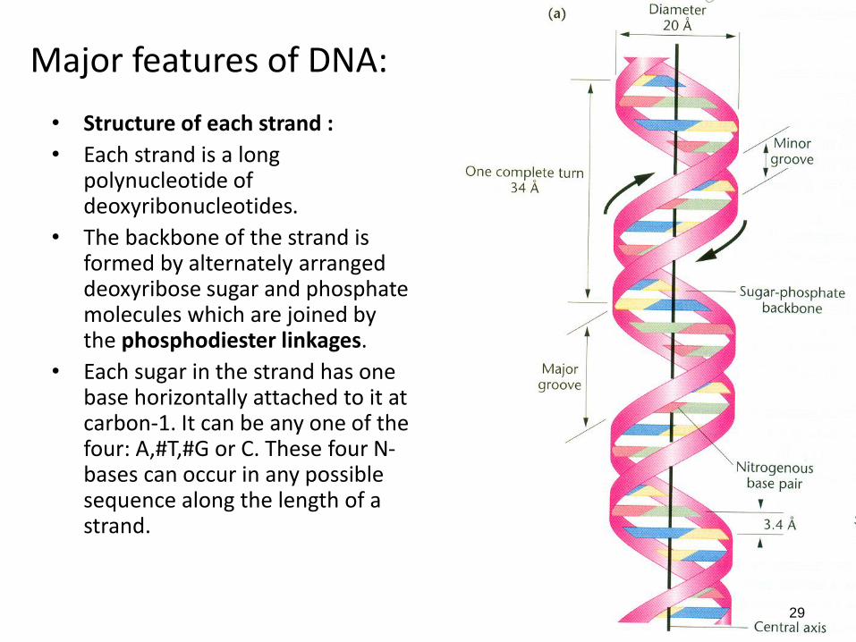
Major features of DNA:
• Structure of each strand :
• Each strand is a long polynucleotide of deoxyribonucleotides.
• The backbone of the strand is formed by alternately arranged deoxyribose sugar and phosphate molecules which are joined by the phosphodiester linkages.
• Each sugar in the strand has one base horizontally attached to it at carbon-1. It can be any one of the four: A,#T,#G or C. These four N-bases can occur in any possible sequence along the length of a strand.
29

Major features of DNA:• Complementary nature of the
strands :
• The two strands are complementary to each other with regards to the arrangement of the bases in the two strands.
• In the double helix, purines and pyrimidines exist in base pairs, i.e., (A and T) and (G and C). As a result, if the base sequence of one strand of DNA is known, the base sequence of its complementary strand can be easily deduced.
30

Major features of DNA:
• Complementary base pairing :
• In each pair, the two bases of the opposite strands are joined by hydrogen bonds.
• A and T are joined by two hydrogen bonds
• G and C are joined by three hydrogen bonds.
• This is called complementary base pairing.
31

Major features of DNA:
• Purine : Pyrimidine ratio :
• Because of the fixed or complementary base pairing in the DNA molecule, the total number of A is equal to the total number of T and the total number of G is equal to the total number of C.
• (A+G)= (T+C)
• Hence, purines: pyrimidines ratio is 1:1.
32

Major features of DNA:
• C-3 and C-5 ends of the strand :
• In each strand one end of the strand has one free phosphate group on carbon-5 of the sugar molecule.
• This is the end of the strand is called C-5 (or 5') end.
• The other end of the strand has a free -OH on carbon-3 of the sugar molecule.
• This is called C-3 (or 3') end of the strand
33

Major features of DNA:
• Antiparallel nature of strands:
• The two strands are oppositely oriented and hence are called antiparallel.
• The 3' end of one strand is adjacent to the 5' end of the other strand.
• This is because, the phosphate-sugar linkages run in opposite directions in the two strands.
34

• Dimensions: • The diameter of the DNA
double helix is 20 Ao (2.0nm). • The length of each complete
spiral (turn or pitch) of the molecule measures 34 Ao
(3.4nm). • 10 pairs of nucleotides are
present in each complete spiral.
• Therefore, each nucleotide in the strand occupies a distance of 3.4A0 (0.34nm).
35
Major features of DNA:

36
DNA (deoxyribonucleic acid) - serves as the genetic
material in all living organisms
DNA is organized into genes and stores genetic
information
Information contained in genes can be transmitted by
parents through gametes to their offspring
Genetic information in DNA will be transferred to a
closely related nucleic acid, RNA (ribonucleic acid)
RNA carries genetic information from nucleus into the
cytoplasm of the cell
Information in RNA will be translated into proteins
GENE EXPRESSION

GENE EXPRESSION
• The molecular expression of genes within cells leads to anorganism’s traits
• The information within the DNA is accessed during the process ofgene expression
• Genetic information is transcribed from DNA to RNA, with the antisense strand of the DNA serving as a template for synthesis ofan RNA with the same base sequence (5' to 3') as the sensestrand of the double helical DNA.
• Genetic information contained in messenger RNA (mRNA) istranslated into a sequence of amino acids in a polypeptide chainduring protein synthesis (translation).
• During translation, the RNA strand is used to specify thesequence of amino acids within a protein
• This protein functions within the cell influencing organism’s trait
37
38
Why would the cell want to
have an intermediate between
DNA and the proteins it
encodes?

Because….
• The DNA can stay pristine and protected, away
from the caustic chemistry of the cytoplasm.
• Gene information can be amplified by having
many copies of an RNA made from one copy of
DNA.
• Regulation of gene expression can be effected
by having specific controls at each element of
the pathway between DNA and proteins. The
more elements there are in the pathway, the
more opportunities there are to control it in
different circumstances. 40
41
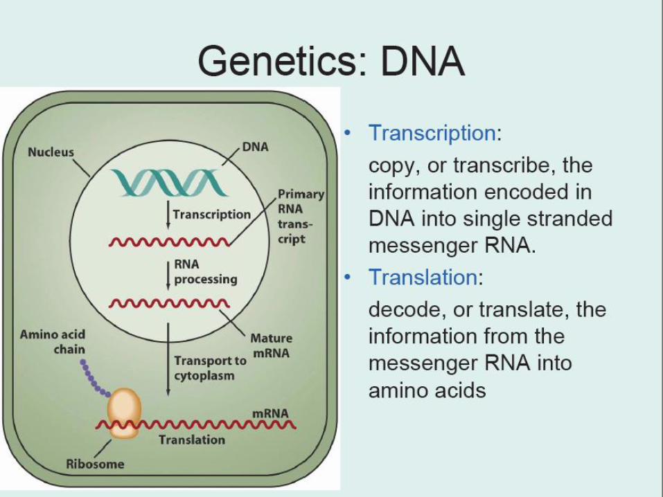
Process of storage and expression of genetic information
can be summarized as:
DNA makes RNA, which makes proteins
Process transferring information from DNA to RNA –
transcription
Subsequent conversion of the genetic information in RNA
into a protein – translation
Molecular level → Cellular level → Organism level →
Population level
GENE EXPRESSION

Relationship between Genes and Traits
42
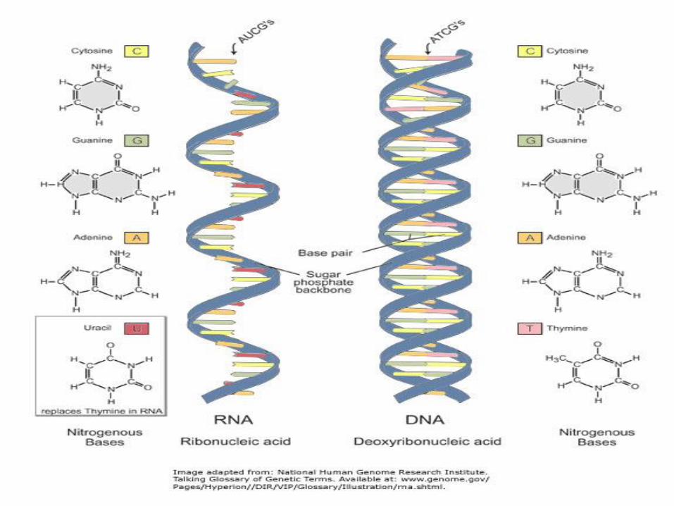
RNA basic unit is a ribonucleotide
RNA comprises of
i) sugar: ribose
ii) phosphate group
iii) nitrogenous base: adenine (A), uracil (U), guanine (G) andcytosine (C)
A base pairs with U; G base pairs with C
at C2, the sugar ribose differs from the deoxyribose by having a OHgroup (hydroxyl)
Because of the extra hydroxyl group on the sugar, RNA is too bulky toform a stable double helix.
RNA exists as a single-stranded molecule. However, regions ofdouble helix can form where there is some base paircomplementation (U and A , G and C), resulting in hairpin loops.The RNA molecule with its hairpin loops is said to have a secondarystructure.
43
44
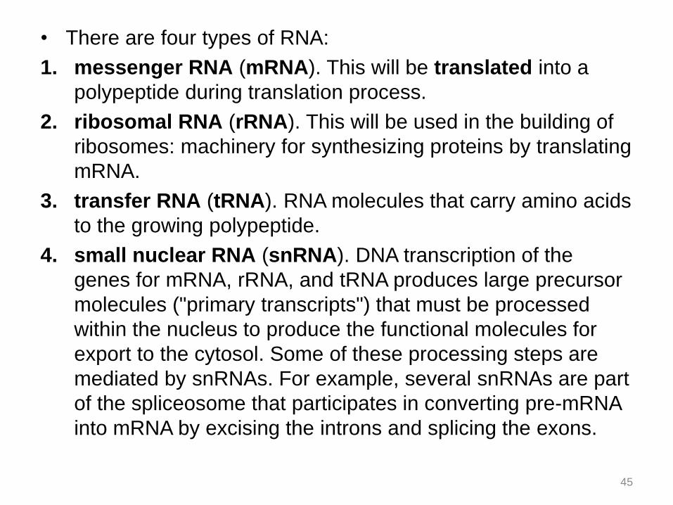
• There are four types of RNA:
1. messenger RNA (mRNA). This will be translated into a
polypeptide during translation process.
2. ribosomal RNA (rRNA). This will be used in the building of
ribosomes: machinery for synthesizing proteins by translating
mRNA.
3. transfer RNA (tRNA). RNA molecules that carry amino acids
to the growing polypeptide.
4. small nuclear RNA (snRNA). DNA transcription of the
genes for mRNA, rRNA, and tRNA produces large precursor
molecules ("primary transcripts") that must be processed
within the nucleus to produce the functional molecules for
export to the cytosol. Some of these processing steps are
mediated by snRNAs. For example, several snRNAs are part
of the spliceosome that participates in converting pre-mRNA
into mRNA by excising the introns and splicing the exons.
45
• RNA sequences are read 5' to 3' in thesame manner as DNA.
• The coding sequence in mRNA is thesame as that of the sense strand of DNA,except that U replaces T.
46

COMPARISON BETWEEN DNA &
RNA
DNA
• Double-stranded
• Basic unit is nucleotide
• The sugar is deoxyribose
• The bases are A, T, G, C
• Normally long
• Stable
• Present only in nucleus
RNA
• Single-stranded
• Basic unit is ribonucleotide
• The sugar is ribose
• The bases are A, U, G, C
• Normally short
• Not stable
• Present in both nucleus
and cytoplasm
47