Dlx5regulates regional development of the branchial arches and … · neurocranium, the skeleton...
16
INTRODUCTION The mammalian skull is an assemblage of skeletal elements with diverse developmental origins which encases the brain, its associated primary sensory organs (olfactory, visual, auditory, vestibular and gustatory), and the oral and respiratory cavities. The embryologically earliest skeletal structures to appear are the cartilaginous (chondrocranial) elements whose development appears to reflect the ancient bauplan of the vertebrate skull (Goodrich, 1958; Youssef, 1966; Barghusen and Hopson, 1979; Moore, 1981; De Beer, 1985; Zeller, 1987; Thorogood, 1988; Hanken and Thorogood, 1993; Novacek, 1993; Kuratani et al., 1997). Historically, two chondrocranial components have been distinguished: the first is the neurocranium, the skeleton built around the nose, eye, inner ear and base of the brain, and the second is the splanchnocranium, the skeleton of the branchial arches. While most of these structures undergo endochondral ossification, some undergo regression and/or non-osseous replacement. The development of the embryonic chondrocranium is coordinated with the development of the dermatocranium, the later-forming cranial skeleton, which undergoes intramembranous ossification over the brain and adjacent to the surfaces of many chondrocranial elements. Experimental evidence suggests that both mesenchyme and ectoderm play key roles in controlling of the number, shape, size and differentiation of the cranial anlagen (Noden, 1983, 1991; Hall, 1991; Hanken and Hall, 1993; Thorogood, 1993; Francis-West et al., 1998). Fate-mapping studies in amphibians and birds indicate that the dermatocranium and splanchnocranium ossify from cranial neural crest (CNC)- derived mesenchyme (ectomesenchyme) (Le Douarin, 1982; Noden, 1983, 1991; Hall, 1988; Serbedzija et al., 1992; Couly et al., 1993; Osumi-Yamashita et al., 1994; Köntges and Lumsden, 1996). This evidence further suggests that the CNC produces the mesenchyme that forms the rostral neurocranium: 3831 Development 126, 3831-3846 (1999) Printed in Great Britain © The Company of Biologists Limited 1999 DEV4175 We report the generation and analysis of mice homozygous for a targeted deletion of the Dlx5 homeobox gene. Dlx5 mutant mice have multiple defects in craniofacial structures, including their ears, noses, mandibles and calvaria, and die shortly after birth. A subset (28%) exhibit exencephaly. Ectodermal expression of Dlx5 is required for the development of olfactory and otic placode-derived epithelia and surrounding capsules. The nasal capsules are hypoplastic (e.g. lacking turbinates) and, in most cases, the right side is more severely affected than the left. Dorsal otic vesicle derivatives (e.g. semicircular canals and endolymphatic duct) and the surrounding capsule, are more severely affected than ventral (cochlear) structures. Dlx5 is also required in mandibular arch ectomesenchyme, as the proximal mandibular arch skeleton is dysmorphic. Dlx5 may control craniofacial development in part through the regulation of the goosecoid homeobox gene. goosecoid expression is greatly reduced in Dlx5 mutants, and both goosecoid and Dlx5 mutants share a number of similar craniofacial malformations. Dlx5 may perform a general role in skeletal differentiation, as exemplified by hypomineralization within the calvaria. The distinct focal defects within the branchial arches of the Dlx1, Dlx2 and Dlx5 mutants, along with the nested expression of their RNAs, support a model in which these genes have both redundant and unique functions in the regulation of regional patterning of the craniofacial ectomesenchyme. Key words: Branchial arch, Dlx5, Placode, Mouse, Craniofacial defect SUMMARY Dlx5 regulates regional development of the branchial arches and sensory capsules Michael J. Depew 1, *, Jen Kuei Liu 1, * ,‡ , Jason E. Long 1, *, Robert Presley 2 , Juanito J. Meneses 3 , Roger A. Pedersen 3 and John L. R. Rubenstein 1,§ 1 Nina Ireland Laboratory of Developmental Neurobiology, Center for Neurobiology and Psychiatry, Department of Psychiatry and Programs in Neuroscience, Developmental Biology, Oral Biology and Biomedical Sciences, 401 Parnassus Avenue, University of California at San Francisco, S.F., CA, 94143-0984, USA 2 Anatomy Unit, University of Wales, Cardiff, UK 3 Reproductive Genetics Unit, Department of Obstetrics, Gynecology and Reproductive Sciences, University of California at San Francisco, S.F., CA, 94143, USA *Joint first authors ‡ Present address: Sentinel Biosciences, 855 South California Avenue, Suite C, Palo Alto CA 94304, USA § Author for correspondence (email: [email protected]) Accepted 31 May; published on WWW 5 August 1999
Transcript of Dlx5regulates regional development of the branchial arches and … · neurocranium, the skeleton...
INTRODUCTION
The mammalian skull is an assemblage of skeletal elementswith diverse developmental origins which encases the brain, itsassociated primary sensory organs (olfactory, visual, auditory,vestibular and gustatory), and the oral and respiratory cavities.The embryologically earliest skeletal structures to appearare the cartilaginous (chondrocranial) elements whosedevelopment appears to reflect the ancient bauplan of thevertebrate skull (Goodrich, 1958; Youssef, 1966; Barghusenand Hopson, 1979; Moore, 1981; De Beer, 1985; Zeller, 1987;Thorogood, 1988; Hanken and Thorogood, 1993; Novacek,1993; Kuratani et al., 1997). Historically, two chondrocranialcomponents have been distinguished: the first is theneurocranium, the skeleton built around the nose, eye, inner earand base of the brain, and the second is the splanchnocranium,the skeleton of the branchial arches. While most of thesestructures undergo endochondral ossification, some undergo
regression and/or non-osseous replacement. The developmentof the embryonic chondrocranium is coordinated with thedevelopment of the dermatocranium, the later-forming cranialskeleton, which undergoes intramembranous ossification overthe brain and adjacent to the surfaces of many chondrocranialelements.
Experimental evidence suggests that both mesenchyme andectoderm play key roles in controlling of the number, shape,size and differentiation of the cranial anlagen (Noden, 1983,1991; Hall, 1991; Hanken and Hall, 1993; Thorogood, 1993;Francis-West et al., 1998). Fate-mapping studies in amphibiansand birds indicate that the dermatocranium andsplanchnocranium ossify from cranial neural crest (CNC)-derived mesenchyme (ectomesenchyme) (Le Douarin, 1982;Noden, 1983, 1991; Hall, 1988; Serbedzija et al., 1992; Coulyet al., 1993; Osumi-Yamashita et al., 1994; Köntges andLumsden, 1996). This evidence further suggests that the CNCproduces the mesenchyme that forms the rostral neurocranium:
3831Development 126, 3831-3846 (1999)Printed in Great Britain © The Company of Biologists Limited 1999DEV4175
We report the generation and analysis of mice homozygousfor a targeted deletion of the Dlx5 homeobox gene. Dlx5mutant mice have multiple defects in craniofacialstructures, including their ears, noses, mandibles andcalvaria, and die shortly after birth. A subset (28%) exhibitexencephaly. Ectodermal expression of Dlx5 is required forthe development of olfactory and otic placode-derivedepithelia and surrounding capsules. The nasal capsules arehypoplastic (e.g. lacking turbinates) and, in most cases, theright side is more severely affected than the left. Dorsalotic vesicle derivatives (e.g. semicircular canals andendolymphatic duct) and the surrounding capsule, aremore severely affected than ventral (cochlear) structures.Dlx5 is also required in mandibular arch ectomesenchyme,as the proximal mandibular arch skeleton is dysmorphic.
Dlx5 may control craniofacial development in part throughthe regulation of the goosecoid homeobox gene. goosecoidexpression is greatly reduced in Dlx5 mutants, and bothgoosecoid and Dlx5 mutants share a number of similarcraniofacial malformations. Dlx5 may perform a generalrole in skeletal differentiation, as exemplified byhypomineralization within the calvaria. The distinct focaldefects within the branchial arches of the Dlx1, Dlx2 andDlx5 mutants, along with the nested expression of theirRNAs, support a model in which these genes have bothredundant and unique functions in the regulation ofregional patterning of the craniofacial ectomesenchyme.
Key words: Branchial arch, Dlx5, Placode, Mouse, Craniofacialdefect
SUMMARY
Dlx5 regulates regional development of the branchial arches and sensory
capsules
Michael J. Depew1,*, Jen Kuei Liu1,*,‡, Jason E. Long1,*, Robert Presley2, Juanito J. Meneses3, Roger A. Pedersen3 and John L. R. Rubenstein1,§
1Nina Ireland Laboratory of Developmental Neurobiology, Center for Neurobiology and Psychiatry, Department of Psychiatry andPrograms in Neuroscience, Developmental Biology, Oral Biology and Biomedical Sciences, 401 Parnassus Avenue, University ofCalifornia at San Francisco, S.F., CA, 94143-0984, USA2Anatomy Unit, University of Wales, Cardiff, UK3Reproductive Genetics Unit, Department of Obstetrics, Gynecology and Reproductive Sciences, University of California at SanFrancisco, S.F., CA, 94143, USA*Joint first authors‡Present address: Sentinel Biosciences, 855 South California Avenue, Suite C, Palo Alto CA 94304, USA§Author for correspondence (email: [email protected])
Accepted 31 May; published on WWW 5 August 1999
3832
the nasal capsules, orbital cartilages and cranial base anteriorto the pituitary (trabecular plate cartilage).
Patterning and differentiation of ectomesenchyme isregulated by adjacent epithelial tissues (Mina and Koller, 1987;
Lumsden, 1988; Hall, 1991; Thorogood, 1993; Webb andNoden, 1993; Thesleff et al., 1995; Helms et al., 1997;Neubüser et al., 1997; Thesleff and Sharpe, 1997; Tucker et al.,1998, 1999; Hu and Helms, 1999). Specialized thickenings
M. J. Depew and others
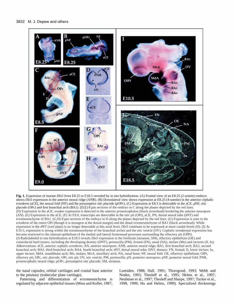
Fig. 1. Expression of murine Dlx5 from E8.25 to E18.5 revealed by in situ hybridization. (A) Frontal view of an E8.25 (2-somite) embryoshows Dlx5 expression in the anterior neural ridge (ANR). (B) Dorsolateral view shows expression at E8.25 (4-somite) in the anterior cephalicectoderm (aCE), the neural fold (NF) and the presumptive otic placode (pOPc). (C) Expression at E8.5 is detectable in the aCE, pNF, oticplacode (OPc) and first branchial arch (BA1). (D,E) 8 µm sections of the embryo in C along the planes depicted by the red lines.(D) Expression in the aCE; weaker expression is detected in the anterior prosencephalon (black arrowhead) bordering the anterior neuropore(AN). (E) Expression in the aCE. (F) At E9.0, transcripts are detectable in the otic pit (OPt), aCE, PN, dorsal neural tube (dNT) andectomesenchyme of BA1. (G,H) 8 µm sections of the embryo in D along the planes depicted by the red lines. (G) Expression is seen in theectoderm of the entire OPt (though it is strongest at the dorsal margin) and the distal ectomesenchyme of BA1 (black arrowhead). Whileexpression in the dNT (roof plate) is no longer detectable at this axial level, Dlx5 continues to be expressed at more caudal levels (H). (I) AtE10.5, expression is strong within the ectomesenchyme of the branchial arches and the otic vesicle (OV). Cephalic ectodermal expression hasbecome restricted to the internal epithelium of the medial and lateral frontonasal processes surrounding the olfactory pit (OfPt).(J) Radiolabeled in situ hybridization at E18.5 reveals Dlx5 expression in the forebrain (striatum, SM), olfactory epithelium (OE) andcraniofacial hard tissues, including the developing dentary (DNT), premaxilla (PM), frontal (FN), nasal (NA), molars (Mo) and incisors (Il, Iu).Abbreviations: aCE, anterior cephalic ectoderm; AN, anterior neuropore; ANR, anterior neural ridge; BA1, first branchial arch; BA2, secondbranchial arch; BA3, third branchial arch; BA4, fourth branchial arch; dNT, dorsal neural tube; DNT, dentary; FN, frontal; Il, lower incisor; Iu,upper incisor; MdA, mandibular arch; Mo, molars; MxA, maxillary arch; NA, nasal bone; NF, neural fold; OE, olfactory epithelium; OfPt,olfactory pit; OPc, otic placode; OPt, otic pit; OV, otic vesicle; PM, premaxilla; pN, posterior neuropore; pNF, posterior neural fold; PNR,prosencephalic neural ridge; pOPc, presumptive otic placode; SM, striatum.
3833Dlx5 regulation of craniofacial development
(placodes) in the surface ectoderm focally regulate skeletal anddental development within the underlying mesenchyme. Forexample, extirpation and transplantation studies suggest thatthe olfactory and otic placodes regulate the development of thecartilaginous nasal and otic capsules, respectively (Corsin,1971; Frenz and Van de Water, 1991; Webb and Noden, 1993).
Craniofacial ectomesenchyme appears to be endowed withsome positional information. Coherent streams of CNCmigrate along distinct pathways from the dorsal margins of theposterior diencephalon, mesencephalon and metencephalon tocontribute skeletogenic mesenchyme to the rostral cranium anddistinct domains of the branchial arches (Tosney, 1982;Serbedzija et al., 1992; Osumi-Yamashita et al., 1994, 1997;Imai et al., 1996; Köntges and Lumsden, 1996). Bothtransplantation and genetic studies provide evidence that A/Pspecification processes within the neural plate and brain haveimportant roles in imparting regional identity to the CNC,which in turn controls its later development (Noden, 1983,
1991; Gendron-Maguire et al., 1993; Rijli et al., 1993, 1998;Matsuo et al., 1995; Kuratani et al., 1997). While certain genes(e.g. Otx2 and the Hox genes) may regulate A/P specificationof the CNC, other homeobox transcription factors appear toinfluence craniofacial morphology through regulation of CNCdevelopment along other axes (e.g. Dlx, Gsc, Msx, Pax and Prxgenes).
Members of the Dlx gene family are expressed in bothcraniofacial ectoderm and CNC (Dollé et al., 1992; Bulfone etal., 1993; Akimenko et al., 1994; Robinson and Mahon, 1994;Simeone et al., 1994; Ellies et al., 1997; Qiu et al., 1997; Yanget al., 1998). Analysis of the expression and function of themurine Dlx gene family suggests that these genes might specifyregional fate of the branchial arch ectomesenchyme (Qiu et al.,1997). In the first arch, Dlx1 and Dlx2 are expressed in boththe maxillary and mandibular components, whereas Dlx3, Dlx5and Dlx6 are expressed only in the mandibular. In the second(hyoid) arch, Dlx1 and Dlx2 are expressed throughout the
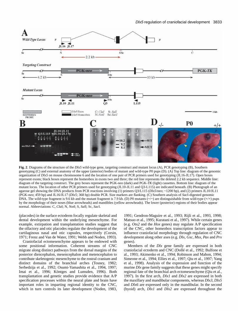
Fig. 2. Diagrams of the structure of the Dlx5 wild-type gene, targeting construct and mutant locus (A), PCR genotyping (B), Southerngenotyping (C) and external anatomy of the upper (anterior) bodies of mutant and wild-type P0 pups (D). (A) Top line: diagram of the genomicorganization of Dlx5 on mouse chromosome 6 and the location of one pair of PCR primers used for genotyping (JL16-JL17). Open boxesrepresent exons; black boxes represent the homeobox in exons two and three; the red line represents the deleted 2.2 kb sequence. Middle line:diagram of the targeting construct. The grey boxes represent the PGK-neo (dark) and PGK-TK (light) cassettes. Bottom line: diagram of themutant locus. The location of other PCR primers used for genotyping (JL10-JL11 and Q3-L115) are indicated beneath. (B) Photograph of anagarose gel showing the DNA products from PCR reactions involving (1) primers Q3/L115 (Dlx5/neo; ~1200 bp), and (2) primers JL10/JL11(PGK-neo; 459 bp) and JL16/JL17 (Dlx5; 368 bp) double PCR. Size markers are flanking. (C) Southern analysis of SacI-digested genomicDNA. The wild-type fragment is 9.6 kb and the mutant fragment is 7.9 kb. (D) P0 mutants (−/−) are distinguishable from wild-type (+/+) pupsby the morphology of their noses (blue arrowheads) and mandibles (yellow arrowheads). The lower (posterior) regions of their bodies appearnormal. Abbreviations: C, ClaI; N, NotI; S, SalI; Sc, SacI.
3834
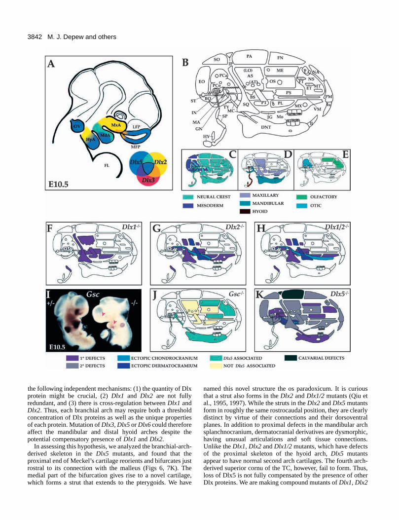
proximodistal axis, while Dlx3, Dlx5 and Dlx6 are onlyexpressed in more distal regions. Mice lacking Dlx1 or Dlx2have deletions and/or morphological transformations ofmaxillary and proximal hyoid arch-derived structures; theskeleton of the mandibular and distal hyoid arch, however,appears to be normal (Qiu et al., 1995, 1997). The finding thatDlx1 and Dlx2 expression is not required in the mandibular anddistal hyoid arches suggests that other Dlx genes compensatein these regions (Qiu et al., 1997).
Herein, we provide evidence that Dlx5 regulates craniofacialdevelopment both through its expression in ectodermalplacodes and ectomesenchyme. Dlx5 is expressed both in theolfactory and otic placodes, their derived epithelia and theCNC of the mandibular arch (Simeone et al., 1994; Qiu et al.,1997; Yang et al., 1998). Mice homozygous for Dlx5 mutantalleles die shortly after birth, some with exencephaly. Non-exencephalic mutant mice have hypomineralized calvaria, andall mutants have regional defects in their nasal and oticcapsules and proximal mandibles. We hypothesize that Dlx5acts in concert with other Dlx genes in patterning theectomesenchyme of the branchial arches. A number of theobserved malformations may be due to decreased expressionof the goosecoid (Gsc) homeobox gene.
MATERIALS AND METHODS
Generation of Dlx5 mutant miceGenomic DNA encoding the Dlx5 gene was isolated by screening agenomic library (from Anton Berns, Amsterdam) made from liverDNA isolated from strain 129-J mice using a Dlx5 cDNA (Liu et al.,1997) as a probe. The targeting construct was made using the pBSKS− cloning vector. The organization of the targeting vector is shownin Fig. 2. Briefly, a 4.2 kb NotI Dlx5 genomic fragment (upstream ofthe NotI site in the first exon of Dlx5) was ligated 5′ to the positiveselection gene cassette (PGK-neo; Tybulewicz et al., 1991). A 10 kbClaI-HincII Dlx5 genomic fragment was subcloned 3′ of the PGK-neo cassette. Finally, a negative selection gene cassette (PGK-thymidine kinase (PGK-TK); Johnson et al., 1989) was inserted 3′ tothis 10 kb fragment. The Dlx5 targeting construct contains a 2.2 kbdeletion of exons 1 and 2, which spans the entire N terminus of theprotein and includes the 5′ end of the homeobox.
The targeting vector was introduced into the JM-1 line of ES cells(Qiu et al., 1995). Genotyping of the clones was performed bydigesting the genomic DNA with SacI, separation on a 0.5% agarosegel, transfer to a Duralon UV membrane (Stratagene) and highstringency Southern analysis (Sambrook et al., 1989). The Southernprobe was a 1.5 kb NheI-KpnI fragment from Dlx5 genomic clone6N6#1 (schematized in Fig. 2). The wild-type fragment appears as a9.6 kb band and the mutant as a 7.9 kb band. PCR conditions, usingTaq polymerase (Qiagen), are as follows: 97°C, 2 minutes (1 cycle);97°C, 1 minute, 55°C, 1 minute, 72°C, 1 minute (30 cycles); 72°C, 5minutes (1 cycle). Primers JL16 (CAG TAG AAG AAC AGC CAC)and JL17 (ACT CGG GAC GCG GTT GTA) were used to generatea 368 bp PCR fragment to determine the presence of the first exon ofDlx5. Primers JL10 (CAA GAT GGA TTG CAC GCA G) and JL11(CAT CCT GAT CGA CAA GAC) were used to generate a 459 bpPCR fragment to determine the presence of the PGK-neo cassette.Primers Q3 (TCG CAG CGC ATC GCC TTC TAT CGC) and L115(CCC GTT TTT CAT GAT CTT C) were used to generate an ~1.2 kbPCR fragment to verify the presence of the PGK-neo cassette insertedinto the Dlx5 locus.
The ES cell lines containing mutations in one of the Dlx5 alleleswere karyotyped, injected into blastocysts isolated from C57BL/6J
mice and then re-implanted into the uteri of pseudopregnant CD-1mice (Joyner, 1993). Male chimeras were then mated with C57BL/6Jfemales. Germline transmission was assessed by scoring the F1offspring for the agouti phenotype and then verified by Southern andPCR analysis (see above). Dlx5 homozygotes from two independentcell lines (#s 19, 26) had defects in the same structures.
Anatomical analysesAnimals were fixed in 4% paraformaldehyde (PFA) in phosphate-buffered saline (PBS), embedded in paraffin (E10.5-E16.5) or OCT(P0), and then cut in 8 to 20 µm thick serial sections. Sections werethen stained with either Gimori trichrome (E10.5-E16.5) or Cresylviolet (P0). Differential staining of cartilage and bone was achievedthrough the use of Alcian blue and Alizarin red S (McLeod, 1980).
Scanning electron microscopyEmbryos were taken from timed pregnancies and fixed at 4°Covernight in 4% PFA in PBS. The embryos were then rinsed threetimes in PBS and dehydrated in an increasing series of EtOH. Theywere critically point dried, sputter coated with gold-palladium andphotographed in a JEOL 840 scanning electron microscope.
RNA in situ hybridizationIn situ hybridization was performed as described by Shimamura et al.(1995) and Qiu et al. (1997). The Dlx5 probe was produced from a 1kb cDNA containing the homeobox and 3′′ sequence (Qiu et al., 1997).The Gsc probe was obtained from Dr E. M. De Robertis. For the Gscin situs, hemisected mutant and wild-type embryos were assayed inthe same tube. The contralateral hemisections were hybridized withpositive control probes (e.g. Msx2).
Whole-mount immunohistochemistryNeurofilaments were detected with a 4:1 dilution of the 2H3 155 kDaneurofilament antibody (Developmental Studies Hybridoma Bank,University of Iowa) using whole-mount immunohistochemistry,utilizing the Vector ABC kit and following the general proceduresdescribed by Qiu et al. (1995).
RESULTS
Expression of Dlx5Dlx5 mRNA is detected in distinct ectodermal tissues(Akimenko et al., 1994; Simeone et al., 1994; Zhao et al., 1994;Liu et al., 1997; Yang et al., 1998). At E8.25 (2- to 4-somitestage), Dlx5 transcripts are found in the neural ridge at all axiallevels (Fig. 1). The anterior neural ridge (ANR), whichexpresses high levels of Dlx5 (Fig. 1A), contains the primordiaof the forebrain, olfactory placodes, Rathke’s pouch andmidfacial ectoderm (Couly and Le Douarin, 1985, 1987;Eagleson et al., 1995; Rubenstein et al., 1998), and may be anorganizing center for forebrain and facial development(Shimamura and Rubenstein, 1997). During later stages, Dlx5expression is maintained in the olfactory placode (Fig. 1F),invaginating olfactory pit (Fig. 1I) and forebrain (Fig. 1J;Simeone et al., 1994; Liu et al., 1997; Yang et al., 1998). Theneural ridge, which produces the neural crest and forms thedorsal midline (roof plate), continues to express Dlx5 at leastthrough E9.0 (Fig. 1C,F,H). Dlx5 transcripts are detected in theotic placode and its derivative, the otic vesicle (OV; Fig.1C,F,G). Its expression is highest in the dorsal regions that arethe anlagen of the endolymphatic duct (ED) and the vestibularapparatus (Sher, 1971; Van de Water, 1984). Dlx5 is alsoexpressed in postmigratory CNC cells in the branchial arches
M. J. Depew and others
3835Dlx5 regulation of craniofacial development
(mandibular first arch and distal parts of the caudal arches)(Fig. 1C,F,G,I; Simeone et al., 1994; Zhao et al., 1994; Qiu etal., 1997). Dlx5 expression can be seen at later stages (Fig. 1J)during organogenesis of the skeleton (Simeone et al., 1994;Zhao et al., 1994; Chen et al., 1996).
Generating Dlx5 mutant miceWe used the JM-1 line (strain 129) embryonic stem (ES) cells(Qiu et al., 1995) for targeted mutagenesis of murine Dlx5. Thetargeting vector was designed to make an approximately 2.2 kbdeletion to eliminate the N terminus of Dlx5, including 43amino acids of the homeodomain (Fig. 2). Four ES clonesharboring one Dlx5 mutant allele were identified. We madechimeric mice from two of these clones. The chimeras wereout-bred with C57BL/6J mice to generate heterozygotes.Inbreeding of these mice produced exencephalic newborns(7/259, or ~3%) which were heterozygous for the mutant Dlx5allele. The frequency of exencephalic heterozygotes has greatlydiminished with further outbreeding to C57BL/6J mice. Otherthan exencephaly, no other phenotypes have been noted inheterozygotes – except that these mice do not breed readily.Inbreeding of heterozygotes was used to generate Dlx5homozygous mutant animals. Of 29 litters tested, a genotypicratio of 31%:51%:18% (81 +/+:132 +/−:46 −/−) wasdetermined. Newborn Dlx5 mutant mice are slightly smallerthan their wild-type littermates (Fig. 2D) and lack milk in theirstomachs. All Dlx5 homozygous mutants die shortly afterbirth.
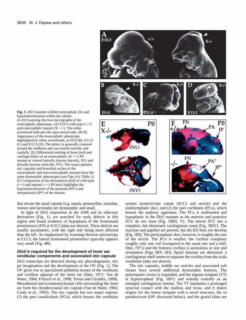
Dlx5 homozygote mutant newborns have cranialdefectsSeveral external craniofacial defects are apparent in the Dlx5homozygous mutant newborns, including decreased nasalcapsular breadth, micrognathia and malformed auditory pinnae(Fig. 2D). Exencephaly was evident in 28% of the animals (Fig.3; Table 1). Scanning electron microscopic analysis of Dlx5mutant embryos suggests that the neural tube closure defect iscentered in the midbrain but can include the forebrain andhindbrain in severe cases (Fig. 3A-D). The calottes (calvarialroofs; White and Folkens, 1991) of non-exencephalic Dlx5mutants have small, hypomineralized parietal and interparietalbones (Fig. 3F). The supraoccipital, though reduced in size, isless affected. The occipital arch cartilages are thicker andexpanded rostrocaudally. Increased amounts of secondarycartilage form in the sagittal and lambdoidal sutures (data notshown). The remainder of the calotte (e.g. frontals) appears tohave normal mineralization as assayed by Alizarin Red Sstaining.
All cranial bones have at least subtle dysmorphologies, whichwe suggest are due to epigenetic factors (e.g. biomechanicalinfluences) and/or altered skeletal differentiation. Because Dlx5is expressed in developing skeletal elements (Fig. 1J; Simeoneet al., 1994; Zhao et al., 1994; Ferrari et al., 1995; Chen et al.,1996), these malformations may result from a lack of Dlx5functioning during skeletogenesis. For example, the primaryand secondary palates exhibit variable degrees of clefting andcomponents of the cranial sidewall (e.g. the alisphenoid andsquamosal) have slight morphologic alterations (data notshown). Although Dlx5 is expressed in the post-cranial axialand appendicular skeleton (Simeone et al., 1994; Zhao et al.,1994; Ferarri et al., 1995; Chen et al., 1996), we detected few
abnormalities in these structures. The angle of the ribs, forexample, appeared sinusoidal in 27% of cases (data not shown).Dlx5 mutant animals – with or without exencephaly – exhibitthe same set of additional craniofacial abnormalities (see belowand Table 1).
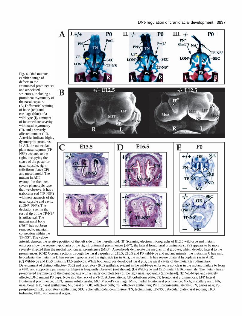
Dlx5 mutants have defects in olfactory placode andfrontonasal prominence derivativesIn Dlx5 mutant newborns (P0), chondrocranial anddermatocranial defects are observed in and around the nasalcapsule and mesethmoid (Fig. 4). In the most severe cases,there is symmetric, near aplasia of the capsule and mesethmoid(Fig. 4AIII,E). A midline cartilaginous rod-like extension ofthe rostral trabecular cartilage forms but fails to develop adorsoventrally expanded nasal septum. Small, cartilaginousspicules are the only remnants of the side walls of the capsule(paries nasi, prominentia lateralis and associated turbinates).The posterior capsule and mesethmoid are reduced to thin,planar orbitonasal laminae (Fig. 4AIII,E): in these cases, thereis no evidence for the development of a nasal epithelium, norof a true tectum or solum [including the paraseptal cartilagesand associated vomeronasal organ (VNO)] of the nasal capsule.
In cases of intermediate severity (the majority of casesobserved), a pronounced asymmetry, with the right side beingmore hypoplastic, is seen (Fig. 4AII,D; Table 1). The trabecularplate-nasal septum, which is compressed dorsoventrally,deviates to the right to occupy the position of the right cribriformplate and posterior nasal capsular wall (Fig. 4AII). No foraminacribrosa are seen on the right and very few on the left. The tectumnasi, solum and paries nasi prominences and associatedturbinates (e.g. frontoturbinates and ethmoturbinates) arehypoplastic. In these less severe cases, rudimentary branches ofthe nasal epithelium are present (Fig. 4D), as are hypoplasticrostral turbinate cartilages (data not shown). Cartilages in thefloor of the nasal capsule (e.g. lamina transversus anterior andparaseptal cartilages) are hypoplastic and sometimes associatedwith a rudimentary VNO (data not shown). The dermal bones
Table 1. Phenotypic penetrance of defects in Dlx-5−−/−−
skulls*Calotte
Hypomineralized 100% (18/18)‡Exencephalic 28% (6/24)
Otic capsulePars Canalicularis 100% (23/23)Pars Cochlearis 100% (21/21)Tegmen Tympani 100% (18/18)
Nasal capsuleParies Nasi/Turbinates 100% (30/30)Asymmetry 85% (25/30)
>Hypoplasia on R 88% (22/25)TP-NS rod-like 30% (9/29)
Brachial archMandibular 100% (31/31)Os Paradoxicum 100% (31/31)
PalateClefting 88% (22/25)Parapalatines 88% (22/25)
*Based upon evaluation of bone and cartilage preparations.‡(x,y) where x/y represents a total of P0 and E15.5 animals examined for
which scoring was possible.
3836
that encase the nasal capsule (e.g. nasals, premaxillae, maxillae,vomers and lacrimals) are dysmorphic and small.
In light of Dlx5 expression in the ANR and its olfactoryderivatives (Fig. 1), we searched for early defects in thisregion and found evidence of hypoplasia of the frontonasalprominences (FP) at E10.5 (data not shown). These defects areusually asymmetric, with the right side being more affectedthan the left. As emphasized by scanning electron microscopyat E12.5, the lateral frontonasal prominence typically appearsvery small (Fig. 4B).
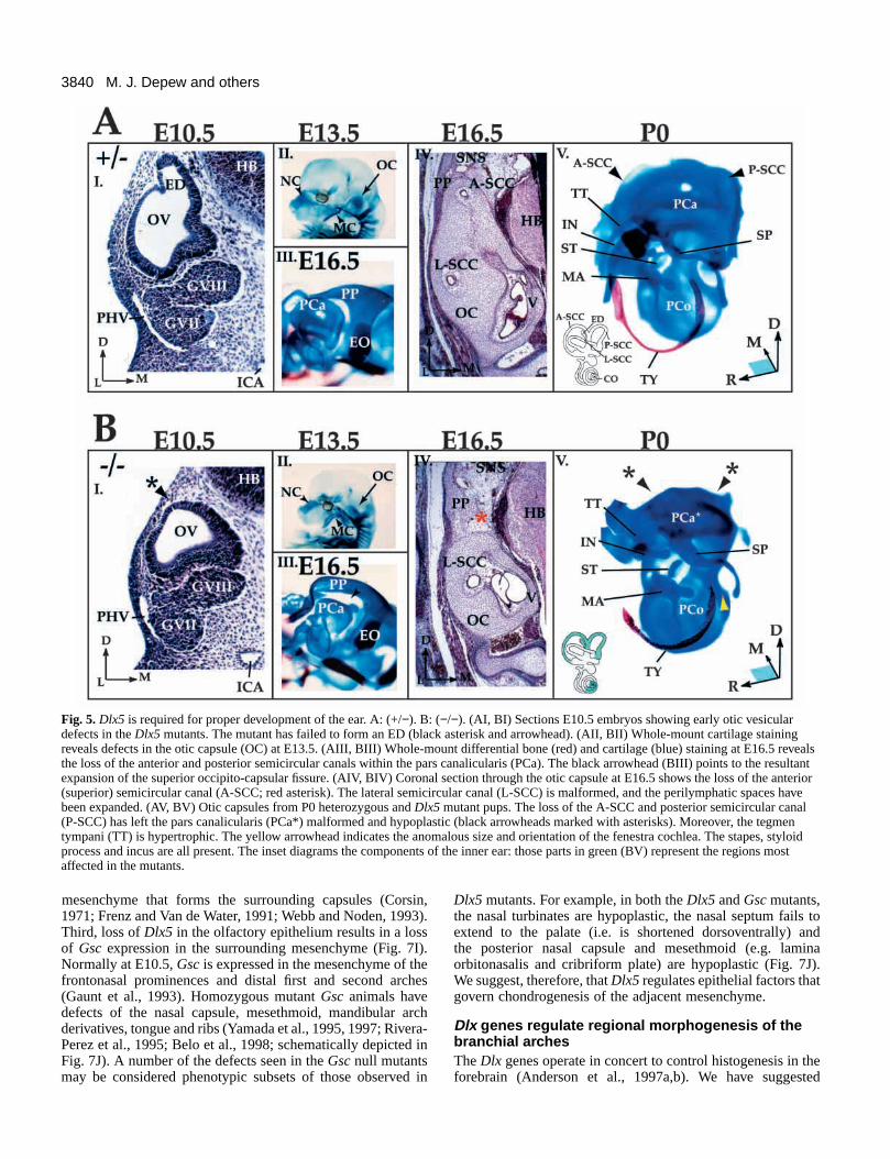
Dlx5 is required for the development of inner earvestibular components and associated otic capsuleDlx5 transcripts are detected during otic placodogenesis, oticpit invagination and the development of the OV (Fig. 1). TheOV gives rise to specialized epithelial tissues of the vestibularand cochlear apparati of the inner ear (Sher, 1971; Van deWater, 1984; Fritzsch et al., 1998; Torres and Giraldez, 1998).Mesodermal and ectomesenchymal cells surrounding the innerear form the chondrocranial otic capsule (Van de Water, 1984;Couly et al., 1993). The otic capsule has two major regions:(1) the pars canalicularis (PCa), which houses the vestibular
system (semicircular canals (SCC) and utricle) and theendolymphatic duct, and (2) the pars cochlearis (PCo), whichhouses the auditory apparatus. The PCa is malformed andhypoplastic in the Dlx5 mutants as the anterior and posteriorSCC do not form (Fig. 5BIII, V). The lateral SCC has acomplete, but shortened, cartilaginous canal (Fig. 5BIV). Themaculae and papillae are present, but the ED does not develop(Fig. 5BI). The perilymphatic duct, however, is roughly the sizeof the utricle. The PCo is smaller: the cochlea completesroughly only one coil (compared to the usual one and a half;Sher, 1971) and the fenestra cochlea is anomalous in size andorientation (Figs 5BV, 6D). Spiral laminae are abnormal: acartilaginous shelf seems to separate the cochlea from the scalavestibulae (data not shown).
The otic capsules, middle ear ossicles and associated softtissues have several additional dysmorphic features. Theepitympanic recess is expanded, and the tegmen tympani (TT)is hypertrophied (Fig. 5BV) and extends rostrally as anenlarged cartilaginous lamina. The TT maintains a prolongedsynovial contact with the malleus and incus, and it sharesorigins for the tensor tympani with a novel structure, the osparadoxicum (OP; discussed below), and the gonial (data not
M. J. Depew and others
Fig. 3. Dlx5 mutants exhibit exencephaly (X) andhypomineralization within the calotte. (A-D) Scanning electron micrographs of theexencephalic phenotype. (A) E10.5 wild-type (+/+)and exencephalic mutant (X −/−). The whitearrowhead indicates the open neural tube. (B-D)Appearance of the exencephalic phenotype,highlighted by white arrowheads, at E9.0 (B), E11.0(C) and E13.5 (D). The defect is generally centeredaround the midbrain and can extend rostrally andcaudally. (E) Differential staining of bone (red) andcartilage (blue) of an exencephalic (X −/−) P0mutant as viewed laterally (norma lateralis, NL) anddorsally (norma verticalis, NV). The nasal capsules,otic capsules and branchial arches of theexencephalic and non-exencephalic mutants have thesame dysmorphic phenotypes (see Figs 4-6; Table 1).(F) Comparison of the dorsolateral skull of wild-type(+/+) and mutant (−/−) P0 mice highlights thehypomineralization of the parietals (PA*) andinterparietals (IP*) of the mutant.
3837Dlx5 regulation of craniofacial development
Fig. 4. Dlx5 mutantsexhibit a range ofdefects in thefrontonasal prominencesand associatedstructures, including aprominent asymmetry ofthe nasal capsule.(A) Differential stainingof bone (red) andcartilage (blue) of awild-type (I), a mutantof intermediate severitywith nasal asymmetry(II), and a severelyaffected mutant (III).Asterisks indicate highlydysmorphic structures.In AII, the trabecularplate-nasal septum (TP-NS*) deviates to theright, occupying thespace of the posteriornasal capsule, rightcribriform plate (CP)and mesethmoid. Themutant in AIIIexemplifies the mostsevere phenotypic typethat we observe: it has atrabecular rod (TP-NS*)with near agenesis of thenasal capsule and cavity(LON*, PN*). Thedeviation seen in therostral tip of the TP-NS*is artifactual. Themutant nasal bone(NA*) has not beenremoved to maintainconnection within theTP-NS*. The yellowasterisk denotes the relative position of the left side of the mesethmoid. (B) Scanning electron micrographs of E12.5 wild-type and mutantembryos show the severe hypoplasia of the right frontonasal prominences (FP*); the lateral frontonasal prominence (LFP) appears to be moreseverely affected than the medial frontonasal prominence (MFP). Arrowheads demarcate the nasolacrimal grooves, which develop lateral to theprominences. (C-E) Coronal sections through the nasal capsules of E13.5, E16.5 and P0 wild-type and mutant animals: the mutant in C has mildhypoplasia; the mutant in D has severe hypoplasia of the right side (as in AII); the mutant in E has severe bilateral hypoplasia (as in AIII).(C) Wild-type and Dlx5 mutant E13.5 embryos. While both embryos developed nasal pits, the nasal cavity of the mutant is rudimentary.Development of distinct olfactory (OE) and respiratory (RE) epithelia, evident in the wild-type embryo, is not clear in the mutant. Failure to forma VNO and supporting paranasal cartilages is frequently observed (not shown). (D) Wild-type and Dlx5 mutant E16.5 animals. The mutant has apronounced asymmetry of the nasal capsule with a nearly complete loss of the right nasal apparatus (arrowhead). (E) Wild-type and severelyaffected Dlx5 mutant P0 pups. Note also the lack of a VNO. Abbreviations: CP, cribriform plate; FP, frontonasal prominences; LFP, lateralfrontonasal prominence; LON, lamina orbitonasalis; MC, Meckel’s cartilage; MFP, medial frontonasal prominence; MxA, maxillary arch; NA,nasal bone; NE, nasal epithelium; NP, nasal pit; OB, olfactory bulb; OE, olfactory epithelium; PmL, prominentia lateralis; PN, paries nasi; PS,presphenoid; RE, respiratory epithelium; SEC, sphenethmoidal commissure; TN, tectum nasi; TP-NS, trabecular plate-nasal septum; TRB,turbinate; VNO, vomeronasal organ.
3838
shown). The TT is also linked to the alicochlear commissurevia an anomalous fibrous tract, and the belly of thestylopharyngeus lies ventral and superficial to the ear drum,most likely precluding functionality (data not shown). Theexternal acoustic meatus is shortened and the tubal cartilagedevelops in nodules (data not shown).
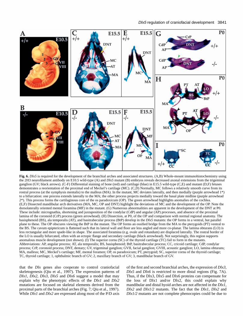
Dlx5 is required for proper development of branchialarch derivativesIn Dlx5 mutants, the chondrocranial element of the mandibulararch, Meckel’s cartilage (MC), is shortened (Fig. 6D,F). Asearly as E13.5, the proximal shaft of MC is extremelydysmorphic; this is clearly seen at E15.5 (Fig. 6D,F). Theorientation of MC deviates twice (n=5/5 at E15.5). Initiallyprojecting caudolaterally, MC sharply deviates laterad at apoint near its proximocaudal end adjacent to the proximal endof the dentary (DNT; Fig. 6D,F). It then abruptly reorientscaudomedially for a short distance whereupon it splits: amedial branch forms an ectopic strut towards the pterygoid andbasisphenoid; the other branch continues caudolaterally intothe malleus. By P0, ectopic intramembranous bones invest thecartilaginous strut (Fig. 6H). We have named this novel ectopicstrut and associated ossification, the os paradoxicum (OP; Fig.6D,F,H). The OP may invest, or form a synovial joint with, thepterygoids. It also forms a synovial joint with the misshapengonial and sutures with the anterior crus of the tympanic. Themalleus has a smaller than normal processus brevis and iscaudally extended and thickened at the level of the manubrium.The tympanic is slightly smaller and thicker (data not shown).
A short and dysmorphic DNT develops around the abnormalMC (Fig. 6D,F,G). The proximal lamina of the coronoid isabsent, and the condylar and angular processes are shortened,misshapen and juxtaposed (Fig. 6G). Often an ectopic,proximodistally oriented cartilaginous and osseous spiculedevelops along the buccal surface of the condylar process (datanot shown). This appears to run toward (and form a synovialjoint with) the hypotrophic jugal bone – itself reoriented towardthe condyle at the expense of its articulation with the zygomaticprocess of the squamosal (not shown). Buccally, the temporalismuscles are attached to what might represent a displaced,vestigial distal coronoid process (minus the proximalextension), which runs parallel to the mandibular body, lateralto the molar teeth (not shown). Extensive chondroid bonedevelops along the incisive alveolar bone at the tips of thedentary; the incisivium itself is broader and shorter than normal.The dental canal/lingula is reflected lingually (medially) at theangular end (with the mylohyoid line extended caudad). Themolar alveolae are shorter, broader and deeper. The alveolaropenings are constricted distally and broadened proximally. Thebuccal molar walls are less robust and ventrally reflected.
Not surprisingly, this region supports anomalous muscledevelopment. The OP acts as an origin for the medial pterygoid(which is hypertrophied) and tensor palati muscles; normally,these muscles originate from fibrous connective tissue inregions where the alisphenoid and the tegmen tympani of theotic capsule ossify (data not shown). The medial pterygoidinsertion groove is occluded by a broad origin for an abnormalmylohyoid. Moreover, the rostral limit of the genioglossus andgeniohyoid muscles are displaced caudad along the mylohyoidline, and there are numerous, ectopic connections to the hardtissue (data not shown). Dorsocaudal to the mylohyoid,
anomalous fascicles of the genioglossus muscle run from themylohyoid, lingula, OP and gonial into the tongue. Thebuccinator muscles attach mandibulae to maxillae such thatthere is no slack in the buccal pouch (data not shown).
There is variable palatal clefting exemplified by defects inpalatine and maxillary morphology (data not shown). Ectopicmembrane bones, here referred to as parapalatines (PrP),consistently (88%, Table 1) develop caudal to the palatineshelves and rostral to dysmorphic pterygoids. The pterygoidshave an abnormal amount of chondroid bone and secondarycartilage. The right side of the rostral tongue is generallyhypertrophied vertically, filling in the right side of the palateand leaving the left side to fill in under the incisors (not shown).
There are additional anomalies of soft-tissue anatomy, oftenasymmetric, not detailed here, that variably affect the coursesof the stapedial artery, the lingual duct and the fascicle patternof the intrinsic tongue musculature. The larynx, furthermore,is displaced caudally and occupies a position well back fromthe pterygoids and tympanic cavity (data not shown). Thesuperior cornu (SC) of the thyroid cartilage (TC; derived fromthe fourth branchial arch, Patten and Carlson, 1988)consistently fail to form (Fig. 6I).
Dlx5 is expressed during dental development (Simeone etal., 1994; Weiss et al., 1994, 1995, 1998). Unlike the Dlx1/2mutants, which lack maxillary molars, molar teeth are presentin the Dlx5 mutants (except for the maxillary and mandibularthird molars, which are frequently absent in wild-type mice;Grüneberg, 1963). The mandibular and maxillary molarsappear to have malformed and poorly mineralized crowns, andboth sets of incisors are shortened and misshapen (data notshown).
To assess whether disruption of craniofacial patterning in theDlx5 mutant affects the growth of the cranial nerves, wecompared the axonal trajectories in wild-type (n=5) and mutant(n=2) embryos at E10.5 using the 2H3 neurofilament antibody(Fig. 6A,B). In the mutant embryos, the trigeminal ganglion issmaller (Fig. 6B), its root appears malformed and it hasreduced axonal projections into the surrounding mesenchyme(especially V2 and V3). Other axonal defects were found in theprocesses of the hypoglossal nerve, the autonomic ganglionicchain (AGC) and the dorsal root ganglion (Fig. 6B and datanot shown). At later stages, trigeminal nerve axons extendfurther into the branchial arches, although their orientations areabnormal based upon the morphology of their foramina [e.g.the mental foramen (Fig. 6F)].
Dlx5 regulates the expression of GscBecause Gsc mutants lack turbinates within their nasalcapsules, and have altered mandibular branchial arch, ear andmesethmoid morphologies (Rivera-Perez et al., 1995; Yamadaet al., 1995, 1997; Belo et al., 1998), we compared theexpression of Gsc in wild-type (n=2) and Dlx5 mutant (n=2)embryos. In situ hybridization of hemisected control andmutant embryos at E10.5 revealed a decrease, or loss, of Gscwithin the mesenchyme of the branchial arches and thefrontonasal prominences of the olfactory pits (Fig. 7I).
DISCUSSION
The development of the skull depends upon interactionsbetween epithelial (surface ectoderm, neural tube and
M. J. Depew and others
3839Dlx5 regulation of craniofacial development
endoderm) and mesenchymal (CNC and mesoderm) tissues(Hall, 1991; Hanken and Hall, 1993; Hanken and Thorogood,1993; Francis-West et al., 1998). Evidence described hereinsuggests that Dlx5 expression is required in specializedepithelial tissues (placodes) for nose and ear development,whereas it is required in the ectomesenchyme of themandibular arch for development of the proximal mandible.Dlx5 further appears to exert an influence in the differentiationof particular skeletal elements.
Animals that are heterozygous for the Dlx5 deletiongenerally appear indistinguishable from wild-type animals,suggesting that this mutation does not act in a dominantmanner. We did, however, observe exencephaly in a fraction ofDlx5 heterozygous animals (~3%) during early passage of thismutant, implying that rostral neural tube closure is sensitiveto Dlx5 dosage in 129-strain mice and that there arecompensatory genes in C57BL/6J-strain mice. The non-Mendelian ratios of heterozygous and homozygous newbornssuggests that some animals die in utero. A definitive cause ofneonatal death has not been determined.
Role of Dlx5 in skeletal differentiationDlx5 is expressed in condensing, differentiating and developingcartilages, bones and teeth (Fig. 1J; Simeone et al., 1994; Weisset al., 1994, 1995, 1998; Zhao et al., 1994; Chen et al., 1996;Ferrari et al., 1995; Newberry et al., 1998; Ryoo et al., 1997;Thomas et al., 1997, 1998; Xu et al., 1999), implying that itmay have a general role in regulating hard tissue development.Three lines of evidence suggest that the Dlx genes play roles inosteogenesis and chondrogenesis. First, Dlx1, Dlx2, Dlx1/2 andDlx5 mutants each have abnormal bone and cartilagemorphogenesis in the skull. Second, Dlx5 is expressed in astage-specific manner in differentiating osteoblasts in vitro,where it may play a role in regulating osteocalcin expression,perhaps through inhibition of the Msx2 protein (Ryoo et al.,1997; Newberry et al., 1998). Third, bone morphogeneticprotein 2 (BMP-2) can increase Dlx5 and Dlx2 expressionin osteoblasts and chondroblasts (S. Harris, personalcommunication). Moreover, antisense oligonucleotides to Dlx2repress the transcription induced by BMP-2 of pro-alpha1(II)collagen in chondrocytes (Xu et al., 1999).
Consistent with these observations, most of the cranial bonesand the teeth in Dlx5 mutants are dysmorphic. In addition,there are minor defects in the ribs (data not shown). While wesuggest that these morphological changes may be due in partto abnormal skeletal differentiation, it is likely that epigeneticfactors contribute to the phenotypic abnormalities. Forinstance, biomechanical effects resulting from malformationsof the capsules, splanchnocranium and associated soft tissuesmay have contributed to abnormal morphogenesis of othercranial structures. We suggest that the mechanisms underlyingthese general defects in skeletal morphogenesis are distinctfrom the mechanisms underlying earlier focal defects in thecartilages of the nasal capsule, otic capsules and the proximalmandibular arch (discussed below).
Dlx5 regulates the development of structuresderived from the olfactory and otic sensoryplacodesPlacodes are thickenings in the primitive ectoderm that give riseto a number of structures, including cranial sensory tissues
(Jacobson, 1963, 1966; Verwoerd and van Oostrom, 1979; Webband Noden, 1993). Some placodes invaginate. For instance, theolfactory placodes involute to give rise to the olfactoryneuroepithelium, the VNO and the respiratory epithelium of thenasal cavity, and the otic placodes involute to form the vestibularand auditory sense organs. Other placodes contribute cells tocranial sensory ganglia through a process of delamination(Begbie et al., 1999). The olfactory and otic placodes do both(Webb and Noden, 1993). The requirement of Dlx5 in thedevelopment of both olfactory and otic structures suggests thatthe induction and histogenesis of these tissues may share similarregulatory mechanisms. Although Dlx5 is expressed at earlystages of olfactory and otic placode formation, it does not appearto be required for their initial specification.
Of the defects observed in the Dlx5 mutants, only thoseassociated with the nasal capsule showed variable severity andasymmetry (Table 1). 85% of Dlx5 mutants have asymmetricnasal development, of which nearly 90% have greater hypoplasiaon the right. This can be seen as early as E10.5 (data not shown)and, by E12.5, it appears as if a lateral frontonasal prominence(LFP) has not formed (Fig. 4B). While there has been recentsuccess in identifying the molecular bases for left-rightasymmetry in several organ systems (Levin and Mercola, 1998;Ramsdell and Yost, 1998), it is not yet clear how these findingsexplain the asymmetry in the Dlx5 mutants. Hesx1 mutants alsohave asymmetric development of the nasal cavity, but we areunaware whether or not the sidedness of the asymmetry israndom (Dattani et al., 1998). There are species, moreover, inwhich midline FP-derived structures are asymmetrically located(e.g. the blow hole of certain whales; Raven and Gregory, 1933;Klima, 1987), or have asymmetric development of the FP-derived upper incisors (e.g. the narwhal, Monodon monocerosL.; Eales, 1950). Hence, the developmental potential forasymmetric development of the FP appears to exist.
The induction and invagination of the otic placode isregulated by interactions with endoderm and hindbrain(Jacobson, 1963, 1966; Fritzsch et al., 1998; Torres andGiraldez, 1998). As with the olfactory epithelium, the oticepithelium has been implicated in regulating chondrogenesisof the adjacent mesenchyme to form the otic capsule(Grobstein and Holtzer, 1955; Van de Water, 1984; Van deWater and Galinovic-Schwartz, 1987; Frenz and Van de Water,1991; Thorogood, 1993; Legan and Richardson, 1997; Fritzschet al., 1998). Morphological defects can be seen as early asE10.5 in the otic vesicles of Dlx5 mutants, in particular, thefailure to form the dorsally located endolymphatic duct (Fig.5B). By E13.5, other dorsal derivatives of the pars canalicularisare not detectable, including the anterior and superiorsemicircular canals and the cartilaginous capsule surroundingthem.
Although there is variable penetrance and asymmetry of thedysmorphic features of the nasal capsule, we hypothesize thatthe defects in both olfactory and otic capsules stem fromprimary defects in the placode-derived epithelium: an aberrantepithelium forms, but it cannot support normal chondrogenesisin the underlying mesenchyme. This hypothesis is based uponthree observations. First, Dlx5 is expressed in the ectoderm andnot the mesenchyme (until after capsulogenesis has begun; Fig.1 and Simeone et al., 1994; Zhao et al., 1994; Yang et al.,1998). Second, extirpation experiments suggest that theolfactory pit and otic vesicle induce chondrogenesis in the
3840
mesenchyme that forms the surrounding capsules (Corsin,1971; Frenz and Van de Water, 1991; Webb and Noden, 1993).Third, loss of Dlx5 in the olfactory epithelium results in a lossof Gsc expression in the surrounding mesenchyme (Fig. 7I).Normally at E10.5, Gsc is expressed in the mesenchyme of thefrontonasal prominences and distal first and second arches(Gaunt et al., 1993). Homozygous mutant Gsc animals havedefects of the nasal capsule, mesethmoid, mandibular archderivatives, tongue and ribs (Yamada et al., 1995, 1997; Rivera-Perez et al., 1995; Belo et al., 1998; schematically depicted inFig. 7J). A number of the defects seen in the Gsc null mutantsmay be considered phenotypic subsets of those observed in
Dlx5 mutants. For example, in both the Dlx5 and Gsc mutants,the nasal turbinates are hypoplastic, the nasal septum fails toextend to the palate (i.e. is shortened dorsoventrally) andthe posterior nasal capsule and mesethmoid (e.g. laminaorbitonasalis and cribriform plate) are hypoplastic (Fig. 7J).We suggest, therefore, that Dlx5 regulates epithelial factors thatgovern chondrogenesis of the adjacent mesenchyme.
Dlx genes regulate regional morphogenesis of thebranchial archesThe Dlx genes operate in concert to control histogenesis in theforebrain (Anderson et al., 1997a,b). We have suggested
M. J. Depew and others
Fig. 5. Dlx5 is required for proper development of the ear. A: (+/−). B: (−/−). (AI, BI) Sections E10.5 embryos showing early otic vesiculardefects in the Dlx5 mutants. The mutant has failed to form an ED (black asterisk and arrowhead). (AII, BII) Whole-mount cartilage stainingreveals defects in the otic capsule (OC) at E13.5. (AIII, BIII) Whole-mount differential bone (red) and cartilage (blue) staining at E16.5 revealsthe loss of the anterior and posterior semicircular canals within the pars canalicularis (PCa). The black arrowhead (BIII) points to the resultantexpansion of the superior occipito-capsular fissure. (AIV, BIV) Coronal section through the otic capsule at E16.5 shows the loss of the anterior(superior) semicircular canal (A-SCC; red asterisk). The lateral semicircular canal (L-SCC) is malformed, and the perilymphatic spaces havebeen expanded. (AV, BV) Otic capsules from P0 heterozygous and Dlx5 mutant pups. The loss of the A-SCC and posterior semicircular canal(P-SCC) has left the pars canalicularis (PCa*) malformed and hypoplastic (black arrowheads marked with asterisks). Moreover, the tegmentympani (TT) is hypertrophic. The yellow arrowhead indicates the anomalous size and orientation of the fenestra cochlea. The stapes, styloidprocess and incus are all present. The inset diagrams the components of the inner ear: those parts in green (BV) represent the regions mostaffected in the mutants.
3841Dlx5 regulation of craniofacial development
that the Dlx genes also cooperate to control craniofacialskeletogenesis (Qiu et al., 1997). The expression patterns ofDlx1, Dlx2, Dlx3, Dlx5 and Dlx6 suggest a model that mayexplain why the phenotypic effects of the Dlx1 and Dlx2mutations are focused on skeletal elements derived from theproximal parts of the branchial arches (Fig. 7; Qiu et al., 1997).While Dlx1 and Dlx2 are expressed along most of the P/D axis
of the first and second branchial arches, the expression of Dlx3,Dlx5 and Dlx6 is restricted to more distal regions (Fig. 7A).Thus, if the Dlx3, Dlx5 and Dlx6 proteins can compensate forthe loss of Dlx1 and/or Dlx2, this could explain whymandibular and distal hyoid arches are not affected in the Dlx1,Dlx2 and Dlx1/2 mutants. The fact that the Dlx1, Dlx2 andDlx1/2 mutants are not complete phenocopies could be due to
Fig. 6. Dlx5 is required for the development of the branchial arches and associated structures. (A,B) Whole-mount immunohistochemistry usingthe 2H3 neurofilament antibody on E10.5 wild-type (A) and Dlx5 mutant (B) embryos reveals decreased axonal extensions from the trigeminalganglion (GV; black arrows). (C-F) Differential staining of bone (red) and cartilage (blue) in E15.5 wild-type (C,E) and mutant (D,F) fetusesdemonstrates a reorientation of the proximal end of Meckel’s cartilage (MC). (C,D) Normally, MC follows a relatively smooth curve from itsrostral process (at the symphysis mentalis) to the malleus (MA). In the mutant, MC deviates laterally, and then medially (purple arrowhead 1*)to a bifurcation: one process extends laterally to the MA; the other process projects medially toward the basal plate midline (purple arrowhead2*). This process forms the cartilaginous core of the os paradoxicum (OP). The green arrowhead highlights anomalies of the cochlea.(E,F) Dissected mandibular arch derivatives (MA, MC, OP and DNT) highlight the deviations of MC and the development of the OP. Note thedorsolaterally oriented mental foramina (MF) in the mutant. (G) Numerous abnormalities are apparent in the development of the DNT at P0.These include: micrognathia, shortening and juxtaposition of the condylar (CdP) and angular (AP) processes, and absence of the proximallamina of the coronoid (CrP) process (green arrowhead). (H) Dissection, at P0, of the OP and comparison with normal regional anatomy. Thebasisphenoid (BS), ala temporalis (AT), and basitrabecular process (BtP) develop in the Dlx5 mutants: the OP forms in a ventral, but parallelplane to these. The OP obscures viewing the BtP in the mutant. The OP forms an ossified bridge from the MA to the pterygoids (PT) ventral tothe BS. The cavum epiptericum is flattened such that its lateral wall and floor are less angled and more co-planar. The lamina obturans (LO) isless rectangular and more spade-like in shape. The associated foramina (e.g. ovale and rotundum) are displaced laterally. The rostral border ofthe LO is usually bifurcated, often with an ectopic flange and secondary cartilage (black arrowhead). Not surprisingly, this region supportsanomalous muscle development (not shown). (I) The superior cornu (SC) of the thyroid cartilage (TC) fail to form in the mutants.Abbreviations: AP, angular process; AT, ala temporalis; BS, basisphenoid; BtP, basitrabecular process; CC, cricoid cartilage; CdP, condylarprocess; CrP, coronoid process; DNT, dentary; GV, trigeminal ganglion; GVII, facial ganglion; GVIII, acoustic ganglion; LO, lamina obturans;MA, malleus; MC, Meckel’s cartilage; MF, mental foramen; OP, os paradoxicum; PT, pterygoid; SC, superior cornu of the thyroid cartilage;TC, thyroid cartilage; 1, ophthalmic branch of GV; 2, maxillary branch of GV; 3, mandibular branch of GV.
3842
the following independent mechanisms: (1) the quantity of Dlxprotein might be crucial, (2) Dlx1 and Dlx2 are not fullyredundant, and (3) there is cross-regulation between Dlx1 andDlx2. Thus, each branchial arch may require both a thresholdconcentration of Dlx proteins as well as the unique propertiesof each protein. Mutation of Dlx3, Dlx5 or Dlx6 could thereforeaffect the mandibular and distal hyoid arches despite thepotential compensatory presence of Dlx1 and Dlx2.
In assessing this hypothesis, we analyzed the branchial-arch-derived skeleton in the Dlx5 mutants, and found that theproximal end of Meckel’s cartilage reorients and bifurcates justrostral to its connection with the malleus (Figs 6, 7K). Themedial part of the bifurcation gives rise to a novel cartilage,which forms a strut that extends to the pterygoids. We have
named this novel structure the os paradoxicum. It is curiousthat a strut also forms in the Dlx2 and Dlx1/2 mutants (Qiu etal., 1995, 1997). While the struts in the Dlx2 and Dlx5 mutantsform in roughly the same rostrocaudal position, they are clearlydistinct by virtue of their connections and their dorsoventralplanes. In addition to proximal defects in the mandibular archsplanchnocranium, dermatocranial derivatives are dysmorphic,having unusual articulations and soft tissue connections.Unlike the Dlx1, Dlx2 and Dlx1/2 mutants, which have defectsof the proximal skeleton of the hyoid arch, Dlx5 mutantsappear to have normal second arch cartilages. The fourth arch-derived superior cornu of the TC, however, fail to form. Thus,loss of Dlx5 is not fully compensated by the presence of otherDlx proteins. We are making compound mutants of Dlx1, Dlx2
M. J. Depew and others
3843Dlx5 regulation of craniofacial development
and Dlx5 to further test predictions about the roles of each genein patterning the branchial arch skeleton.
The Dlx genes have dynamic expression patterns andessential regulatory roles during tooth formation (Weiss et al.,1994, 1995, 1998; Qiu et al., 1997; Thomas et al., 1997, 1998).A frame-shift mutation in DLX3 is associated withtaurodontism and enamel hypoplasia in humans with Tricho-dento-osseous (TDO) syndrome (Price et al., 1998). TDOpatients also have a thickening of the cranial bones. Moreover,the Dlx1/2 double mutants lack maxillary molars (Qiu et al.,1997; Thomas et al., 1997). Unlike Dlx1 and Dlx2 singlemutants, however, whose teeth appear to be normal, the Dlx5mutation affects, to varying degrees, development of all of theteeth (see results).
Decreased expression at E10.5 of Gsc suggests that Dlx5may function in one of several recently recognized pathwayswhich appear to regulate Gsc expression. Current evidencesuggests that Gsc expression is sensitive to signalling through
both Fgf8 and endothelin-1 (ET-1) via its receptor, ET-A. Forexample, experimental evidence using mandibular arch ex vivocultures has suggested that Gsc expression is either directly ofindirectly inducible by Fgf8 (Tucker et al., 1999). Moreover,gene targetted loss of ET-A results in a loss of expression ofGsc within the first branchial arch (Clouthier et al., 1998). Itremains to be determined if Dlx5 mediates transcription of Gscthrough either of these signalling systems.
Dlx1, Dlx2 and Dlx5 mutants have normal limbsDlx genes are also expressed in the apical ectodermal ridge ofvertebrate limb buds (Dollé et al., 1992; Robinson and Mahon,1994; Ferrari et al., 1995) and their invertebrate homologuesare expressed in, and required for, developing appendages(Stock et al., 1996; Panganiban et al., 1997; Williams, 1998;Wu and Cohen, 1999). Thus, it is surprising that none of thereported Dlx mutants (Dlx1, Dlx2, Dlx1/2 and here Dlx5) haveobvious defects of limb development. The most-likelyexplanation is that of genetic compensation. In this regard,there is evidence implicating DLX5 and DLX6 in ectrodactylyin humans (Scherer et al., 1994; Crackower et al., 1996). Often,these patients also have cleft palate and deafness (Ignatius etal., 1996), defects that are consistent with the functions of Dlx5described herein. Thus, it would be instructive to study the limbphenotypes of Dlx compound mutants, particularly the Dlx5/6double mutants. In addition, people with ectrodactyly can havemental retardation (Ignatius et al., 1996). Perhaps this is dueto the expression of Dlx5 in the developing brain (Simeone etal., 1994; Liu et al., 1997).
M. J. D. would like to thank Dr Michael Novacek of the AmericanMuseum of Natural History, Dr Philip Crossley of the University ofCalifornia, San Francisco, and Drs James Patton and David Wake, ofthe University of California, Berkeley, for their insightful comments.This work was supported by the research grants to J. L. R. R. fromNina Ireland, the March of Dimes, NARSAD, and NIH GrantMH51561 and K02 MH01046; to R. A. P. from NIH Grant HD26732,and United States Department of Energy /OHER Contract No. DE-ACO3-76-SF01012; and to M. J. D. from NIDR training grant T32DE07204 and ARCS.
REFERENCES
Akimenko, M. A., Ekker, M., Wegner, J., Lin, W. and Westerfield, M.(1994) Combinatorial expression of three zebrafish genes related to distal-less: part of a homeobox gene code for the head. J. Neurosci. 14, 3475-3486.
Anderson, S. A., Eisenstat, D. D., Shi, L. and Rubenstein, J. L. R. (1997a)Interneuron migration from basal forebrain to neocortex: dependence on Dlxgenes. Science 278, 474-476.
Anderson, S. A., Qiu, M., Bulfone, A., Eisenstat, D. D., Meneses, J.,Pedersen, R. and Rubenstein, J. L. R. (1997b) Mutations of the homeoboxgenes Dlx-1 and Dlx-2 disrupt the striatal subventricular zone anddifferentiation of late born striatal neurons. Neuron 19, 27-37.
Barghusen, H. R. and Hopson, A. (1979) The endoskeleton: The comparativeanatomy of the skull and visceral skeleton. In Hyman’s ComparativeAnatomy. (ed. M. Wake) pp. 265-326. Chicago, IL: The University ofChicago Press.
Begbie, J., Brunet, J. F., Rubenstein, J. L. R. and Graham, A. (1999)Induction of the epibranchial placodes. Development 126, 895-902.
Belo, J. A., Leyns, L., Yamada, G. and De Robertis, E. M. (1998) Theprechordal midline of the chondrocranium is defective in Goosecoid-1mouse mutants. Mechan. Dev. 72, 15-25.
Broom, R. (1930) The Origin of the Human Skeleton: an Introduction toHuman Osteology. London: H. F. & G. Witherby.
Bulfone, A., Puelles, L., Porteus, M. H., Frohman, M. A., Martin, G. R.
Fig. 7. Schemata of Dlx gene expression at E10.5 and regionalcraniofacial defects in Dlx and Gsc mutations. (A) Schema depictingDlx2, Dlx3 and Dlx5 expression in an E10.5 embryo. Overlappingexpression of these genes is defined by the color wheel. Dlx1 andDlx6 have similar expression patterns to Dlx2 and Dlx5, respectively(Qiu et al., 1997). (B) Model of skeletal elements of theprotovertebrate skull (after Flower, 1885; Broom, 1930). (C) Schemadepicting the elements theoretically formed by CNC (green) ormesoderm (purple). (D) Elements from the maxillary (lavender) firstarch, mandibular (blue) first arch and hyoid (brown) arch.(E) Elements with major contributions to the otic (blue) and nasal(green) capsules. (F) Elements altered in Dlx1 mutants. It issuggested that elements having primary defects (in purple) are due toabnormalities in patterning. (G) Elements altered in Dlx2 mutants.Those in light blue represent ectopic chondrocranial elements; thosein dark blue represent ectopic dermatocranial elements. (H) Elementsaltered in Dlx1/2 double mutants. (I) Whole-mount in situhybridization of Gsc transcripts in heterozygote (left) and Dlx5mutant (right) E10.5 embryos. Gsc signal is reduced in themesenchyme of the frontonasal prominences and in the mandibularand hyoid arch mesenchyme (red arrowheads). (J) Our interpretationof the reported Gsc mutant phenotype. Those elements in green havephenotypes similar to the Dlx5 mutants. Elements in light yellowmay not be linked to Dlx5 function at E10.5. (K) Depiction of theelements altered by the loss of Dlx5 expression. Primary defectsdefined as above. Elements with secondary defects are defined asthose whose abnormalities may be due to defects in skeletaldifferentiation and/or to biomechanical effects. Hypomineralizationdefects of the parietal (PA) and interparietal (IP) are indicated inblack. Change in elements within the nasal capsule and the mandibleare hypothesized as due, in part, to a loss of Gsc expression.Abbreviations: AS, alisphenoid; AT, ala temporalis; BO,basioccipital; BS, basisphenoid; DNT, dentary; EO, exoccipital; ET,ethmoturbinate; FL, forelimb; FN, frontal; FT, frontoturbinate; GN,gonial; HY, hyoid; HyA, hyoid arch; Il, lower incisor; IN, incus; IP,interparietal; Iu, upper incisor; JG, jugal; LA, lachrimal; LFP, lateralfrontonasal prominence; LO, lamina obturans; MA, malleus; MC,Meckel’s cartilage; MdA, mandibular arch; ME, mesethmoid; MFP,medial frontonasal prominence; Mo, molars; MT, maxilloturbinate;MxA, maxillary arch; MX, maxilla; NA, nasal bone; NS, nasalseptum; OC, otic capsule; OS, orbitosphenoid; OV, otic vesicle; PA,parietal; PCa, pars canalicularis; PCo, pars cochlearis; PL, palatine;PM, premaxilla; PS, presphenoid; PT, pterygoid; SO, supraoccipital;SP, styloid process; SQ, squamosal; ST, stapes; TY, tympanic; VM,vomer.
3844
and Rubenstein, J. L. R. (1993) Spatially restricted expression of Dlx-1,Dlx-2 (Tes-1), Gbx-2, and Wnt-3 in the embryonic day 12. 5 mouseforebrain defines potential transverse and longitudinal segmentalboundaries. J. Neurosci. 13, 3155-3172.
Chen, X., Li, X., Wang, W. and Lufkin, T. (1996) Dlx5 and Dlx6: anevolutionary conserved pair of murine homeobox genes expressed in theembryonic skeleton. Ann. New York Acad. Sci. 785, 38-47.
Clouthier, D. E., Hosoda, K., Richardson, J. A., Williams, S. C.,Yanagisawa, H., Kuwaki, T., Kumada, M., Hammer, R. E. andYanagisawa, M. (1998) Cranial and cardiac neural crest defects inendothelin-A receptor-deficient mice. Development 125, 813-824.
Corsin, J. (1971) Influence des placodes olfactives et des ebauches optiquessur la morphagenese du squelette cranien chez Pleurodeles waltii michah.Annales d’Embryologie et de morphogenese 1, 41-48.
Couly, G. F. and Le Douarin, N. M. (1985) Mapping of the early neuralprimordium in quail-chick chimeras. I. Developmental relationshipsbetween placodes, facial ectoderm, and prosencephalon. Dev. Biol. 110,422-439.
Couly, G. F. and Le Douarin, N. M. (1987) Mapping of the early neuralprimordium in quail-chick chimeras. II. The prosencephalic neural plate andneural folds: implications for the genesis of cephalic human congenitalabnormalities. Dev. Biol. 120, 198-214.
Couly, G. F., Coltey, P. M. and Le Douarin, N. M. (1993) The triple originof skull in higher vertebrates: a study in quail-chick chimeras. Development117, 409-429.
Crackower, M. A., Scherer, S. W., Rommens, J. M., Hui, C. C., Poorkaj,P., Soder, S., Cobben, J. M., Hudgins, L., Evans, J. P. and Tsui, L. C.(1996) Characterization of the split hand/split foot malformation locusSHFM1 at 7q21. 3-q22. 1 and analysis of a candidate gene for its expressionduring limb development. Human Molecular Genetics 5, 571-579.
Dattani, M. T., Martinez-Barbera, J. P., Thomas, P. Q., Brickman, J. M.,Gupta, R., Mårtensson, I. L., Toresson, H., Fox, M., Wales, J. K.,Hindmarsh, P. C., Krauss, S., Beddington, R. S. and Robinson, I. C.(1998) Mutations in the homeobox gene HESX1/Hesx1 associated withsepto-optic dysplasia in human and mouse. Nature Genetics 19, 125-133.
De Beer, G. (1985) The Development of the Vertebrate Skull. Chicago:Univeristy of Chicago Press.
Dollé, P., Price, M. and Duboule, D. (1992) Expression of the murine Dlx-1homeobox gene during facial, ocular and limb development. Differentiation49, 93-99.
Eagleson, G., Ferreiro, B. and Harris, W. A. (1995) Fate of the anteriorneural ridge and the morphogenesis of the Xenopus forebrain. J. Neurobiol.28, 146-158.
Eales, N. B. (1950) The skull of the foetal narwhal, Monodon monoceros L.Phil. Trans. Roy. Soc. London, B 235, 1-33.
Ellies, D. L., Langille, R. M., Martin, C. C., Akimenko, M. A. and Ekker,M. (1997) Specific craniofacial cartilage dysmorphogenesis coincides witha loss of dlx gene expression in retinoic acid-treated zebrafish embryos.Mech. Dev. 61, 23-36.
Ferrari, D., Sumoy, L., Gannon, J., Sun, H., Brown, A. M., Upholt, W. B.and Kosher, R. A. (1995) The expression pattern of the Distal-lesshomeobox-containing gene Dlx-5 in the developing chick limb bud suggestsits involvement in apical ectodermal ridge activity, pattern formation, andcartilage differentiation. Mech. Dev. 52, 257-264.
Flower, W. H. (1885) An Introduction to the Osteology of the Mammalia.London: Macmillan.
Francis-West, P., Ladher, R., Barlow, A. and Graveson, A. (1998)Signalling interactions during facial development. Mech. Dev. 75, 3-28.
Frenz, D. A. and Van De Water, T. R. (1991) Epithelial control of perioticmesenchyme chondrogenesis. Dev. Biol. 144, 38-46.
Fritzsch, B., Barald, K. F. and Lomax, M. I. (1998) Early embryology ofthe vertebrate ear. In Development of the Auditory System. (ed. E. W. Rubel,A. N. Proper and R. R. Fay). New York: Springer-Verlag.
Gaunt, S. J., Blum, M. and De Robertis, E. M. (1993) Expression of themouse goosecoid gene during mid-embryogenesis may mark mesenchymalcell lineages in the developing head, limbs and body wall. Development 117,769-778.
Gendron-Maguire, M., Mallo, M., Zhang, M. and Gridley, T. (1993) Hoxa-2 mutant mice exhibit homeotic transformation of skeletal elements derivedfrom cranial neural crest. Cell 75, 1317-1331.
Goodrich, E. S. (1958) Studies on the Structure and Development ofVertebrates. New York: Dover Publications.
Grobstein, C. and Holtzer, H. (1955) ‘In vitro’ studies of cartilage inductionin mouse somite mesoderm. Exp. Zool. 28, 333-357.
Grüneberg, H. (1963). The Pathology of Development: a study of inheritedskeletal disorders in animals. New York: John Wiley & Sons Inc.
Hall, B. K. (1988) The Neural Crest. Oxford University Press, London. Hall, B. K. (1991) Cellular interactions during cartilage and bone
development. J. Craniofacial Genet. Dev. Biol. 11, 238-250. Hanken, J. and Hall, B. K. (1993) Mechanisms of skull diversity and
evolution. In The Skull, Volume 3: Functional and EvolutionaryMechanisms. (ed. J. Hanken and B. K. Hall). pp. 1-36. Chicago: Universityof Chicago Press.
Hanken, J. and Thorogood, P. (1993) Evolution and development of thevertebrate skull: the role of pattern formation. Trends in Ecology &Evolution 8, 9-15.
Helms, J. A., Kim, C. H., Hu, D., Minkoff, R., Thaller, C. and Eichele,G. (1997) Sonic hedgehog participates in craniofacial morphogenesis andis down-regulated by teratogenic doses of retinoic acid. Dev. Biol. 187,25-35.
Hu, D. and Helms, J. A. (1999). Roles for Sonic Hedgehog in normal andabnormal craniofacial morphogenesis. Development (in press).
Ignatius, J., Knuutila, S., Scherer, S. W., Trask, B. and Kere, J. (1996) Splithand/split foot malformation, deafness, and mental retardation with acomplex cytogenetic rearrangement involving 7q21. 3. J. Med. Genet. 33,507-510.
Imai, H., Osumi-Yamashita, N., Ninomiya, Y. and Eto, K. (1996)Contribution of early-emigrating midbrain crest cells to the dentalmesenchyme of mandibular molar teeth in rat embryos. Dev. Biol. 176, 151-165.
Jacobson, A. G. (1963) The determination and positioning of the nose, lensand ear. I. Interactions within the ectoderm, and between the ectoderm andunderlying tissues. J. Exp. Zool. 154, 273-284.
Jacobson, A. G. (1966) Inductive processes in embryonic development.Science 152, 25-34.
Johnson, R. S., Sheng, M., Greenberg, M. E., Kolodner, R. D.,Papaioannou, V. E. and Spiegelman, B. M. (1989) Targeting ofnonexpressed genes in embryonic stem cells via homologous recombination.Science 245, 1234-1236.
Joyner, A. L. (1993) Gene Targeting: a Practical Approach. Oxford: IRLPress at Oxford University Press.
Klima, M. (1987) Morphogenesis of the nasal structures of the skulls intoothed whales (Odontoceti). In Morphogenesis of the Mammalian Skull.(ed. H. Kuhn and U. Zeller), pp. 105-122. Hamburg: Verlag Paul Paray,
Köntges, G. and Lumsden, A. (1996) Rhombencephalic neural crestsegmentation is preserved throughout craniofacial ontogeny. Development122, 3229-3242.
Kuratani, S., Matsuo, I. and Aizawa, S. (1997) Developmental patterningand evolution of the mammalian viscerocranium: genetic insights intocomparative morphology. Dev. Dynamics 209, 139-155.
Le Douarin, N. (1982) The Neural Crest. Cambridge Univeristy Press,London.
Legan, P. K. and Richardson, G. P. (1997) Extracellular matrix and celladhesion molecules in the developing inner ear. Seminars in Cell &Developmental Biology 8, 217-224.
Levin, M. and Mercola, M. (1998) The compulsion of chirality: toward anunderstanding of left-right asymmetry. Genes Dev. 12, 763-769.
Liu, J. K., Ghattas, I., Liu, S., Chen, S. and Rubenstein, J. L. R. (1997)Dlx genes encode DNA-binding proteins that are expressed in anoverlapping and sequential pattern during basal ganglia differentiation. Dev.Dynamics 210, 498-512.
Lumsden, A. G. (1988) Spatial organization of the epithelium and the role ofneural crest cells in the initiation of the mammalian tooth germ.Development 103 Supplement, 155-169.
Matsuo, I., Kuratani, S., Kimura, C., Takeda, N. and Aizawa, S. (1995)Mouse Otx2 functions in the formation and patterning of rostral head. GenesDev. 9, 2646-2658.
McLeod, M. J. (1980) Differential staining of cartilage and bone in wholemouse fetuses by alcian blue and alizarin red S. Teratology 22, 299-301.
Mina, M. and Kollar, E. J. (1987) The induction of odontogenesis in non-dental mesenchyme combined with early murine mandibular archepithelium. Arch. Oral Biol. 32, 123-127.
Moore, W. J. (1981) The Mammalian Skull. Cambridge University Press,Cambridge.
Neubüser, A., Peters, H., Balling, R. and Martin, G. R. (1997) Antagonisticinteractions between FGF and BMP signaling pathways: a mechanism forpositioning the sites of tooth formation. Cell 90, 247-255.
Newberry, E. P., Latifi, T. and Towler, D. A. (1998) Reciprocal regulation
M. J. Depew and others
3845Dlx5 regulation of craniofacial development
of osteocalcin transcription by the homeodomain proteins Msx2 and Dlx5.Biochemistry 37, 16360-16368.
Noden, D. M. (1983) The role of the neural crest in patterning of avian cranialskeletal, connective, and muscle tissues. Dev. Biol. 96, 144-165.
Noden, D. M. (1991) Vertebrate craniofacial development: the relationbetween ontogenetic process and morphological outcome. Brain, Behaviorand Evolution 38, 190-225.
Novacek, M. J. (1993) Patterns of diversity in the mammalian skull. In TheSkull, Volume 2: Patterns of Structural and Systematic Diversity. (ed. J.Hanken and B. K. Hall), pp. 438-545. Chicago: University of Chicago Press.
Osumi-Yamashita, N., Kuratani, S., Ninomiya, Y., Aoki, K., Iseki, S.,Chareonvit, S., Doi, H., Fujiwara, M., Watanabe, T. and Eto, K. (1997)Cranial anomaly of homozygous rSey rat is associated with a defect in themigration pathway of midbrain crest cells. Development, Growth andDifferentiation 39, 53-67.
Osumi-Yamashita, N., Ninomiya, Y., Doi, H. and Eto, K. (1994) Thecontribution of both forebrain and midbrain crest cells to the mesenchymein the frontonasal mass of mouse embryos. Dev. Biol. 164, 409-419.
Panganiban, G., Irvine, S. M., Lowe, C., Roehl, H., Corley, L. S., Sherbon,B., Grenier, J. K., Fallon, J. F., Kimble, J., Walker, M., Wray, G. A.,Swalla, B. J., Martindale, M. Q. and Carroll, S. B. (1997) The originand evolution of animal appendages. Proc. Nat. Acad. Sci. USA 94, 5162-5166.
Patten, B. M. and Carlson, B. M. (1988) Patten’s Foundations ofEmbryology. New York: McGraw-Hill.
Price, J. A., Bowden, D. W., Wright, J. T., Pettenati, M. J. and Hart, T. C.(1998) Identification of a mutation in DLX3 associated with tricho-dento-osseous (TDO) syndrome. Human Molecular Genetics 7, 563-569.
Qiu, M., Bulfone, A., Ghattas, I., Meneses, J. J., Christensen, L., Sharpe,P. T., Presley, R., Pedersen, R. A. and Rubenstein, J. L. R. (1997) Roleof the Dlx homeobox genes in proximodistal patterning of the branchialarches: mutations of Dlx-1, Dlx-2, and Dlx-1 and -2 alter morphogenesis ofproximal skeletal and soft tissue structures derived from the first and secondarches. Dev. Biol. 185, 165-184.
Qiu, M., Bulfone, A., Martinez, S., Meneses, J. J., Shimamura, K.,Pedersen, R. A. and Rubenstein, J. L. R. (1995) Null mutation of Dlx-2results in abnormal morphogenesis of proximal first and second branchialarch derivatives and abnormal differentiation in the forebrain. Genes Dev.9, 2523-2538.
Ramsdell, A. F. and Yost, H. J. (1998) Molecular mechanisms of vertebrateleft-right develoment. Trends in Genetics 14, 459-464.
Raven, H. C. and Gregory, W. K. (1933) The spermaceti organ and nasalpassages of the sperm whale (Physeter catodon) and other odontocetes.American Museum Novitates 677, 1-18.
Rijli, F. M., Gavalas, A. and Chambon, P. (1998) Segmentation andspecification in the branchial region of the head: the role of the Hox selectorgenes. Int. J. Dev. Biol. 42, 393-401.
Rijli, F. M., Mark, M., Lakkaraju, S., Dierich, A., Dolle, P. and Chambon,P. (1993) A homeotic transformation is generated in the rostral branchialregion of the head by disruption of Hoxa-2, which acts as a selector gene.Cell 75, 1333-1349.
Rivera-Pérez, J. A., Mallo, M., Gendron-Maguire, M., Gridley, T. andBehringer, R. R. (1995) Goosecoid is not an essential component of themouse gastrula organizer but is required for craniofacial and ribdevelopment. Development 121, 3005-3012.
Robinson, G. W. and Mahon, K. A. (1994) Differential and overlappingexpression domains of Dlx-2 and Dlx-3 suggest distinct roles for Distal-lesshomeobox genes in craniofacial development. Mech. Dev. 48, 199-215.
Rubenstein, J. L. R., Shimamura, K., Martinez, S. and Puelles, L. (1998)Regionalization of the prosencephalic neural plate. Ann. Review Neurosci.21, 445-477.
Ryoo, H. M., Hoffmann, H. M., Beumer, T., Frenkel, B., Towler, D. A.,Stein, G. S., Stein, J. L., van Wijnen, A. J. and Lian, J. B. (1997) Stage-specific expression of Dlx-5 during osteoblast differentiation: involvementin regulation of osteocalcin gene expression. Molec. Endocrin. 11, 1681-1694.
Sambrook, J., Maniatis, T. and Fritsch, E. F. (1989) Molecular Cloning: aLaboratory Manual. Cold Spring Harbor, NY: Cold Spring HarborLaboratory.
Scherer, S. W., Poorkaj, P., Massa, H., Soder, S., Allen, T., Nunes, M.,Geshuri, D., Wong, E., Belloni, E., Little, S. and et al. (1994) Physicalmapping of the split hand/split foot locus on chromosome 7 and implicationin syndromic ectrodactyly. Human Molecular Genetics 3, 1345-1354.
Serbedzija, G. N., Bronner-Fraser, M. and Fraser, S. E. (1992) Vital dye
analysis of cranial neural crest cell migration in the mouse embryo.Development 116, 297-307.
Sher, A. E. (1971) The embryonic and postnatal development of the inner earof the mouse. Acta oto-Laryngologica Suppl. 285, 1-77.
Shimamura, K., Hartigan, D. J., Martinez, S., Puelles, L. and Rubenstein,J. L. R. (1995) Longitudinal organization of the anterior neural plate andneural tube. Development 121, 3923-3933.
Shimamura, K. and Rubenstein, J. L. R. (1997) Inductive interactions directearly regionalization of the mouse forebrain. Development 124, 2709-2718.
Simeone, A., Acampora, D., Pannese, M., D’Esposito, M., Stornaiuolo, A.,Gulisano, M., Mallamaci, A., Kastury, K., Druck, T., Huebner, K. andet al. (1994) Cloning and characterization of two members of the vertebrateDlx gene family. Proc. Nat. Acad. Sci. USA 91, 2250-2254.
Stock, D. W., Ellies, D. L., Zhao, Z., Ekker, M., Ruddle, F. H. and Weiss,K. M. (1996) The evolution of the vertebrate Dlx gene family. Proc. Nat.Acad. Sci. USA 93, 10858-10863.
Thesleff, I. and Sharpe, P. (1997) Signalling networks regulating dentaldevelopment. Mech. Dev. 67, 111-123.
Thesleff, I., Vaahtokari, A. and Partanen, A. M. (1995) Regulation oforganogenesis. Common molecular mechanisms regulating the developmentof teeth and other organs. Int. J. Dev. Biol. 39, 35-50.
Thomas, B. L., Tucker, A. S., Ferguson, C., Qiu, M., Rubenstein, J. L. R.and Sharpe, P. T. (1998) Molecular control of odontogenic patterning:positional dependent initiation and morphogenesis. Eur. J. Oral Sci. 106Suppl 1, 44-47.
Thomas, B. L., Tucker, A. S., Qui, M., Ferguson, C. A., Hardcastle, Z.,Rubenstein, J. L. R. and Sharpe, P. T. (1997) Role of Dlx-1 and Dlx-2genes in patterning of the murine dentition. Development 124, 4811-4818.
Thorogood, P. (1988) The developmental specification of the vertebrate skull.Development 103 Supplement, 141-153.
Thorogood, P. (1993) Differentiation and morphogenesis of cranial skeletaltissues. In, The Skull, Volume 1: Development. (ed. J. Hanken and B. K.Hall). pp. 112-152. Chicago: University of Chicago Press.
Torres, M. and Giraldez, F. (1998) The development of the vertebrate innerear. Mech. Dev. 71, 5-21.
Tosney, K. W. (1982) The segregation and early migration of cranial neuralcrest cells in the avian embryo. Dev. Biol. 89, 13-24.
Tucker, A. S., Matthews, K. L. and Sharpe, P. T. (1998) Transformation oftooth type induced by inhibition of BMP signaling. Science 282, 1136-1138.
Tucker, A. S., Yamada, G., Grigoriou, M., Pachnis, V. and Sharpe, P. T.(1999) Fgf-8 determines the rostral-caudal polarity in the first branchialarch. Development 51, 51-61.
Tybulewicz, V. L., Crawford, C. E., Jackson, P. K., Bronson, R. T. andMulligan, R. C. (1991) Neonatal lethality and lymphopenia in mice with ahomozygous disruption of the c-abl proto-oncogene. Cell 65, 1153-1163.
Van de Water, T. R. (1984) Developmental mechanisms of mammalian innerear formation. In Berlin, C. I. (ed. ) Hearing Science: Recent Advances. SanDiego: Collage Hill Press.
Van de Water, T. R. and Galinovic-Schwartz, V. (1987) Collagen type II inthe otic extracellular matrix. Effect on inner ear development. HearingResearch 30, 39-47.
Verwoerd, C. and van Oostrom, C. (1979) Cephalic neural crest andplacodes. Advances in Anatomy, Embryology, and Cell Biology 58, 1-71.
Webb, J. F. and Noden, D. M. (1993) Ectodermal placodes: Contributions tothe development of the vertebrate head. American Zoologist 33, 434-447.
Weiss, K. M., Bollekens, J., Ruddle, F. H. and Takashita, K. (1994) Distal-less and other homeobox genes in the development of the dentition. J. Exp.Zool. 270, 273-284.
Weiss, K. M., Ruddle, F. H. and Bollekens, J. (1995) Dlx and otherhomeobox genes in the morphological development of the dentition.Connective Tissue Research 32, 35-40.
Weiss, K. M., Stock, D., Zhao, Z., Buchanan, A., Ruddle, F. andShashikant, C. (1998) Perspectives on genetic aspects of dental patterning.Eur. J. Oral Sci. 106 Suppl 1, 55-63.
White, T. D. and Folkens, P. A. (1991) Human Osteology. San Diego:Academic Press.
Williams, T. A. (1998) Distalless expression in crustaceans and patterning ofbranched limbs. Development, Genes and Evolution 207, 427-434.
Wu, J. and Cohen, S. M. (1999) Proximodistal axis formation in theDrosophila leg: subdivision into proximal and distal domains byHomothorax and Distal-less. Development 126, 109-117.
Xu, C., Harris, A., lefebvre, V., de Crombrugghe, B., Rubenstein, J. L. R.,Mundy, G. R. and Harris, S. E. (1999) Homeobox gene Dlx-2 is required

3846
for Bone Morphogenetic Protein-2 (BMP2) signalling to the Pro-alpha1 (II)Collagen in chondroblasts. Molecular Endocrinology in press.
Yamada, G., Mansouri, A., Torres, M., Stuart, E. T., Blum, M., Schultz,M., De Robertis, E. M. and Gruss, P. (1995) Targeted mutation of themurine goosecoid gene results in craniofacial defects and neonatal death.Development 121, 2917-2922.
Yamada, G., Ueno, K., Nakamura, S., Hanamure, Y., Yasui, K., Uemura,M., Eizuru, Y., Mansouri, A., Blum, M. and Sugimura, K. (1997) Nasaland pharyngeal abnormalities caused by the mouse goosecoid genemutation. Biochem. Biophys. Res. Commun. 233, 161-165.
Yang, L., Zhang, H., Hu, G., Wang, H., Abate-Shen, C. and Shen, M. M.
(1998) An early phase of embryonic Dlx5 expression defines the rostralboundary of the neural plate. J. Neurosci. 18, 8322-8330.
Youssef, E. H. (1966) The chondrocranium of the albino rat. Acta Anatomica64, 586-617.
Zeller, U. (1987) Morphogenesis of the mammalian skull with specialreference to Tupaia. In Morphogenesis of the Mammalian Skull (ed. H. Kuhnand U. Zeller). pp. 17-50. Hamburg: Verlag Paul Paray.
Zhao, G. Q., Zhao, S., Zhou, X., Eberspaecher, H., Solursh, M. and deCrombrugghe, B. (1994) rDlx, a novel distal-less-like homeoprotein isexpressed in developing cartilages and discrete neuronal tissues. Dev. Biol.164, 37-51.
M. J. Depew and others