DISTURBANCES OF CIRCULATION - University of Prince...
26
1 DISTURBANCES OF CIRCULATION Lisa Miller – VPM 152 General Pathology PREFACE: In this portion of General Pathology normal hemostasis will be reviewed. The pathological processes which may be associated with abnormal hemostasis will be discussed. A good understanding of how fluids normally pass from tissue into the vascular system and back again is important for the study of disease, understanding the pathogenesis of disease, and treatment of disease processes. Definition: Hemostasis - arrest bleeding, either by the physiological properties of vasoconstriction and coagulation or by surgical means. Homeostasis - Normal fluid homeostasis encompasses maintenance of vessel wall integrity as well as intravascular pressure and osmolarity within certain physiologic ranges. NOTES: "The metabolism of organs and cells depends on an intact circulation for continuous delivery of oxygen, nutrients, hormones, electrolytes, and water; and for the removal of metabolic waste and carbon dioxide. Delivery and elimination at the cellular level are controlled by exchanges between the intravascular space, interstitial space, cellular space, and lymphatic space." 'The survival of cells and tissues is exquisitely dependent on the oxygen provided in a normal blood supply and therefore on delivery of sufficient blood through a patent circulatory system.' “The well-being of tissues requires normal fluid balance. Abnormalities in vascular permeability and hemostasis can result in cellular injury even if the blood supply is intact.” REFERENCES: VASCULAR DISORDERS and THROMBOSIS – Chapter 2 (Mosier) In: Pathological Basis of Veterinary Disease , 4 th ed. McGavin MD, Zachary JF, 2006, pp 63-99. DISTURBANCES OF BLOOD FLOW & CIRCULATION - Chapter 3 In: Mechanisms Of Disease : Slauson, DO, Cooper, BJ, 2002, PP: 77-139. HEMODYNAMIC DISORDERS, THROMBOMBOLIC DISEASE And SHOCK Chapter 4 (Mitchell), In: ROBBINS And Cotran Pathologic Basis Of Disease 7 th ed, Kumar, Abbas and Fausto, 2004, pp 119-144.
Transcript of DISTURBANCES OF CIRCULATION - University of Prince...
1
DISTURBANCES OF CIRCULATION Lisa Miller – VPM 152 General Pathology
PREFACE: In this portion of General Pathology normal hemostasis will be reviewed. The pathological processes which may be associated with abnormal hemostasis will be discussed. A good understanding of how fluids normally pass from tissue into the vascular system and back again is important for the study of disease, understanding the pathogenesis of disease, and treatment of disease processes.
Definition: Hemostasis - arrest bleeding, either by the physiological properties of vasoconstriction and coagulation or by surgical means. Homeostasis - Normal fluid homeostasis encompasses maintenance of vessel wall integrity as well as intravascular pressure and osmolarity within certain physiologic ranges.
NOTES: "The metabolism of organs and cells depends on an intact circulation for continuous delivery of oxygen, nutrients, hormones, electrolytes, and water; and for the removal of metabolic waste and carbon dioxide. Delivery and elimination at the cellular level are controlled by exchanges between the intravascular space, interstitial space, cellular space, and lymphatic space." 'The survival of cells and tissues is exquisitely dependent on the oxygen provided in a normal blood supply and therefore on delivery of sufficient blood through a patent circulatory system.' “The well-being of tissues requires normal fluid balance. Abnormalities in vascular permeability and hemostasis can result in cellular injury even if the blood supply is intact.” REFERENCES: VASCULAR DISORDERS and THROMBOSIS – Chapter 2 (Mosier) In: Pathological Basis of Veterinary Disease, 4th ed. McGavin MD, Zachary JF, 2006, pp 63-99. DISTURBANCES OF BLOOD FLOW & CIRCULATION - Chapter 3 In: Mechanisms Of Disease: Slauson, DO, Cooper, BJ, 2002, PP: 77-139.
HEMODYNAMIC DISORDERS, THROMBOMBOLIC DISEASE And SHOCK Chapter 4 (Mitchell), In: ROBBINS And Cotran Pathologic Basis Of Disease 7th ed, Kumar, Abbas and Fausto, 2004, pp 119-144.
VPM 152 – Winter 2007 Circulatory Disturbances General Pathology
2
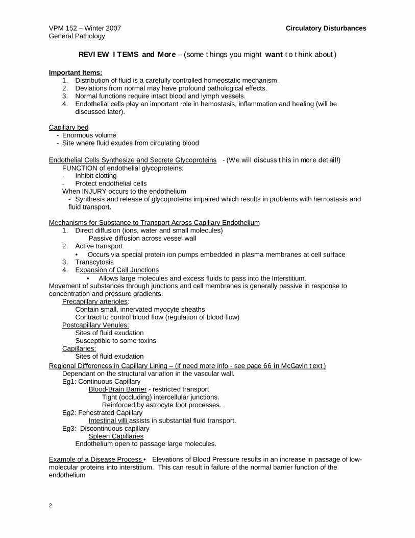
REVIEW ITEMS and More – (some things you might want to think about)
Important Items: 1. Distribution of fluid is a carefully controlled homeostatic mechanism. 2. Deviations from normal may have profound pathological effects. 3. Normal functions require intact blood and lymph vessels. 4. Endothelial cells play an important role in hemostasis, inflammation and healing (will be
discussed later). Capillary bed - Enormous volume - Site where fluid exudes from circulating blood Endothelial Cells Synthesize and Secrete Glycoproteins - (We will discuss this in more detail!) FUNCTION of endothelial glycoproteins: - Inhibit clotting
- Protect endothelial cells When INJURY occurs to the endothelium
- Synthesis and release of glycoproteins impaired which results in problems with hemostasis and fluid transport.
Mechanisms for Substance to Transport Across Capillary Endothelium 1. Direct diffusion (ions, water and small molecules) Passive diffusion across vessel wall
2. Active transport • Occurs via special protein ion pumps embedded in plasma membranes at cell surface
3. Transcytosis 4. Expansion of Cell Junctions
• Allows large molecules and excess fluids to pass into the Interstitium. Movement of substances through junctions and cell membranes is generally passive in response to concentration and pressure gradients. Precapillary arterioles: Contain small, innervated myocyte sheaths Contract to control blood flow (regulation of blood flow) Postcapillary Venules: Sites of fluid exudation Susceptible to some toxins Capillaries: Sites of fluid exudation Regional Differences in Capillary Lining – (if need more info - see page 66 in McGavin text) Dependant on the structural variation in the vascular wall. Eg1: Continuous Capillary Blood-Brain Barrier - restricted transport Tight (occluding) intercellular junctions. Reinforced by astrocyte foot processes. Eg2: Fenestrated Capillary Intestinal villi assists in substantial fluid transport. Eg3: Discontinuous capillary Spleen Capillaries Endothelium open to passage large molecules. Example of a Disease Process • Elevations of Blood Pressure results in an increase in passage of low-molecular proteins into interstitium. This can result in failure of the normal barrier function of the endothelium
VPM 152 – Winter 2007 Circulatory Disturbances General Pathology
3
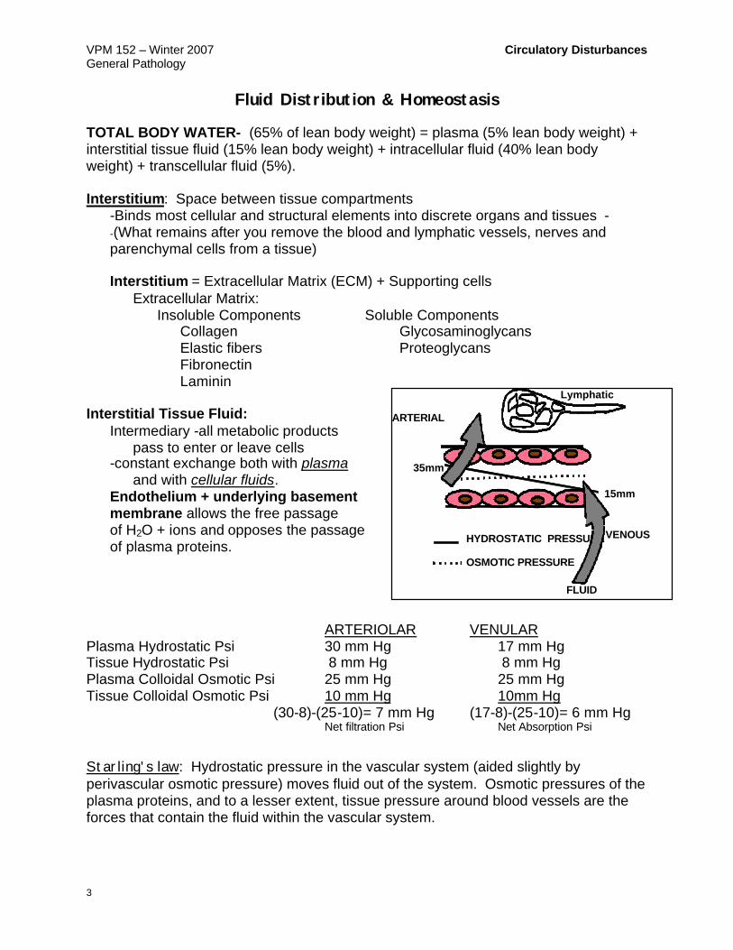
Fluid Distribution & Homeostasis TOTAL BODY WATER- (65% of lean body weight) = plasma (5% lean body weight) + interstitial tissue fluid (15% lean body weight) + intracellular fluid (40% lean body weight) + transcellular fluid (5%). Interstitium: Space between tissue compartments -Binds most cellular and structural elements into discrete organs and tissues -
-(What remains after you remove the blood and lymphatic vessels, nerves and parenchymal cells from a tissue)
Interstitium = Extracellular Matrix (ECM) + Supporting cells Extracellular Matrix: Insoluble Components Soluble Components Collagen Glycosaminoglycans Elastic fibers Proteoglycans Fibronectin Laminin Interstitial Tissue Fluid: Intermediary -all metabolic products pass to enter or leave cells -constant exchange both with plasma and with cellular fluids. Endothelium + underlying basement membrane allows the free passage of H2O + ions and opposes the passage of plasma proteins. ARTERIOLAR VENULAR Plasma Hydrostatic Psi 30 mm Hg 17 mm Hg Tissue Hydrostatic Psi 8 mm Hg 8 mm Hg Plasma Colloidal Osmotic Psi 25 mm Hg 25 mm Hg Tissue Colloidal Osmotic Psi 10 mm Hg 10mm Hg (30-8)-(25-10)= 7 mm Hg (17-8)-(25-10)= 6 mm Hg Net filtration Psi Net Absorption Psi
Starling's law: Hydrostatic pressure in the vascular system (aided slightly by perivascular osmotic pressure) moves fluid out of the system. Osmotic pressures of the plasma proteins, and to a lesser extent, tissue pressure around blood vessels are the forces that contain the fluid within the vascular system.
HYDROSTATIC PRESSURE OSMOTIC PRESSURE
ARTERIAL
VENOUS
35mm
15mm
FLUID
Lymphatic
VPM 152 – Winter 2007 Circulatory Disturbances General Pathology
4
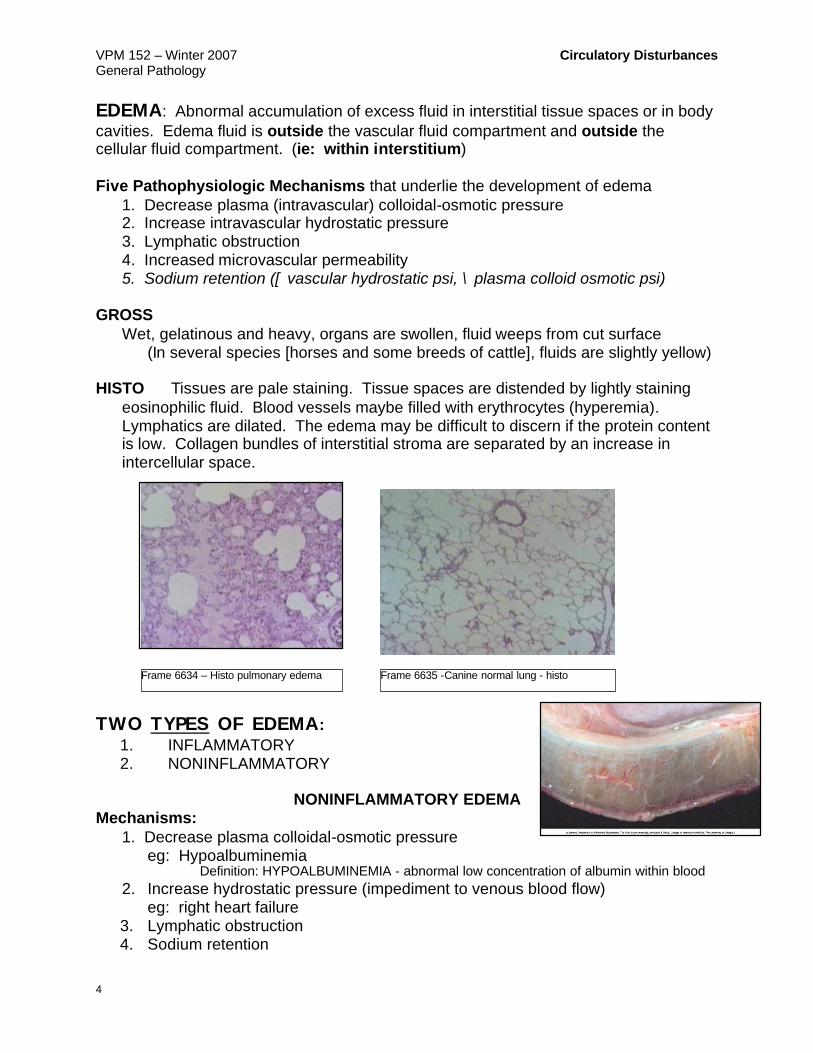
EDEMA: Abnormal accumulation of excess fluid in interstitial tissue spaces or in body cavities. Edema fluid is outside the vascular fluid compartment and outside the cellular fluid compartment. (ie: within interstitium) Five Pathophysiologic Mechanisms that underlie the development of edema 1. Decrease plasma (intravascular) colloidal-osmotic pressure 2. Increase intravascular hydrostatic pressure 3. Lymphatic obstruction 4. Increased microvascular permeability 5. Sodium retention ([ vascular hydrostatic psi, \ plasma colloid osmotic psi) GROSS Wet, gelatinous and heavy, organs are swollen, fluid weeps from cut surface (In several species [horses and some breeds of cattle], fluids are slightly yellow) HISTO Tissues are pale staining. Tissue spaces are distended by lightly staining
eosinophilic fluid. Blood vessels maybe filled with erythrocytes (hyperemia). Lymphatics are dilated. The edema may be difficult to discern if the protein content is low. Collagen bundles of interstitial stroma are separated by an increase in intercellular space.
TWO TYPES OF EDEMA:
1. INFLAMMATORY 2. NONINFLAMMATORY
NONINFLAMMATORY EDEMA
Mechanisms: 1. Decrease plasma colloidal-osmotic pressure eg: Hypoalbuminemia
Definition: HYPOALBUMINEMIA - abnormal low concentration of albumin within blood 2. Increase hydrostatic pressure (impediment to venous blood flow)
eg: right heart failure 3. Lymphatic obstruction 4. Sodium retention
Frame 6634 – Histo pulmonary edema Frame 6635 -Canine normal lung - histo
VPM 152 – Winter 2007 Circulatory Disturbances General Pathology
5
Fluid Characteristics: "protein poor" Transudate - low protein content < 30 g/L - specific gravity below 1.017 - total nucleated cell count < 1.5 X 10⁹/L
INFLAMMATORY EDEMA
Mechanism: Increased Vascular Permeability - Endothelial damage Fluid Characteristics: "protein rich" Exudate - high concentration of protein > 30 g/L - 8 specific gravity > 1.025 - total nucleated cell count > 7.0 X 10⁹/L LOCAL EDEMA Mechanisms: Local Increase in hydrostatic pressure Lymphatic obstruction Inflammation Etio: impaired venous drainage or lymphatic blockade or inflammation eg1: Improperly bandaged limb resulting in venous occlusion eg2: Damage to lymphatics (surgery, neoplasm, or intravascular parasites) eg3: Inflammation may also affect lymphatics (lymphangitis) GENERALIZED EDEMA Mechanism: 1. Increased hydrostatic pressure of blood
2. Decreased colloid osmotic pressure of plasma proteins 3. Sodium retention
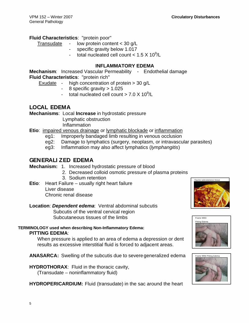
Etio: Heart Failure – usually right heart failure Liver disease Chronic renal disease Location: Dependent edema: Ventral abdominal subcutis Subcutis of the ventral cervical region Subcutaneous tissues of the limbs
TERMINOLOGY used when describing Non-Inflammatory Edema: PITTING EDEMA: When pressure is applied to an area of edema a depression or dent results as excessive interstitial fluid is forced to adjacent areas. ANASARCA: Swelling of the subcutis due to severe generalized edema HYDROTHORAX: Fluid in the thoracic cavity, (Transudate – noninflammatory fluid) HYDROPERICARDIUM: Fluid (transudate) in the sac around the heart
Frame 9993
Pitting Edema
Frame 9994 Pitting Edema
Equine subcutaneous tissue
VPM 152 – Winter 2007 Circulatory Disturbances General Pathology
6
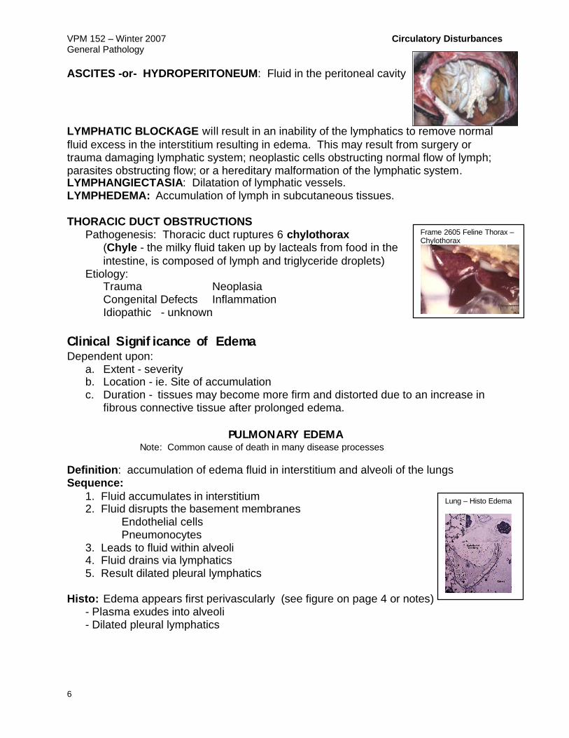
ASCITES -or- HYDROPERITONEUM: Fluid in the peritoneal cavity LYMPHATIC BLOCKAGE will result in an inability of the lymphatics to remove normal fluid excess in the interstitium resulting in edema. This may result from surgery or trauma damaging lymphatic system; neoplastic cells obstructing normal flow of lymph; parasites obstructing flow; or a hereditary malformation of the lymphatic system. LYMPHANGIECTASIA: Dilatation of lymphatic vessels. LYMPHEDEMA: Accumulation of lymph in subcutaneous tissues. THORACIC DUCT OBSTRUCTIONS
Pathogenesis: Thoracic duct ruptures 6 chylothorax (Chyle - the milky fluid taken up by lacteals from food in the intestine, is composed of lymph and triglyceride droplets) Etiology:
Trauma Neoplasia Congenital Defects Inflammation Idiopathic - unknown
Clinical Significance of Edema Dependent upon: a. Extent - severity b. Location - ie. Site of accumulation
c. Duration - tissues may become more firm and distorted due to an increase in fibrous connective tissue after prolonged edema.
PULMONARY EDEMA
Note: Common cause of death in many disease processes Definition: accumulation of edema fluid in interstitium and alveoli of the lungs Sequence: 1. Fluid accumulates in interstitium 2. Fluid disrupts the basement membranes Endothelial cells Pneumonocytes 3. Leads to fluid within alveoli 4. Fluid drains via lymphatics 5. Result dilated pleural lymphatics Histo: Edema appears first perivascularly (see figure on page 4 or notes) - Plasma exudes into alveoli - Dilated pleural lymphatics
Frame 2605 Feline Thorax – Chylothorax
Lung – Histo Edema
VPM 152 – Winter 2007 Circulatory Disturbances General Pathology
7
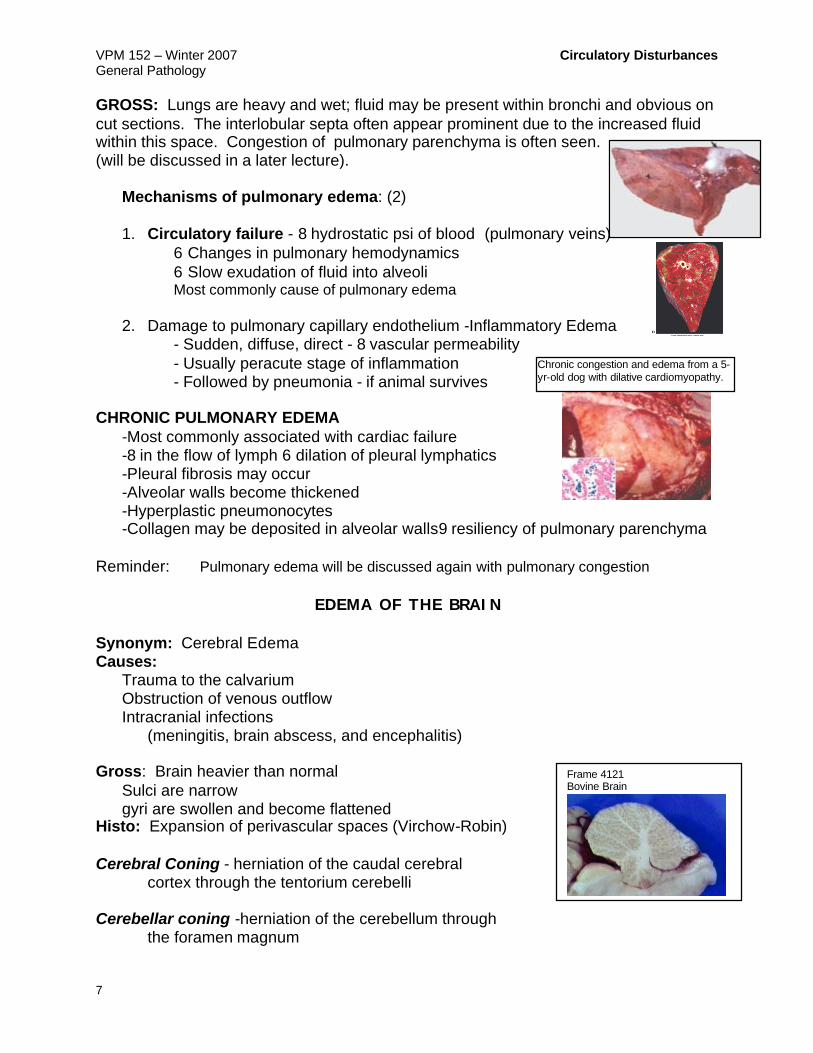
GROSS: Lungs are heavy and wet; fluid may be present within bronchi and obvious on cut sections. The interlobular septa often appear prominent due to the increased fluid within this space. Congestion of pulmonary parenchyma is often seen. (will be discussed in a later lecture). Mechanisms of pulmonary edema: (2)
1. Circulatory failure - 8 hydrostatic psi of blood (pulmonary veins) 6 Changes in pulmonary hemodynamics 6 Slow exudation of fluid into alveoli Most commonly cause of pulmonary edema
2. Damage to pulmonary capillary endothelium -Inflammatory Edema - Sudden, diffuse, direct - 8 vascular permeability - Usually peracute stage of inflammation - Followed by pneumonia - if animal survives CHRONIC PULMONARY EDEMA -Most commonly associated with cardiac failure -8 in the flow of lymph 6 dilation of pleural lymphatics -Pleural fibrosis may occur -Alveolar walls become thickened -Hyperplastic pneumonocytes -Collagen may be deposited in alveolar walls9 resiliency of pulmonary parenchyma Reminder: Pulmonary edema will be discussed again with pulmonary congestion
EDEMA OF THE BRAIN
Synonym: Cerebral Edema Causes:
Trauma to the calvarium Obstruction of venous outflow Intracranial infections (meningitis, brain abscess, and encephalitis) Gross: Brain heavier than normal
Sulci are narrow gyri are swollen and become flattened
Histo: Expansion of perivascular spaces (Virchow-Robin) Cerebral Coning - herniation of the caudal cerebral
cortex through the tentorium cerebelli Cerebellar coning -herniation of the cerebellum through the foramen magnum
Frame 4121 Bovine Brain
Chronic congestion and edema from a 5-yr-old dog with dilative cardiomyopathy.
VPM 152 – Winter 2007 Circulatory Disturbances General Pathology
8
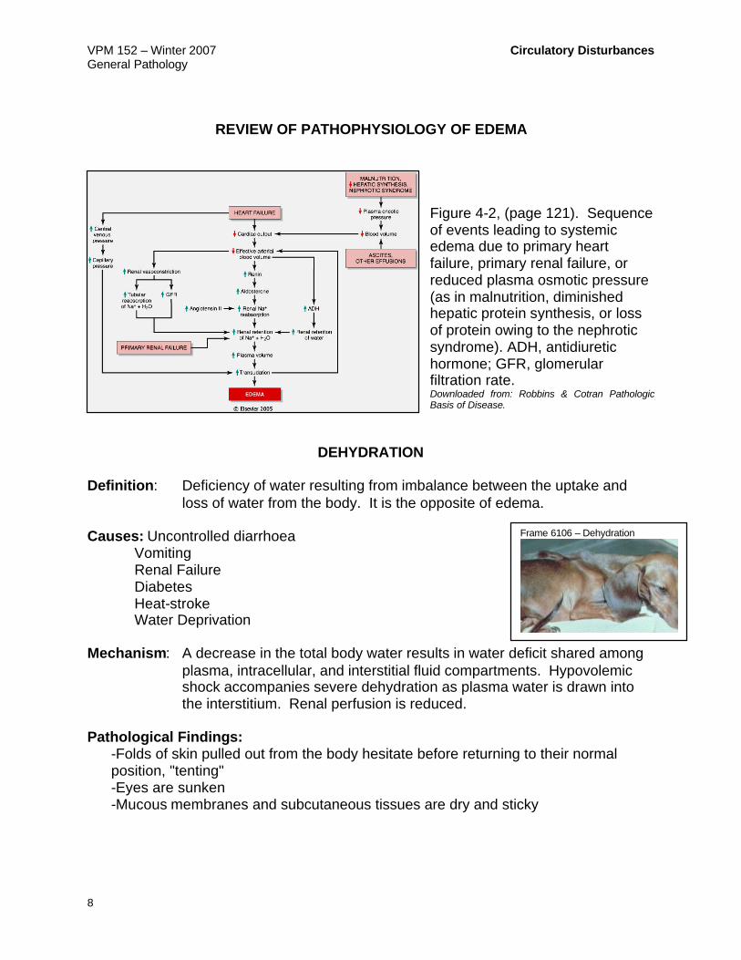
REVIEW OF PATHOPHYSIOLOGY OF EDEMA
Figure 4-2, (page 121). Sequence of events leading to systemic edema due to primary heart failure, primary renal failure, or reduced plasma osmotic pressure (as in malnutrition, diminished hepatic protein synthesis, or loss of protein owing to the nephrotic syndrome). ADH, antidiuretic hormone; GFR, glomerular filtration rate. Downloaded from: Robbins & Cotran Pathologic Basis of Disease.
DEHYDRATION
Definition: Deficiency of water resulting from imbalance between the uptake and
loss of water from the body. It is the opposite of edema. Causes: Uncontrolled diarrhoea Vomiting Renal Failure Diabetes Heat-stroke Water Deprivation Mechanism: A decrease in the total body water results in water deficit shared among
plasma, intracellular, and interstitial fluid compartments. Hypovolemic shock accompanies severe dehydration as plasma water is drawn into the interstitium. Renal perfusion is reduced.
Pathological Findings:
-Folds of skin pulled out from the body hesitate before returning to their normal position, "tenting" -Eyes are sunken
-Mucous membranes and subcutaneous tissues are dry and sticky
Frame 6106 – Dehydration
VPM 152 – Winter 2007 Circulatory Disturbances General Pathology
9
ALTERATIONS IN BLOOD FLOW AND PERFUSION
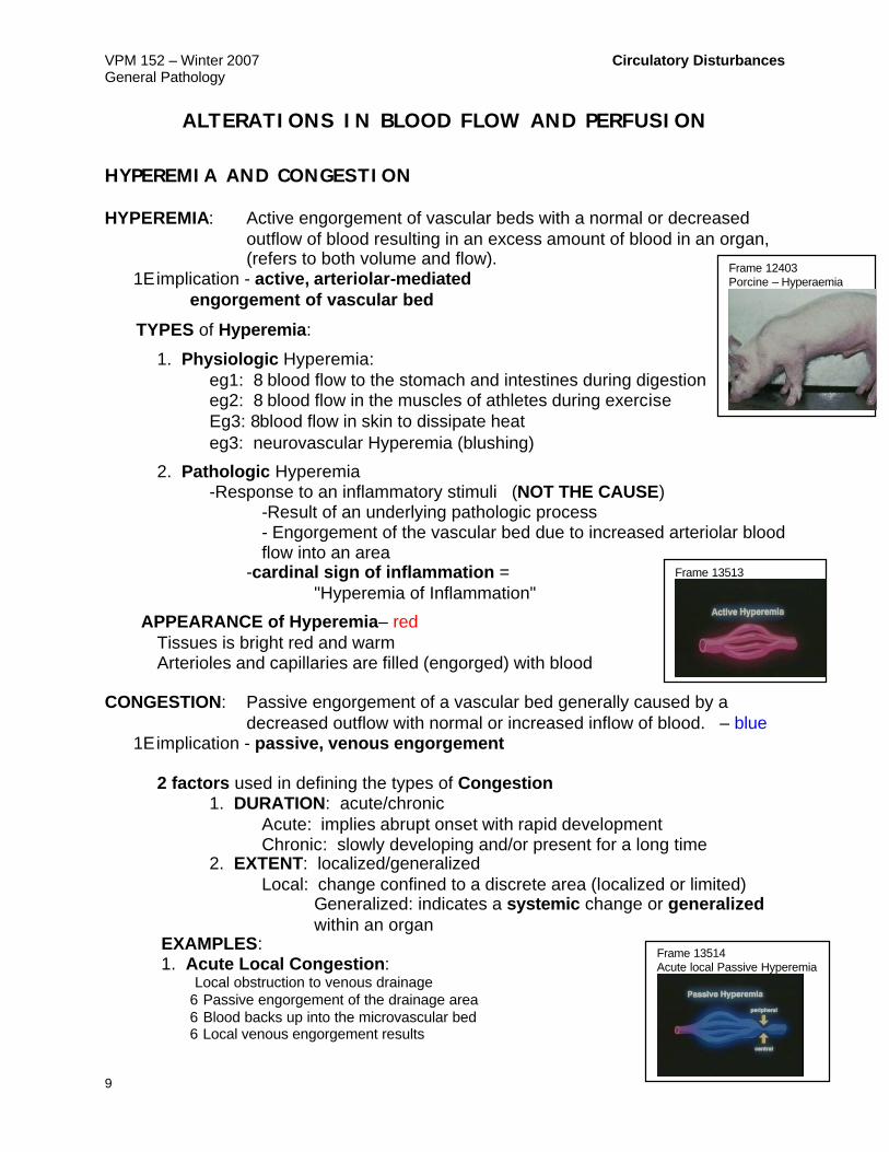
HYPEREMIA AND CONGESTION HYPEREMIA: Active engorgement of vascular beds with a normal or decreased
outflow of blood resulting in an excess amount of blood in an organ, (refers to both volume and flow). 1E implication - active, arteriolar-mediated engorgement of vascular bed
TYPES of Hyperemia:
1. Physiologic Hyperemia: eg1: 8 blood flow to the stomach and intestines during digestion eg2: 8 blood flow in the muscles of athletes during exercise Eg3: 8blood flow in skin to dissipate heat eg3: neurovascular Hyperemia (blushing) 2. Pathologic Hyperemia -Response to an inflammatory stimuli (NOT THE CAUSE) -Result of an underlying pathologic process
- Engorgement of the vascular bed due to increased arteriolar blood flow into an area
-cardinal sign of inflammation = "Hyperemia of Inflammation"
APPEARANCE of Hyperemia– red Tissues is bright red and warm Arterioles and capillaries are filled (engorged) with blood
CONGESTION: Passive engorgement of a vascular bed generally caused by a
decreased outflow with normal or increased inflow of blood. – blue 1E implication - passive, venous engorgement
2 factors used in defining the types of Congestion 1. DURATION: acute/chronic Acute: implies abrupt onset with rapid development Chronic: slowly developing and/or present for a long time 2. EXTENT: localized/generalized Local: change confined to a discrete area (localized or limited)
Generalized: indicates a systemic change or generalized within an organ
EXAMPLES: 1. Acute Local Congestion: Local obstruction to venous drainage 6 Passive engorgement of the drainage area 6 Blood backs up into the microvascular bed 6 Local venous engorgement results
Frame 12403 Porcine – Hyperaemia
Frame 13513
Frame 13514 Acute local Passive Hyperemia
VPM 152 – Winter 2007 Circulatory Disturbances General Pathology
10
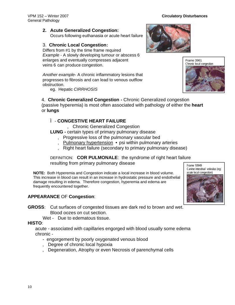
2. Acute Generalized Congestion: Occurs following euthanasia or acute heart failure 3. Chronic Local Congestion: Differs from #1 by the time frame required
Example - A slowly developing tumour or abscess 6 enlarges and eventually compresses adjacent veins 6 can produce congestion. Another example- A chronic inflammatory lesions that progresses to fibrosis and can lead to venous outflow obstruction. eg. Hepatic CIRRHOSIS
4. Chronic Generalized Congestion - Chronic Generalized congestion (passive hyperemia) is most often associated with pathology of either the heart or lungs
Ì - CONGESTIVE HEART FAILURE ¸ Chronic Generalized Congestion LUNG - certain types of primary pulmonary disease ¸ Progressive loss of the pulmonary vascular bed ¸ Pulmonary hypertension • psi within pulmonary arteries ¸ Right heart failure (secondary to primary pulmonary disease)
DEFINITION: COR PULMONALE: the syndrome of right heart failure resulting from primary pulmonary disease
NOTE: Both Hyperemia and Congestion indicate a local increase in blood volume. This increase in blood can result in an increase in hydrostatic pressure and endothelial damage resulting in edema. Therefore congestion, hyperemia and edema are frequently encountered together.
APPEARANCE OF Congestion: GROSS: Cut surfaces of congested tissues are dark red to brown and wet. Blood oozes on cut section. Wet - Due to edematous tissue. HISTO: acute - associated with capillaries engorged with blood usually some edema chronic - - engorgement by poorly oxygenated venous blood ¸ Degree of chronic local hypoxia ¸ Degeneration, Atrophy or even Necrosis of parenchymal cells
Frame 10949 Canine intestinal volvulus (eg: acute local congestion)
Frame 3961 Chronic local congestion
VPM 152 – Winter 2007 Circulatory Disturbances General Pathology
11
LUNG Cause: Can be acute or chronic. If acute see diffuse pulmonary congestion and edema (for review see pages 4&6) . Chronic failure of left ventricle impedes the flow of blood from the lungs to the heart ¸ chronic passive congestion ¸ 8 psi in alveolar capillaries and alveolar capillaries become engorged with blood. 4 consequences - of chronic pulmonary congestion (hyperemia) 1. Microhemorrhages
Small capillaries rupture ¸ intra-alveolar hemorrhages ¸ extravascular red cells are phagocytized by alveolar macrophages ¸ hemosiderin pigment "heart failure cells"
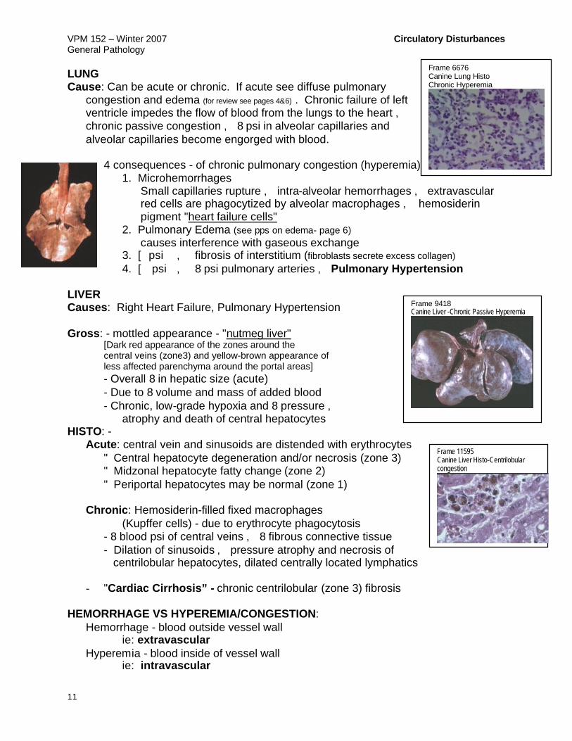
2. Pulmonary Edema (see pps on edema- page 6) causes interference with gaseous exchange 3. [ psi ¸ fibrosis of interstitium (fibroblasts secrete excess collagen) 4. [ psi ¸ 8 psi pulmonary arteries ¸ Pulmonary Hypertension LIVER Causes: Right Heart Failure, Pulmonary Hypertension Gross: - mottled appearance - "nutmeg liver" [Dark red appearance of the zones around the central veins (zone3) and yellow-brown appearance of less affected parenchyma around the portal areas] - Overall 8 in hepatic size (acute) - Due to 8 volume and mass of added blood - Chronic, low-grade hypoxia and 8 pressure ¸ atrophy and death of central hepatocytes HISTO: - Acute: central vein and sinusoids are distended with erythrocytes " Central hepatocyte degeneration and/or necrosis (zone 3) " Midzonal hepatocyte fatty change (zone 2) " Periportal hepatocytes may be normal (zone 1) Chronic: Hemosiderin-filled fixed macrophages (Kupffer cells) - due to erythrocyte phagocytosis - 8 blood psi of central veins ¸ 8 fibrous connective tissue - Dilation of sinusoids ¸ pressure atrophy and necrosis of centrilobular hepatocytes, dilated centrally located lymphatics
- "Cardiac Cirrhosis” - chronic centrilobular (zone 3) fibrosis HEMORRHAGE VS HYPEREMIA/CONGESTION: Hemorrhage - blood outside vessel wall
ie: extravascular Hyperemia - blood inside of vessel wall
ie: intravascular
Frame 6676 Canine Lung Histo Chronic Hyperemia
Frame 9418 Canine Liver -Chronic Passive Hyperemia
Frame 11595 Canine Liver Histo-Centrilobular congestion
VPM 152 – Winter 2007 Circulatory Disturbances General Pathology
12
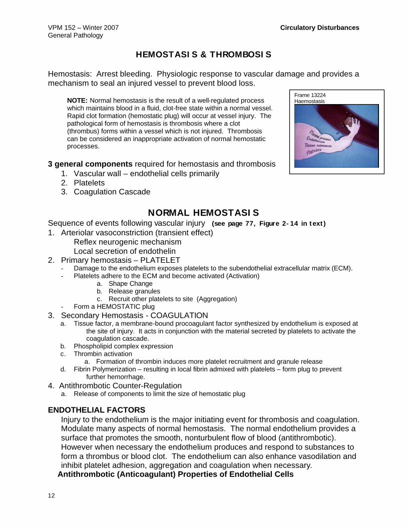
HEMOSTASIS & THROMBOSIS Hemostasis: Arrest bleeding. Physiologic response to vascular damage and provides a mechanism to seal an injured vessel to prevent blood loss.
NOTE: Normal hemostasis is the result of a well-regulated process which maintains blood in a fluid, clot-free state within a normal vessel. Rapid clot formation (hemostatic plug) will occur at vessel injury. The pathological form of hemostasis is thrombosis where a clot (thrombus) forms within a vessel which is not injured. Thrombosis can be considered an inappropriate activation of normal hemostatic processes.
3 general components required for hemostasis and thrombosis 1. Vascular wall – endothelial cells primarily 2. Platelets 3. Coagulation Cascade
NORMAL HEMOSTASIS Sequence of events following vascular injury (see page 77, Figure 2-14 in text) 1. Arteriolar vasoconstriction (transient effect) Reflex neurogenic mechanism Local secretion of endothelin 2. Primary hemostasis – PLATELET
- Damage to the endothelium exposes platelets to the subendothelial extracellular matrix (ECM). - Platelets adhere to the ECM and become activated (Activation)
a. Shape Change b. Release granules c. Recruit other platelets to site (Aggregation)
- Form a HEMOSTATIC plug 3. Secondary Hemostasis - COAGULATION
a. Tissue factor, a membrane-bound procoagulant factor synthesized by endothelium is exposed at the site of injury. It acts in conjunction with the material secreted by platelets to activate the coagulation cascade.
b. Phospholipid complex expression c. Thrombin activation
a. Formation of thrombin induces more platelet recruitment and granule release d. Fibrin Polymerization – resulting in local fibrin admixed with platelets – form plug to prevent
further hemorrhage. 4. Antithrombotic Counter-Regulation a. Release of components to limit the size of hemostatic plug ENDOTHELIAL FACTORS
Injury to the endothelium is the major initiating event for thrombosis and coagulation. Modulate many aspects of normal hemostasis. The normal endothelium provides a surface that promotes the smooth, nonturbulent flow of blood (antithrombotic). However when necessary the endothelium produces and respond to substances to form a thrombus or blood clot. The endothelium can also enhance vasodilation and inhibit platelet adhesion, aggregation and coagulation when necessary.
Antithrombotic (Anticoagulant) Properties of Endothelial Cells
Frame 13224 Haemostasis
VPM 152 – Winter 2007 Circulatory Disturbances General Pathology
13
Antiplatelet 1. Barrier to subendothelial collagen - prevent platelets and plasma factors from exposure
2. Prostacyclin - PGI2 , and Nitric Oxide - inhibit platelet adhesion and aggregation and maintains vascular relaxation
3. Express adenosine diphosphatase to degrade ADP (ADP promotes platelet aggregation)
Anticoagulant properties 1. Membrane associated, heparin-like molecules Cofactors which allow antithrombin III to inactivate thrombin + factor Xa + other factors 2. Thrombomodulin - specific thrombin receptor -Binds to thrombin making it an anticoagulant which can activate protein C º activeProtein C - inhibits clotting by cleaving factors Va and VIIIa Requires protein S - synthesized by endothelial cells
3. Synthesizes tissue factor pathway inhibitor – complexes and inhibits Factors VIIa and Xa 4. Plasminogen activators which promote fibrinolytic activity to clear fibrin deposits from endothelium
Prothrombotic (Procoagulant) Properties of Endothelial cells Endothelial cells may be activated by infectious agents, hemodynamic factors, plasma mediators and cytokines or injured indirectly.
1. Synthesize, store, and release von Willebrand factor (vWF) - essential cofactor for platelet binding to collagen and other surfaces. Stored in Weibel-Palade bodies.
2. Endothelial cells are also induced by cytokines (eg: TNF, or IL-1) or bacterial endotoxin - to secrete tissue factor (Factor VII) which activates the extrinsic clotting pathway.
3. Endothelial cells bind IXa and Xa and increase their catalytic activities 4. Secrete plasminogen activator inhibitors - to depress fibrinolysis Vascular Repair
1. Platelet Derived Growth Factor (PDGF) stimulates smooth muscle and fibroblasts proliferation
2. Fibroblast Growth Factor (FGF) 3. Transforming Growth Factor-• (TFG-•) modulates vascular repair
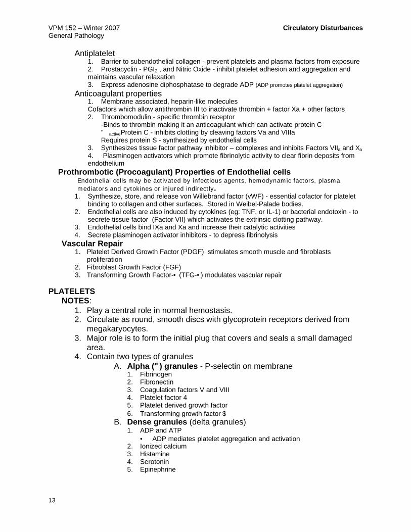
PLATELETS NOTES:
1. Play a central role in normal hemostasis. 2. Circulate as round, smooth discs with glycoprotein receptors derived from
megakaryocytes. 3. Major role is to form the initial plug that covers and seals a small damaged
area. 4. Contain two types of granules
A. Alpha (") granules - P-selectin on membrane 1. Fibrinogen 2. Fibronectin 3. Coagulation factors V and VIII 4. Platelet factor 4 5. Platelet derived growth factor 6. Transforming growth factor $
B. Dense granules (delta granules) 1. ADP and ATP
• ADP mediates platelet aggregation and activation 2. Ionized calcium 3. Histamine 4. Serotonin 5. Epinephrine
VPM 152 – Winter 2007 Circulatory Disturbances General Pathology
14
Platelet Response Vascular injury Yexposes Extracellular Matrix (ECM) Normally hidden by intact endothelium. Composed of - Collagen, proteoglycans, fibronectin, and other materials. Platelets + ECM Y 3 reactions
1. Adhesion and shape change Mediated via interactions with vWF - acts as bridge for platelets and ECM. 2. Secretion (release reaction) of both granule types.
Release of dense granules is very important because calcium is required for coagulation cascade.
ADP is a very important mediator of platelet aggregation. Leads to surface expression of a phospholipid complex. Needed binding site for calcium and coagulation factors. 3. Aggregation
Thromboxane A2 (TxA2) secreted by platelets and induces vasoconstriction and necessary for platelet aggregation.
ADP + TxA2 start reaction which leads to enlarging platelet aggregation = 1o hemostatic plug. Activates coagulation generated thrombin increasing aggregation. Platelet contraction - fused mass of platelets, fibrin formed cements mass = 2o hemostatic plug.
THROMBOCYTOPENIA (pp 83-84 McGavin) Definition: Drop below 100 X 109 platelets/L Most species bleed < 50 X 109 platelets/L dogs < 30 X 109 platelets/L Diagnosis: history of bleeding low platelet counts increased mucosal bleeding times Mechanisms: Deficient formation of platelets (eg: estrogen toxicoses) Excessive utilization (eg: consumptive coagulopathies) Premature destruction (eg: antibodies to Platelets) THROMBOCYTOPATHY Definition: Defective platelet function.
Mechanisms: Defect in adhesion - (eg: von WIllebrand’s disesase), aggregation, or release of granules.
VPM 152 – Winter 2007 Circulatory Disturbances General Pathology
15
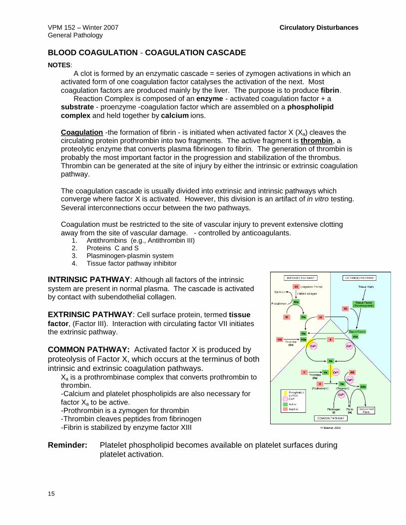
BLOOD COAGULATION - COAGULATION CASCADE NOTES:
A clot is formed by an enzymatic cascade = series of zymogen activations in which an activated form of one coagulation factor catalyses the activation of the next. Most coagulation factors are produced mainly by the liver. The purpose is to produce fibrin. Reaction Complex is composed of an enzyme - activated coagulation factor + a substrate - proenzyme -coagulation factor which are assembled on a phospholipid complex and held together by calcium ions.
Coagulation -the formation of fibrin - is initiated when activated factor X (Xa) cleaves the circulating protein prothrombin into two fragments. The active fragment is thrombin, a proteolytic enzyme that converts plasma fibrinogen to fibrin. The generation of thrombin is probably the most important factor in the progression and stabilization of the thrombus. Thrombin can be generated at the site of injury by either the intrinsic or extrinsic coagulation pathway.
The coagulation cascade is usually divided into extrinsic and intrinsic pathways which converge where factor X is activated. However, this division is an artifact of in vitro testing. Several interconnections occur between the two pathways.
Coagulation must be restricted to the site of vascular injury to prevent extensive clotting away from the site of vascular damage. - controlled by anticoagulants.
1. Antithrombins (e.g., Antithrombin III) 2. Proteins C and S 3. Plasminogen-plasmin system 4. Tissue factor pathway inhibitor
INTRINSIC PATHWAY: Although all factors of the intrinsic system are present in normal plasma. The cascade is activated by contact with subendothelial collagen. EXTRINSIC PATHWAY: Cell surface protein, termed tissue factor, (Factor III). Interaction with circulating factor VII initiates the extrinsic pathway. COMMON PATHWAY: Activated factor X is produced by proteolysis of Factor X, which occurs at the terminus of both intrinsic and extrinsic coagulation pathways.
Xa is a prothrombinase complex that converts prothrombin to thrombin. -Calcium and platelet phospholipids are also necessary for factor Xa to be active.
-Prothrombin is a zymogen for thrombin -Thrombin cleaves peptides from fibrinogen -Fibrin is stabilized by enzyme factor XIII Reminder: Platelet phospholipid becomes available on platelet surfaces during
platelet activation.
VPM 152 – Winter 2007 Circulatory Disturbances General Pathology
16
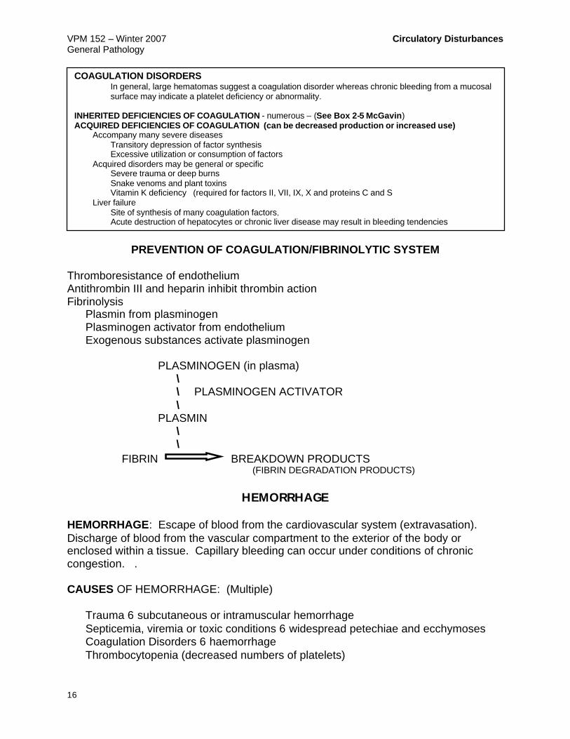
PREVENTION OF COAGULATION/FIBRINOLYTIC SYSTEM
Thromboresistance of endothelium Antithrombin III and heparin inhibit thrombin action Fibrinolysis Plasmin from plasminogen Plasminogen activator from endothelium Exogenous substances activate plasminogen PLASMINOGEN (in plasma) \ \ PLASMINOGEN ACTIVATOR \ PLASMIN \ \ FIBRIN BREAKDOWN PRODUCTS (FIBRIN DEGRADATION PRODUCTS)
HEMORRHAGE
HEMORRHAGE: Escape of blood from the cardiovascular system (extravasation). Discharge of blood from the vascular compartment to the exterior of the body or enclosed within a tissue. Capillary bleeding can occur under conditions of chronic congestion. . CAUSES OF HEMORRHAGE: (Multiple) Trauma 6 subcutaneous or intramuscular hemorrhage Septicemia, viremia or toxic conditions 6 widespread petechiae and ecchymoses Coagulation Disorders 6 haemorrhage Thrombocytopenia (decreased numbers of platelets)
COAGULATION DISORDERS In general, large hematomas suggest a coagulation disorder whereas chronic bleeding from a mucosal surface may indicate a platelet deficiency or abnormality.
INHERITED DEFICIENCIES OF COAGULATION - numerous – (See Box 2-5 McGavin) ACQUIRED DEFICIENCIES OF COAGULATION (can be decreased production or increased use) Accompany many severe diseases Transitory depression of factor synthesis Excessive utilization or consumption of factors Acquired disorders may be general or specific Severe trauma or deep burns Snake venoms and plant toxins Vitamin K deficiency (required for factors II, VII, IX, X and proteins C and S Liver failure Site of synthesis of many coagulation factors, Acute destruction of hepatocytes or chronic liver disease may result in bleeding tendencies
VPM 152 – Winter 2007 Circulatory Disturbances General Pathology
17
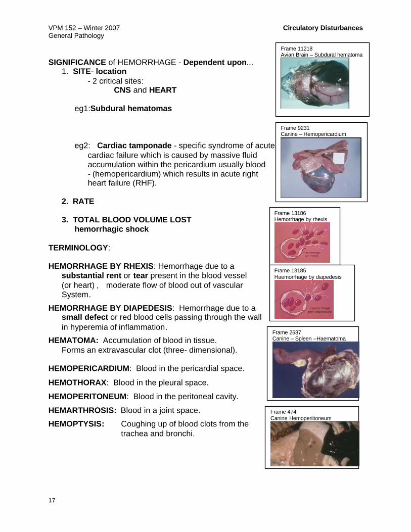
SIGNIFICANCE of HEMORRHAGE - Dependent upon...
1. SITE- location - 2 critical sites: CNS and HEART eg1:Subdural hematomas
eg2: Cardiac tamponade - specific syndrome of acute
cardiac failure which is caused by massive fluid accumulation within the pericardium usually blood - (hemopericardium) which results in acute right heart failure (RHF). 2. RATE 3. TOTAL BLOOD VOLUME LOST hemorrhagic shock TERMINOLOGY: HEMORRHAGE BY RHEXIS: Hemorrhage due to a
substantial rent or tear present in the blood vessel (or heart) ¸ moderate flow of blood out of vascular System.
HEMORRHAGE BY DIAPEDESIS: Hemorrhage due to a small defect or red blood cells passing through the wall in hyperemia of inflammation. HEMATOMA: Accumulation of blood in tissue. Forms an extravascular clot (three- dimensional). HEMOPERICARDIUM: Blood in the pericardial space.
HEMOTHORAX: Blood in the pleural space.
HEMOPERITONEUM: Blood in the peritoneal cavity.
HEMARTHROSIS: Blood in a joint space.
HEMOPTYSIS: Coughing up of blood clots from the trachea and bronchi.
Frame 11218 Avian Brain – Subdural hematoma
Frame 9231 Canine – Hemopericardium
Frame 13186 Hemorrhage by rhexis
Frame 13185 Haemorrhage by diapedesis
Frame 2687 Canine – Spleen –Haematoma
Frame 474 Canine Hemoperiitoneum
VPM 152 – Winter 2007 Circulatory Disturbances General Pathology
18
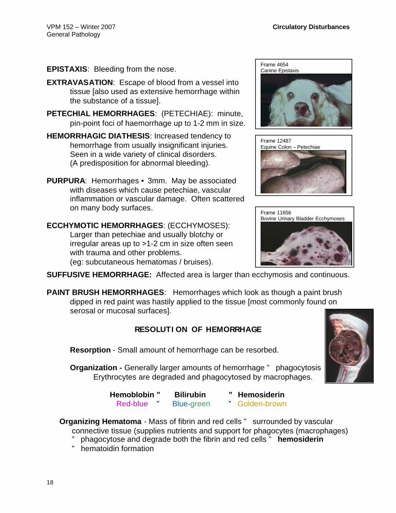
EPISTAXIS: Bleeding from the nose.
EXTRAVASATION: Escape of blood from a vessel into tissue [also used as extensive hemorrhage within
the substance of a tissue]. PETECHIAL HEMORRHAGES: (PETECHIAE): minute, pin-point foci of haemorrhage up to 1-2 mm in size. HEMORRHAGIC DIATHESIS: Increased tendency to hemorrhage from usually insignificant injuries. Seen in a wide variety of clinical disorders. (A predisposition for abnormal bleeding). PURPURA: Hemorrhages • 3mm. May be associated
with diseases which cause petechiae, vascular inflammation or vascular damage. Often scattered on many body surfaces.
ECCHYMOTIC HEMORRHAGES: (ECCHYMOSES): Larger than petechiae and usually blotchy or
irregular areas up to >1-2 cm in size often seen with trauma and other problems.
(eg: subcutaneous hematomas / bruises). SUFFUSIVE HEMORRHAGE: Affected area is larger than ecchymosis and continuous. PAINT BRUSH HEMORRHAGES: Hemorrhages which look as though a paint brush
dipped in red paint was hastily applied to the tissue [most commonly found on serosal or mucosal surfaces].
RESOLUTION OF HEMORRHAGE
Resorption - Small amount of hemorrhage can be resorbed. Organization - Generally larger amounts of hemorrhage º phagocytosis Erythrocytes are degraded and phagocytosed by macrophages. Hemoblobin º Bilirubin º Hemosiderin Red-blue º Blue-green º Golden-brown
Organizing Hematoma - Mass of fibrin and red cells º surrounded by vascular
connective tissue (supplies nutrients and support for phagocytes (macrophages) º phagocytose and degrade both the fibrin and red cells º hemosiderin º hematoidin formation
Frame 4654 Canine Epistaxis
Frame 12487 Equine Colon – Petechiae
Frame 11656 Bovine Urinary Bladder Ecchymoses
VPM 152 – Winter 2007 Circulatory Disturbances General Pathology
19
THROMBOSIS and INFARCTION PATHOGENESIS: - 3 primary influences - Virchow’s triad
1. Endothelial injury Dominant influence = can lead to thrombosis by itself eg: inflammation of heart valves
ºExpose of subendothelial ECM º platelet adherence º release of tissue factor ºDepletion of prostacyclin º primary and secondary hemostatic plug formation. 2 Alterations in normal blood flow - turbulence or stasis Normal blood flow is laminar - cellular elements in the middle, surrounded by plasma. Disrupt normal laminar flow º allows platelets to contact endothelium ºPrevents dilution of activated clotting factors by fresh-flowing blood º allows the build up of thrombi (slows the inflow of anticoagulants) º promotes endothelial cell activation.
3. Hypercoaguability Definition: any alteration of the coagulation pathways that predisposes to thombosis. 8 Coagulation factors 9 Inhibitory factors
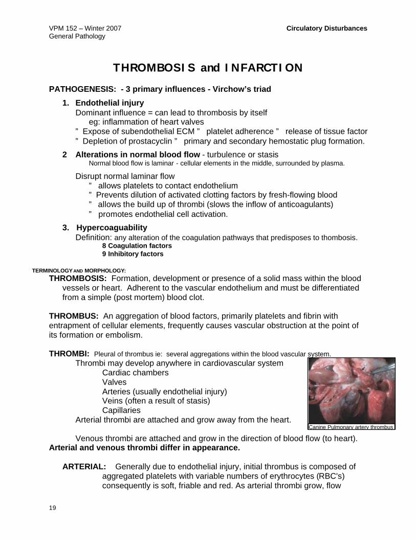
TERMINOLOGY AND MORPHOLOGY: THROMBOSIS: Formation, development or presence of a solid mass within the blood vessels or heart. Adherent to the vascular endothelium and must be differentiated from a simple (post mortem) blood clot. THROMBUS: An aggregation of blood factors, primarily platelets and fibrin with entrapment of cellular elements, frequently causes vascular obstruction at the point of its formation or embolism. THROMBI: Pleural of thrombus ie: several aggregations within the blood vascular system. Thrombi may develop anywhere in cardiovascular system Cardiac chambers Valves Arteries (usually endothelial injury) Veins (often a result of stasis) Capillaries
Arterial thrombi are attached and grow away from the heart.
Venous thrombi are attached and grow in the direction of blood flow (to heart). Arterial and venous thrombi differ in appearance.
ARTERIAL: Generally due to endothelial injury, initial thrombus is composed of aggregated platelets with variable numbers of erythrocytes (RBC's) consequently is soft, friable and red. As arterial thrombi grow, flow
Canine Pulmonary artery thrombus
VPM 152 – Winter 2007 Circulatory Disturbances General Pathology
20
patterns adjacent to the thrombi cause fibrin to be deposited and the platelet mass that persists is transformed into a fibrin mass. Fibrin strands polymerize between the separating and degenerating platelets. The alternating lines of yellow platelets and fibrin separating RBC's forms the lines of Zahn.
VENOUS: A venous thrombi is composed of fibrin strands with entrapped RBC's, since the dominant mechanism of formation is coagulation.
Morphological Differentiation of Thrombi Vs Post Mortem Clots
Arterial Thrombus
Venous Thrombus
Post Mortem Clot
Attributes
Grey, pale, white Red Yellow = chicken fat
Colour
+ Not frequent - Lamination
+ + - Attachment
Small - may be mural often fill lumen fill lumen Size and location
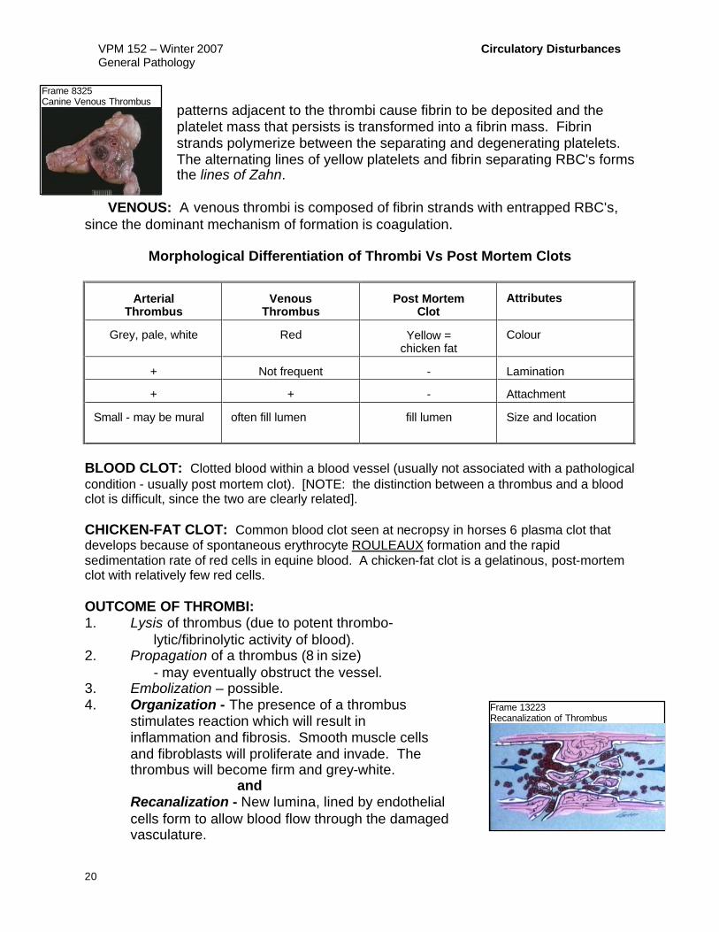
BLOOD CLOT: Clotted blood within a blood vessel (usually not associated with a pathological condition - usually post mortem clot). [NOTE: the distinction between a thrombus and a blood clot is difficult, since the two are clearly related]. CHICKEN-FAT CLOT: Common blood clot seen at necropsy in horses 6 plasma clot that develops because of spontaneous erythrocyte ROULEAUX formation and the rapid sedimentation rate of red cells in equine blood. A chicken-fat clot is a gelatinous, post-mortem clot with relatively few red cells. OUTCOME OF THROMBI: 1. Lysis of thrombus (due to potent thrombo- lytic/fibrinolytic activity of blood). 2. Propagation of a thrombus (8 in size) - may eventually obstruct the vessel. 3. Embolization – possible. 4. Organization - The presence of a thrombus
stimulates reaction which will result in inflammation and fibrosis. Smooth muscle cells and fibroblasts will proliferate and invade. The thrombus will become firm and grey-white.
and Recanalization - New lumina, lined by endothelial cells form to allow blood flow through the damaged vasculature.
Frame 8325 Canine Venous Thrombus
Frame 13223 Recanalization of Thrombus
VPM 152 – Winter 2007 Circulatory Disturbances General Pathology
21
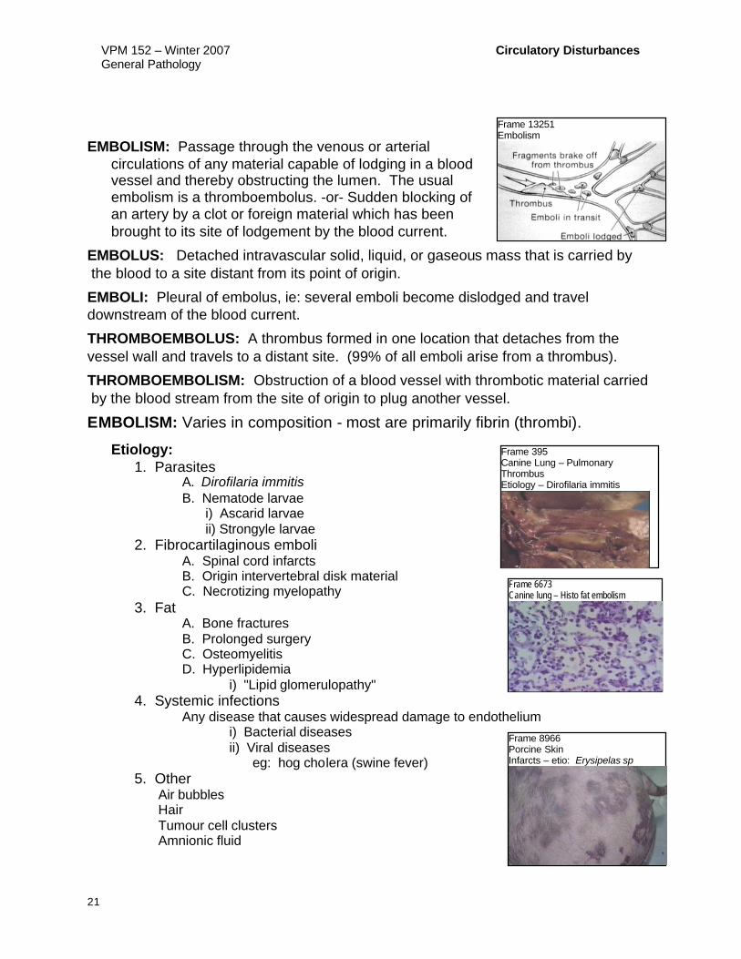
EMBOLISM: Passage through the venous or arterial circulations of any material capable of lodging in a blood vessel and thereby obstructing the lumen. The usual embolism is a thromboembolus. -or- Sudden blocking of an artery by a clot or foreign material which has been brought to its site of lodgement by the blood current. EMBOLUS: Detached intravascular solid, liquid, or gaseous mass that is carried by the blood to a site distant from its point of origin. EMBOLI: Pleural of embolus, ie: several emboli become dislodged and travel downstream of the blood current. THROMBOEMBOLUS: A thrombus formed in one location that detaches from the vessel wall and travels to a distant site. (99% of all emboli arise from a thrombus). THROMBOEMBOLISM: Obstruction of a blood vessel with thrombotic material carried by the blood stream from the site of origin to plug another vessel.
EMBOLISM: Varies in composition - most are primarily fibrin (thrombi). Etiology: 1. Parasites A. Dirofilaria immitis B. Nematode larvae i) Ascarid larvae ii) Strongyle larvae 2. Fibrocartilaginous emboli A. Spinal cord infarcts B. Origin intervertebral disk material C. Necrotizing myelopathy 3. Fat A. Bone fractures B. Prolonged surgery C. Osteomyelitis D. Hyperlipidemia i) "Lipid glomerulopathy" 4. Systemic infections Any disease that causes widespread damage to endothelium i) Bacterial diseases ii) Viral diseases eg: hog cholera (swine fever) 5. Other Air bubbles Hair Tumour cell clusters Amnionic fluid
Frame 13251 Embolism
Frame 395 Canine Lung – Pulmonary Thrombus Etiology – Dirofilaria immitis
Frame 6673 Canine lung – Histo fat embolism
Frame 8966 Porcine Skin Infarcts – etio: Erysipelas sp
VPM 152 – Winter 2007 Circulatory Disturbances General Pathology
22
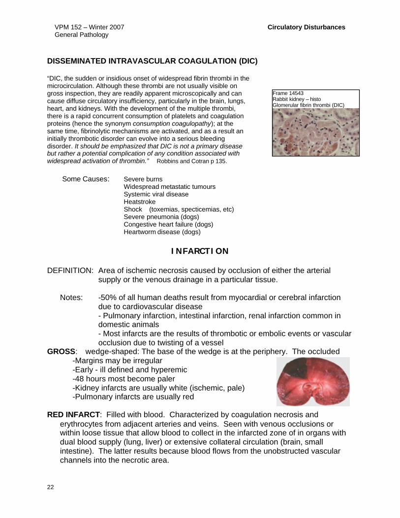
DISSEMINATED INTRAVASCULAR COAGULATION (DIC) “DIC, the sudden or insidious onset of widespread fibrin thrombi in the microcirculation. Although these thrombi are not usually visible on gross inspection, they are readily apparent microscopically and can cause diffuse circulatory insufficiency, particularly in the brain, lungs, heart, and kidneys. With the development of the multiple thrombi, there is a rapid concurrent consumption of platelets and coagulation proteins (hence the synonym consumption coagulopathy); at the same time, fibrinolytic mechanisms are activated, and as a result an initially thrombotic disorder can evolve into a serious bleeding disorder. It should be emphasized that DIC is not a primary disease but rather a potential complication of any condition associated with widespread activation of thrombin.” Robbins and Cotran p 135. Some Causes: Severe burns Widespread metastatic tumours Systemic viral disease Heatstroke Shock (toxemias, specticemias, etc) Severe pneumonia (dogs) Congestive heart failure (dogs) Heartworm disease (dogs)
INFARCTION DEFINITION: Area of ischemic necrosis caused by occlusion of either the arterial
supply or the venous drainage in a particular tissue. Notes: -50% of all human deaths result from myocardial or cerebral infarction
due to cardiovascular disease - Pulmonary infarction, intestinal infarction, renal infarction common in domestic animals - Most infarcts are the results of thrombotic or embolic events or vascular occlusion due to twisting of a vessel
GROSS: wedge-shaped: The base of the wedge is at the periphery. The occluded -Margins may be irregular -Early - ill defined and hyperemic -48 hours most become paler
-Kidney infarcts are usually white (ischemic, pale) -Pulmonary infarcts are usually red
RED INFARCT: Filled with blood. Characterized by coagulation necrosis and
erythrocytes from adjacent arteries and veins. Seen with venous occlusions or within loose tissue that allow blood to collect in the infarcted zone of in organs with dual blood supply (lung, liver) or extensive collateral circulation (brain, small intestine). The latter results because blood flows from the unobstructed vascular channels into the necrotic area.
Frame 14543 Rabbit kidney – histo Glomerular fibrin thrombi (DIC)
VPM 152 – Winter 2007 Circulatory Disturbances General Pathology
23
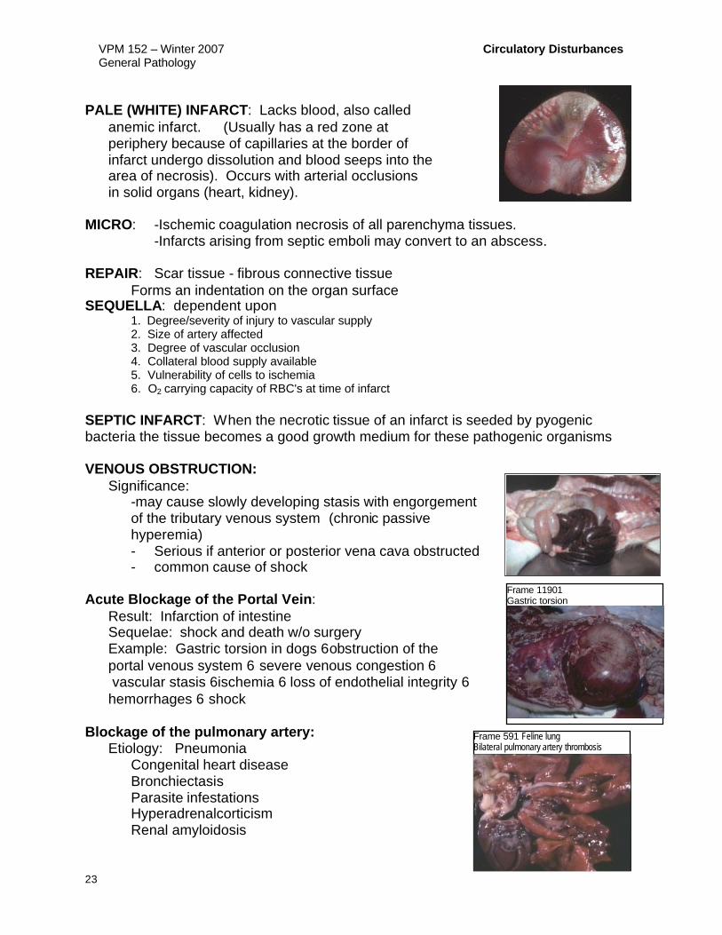
PALE (WHITE) INFARCT: Lacks blood, also called anemic infarct. (Usually has a red zone at periphery because of capillaries at the border of infarct undergo dissolution and blood seeps into the area of necrosis). Occurs with arterial occlusions in solid organs (heart, kidney). MICRO: -Ischemic coagulation necrosis of all parenchyma tissues.
-Infarcts arising from septic emboli may convert to an abscess. REPAIR: Scar tissue - fibrous connective tissue Forms an indentation on the organ surface SEQUELLA: dependent upon
1. Degree/severity of injury to vascular supply 2. Size of artery affected 3. Degree of vascular occlusion 4. Collateral blood supply available 5. Vulnerability of cells to ischemia 6. O2 carrying capacity of RBC's at time of infarct SEPTIC INFARCT: When the necrotic tissue of an infarct is seeded by pyogenic bacteria the tissue becomes a good growth medium for these pathogenic organisms VENOUS OBSTRUCTION: Significance:
-may cause slowly developing stasis with engorgement of the tributary venous system (chronic passive hyperemia)
- Serious if anterior or posterior vena cava obstructed - common cause of shock Acute Blockage of the Portal Vein:
Result: Infarction of intestine Sequelae: shock and death w/o surgery Example: Gastric torsion in dogs 6obstruction of the
portal venous system 6 severe venous congestion 6 vascular stasis 6ischemia 6 loss of endothelial integrity 6 hemorrhages 6 shock Blockage of the pulmonary artery: Etiology: Pneumonia Congenital heart disease Bronchiectasis Parasite infestations Hyperadrenalcorticism Renal amyloidosis
Frame 11901 Gastric torsion
Frame 591 Feline lung Bilateral pulmonary artery thrombosis
VPM 152 – Winter 2007 Circulatory Disturbances General Pathology
24
Result:- If sudden and large artery - death - If incomplete and smaller arteries 6 Anastomoses develop between pulmonary arteries and bronchial arteries Blockage of the posterior vena cava: Etiology: Hepatic abscesses in ruminants Dirofilariasis in dogs - overwhelming infections Pathogenesis: 1. Acute, complete occlusion 6death 2. Collateral circulation could develop (azygous vein)
SHOCK
Shock is the final common pathway for many potentially lethal clinical events which include microbial sepsis, severe hemorrhage, extensive trauma or burns, myocardial infarction, and mass pulmonary embolism. Whatever the cause the result is a decreased perfusion due to either decreased cardiac output or blood volume. Dr. Mosier in your veterinary pathology text refers to this as “circulatory dyshomeostasis.” The end result is hypotension which results in impaired tissue perfusion and cellular hypoxia. Definition: A syndrome resulting from a disproportion between the amount of blood volume present
and the volume of the circulatory system. In other words, an acute generalized failure of the capillary bed. -or - A condition of profound hemodynamic and metabolic disturbance characterized by failure of the circulatory system to maintain adequate perfusion of vital organs (circulatory dyshomeostasis).
Fundamental Disturbance: The blood volume is too small to fill the vascular system resulting in peripheral circulatory failure and cell damage due to hypoxia from inadequate tissue perfusion. Either not enough volume or blood flow is impaired. Three general categories of shock 1. Cardiogenic 2. Hypovolemic 3. Blood Maldistribution Note: Neurogenic and Anaphylactic shock both result in widespread vasodilation Cardiogenic Shock Results from failure of the heart to adequately pump blood. Can occur due to myocardial infarction, ventricular tachycardia, arrhythmias, cardiomyopathy, or an obstruction of the flow of blood from the heart. The stroke volume and cardiac output is decreased. Hypovolemic Shock Results from decreased circulating blood volume, can be due to blood loss from hemorrhage or fluid loss secondary to vomiting, diarrhea or burns.
VPM 152 – Winter 2007 Circulatory Disturbances General Pathology
25
Blood Maldistribution Characterized by a decrease in peripheral vascular resistance and resultant pooling of blood in peripheral tissues. Caused by neural or cytokine induced vasodilation. Trauma, emotional stress, systemic hypersensitivity to allergens, or endotoxemia can cause blood maldistribution. 1. Anaphylactic Shock – generalized type I hypersensitivity. 2. Neurogenic Shock 3. Septic Shock Pathogenesis of Septic Shock ~ 70% of septic shock are caused by endotoxin-producing gram-negative bacilli reason for the term endotoxic shock. Endotoxins are bacterial wall lipopoysaccharides (LPSs). These are released when bacterial cell walls are degraded. LPS consists of a toxic fatty acid (lipid A) core surrounded by a complex polysaccharide coat which is unique to the particular bacteria. Similar molecules can also be found on gram + bacteria and fungi. LPS injected into the blood stream can result in shock. LPS + LPS binding protein together bind to a cell surface receptor. This reaction can directly activate endothelial cells (makes them prothrombotic), WBC’s to release cytokines, activate complement mediated reactions. The brain and heart are the most susceptible organs. (NOTE: amount of O2 removed during blood flow varies average is approximately 25%, myocardium removes 75%) Stages of shock 3 stages 1. Nonprogressive shock 2. Progressive shock 3. Irreversible shock. Defined as a refractory state of circulatory control with inability to control the clinical disease. Shock is characterized by failure of multiple organ systems. Lesions: Pulmonary edema (cattle/horses - prominent shock organ) Liver Congestion: (dogs - prominent shock organ) Kidneys: Acute tubular necrosis Heart: Subendocardial hemorrhage and necrosis
Zonal lesions deep in myocardium Brain – Neuronal cell death
Adrenal glands: Cortical cell lipid depletion Degranulation of adrenalin-producing cells hemorrhagic with foci of necrosis " Gastrointestinal tract: hyperemia of mucosa with erosions are possible
Skeletal muscle: Pallor (peripheral vasoconstriction) severe crush injuries
VPM 152 – Winter 2007 Circulatory Disturbances General Pathology
26
INDEX
Anasarca, 5 Arterial Thrombi, 19 Blood Clot, 20 Cardiac Cirrhosis, 11 Cardiac Tamponade, 17 Cerebellar Coning, 7 Cerebral Coning, 7 Cerebral Edema, 7 Chicken-Fat Clot, 20 Chylothorax, 6 Coagulation, 15 Common Pathway, 15 Congestion, 9 Cor Pulmonale, 10 Plasma Colloidal-Osmotic Pressure, 4 Dehydration, 8 Dependent Edema, 5 Ecchymoses, 18 Ecchymotic Hemorrhages, 18 Edema, 3, 4, 5, 6, 7 Emboli 21 Embolism, 21 Embolus, 21 Endothelial Cells, 2 Epistaxis, 18 Extravasation, 18 Extravascular, 11 Extrinsic Coagulation Pathway, 15 Fibrocartilaginous Emboli, 21 Heart Failure Cells, 11 Hemarthrosis, 17 Hematoma, 17 Hemopericardium , 17 Hemoperitoneum, 17 HemoptysisIS, 17 Hemorrhage, 11, 16 Hemorrhage by Diapedesis, 17
Hemorrhage by Rhexis, 17 Hemorrhagic Diathesis, 18 Hemostasis, 1, 12 Hemothorax, 17 Hemosiderin, 18 Hydropericardium, 5 Hydroperitoneum, 6 Hydrothorax, 5 Hyperaemia, 11 Hyperemia, 9 Hypoalbuminemia, 4 Infarction, 22 Interstitium, 3, 4 Intravascular, 11 Lymphangiectasia, 6 Nutmeg Liver, 11 Organizing Haematoma, 18 Paint Brush Hemorrhages, 18 Petechiae, 18 Petechial Hemorrhages, 18 Pitting Edema, 5 Plasma Colloidal-Osmotic Psi, 3 Plasma Hydrostatic Psi, 3 Pulmonary Edema, 7, 25 Purpura, 18 Red Infarct, 22 Starling's Law, 3 Thrombi, 19 Thrombin, 15 Thromboembolism, 21 Thrombosis, 19 Thrombus, 19 Tissue Colloidal- Osmotic Psi, 3 Tissue Hydrostatic Psi, 3 Transudate, 5 Venous Thrombi, 20 White Infarct, 22