Disturbance of Circulation Hemodynamic Disorder · Disturbance of Circulation Hemodynamic Disorder...
48
Disturbance of Circulation Hemodynamic Disorder By Dr. Hemn Hassan Othman PhD, Pathology Fall 2016 2/9/2017 1
Transcript of Disturbance of Circulation Hemodynamic Disorder · Disturbance of Circulation Hemodynamic Disorder...
Disturbance of Circulation
Hemodynamic Disorder
By Dr. Hemn Hassan Othman
PhD, Pathology Fall 2016 2/9/2017 1
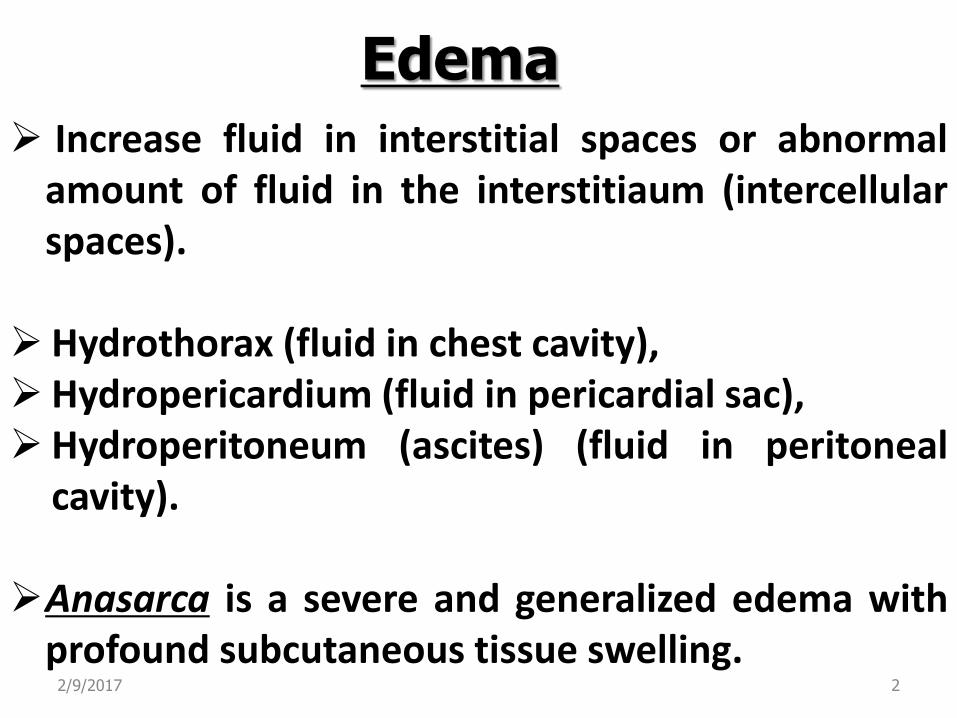
Edema
Increase fluid in interstitial spaces or abnormal amount of fluid in the interstitiaum (intercellular spaces). Hydrothorax (fluid in chest cavity), Hydropericardium (fluid in pericardial sac), Hydroperitoneum (ascites) (fluid in peritoneal
cavity).
Anasarca is a severe and generalized edema with profound subcutaneous tissue swelling.
2/9/2017 2
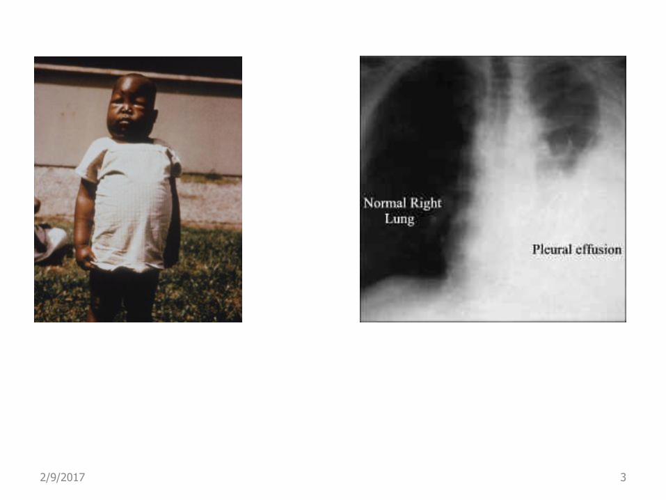
2/9/2017 3
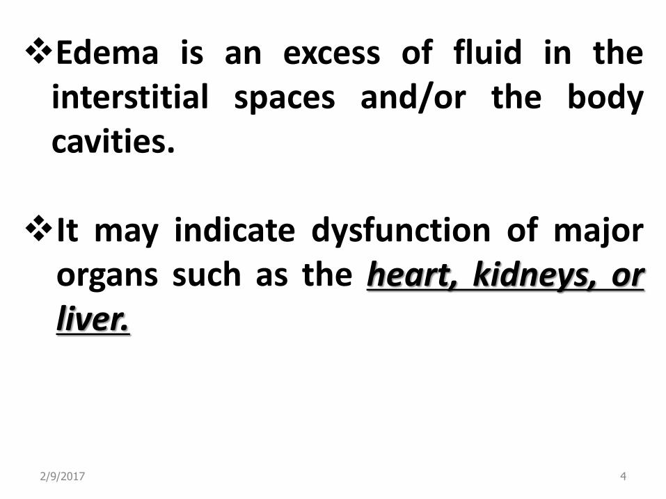
Edema is an excess of fluid in the interstitial spaces and/or the body cavities.
It may indicate dysfunction of major organs such as the heart, kidneys, or liver.
2/9/2017 4

Normal Fluid balance in the tissues
2/9/2017 5
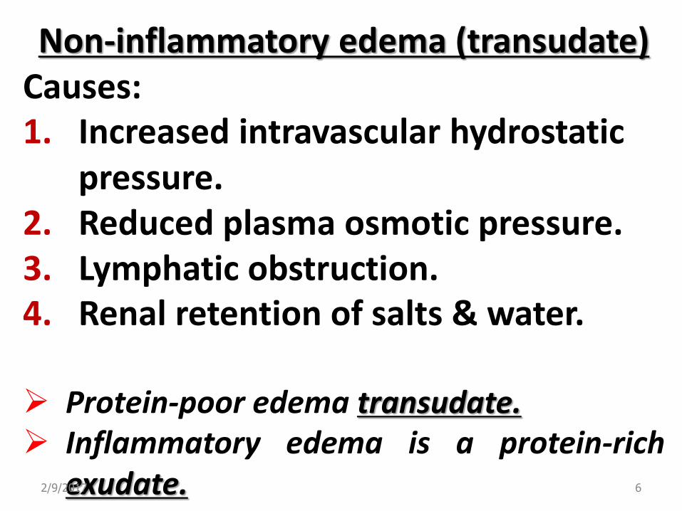
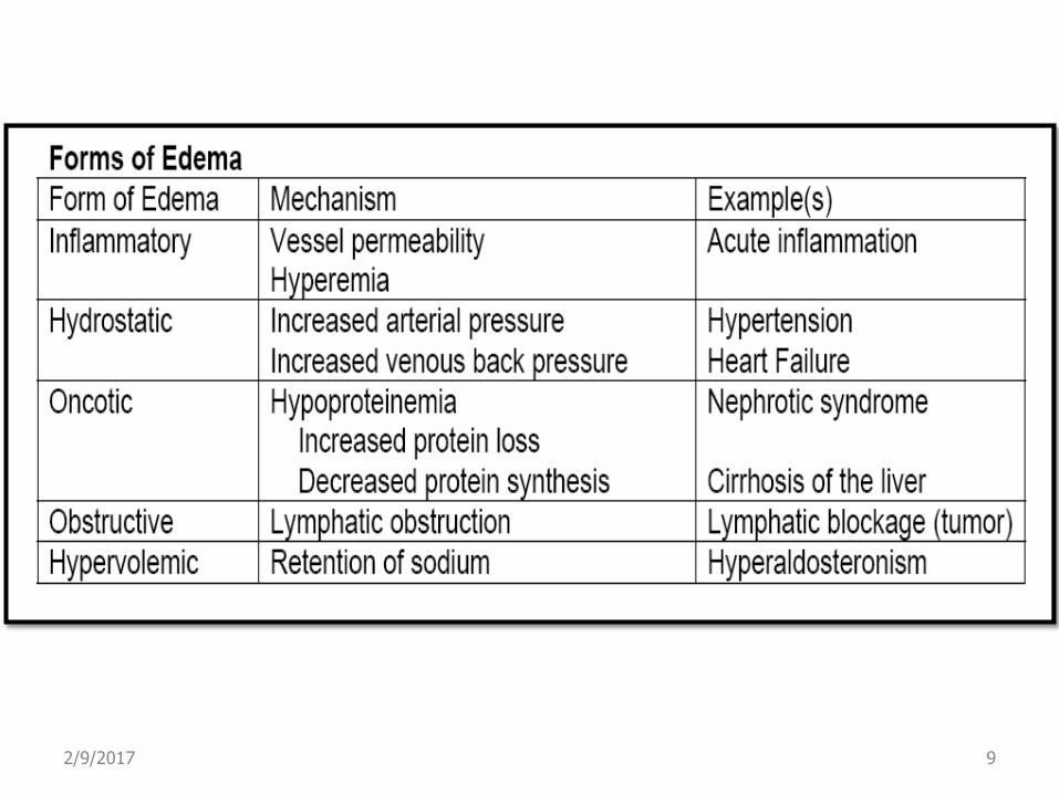
Non-inflammatory edema (transudate)
Causes: 1. Increased intravascular hydrostatic
pressure. 2. Reduced plasma osmotic pressure. 3. Lymphatic obstruction. 4. Renal retention of salts & water.
Protein-poor edema transudate. Inflammatory edema is a protein-rich
exudate. 2/9/2017 6

2/9/2017 7

Non inflammatory edema (transudate) Inflammatory edema (exudate)
2/9/2017 8
2/9/2017 9
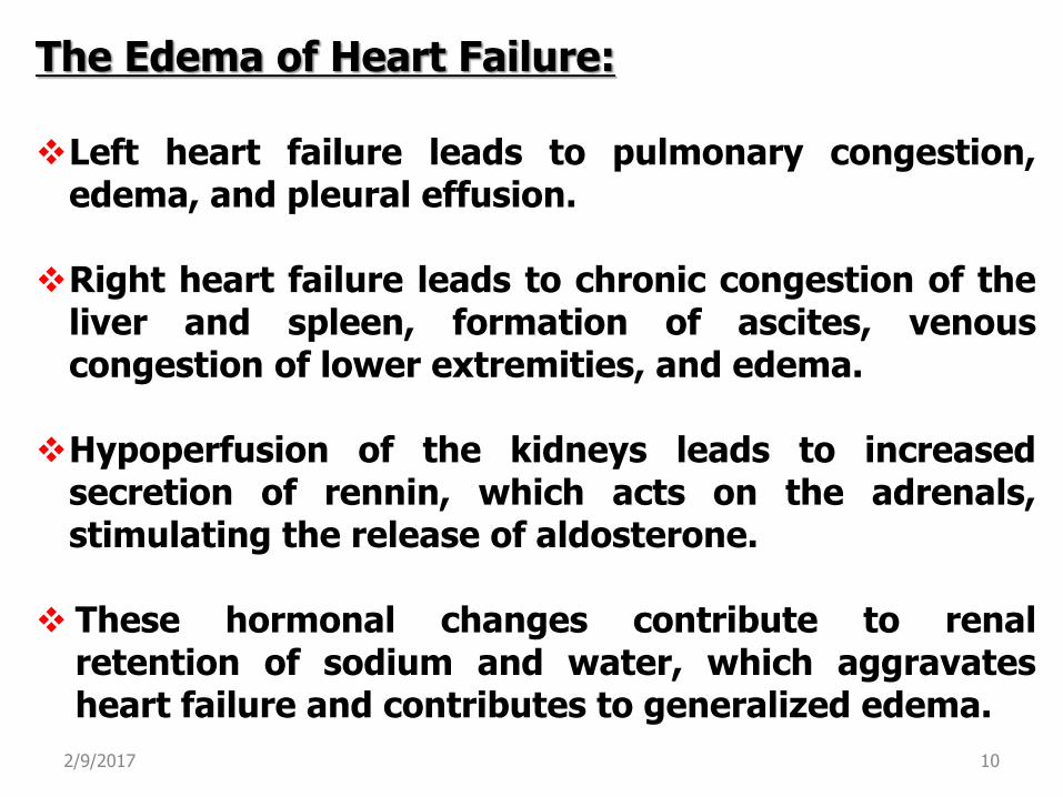
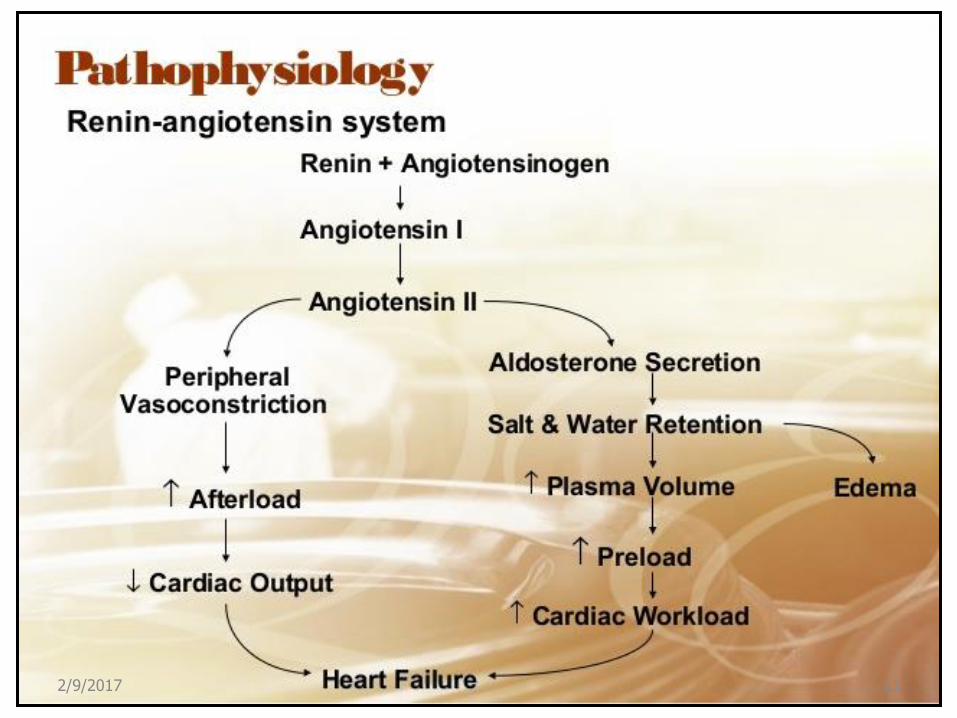
The Edema of Heart Failure: Left heart failure leads to pulmonary congestion,
edema, and pleural effusion.
Right heart failure leads to chronic congestion of the liver and spleen, formation of ascites, venous congestion of lower extremities, and edema.
Hypoperfusion of the kidneys leads to increased secretion of rennin, which acts on the adrenals, stimulating the release of aldosterone.
These hormonal changes contribute to renal retention of sodium and water, which aggravates heart failure and contributes to generalized edema.
2/9/2017 10
2/9/2017 11

2/9/2017 12

2/9/2017 13
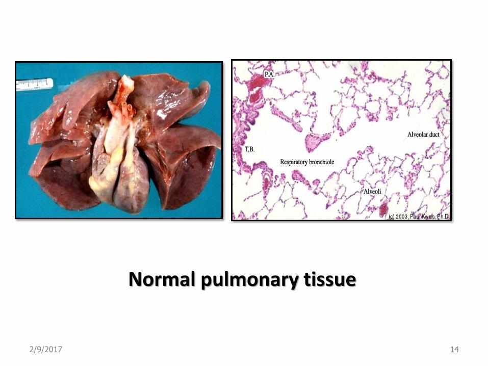
Normal pulmonary tissue
2/9/2017 14
Pulmonary edema due to heart failure
2/9/2017 15
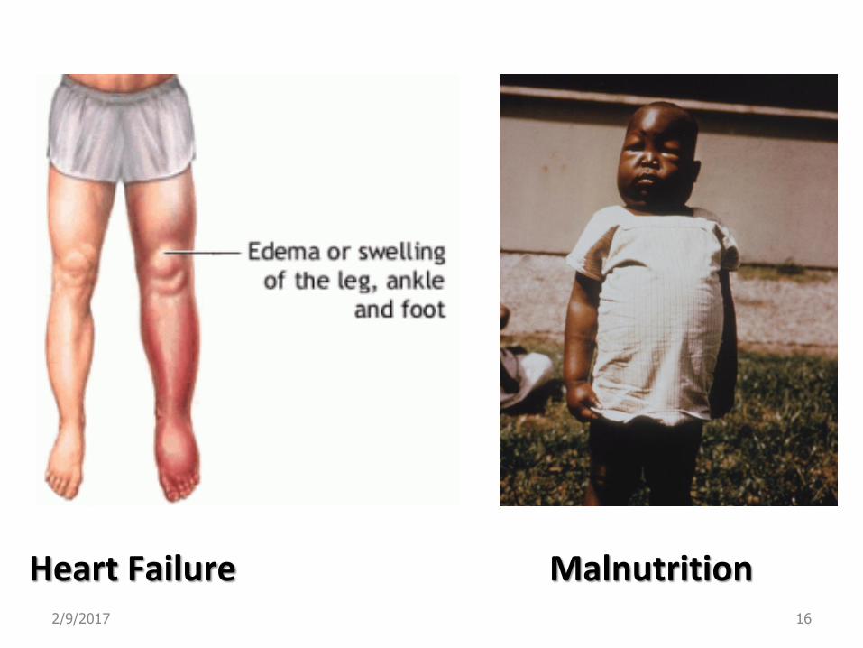
Heart Failure Malnutrition 2/9/2017 16

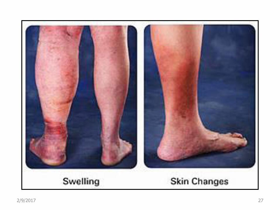
Venous Thrombi Limb edema 2/9/2017 17
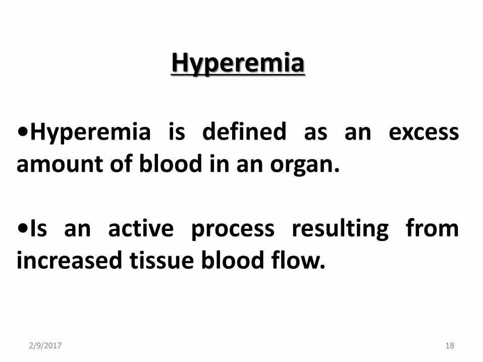
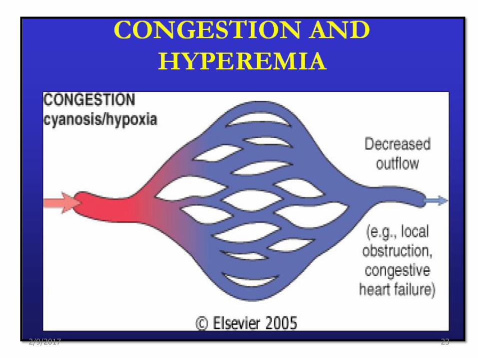
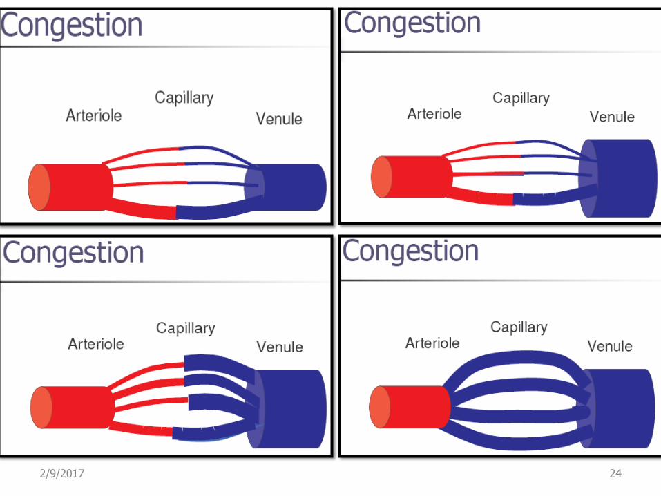
Hyperemia
•Hyperemia is defined as an excess amount of blood in an organ. •Is an active process resulting from increased tissue blood flow.
2/9/2017 18
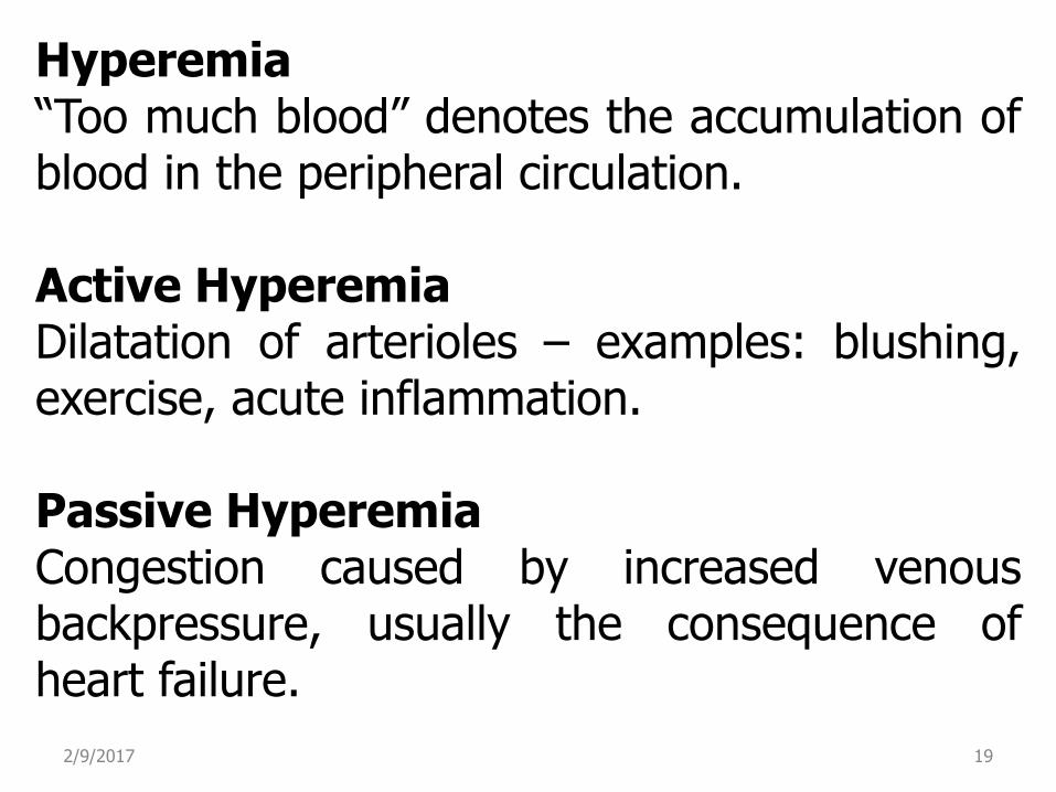
Hyperemia “Too much blood” denotes the accumulation of blood in the peripheral circulation. Active Hyperemia Dilatation of arterioles – examples: blushing, exercise, acute inflammation. Passive Hyperemia Congestion caused by increased venous backpressure, usually the consequence of heart failure.
2/9/2017 19
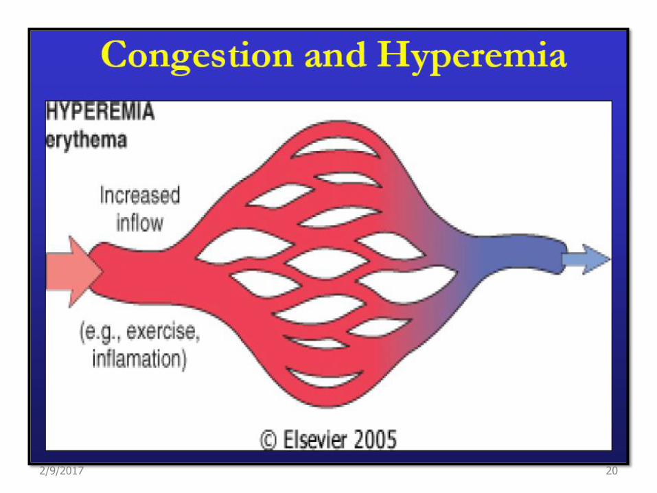
2/9/2017 20
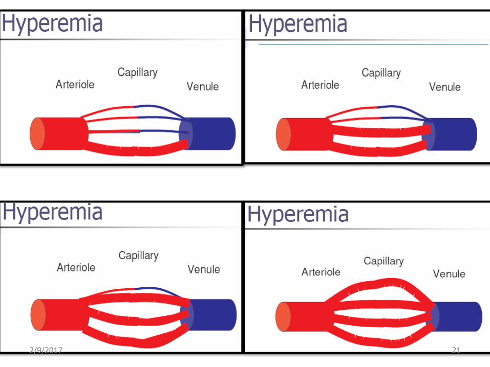
2/9/2017 21

Passive Hyperemia (Congestion): Passive hyperemia or congestion refers to the engorgement of an organ with venous blood. Acute passive congestion is clinically a consequence of acute failure of the left ventricle. The resulting venous engorgement of the lung leads to the accumulation of a transudate in the alveoli, a condition termed pulmonary edema.
2/9/2017 22
2/9/2017 23
2/9/2017 24
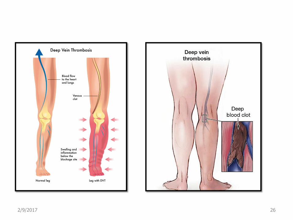
Passive congestion may also be confined (limited) to a limb or an organ as a result of more localized obstruction to the venous drainage. Examples include deep venous thrombosis of the leg, with resulting edema of the lower extremity,
2/9/2017 25
2/9/2017 26
2/9/2017 27
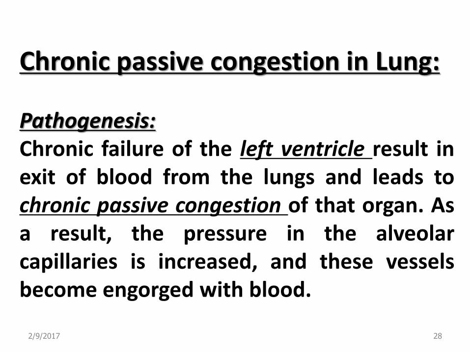
Chronic passive congestion in Lung: Pathogenesis: Chronic failure of the left ventricle result in exit of blood from the lungs and leads to chronic passive congestion of that organ. As a result, the pressure in the alveolar capillaries is increased, and these vessels become engorged with blood.
2/9/2017 28

The increased pressure in the alveolar capillaries leads to microhemorrhages and release erythrocytes into the alveolar spaces, where they are phagocytosed and degraded by alveolar macrophages. The released iron, in the form of
hemosiderin, remains in the macrophages, which are then called “heart failure cells”.
2/9/2017 29
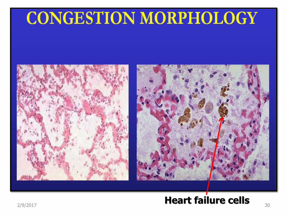
Heart failure cells 2/9/2017 30
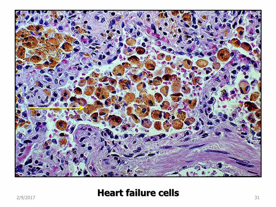
Heart failure cells 2/9/2017 31

2/9/2017 32
Chronic passive congestion in liver: Pathogenesis: Chronic passive hepatic congestion result
from right side heart failure. The blood will flow back into the inferior
vena cava, then central veins of the hepatic lobule become dilated, eventually hepatic sinusoid will engorged with venous blood, result in increase venous pressure and edema into the peritoneal cavity.
2/9/2017 33
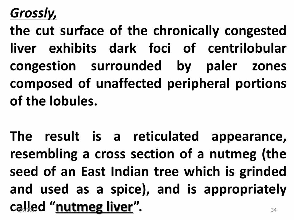
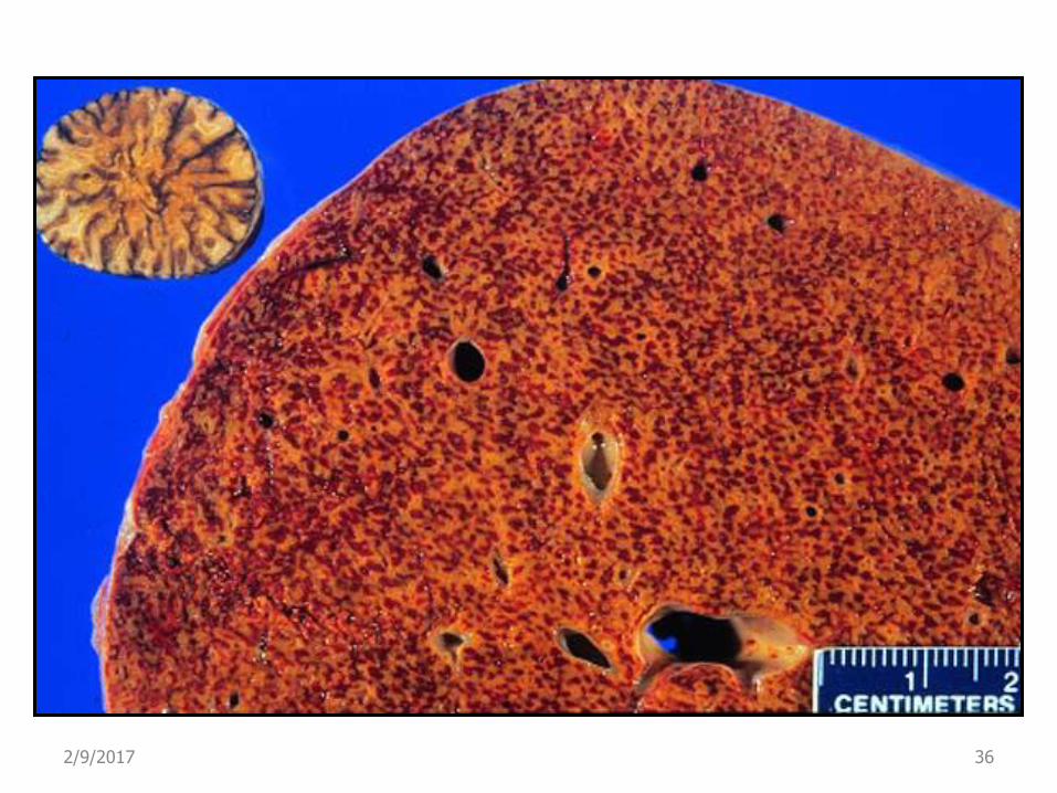
Grossly, the cut surface of the chronically congested liver exhibits dark foci of centrilobular congestion surrounded by paler zones composed of unaffected peripheral portions of the lobules. The result is a reticulated appearance, resembling a cross section of a nutmeg (the seed of an East Indian tree which is grinded and used as a spice), and is appropriately called “nutmeg liver”. 2/9/2017 34
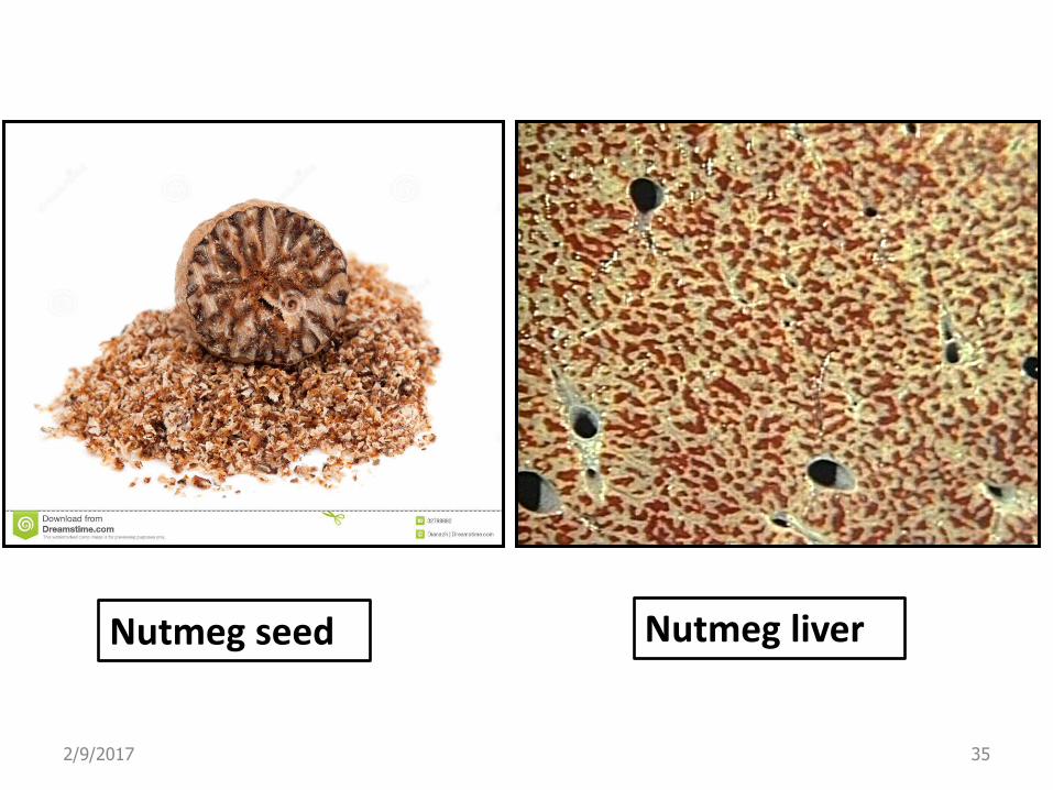
Nutmeg seed Nutmeg liver
2/9/2017 35
2/9/2017 36
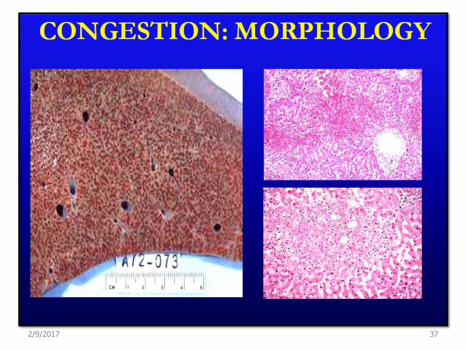
2/9/2017 37

Hemorrhage Its an extravasation of blood outside the cardiovascular system because of vessel rupture.
Capillary bleeding can occur in chronic congestion.
Rupture of large artery or vein is usually due to vascular injury such as, trauma, arthrosclerosis, inflammation or neoplastic erosion of blood vessel wall.
2/9/2017 38
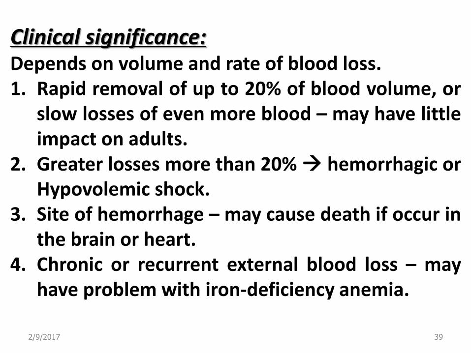
Clinical significance: Depends on volume and rate of blood loss. 1. Rapid removal of up to 20% of blood volume, or
slow losses of even more blood – may have little impact on adults.
2. Greater losses more than 20% hemorrhagic or Hypovolemic shock.
3. Site of hemorrhage – may cause death if occur in the brain or heart.
4. Chronic or recurrent external blood loss – may have problem with iron-deficiency anemia.
2/9/2017 39
2/9/2017 40
Hemothorax: Hemorrhage in the thoracic (pleural) cavity.
Hemopericarduim: Hemorrhage in the pericardial sac.
Hemoperitoneum: bleeding in the peritoneal cavity.
Hemothrosis: Bleeding into joint spaces.
2/9/2017 41
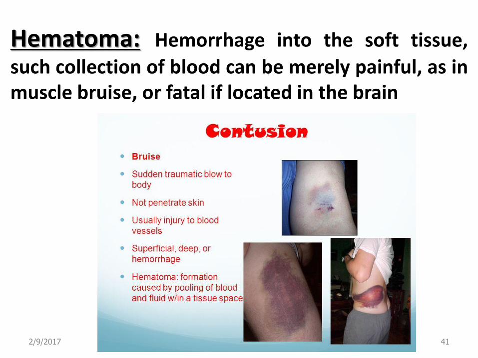
Hematoma: Hemorrhage into the soft tissue,
such collection of blood can be merely painful, as in muscle bruise, or fatal if located in the brain
2/9/2017 42
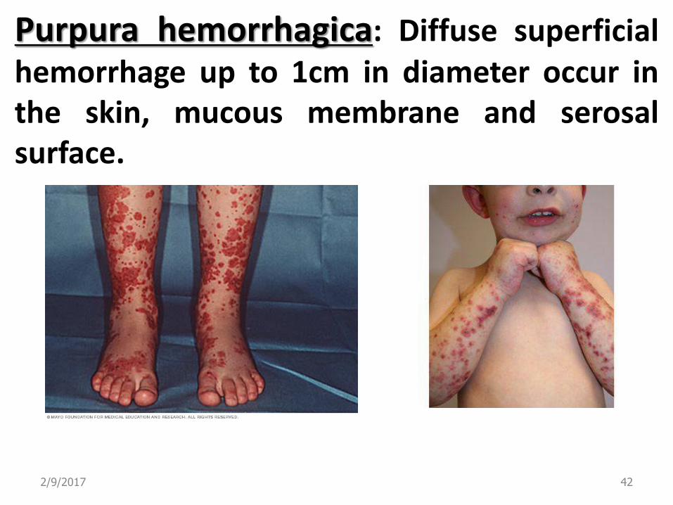
Purpura hemorrhagica: Diffuse superficial hemorrhage up to 1cm in diameter occur in the skin, mucous membrane and serosal surface.
2/9/2017 43
2/9/2017 44
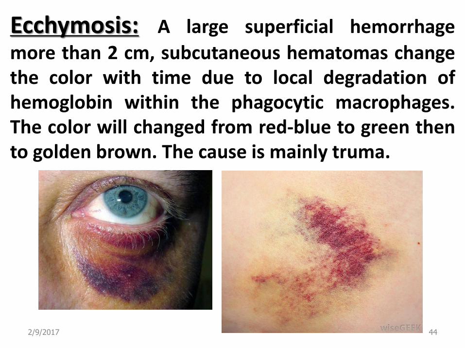
Ecchymosis: A large superficial hemorrhage
more than 2 cm, subcutaneous hematomas change the color with time due to local degradation of hemoglobin within the phagocytic macrophages. The color will changed from red-blue to green then to golden brown. The cause is mainly truma.
2/9/2017 45
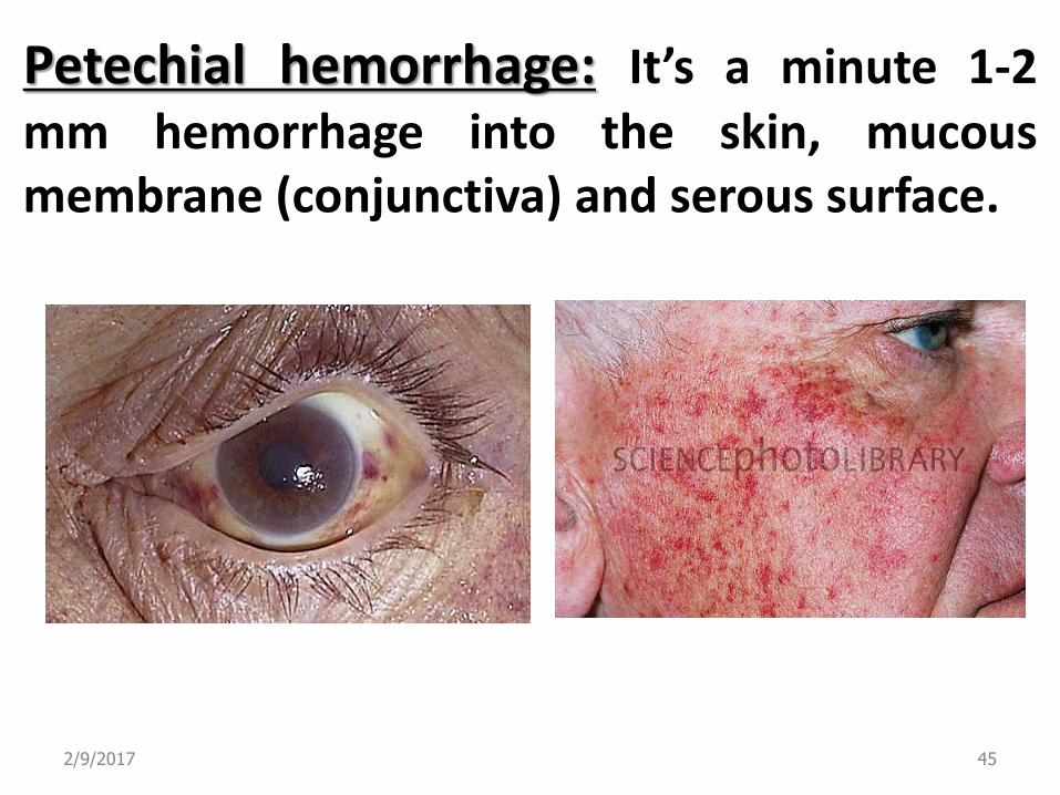
Petechial hemorrhage: It’s a minute 1-2 mm hemorrhage into the skin, mucous membrane (conjunctiva) and serous surface.
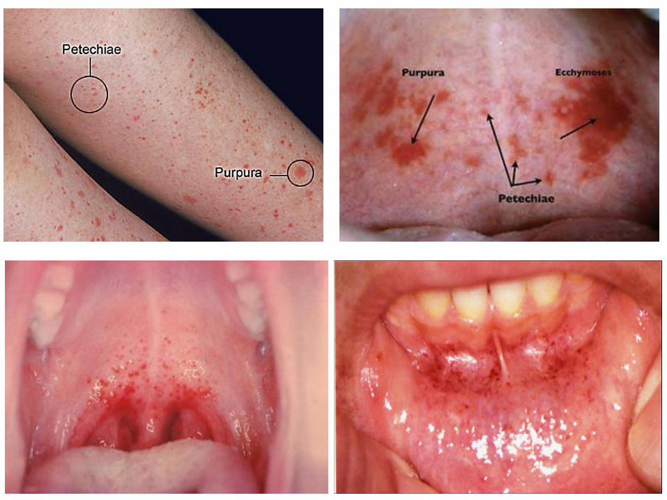
2/9/2017 46

Slide 5.4
Petechial hemorrhages in the intestine
Hematoma in the brain
2/9/2017 47
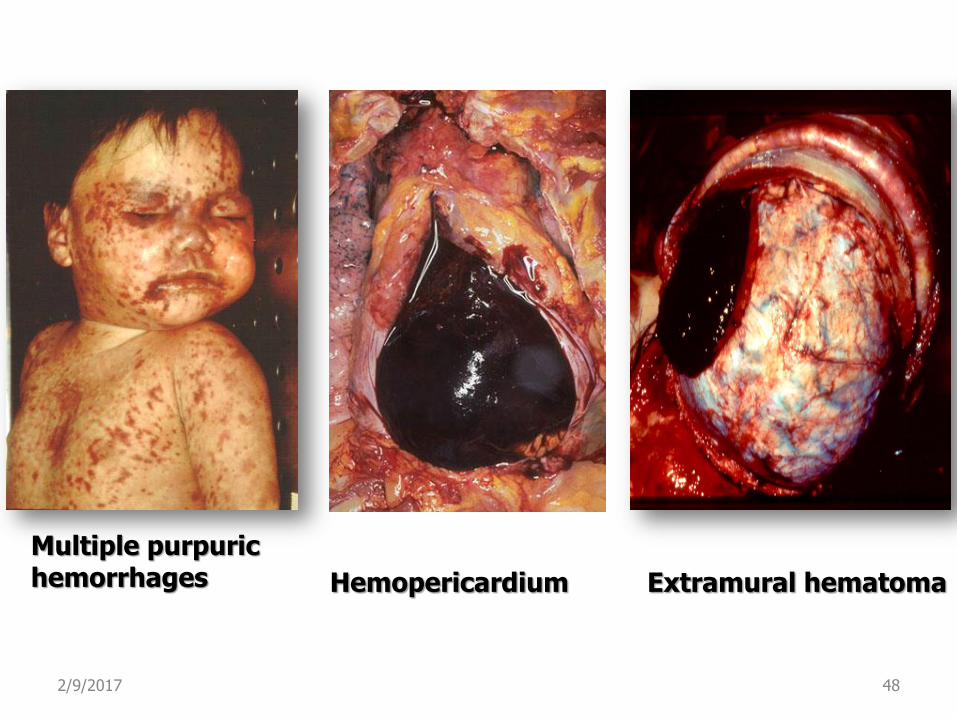
Multiple purpuric hemorrhages
Hemopericardium Extramural hematoma
2/9/2017 48


![AnestheticConsiderationsinHepatectomiesunderHepatic ...downloads.hindawi.com/archive/2012/720754.pdf · 0.5mL/kg/h can be used safely with minor hemodynamic disturbance [32]. If fluid](https://static.fdocuments.us/doc/165x107/5ebc015bb7d7875d803b19c8/anestheticconsiderationsinhepatectomiesunderhepatic-05mlkgh-can-be-used-safely.jpg)