Distributed cortical adaptation during learning of a … · Distributed cortical adaptation during...
6
Distributed cortical adaptation during learning of a brain–computer interface task Jeremiah D. Wander a,1 , Timothy Blakely a , Kai J. Miller b,2 , Kurt E. Weaver c , Lise A. Johnson d , Jared D. Olson e , Eberhard E. Fetz a,b,f , Rajesh P. N. Rao a,b,g , and Jeffrey G. Ojemann b,c,d Departments of a Bioengineering, c Radiology, d Neurological Surgery, e Rehabilitation Medicine, f Physiology and Biophysics, and g Computer Science and Engineering and b Program in Neurobiology and Behavior, University of Washington, Seattle, WA 98195 Edited by Terrence J. Sejnowski, Salk Institute for Biological Studies, La Jolla, CA, and approved April 30, 2013 (received for review December 4, 2012) The majority of subjects who attempt to learn control of a brain– computer interface (BCI) can do so with adequate training. Much like when one learns to type or ride a bicycle, BCI users report transitioning from a deliberate, cognitively focused mindset to near automatic control as training progresses. What are the neural correlates of this process of BCI skill acquisition? Seven subjects were implanted with electrocorticography (ECoG) electrodes and had multiple opportunities to practice a 1D BCI task. As subjects became proficient, strong initial task-related activation was fol- lowed by lessening of activation in prefrontal cortex, premotor cortex, and posterior parietal cortex, areas that have previously been implicated in the cognitive phase of motor sequence learning and abstract task learning. These results demonstrate that, although the use of a BCI only requires modulation of a local pop- ulation of neurons, a distributed network of cortical areas is in- volved in the acquisition of BCI proficiency. brain–machine interface | motor learning | plasticity | electrophysiology | high gamma O ver the last 50 years, it has been demonstrated that, when given feedback, the brain can learn to volitionally modulate the activity of single neurons (1–5) and populations of neurons (6–9). This modulation can occur in the absence of overt movement (6, 9) or even when overt movement is not possible (3, 10). The use of these signals to control external devices for restoration of lost function or as potential feedback signals for rehabilitation holds great promise for the future; however, our understanding of the underlying processes that drive this activity modulation is incomplete. The ability to voluntarily modulate neural activity to control a brain–computer interface (BCI) appears to be a learned skill, similar to learning to ride a bike or swing a golf club. Inves- tigators have repeatedly documented that task performance typically increases over the course of practice (11–15), which is indicative of a learning process taking place in the brain. In many cases, human BCI users have anecdotally reported transitioning from a very deliberate, cognitive approach to nearly automatic execution. Whereas they were initially focused on the cognitive task (e.g., motor imagery) that drives neural activity and in turn controls the end effector, after training, they report a goal-directed approach focused directly on the end effector itself. BCI use is interesting in that it shares characteristics with both concrete motor tasks and abstract cognitive tasks (16). Users of BCIs are manipulating their physical environment, but doing so without necessarily moving their bodies, thus depriving their brains of the multiple modalities of sensory feedback that have been demonstrated to be vital to learning a motor skill (17). The transition from a cognitive to an automatic phase, com- bined with the fact that the majority of single-unit and field- potential BCIs are driven by signals recorded from motor areas, suggests the hypothesis that BCI skill learning shares many commonalities with motor sequence learning. Indeed, in most cases, large initial gains in performance are followed by gradually diminishing improvements, congruent with Fitts’ well-known work on motor performance during skill learning (18). Following results on neural activity patterns during motor skill learning (19, 20), one would therefore expect significant involvement of motor, prefrontal, and parietal cortical areas, as well as the striatum and cerebellum during the cognitive phase of BCI skill acquisition. To investigate the role of distributed cortical networks in BCI skill acquisition, we recorded population-scale neural activity simultaneously from various locations across the cortex using electrocorticography (ECoG) while subjects performed a BCI task. The task required subjects to modulate a subrange (∼70– 100 Hz) of the high-gamma band (HG; 70–200 Hz) in ECoG surface potentials to control the position of a cursor on a com- puter monitor. HG activity was used because it has been pre- viously shown to correspond to an increase in the firing rate and/ or coherence of underlying local neural populations (21). We investigated two hypotheses: (i ) that motor learning networks across the cortex participate in BCI learning even though the task only requires modulation of a localized neural population and (ii ) that activity in these distributed cortical networks changes with increasing BCI task proficiency. Results Seven human subjects (two women; mean age, 25.6 y; range, 18– 32 y) with intractable epilepsy were implanted for long-term (4– 10 d) monitoring. During the monitoring period, these subjects participated in multiple experimental recording sessions that took place over the course of 1–3 d. The first session began with a series of two basic functional screening tasks, in which subjects performed gross motor movement or imagined motor movement in response to a visual cue. Functional screening was conducted to determine the electrodes that exhibited statistically significant increase in HG activity during actual movement or imagined movement compared with rest (9, 22). The controlling electrode that was used for online control was selected from these elec- trodes based on the magnitude of observed power change and anatomical relevance. The BCI task was the 1D, two-target right-justified box task (23). In this task, the subject is presented one of two targets along the right side of a monitor and a cursor that starts on the left side of the monitor and travels to the right at a fixed hori- zontal velocity. During each trial, the subject modulates HG activity to control the vertical velocity of the cursor such that the cursor comes in contact with the target. HG activity recorded from the controlling electrode is mapped to vertical cursor Author contributions: J.D.W. and J.G.O. designed research; J.D.W., T.B., K.J.M., and L.A.J. performed research; J.D.W. analyzed data; and J.D.W., K.E.W., J.D.O., E.E.F., R.P.N.R., and J.G.O. wrote the paper. The authors declare no conflict of interest. This article is a PNAS Direct Submission. 1 To whom correspondence should be addressed. E-mail: [email protected]. 2 Present address: Department of Neurosurgery, Stanford University, Stanford, CA 94305. This article contains supporting information online at www.pnas.org/lookup/suppl/doi:10. 1073/pnas.1221127110/-/DCSupplemental. 10818–10823 | PNAS | June 25, 2013 | vol. 110 | no. 26 www.pnas.org/cgi/doi/10.1073/pnas.1221127110
Transcript of Distributed cortical adaptation during learning of a … · Distributed cortical adaptation during...
Distributed cortical adaptation during learningof a brain–computer interface taskJeremiah D. Wandera,1, Timothy Blakelya, Kai J. Millerb,2, Kurt E. Weaverc, Lise A. Johnsond, Jared D. Olsone,Eberhard E. Fetza,b,f, Rajesh P. N. Raoa,b,g, and Jeffrey G. Ojemannb,c,d
Departments of aBioengineering, cRadiology, dNeurological Surgery, eRehabilitation Medicine, fPhysiology and Biophysics, and gComputer Science andEngineering and bProgram in Neurobiology and Behavior, University of Washington, Seattle, WA 98195
Edited by Terrence J. Sejnowski, Salk Institute for Biological Studies, La Jolla, CA, and approved April 30, 2013 (received for review December 4, 2012)
The majority of subjects who attempt to learn control of a brain–computer interface (BCI) can do so with adequate training. Muchlike when one learns to type or ride a bicycle, BCI users reporttransitioning from a deliberate, cognitively focused mindset tonear automatic control as training progresses. What are the neuralcorrelates of this process of BCI skill acquisition? Seven subjectswere implanted with electrocorticography (ECoG) electrodes andhad multiple opportunities to practice a 1D BCI task. As subjectsbecame proficient, strong initial task-related activation was fol-lowed by lessening of activation in prefrontal cortex, premotorcortex, and posterior parietal cortex, areas that have previouslybeen implicated in the cognitive phase of motor sequence learningand abstract task learning. These results demonstrate that,although the use of a BCI only requires modulation of a local pop-ulation of neurons, a distributed network of cortical areas is in-volved in the acquisition of BCI proficiency.
brain–machine interface | motor learning | plasticity | electrophysiology |high gamma
Over the last 50 years, it has been demonstrated that, whengiven feedback, the brain can learn to volitionally modulate
the activity of single neurons (1–5) and populations of neurons(6–9). This modulation can occur in the absence of overtmovement (6, 9) or even when overt movement is not possible (3,10). The use of these signals to control external devices forrestoration of lost function or as potential feedback signals forrehabilitation holds great promise for the future; however, ourunderstanding of the underlying processes that drive this activitymodulation is incomplete.The ability to voluntarily modulate neural activity to control
a brain–computer interface (BCI) appears to be a learned skill,similar to learning to ride a bike or swing a golf club. Inves-tigators have repeatedly documented that task performancetypically increases over the course of practice (11–15), which isindicative of a learning process taking place in the brain. In manycases, human BCI users have anecdotally reported transitioningfrom a very deliberate, cognitive approach to nearly automaticexecution. Whereas they were initially focused on the cognitivetask (e.g., motor imagery) that drives neural activity and in turncontrols the end effector, after training, they report a goal-directedapproach focused directly on the end effector itself.BCI use is interesting in that it shares characteristics with both
concrete motor tasks and abstract cognitive tasks (16). Users ofBCIs are manipulating their physical environment, but doing sowithout necessarily moving their bodies, thus depriving theirbrains of the multiple modalities of sensory feedback that havebeen demonstrated to be vital to learning a motor skill (17).The transition from a cognitive to an automatic phase, com-
bined with the fact that the majority of single-unit and field-potential BCIs are driven by signals recorded from motor areas,suggests the hypothesis that BCI skill learning shares manycommonalities with motor sequence learning. Indeed, in mostcases, large initial gains in performance are followed by graduallydiminishing improvements, congruent with Fitts’ well-known
work on motor performance during skill learning (18). Followingresults on neural activity patterns during motor skill learning (19,20), one would therefore expect significant involvement of motor,prefrontal, and parietal cortical areas, as well as the striatum andcerebellum during the cognitive phase of BCI skill acquisition.To investigate the role of distributed cortical networks in BCI
skill acquisition, we recorded population-scale neural activitysimultaneously from various locations across the cortex usingelectrocorticography (ECoG) while subjects performed a BCItask. The task required subjects to modulate a subrange (∼70–100 Hz) of the high-gamma band (HG; 70–200 Hz) in ECoGsurface potentials to control the position of a cursor on a com-puter monitor. HG activity was used because it has been pre-viously shown to correspond to an increase in the firing rate and/or coherence of underlying local neural populations (21). Weinvestigated two hypotheses: (i) that motor learning networksacross the cortex participate in BCI learning even though thetask only requires modulation of a localized neural populationand (ii) that activity in these distributed cortical networkschanges with increasing BCI task proficiency.
ResultsSeven human subjects (two women; mean age, 25.6 y; range, 18–32 y) with intractable epilepsy were implanted for long-term (4–10 d) monitoring. During the monitoring period, these subjectsparticipated in multiple experimental recording sessions thattook place over the course of 1–3 d. The first session began witha series of two basic functional screening tasks, in which subjectsperformed gross motor movement or imagined motor movementin response to a visual cue. Functional screening was conductedto determine the electrodes that exhibited statistically significantincrease in HG activity during actual movement or imaginedmovement compared with rest (9, 22). The controlling electrodethat was used for online control was selected from these elec-trodes based on the magnitude of observed power change andanatomical relevance.The BCI task was the 1D, two-target right-justified box task
(23). In this task, the subject is presented one of two targetsalong the right side of a monitor and a cursor that starts on theleft side of the monitor and travels to the right at a fixed hori-zontal velocity. During each trial, the subject modulates HGactivity to control the vertical velocity of the cursor such that thecursor comes in contact with the target. HG activity recordedfrom the controlling electrode is mapped to vertical cursor
Author contributions: J.D.W. and J.G.O. designed research; J.D.W., T.B., K.J.M., and L.A.J.performed research; J.D.W. analyzed data; and J.D.W., K.E.W., J.D.O., E.E.F., R.P.N.R., andJ.G.O. wrote the paper.
The authors declare no conflict of interest.
This article is a PNAS Direct Submission.1To whom correspondence should be addressed. E-mail: [email protected] address: Department of Neurosurgery, Stanford University, Stanford, CA 94305.
This article contains supporting information online at www.pnas.org/lookup/suppl/doi:10.1073/pnas.1221127110/-/DCSupplemental.
10818–10823 | PNAS | June 25, 2013 | vol. 110 | no. 26 www.pnas.org/cgi/doi/10.1073/pnas.1221127110
velocity using a simple linear decoder that was fixed in the firstrecording session, such that execution of motor imagery or overtmotor movement causes the cursor to travel up and remaining atrest causes the cursor to travel down. The task consists of fourperiods: rest, targeting, feedback, and reward. Because the hor-izontal velocity of the cursor is fixed, the length of the feedbackperiod for each trial is also fixed to 3 s. In subsequent discussionof this task, targets requiring an increase of HG activity are re-ferred to as “up targets” and targets requiring a decrease of HGactivity are referred to as “down targets.”
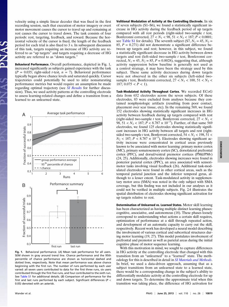
Behavioral Performance. Overall performance, depicted in Fig. 1,increased significantly as subjects gained experience with the task(P = 0.035; right-sided t-test, n = 7). Behavioral performancetypically began above chance levels and saturated quickly. Cursortrajectories could potentially be used to infer nonsaturatingperformance metrics but would require an assumption be maderegarding optimal trajectory (see SI Results for further discus-sion). Thus, we used activity patterns at the controlling electrodeto assess learning-related changes and define a transition from alearned to an unlearned state.
Volitional Modulation of Activity at the Controlling Electrode. In sixof seven subjects (S1–S6), we found a statistically significant in-crease in HG activity during the feedback period of up targetscompared with all rest periods (right-sided two-sample t test,Bonferonni corrected, 27 < N1 < 98, 51 < N2 < 187; P < 0.0001;see Table S1 for details). The seventh subject (S7, N1 = 45, N2 =85, P = 0.271) did not demonstrate a significant difference be-tween up targets and rest; however, in this subject, we founda statistically significant decrease in HG activity between downtargets and rest (left-sided two-sample t test, Bonferonni cor-rected, N1 = 45, N2 = 85, P = 0.0028), suggesting that, althoughactivity suppression below baseline is generally not used asa control strategy, it may have been the strategy used by thissubject. These same activity decreases during down targetswere not observed in the other six subjects (left-sided two-sample t test, Bonferonni corrected, 27 < N1 < 100, 51 < N2 <187; 0.075 < P < 1).
Task-Modulated Activity Throughout Cortex. We recorded ECoGdata from 652 electrodes across the seven subjects. Of theseelectrodes, 83 were excluded from analyses because they con-tained nonphysiologic artifacts (resulting from poor contact,placement over scar tissue, etc). In the remaining 569, we found152 electrodes showing statistically significant increases in HGactivity between feedback during up targets compared with rest(right-sided two-sample t test, Bonferroni corrected, 27 < N1 <98, 51 < N2 < 187; P < 8.787 × 10⁻⁵). Further, of that same 569electrodes, we found 125 electrodes showing statistically signifi-cant increases in HG activity between all targets and rest (right-sided two-sample t test, Bonferroni corrected, 54 < N1 < 198, 51 <N2 < 187; P < 8.787 × 10⁻⁵). Electrodes showing significant ac-tivity increase were concentrated in cortical areas previouslyknown to be associated with motor learning: primary motor cortex(MC), primary somatosensory cortex (SC), dorsolateral prefrontalcortex (PFC), and dorsal/ventral premotor cortices (PMd/PMv)(24, 25). Additionally, electrodes showing increases were found inposterior parietal cortex (PPC), an area associated with sensori-motor tasks involving visual feedback (26). Additional task-mod-ulated electrodes were found in other cortical areas, such as thetemporal parietal junction and the inferior temporal gyrus, al-though to a lesser extent. Task-modulated activity in supplemen-tary motor area (SMA) was noted in the only subject with SMAcoverage, but this finding was not included in our analyses as itcould not be verified in multiple subjects. Fig. 2A illustrates thespatial distribution of electrodes showing significant activation forup targets relative to rest.
Determination of Unlearned vs. Learned States.Motor skill learninghas been characterized as having multiple distinct learning phases:cognitive, associative, and autonomous (18). These phases looselycorrespond to understanding what actions a certain skill requires,optimization of performance at a skill through repeated action,and development of an automatic capacity to carry out the skill,respectively. Recent work has developed a neural model describingthe involvement of various cortical and subcortical structures dur-ing motor learning (19, 27). This model postulates involvement ofprefrontal and premotor as well as parietal areas during the initialcognitive phase of motor sequence learning.With this motivation in mind, we sought to capture differences
in HG activity at the controlling electrode that changed with thistransition from an “unlearned” to a “learned” state. The meth-odology for this is described in detail in SI Materials and Methods.In brief, we used a data-driven approach, assuming that as asubject transitions from an unlearned state to a learned state,there would be a corresponding change in the subject’s ability todifferentially modulate activity at the controlling electrode for upand down targets. To determine the approximate trial when thistransition was taking place, the difference of HG activation for
A
B
Fig. 1. Behavioral performance. (A) Mean task performance for all users.SEM shown in gray around trend line. Chance performance and the 95thpercentile of chance performance are shown as horizontal dashed anddotted lines, respectively. Note that mean performance was above chancebeginning with the first run. The number of runs performed by each uservaried: all seven users contributed to data for the first three runs, six userscontributed through the first five runs, and four contributed to the sixth run.See Table S1 for additional details. (B) Comparison of performance for thefirst and last runs performed by each subject. Significant differences (P <0.05) denoted with an asterisk.
Wander et al. PNAS | June 25, 2013 | vol. 110 | no. 26 | 10819
NEU
ROSC
IENCE

up and down targets was estimated and divided into unlearnedand learned trials, occurring before and after an assumed tran-sition trial, respectively. With the location of this assumed tran-sition trial being the single free parameter, we then fit a model oftwo Gaussian distributions to the unlearned and learned trialssuch that the distance (see SI Materials and Methods) betweenthese two distributions was maximized. In comparing unlearnedand learned distributions, distance value magnitudes close to
one implied that the majority of the variance in the jointdistribution can be explained by the difference in the means ofthe two subdistributions. The sign of the resulting distancemeasure demonstrates the direction of shift in the means ofthe two subdistributions, where a positive value implies thatthe mean of the learned distribution is greater than the meanof the unlearned distribution. Within the context of this task,a large, positive distance value for a given transition trialimplies that the subject’s ability to differentially modulate activityduring up targets compared with down targets was much greaterafter the transition trial compared with before. Furthermore,a relatively large distance value implies that one of the solutionstrategies used was to increase the difference in activity at thecontrolling electrode in up targets relative to down targets. Thispossibility is compared with the alternative that the user onlyreduces the variability of their control signal as they gain experi-ence. If this was the case, we would have observed relatively lowdistances between the unlearned and learned distributions.In all seven subjects, relatively large distances between the
distributions of power separation for learned and unlearnedstates demonstrated that the applied model effectively separatedthose two states (0.2039 < distance < 0.8689). Accordingly, thedifferences in distributions of power separation for unlearnedand learned states were highly significant (both-sided two-samplet test, Bonferonni corrected, P < 0.0001; see Table S1 for details).
Cortexwide Changes in Activation from Unlearned to Learned States.HG activation of all noncontrolling electrodes was visualized ona trial-by-trial basis to observe activity dynamics at these elec-trodes. Fig. 3 shows example trial-by-trial plots for electrodeslocated throughout motor-learning-associated cortical areas (seeFig. S1 for similar plots separated by subject). MC/SC and PMd/PMv electrodes demonstrated activation primarily during uptargets, and PFC and PPC demonstrated activation during bothup and down targets. As can be seen in Fig. S1, changes in ac-tivation in these electrodes over the course of many trials wereoften, although not always, well aligned with the transition fromunlearned to learned state as defined solely by HG power at thecontrolling electrode. It is notable that in some subjects, tran-sitions in HG activity patterns were approximately temporallyaligned with breaks in experimental sessions, suggesting thatoffline learning was taking place during these periods. This resultis complimentary to previous findings of increased sleep spindledensity local to the controlling electrode correlated with trainingon an ECoG BCI (28).Dynamics of activity in remote electrodes were quantitatively
evaluated by comparing mean activity during feedback in theseelectrodes in the unlearned and learned states. Sixty-sevenelectrodes showed a significant change in HG activity during alltargets from the unlearned to learned states (both-sided two-sample t test, Bonferroni corrected, P < 8.787 × 10⁻⁵, 20 < N1 <75, 15 < N2 < 166). A large portion of these changes corre-sponded to significant lessening in activation in the frontal cor-tex, superior parietal cortex, and posterior parietal cortex, asdepicted in Fig. 4A. A smaller portion corresponded to signifi-cant increases of activation in areas surrounding the controllingelectrode. To determine anatomically relevant patterns of ac-tivity dynamics, electrodes were assigned to cortical areas usingthe human motor area template (HMAT) atlas (29) and theTalairach Daemon (30, 31). Significant lessening of activationwas found in 31 electrodes corresponding to labels of PMd, PMv,PFC, and PPC, as shown in Fig. 4B.
DiscussionOur results demonstrate that when learning a BCI cursor-controltask based on signals recorded from a single electrode over motorcortex, there is a distributed network of cortical areas that is in-volved in acquisition of skill in the task. This network includes, but
A
B
C
Fig. 2. Cortexwide activity during BCI use and modeling of early vs. late ac-tivity patterns. (A) Activation during up targets for all lateral electrodes for allsubjects (left coverage projected to right hemisphere) shown on the Talairachbrain. Note widespread cortical activation including frontal, middle-parietal,and posterior-parietal areas. Controlling electrodes are circled in blue. (B)Activation for an example subject (S4) during each feedback period normal-ized against log HG power during rest. Each dot represents one trial; up anddown targets are shown in red and blue, respectively. Thick red and blue linesrepresent mean activation for all trials, respectively. (C) Early-late trial divisionshown for subject S4. Separability of high-gamma activity during up and downtrials was used as a measurement of task proficiency. A separabililty measure(black dotted line) was modeled as two Gaussian distributions and fit to thedata such that the distance between the distributions was maximized.
10820 | www.pnas.org/cgi/doi/10.1073/pnas.1221127110 Wander et al.

is not limited to, surrounding sensorimotor and visuomotor areas:PMd/PMv, PFC, and PPC. We further demonstrated that as a userdevelops proficiency with the BCI, activity in parts of this network(PMd, PFC, and PPC) decreases, a finding that is indicative of themental shift from cognitive to automatic task execution oftenanecdotally reported in human BCI studies.
Controlling a BCI with volitional modulation of activity frommotor areas is intriguing in that it is arguably a nonmotor or atleast quasi-motor task. One might expect that the same cortical(PMd/PMv, PFC, PPC) (24, 25) and subcortical areas (basalganglia, cerebellum) (32) that are involved in motor tasks wouldalso be involved in the control of a BCI. However, the two tasksare significantly different in that, with the exception of visualfeedback, the afferent activity that is critical to successful ac-quisition of traditional motor tasks is not present during the useof a BCI. Thus, motor-based BCI may provide an opportunity tostudy these networks in the absence of or with manipulatedsensory feedback.In this study, we sought to investigate changes taking place
in motor networks as users developed both their control strategyand execution proficiency. To do so, we used a data-driven ap-proach that relied solely on the signal that each subject was usingto control the BCI. We found that dorsal premotor, prefrontal,and posterior parietal cortices exhibited decreased task-modulatedactivity as users transitioned from a naive to more experienced
A
B
C
D
E
Fig. 3. Time-by-trial high-gamma activation. (A) Distribution of electrodesshown in B–E on the Talairach brain. The subject from which each electrodewas taken is noted; not all subjects had coverage in all areas (Fig. S2). (B–E)Subplot 1: average HG activation in a given electrode for all trials separatedby up and down targets. Subject is specified in the subplot title. Phases ofthe task (ordered from L to R: inter-stimulus interval, target presentation,feedback, reward) are separated by vertical bars. Dotted line represents SEM.Subplots 2 and 3: trial-by-trial HG activation for all trials, separated by up(subplot 2) and down (subplot 3) targets. Trial count is shown on the verticalaxis. Breaks in the experimental session of more than 8 h are denoted withan asterisk (*). Time, as described for subplot 1, is shown on the horizontalaxis. The black horizontal bar represents the transition trial, and the early andlate trials relative to this point are denoted with black arrows. (B) MC/SCelectrode shows continued increase in activation for up targets, congruentwith changes at the controlling electrode. (C) PFC electrode shows activationfor both up and down targets, decreasing near the point of model separa-tion. (D) PMd electrode shows activation for up targets, decreasing near thepoint of model separation. (E) PPC electrode shows subtle activation duringup and down targets, decreasing near the point of model separation.
Fig. 4. Cortexwide activation changes. (A) Spatial distribution of change inmean activation comparing early to late trials for all targets for all subjects.Activations for individual electrodes are normalized against rest periods fromthe same electrode for a given run. Activation change values are blurredusing a 12-cm FWHM Gaussian filter. Frontal areas and posterior parietalareas exhibited lessening in task-related activation over the course of BCI use.(B) Change in mean activation for all electrodes showing significant changefrom early to late trials, classified into approximate cortical areas.
Wander et al. PNAS | June 25, 2013 | vol. 110 | no. 26 | 10821
NEU
ROSC
IENCE

state. This transition occurred quickly and was contemporaneouswith changes taking place in the control signal.It is interesting to note the patterns of distributed activity
observed across multiple subjects. The time course of activationin premotor electrodes parallels tightly that of correspondingMC/SC electrodes. Further, premotor areas were primarily moreactivated for up targets than down targets, instances in which thecontrolling electrodes were required to exhibit the same activity.These observations suggest a direct relationship between pre-motor areas and the controlling electrode. This pattern can becontrasted with activity patterns observed in prefrontal and pos-terior parietal areas, which exhibited fairly equivalent activationfor both up and down targets. These activity patterns are a re-flection of effort or engagement on the part of the subject, andcorrespondingly decrease as the subject gains task automaticity.In contrast to ECoG BCIs, noninvasive, EEG-based BCIs
typically harness power changes occurring in lower frequenciesto achieve control. When EEG subjects are performing motorimagery, the typical frequency band chosen is in the mu-betarange (12–30 Hz), because power changes in this band have beendemonstrated to be anticorrelated with movement and motorimagery (33). When using the methods described above to assesswhether similar changes were taking place in the mu-beta rangeas subjects develop experience with an ECoG BCI, we foundthat, although activity in the mu-beta band was strongly taskmodulated, it did not undergo the same spatiotemporal changeswe observed in HG activity. It is important to note, however, thatas the cursor was not being driven by mu-beta activity changes,the impact of this observation to noninvasive BCIs will requirefurther investigation. Details can be found in the SI Results.A logical and necessary extension of these findings is to in-
vestigate the roles that subcortical networks play in this samelearning process. Previous studies have demonstrated the vitaland differential roles of the basal ganglia and cerebellum duringmotor sequence learning and motor adaptation (27, 32), but theinvolvement of these subcortical networks in the process of BCIskill acquisition remains an open question, with implications forboth fundamental neuroscience and the incremental improve-ment of BCI frameworks. Recent work performed by Koralekand colleagues (5) has demonstrated that the striatum is involvedin and critical to development of proficiency with a BCI in a ratmodel. This finding was notable, given that effective use of theBCI in that study did not explicitly require recruitment of themotor system outside of motor cortex, yet task performance wasdegraded in subjects with impaired cortico-striatal interaction. In-vestigation of the role of these structures in humans will have to beleft to other recording modalities, as ECoG provides informationonly regarding activity near the cortical surface and is subject tospatial undersampling based on the distribution of electrodes.The distributed dynamics in cortical activity that we demon-
strated here have significant implications in development ofcoadaptive BCI frameworks. Investigators have recognized theneed for BCI architectures that accommodate the dynamic natureof the neural signals used as inputs (34, 35), but our findingssuggest that a number of the most commonly used methods [e.g.,common spatial patterns (36)] may require updating to handleboth the spatial and temporal variability in input signals.The ability to detect correlates of cognitive load during BCI
task learning and execution holds great potential for expandingthe current limitations of BCIs. Continuous control of a BCI hasbeen demonstrated in two dimensions using ECoG (13) andEEG (14), with degrading performance as users attempt controlin more dimensions. Real-time monitoring of cognitive loadduring BCI skill acquisition would allow for titrated increases intask complexity, potentially facilitating an increase in the totalnumber of dimensions that could be simultaneously controlled,thus allowing operation of more complex devices.
Our results also demonstrate the potential that BCI holds asa technique for probing neural systems in vivo (37). By applyingspecific task requirements but allowing users to use nativelearning strategies, we observed that distributed cortical net-works are involved in the cognitive phase of BCI skill acquisition,but the degree of involvement of these networks lessens as userstransition to automatic execution of the task. Future work willallow us to probe whether activity in this network is renewedwhen task dynamics are perturbed and the user is required toadapt to novel task conditions.
Materials and MethodsSubjects. Seven subjects with intractable epilepsy were implanted withplatinum subdural ECoG electrodes for the purpose of seizure focus locali-zation at Harborview Medical Center and Children’s Hospital (Seattle, WA).Research was conducted under the oversite of Seattle Children’s HospitalInstitutional review board FWA00002443, protocol number 12193. Thespecific location of the grids was determined based on clinical indication.Electrode grids were constructed of 3-mm-diameter platinum pads spaced at1 cm center-to-center and embedded in silastic (AdTech). Electrodes werearranged in 8 × 2 or 8 × 8 grids or 1 × 4, 1 × 6, or 1 × 8 strips. Subjectsprovided informed consent in accordance with the Institutional ReviewBoard’s direction, and patient data were anonymized in accordance withHealth Insurance Portability and Accountability Act mandate. None of thesubjects had previous experience with BCI. Subjects were being observed forseizure focus localization over the course of 4–10 d, although participationin research studies typically did not begin until the third postoperative day.
Recordings. Experimental recordings were performed at the patient’s bedsidewithout interrupting the clinical recording systems. Synamps2 (Neuroscan)and g.USBamps (GugerTec) sampled at 1,000 and 1,200 Hz, respectively,were used for recording. Cortical potentials referenced against a scalpelectrode and were digitized and processed using the BCI2000 software suite(38), which provided real-time feedback to the user.
BCI Task. BCI paradigms were driven by spectral power changes in a portion ofthe HG frequency band of a single electrode determined to be modulated bymotor imagery by an initial screening task as is described in SI Materials andMethods. Only a subset of the HG range was used during online control(∼75–100 Hz) for computational tractability and to eliminate the need forreal-time notch filtering to reduce line noise harmonics. HG activity waschosen as the control feature as it has been previously postulated that HGactivity is a correlate of underlying population level firing rates or coherencein firing (21). All subjects performed the standard right justified box task(23), depicted in Fig. S3. Subjects were instructed to conduct or imagine themovement associated with the chosen controlling electrode when they werepresented with a target filling the top half of the right most side of thescreen (an up target) and were instructed to rest when they were presentedwith a target filling the bottom half of the right most side of the screen (adown target). The cursor’s vertical velocity was updated every 40 ms andcontrolled by changes in HG activity at the controlling electrode as calcu-lated by an autoregressive filter using the previous 500 ms of data. This time-variant estimate of HG activity was normalized against 6 s of stored data forthe current target type and then mapped to cursor velocity. The normalizerwas typically adapting (collecting reference data and updating normaliza-tion parameters) only during the first run (18 trials); however, in cases wherenonstationarity of the signals showed obvious bias, the normalizer wasallowed to recalibrate. Subjects participated in the experiment over thecourse of multiple days. Duration of the recording sessions was dictated bythe subjects’ willingness and capability to participate.
Anatomical Labeling. The process by which electrodes were associated withanatomical labels is discussed in SI Materials and Methods; in brief, pre-operative MRI was coregistered with postoperative CT scans to allow forlocalization of electrodes with respect to each subject’s brain. These elec-trode locations were then mapped to Talairach coordinates. Anatomicallabels were estimated using the HMAT (29) and the Talairach Daemon (30,31). The HMAT atlas estimates functional areas corresponding to coor-dinates in Talairach space; as it was constructed from the meta-analysis of126 motor-based functional MRI studies, it does not include posterior pari-etal cortex or prefrontal cortex. These areas were identified as consisting ofBrodmann areas 7/40 and 8/9/46, respectively. All offline data analyses wereperformed using custom Matlab software.
10822 | www.pnas.org/cgi/doi/10.1073/pnas.1221127110 Wander et al.

Preprocessing. For each implanted grid or strip, signals were common averagerereferenced by subtracting the average signal recorded at all electrodeswithin that grid or strip. This rereferencing was done to eliminate anycommon noise introduced by activity recorded at the reference electrode.Rereferenced signals were band pass filtered for the HG range (70–200 Hz)using a fourth-order Butterworth filter, and a time-variant estimate of bandpower was calculated using the square of the magnitude of the Hilberttransform. Data were then log transformed to become approximately nor-mally distributed such that the assumptions made by statistical tests used infurther analyses would be met. To account for changes in recording char-acteristics from session to session, the resulting log-power estimate wasz-normalized with respect to the rest samples only per channel and session.Throughout the remainder of this section, the signal resulting from thepreprocessing steps will be referred to as HG activation.
Quantification of Average Responses. Trials were divided into four periods:rest, targeting, feedback, and reward. These periods were 1, 2, 3, and 1 s,respectively, resulting in a trial length of 7 s. The HG activation for each ofthese periods was calculated by averaging all HG activation samples withina given period. Fig. 2B shows mean HG activations during feedback on atrial-by-trial basis for a single subject (S4). Average activation for a givenelectrode across all trials of a given type was calculated as the mean HGactivation during all feedback periods for trials of a given type minus themean HG activation during all rest periods.
Estimation of HG Activation Separability and Learning States. As was describedbriefly in Results, we sought to quantify differences in activation observed atthe controlling electrode that were indicative of a transition from an un-learned to a learned state. The method for this is described at length in SIMaterials and Methods; briefly, we developed a running estimate of eachsubject’s ability to separate HG activation in up targets as opposed to downtargets. This estimate was divided into two distributions such that thedistance between those distributions was maximized. The trial that divides
these two distributions was used as the transition trial for the comparison ofdata from the unlearned to the learned state.
Generation of Time Course Activations. The average time course of HG acti-vation for all trials of a given type, like those shown in Fig. 3 B–E, wereconstructed by averaging the time course of HG activation for a given targetand smoothing with a Gaussian kernel [250-ms full width at half maximum(FWHM)]. SEs were calculated before smoothing and were also smoothedusing a Gaussian kernel (250-ms FWHM). Trial-by-trial progressions of thetime course of HG activation were generated by aligning all trials into anM×T matrix, where M was the number of trials of a given type and T wasthe length of a trial in samples. This matrix was then smoothed simulta-neously in both dimensions using a 2D Gaussian kernel (300-ms and seven-trial FWHM). Fig. S1 shows average and trial-by-trial time course activationsfor all subjects.
Comparison of Early and Late Activations. Shifts in activation from early to latelearning states were calculated in much the same way that average responseswere calculated, except that instead of comparing activation during thefeedback period to the activation during rest, activation during the feedbackperiod of early trials was compared with activation during the feedbackperiod of late trials. These changes were summarized by subtracting averageearly activation from average late activation such that a negative change as isshown in Fig. 4B for the majority of PMd/PMv, PFC, and PPC electrodes isindicative of a decrease in activation from early to late trials.
ACKNOWLEDGMENTS. This work was supported by National Institutes ofHealth Grants T90 DA023436-02, NS065186-01, K12 2K12HD001097-16, andNational Institute of Mental Health K01 MH086118; Army Research OfficeAward W911NF-11-1-0307; National Science Foundation Award 0930908; theOffice of Naval Research; the Keck Foundation; and the National ScienceFoundation Grant EEC-1028725.
1. Fetz EE (1969) Operant conditioning of cortical unit activity. Science 163(3870):955–958.
2. Fetz EE, Baker MA (1973) Operantly conditioned patterns on precentral unit activityand correlated responses in adjacent cells and contralateral muscles. J Neurophysiol36(2):179–204.
3. Kennedy PR, Bakay RA (1998) Restoration of neural output from a paralyzed patientby a direct brain connection. Neuroreport 9(8):1707–1711.
4. Chapin JK, Moxon KA, Markowitz RS, Nicolelis MA (1999) Real-time control of a robotarm using simultaneously recorded neurons in the motor cortex. Nat Neurosci 2(7):664–670.
5. Koralek AC, Jin X, Long JD, 2nd, Costa RM, Carmena JM (2012) Corticostriatal plasticityis necessary for learning intentional neuroprosthetic skills. Nature 483(7389):331–335.
6. Leuthardt EC, Schalk G, Wolpaw JR, Ojemann JG, Moran DW (2004) A brain-computerinterface using electrocorticographic signals in humans. J Neural Eng 1(2):63–71.
7. Wolpaw JR, McFarland DJ, Neat GW, Forneris CA (1991) An EEG-based brain-computerinterface for cursor control. Electroencephalogr Clin Neurophysiol 78(3):252–259.
8. Fabiani GE, McFarland DJ, Wolpaw JR, Pfurtscheller G (2004) Conversion of EEG ac-tivity into cursor movement by a brain-computer interface (BCI). IEEE Trans NeuralSyst Rehab Eng 12(3):331–338.
9. Miller KJ, et al. (2010) Cortical activity during motor execution, motor imagery, andimagery-based online feedback. Proc Natl Acad Sci USA 107(9):4430–4435.
10. Hochberg LR, et al. (2006) Neuronal ensemble control of prosthetic devices by a hu-man with tetraplegia. Nature 442(7099):164–171.
11. Carmena JM, et al. (2003) Learning to control a brain-machine interface for reachingand grasping by primates. PLoS Biol 1(2):E42.
12. Ganguly K, Carmena JM (2009) Emergence of a stable cortical map for neuro-prosthetic control. PLoS Biol 7(7):e1000153.
13. Schalk G, et al. (2008) Two-dimensional movement control using electrocortico-graphic signals in humans. J Neural Eng 5(1):75–84.
14. Wolpaw JR, McFarland DJ (2004) Control of a two-dimensional movement signal bya noninvasive brain-computer interface in humans. Proc Natl Acad Sci USA 101(51):17849–17854.
15. Moritz CT, Perlmutter SI, Fetz EE (2008) Direct control of paralysed muscles by corticalneurons. Nature 456(7222):639–642.
16. Green AM, Kalaska JF (2011) Learning to move machines with the mind. TrendsNeurosci 34(2):61–75.
17. Wolpaw JR (2007) Brain-computer interfaces as new brain output pathways. J Physiol579(Pt 3):613–619.
18. Fitts PM (1954) The information capacity of the human motor system in controllingthe amplitude of movement. J Exp Psychol 47(6):381–391.
19. Doyon J, Ungerleider L (2002) Neuropsychology of Memory (Guilford Press, NewYork), 3rd Ed.
20. van Mier H, et al. (1998) Changes in brain activity during motor learning measuredwith PET: Effects of hand of performance and practice. J Neurophysiol 80(4):2177–2199.
21. Ray S, Crone NE, Niebur E, Franaszczuk PJ, Hsiao SS (2008) Neural correlates of high-gamma oscillations (60-200 Hz) in macaque local field potentials and their potentialimplications in electrocorticography. J Neurosci 28(45):11526–11536.
22. Crone NE, Miglioretti DL, Gordon B, Lesser RP (1998) Functional mapping of humansensorimotor cortex with electrocorticographic spectral analysis. II. Event-relatedsynchronization in the gamma band. Brain 121(12):2301–2315.
23. Wolpaw JR, McFarland DJ, Vaughan TM, Schalk G (2003) The Wadsworth Centerbrain-computer interface (BCI) research and development program. IEEE Trans NeuralSyst Rehab Eng 11(2):204–207.
24. Schlaug G, Knorr U, Seitz R (1994) Inter-subject variability of cerebral activations inacquiring a motor skill: A study with positron emission tomography. Exp Brain Res98(3):523–534.
25. Jenkins IH, Brooks DJ, Nixon PD, Frackowiak RS, Passingham RE (1994) Motor sequencelearning: A study with positron emission tomography. J Neurosci 14(6):3775–3790.
26. Buneo CA, Andersen RA (2006) The posterior parietal cortex: Sensorimotor interfacefor the planning and online control of visually guided movements. Neuropsychologia44(13):2594–2606.
27. Doyon J, Benali H (2005) Reorganization and plasticity in the adult brain duringlearning of motor skills. Curr Opin Neurobiol 15(2):161–167.
28. Johnson L, et al. (2012) Sleep spindles are locally modulated by training on a brain-computer interface. Proc Natl Acad Sci USA 109(45):18583–18588.
29. Mayka MA, Corcos DM, Leurgans SE, Vaillancourt DE (2006) Three-dimensional lo-cations and boundaries of motor and premotor cortices as defined by functional brainimaging: a meta-analysis. Neuroimage 31(4):1453–1474.
30. Lancaster JL, et al. (2000) Automated Talairach atlas labels for functional brainmapping. Hum Brain Mapp 10(3):120–131.
31. Lancaster JL, et al. (1997) Automated labeling of the human brain: A preliminaryreport on the development and evaluation of a forward-transform method. HumBrain Mapp 5(4):238–242.
32. Hikosaka O, Nakamura K, Sakai K, Nakahara H (2002) Central mechanisms of motorskill learning. Curr Opin Neurobiol 12(2):217–222.
33. McFarland DJ, Miner LA, Vaughan TM, Wolpaw JR (2000) Mu and beta rhythm top-ographies during motor imagery and actual movements. Brain Topogr 12(3):177–186.
34. Vidaurre C, Sannelli C, Müller K-R, Blankertz B (2010) Machine-Learning-Based Co-adaptive Calibration for Brain-Computer Interfaces. Neural Comput 23:791–816.
35. Vidaurre C, Sannelli C, Müller K-R, Blankertz B (2011) Co-adaptive calibration to im-prove BCI efficiency. J Neural Eng 8(2):025009.
36. Müller-Gerking J, Pfurtscheller G, Flyvbjerg H (1999) Designing optimal spatial filtersfor single-trial EEG classification in a movement task. Clin Neurophysiol 110(5):787–798.
37. Moran D (2010) Evolution of brain-computer interface: Action potentials, local fieldpotentials and electrocorticograms. Curr Opin Neurobiol 20(6):741–745.
38. Schalk G, McFarland DJ, Hinterberger T, Birbaumer N, Wolpaw JR (2004) BCI2000: Ageneral-purpose brain-computer interface (BCI) system. IEEE Trans Biomed Eng 51(6):1034–1043.
Wander et al. PNAS | June 25, 2013 | vol. 110 | no. 26 | 10823
NEU
ROSC
IENCE