Distinctive Morphological Features of a Subset of Cortical ... · PDF fileDistinctive...
15
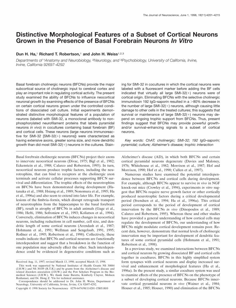
Distinctive Morphological Features of a Subset of Cortical Neurons Grown in the Presence of Basal Forebrain Neurons In Vitro Dun H. Ha, 1 Richard T. Robertson, 1 and John H. Weiss 1,2,3 Departments of 1 Anatomy and Neurobiology, 2 Neurology, and 3 Psychobiology, University of California, Irvine, Irvine, California 92697-4292 Basal forebrain cholinergic neurons (BFCNs) provide the major subcortical source of cholinergic input to cerebral cortex and play an important role in regulating cortical activity. The present study examined the ability of BFCNs to influence neocortical neuronal growth by examining effects of the presence of BFCNs on certain cortical neurons grown under the controlled condi- tions of dissociated cell culture. Initial experiments demon- strated distinctive morphological features of a population of neurons (labeled with SMI-32, a monoclonal antibody to non- phosphorylated neurofilament proteins that labels pyramidal neurons in vivo) in cocultures containing basal forebrain (BF) and cortical cells. These neurons (large neurons immunoreac- tive for SMI-32 [SMI-32(1) neurons]) were characterized as having extensive axons, greater soma size, and more dendritic growth than did most SMI-32(1) neurons in the cultures. Stain- ing for SMI-32 in cocultures in which the cortical neurons were labeled with a fluorescent marker before adding the BF cells indicated that virtually all large SMI-32(1) neurons were of cortical origin. Eliminating BFCNs with the selective cholinergic immunotoxin 192 IgG–saporin resulted in a .80% decrease in the number of large SMI-32(1) neurons, although causing little damage to other cells in the treated cultures; this suggests that survival or maintenance of large SMI-32(1) neurons may de- pend on ongoing trophic support from BFCNs. Thus, present findings suggest that BFCNs may provide powerful growth- and/or survival-enhancing signals to a subset of cortical neurons. Key words: ChAT; cholinergic; SMI-32; 192 IgG–saporin; pyramidal; culture; Alzheimer’s disease; trophic interaction Basal forebrain cholinergic neurons (BFCNs) project their axons to innervate neocortical neurons (Divac, 1975; Bigl et al., 1982; Eckenstein et al., 1988; Calarco and Roberston, 1995). In turn, neocortical neurons produce trophic factors, including the neu- rotrophins, that can bind to receptors at the cholinergic axon terminals and activate cellular processes supporting BFCN sur- vival and differentiation. The trophic effects of the neurotrophins on BFCNs have been demonstrated during development (Ha- tanaka et al., 1988; Hsiang et al., 1989; Nonomura et al., 1995; Ha et al., 1996a) and also seem to extend into later life. For example, lesions of the fimbria–fornix, which disrupt retrograde transport of neurotrophins from the hippocampus to the basal forebrain (BF), result in atrophy of BFCNs in adult animals (Gage et al., 1986; Hefti, 1986; Sofroniew et al., 1993; Koliatsos et al., 1994). Conversely, elimination of BFC Ns induces changes in neocortical neurons, including reductions in cell number, cell size, and den- dritic length of some cortical neurons (Arendash et al., 1987; Hohmann et al., 1991; Wellman and Sengelaub, 1991, 1995; Roßner et al., 1995; Robertson et al., 1998). Collectively, these results indicate that BFCNs and cortical neurons are functionally interdependent and suggest that a breakdown in the function of one population may adversely effect the other. Such interdepen- dence could be evidenced in degenerative conditions such as Alzheimer’s disease (AD), in which both BFCNs and certain cortical pyramidal neurons degenerate (Davies and Maloney, 1976; Whitehouse et al., 1982; Morrison et al., 1987; Hof and Morrison, 1990; Hof et al., 1990; Cullen et al., 1997). Numerous studies have examined the potential interdepen- dence between BFCNs and cortical cells during development. For example, although BFCNs appear to survive in vivo in NGF knock-out mice (Crowley et al., 1994), experiments in vitro sug- gest that BFCNs require nerve growth factor or other cortically produced neurotrophic factors during a critical developmental period (Svendsen et al., 1994; Ha et al., 1996a). This critical period corresponds to the period of development of cortical innervation by the BFCNs in vivo (Dinopoulos et al., 1989; C alarco and Robertson, 1995). Whereas these and other studies have provided a general understanding of how cortical cells may regulate the development of BFCNs, our understanding of how BFC Ns might modulate cortical development remains poor. Re- cent data, however, demonstrate that normal levels of cholinergic innervation may be important for development of dendritic fea- tures of some cortical pyramidal cells (Hohmann et al., 1991; Robertson et al., 1998). In a previous study, we examined interactions between BFCNs and cortical neurons by growing dissociated BF and cortical cells together in cocultures. BFCNs in this highly simplified system form synapses with cortical neurons and display increased sur- vival and enhancement of morphological features (Ha et al., 1996a). In the present study, a similar coculture system was used to examine effects of the presence of BFC Ns on the phenotype of a subset of developing cortical neurons. Because BFCNs inner- vate cortical pyramidal neurons in vivo (Wainer et al., 1984; Houser et al., 1985; Houser, 1990) and elimination of the BFCNs Received Aug. 11, 1997; revised March 13, 1998; accepted March 13, 1998. This work was supported by National Institutes of Health Grants NS 30884 (J.H.W.) and NS 30109 (R.T.R.) and by grants from the Alzheimer’s disease and related disorders association (J.H.W.) and the Pew Scholars Program in the Bio- medical Sciences (J.H.W.). We thank Janie Baratta, Kimberly Claytor, Mohsen Roshanaei, and Dr. Hong Z. Yin for technical assistance. Correspondence should be addressed to Dr. John H. Weiss, Department of Neurology, University of California, Irvine, Irvine, CA 92697-4292. Copyright © 1998 Society for Neuroscience 0270-6474/98/114201-15$05.00/0 The Journal of Neuroscience, June 1, 1998, 18(11):4201–4215
Transcript of Distinctive Morphological Features of a Subset of Cortical ... · PDF fileDistinctive...
Distinctive Morphological Features of a Subset of Cortical NeuronsGrown in the Presence of Basal Forebrain Neurons In Vitro
Dun H. Ha,1 Richard T. Robertson,1 and John H. Weiss1,2,3
Departments of 1Anatomy and Neurobiology, 2Neurology, and 3Psychobiology, University of California, Irvine,Irvine, California 92697-4292
Basal forebrain cholinergic neurons (BFCNs) provide the majorsubcortical source of cholinergic input to cerebral cortex andplay an important role in regulating cortical activity. The presentstudy examined the ability of BFCNs to influence neocorticalneuronal growth by examining effects of the presence of BFCNson certain cortical neurons grown under the controlled condi-tions of dissociated cell culture. Initial experiments demon-strated distinctive morphological features of a population ofneurons (labeled with SMI-32, a monoclonal antibody to non-phosphorylated neurofilament proteins that labels pyramidalneurons in vivo) in cocultures containing basal forebrain (BF)and cortical cells. These neurons (large neurons immunoreac-tive for SMI-32 [SMI-32(1) neurons]) were characterized ashaving extensive axons, greater soma size, and more dendriticgrowth than did most SMI-32(1) neurons in the cultures. Stain-
ing for SMI-32 in cocultures in which the cortical neurons werelabeled with a fluorescent marker before adding the BF cellsindicated that virtually all large SMI-32(1) neurons were ofcortical origin. Eliminating BFCNs with the selective cholinergicimmunotoxin 192 IgG–saporin resulted in a .80% decrease inthe number of large SMI-32(1) neurons, although causing littledamage to other cells in the treated cultures; this suggests thatsurvival or maintenance of large SMI-32(1) neurons may de-pend on ongoing trophic support from BFCNs. Thus, presentfindings suggest that BFCNs may provide powerful growth-and/or survival-enhancing signals to a subset of corticalneurons.
Key words: ChAT; cholinergic; SMI-32; 192 IgG–saporin;pyramidal; culture; Alzheimer’s disease; trophic interaction
Basal forebrain cholinergic neurons (BFCNs) project their axonsto innervate neocortical neurons (Divac, 1975; Bigl et al., 1982;Eckenstein et al., 1988; Calarco and Roberston, 1995). In turn,neocortical neurons produce trophic factors, including the neu-rotrophins, that can bind to receptors at the cholinergic axonterminals and activate cellular processes supporting BFCN sur-vival and differentiation. The trophic effects of the neurotrophinson BFCNs have been demonstrated during development (Ha-tanaka et al., 1988; Hsiang et al., 1989; Nonomura et al., 1995; Haet al., 1996a) and also seem to extend into later life. For example,lesions of the fimbria–fornix, which disrupt retrograde transportof neurotrophins from the hippocampus to the basal forebrain(BF), result in atrophy of BFCNs in adult animals (Gage et al.,1986; Hefti, 1986; Sofroniew et al., 1993; Koliatsos et al., 1994).Conversely, elimination of BFCNs induces changes in neocorticalneurons, including reductions in cell number, cell size, and den-dritic length of some cortical neurons (Arendash et al., 1987;Hohmann et al., 1991; Wellman and Sengelaub, 1991, 1995;Roßner et al., 1995; Robertson et al., 1998). Collectively, theseresults indicate that BFCNs and cortical neurons are functionallyinterdependent and suggest that a breakdown in the function ofone population may adversely effect the other. Such interdepen-dence could be evidenced in degenerative conditions such as
Alzheimer’s disease (AD), in which both BFCNs and certaincortical pyramidal neurons degenerate (Davies and Maloney,1976; Whitehouse et al., 1982; Morrison et al., 1987; Hof andMorrison, 1990; Hof et al., 1990; Cullen et al., 1997).
Numerous studies have examined the potential interdepen-dence between BFCNs and cortical cells during development.For example, although BFCNs appear to survive in vivo in NGFknock-out mice (Crowley et al., 1994), experiments in vitro sug-gest that BFCNs require nerve growth factor or other corticallyproduced neurotrophic factors during a critical developmentalperiod (Svendsen et al., 1994; Ha et al., 1996a). This criticalperiod corresponds to the period of development of corticalinnervation by the BFCNs in vivo (Dinopoulos et al., 1989;Calarco and Robertson, 1995). Whereas these and other studieshave provided a general understanding of how cortical cells mayregulate the development of BFCNs, our understanding of howBFCNs might modulate cortical development remains poor. Re-cent data, however, demonstrate that normal levels of cholinergicinnervation may be important for development of dendritic fea-tures of some cortical pyramidal cells (Hohmann et al., 1991;Robertson et al., 1998).
In a previous study, we examined interactions between BFCNsand cortical neurons by growing dissociated BF and cortical cellstogether in cocultures. BFCNs in this highly simplified systemform synapses with cortical neurons and display increased sur-vival and enhancement of morphological features (Ha et al.,1996a). In the present study, a similar coculture system was usedto examine effects of the presence of BFCNs on the phenotype ofa subset of developing cortical neurons. Because BFCNs inner-vate cortical pyramidal neurons in vivo (Wainer et al., 1984;Houser et al., 1985; Houser, 1990) and elimination of the BFCNs
Received Aug. 11, 1997; revised March 13, 1998; accepted March 13, 1998.This work was supported by National Institutes of Health Grants NS 30884
(J.H.W.) and NS 30109 (R.T.R.) and by grants from the Alzheimer’s disease andrelated disorders association (J.H.W.) and the Pew Scholars Program in the Bio-medical Sciences (J.H.W.). We thank Janie Baratta, Kimberly Claytor, MohsenRoshanaei, and Dr. Hong Z. Yin for technical assistance.
Correspondence should be addressed to Dr. John H. Weiss, Department ofNeurology, University of California, Irvine, Irvine, CA 92697-4292.Copyright © 1998 Society for Neuroscience 0270-6474/98/114201-15$05.00/0
The Journal of Neuroscience, June 1, 1998, 18(11):4201–4215
seems to affect pyramidal target neurons, we set out to investigateeffects of BFCNs on putative cortical pyramidal neurons in cul-ture. As a neuronal marker, we chose the anti-nonphosphorylatedneurofilament antibody SMI-32 that provides extensive morpho-logical detail needed for these studies. In vivo, SMI-32 labels largesubsets of pyramidal neurons, including those that are prone todegenerate in AD (Morrison et al., 1987; Hof and Morrison,1990; Hof et al., 1990) and many of which express acetylcholines-terase (AChE), suggesting that they may be important physio-logical targets of BFCN projections in cortex (Mesulam andGeula, 1991).
Parts of this paper have been published previously (Ha et al.,1996b).
MATERIALS AND METHODSAnimalsBrain tissues used for preparing cell cultures were derived from fetusesand neonates of timed-pregnant Sprague Dawley rats (Simonsen Labs).Pregnant dams were killed by lethal injections of sodium pentobarbital,and the fetuses were rapidly removed into cold medium; the neonateswere killed by decapitation. All animals were deeply anesthetized withHalothane before death. The use of animals was conducted in accor-dance with the National Institutes of Health Guide for the Care and Use ofLaboratory Animals.
Cell culture preparationThe general preparation and maintenance of cultures were performedprimarily as described by Ha et al. (1996a). Briefly, the neocortex and theBF were dissected from the brains of fetuses (gestational age, 16–17 d)that were removed from timed-pregnant rats. The dissected cortex andBF were kept in separate dishes, minced, and incubated in trypsin for 30min at 37°C. Further dissociation of the brain tissues was accomplishedby trituration using Pasteur pipettes with decreasing bore sizes. Theresulting cell suspensions were diluted in a plating medium (PM) con-sisting of Eagle’s minimal essential medium (MEM-Earle’s salts, sup-plied glutamine-free) supplemented with 10% heat-inactivated horseserum, 10% fetal bovine serum, glutamine (2 mM), and glucose (total, 25mM). The diluted cell suspensions were plated on previously establishedmonolayers of cortical astrocytes in 24-well tissue culture plates at1.0–2.0 3 10 5 cells per cm 2 for pure BF or pure cortical cultures and at2.0–4.0 3 10 5 cells per cm 2 for cocultures (see below) and were main-tained at 37°C in a 5% CO2 incubator. After 5–7 d, cultures were treatedwith 10 25 M cytosine arabinoside to reduce non-neuronal cell division.Cultures were fed with a maintenance medium that is similar to the PMbut lacks fetal bovine serum. The astrocyte cultures used to plate neuronswere prepared by plating cortical cells, taken from postnatal rats (day1–3), directly on Falcon Primaria culture plates in medium supplementedwith epidermal growth factor (10 ng/ml).
Several types of dissociated cell cultures were used in this study.Pure BF or pure cortical cultures. For some experiments, pure BF and
pure cortical cultures consisting of only BF and cortical cells, respec-tively, were prepared as described by Hartikka and Hefti (1988), Rose etal. (1993), and Ha et al. (1996a). Great care was taken to removeunwanted neighboring tissue to make these cultures as pure as possible.Although the BF dissection procedure may take much tissue from themedial septum/diagonal band, and less from the nucleus basalis magno-cellularis itself, recent studies using organotypic basal forebrain slicescocultured with either neocortex or hippocampus demonstrate that thecholinergic neurons do not discriminate and that cells from either regionof BF project equally well to both neocortex and hippocampus (Barattaet al., 1996).
Mixed BF–cortical cocultures. These were prepared by plating previ-ously separated BF and cortical cells together. Two types of cocultureswere prepared. The first consisted of BF and cortical cells that werecombined, plated concurrently, and grown for a varied number of days(usually 18–20) depending on the experiment. When these cocultureswere grown for only 5 d, they are referred to as 5CB cocultures. In thesecond type of cocultures, the cortical cells were plated first and grownalone for 5 or 12 d, after which BF cells were added for an additional 5 d(referred to as 5C5CB and 12C5CB cocultures, respectively). The con-verse setups, with the BF cells grown first for 5 d (5B5BC) or 12 d
(12B5BC cocultures) followed by the addition of cortical cells, were alsoprepared.
Tandem cultures. To address issues regarding contact-mediated effects,we divided culture wells into two compartments using glass rings (8 mm;Bellco Glass, Vineland, NJ) coated with sterile vacuum grease. Thedissociated BF cells were plated into the center compartment and al-lowed to attach. After 2–3 d, the rings were removed, and dissociatedcortical cells were plated into the entire well. This resulted in tandemcultures that have regions consisting of only cortical cells as well asregions consisting of both cortical and BF cells but sharing a commonastrocyte substratum and culture medium.
CellTracker labelingPure cortical cultures (12-d-old) were rinsed twice with a definedmedium before the addition of the fluorescent cell marker5-chloromethylfluorescein diacetate [CellTracker Green (CT); 10 mM;Molecular Probes, Eugene, OR]. The CT is taken up into cells in whichit is de-esterified, producing stable intracellular fluorescence that can bedetected with a fluorescein optical filter (excitation, 492 nm; emission,516 nm). The cultures were incubated with the CT at 37°C until virtuallyall of the cortical neurons were clearly labeled (usually 1 hr), followed byremoval of the CT with three media rinses. BF cells were then plated intothese cultures and grown for 5 d as described above (12C5CB cocultures).
ImmunocytochemistryCultures were fixed for 45 min in 4% paraformaldehyde at room tem-perature, followed by three PBS rinses. Cultures were then incubated ina blocking solution consisting of 10% horse serum in PBS for 1 hr at25°C. Exposure to the appropriate primary antibodies was performed infresh blocking solution for 24 hr at 4°C. Primary antibodies includedSMI-32 (1:5000 in PBS with 0.2% Triton X-100; made in mouse; Stern-berger Monoclonals Inc., Baltimore, MA), anti-choline acetyltransferase(ChAT) (1:2000; made in goat; Chemicon, Temecula, CA), anti-p75(1:5000; made in rabbit; Chemicon), or anti-GABA (1:10,000; made inrabbit; Sigma, St. Louis, MO). After the primary antibodies were re-moved with three PBS rinses, the cultures were incubated in secondaryantibodies. For single staining, the appropriate biotinylated anti-mouse,anti-goat, or anti-rabbit secondary antibody was used (1:200; 1 hr; 25°C;Vector Laboratories, Burlingame, CA). After washout with PBS, avidin–horseradish peroxidase (ABC solution; Vector Laboratories) was added(1 hr; 25°C), and labeled cells were visualized using 3-amino-9-ethylcarbazole and 0.003% H2O2 in acetate buffer (50 mM), pH 5.0. Fordouble staining, cultures were first stained for ChAT, p75, or GABAusing the procedure described above. After these stains were developed,the cultures were then incubated with the SMI-32 antibody (1:2500 inPBS with 0.2% Triton X-100; 24 hr; 4°C). To avoid cross-reaction, weused an anti-mouse IgG–Cy3 secondary antibody (1:200; Jackson Immu-noResearch, West Grove, PA) to detect SMI-32 immunoreactive cellsunder fluorescent microscopy using a Cy3 optical filter (excitation, 510–560 nm; emission, .590 nm).
For stains of brain slices, two rat pups were killed at ages postnatal day0 (P0), P7, and P14, and tissue was fixed by perfusion with 4% parafor-
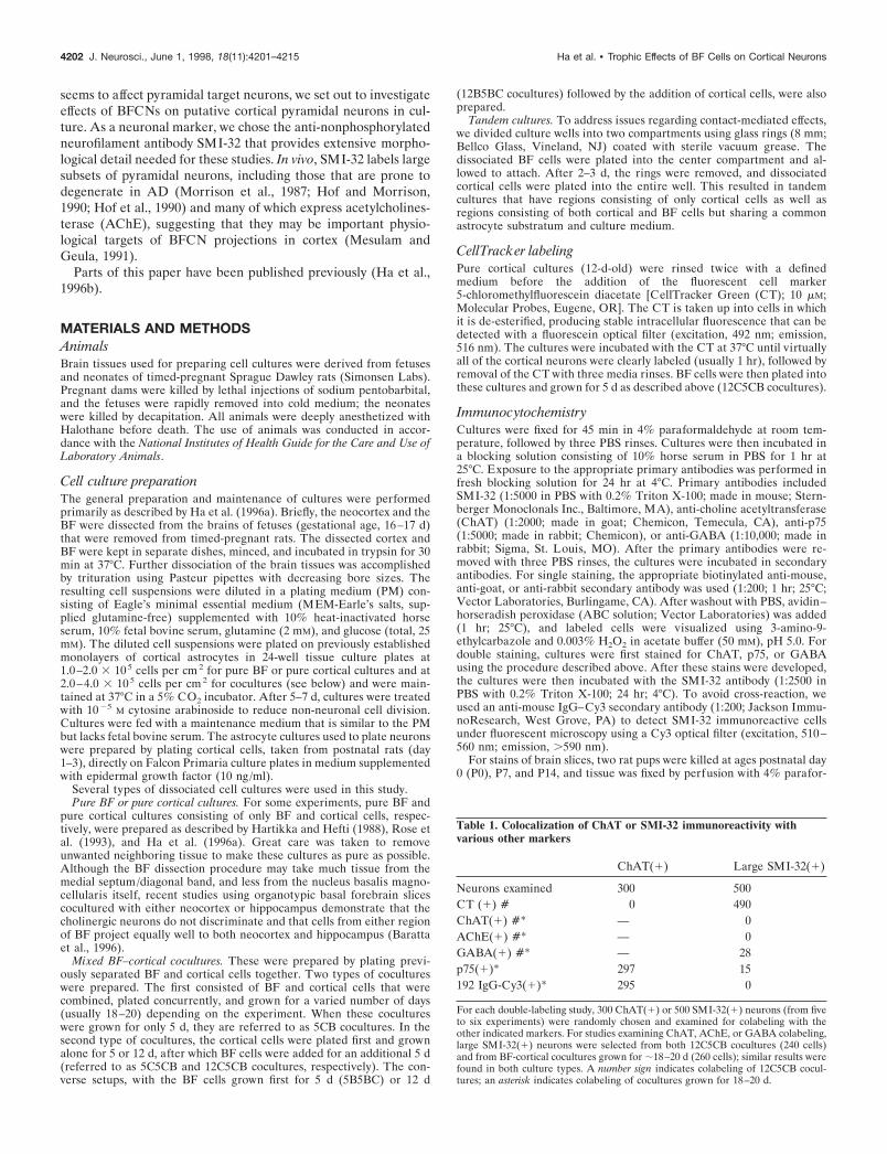
Table 1. Colocalization of ChAT or SMI-32 immunoreactivity withvarious other markers
ChAT(1) Large SMI-32(1)
Neurons examined 300 500CT (1) # 0 490ChAT(1) #* — 0AChE(1) #* — 0GABA(1) #* — 28p75(1)* 297 15192 IgG-Cy3(1)* 295 0
For each double-labeling study, 300 ChAT(1) or 500 SMI-32(1) neurons (from fiveto six experiments) were randomly chosen and examined for colabeling with theother indicated markers. For studies examining ChAT, AChE, or GABA colabeling,large SMI-32(1) neurons were selected from both 12C5CB cocultures (240 cells)and from BF-cortical cocultures grown for ;18–20 d (260 cells); similar results werefound in both culture types. A number sign indicates colabeling of 12C5CB cocul-tures; an asterisk indicates colabeling of cocultures grown for 18–20 d.
4202 J. Neurosci., June 1, 1998, 18(11):4201–4215 Ha et al. • Trophic Effects of BF Cells on Cortical Neurons
maldehyde. Frozen sections (50 mm) were then processed for SMI-32immunocytochemistry as described above.
Acetylcholinesterase histochemistryAChE staining was performed as described by Tago et al. (1986) withminor modifications. Cultures were fixed with 4% paraformaldehyde for;40 min, after which they were rinsed three times with 0.1 M maleatebuffer, pH 6.0. The cultures were then incubated in a fresh solutionconsisting of 300 mM copper sulfate, 500 mM sodium citrate, 50 mMpotassium ferricyanide, and 30 mM acetylthiocholine iodide in 0.1 Mmaleate buffer for 1–2 hr in the dark. After being rinsed with PBS fivetimes, the cultures were incubated in an intensification solution (0.04%DAB, 0.3% nickel ammonium sulfate, and 0.003% H2O2 in PBS) untilcells were clearly stained (15–30 min). For AChE and SMI-32 doublelabeling, the AChE histochemistry was performed first, followed bySMI-32 immunocytochemistry. Again, an anti-mouse IgG–Cy3 second-
ary antibody was used to visualize cells labeled with the SMI-32 primaryantibody.
192 IgG–saporin treatmentTandem cultures were prepared and maintained as described above. Onday 12 and again on day 16 of a 22 d culturing period, some of thecultures were treated with the 192 IgG–saporin (35–40 ng/ml; Chemi-con). The cultures were then stained with the SMI-32 antibody on day 22.In investigations of effects of 192 IgG–saporin on BFCNs, pure BFcultures were grown for 14 d before treating with the 192 IgG–saporinfor a varied number of days and staining for ChAT as described above.For experiments examining effects of 192 IgG–saporin treatment on pureBF cultures before addition of cortical cells, E17 BF cells were grown for6–8 d before addition of the 192 IgG–saporin for 3 d. The cultures werethen thoroughly washed, and E17 cortical cells were added for an addi-tional 14 d before staining for ChAT or for SMI-32.
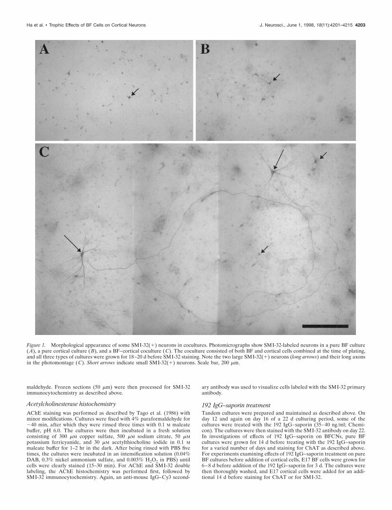
Figure 1. Morphological appearance of some SMI-32(1) neurons in cocultures. Photomicrographs show SMI-32-labeled neurons in a pure BF culture(A), a pure cortical culture (B), and a BF–cortical coculture (C). The coculture consisted of both BF and cortical cells combined at the time of plating,and all three types of cultures were grown for 18–20 d before SMI-32 staining. Note the two large SMI-32(1) neurons (long arrows) and their long axonsin the photomontage (C). Short arrows indicate small SMI-32(1) neurons. Scale bar, 200 mm.
Ha et al. • Trophic Effects of BF Cells on Cortical Neurons J. Neurosci., June 1, 1998, 18(11):4201–4215 4203

To study which cells in culture took up the 192 IgG–saporin, weconjugated the same 192 IgG antibody to the fluorescent marker Cy3(Chemicon) instead of saporin. Cocultures were treated with the 192IgG–Cy3 (50–100 ng/ml) for 1–2 hr before fixation and staining with theSMI-32 antibody. In this case, the neurons immunoreactive for SMI-32[SMI-32(1) neurons] were visualized with a secondary antibody labeledwith the fluorescent marker Cy2 (seen with a fluorescein optical filter;1:200; Jackson ImmunoResearch).
Quantitative analysisFor morphological measurements, stained cultures were viewed underbright-field microscopy (1003), and fields were randomly selected byblindly scrolling through a grid pattern of approximately seven by sevenfields and randomly stopping at intervals of three to four fields, such that12–14 fields representing all areas of the dish and constituting ;25% oftotal dish area were selected for imaging and analysis. A total of 30–40fields were imaged from three to four sister cultures per condition foreach experiment. The images were imported into a computer in whichSMI-32(1) cells within each field were counted and their morphologicalfeatures were measured using COMOS software from Bio-Rad (Her-cules, CA). The parameters measured were cell area, total dendriticlength, number of first-, second-, and third-order dendrites, and thepresence or absence of a long (.1500 mm) axon. Evaluation of datarevealed that the presence of a long axon was the single trait that mostreliably distinguished the large SMI-32(1) neurons from other SMI-32(1) neurons.
To assess double-labeling experiments, we first identified large SMI-32(1) neurons based on morphological criteria, including long axons,large soma size, and extensive dendritic arbor. Other experiments re-quired the initial identification of ChAT(1) neurons. These large SMI-32(1) or ChAT(1) neurons were then examined for the presence of labelfor other markers, as indicated in Table 1. For each double-labelingstudy, 500 large SMI-32(1) or 300 ChAT(1) neurons from five to sixexperiments were examined.
The effects of 192 IgG–saporin treatment were assessed by comparingcell counts of ChAT(1) or SMI-32(1) neurons in treated and untreatedcultures. In each culture well, the counts were determined from 52consecutive, nonoverlapping microscope fields, covering over 95% of thewell area, using low-power (1003) bright-field optics. Neurons wereconsidered ChAT(1) or SMI-32(1) if they were clearly stained and if atleast two neurites could be identified. When the labeled cells displayedatrophic cell bodies, disrupted cell membranes, and broken processes,they were excluded from the counts. In each experiment, the percent lossof ChAT(1) or SMI-32(1) neurons was calculated by comparing themean number of intact stained cells in several (three to four) control(untreated) cultures with the mean number in several experimental (192IgG–saporin-treated) cultures. In all experiments, both control and ex-perimental conditions were on the same multiwell culture plate andderived from the same plating.
Values are given as the mean 6 SEM, normalized to control conditionsin each experiment. Significance of the data was determined by ANOVA,with the Bonferroni post hoc test, using Instat software (Graph Pad, SanDiego, CA).
RESULTSLarge SMI-32(1) neurons are only found inBF–cortical coculturesInitial experiments were conducted to characterize the popula-tion of SMI-32(1) neurons in three different types of cultures(pure BF cultures, pure cortical cultures, and mixed BF–corticalcocultures) that were plated at gestational age 17 d (G-17) andgrown for 18–20 d (see Materials and Methods). As illustrated inFigure 1, SMI-32(1) neurons were found in both pure BF (A)and pure cortical (B) cultures; however, total numbers of SMI-32(1) neurons were ;10 times higher in the cortical cultures. TheSMI-32(1) neurons in both of these pure culture types appearedrelatively small, with short isodendritic neurites and no identifi-able axons. In the combined BF–cortical cocultures, SMI-32(1)neurons were found with frequencies similar to those of the purecortical culture but appeared as two morphologically distinctpopulations. The majority displayed morphological features sim-
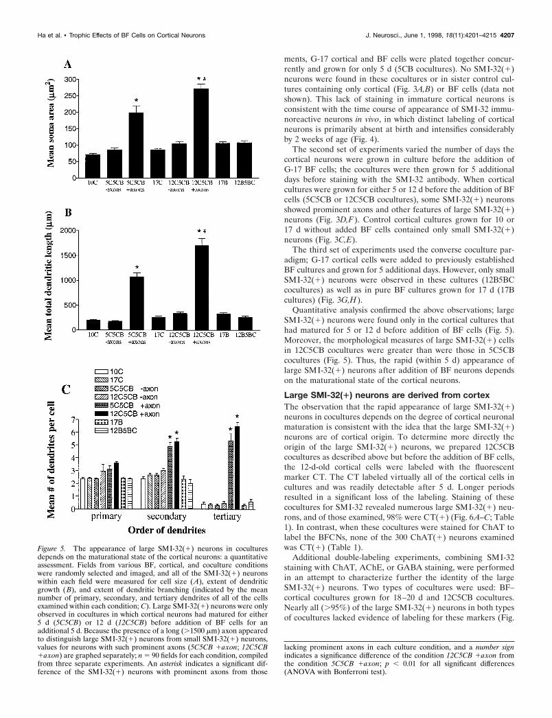
Figure 2. The morphological features of SMI-32(1) neurons in cocul-tures: a quantitative assessment. Fields from BF and cortical (Cor) cul-tures and from BF–cortical cocultures such as those illustrated in Figure1 were randomly selected and imaged, and all of the SMI-32(1) neuronswithin each field were measured for cell size (A), extent of dendriticgrowth (B), and extent of dendritic branching (indicated by the meannumber of primary, secondary, and tertiary dendrites of all of the cellsexamined within each condition; C). In cocultures, the presence of a long(.1500 mm) axon appeared to distinguish the minority (;5–10%) ofSMI-32(1) neurons (BF–Cor 1axons) that displayed enhanced morpho-logical features from the remaining small SMI-32(1) neurons (BF–Cor2axons) that were generally indistinguishable from those in pure corticalcultures. Values for neurons with such prominent axons are thereforegraphed separately. Data are presented as the mean 6 SEM; n 5 120fields for each condition, compiled from three experiments. An asteriskindicates a significant difference of the SMI-32(1) neurons with promi-nent axons from those lacking prominent axons in each culture condition;p , 0.001 (ANOVA with Bonferroni post hoc test).
4204 J. Neurosci., June 1, 1998, 18(11):4201–4215 Ha et al. • Trophic Effects of BF Cells on Cortical Neurons

Figure 3. The appearance of large SMI-32(1) neurons in cocultures depends on the maturational state of the cortical neurons. Photomicrographs onthe lef t show representative SMI-32-stained fields from pure cortical cultures grown for 5 d ( A), 10 d ( C), or 17 d ( E) and from pure BF cultures grownfor 17 d (G). Photomicrographs on the right show fields from cultures identical to those shown on the lef t except that either BF (B, D, F ) or cortical (H )neurons were added for the last 5 d before staining. Note the presence of large SMI-32(1) neurons only in cocultures in which the cortical neurons weregrown for at least 5 d before the addition of BF cells (D, F ). Scale bar, 100 mm.
Ha et al. • Trophic Effects of BF Cells on Cortical Neurons J. Neurosci., June 1, 1998, 18(11):4201–4215 4205

ilar to SMI-32(1) neurons found in pure cortical cultures [re-ferred to as small SMI-32(1) neurons], whereas a minority [re-ferred to as large SMI-32(1) neurons] displayed prominent axonsthat can often be traced for long distances as well as large cellbodies and extensive dendritic arbors (Fig. 1C).
Morphological characterization of SMI-32(1) neurons in ran-domly selected fields of the cocultures showed that SMI-32(1)neurons with long (.1500 mm) axons [5–10% of total SMI-32(1)neurons] also had substantially larger somata, longer dendrites,and greater dendritic branching than did most SMI-32(1) neu-rons (Fig. 2). Thus, the presence of long axons seemed to be a
single criterion that could distinguish virtually all of the largefrom the small SMI-32(1) neurons.
Phenotypic characteristics of SMI-32(1) neurons incocultures is dependent on the age of cortical cellsInitial experiments demonstrated the distinctive morphologicalfeatures of a subset of SMI-32(1) neurons in BF–cortical cocul-tures grown for 18–20 d. Three sets of additional experimentswere performed to examine whether the appearance of neuronswith these morphological features was dependent on the age ofeither the cortical or the BF neurons. In the first set of experi-
Figure 4. Patterns of SMI-32(1) neurons in developing neocortex. In animals killed at P0 (A, D), no SMI-32(1) neurons are detected, although somelabeling of capillaries can be seen. By P7 (B, E), pyramidal neurons in layer V show prominent SMI-32 reactivity. By P14 (C, F ), pyramidal neuronspredominantly in layers V and III are SMI-32(1). Scale bars: A–C, 200 mm; D–F, 50 mm.
4206 J. Neurosci., June 1, 1998, 18(11):4201–4215 Ha et al. • Trophic Effects of BF Cells on Cortical Neurons
ments, G-17 cortical and BF cells were plated together concur-rently and grown for only 5 d (5CB cocultures). No SMI-32(1)neurons were found in these cocultures or in sister control cul-tures containing only cortical (Fig. 3A,B) or BF cells (data notshown). This lack of staining in immature cortical neurons isconsistent with the time course of appearance of SMI-32 immu-noreactive neurons in vivo, in which distinct labeling of corticalneurons is primarily absent at birth and intensifies considerablyby 2 weeks of age (Fig. 4).
The second set of experiments varied the number of days thecortical neurons were grown in culture before the addition ofG-17 BF cells; the cocultures were then grown for 5 additionaldays before staining with the SMI-32 antibody. When corticalcultures were grown for either 5 or 12 d before the addition of BFcells (5C5CB or 12C5CB cocultures), some SMI-32(1) neuronsshowed prominent axons and other features of large SMI-32(1)neurons (Fig. 3D,F). Control cortical cultures grown for 10 or17 d without added BF cells contained only small SMI-32(1)neurons (Fig. 3C,E).
The third set of experiments used the converse coculture par-adigm; G-17 cortical cells were added to previously establishedBF cultures and grown for 5 additional days. However, only smallSMI-32(1) neurons were observed in these cultures (12B5BCcocultures) as well as in pure BF cultures grown for 17 d (17Bcultures) (Fig. 3G,H).
Quantitative analysis confirmed the above observations; largeSMI-32(1) neurons were found only in the cortical cultures thathad matured for 5 or 12 d before addition of BF cells (Fig. 5).Moreover, the morphological measures of large SMI-32(1) cellsin 12C5CB cocultures were greater than were those in 5C5CBcocultures (Fig. 5). Thus, the rapid (within 5 d) appearance oflarge SMI-32(1) neurons after addition of BF neurons dependson the maturational state of the cortical neurons.
Large SMI-32(1) neurons are derived from cortexThe observation that the rapid appearance of large SMI-32(1)neurons in cocultures depends on the degree of cortical neuronalmaturation is consistent with the idea that the large SMI-32(1)neurons are of cortical origin. To determine more directly theorigin of the large SMI-32(1) neurons, we prepared 12C5CBcocultures as described above but before the addition of BF cells,the 12-d-old cortical cells were labeled with the fluorescentmarker CT. The CT labeled virtually all of the cortical cells incultures and was readily detectable after 5 d. Longer periodsresulted in a significant loss of the labeling. Staining of thesecocultures for SMI-32 revealed numerous large SMI-32(1) neu-rons, and of those examined, 98% were CT(1) (Fig. 6A–C; Table1). In contrast, when these cocultures were stained for ChAT tolabel the BFCNs, none of the 300 ChAT(1) neurons examinedwas CT(1) (Table 1).
Additional double-labeling experiments, combining SMI-32staining with ChAT, AChE, or GABA staining, were performedin an attempt to characterize further the identity of the largeSMI-32(1) neurons. Two types of cocultures were used: BF–cortical cocultures grown for 18–20 d and 12C5CB cocultures.Nearly all (.95%) of the large SMI-32(1) neurons in both typesof cocultures lacked evidence of labeling for these markers (Fig.
Figure 5. The appearance of large SMI-32(1) neurons in coculturesdepends on the maturational state of the cortical neurons: a quantitativeassessment. Fields from various BF, cortical, and coculture conditionswere randomly selected and imaged, and all of the SMI-32(1) neuronswithin each field were measured for cell size (A), extent of dendriticgrowth (B), and extent of dendritic branching (indicated by the meannumber of primary, secondary, and tertiary dendrites of all of the cellsexamined within each condition; C). Large SMI-32(1) neurons were onlyobserved in cocultures in which cortical neurons had matured for either5 d (5C5CB) or 12 d (12C5CB) before addition of BF cells for anadditional 5 d. Because the presence of a long (.1500 mm) axon appearedto distinguish large SMI-32(1) neurons from small SMI-32(1) neurons,values for neurons with such prominent axons (5C5CB 1axon; 12C5CB1axon) are graphed separately; n 5 90 fields for each condition, compiledfrom three separate experiments. An asterisk indicates a significant dif-ference of the SMI-32(1) neurons with prominent axons from those
lacking prominent axons in each culture condition, and a number signindicates a significance difference of the condition 12C5CB 1axon fromthe condition 5C5CB 1axon; p , 0.01 for all significant differences(ANOVA with Bonferroni test).
Ha et al. • Trophic Effects of BF Cells on Cortical Neurons J. Neurosci., June 1, 1998, 18(11):4201–4215 4207
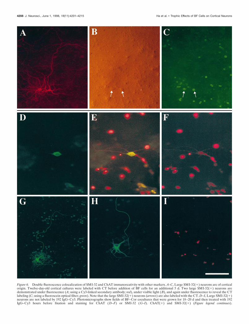
Figure 6. Double fluorescence colocalization of SMI-32 and ChAT immunoreativity with other markers. A–C, Large SMI-32(1) neurons are of corticalorigin. Twelve-day-old cortical cultures were labeled with CT before addition of BF cells for an additional 5 d. Two large SMI-32(1) neurons aredemonstrated under fluorescence (A; using a Cy3-linked secondary antibody; red), under visible light (B), and again under fluorescence to reveal the CTlabeling (C; using a fluorescein optical filter; green). Note that the large SMI-32(1) neurons (arrows) are also labeled with the CT. D–I, Large SMI-32(1)neurons are not labeled by 192 IgG–Cy3. Photomicrographs show fields of BF–Cor cocultures that were grown for 18–20 d and then treated with 192IgG–Cy3 hours before fixation and staining for ChAT ( D–F) or SMI-32 ( G–I). ChAT(1) and SMI-32(1) (Figure legend continues).
4208 J. Neurosci., June 1, 1998, 18(11):4201–4215 Ha et al. • Trophic Effects of BF Cells on Cortical Neurons
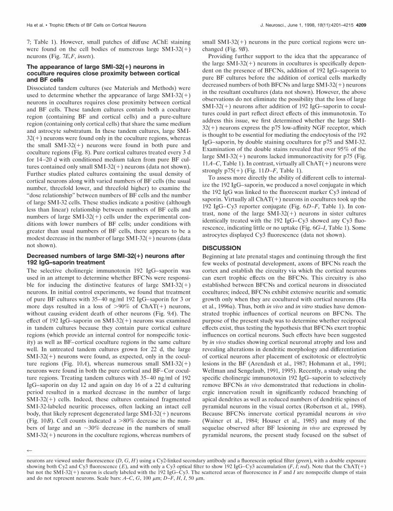
7; Table 1). However, small patches of diffuse AChE stainingwere found on the cell bodies of numerous large SMI-32(1)neurons (Fig. 7E,F, insets).
The appearance of large SMI-32(1) neurons incoculture requires close proximity between corticaland BF cellsDissociated tandem cultures (see Materials and Methods) wereused to determine whether the appearance of large SMI-32(1)neurons in cocultures requires close proximity between corticaland BF cells. These tandem cultures contain both a cocultureregion (containing BF and cortical cells) and a pure-cultureregion (containing only cortical cells) that share the same mediumand astrocyte substratum. In these tandem cultures, large SMI-32(1) neurons were found only in the coculture regions, whereasthe small SMI-32(1) neurons were found in both pure andcoculture regions (Fig. 8). Pure cortical cultures treated every 3 dfor 14–20 d with conditioned medium taken from pure BF cul-tures contained only small SMI-32(1) neurons (data not shown).Further studies plated cultures containing the usual density ofcortical neurons along with varied numbers of BF cells (the usualnumber, threefold lower, and threefold higher) to examine the“dose relationship” between numbers of BF cells and the numberof large SMI-32 cells. These studies indicate a positive (althoughless than linear) relationship between numbers of BF cells andnumbers of large SMI-32(1) cells under the experimental con-ditions with lower numbers of BF cells; under conditions withgreater than usual numbers of BF cells, there appears to be amodest decrease in the number of large SMI-32(1) neurons (datanot shown).
Decreased numbers of large SMI-32(1) neurons after192 IgG–saporin treatmentThe selective cholinergic immunotoxin 192 IgG–saporin wasused in an attempt to determine whether BFCNs were responsi-ble for inducing the distinctive features of large SMI-32(1)neurons. In initial control experiments, we found that treatmentof pure BF cultures with 35–40 ng/ml 192 IgG–saporin for 3 ormore days resulted in a loss of .90% of ChAT(1) neurons,without causing evident death of other neurons (Fig. 9A). Theeffect of 192 IgG–saporin on SMI-32(1) neurons was examinedin tandem cultures because they contain pure cortical cultureregions (which provide an internal control for nonspecific toxic-ity) as well as BF–cortical coculture regions in the same culturewell. In untreated tandem cultures grown for 22 d, the largeSMI-32(1) neurons were found, as expected, only in the cocul-ture regions (Fig. 10A), whereas numerous small SMI-32(1)neurons were found in both the pure cortical and BF–Cor cocul-ture regions. Treating tandem cultures with 35–40 ng/ml of 192IgG–saporin on day 12 and again on day 16 of a 22 d culturingperiod resulted in a marked decrease in the number of largeSMI-32(1) cells. Indeed, these cultures contained fragmentedSMI-32-labeled neuritic processes, often lacking an intact cellbody, that likely represent degenerated large SMI-32(1) neurons(Fig. 10B). Cell counts indicated a .80% decrease in the num-bers of large and an ;30% decrease in the numbers of smallSMI-32(1) neurons in the coculture regions, whereas numbers of
small SMI-32(1) neurons in the pure cortical regions were un-changed (Fig. 9B).
Providing further support to the idea that the appearance ofthe large SMI-32(1) neurons in cocultures is specifically depen-dent on the presence of BFCNs, addition of 192 IgG–saporin topure BF cultures before the addition of cortical cells markedlydecreased numbers of both BFCNs and large SMI-32(1) neuronsin the resultant cocultures (data not shown). However, the aboveobservations do not eliminate the possibility that the loss of largeSMI-32(1) neurons after addition of 192 IgG–saporin to cocul-tures could in part reflect direct effects of this immunotoxin. Toaddress this issue, we first determined whether the large SMI-32(1) neurons express the p75 low-affinity NGF receptor, whichis thought to be essential for mediating the endocytosis of the 192IgG–saporin, by double staining cocultures for p75 and SMI-32.Examination of the double stains revealed that over 95% of thelarge SMI-32(1) neurons lacked immunoreactivity for p75 (Fig.11A–C, Table 1). In contrast, virtually all ChAT(1) neurons werestrongly p75(1) (Fig. 11D–F, Table 1).
To assess more directly the ability of different cells to internal-ize the 192 IgG–saporin, we produced a novel conjugate in whichthe 192 IgG was linked to the fluorescent marker Cy3 instead ofsaporin. Virtually all ChAT(1) neurons in cocultures took up the192 IgG–Cy3 reporter conjugate (Fig. 6D–F, Table 1). In con-trast, none of the large SMI-32(1) neurons in sister culturesidentically treated with the 192 IgG–Cy3 showed any Cy3 fluo-rescence, indicating little or no uptake (Fig. 6G–I, Table 1). Someastrocytes displayed Cy3 fluorescence (data not shown).
DISCUSSIONBeginning at late prenatal stages and continuing through the firstfew weeks of postnatal development, axons of BFCNs reach thecortex and establish the circuitry via which the cortical neuronscan exert trophic effects on the BFCNs. This circuitry is alsoestablished between BFCNs and cortical neurons in dissociatedcocultures; indeed, BFCNs exhibit extensive neuritic and somaticgrowth only when they are cocultured with cortical neurons (Haet al., 1996a). Thus, both in vivo and in vitro studies have demon-strated trophic influences of cortical neurons on BFCNs. Thepurpose of the present study was to determine whether reciprocaleffects exist, thus testing the hypothesis that BFCNs exert trophicinfluences on cortical neurons. Such effects have been suggestedby in vivo studies showing cortical neuronal atrophy and loss andrevealing alterations in dendritic morphology and differentiationof cortical neurons after placement of excitotoxic or electrolyticlesions in the BF (Arendash et al., 1987; Hohmann et al., 1991;Wellman and Sengelaub, 1991, 1995). Recently, a study using thespecific cholinergic immunotoxin 192 IgG–saporin to selectivelyremove BFCNs in vivo demonstrated that reductions in cholin-ergic innervation result in significantly reduced branching ofapical dendrites as well as reduced numbers of dendritic spines ofpyramidal neurons in the visual cortex (Robertson et al., 1998).Because BFCNs innervate cortical pyramidal neurons in vivo(Wainer et al., 1984; Houser et al., 1985) and many of thesequelae observed after BF lesioning in vivo are expressed bypyramidal neurons, the present study focused on the subset of
4
neurons are viewed under fluorescence (D, G, H ) using a Cy2-linked secondary antibody and a fluorescein optical filter (green), with a double exposureshowing both Cy2 and Cy3 fluorescence (E), and with only a Cy3 optical filter to show 192 IgG–Cy3 accumulation (F, I; red). Note that the ChAT(1)but not the SMI-32(1) neuron is clearly labeled with the 192 IgG–Cy3. The scattered areas of fluorescence in F and I are nonspecific clumps of stainand do not represent neurons. Scale bars: A–C, G, 100 mm; D–F, H, I, 50 mm.
Ha et al. • Trophic Effects of BF Cells on Cortical Neurons J. Neurosci., June 1, 1998, 18(11):4201–4215 4209
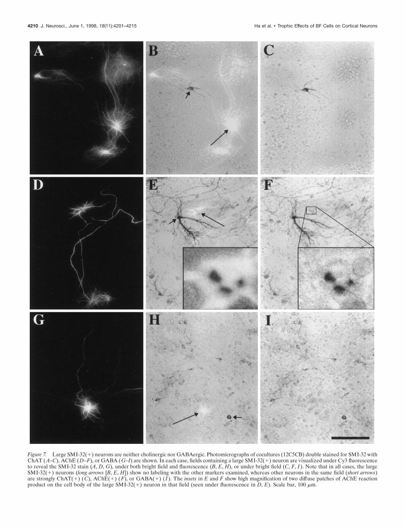
Figure 7. Large SMI-32(1) neurons are neither cholinergic nor GABAergic. Photomicrographs of cocultures (12C5CB) double stained for SMI-32 withChAT (A–C), AChE (D–F), or GABA (G–I) are shown. In each case, fields containing a large SMI-32(1) neuron are visualized under Cy3 fluorescenceto reveal the SMI-32 stain (A, D, G), under both bright field and fluorescence (B, E, H), or under bright field (C, F, I ). Note that in all cases, the largeSMI-32(1) neurons (long arrows [B, E, H]) show no labeling with the other markers examined, whereas other neurons in the same field (short arrows)are strongly ChAT(1) ( C), AChE(1) ( F), or GABA(1) ( I ). The insets in E and F show high magnification of two diffuse patches of AChE reactionproduct on the cell body of the large SMI-32(1) neuron in that field (seen under fluorescence in D, E). Scale bar, 100 mm.
4210 J. Neurosci., June 1, 1998, 18(11):4201–4215 Ha et al. • Trophic Effects of BF Cells on Cortical Neurons

neurons in culture labeled with the monoclonal antibody SMI-32that labels subsets of cortical pyramidal neurons in vivo (Morrisonet al., 1987; Hof and Morrison, 1990; Hof et al., 1990) (see alsoFig. 4).
The primary finding of this study is that cocultures containingboth BF and cortical cells display a subset of SMI-32-immunoreactive neurons with distinctive morphological features:prominent axons, large somata, and long-branching dendrites.Thus, the appearance of these large SMI-32(1) neurons in co-cultures is consistent with our working hypothesis that BFCNsmay induce growth and/or increase survival of subsets of corticalpyramidal neurons in culture. Present data do not distinguishbetween the possibility that the appearance of the large SMI-32(1) neurons reflects morphological enhancement of smallSMI-32(1) cells present in the cultures or induction of a newpopulation of neurons that would either not survive or not beobserved by SMI-32 staining in pure cortical cultures.
Phenotypic identity and cortical origin of largeSMI-32(1) neuronsSeveral lines of evidence support the hypothesis that the largeSMI-32(1) cells are a subset of cortical pyramidal neurons. First,studies using the fluorescent marker CT provided direct evidencethat virtually all of the large SMI-32(1) neurons are derived fromcortex and not from BF. Specificity of the CT labeling for cortical
neurons was achieved by complete removal of the CT with mediarinses before the addition of BF cells and also by the enzymaticcleavage of the CT, after it is inside the cell, to cell membrane-impermeable fluorescent products.
Second, the finding that large SMI-32(1) neurons do notexpress the enzymes ChAT or AChE is consistent with theirpresumed cortical origin and a noncholinergic identity. Interest-ingly, many of these neurons had small patches of AChE reactionproduct on their somata. This pattern of staining is entirelydistinct from the extensive somatic and neuritic labeling shown bythe BFCNs and is suggestive of a pyramidal identity, becausemany pyramidal neurons in neocortical layers III and V expressAChE and are strongly SMI-32-immunoreactive (Mesulam andGeula, 1991). Further support for a pyramidal identity comesfrom studies demonstrating that large SMI-32(1) neurons areGABA-negative.
Conditions under which large SMI-32(1) neuronsare presentAs large SMI-32(1) neurons are only found in cocultures, ex-pression of the distinctive morphological features of these neu-rons seems to be mediated via interactions between BF andcortical cells. In addition, the observation that destruction of mostBFCNs in BF cultures by 192 IgG–saporin exposure before theaddition of cortical cells results in markedly decreased numbers of
Figure 8. Large SMI-32(1) neurons are found only in coculture regions of tandem cultures. Photomicrographs show low and high magnification ofrepresentative SMI-32(1) neurons in the pure cortical (A, B) and the coculture (C, D) regions of a tandem culture. Note the large SMI-32(1) neuron(long arrows) in the coculture region and the small SMI-32(1) neurons (short arrows) in both pure and coculture regions of the tandem culture; no largeSMI-32(1) neurons were present in the pure cortical regions of the cultures. Scale bars: A, C, 200 mm; B, D, 50 mm.
Ha et al. • Trophic Effects of BF Cells on Cortical Neurons J. Neurosci., June 1, 1998, 18(11):4201–4215 4211
large SMI-32(1) neurons suggests that the growth- and/orsurvival-stimulating factors depend specifically on the presence ofBFCNs. The further observation that large SMI-32(1) neuronswere only present in the coculture regions of tandem culturesindicates that close proximity between BFCNs (or their axonalprocesses) and cortical cells may be necessary for any growth- orsurvival-stimulating signals to be transmitted. Thus, the effectscould be mediated by direct interactions between BFCNs andSMI-32(1) neurons, as through synaptically transmitted signals,or by actions of cell surface proteins. Alternatively, soluble fac-tors that are effective only over short distances could be pro-duced. Although the suggestion that BFCNs may influence SMI-32(1) central neurons directly is an attractive one, at present wecannot eliminate the possibility that BFCNs affect an intermedi-
ate population of cells, which in turn affect the SMI-32(1)neurons.
The morphological parameters of large SMI-32(1) neuronsincreased with increasing age of the cortical neurons, suggestingthat the cortically derived large SMI-32(1) neurons becomemore responsive to BF-dependent factors with increasing matu-ration. The age in vitro at which cortical neurons appear tobecome responsive to the BF cells is ;5–10 d in culture. Thismaturational level corresponds both to the time of appearance ofSMI-32-immunoreactivity in cortical pyramidal neurons (see Fig.4) and to the time at which cortical neurons are becoming differ-entiated and innervated by BFCNs in vivo (Dinopoulos et al.,1989; Gould et al., 1991; Calarco and Robertson, 1995; De Carloset al., 1995).
Maintenance of morphological features of largeSMI-32(1) neurons by BFCNsResults from experiments using the cholinergic immunotoxin 192IgG–saporin to eliminate BFCNs support the hypothesis that thepresence of large SMI-32(1) neurons in cocultures requires theongoing presence of BFCNs. Several studies have demonstratedthe effectiveness and specificity with which 192 IgG–saporin kills
Figure 9. Loss of ChAT(1) and large SMI-32(1) neurons in culturestreated with 192 IgG–saporin. A, Fourteen-day-old pure BF cultures weretreated with 192 IgG–saporin (35–40 ng/ml) for 1–5 d, followed by ChATstaining and evaluation of cell loss (by comparison with untreated sistercultures). Note the marked ChAT(1) neuronal cell loss after 3 or moredays of exposure to 192 IgG–saporin; n 5 12 cultures per condition fromthree experiments. B, Tandem cultures were treated with the 192 IgG–saporin on day 12 and again on day 16, followed by SMI-32 staining onday 22. Cell loss was evaluated in relation to untreated sister cultures.Note the loss of most large SMI-32(1) neurons (present in the cocultureor BF–Cor regions of the tandem cultures), the minimal loss of smallSMI-32(1) neurons in pure cortical regions, and the partial loss of smallSMI-32(1) neurons in the coculture regions; n 5 9 cultures per condition,compiled from three experiments. An asterisk indicates significant differ-ences from BF–Cor cocultures, and a number sign indicates significantdifferences from small SMI-32(1) neurons; p , 0.001 for all significantdifferences (ANOVA with Bonferroni test).
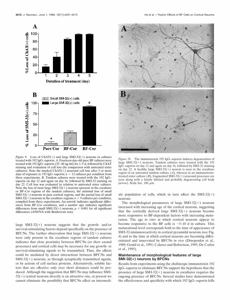
Figure 10. The immunotoxin 192 IgG–saporin induces degeneration oflarge SMI-32(1) neurons. Tandem cultures were treated with the 192IgG–saporin on day 12 and again on day 16, followed by SMI-32 stainingon day 22. A healthy large SMI-32(1) neuron is seen in the cocultureregion of an untreated tandem culture (A), whereas in an immunotoxin-treated sister culture ( B), fragmented SMI-32(1) neuronal processes areseen along with a faintly labeled and probably degenerating cell body(arrow). Scale bar, 100 mm.
4212 J. Neurosci., June 1, 1998, 18(11):4201–4215 Ha et al. • Trophic Effects of BF Cells on Cortical Neurons

the BFCNs in vivo (Wiley et al., 1991; Book et al., 1992, 1995;Heckers et al., 1994; Roßner et al., 1995). In cultures, the 192IgG–saporin appears to have similar specificity; that is, treatingcocultures with this immunotoxin resulted in near complete lossof the BFCNs, whereas the background cells did not appear to beaffected. However, this treatment resulted in a significant reduc-tion in the number of large SMI-32(1) neurons in the cocultureregions of tandem cultures. If we accept for the moment that theprimary effect of 192 IgG–saporin is on BFCNs (discussed in thenext paragraph), the decrease in numbers of large SMI-32(1)neurons could reflect a secondary effect, either from a loss ofgrowth stimulation or from degeneration or atrophy of largeSMI-32(1) neurons that were already present after removal ofBFCNs. Evidence of fragmented axons and damaged somata oflarge SMI-32(1) neurons suggests that the latter mechanismplays a role. These findings are compatible with in vivo studiesshowing various altered cortical phenotypes (Arendash et al.,1987; Hohmann et al., 1991; Wellman and Sengelaub, 1991, 1995)
and specific structural abnormalities of developing pyramidalneurons (Robertson et al., 1998) after lesioning of the BFCNs.
The conclusion that the decrease in number of large SMI-32(1) neurons is secondary to 192 IgG–saporin-induced loss ofBFCNs requires the demonstration that the toxic effects of the192 IgG–saporin are highly selective. Although present resultscannot completely eliminate possible direct toxic effects of 192IgG–saporin on large SMI-32(1) neurons, three lines of experi-mental data support this contention. First, specificity of the 192IgG–saporin for BFCNs is dependent on the presence of the p75low-affinity neurotrophin receptor, possibly in combination withthe high affinity neurotrophin tyrosine kinase receptors, whichare necessary for binding and internalization of the 192 IgG–saporin (Chandler et al., 1984; Taniuchi and Johnson, 1985; Wileyet al., 1991; Gargano et al., 1997). Thus, the observation that allof the ChAT(1) neurons in culture were found to be p75(1),whereas the large SMI-32(1) neurons were nearly all p75-negative, would favor the selective ability of BFCNs to bind
Figure 11. Large SMI-32(1) neurons do not express the p75 receptor. Photomicrographs show 18-d-old BF–Cor cocultures double stained for p75 andSMI-32 (A–C) or ChAT (D–F). Note that the large SMI-32(1) neurons (A, B; under fluorescence) lack p75 immunoreactivity (B, C). The short arrowin B indicates a p75(1) neuron in proximity to the large SMI-32(1) cell (long arrow). In contrast, the ChAT(1) neuron (D, E) is strongly p75(1) ( F).Scale bar, 100 mm.
Ha et al. • Trophic Effects of BF Cells on Cortical Neurons J. Neurosci., June 1, 1998, 18(11):4201–4215 4213
directly and take up the 192 IgG–saporin. Second, labeling of allof the ChAT(1) cells, but none of the large SMI-32(1) neurons,with the 192 IgG–Cy3 provides more direct evidence that BF-CNs, rather than large SMI-32(1) neurons, readily take up 192IgG conjugates. The significance of the 192 IgG–Cy3 labeling ofsome astrocytes is uncertain, because in vivo studies have dem-onstrated mild or no astroglial reaction to 192 IgG–saporin treat-ment (Book et al., 1995; Roßner et al., 1995). Finally, the lack ofdegeneration of small SMI-32(1) neurons in the pure corticalregions of tandem cultures after 192 IgG–saporin treatment pro-vides evidence against nonspecific toxic effects on the overallSMI-32(1) neuronal population. The loss of some small SMI-32(1) neurons only in coculture regions of treated tandem cul-tures could indicate the presence at the time of staining of someneurons that have been affected by the presence of the BFCNs,although not manifesting the distinctive morphological featuresof typical large SMI-32(1) neurons.
ConclusionsThe present data suggest that BFCNs induce powerful survival-and/or growth-enhancing effects on a distinct subpopulation ofcortical neurons. Furthermore, selective destruction of BFCNsseems to result in a secondary loss or atrophy of these corticalneurons. These observations are consistent with several in vivostudies demonstrating morphological alterations in certain corti-cal neurons after BF lesions. Although the demonstration of theseeffects in a highly simplified dissociated culture system lendssupport to the idea that the stimulatory signals are provided tocortical neurons directly by BFCNs, present results do not elim-inate indirect effects. Possible direct mediators might include theneurotransmitter acetylcholine, other anterogradely transportedtrophic molecules (Corfas et al., 1995), or membrane-boundmolecules. Possible indirect mediators could include other neu-rons or cortical glial cells. Present results, taken together with ourprevious study demonstrating survival- and growth-enhancingeffects of cortical neurons on BFCNs (Ha et al., 1996a), suggestthe existence of reciprocal interactions between BFCNs andcertain cortical neurons that can be modeled in a simplifiedculture system. Because these effects appear to be maximal atculture ages corresponding to the period during which BFCNsgrow into and innervate cortex, they may well be relevant to thedevelopment of BFCN–cortical projections in vivo. In addition,the present culture system may prove useful for studies relevant toAlzheimer’s disease or other conditions in which there is degen-eration of both BFCNs and SMI-32-immunoreactive corticalpyramidal neurons (Davies and Maloney, 1976; Whitehouse et al.,1982; Morrison et al., 1987; Hof et al., 1990; Cullen et al., 1997).
REFERENCESArendash GW, Millard WJ, Dunn AJ, Meyer EM (1987) Long-term
neuropathological and neurochemical effects of nucleus basalis lesionsin the rat. Science 238:952–956.
Baratta J, Ha DH, Weiss JH, Yu J, Robertson RT (1996) Cholinergicneurons from different subdivisions of the basal forebrain lack connec-tional specificity for cerebral cortical target sites in vitro. Dev Brain Res97:143–147.
Bigl V, Woolf NJ, Butcher LL (1982) Cholinergic projections from thebasal forebrain to frontal, parietal, temporal, occipital, and cingulatecortices: a combined fluorescent tracer and acetylcholinesterase anal-ysis. Brain Res Bull 8:727–749.
Book AA, Wiley RG, Schweitzer JB (1992) Specificity of 192 IgG–saporin for NGF receptor-positive cholinergic basal forebrain neuronsin the rat. Brain Res 590:350–355.
Book AA, Wiley RG, Schweitzer JB (1995) 192 IgG–saporin 2. Neuro-pathology in the rat brain. Acta Neuropathol (Berl) 89:519–526.
Calarco CA, Robertson RT (1995) Development of basal forebrain pro-jections to visual cortex: DiI studies in rat. J Comp Neurol 354:608–626.
Chandler CE, Parsons LM, Hosang M, Shooter EM (1984) A monoclo-nal antibody modulates the interaction of nerve growth factor withPC12 cells. J Biol Chem 259:6882–6889.
Corfas G, Rosen KM, Aratake H, Krauss R, Fischbach GD (1995)Differential expression of ARIA isoforms in the rat brain. Neuron14:103–115.
Crowley C, Spencer SD, Nishimura MC, Chen KS, Pitts-Meek S, Arma-nini MP, Ling LH, MacMahon SB, Shelton DL, Levinson AD, PhillipsHS (1994) Mice lacking nerve growth factor display perinatal loss ofsensory and sympathetic neurons yet develop basal forebrain cholin-ergic neurons. Cell 76:1001–1011.
Cullen KM, Halliday GM, Double KL, Brooks WS, Creasey H, Broe GA(1997) Cell loss in the nucleus basalis is related to regional corticalatrophy in Alzheimer’s disease. Neuroscience 78:641–652.
Davies P, Maloney AJ (1976) Selective loss of central cholinergic neu-rons in Alzheimer’s disease. Lancet 2:1403.
De Carlos JA, Schlaggar BL, O’Leary DDM (1995) Development ofacetylcholinesterase-positive thalamic and basal forebrain afferents toembryonic rat neocortex. Exp Brain Res 104:385–401.
Dinopoulos A, Eadie LA, Dori I, Parnavelas JG (1989) The develop-ment of basal forebrain projections to the rat visual cortex. Exp BrainRes 76:563–571.
Divac I (1975) Magnocellular nuclei of the basal forebrain project toneocortex, brain stem, and olfactory bulb. Review of some functionalcorrelates. Brain Res 93:385–398.
Eckenstein FP, Baughman RW, Quinn J (1988) An anatomical study ofcholinergic innervation in the rat cerebral cortex. Neuroscience25:457–474.
Gage FH, Wictorin K, Fischer W, Williams LR, Varon S, Bjorklund A(1986) Retrograde cell changes in the medial septum and diagonalband following fimbria-fornix transection: quantitative temporal anal-ysis. Neuroscience 19:241–255.
Gargano N, Levi A, Alema S (1997) Modulation of nerve growth factorinternalization by direct interaction between p75 and Trk A receptors.J Neurosci Res 50:1–12.
Gould E, Woolf NJ, Butcher LL (1991) Postnatal development of cho-linergic neurons in the rat. I. Forebrain. Brain Res Bull 27:767–789.
Ha DH, Robertson RT, Ribak CE, Weiss JH (1996a) Cultured basalforebrain cholinergic neurons in contact with cortical cells displaysynapses, enhanced morphological features, and decreased dependenceon nerve growth factor. J Comp Neurol 373:451–465.
Ha DH, Robertson RT, Weiss JH (1996b) Trophic interaction betweenbasal forebrain neurons and cortical neurons in dissociated co-cultures.Soc Neurosci Abstr 22:744.
Hartikka J, Hefti F (1988) Development of septal cholinergic neurons inculture: plating density and glial cells modulate effects of NGF onsurvival, fiber growth, and expression of transmitter-specific enzymes.J Neurosci 8:2967–2985.
Hatanaka H, Tsukui H, Nihonmatsu I (1988) Developmental change inthe nerve growth factor action from induction of choline acetyltrans-ferase to promotion of cell survival in cultured basal forebrain cholin-ergic neurons from postnatal rats. Dev Brain Res 39:85–95.
Heckers S, Ohtake T, Wiley RG, Lappi DA, Geula C, Mesulam M (1994)Complete and selective cholinergic denervation of rat neocortex andhippocampus but not amygdala by an immunotoxin against the p75NGF receptor. J Neurosci 14:1271–1289.
Hefti F (1986) Nerve growth factor promotes survival of septal cholin-ergic neurons after fimbrial transections. J Neurosci 6:2155–2162.
Hof PR, Morrison JH (1990) Quantitative analysis of a vulnerable sub-set of pyramidal neurons in Alzheimer’s disease. II. Primary andsecondary visual cortex. J Comp Neurol 301:55–64.
Hof PR, Cox K, Morrison JH (1990) Quantitative analysis of a vulner-able subset of pyramidal neurons in Alzheimer’s disease. I. Superiorfrontal and inferior temporal cortex. J Comp Neurol 301:44–54.
Hohmann CF, Kwiterovich KK, Oster-Granite ML, Coyle JT (1991)Newborn basal forebrain lesions disrupt cortical cytodifferentiation asvisualized by rapid Golgi staining. Cereb Cortex 1:143–157.
Houser CR (1990) Cholinergic synapses in the central nervous system:studies of the immunocytochemical localization of choline acetyltrans-ferase. J Electron Microsc Tech 15:2–19.
Houser CR, Crawford GD, Salvaterra PM, Vaughn JE (1985) Immuno-cytochemical localization of choline acetyltransferase in rat cerebral
4214 J. Neurosci., June 1, 1998, 18(11):4201–4215 Ha et al. • Trophic Effects of BF Cells on Cortical Neurons
cortex: a study of cholinergic neurons and synapses. J Comp Neurol234:17–34.
Hsiang J, Heller A, Hoffmann PC, Mobley WC, Wainer BH (1989) Theeffects of nerve growth factor on the development of septal cholinergicneurons in reaggregate cell cultures. Neuroscience 29:209–223.
Koliatsos VE, Price DL, Gouras GK, Cayoutte MH, Burton LE, Win-slow JW (1994) Highly selective effects of nerve growth factor, brain-derived neurotrophic factor, and neurotrophin-3 on intact and injuredbasal forebrain magnocellular neurons. J Comp Neurol 343:247–262.
Mesulam M, Geula C (1991) Differential distribution of a neurofilamentprotein epitope in acetylcholinesterase-rich neurons of human cerebralcortex. Brain Res 544:169–173.
Morrison JH, Lewis DA, Campbell MJ, Huntley GW, Benson DL,Bouras C (1987) A monoclonal antibody to non-phosphorylated neu-rofilament protein marks the vulnerable cortical neurons in Alzhei-mer’s disease. Brain Res 416:331–336.
Nonomura T, Nishio C, Lindsay RM, Hatanaka H (1995) Cultured basalforebrain cholinergic neurons from postnatal rats show both overlap-ping and non-overlapping responses to the neurotrophins. Brain Res683:129–139.
Robertson RT, Gallardo KA, Claytor KJ, Ha DH, Ku K, Yu BP,Lauterborn JC, Wiley RG, Yu J, Gall CM, Leslie FM (1998) Neonataltreatment with 192 IgG–saporin produces long term forebrain cholin-ergic deficits and reduces dendritic branching and spine density ofneocortical pyramidal neurons. Cereb Cortex, 8:142–155.
Rose K, Goldberg MP, Choi DW (1993) Cytotoxicity in murine corticalcell culture. In: In vitro biological methods (Tyson CA, Frazier JM,eds), pp 46–60. San Diego: Academic.
Roßner S, Hartig W, Schliebs R, Bruckner G, Brauer K, Perez-Polo JR,Wiley RG, Bigl V (1995) 192 IgG–saporin immunotoxin-induced lossof cholinergic cells differentially activates microglia in rat basal fore-brain nuclei. J Neurosci Res 41:335–346.
Sofroniew MV, Cooper JD, Svendsen CN, Crossman P, Ip NY, LindsayRM, Zafra F, Lindholm D (1993) Atrophy but not death of adultseptal cholinergic neurons after ablation of target capacity to producemRNAs for NGF, BDNF, and NT3. J Neurosci 13:5263–5276.
Svendsen CN, Kew JN, Staley K, Sofroniew MV (1994) Death of devel-oping septal cholinergic neurons following NGF withdrawal in vitro:protection by protein synthesis inhibition. J Neurosci 14:75–87.
Tago H, Kimura H, Maeda T (1986) Visualization of detailed acetyl-cholinesterase fiber and neuron staining in rat brain by a sensitivehistochemical procedure. J Histochem Cytochem 34:1431–1438.
Taniuchi M, Johnson Jr EM (1985) Characterization of the bindingproperties and retrograde axonal transport of a monoclonal antibodydirected against the rat nerve growth factor receptor. J Cell Biol101:1100–1106.
Wainer BH, Mesulam MM, Mufson EJ, Saper CB (1984) Cortical pro-jections arising from the basal forebrain: a study of cholinergic andnon-cholinergic compounds employing combined retrograde tracingand immunohistochemical localization of choline acetyltransferase.Neuroscience 13:627–643.
Wellman CL, Sengelaub DR (1991) Cortical neuroanatomical correlatesof behavioral deficits produced by lesion of the basal forebrain in rats.Behav Neural Biol 56:1–24.
Wellman CL, Sengelaub DR (1995) Alterations in dendritic morphologyof frontal cortical neurons after basal forebrain lesions in adult andaged rats. Brain Res 669:48–58.
Whitehouse PJ, Price DL, Struble RG, Clark AW, Coyle JT, Delon MR(1982) Alzheimer’s disease and senile dementia: loss of neurons in thebasal forebrain. Science 215:1237–1239.
Wiley RG, Oeltmann TN, Lappi DA (1991) Immunolesioning: selectivedestruction of neurons using immunotoxin to rat NGF receptor. BrainRes 562:149–153.
Ha et al. • Trophic Effects of BF Cells on Cortical Neurons J. Neurosci., June 1, 1998, 18(11):4201–4215 4215