Distinct neural processes are engaged in the modulation...
19
Special issue: Research report Distinct neural processes are engaged in the modulation of mimicry by social group-membership and emotional expressions Birgit Rauchbauer a,b , Jasminka Majdand zi c a,b , Allan Hummer c,d , Christian Windischberger c,d and Claus Lamm a,b,* a Social, Cognitive and Affective Neuroscience Unit, Department of Basic Psychological Research and Research Methods, Faculty of Psychology, University of Vienna, Vienna, Austria b Cognitive Science Research Platform, University of Vienna, Vienna, Austria c MR Center of Excellence, Medical University of Vienna, Vienna, Austria d Center for Medical Physics and Biomedical Engineering, Medical University of Vienna, Vienna, Austria article info Article history: Received 30 September 2014 Reviewed 18 November 2014 Revised 22 January 2015 Accepted 4 March 2015 Published online 20 March 2015 Keywords: Imitation Mimicry Chameleon effect Emotional expressions Affiliation abstract People often spontaneously engage in copying each other's postures and mannerisms, a phenomenon referred to as behavioral mimicry. Social psychology experiments indicate that mimicry denotes an implicit affiliative signal flexibly regulated in response to social requirements. Yet, the mediating processes and neural underpinnings of such regulation are largely unexplored. The present functional magnetic resonance imaging (fMRI) study examined mimicry regulation by combining an automatic imitation task with facial stimuli, varied on two social-affective dimensions: emotional expression (angry vs happy) and ethnic group membership (in- vs out-group). Behavioral data revealed increased mimicry when happy and when out-group faces were shown. Imaging results revealed that mimicry regulation in response to happy faces was associated with increased activation in the right temporo-parietal junction (TPJ), right dorsal premotor cortex (dPMC), and right superior parietal lobule (SPL). Mimicry regulation in response to out-group faces was related to increased activation in the left ventral premotor cortex (vPMC) and inferior pa- rietal lobule (IPL), bilateral anterior insula, and mid-cingulate cortex (MCC). We suggest that mimicry in response to happy and to out-group faces is driven by distinct affiliative goals, and that mimicry regulation to attain these goals is mediated by distinct neuro-cognitive processes. Higher mimicry in response to happy faces seems to denote reciprocation of an affiliative signal. Higher mimicry in response to out-group faces, reflects an appease- ment attempt towards an interaction partner perceived as threatening (an interpretation supported by implicit measures showing that out-group members are more strongly associated with threat). Our findings show that subtle social cues can result in the implicit regulation of mimicry. This regulation serves to achieve distinct affiliative goals, is * Corresponding author. Social, Cognitive and Affective Neuroscience Unit, Department of Basic Psychological Research and Research Methods, Liebiggasse 5, 1010 Vienna, Austria. E-mail address: [email protected] (C. Lamm). Available online at www.sciencedirect.com ScienceDirect Journal homepage: www.elsevier.com/locate/cortex cortex 70 (2015) 49 e67 http://dx.doi.org/10.1016/j.cortex.2015.03.007 0010-9452/© 2015 Elsevier Ltd. All rights reserved.
-
Upload
duongtuyen -
Category
Documents
-
view
217 -
download
1
Transcript of Distinct neural processes are engaged in the modulation...
www.sciencedirect.com
c o r t e x 7 0 ( 2 0 1 5 ) 4 9e6 7
Available online at
ScienceDirect
Journal homepage: www.elsevier.com/locate/cortex
Special issue: Research report
Distinct neural processes are engaged in themodulation of mimicry by socialgroup-membership and emotional expressions
Birgit Rauchbauer a,b, Jasminka Majdand�zi�c a,b, Allan Hummer c,d,Christian Windischberger c,d and Claus Lamm a,b,*
a Social, Cognitive and Affective Neuroscience Unit, Department of Basic Psychological Research and Research
Methods, Faculty of Psychology, University of Vienna, Vienna, Austriab Cognitive Science Research Platform, University of Vienna, Vienna, Austriac MR Center of Excellence, Medical University of Vienna, Vienna, Austriad Center for Medical Physics and Biomedical Engineering, Medical University of Vienna, Vienna, Austria
a r t i c l e i n f o
Article history:
Received 30 September 2014
Reviewed 18 November 2014
Revised 22 January 2015
Accepted 4 March 2015
Published online 20 March 2015
Keywords:
Imitation
Mimicry
Chameleon effect
Emotional expressions
Affiliation
* Corresponding author. Social, Cognitive anMethods, Liebiggasse 5, 1010 Vienna, Austri
E-mail address: [email protected]://dx.doi.org/10.1016/j.cortex.2015.03.0070010-9452/© 2015 Elsevier Ltd. All rights rese
a b s t r a c t
People often spontaneously engage in copying each other's postures and mannerisms, a
phenomenon referred to as behavioral mimicry. Social psychology experiments indicate
that mimicry denotes an implicit affiliative signal flexibly regulated in response to social
requirements. Yet, the mediating processes and neural underpinnings of such regulation
are largely unexplored. The present functional magnetic resonance imaging (fMRI) study
examined mimicry regulation by combining an automatic imitation task with facial
stimuli, varied on two social-affective dimensions: emotional expression (angry vs happy)
and ethnic group membership (in- vs out-group). Behavioral data revealed increased
mimicry when happy and when out-group faces were shown. Imaging results revealed that
mimicry regulation in response to happy faces was associated with increased activation in
the right temporo-parietal junction (TPJ), right dorsal premotor cortex (dPMC), and right
superior parietal lobule (SPL). Mimicry regulation in response to out-group faces was
related to increased activation in the left ventral premotor cortex (vPMC) and inferior pa-
rietal lobule (IPL), bilateral anterior insula, andmid-cingulate cortex (MCC). We suggest that
mimicry in response to happy and to out-group faces is driven by distinct affiliative goals,
and that mimicry regulation to attain these goals is mediated by distinct neuro-cognitive
processes. Higher mimicry in response to happy faces seems to denote reciprocation of
an affiliative signal. Higher mimicry in response to out-group faces, reflects an appease-
ment attempt towards an interaction partner perceived as threatening (an interpretation
supported by implicit measures showing that out-group members are more strongly
associated with threat). Our findings show that subtle social cues can result in the implicit
regulation of mimicry. This regulation serves to achieve distinct affiliative goals, is
d Affective Neuroscience Unit, Department of Basic Psychological Research and Researcha.(C. Lamm).
rved.

c o r t e x 7 0 ( 2 0 1 5 ) 4 9e6 750
mediated by different regulatory processes, and relies on distinct parts of an overarching
network of task-related brain areas. Our findings shed new light on the neural mechanisms
underlying the interplay between implicit action control and social cognition.
© 2015 Elsevier Ltd. All rights reserved.
1 Note that although the term “racial” has been mostly used inprevious work, this term and its use has some problematic con-notations in its public use (for instance motivating measuresagainst certain racial groups based on their presumed “biologi-cally determined” inferiority). We therefore prefer to use the term“ethnicity” as a more neutral description of what we are dealingwith e i.e., differences between individuals in socio-cultural andphysical, but not in biological-genetic terms (AAPA, 1996; see also(Lamm & Majdand�zi�c, 2015; Rie�cansky, Paul, K€olble, Stieger, &Lamm, 2014).
1. Introduction
Imagine yourself in a conversation with a friend, or even
somebody you have just met. You laugh and have a good time
and then you might come to notice that you're sitting in the
sameposition:youbothhaveyour legscrossedand leanforward
in your chair. In many social interactions, individuals uncon-
sciously align their body postures ormannerisms to each other.
This engagement in behavioral mimicry has been termed the
Chameleon effect (Chartrand & Bargh, 1999), referring to the
chameleon-likeway inwhich interactionpartners “merge”with
their social surroundings (Chartrand&Bargh, 1999; Chartrand&
Lakin, 2013; Heyes, 2011; Lakin & Chartrand, 2003). The
Chameleon effect has been ascribedmultiple socially beneficial
functions, such as affiliating and bonding with others (Lakin &
Chartrand, 2003; Lakin, Jefferis, Cheng, & Chartrand, 2003; Stel
& Vonk, 2010), stabilizing group cohesiveness (Lakin, et al.,
2003), and enhancing prosocial behavior (Van Baaren, Holland,
Kawakami, & Knippenberg, 2004; Van Baaren, Janssen,
Chartrand, & Dijksterhuis, 2009). Moreover, contextual factors
such as liking of the interaction partner (Stel et al., 2010), or the
goal to affiliate with him or her (Lakin & Chartrand, 2003), have
been shown to enhance behavioral mimicry. Conversely,
decreasedmimicryhasbeenobserved insituations inwhich it is
advantageous to inhibit mimicry (Brass, Zysset, & von Cramon,
2001; Spengler, von Cramon,& Brass, 2009), such as disliking an
interaction partner (Stel, et al., 2010) or not wanting to affiliate
with him or her (Johnston, 2002).
Mimicry thus seems to be regulated in a versatile fashion to
different affiliative motives. The present study aimed to iden-
tify the (neural) processes engaged in such a flexible regulation
ofmimicry, in order to gain a better understanding of the role of
mimicry in the implicit regulation of social interaction. To this
end, we investigated whether and how mimicry of arbitrary
finger lifting movements is modulated by salient social signals,
i.e., the emotional expressions (happy vs angry) and group-
membership (in- vs out-group) of putative interaction partners.
However, behavioral mimicry has thus far mostly been
studied by social psychologist, using naturalistic paradigms,
which usually manipulated or measured the frequency of
mimicking acts in interactions between a participant and a
confederate (Chartrand & Bargh, 1999; Lakin, Chartrand, &
Arkin, 2008; Stel, et al., 2010; Stel & Vonk, 2010; Van Baaren,
et al., 2004; Van Baaren, et al., 2009). While such naturalistic
paradigms have high ecological validity, they suffer from a
number of limitations. For one, they are limited in their ability
to experimentally control social cues relevant for social in-
teractions, such as emotion displays or eye contact. Secondly,
measuring the frequency of mimicry provides only a crude
quantification of the extent of behavioral mimicry. Also,
behavioral measures alone are limited in identifying the
underlying processes regulating mimicry. While neural mea-
sures would be more informative in this respect, naturalistic
paradigms are hardly suitable for use in neuroimaging experi-
ments, which usually require repeated trials, and whose mea-
surement constraints mostly preclude the investigation of
naturalistic social interaction.
Automatic imitation paradigms have therefore been pro-
posed as laboratorymodels ofmimicry (Heyes, 2011), providing
an intriguing possibility to study the neural bases of
chameleon-like mimicry effects to varying social cues (Heyes,
2011; Klapper, Ramsey, Wigboldus, & Cross, 2014; Wang &
Hamilton, 2012, 2014, 2015; Wang, Newport, & Hamilton, 2011;
Wang, Ramsey, & Hamilton, 2011). Central to automatic imita-
tion paradigms is the notion that the mere observation of a
movement triggers motor resonance processes that facilitate
the execution of this very movement (Brass, Bekkering,
Wohlschl€ager, & Prinz, 2000). The label “automatic”, in this
context, refers to the fact that the perception-action link oper-
ates independently of the explicit intentions of the individual
exerting it, as participants are instructed to respond to a num-
ber cue (e.g., with a finger-liftingmovement (Brass et al., 2000)),
but are “automatically” influenced by a simultaneously dis-
played movement (e.g., a congruent or incongruent finger-
lifting movement) acting as a distractor irrelevant to the task
at hand (Heyes, 2011).
Notably, there is consistent evidence that situational and
contextual variables implicitly modify automatic imitation
(Grecucci, Koch, & Rumiati, 2011; Klapper et al., 2014; Leighton,
Bird, Orsini,& Heyes, 2010; Wang& Hamilton, 2012, 2014, 2015;
Wang, Newport, et al., 2011; Wang, Ramsey, et al., 2011). For
instance, automatic imitation has been shown to bemodulated
by pro-versus antisocial primes (Leighton, et al., 2010; Wang &
Hamilton, 2015), the social status of the interaction partner
(Wang & Hamilton, 2012), or the occurrence of direct eye-
contact (Wang, Newport, et al., 2011; Wang, Ramsey, et al.,
2011). Studies by Losin, Iacoboni, Martin, Cross, and Dapretto
(2012) & Losin, Cross, Iacoboni, and Dapretto (2014) have
investigated the modulation of conscious imitation (i.e.,
instructed imitation of gestures) by group-membership.
Importantly, the results suggest that it is the implicit percep-
tion of the out-group's social status and not ethnic1 similarity
per se which modulates conscious imitation and underlying
c o r t e x 7 0 ( 2 0 1 5 ) 4 9e6 7 51
neural processes (Losin et al., 2012; Losin et al., 2014). This is
vital to notice, as it suggests that it might be first and foremost
implicit perceptions and the relevance of the stimuli for one'sown current social interest that are crucial for guidingmimicry
in a goal-directed fashion. Overall, the stark analogy with the
malleability of mimicry in the Chameleon effect renders auto-
matic imitation paradigms a promising tool to investigate
mimicry's versatility in implicitly supporting and improving
social interaction. As we are using automatic imitation as a
laboratory model of mimicry, we will furthermore refer to
automatic imitation assessed with this paradigm as mimicry.
The term mimicry effect will henceforth be used to refer specif-
ically to the difference in reaction times from incongruent
versus congruent trials in such automatic imitation paradigms.
Due to their high experimental control and the repeated
measurement over multiple trials automatic imitation para-
digms also enable studies of mimicry with neuroimaging
methods. This allows assessments of the neuro-cognitive
mechanisms underpinning the regulation of mimicry in
different social contexts and in response to different social
signals. In this way some central questions on how the mod-
ulation of mimicry is implemented functionally can be
addressed. For instance, as proposed by Heyes (2011), the
mimicry response could be altered on the one hand by
enhanced attention to the task-irrelevant hand stimuli, thus
modulatingmimicry via inputmodulation. On the other hand,
social-cognitive variables might alter mimicry via modulating
its overt behavioral output (i.e., output modulation) (Heyes,
2011). Conducting a functional neuroimaging study might
thus allow us to also shedmore light on the involvement of in-
and output modulation in the course of tailoring mimicry to
affiliative goals.
Neuroimaging studies on this topic have thus far mainly
focused on understanding the phenomenon of automatic
imitation itself (Brass, Derrfuss, & von Cramon, 2005; Catmur
& Heyes, 2011, 2013; Catmur, Mars, Rushworth, & Heyes, 2011;
Cooper, Catmur, & Heyes, 2013; Sowden & Catmur, 2015;
Spengler, et al., 2009). In these studies, the processes under-
lying mimicry regulation are typically assessed by contrasting
incongruent with congruent trials. In this way it has been
shown that mimicry regulation using the imitation inhibition
task mentioned above has repeatedly revealed increased
activation of the medial prefrontal cortex (mPFC) and the
temporo-parietal junction (TPJ) (Brass & Heyes, 2005; Brass,
Ruby, & Spengler, 2009; Spengler, Brass, Kuhn, & Schutz-
Bosbach, 2010; Spengler, et al., 2009). Only few studies have
investigated the neural processes associated with the regu-
lation ofmimicry when presentedwith different social signals
(e.g., Klapper et al., 2014; Wang & Hamilton, 2014, 2015; Wang,
Ramsey, et al., 2011). For instance, the modulation of mimicry
when priming participants with pro- and anti-social words
has been associated with activation changes in the anterior
medial prefrontal cortex (amPFC) (Wang & Hamilton, 2015).
Moreover, modulation of mimicry while an “interaction part-
ner” was presented who was either engaging in direct or
averted eye-contact was associated with a network
comprising the mPFC, the inferior frontal gyrus (IFG) and the
superior temporal sulcus (STS) e which all showed higher
activation in the direct-gaze condition (Wang & Hamilton,
2012; Wang, Ramsey, et al., 2011). These studies provide
important first insights into the psychological and neural
mechanisms of the regulation ofmimicry. Nevertheless, given
the remarkable flexibility of mimicry, different behavioral
goals evoked through relevant information in the social
environment may engage different social-cognitive and
behavior (mimicry) regulation processes. This should be re-
flected in distinct neural pathways.
Direct eye-gaze in a social interaction represents an
important social signal, as it may express affiliative intent
(Wang, Newport, et al., 2011; Wang, Ramsey, et al., 2011). Be-
side eye contact, emotional expressions likewise are highly
salient social cues which might enhance or temper mimicry,
respectively. A smile, for example, inherently signals affili-
ative intention, whereas an angry facial expression rather
indicates dominance motives (Bourgeois & Hess, 2008; Van
Kleef, De Dreu, & Manstead, 2004, 2010). Hence mimicry
should be enhanced in response to smiling others, and
decreased when confronted with an angry interaction part-
ner. Another powerful factor in human social life is belonging
to a cohesive and functional social group (Dunbar, 2012;
Dunbar & Shultz, 2010; Machin & Dunbar, 2011). To signal
and to reciprocate affiliation, in order to stabilize social bonds,
mimicry might therefore be enhancedmore towards in-group
members, while out-group members might be imitated less.
This has indeed been demonstrated previously (Van der
Schalk et al., 2011; Yabar, Johnston, Miles, & Peace, 2006).
On the other hand, some studies have shown opposite ef-
fects, suggesting that negative social signals may enhance
mimicry. For instance, Grecucci et al. (2011) have shown
enhanced mimicry to stimuli with non-social negative
valence. Lakin et al. (2008) demonstrated enhanced mimicry
after experiencing social rejection, which was interpreted as
an attempt to achieve the affiliative goal of regaining social
inclusion. Asmentioned above, Losin et al. (2012)& Losin et al.
(2014) have demonstrated that implicit perceptions of an out-
group of low social status modulates conscious imitation and
associate neural activations. Thus, implicit social perceptions
of an out-group and their relevance for current affiliative goals
might also guide mimicry (i.e., unconscious imitation). More
specifically, mimicry as an affiliative signal might be up-
regulated in response to negative social cues to soothe
potentially harmful conflicts (de Waal, 1986, 2003; Keltner,
Young, & Buswell, 1997). It has been reported for other pri-
mates that, depending on the context, affiliative signals, such
as embracing or kissing, may reflect attempts to soothe a
potential conflict. Primates seem to display these affiliative
behaviors to reduce aggression or prevent a potentially
harmful encounter, as an alternative to engaging in with-
drawal or fighting behavior (Keltner et al., 1997; deWaal, 1986,
2003). In many western countries, Black out-group members
are implicitly perceived as posing enhanced (physical) threat
(Amodio, 2004, 2008; Amodio, Devine, & Harmon-Jones, 2008;
Amodio et al., 2004; Neuberg & Cottrell, 2008). This might in
particular be the case if they display anger. This notion seems
to be confirmed by previous behavioral results from our group
(Rauchbauer, Majdand�zi�c, Stieger, & Lamm, 2014, 2015). In a
series of experiments with more than 180 participants, we
revealed enhanced mimicry towards angry Black out-group
members, as compared to angry in-group members. Based
on these considerations, we suggest that the implicit

c o r t e x 7 0 ( 2 0 1 5 ) 4 9e6 752
perception of angry Black out-group members as threatening
might lead to an up-regulation of mimicry for the affiliative
goal of appeasement (Rauchbauer et al., 2014, 2015). That is,
rather than eliciting reducedmimicry, reflecting a withdrawal
or fight signal, angry out-group members might evoke an up-
regulation of mimicry which serves to appease the interaction
partner and to de-escalate a potential conflict.
These observations suggest that the fine-tuning ofmimicry
(as an affiliative response) might be even more delicate than
previously shown. Moreover, the question emerges how such
adaptively tailored modulation of mimicry to distinct affili-
ative goals, that is, reciprocation of affiliative intent on the
one, and appeasement of a potentially threatening encounter
on the other hand, is achieved on a process level, and how this
is implemented and reflected on the neural level.
The aim of the present study was, therefore, to investigate
whether and how mimicry (in this case mimicry of arbitrary
finger-lifting movements) is modulated by social-affective
variables such as emotion displays and group membership
of a putative “interaction partner”. To investigate the behav-
ioral and neuro-cognitive processes associated with this
modulation, we used an automatic imitation paradigm while
participants underwent functional magnetic resonance im-
aging (fMRI). We extended the imitation inhibition task
developed by Brass et al. (2000), which we, in the context of
our design, will refer to as mimicry task, by adding social-
affective stimuli (social-affective mimicry task (SAMT)). To
this end, we presented finger-lifting movements simulta-
neously with face stimuli differing in ethnic group member-
ship (White/European Caucasian and Black/African-
American) and emotional expression (happy and angry).
Thus it is important to point out, that we did not measure
facial mimicry itself, but were interested in how mimicry of
arbitrary finger-lifting movements (carrying no inherent af-
fective or social information) were modulated by task-
irrelevant social-affective stimuli. In line with the notion
that mimicry might be used as an affiliative signal which is
flexibly regulated to varying social requirements, we expected
mimicry of the task-irrelevant finger lifting movements to be
higher in response to happy facial expressions (signaling
affiliative intent) than to angry ones (signaling non-affiliative
intent). As for the effects of group-membership previous evi-
dence (reviewed above) suggested an open hypothesis: mim-
icry might either be enhanced towards in-group members,
reflecting affirmation of group bonds, or towards out-group
members, in an attempt to appease a potentially threatening
interaction partner. Moreover, as our design enabled us to
explore interactions between emotion displays and group
membership, we predicted based on previous findings
(Rauchbauer et al., 2014, 2015) that mimicry should be stron-
gest in response to happy in-group faces, in line with its
function to reciprocate affiliative intent, and speculated that it
would be stronger in response to angry out-group faces as
compared to angry in-group faces, in serving an implicit
appeasement function. In the SAMT enhanced mimicry to
different social-affective cues is reflected in a uniform mea-
sure, which is the magnitude of the mimicry effect. This,
however, might not reflect the distinct underlying processes
involved in goal-directed regulation of mimicry. Specifically,
reciprocation of affiliation and appeasement serve distinct
affiliative goals. The former might aim to ensure continuative
positive social interaction with the other (Bourgeois & Hess,
2008; Hess & Fischer, 2013; Van Kleef et al., 2010), whereas
the latter might aim to soothe a potentially negative social
exchange (Keltner et al., 1997). Thus, such distinct types of
goal-directed mimicry regulation might potentially be
underpinned by distinct processes and neural mechanisms.
This could include attentional and sensorimotor, as well as
behavior regulation processes. Moreover, social-cognitive
processes involved in representing and actively discerning
the representation of others' internal states from one's own
current state, as self-other distinction, might be engaged in
the regulation of mimicry. These processes may operate at
different stages, involving both the modification of sensory
input and of behavioral output (see Heyes, 2011).We predicted
that these processes should be reflected in the engagement of
distinct neural networks.
First, the right TPJ has been assigned a crucial role in self-
other distinction, which in the present context is the active
distinction of one's own motor representations or intentions
from those related to observed actions (Brass et al., 2001; Brass
et al., 2005; Brass et al., 2009; Santiesteban, Banissy, Catmur,&
Bird, 2012; Spengler et al., 2009; Spengler et al., 2010). Causal
evidence from a tDCS-study reports that excitatory tDCS
stimulation of the TPJ enhances self-other distinction in the
imitation inhibition task (Santiesteban et al., 2012). The TPJ
has further been shown to be engaged in the distinction of
self-generated movements from those of a human, as
opposed to a non-human agent (Klapper et al., 2014). Thus, the
TPJ might be of particular importance in an experimental
manipulation where salient social signals, as in our case,
interfere with one's task requirements, requiring a higher
effort to disambiguate one's own actions from those of others,
in order to better comply with task demands. The mPFC is
another area that might play a role in mimicry regulation, as
overlapping neural activation in social-cognition tasks
(investigating mentalizing and self-referential judgments)
and in the imitation inhibition task have been shown
(Spengler et al., 2009). Moreover, the mPFC has been linked to
the modulation of mimicry behavior by gaze processing and
social context (Wang & Hamilton, 2012, 2015; Wang, Ramsey,
et al., 2011), and in tasks that require integrating one's own
actions with those of out-group members (specifically
African-American) (Amodio, 2008; Amodio, Kubota, Harmon-
Jones, & Devine, 2006). In the context of controlling mimicry
by eye contact (gaze processing), it is interesting to note that
the mPFC has been reported (by means of dynamic causal
modeling) to modulate sensory input to the STS and the IFG.
Second, taking into account contextual information in
order to regulate mimicry in a goal-directed fashion suggests
the involvement of behavior regulation processes sensitive to
social needs. Behavioral and homeostatic regulation, in
particular in the social domain, have been repeatedly linked to
a network including the anterior insula (AI) and the mid-
cingulate cortex (MCC; e.g., Lamm & Singer, 2010; Medford &
Critchley, 2010; Shackman et al., 2011). In the present
context, these areas might constitute an in- and output
module aimed at the regulation of mimicry. The AI, the input-
module, codes sensory multi-modal information of the social
environment and signals the need for behavioral regulation to
c o r t e x 7 0 ( 2 0 1 5 ) 4 9e6 7 53
the output-module, the MCC. Moreover, recent evidence spe-
cifically reports involvement of the (AI) and anterior parts of
the MCC in the control of mimicry (Cross, Torrisi, Losin, &
Iacoboni, 2013).
Third, modulation of sensorimotor processes might allow
us to clarify how modifications of mimicry are accompanied
by changes in attentional processing of the primary task (the
number cue) versus the (task-irrelevant) hand stimulus. Since
evidence on the malleability of mimicry by salient social cues
is scarce (with the exception of e.g., Wang & Hamilton, 2012,
2014, 2015; Wang, Ramsey, et al., 2011), hypotheses about
the specific roles of these processes in modifying mimicry in
response to our social-affective context manipulation were
open. More in general, in line with the different presumed
functions of mimicry in either reciprocating affiliative intent,
or in appeasing a threatening interaction partner, we pre-
dicted that there is no unitary process engaged in all kinds of
mimicry regulation, but that distinct processes, engaging
different neural networks, underlie mimicry's flexibility.
2. Material and methods
2.1. Participants
Overall we tested 50 white European-Caucasian (predomi-
nantly Austrian and German), healthy right-handed partici-
pants with the SAMT in the MR scanner. The paradigm
reported in this paper was the last task for all subjects, after
completing other tasks and scanning runs (tapping into
empathy and prosocial behavior) which are outside the scope
of this paper. Data from 9 participants had to be discarded
from further analyses (i.e., 3 were excluded due to excessive
head-movement, 3 for failure to complete scanning, and 3 for
lack of compliance with the task instructions). Thus, 41 par-
ticipants remained as the final sample for the fMRI analyses
(23 males; age M ¼ 22.63, SD ¼ 2.86). Behavioral data from the
first 14 participants unfortunately had to be discarded from
behavioral analyses since due to a logging error, button
presses instead of button releases were recorded in this sub-
sample (adding a constant of, on average, about 320 msec to
reaction times (RTs)). Crucially, these participants showed a
modulation of mimicry across conditions that was similar to
the rest of the sample. Moreover, inclusion of these partici-
pants into themain sample did not essentially alter the overall
pattern of behavioral effects, apart from a general shift to-
wards longer RTs across all conditions. Likewise, exclusion of
the sub-sample from our imaging analysis did not alter the
results of the group analyses, apart from minor differences
that we ascribe to a reduced power of analyses with 27 versus
41 participants. Thus, for the sake of higher analytical power,
we included data from all 41 participants in the fMRI analyses,
while restricting our behavioral analyses to the 27 participants
for whom responses had been correctly recorded (15 males;
age: M ¼ 22.61, SD ¼ 3.25).
Participants received detailed information regarding the
scanning procedure and gave their written informed consent
prior to MRI scanning. Participants were paid a flat fee of 35
Euro for participation. All participants had normal or
corrected-to-normal eye-vision and no history of psychiatric
or neurological conditions. The study was approved by the
local ethics committee of the Medical University of Vienna
and performed in accordancewith the Declaration of Helsinki.
2.2. Experimental design, materials and procedure
2.2.1. Social-affective mimicry task (SAMT)During the fMRI scanning procedure participants performed
the social-affective mimicry task (SAMT). This task is a
modification of the imitation inhibition task (Brass et al., 2000)
in which participants are instructed to lift their right index or
middle finger in response to a number cue which is presented
on an image of a hand that mirrors the participant's hand.
Index finger movements are cued by the number “1”, and
middle finger movements by the number “2”. Simultaneously
to presentation of the number cue the hand stimulus shows a
finger lifting movement. This finger movement is irrelevant to
the task and shows either a movement of the same finger as
the one required to lift by the participant (congruent trials), or
movement of the other finger (incongruent trials) (see Fig. 1A).
Although participants are not instructed to consider the task-
irrelevant hand movements in any way, response times are
faster on congruent trials and slowed down on incongruent
trials, due to motor resonance effects either facilitating or
interfering with movement execution. The difference be-
tween the mean response times (in milliseconds (msec)) of
incongruent and congruent trials is therefore taken as an
index of the extent of the mimicry effect (Brass et al., 2000).
We extended this task to investigate modification of the
mimicry response (i.e., mimicry of arbitrary movements) by
social-affective variables. To this endwe presented pictures of
female faces who differed in ethnic group membership (Eu-
ropean-Caucasian and African-American), and displayed
either happy or angry emotional expressions above the hand
stimulus and the number cues (see Fig. 1B). The face stimuli
were taken from the NimStim Set of Facial Expressions
(Tottenham et al., 2009), and depicted a Caucasian and an
African American female expressing happiness and anger.
This yielded a 2 � 2 � 2 full-factorial within-subjects design
with the factors GROUP (In-group, Out-group), EMOTION (Happy,
Angry) and CONGRUENCY (Congruent, Incongruent finger
movements).
The face stimulus was presented above the hand stimulus,
which was shown from a frontal perspective, mirroring the
participant's right hand. Stimuli were presented on a blue
background. In contrast to the original version of the task,
hand stimuli matched the ethnicity of the displayed face. As a
pilot behavioral study (data not shown) had indicated that
low-level perceptual effects of skin color might confound task
effects, due to the differing visual contrast of the execution
cue against a white versus black surrounding stimulus, the
white and the black hands were wearing a beige cotton glove
in our experiment. Skin color (white or black), indicating
ethnic group-membership, however, was visible at the wrist
and matched that of the face stimulus. Participants were
wearing an identical glove as shown on the screen. The
number cue consisted of a “1” (lift index finger) or “2” (lift
middle finger) in black on a grey square, which was shown
between index and middle finger of the hand stimulus (posi-
tion matched for all conditions) (see Fig. 1A).
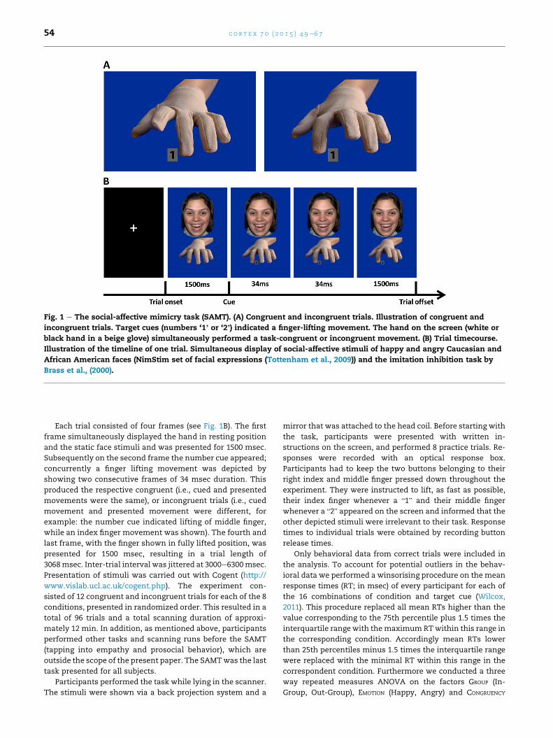
Fig. 1 e The social-affective mimicry task (SAMT). (A) Congruent and incongruent trials. Illustration of congruent and
incongruent trials. Target cues (numbers ‘1’ or ‘2’) indicated a finger-lifting movement. The hand on the screen (white or
black hand in a beige glove) simultaneously performed a task-congruent or incongruent movement. (B) Trial timecourse.
Illustration of the timeline of one trial. Simultaneous display of social-affective stimuli of happy and angry Caucasian and
African American faces (NimStim set of facial expressions (Tottenham et al., 2009)) and the imitation inhibition task by
Brass et al., (2000).
c o r t e x 7 0 ( 2 0 1 5 ) 4 9e6 754
Each trial consisted of four frames (see Fig. 1B). The first
frame simultaneously displayed the hand in resting position
and the static face stimuli and was presented for 1500 msec.
Subsequently on the second frame the number cue appeared;
concurrently a finger lifting movement was depicted by
showing two consecutive frames of 34 msec duration. This
produced the respective congruent (i.e., cued and presented
movements were the same), or incongruent trials (i.e., cued
movement and presented movement were different, for
example: the number cue indicated lifting of middle finger,
while an index finger movement was shown). The fourth and
last frame, with the finger shown in fully lifted position, was
presented for 1500 msec, resulting in a trial length of
3068msec. Inter-trial interval was jittered at 3000e6300msec.
Presentation of stimuli was carried out with Cogent (http://
www.vislab.ucl.ac.uk/cogent.php). The experiment con-
sisted of 12 congruent and incongruent trials for each of the 8
conditions, presented in randomized order. This resulted in a
total of 96 trials and a total scanning duration of approxi-
mately 12 min. In addition, as mentioned above, participants
performed other tasks and scanning runs before the SAMT
(tapping into empathy and prosocial behavior), which are
outside the scope of the present paper. The SAMTwas the last
task presented for all subjects.
Participants performed the task while lying in the scanner.
The stimuli were shown via a back projection system and a
mirror that was attached to the head coil. Before starting with
the task, participants were presented with written in-
structions on the screen, and performed 8 practice trials. Re-
sponses were recorded with an optical response box.
Participants had to keep the two buttons belonging to their
right index and middle finger pressed down throughout the
experiment. They were instructed to lift, as fast as possible,
their index finger whenever a “1” and their middle finger
whenever a “2” appeared on the screen and informed that the
other depicted stimuli were irrelevant to their task. Response
times to individual trials were obtained by recording button
release times.
Only behavioral data from correct trials were included in
the analysis. To account for potential outliers in the behav-
ioral data we performed a winsorising procedure on the mean
response times (RT; in msec) of every participant for each of
the 16 combinations of condition and target cue (Wilcox,
2011). This procedure replaced all mean RTs higher than the
value corresponding to the 75th percentile plus 1.5 times the
interquartile range with themaximum RTwithin this range in
the corresponding condition. Accordingly mean RTs lower
than 25th percentiles minus 1.5 times the interquartile range
were replaced with the minimal RT within this range in the
correspondent condition. Furthermore we conducted a three
way repeated measures ANOVA on the factors GROUP (In-
Group, Out-Group), EMOTION (Happy, Angry) and CONGRUENCY
c o r t e x 7 0 ( 2 0 1 5 ) 4 9e6 7 55
(Congruent, Incongruent). To specifically assess the mimicry
effect we carried out two-way repeated measures ANOVA
with factors GROUP (In-Group, Out-Group) and EMOTION (Happy,
Angry) on the difference between reaction times in incon-
gruent and congruent trials for each condition. In addition, we
explored possible effects of sex/gender by incorporating sex/
gender as an additional variable in these analyses. This was
motivated by the fact that all face stimuli showed females,
hence making it possible that male and female participants
responded differently to them.
2.3. Post-scanning ethnic attitude measures
2.3.1. Threat implicit association task (Threat-IAT)To substantiate the interpretation of our findings, participants
completed a Threat/Security implicit association task (IAT;
Greenwald, McGhee, & Schwartz, 1998) after the scanning
procedure. The IAT is a frequently used measure to assess
implicit ethnic bias (for meta-analytic review see Greenwald,
Poehlman, Uhlmann, & Banaji, 2009). We specifically hy-
pothesized that Black people would be associated with threat
rather than with security, as compared to White people. Thus
we designed an IAT that specifically tested for the strength of
implicit associations between the target concepts of “Blacks”
(ethnic out-group) and “Whites” (in-group) with attributes
describing “Threat” and “Security”. The attributes describing
“Threat” were: threat, fear, violence, danger and attack. The
matched nouns describing “Security” were: security, peace,
shelter, protection and calmness. All terms were presented in
German and matched for word length. The IAT was designed
following the approach by Greenwald et al. (1998) and was
composed of five tasks with separate instructions.
Participants performed the task on a computer running
Adobe Flash Player for stimulus presentation. Across all tasks
they were instructed to keep the letters “E” and “I” on the
keyboard pressed with their index, or middle fingers, to
respond as fast as possible, and to disregard errors, indicated
by a red cross. In the first task pictures of the target-concepts
ofWhites and Blacks had to be sorted to the left and right hand
side, respectively, by lifting the corresponding keys. In the
second task the instructions were to sort the target concepts
of Security to the left, and Threat to the right hand side. The
third task required sorting both the attributes describing Se-
curity and the target concept of Whites to the left, and those
describing Threat and Blacks to the right. The fourth task
reversed themapping of target-conceptsWhites and Blacks to
right, respectively left with regard to the first task. In the fifth
task this reversed mapping of group was combined with the
Threat and Security attributes. Instructions were to sort the
combination of the target-concept of Blacks and the attributes
of Security to the left, respectively Whites and Threat to the
right hand side.
The “D-measure” (for details see Greenwald, Nosek, &
Banaji, 2003) describes the implicit association of Whites
with Security and Blacks with Threat. Firstly the latency
standard deviation (SD) from the combined third and fifth task
was calculated. The “D-measure” was then furthermore
calculated by dividing the difference in reaction times (msec)
of the third and fifth task by this SD. In the case of a stronger
association of Blacks with Threat attributes a positive D-value
is expected.
2.3.2. Attitudes towards Blacks ScaleAfter assessing participants' implicit attitudes using the
Threat/Security IATwe conducted a German translation of the
Attitudes towards Blacks Scale (Brigham, 1993) to assess their
explicit ethnicity bias. The Attitudes towards Blacks Scale
comprises 20 statements concerning the aim of the ques-
tionnaire, which have to be rated from “1” (strongly disagree)
to “7” (strongly agree). Thus a high score indicates highly
positive, whereas low scores reflect highly negative attitudes
towards blacks.
2.4. MRI acquisition
MRI data were acquired using a 3 T Siemens Tim Trio MRI
system (Siemens Medical, Erlangen, Germany) using a 32-
channel head coil for signal reception. Blood oxygen level-
dependent (BOLD) sensitive functional imaging was per-
formed using a multi-band accelerated echoplanar imaging
(EPI) sequence with the following parameters: echo time (TE)/
repetition time (TR) ¼ 33/1800 msec, flip angle 60�, interleavedacquisition, 54 axial slices co-planar the connecting line be-
tween anterior and posterior commissure, FOV
192mm� 192mm� 108mm,matrix size 128� 128, voxel size
1.5 � 1.5 � 2 mm, no interslice gap). Structural images were
acquired after functional scanning using a magnetization-
prepared rapid gradient-echo (MPRAGE) sequence (TE/
TR ¼ 4.21/2300 msec, 160 sagittal slices, voxel
size ¼ 1.0 � 1.0 � 1.1 mm, field of view ¼ 256 mm).
2.5. Analysis of MRI data
MRI data were analyzed using SPM8 (Statistical Parametric
Mapping, http://www.fil.ion.ucl.ac.uk/spm). The first five vol-
umes of each participant's fMRI data were discarded to allow
for T1 equilibration. The time series for each voxel was then
realigned temporally to the acquisition of the first slice in time
to correct for differences in slice time acquisition. The image
time series were spatially realigned using a sinc interpolation
algorithm that estimates rigid body transformations (trans-
lations, rotations) by minimizing head-movements between
each image and the reference image. Subsequently, each
participant's functional image was segmented into gray mat-
ter (GM), white matter (WM), and cerebral spinal fluid (CSF)
using GM, WM, and CSF tissue probability maps provided by
SPM8 and then spatially normalized to the International
Consortium for Brain Mapping (ICBM) space templates (Euro-
pean brains) using both linear and nonlinear transformations.
Finally, the imageswere spatially smoothed using an isotropic
8 mm full-width-at-half-maximum Gaussian kernel.
The fMRI time series were analyzed using an event-related
approach in the context of the General Linear Model (GLM).
Single-subject models consisted of multiple regressors
describing, for each of the 8 conditions resulting from the
2 � 2 � 2 design, the time period from presentation of the
facial stimulus until the time that the stimuli disappeared
from the screen. Duration of a trial was fixed at 3.068 s. Trials
c o r t e x 7 0 ( 2 0 1 5 ) 4 9e6 756
onwhich an incorrect response or no responsewas givenwere
modeled in a separate error regressor.
Each effect was modeled on a trial-by-trial basis as a
concatenation of square-wave functions. Each of these
square-wave functions was then convolved with a canonical
hemodynamic response function, as implemented in SPM8, in
order to generate 9 regressors modeling the main effects
described above.
Head movement effects were accounted for by including
the six rigid-body motion parameters (translation and rota-
tion) as well as two regressors describing signal intensities in
WM and CSF as nuisance covariates.
2.6. Statistical inference
The statistical significance of the estimated evoked hemody-
namic responses was assessed using t-statistics in the context
of general linear model-based analyses, as implemented in
SPM8. We were specifically interested in assessing mimicry
regulation (i.e., Incongruent > Congruent) effects related to
the enhanced behavioral mimicry responses, that is specif-
ically for the conditions Out-group and Happy on brain ac-
tivity during the finger lifting trials. Note therefore that since
the behavioral data analysis showed no interaction effects
between these factors (GROUP � EMOTION), we did not test for
GROUP � EMOTION effects in the fMRI data. Note that inclusion of
the data of the 14 imperfectly logged participants neither
changed the basic pattern of behavioral results, in particular
of the mimicry effect across conditions; nor did this exclusion
essentially alter the results of the imaging analyses. Thus for
the sake of enhanced analytical power we included functional
imaging data of all 41 participants in the fMRI analyses. For
completeness of reporting we also report changes in imaging
results in the sub-sample of 27 participants in the results
section (see section 3.2).
Contrasts of the parameter estimates with respect to
baseline for all trials were calculated, separately for the
different levels of GROUP, EMOTION and CONGRUENCY, with the
intertrial interval being used as an implicit baseline. The
resulting 8 contrasts were entered into a random-effects
second-level analysis using a repeated measures ANOVA, in
order to enable inferences on a population level (Penny &
Holmes, 2004).
In line with the behavioral data analysis, in which we
contrasted reaction times to incongruent and congruent trials,
and based on previous fMRI studies using the imitation inhi-
bition task (Brass et al., 2000; Brass, et al., 2001; Brass, Derrfuss,
Forstmann, et al., 2005; Brass, Derrfuss, & von Cramon, 2005;
Spengler, et al., 2009), we also contrasted incongruent with
congruent trials in our fMRI analysis in order to assess neural
activation related to the regulation of mimicry. To isolate the
parts of this activation specifically related to our main condi-
tions of interest (which were mimicry in response to Happy
faces, or to Out-group faces, respectively), we confined our
analyses to effects that showed stronger activation during
these conditions, as compared to their counterpart. This was
implemented using an implicit masking procedure which
restricted the display of results to those voxels which had
shown higher activation in the condition of interest compared
to its counterpart. Stated explicitly, we tested our effects of
interest (i.e., [Happy Incongruent > Happy Congruent], or
[Out-group Incongruent > Out-group Congruent], and inclu-
sively masked the ensuing effects with the corresponding
interaction effects (i.e., [[Happy Incongruent > Happy
Congruent] > [Angry Incongruent > Angry Congruent]]; or
[[Out-group Incongruent > Out-group Congruent] > [In-group
Incongruent > In-group Congruent]]). In this way we isolated
differential neural responses to incongruent versus congruent
trials that were stronger for happy than angry facial stimuli, or
for out-group than in-group stimuli (and vice versa).
Note that this analysis is not circular (as would be the case
if one analysis was used to assess the significance of a second,
non-independent analysis (Kriegeskorte, Simmons,
Bellgowan, & Baker, 2009)): the masking procedure was part
of a single analysis in which we confined the effects obtained
by our main contrast to effects that were specific for this
condition. Thus, this analysis did not reduce the search vol-
ume of the main effect, and hence left its significance unaf-
fected (which was corrected for multiple comparisons using
cluster-level correction at the whole-brain level). All the
masking approach added to the analysis was to selectively
display only those voxels which were specifically associated
with higher activation in the effect of interest. For complete-
ness and full data reporting, however, we also report themain
effects of all conditions without the masking procedure in the
supplementary material (see Tables S1eS4 in the
supplementary material).
All statistical inferencewas performed using a threshold of
p ¼ .05 corrected for multiple comparisons over the whole
brain, using the Gaussian random fields approach at cluster-
level (Friston, Holmes, Poline, Price, & Frith, 1996) with a
voxel-level intensity threshold of p ¼ .001; the masking
contrast which only served to control for the directionality of
effects but not its significance was thresholded at p ¼ .05 un-
corrected. The SPM Anatomy Toolbox, version v1.8 (Eickhoff
et al., 2005), was used to guide anatomical and probabilistic
cytoarchitectonic localization of the resulting clusters.
To corroborate the functional interpretation of our fMRI
findings, we investigated the relationship between the behav-
ioral and the imaging results by calculating Pearson correla-
tions between the size of the individualmimicry effects and the
corresponding individual fMRI activation. To calculate these
post-hoc brain-behavior correlations, we used the MarsBaR
toolbox v0.43 (http://www.marsbar.sourceforge.net) to create
10 mm spherical functional ROIs centered at peak coordinates
of activation foci related to our contrasts of interest (see Table 1
for details of maximum activations). ROI selection was based
on the maxima of activation clusters yielded by inclusively
masking the [Out-group Incongruent > Out-group Congruent]
contrast with the interaction contrast [[Out-group Incongruent
> Out-group Congruent] > [In-group Incongruent > In-group
Congruent]]. For correlation with the mimicry effect in the out-
group condition ROIs were created in left and right AI; right and
left MCC; and left inferior parietal lobule (IPL) and left IFG.
Respectively ROI selection was based on maxima of activation
clusters revealed by the [Happy Incongruent > Happy
Congruent] contrast inclusively masked with the interaction
contrast [[Happy Incongruent > Happy Congruent] > [Angry
Incongruent > Angry Congruent]]. ROIs for correlation with the
mimicry effect in the happy condition were right superior
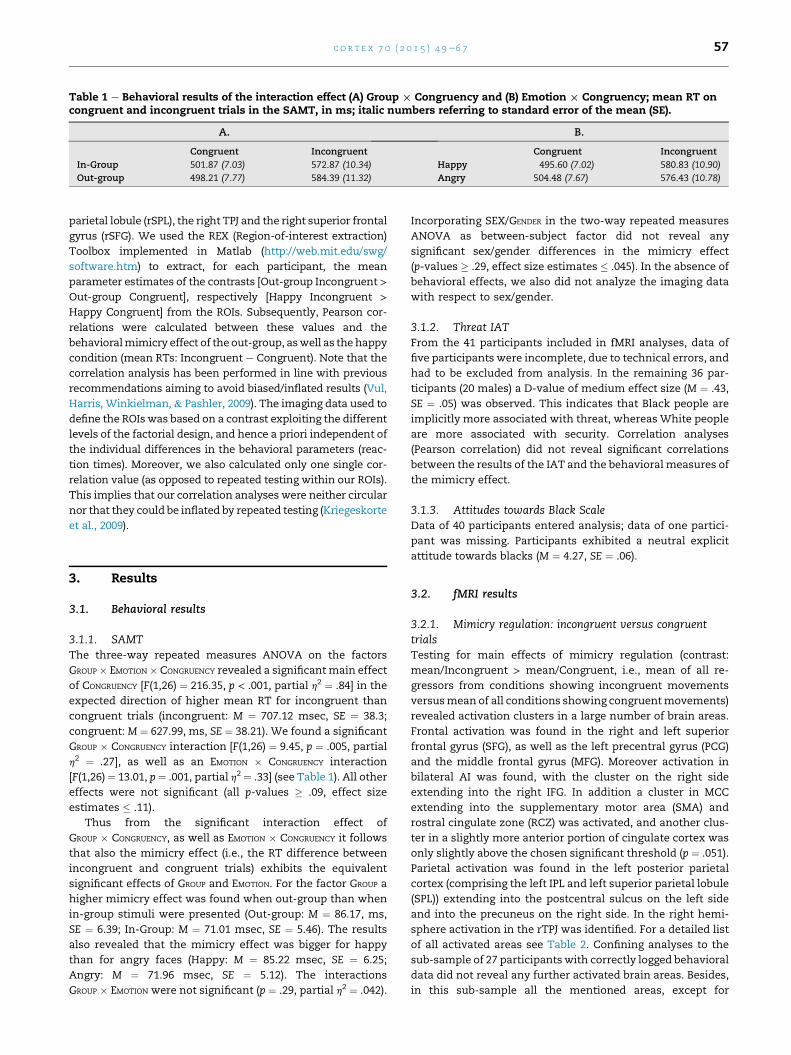
Table 1 e Behavioral results of the interaction effect (A) Group £ Congruency and (B) Emotion £ Congruency; mean RT oncongruent and incongruent trials in the SAMT, in ms; italic numbers referring to standard error of the mean (SE).
A. B.
Congruent Incongruent Congruent Incongruent
In-Group 501.87 (7.03) 572.87 (10.34) Happy 495.60 (7.02) 580.83 (10.90)
Out-group 498.21 (7.77) 584.39 (11.32) Angry 504.48 (7.67) 576.43 (10.78)
c o r t e x 7 0 ( 2 0 1 5 ) 4 9e6 7 57
parietal lobule (rSPL), the right TPJ and the right superior frontal
gyrus (rSFG). We used the REX (Region-of-interest extraction)
Toolbox implemented in Matlab (http://web.mit.edu/swg/
software.htm) to extract, for each participant, the mean
parameter estimates of the contrasts [Out-group Incongruent >Out-group Congruent], respectively [Happy Incongruent >Happy Congruent] from the ROIs. Subsequently, Pearson cor-
relations were calculated between these values and the
behavioralmimicry effect of the out-group, aswell as the happy
condition (mean RTs: Incongruent e Congruent). Note that the
correlation analysis has been performed in line with previous
recommendations aiming to avoid biased/inflated results (Vul,
Harris, Winkielman, & Pashler, 2009). The imaging data used to
define the ROIswas based on a contrast exploiting the different
levels of the factorial design, and hence a priori independent of
the individual differences in the behavioral parameters (reac-
tion times). Moreover, we also calculated only one single cor-
relation value (as opposed to repeated testing within our ROIs).
This implies that our correlation analyses were neither circular
nor that they could be inflated by repeated testing (Kriegeskorte
et al., 2009).
3. Results
3.1. Behavioral results
3.1.1. SAMTThe three-way repeated measures ANOVA on the factors
GROUP � EMOTION � CONGRUENCY revealed a significantmain effect
of CONGRUENCY [F(1,26) ¼ 216.35, p < .001, partial h2 ¼ .84] in the
expected direction of higher mean RT for incongruent than
congruent trials (incongruent: M ¼ 707.12 msec, SE ¼ 38.3;
congruent: M ¼ 627.99, ms, SE ¼ 38.21). We found a significant
GROUP � CONGRUENCY interaction [F(1,26) ¼ 9.45, p ¼ .005, partial
h2 ¼ .27], as well as an EMOTION � CONGRUENCY interaction
[F(1,26)¼ 13.01, p¼ .001, partial h2 ¼ .33] (see Table 1). All other
effects were not significant (all p-values � .09, effect size
estimates � .11).
Thus from the significant interaction effect of
GROUP � CONGRUENCY, as well as EMOTION � CONGRUENCY it follows
that also the mimicry effect (i.e., the RT difference between
incongruent and congruent trials) exhibits the equivalent
significant effects of GROUP and EMOTION. For the factor GROUP a
higher mimicry effect was found when out-group than when
in-group stimuli were presented (Out-group: M ¼ 86.17, ms,
SE ¼ 6.39; In-Group: M ¼ 71.01 msec, SE ¼ 5.46). The results
also revealed that the mimicry effect was bigger for happy
than for angry faces (Happy: M ¼ 85.22 msec, SE ¼ 6.25;
Angry: M ¼ 71.96 msec, SE ¼ 5.12). The interactions
GROUP � EMOTION were not significant (p ¼ .29, partial h2 ¼ .042).
Incorporating SEX/GENDER in the two-way repeated measures
ANOVA as between-subject factor did not reveal any
significant sex/gender differences in the mimicry effect
(p-values � .29, effect size estimates � .045). In the absence of
behavioral effects, we also did not analyze the imaging data
with respect to sex/gender.
3.1.2. Threat IATFrom the 41 participants included in fMRI analyses, data of
five participants were incomplete, due to technical errors, and
had to be excluded from analysis. In the remaining 36 par-
ticipants (20 males) a D-value of medium effect size (M ¼ .43,
SE ¼ .05) was observed. This indicates that Black people are
implicitly more associated with threat, whereas White people
are more associated with security. Correlation analyses
(Pearson correlation) did not reveal significant correlations
between the results of the IAT and the behavioral measures of
the mimicry effect.
3.1.3. Attitudes towards Black ScaleData of 40 participants entered analysis; data of one partici-
pant was missing. Participants exhibited a neutral explicit
attitude towards blacks (M ¼ 4.27, SE ¼ .06).
3.2. fMRI results
3.2.1. Mimicry regulation: incongruent versus congruenttrialsTesting for main effects of mimicry regulation (contrast:
mean/Incongruent > mean/Congruent, i.e., mean of all re-
gressors from conditions showing incongruent movements
versusmean of all conditions showing congruentmovements)
revealed activation clusters in a large number of brain areas.
Frontal activation was found in the right and left superior
frontal gyrus (SFG), as well as the left precentral gyrus (PCG)
and the middle frontal gyrus (MFG). Moreover activation in
bilateral AI was found, with the cluster on the right side
extending into the right IFG. In addition a cluster in MCC
extending into the supplementary motor area (SMA) and
rostral cingulate zone (RCZ) was activated, and another clus-
ter in a slightly more anterior portion of cingulate cortex was
only slightly above the chosen significant threshold (p ¼ .051).
Parietal activation was found in the left posterior parietal
cortex (comprising the left IPL and left superior parietal lobule
(SPL)) extending into the postcentral sulcus on the left side
and into the precuneus on the right side. In the right hemi-
sphere activation in the rTPJ was identified. For a detailed list
of all activated areas see Table 2. Confining analyses to the
sub-sample of 27 participants with correctly logged behavioral
data did not reveal any further activated brain areas. Besides,
in this sub-sample all the mentioned areas, except for
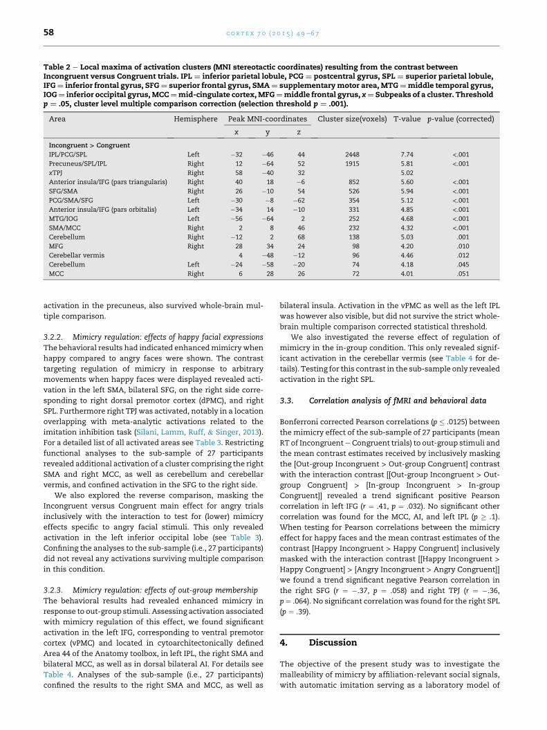
Table 2 e Local maxima of activation clusters (MNI stereotactic coordinates) resulting from the contrast betweenIncongruent versus Congruent trials. IPL ¼ inferior parietal lobule, PCG ¼ postcentral gyrus, SPL ¼ superior parietal lobule,IFG¼ inferior frontal gyrus, SFG¼ superior frontal gyrus, SMA¼ supplementarymotor area, MTG¼middle temporal gyrus,IOG¼ inferior occipital gyrus,MCC¼mid-cingulate cortex,MFG¼middle frontal gyrus, x¼ Subpeaks of a cluster. Thresholdp ¼ .05, cluster level multiple comparison correction (selection threshold p ¼ .001).
Area Hemisphere Peak MNI-coordinates Cluster size(voxels) T-value p-value (corrected)
x y z
Incongruent > Congruent
IPL/PCG/SPL Left �32 �46 44 2448 7.74 <.001Precuneus/SPL/IPL Right 12 �64 52 1915 5.81 <.001xTPJ Right 58 �40 32 5.02
Anterior insula/IFG (pars triangularis) Right 40 18 �6 852 5.60 <.001SFG/SMA Right 26 �10 54 526 5.94 <.001PCG/SMA/SFG Left �30 �8 �62 354 5.12 <.001Anterior insula/IFG (pars orbitalis) Left �34 14 �10 331 4.85 <.001MTG/IOG Left �56 �64 2 252 4.68 <.001SMA/MCC Right 2 8 46 232 4.32 <.001Cerebellum Right �12 2 68 138 5.03 .001
MFG Right 28 34 24 98 4.20 .010
Cerebellar vermis 4 �48 �12 96 4.46 .012
Cerebellum Left �24 �58 �20 74 4.18 .045
MCC Right 6 28 26 72 4.01 .051
c o r t e x 7 0 ( 2 0 1 5 ) 4 9e6 758
activation in the precuneus, also survived whole-brain mul-
tiple comparison.
3.2.2. Mimicry regulation: effects of happy facial expressionsThe behavioral results had indicated enhancedmimicrywhen
happy compared to angry faces were shown. The contrast
targeting regulation of mimicry in response to arbitrary
movements when happy faces were displayed revealed acti-
vation in the left SMA, bilateral SFG, on the right side corre-
sponding to right dorsal premotor cortex (dPMC), and right
SPL. Furthermore right TPJ was activated, notably in a location
overlapping with meta-analytic activations related to the
imitation inhibition task (Silani, Lamm, Ruff, & Singer, 2013).
For a detailed list of all activated areas see Table 3. Restricting
functional analyses to the sub-sample of 27 participants
revealed additional activation of a cluster comprising the right
SMA and right MCC, as well as cerebellum and cerebellar
vermis, and confined activation in the SFG to the right side.
We also explored the reverse comparison, masking the
Incongruent versus Congruent main effect for angry trials
inclusively with the interaction to test for (lower) mimicry
effects specific to angry facial stimuli. This only revealed
activation in the left inferior occipital lobe (see Table 3).
Confining the analyses to the sub-sample (i.e., 27 participants)
did not reveal any activations surviving multiple comparison
in this condition.
3.2.3. Mimicry regulation: effects of out-group membershipThe behavioral results had revealed enhanced mimicry in
response to out-group stimuli. Assessing activation associated
with mimicry regulation of this effect, we found significant
activation in the left IFG, corresponding to ventral premotor
cortex (vPMC) and located in cytoarchitectonically defined
Area 44 of the Anatomy toolbox, in left IPL, the right SMA and
bilateral MCC, as well as in dorsal bilateral AI. For details see
Table 4. Analyses of the sub-sample (i.e., 27 participants)
confined the results to the right SMA and MCC, as well as
bilateral insula. Activation in the vPMC as well as the left IPL
was however also visible, but did not survive the strict whole-
brain multiple comparison corrected statistical threshold.
We also investigated the reverse effect of regulation of
mimicry in the in-group condition. This only revealed signif-
icant activation in the cerebellar vermis (see Table 4 for de-
tails). Testing for this contrast in the sub-sample only revealed
activation in the right SPL.
3.3. Correlation analysis of fMRI and behavioral data
Bonferroni corrected Pearson correlations (p � .0125) between
the mimicry effect of the sub-sample of 27 participants (mean
RT of IncongruenteCongruent trials) to out-group stimuli and
the mean contrast estimates received by inclusively masking
the [Out-group Incongruent > Out-group Congruent] contrast
with the interaction contrast [[Out-group Incongruent > Out-
group Congruent] > [In-group Incongruent > In-group
Congruent]] revealed a trend significant positive Pearson
correlation in left IFG (r ¼ .41, p ¼ .032). No significant other
correlation was found for the MCC, AI, and left IPL (p � .1).
When testing for Pearson correlations between the mimicry
effect for happy faces and the mean contrast estimates of the
contrast [Happy Incongruent > Happy Congruent] inclusively
masked with the interaction contrast [[Happy Incongruent >Happy Congruent] > [Angry Incongruent > Angry Congruent]]
we found a trend significant negative Pearson correlation in
the right SFG (r ¼ �.37, p ¼ .058) and right TPJ (r ¼ �.36,
p¼ .064). No significant correlationwas found for the right SPL
(p ¼ .39).
4. Discussion
The objective of the present study was to investigate the
malleability of mimicry by affiliation-relevant social signals,
with automatic imitation serving as a laboratory model of
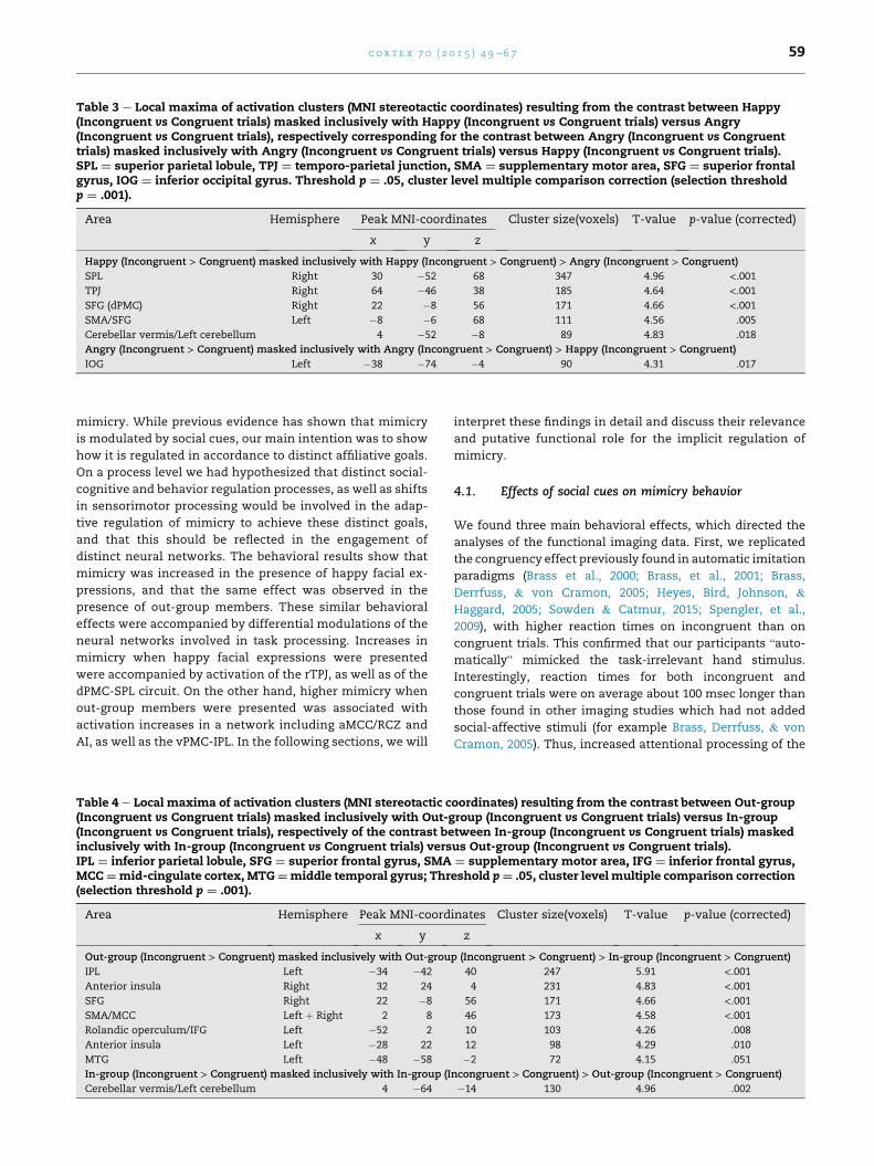
Table 3 e Local maxima of activation clusters (MNI stereotactic coordinates) resulting from the contrast between Happy(Incongruent vs Congruent trials) masked inclusively with Happy (Incongruent vs Congruent trials) versus Angry(Incongruent vs Congruent trials), respectively corresponding for the contrast between Angry (Incongruent vs Congruenttrials) masked inclusively with Angry (Incongruent vs Congruent trials) versus Happy (Incongruent vs Congruent trials).SPL ¼ superior parietal lobule, TPJ ¼ temporo-parietal junction, SMA ¼ supplementary motor area, SFG ¼ superior frontalgyrus, IOG ¼ inferior occipital gyrus. Threshold p ¼ .05, cluster level multiple comparison correction (selection thresholdp ¼ .001).
Area Hemisphere Peak MNI-coordinates Cluster size(voxels) T-value p-value (corrected)
x y z
Happy (Incongruent > Congruent) masked inclusively with Happy (Incongruent > Congruent) > Angry (Incongruent > Congruent)
SPL Right 30 �52 68 347 4.96 <.001TPJ Right 64 �46 38 185 4.64 <.001SFG (dPMC) Right 22 �8 56 171 4.66 <.001SMA/SFG Left �8 �6 68 111 4.56 .005
Cerebellar vermis/Left cerebellum 4 �52 �8 89 4.83 .018
Angry (Incongruent > Congruent) masked inclusively with Angry (Incongruent > Congruent) > Happy (Incongruent > Congruent)
IOG Left �38 �74 �4 90 4.31 .017
c o r t e x 7 0 ( 2 0 1 5 ) 4 9e6 7 59
mimicry. While previous evidence has shown that mimicry
is modulated by social cues, our main intention was to show
how it is regulated in accordance to distinct affiliative goals.
On a process level we had hypothesized that distinct social-
cognitive and behavior regulation processes, as well as shifts
in sensorimotor processing would be involved in the adap-
tive regulation of mimicry to achieve these distinct goals,
and that this should be reflected in the engagement of
distinct neural networks. The behavioral results show that
mimicry was increased in the presence of happy facial ex-
pressions, and that the same effect was observed in the
presence of out-group members. These similar behavioral
effects were accompanied by differential modulations of the
neural networks involved in task processing. Increases in
mimicry when happy facial expressions were presented
were accompanied by activation of the rTPJ, as well as of the
dPMC-SPL circuit. On the other hand, higher mimicry when
out-group members were presented was associated with
activation increases in a network including aMCC/RCZ and
AI, as well as the vPMC-IPL. In the following sections, we will
Table 4 e Local maxima of activation clusters (MNI stereotactic c(Incongruent vs Congruent trials) masked inclusively with Out-g(Incongruent vs Congruent trials), respectively of the contrast beinclusively with In-group (Incongruent vs Congruent trials) versIPL ¼ inferior parietal lobule, SFG ¼ superior frontal gyrus, SMAMCC¼mid-cingulate cortex, MTG¼middle temporal gyrus; Thr(selection threshold p ¼ .001).
Area Hemisphere Peak MNI-coord
x y
Out-group (Incongruent > Congruent) masked inclusively with Out-grou
IPL Left �34 �42
Anterior insula Right 32 24
SFG Right 22 �8
SMA/MCC Left þ Right 2 8
Rolandic operculum/IFG Left �52 2
Anterior insula Left �28 22
MTG Left �48 �58
In-group (Incongruent > Congruent) masked inclusively with In-group (I
Cerebellar vermis/Left cerebellum 4 �64
interpret these findings in detail and discuss their relevance
and putative functional role for the implicit regulation of
mimicry.
4.1. Effects of social cues on mimicry behavior
We found three main behavioral effects, which directed the
analyses of the functional imaging data. First, we replicated
the congruency effect previously found in automatic imitation
paradigms (Brass et al., 2000; Brass, et al., 2001; Brass,
Derrfuss, & von Cramon, 2005; Heyes, Bird, Johnson, &
Haggard, 2005; Sowden & Catmur, 2015; Spengler, et al.,
2009), with higher reaction times on incongruent than on
congruent trials. This confirmed that our participants “auto-
matically” mimicked the task-irrelevant hand stimulus.
Interestingly, reaction times for both incongruent and
congruent trials were on average about 100 msec longer than
those found in other imaging studies which had not added
social-affective stimuli (for example Brass, Derrfuss, & von
Cramon, 2005). Thus, increased attentional processing of the
oordinates) resulting from the contrast between Out-grouproup (Incongruent vs Congruent trials) versus In-grouptween In-group (Incongruent vs Congruent trials) maskedus Out-group (Incongruent vs Congruent trials).¼ supplementary motor area, IFG ¼ inferior frontal gyrus,eshold p¼ .05, cluster level multiple comparison correction
inates Cluster size(voxels) T-value p-value (corrected)
z
p (Incongruent > Congruent) > In-group (Incongruent > Congruent)
40 247 5.91 <.0014 231 4.83 <.001
56 171 4.66 <.00146 173 4.58 <.00110 103 4.26 .008
12 98 4.29 .010
�2 72 4.15 .051
ncongruent > Congruent) > Out-group (Incongruent > Congruent)
�14 130 4.96 .002

c o r t e x 7 0 ( 2 0 1 5 ) 4 9e6 760
facial stimuli might have led to a general increase in reaction
times. Notably, the magnitude of the mimicry effect is com-
parable to the one found in another fMRI studywhich had also
implied contextual manipulations (Klapper et al. (2014), as
well as to behavioral studies using this paradigmwith a social
stress manipulation (Tomova, von Dawans, Heinrichs, Silani,
& Lamm, 2014), and priming with negatively valenced pic-
tures (Grecucci et al., 2011). Thus, it seems that an added social
and/or emotional context leads to generally enhanced mim-
icry effects.
Second, we found higher mimicry of task-irrelevant
movements when confronted with happy faces. This is in
line with the hypothesis that mimicry reflects the implicit
reciprocation of affiliative intent. Similar results have been
found in a study by Bourgeois and Hess (2008), showing that
implicit facial mimicry was similarly enhanced for happy
faces. Notably, this effect was observed for in-group and out-
group faces alike, which has been interpreted in the sense that
the affiliative intent conveyed by happy facial expressions is
reciprocated irrespective of group-membership (Bourgeois &
Hess, 2008; Van Kleef, et al., 2004, 2010). Our findings are in
line with this interpretation (as we did not observe an inter-
action of emotion and groupmembership). They however also
crucially extend it to mimicry of movements that carry no
intrinsic affective information e as we studied mimicry of
arbitrary finger lifting movements, and not of emotion dis-
plays via facial mimicry themselves.
Third, we found enhanced mimicry when out-group faces
were presented. As suggested by evidence in social psychol-
ogy, mimicry might represent an affiliative signal (e.g.,
Chartrand& Bargh, 1999; Chartrand& Lakin, 2013; Lakin et al.,
2003). Also, accounts from behavioral biology suggest that
affiliative displays (“such as kissing and embracing” (de Waal,
2003)) of non-human primates could represent a flexible and
context-dependent behavioral means to de-escalate a socially
threatening situation e as an alternative to engage in a fight
with an aggressor, or to withdraw from him or her (de Waal,
1986, 2003; Keltner et al., 1997). Affiliation signals might thus
have two functions: to strengthen social bonds, and to soothe
a potential conflict. Increased mimicry in response to out-
group faces might, therefore, reflect a regulative response to
avert potential threat. This interpretation is supported by the
results of the Threat IAT, which showed that Blacks are
implicitly more strongly associated with the concept of threat
e a finding that has in a similar fashion also been reported
elsewhere (Amodio, 2004, 2008; Amodio et al., 2008; Amodio
et al., 2004; Amodio et al., 2006; Neuberg & Cottrell, 2008).
Interestingly, the present results suggest that out-group faces
might generally elicit an implicit threat response, indepen-
dently of the emotions they are showing (as, somewhat un-
expectedly, we did not find an interaction effect of emotion
and groupmembershipe implying that being confrontedwith
angry out-group members did not result in even higher
mimicry). We therefore suggest that mimicry, as an affiliative
signal, is, in the case of presentation of out-group members,
enhanced to reach the goal of appeasement. Grecucci et al.
(2011) found enhanced mimicry to negatively valenced pic-
tures, which they interpreted as being related to a fight-or
flight response. Our results extend those of Grecucci et al.
(2011), by suggesting that the implicit perception of threat
(which carries negative valence) might, depending on the so-
cial context, also lead to approach behavior in the form of
appeasement. In a series of three previous behavioral exper-
iments involvingmore than 180 participants, in whichwe also
used the SAMT, we consistently found enhanced mimicry in
response to angry out-group faces (Rauchbauer et al., 2014,
2015). Unexpectedly, as mentioned before, we did not repli-
cate these results with regard to the interaction of emotion
and group-membership (i.e., angry out-groupmembers) in the
present study. Thismight be due to the different experimental
setting (lying supine in a dark, loud scanner environment),
which might have enhanced the focus on the more salient
happy and out-group stimuli. Results of the behavioral studies
consistently showed that mimicry's malleability by social-
affective cues was based on modulation of RTs on congruent
trials, as also found by Grecucci et al., 2011. Although it might
be argued that the enhanced RTs on negatively valenced
congruent trials reflect a general enhanced motor respon-
siveness due to higher arousal, this interpretation seems
implausible. First, enhanced responsiveness to task-irrelevant
motor cues due to negatively valenced stimuli (angry out-
group faces) should have also been reflected in a speeding of
RTs in incongruent trials, which was not the case. Moreover,
no increase in RTs in response to baseline trials (trials without
movement of the other's hand) was observed in our behavioral
study, where such trials had been included. Second, results
from a control experiment included in the latter study, in
which we had assessed effects of our social-affiliative stimuli
on responses in a (non-imitative) Simon task, showed that
negatively valenced stimuli did not affect responses on
congruent trials in this task in the sameway as in themimicry
task. Therefore, the modulation of mimicry behavior by out-
group faces on congruent trials most likely reflects a specific
effect of social context on mimicry behavior, and we propose
that this effect is related to appeasement of a potentially
threatening interaction partner.
In sum, the behavioral results confirm that mimicry was
affected by our social-affective manipulation. Yet, the reac-
tion timemeasures alone are non-informative with respect to
the processes underlying these modulations of mimicry.
Thus, in the next sections we will discuss how different
cognitive processes, and their reflection in distinct neural
activations, might explain the behavioral effects.
4.2. Imaging effects related to regulation of mimicry
4.2.1. Incongruent versus congruent trials (generalizedregulation of mimicry)By contrasting incongruent and congruent trials we aimed to
identify the neural activations specific to the regulation of
mimicry, that is, the activation underlying the increased need
to regulate mimicry if the hand stimulus moved differently
versus similarly with respect to the required movement.
Contrasting activation during incongruent and congruent tri-
als revealed a broad network of brain areas. We observed
activation in the right TPJ, the AI andMCC, aswell as in fronto-
parietal areas involved in sensorimotor control and action
observation (see Table 2).
Involvement of right TPJ has repeatedly been found using
the imitation inhibition task (Brass, Derrfuss, Forstmann,
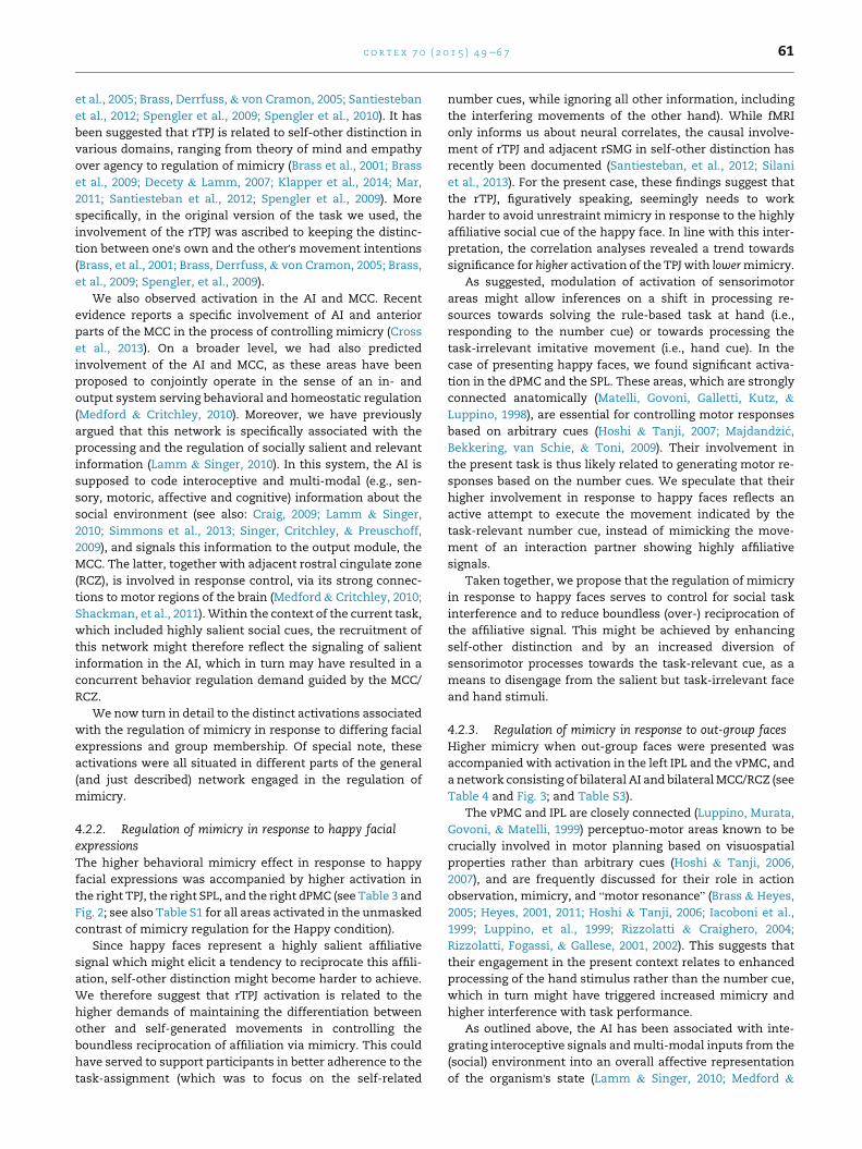
c o r t e x 7 0 ( 2 0 1 5 ) 4 9e6 7 61
et al., 2005; Brass, Derrfuss,& von Cramon, 2005; Santiesteban
et al., 2012; Spengler et al., 2009; Spengler et al., 2010). It has
been suggested that rTPJ is related to self-other distinction in
various domains, ranging from theory of mind and empathy
over agency to regulation of mimicry (Brass et al., 2001; Brass
et al., 2009; Decety & Lamm, 2007; Klapper et al., 2014; Mar,
2011; Santiesteban et al., 2012; Spengler et al., 2009). More
specifically, in the original version of the task we used, the
involvement of the rTPJ was ascribed to keeping the distinc-
tion between one's own and the other's movement intentions
(Brass, et al., 2001; Brass, Derrfuss,& von Cramon, 2005; Brass,
et al., 2009; Spengler, et al., 2009).
We also observed activation in the AI and MCC. Recent
evidence reports a specific involvement of AI and anterior
parts of the MCC in the process of controlling mimicry (Cross
et al., 2013). On a broader level, we had also predicted
involvement of the AI and MCC, as these areas have been
proposed to conjointly operate in the sense of an in- and
output system serving behavioral and homeostatic regulation
(Medford & Critchley, 2010). Moreover, we have previously
argued that this network is specifically associated with the
processing and the regulation of socially salient and relevant
information (Lamm & Singer, 2010). In this system, the AI is
supposed to code interoceptive and multi-modal (e.g., sen-
sory, motoric, affective and cognitive) information about the
social environment (see also: Craig, 2009; Lamm & Singer,
2010; Simmons et al., 2013; Singer, Critchley, & Preuschoff,
2009), and signals this information to the output module, the
MCC. The latter, together with adjacent rostral cingulate zone
(RCZ), is involved in response control, via its strong connec-
tions to motor regions of the brain (Medford & Critchley, 2010;
Shackman, et al., 2011). Within the context of the current task,
which included highly salient social cues, the recruitment of
this network might therefore reflect the signaling of salient
information in the AI, which in turn may have resulted in a
concurrent behavior regulation demand guided by the MCC/
RCZ.
We now turn in detail to the distinct activations associated
with the regulation of mimicry in response to differing facial
expressions and group membership. Of special note, these
activations were all situated in different parts of the general
(and just described) network engaged in the regulation of
mimicry.
4.2.2. Regulation of mimicry in response to happy facialexpressionsThe higher behavioral mimicry effect in response to happy
facial expressions was accompanied by higher activation in
the right TPJ, the right SPL, and the right dPMC (see Table 3 and
Fig. 2; see also Table S1 for all areas activated in the unmasked
contrast of mimicry regulation for the Happy condition).
Since happy faces represent a highly salient affiliative
signal which might elicit a tendency to reciprocate this affili-
ation, self-other distinction might become harder to achieve.
We therefore suggest that rTPJ activation is related to the
higher demands of maintaining the differentiation between
other and self-generated movements in controlling the
boundless reciprocation of affiliation via mimicry. This could
have served to support participants in better adherence to the
task-assignment (which was to focus on the self-related
number cues, while ignoring all other information, including
the interfering movements of the other hand). While fMRI
only informs us about neural correlates, the causal involve-
ment of rTPJ and adjacent rSMG in self-other distinction has
recently been documented (Santiesteban, et al., 2012; Silani
et al., 2013). For the present case, these findings suggest that
the rTPJ, figuratively speaking, seemingly needs to work
harder to avoid unrestraint mimicry in response to the highly
affiliative social cue of the happy face. In line with this inter-
pretation, the correlation analyses revealed a trend towards
significance for higher activation of the TPJ with lowermimicry.
As suggested, modulation of activation of sensorimotor
areas might allow inferences on a shift in processing re-
sources towards solving the rule-based task at hand (i.e.,
responding to the number cue) or towards processing the
task-irrelevant imitative movement (i.e., hand cue). In the
case of presenting happy faces, we found significant activa-
tion in the dPMC and the SPL. These areas, which are strongly
connected anatomically (Matelli, Govoni, Galletti, Kutz, &
Luppino, 1998), are essential for controlling motor responses
based on arbitrary cues (Hoshi & Tanji, 2007; Majdand�zi�c,
Bekkering, van Schie, & Toni, 2009). Their involvement in
the present task is thus likely related to generating motor re-
sponses based on the number cues. We speculate that their
higher involvement in response to happy faces reflects an
active attempt to execute the movement indicated by the
task-relevant number cue, instead of mimicking the move-
ment of an interaction partner showing highly affiliative
signals.
Taken together, we propose that the regulation of mimicry
in response to happy faces serves to control for social task
interference and to reduce boundless (over-) reciprocation of
the affiliative signal. This might be achieved by enhancing
self-other distinction and by an increased diversion of
sensorimotor processes towards the task-relevant cue, as a
means to disengage from the salient but task-irrelevant face
and hand stimuli.
4.2.3. Regulation of mimicry in response to out-group facesHigher mimicry when out-group faces were presented was
accompanied with activation in the left IPL and the vPMC, and
a network consisting of bilateral AI and bilateralMCC/RCZ (see
Table 4 and Fig. 3; and Table S3).
The vPMC and IPL are closely connected (Luppino, Murata,
Govoni, & Matelli, 1999) perceptuo-motor areas known to be
crucially involved in motor planning based on visuospatial
properties rather than arbitrary cues (Hoshi & Tanji, 2006,
2007), and are frequently discussed for their role in action
observation, mimicry, and “motor resonance” (Brass & Heyes,
2005; Heyes, 2001, 2011; Hoshi & Tanji, 2006; Iacoboni et al.,
1999; Luppino, et al., 1999; Rizzolatti & Craighero, 2004;
Rizzolatti, Fogassi, & Gallese, 2001, 2002). This suggests that
their engagement in the present context relates to enhanced
processing of the hand stimulus rather than the number cue,
which in turn might have triggered increased mimicry and
higher interference with task performance.
As outlined above, the AI has been associated with inte-
grating interoceptive signals andmulti-modal inputs from the
(social) environment into an overall affective representation
of the organism's state (Lamm & Singer, 2010; Medford &
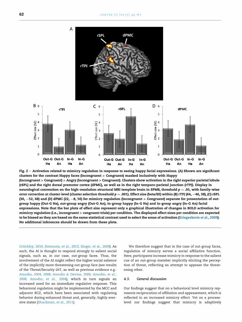
Fig. 2 e Activation related to mimicry regulation in response to seeing happy facial expressions. (A) Shown are significant
clusters for the contrast Happy faces (Incongruent > Congruent) masked inclusively with Happy
(Incongruent > Congruent) > Angry (Incongruent > Congruent). Clusters show activation in the right superior parietal lobule
(rSPL) and the right dorsal premotor cortex (dPMC), as well as in the right temporo-parietal junction (rTPJ). Display in
neurological convention on the high-resolution structural MRI template brain in SPM8, threshold p ¼ .05, with family-wise
error correction at cluster-level (cluster selection threshold p¼ .001). Effect size (beta/SE) within (B) rTPJ (64,¡46, 38), (C) rSPL
(30, ¡52, 68) and (D) dPMC (22, ¡8, 56) for mimicry regulation (Incongruent > Congruent) separate for presentation of out-
group happy (Out-G Ha), out-group angry (Out-G An), in-group happy (In-G Ha) and in-group angry (In-G An) facial
expressions. Note that the bar plots of effect size represent only a graphical illustration of changes in BOLD activation for
mimicry regulation (i.e., incongruent > congruent trials) per condition. The displayed effect sizes per condition are expected
to be biased as they are based on the same statistical contrast used to select the areas of activation (Kriegeskorte et al., 2009).
No additional inferences should be drawn from these plots.
c o r t e x 7 0 ( 2 0 1 5 ) 4 9e6 762
Critchley, 2010; Simmons, et al., 2013; Singer, et al., 2009). As
such, the AI is thought to respond strongly to salient social
signals, such as, in our case, out-group faces. Thus, the
involvement of the AI might reflect the higher social salience
of the implicitly more threatening out-group face (see results
of the Threat/Security IAT, as well as previous evidence e.g.:
Amodio, 2004, 2008; Amodio & Devine, 2006; Amodio, et al.,
2008; Amodio, et al., 2004), which in turn signals an
increased need for an immediate regulative response. This
behavioral regulation might be implemented by the MCC and
adjacent RCZ, which have been associated with regulating
behavior during enhanced threat and, generally, highly aver-
sive states (Shackman, et al., 2011).
We therefore suggest that in the case of out-group faces,
regulation of mimicry serves a social affiliative function.
Here, participants increasemimicry in response to the salient
cue of an out-group member implicitly eliciting the percep-
tion of threat, reflecting an attempt to appease the threat-
ening other.
4.3. General discussion
Our findings suggest that on a behavioral level mimicry rep-
resents reciprocation of affiliation and appeasement, which is
reflected in an increased mimicry effect. Yet on a process-
level our findings suggest that mimicry is adaptively
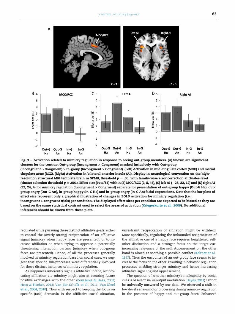
Fig. 3 e Activation related to mimicry regulation in response to seeing out-group members. (A) Shown are significant
clusters for the contrast Out-group (Incongruent > Congruent) masked inclusively with Out-group
(Incongruent > Congruent) > In-group (Incongruent > Congruent). (Left) Activation in mid-cingulate cortex (MCC) and rostral
cingulate zone (RCZ). (Right) Activation in bilateral anterior insula (AI). Display in neurological convention on the high-
resolution structural MRI template brain in SPM8, threshold p ¼ .05, with family-wise error correction at cluster-level
(cluster selection threshold p ¼ .001). Effect size (beta/SE) within (B) MCC/RCZ (2, 8, 46), (C) left AI (¡28, 22, 12) and (D) right AI
(32, 24, 4) for mimicry regulation (Incongruent > Congruent) separate for presentation of out-group happy (Out-G Ha), out-
group angry (Out-G An), in-group happy (In-G Ha) and in-group angry (In-G An) facial expressions. Note that the bar plots of
effect size represent only a graphical illustration of changes in BOLD activation for mimicry regulation (i.e.,
incongruent > congruent trials) per condition. The displayed effect sizes per condition are expected to be biased as they are
based on the same statistical contrast used to select the areas of activation (Kriegeskorte et al., 2009). No additional
inferences should be drawn from these plots.
c o r t e x 7 0 ( 2 0 1 5 ) 4 9e6 7 63
regulated while pursuing these distinct affiliative goals: either
to control the (overly strong) reciprocation of an affiliative
signal (mimicry when happy faces are presented), or to in-
crease affiliation as when trying to appease a potentially
threatening interaction partner (mimicry when out-group
faces are presented). Hence, of all the processes generally
involved in mimicry regulation based on social cues, we sug-
gest that specific sub-processes were differentially involved
for these distinct instances of mimicry regulation.
As happiness inherently signals affiliative intent, recipro-
cating affiliation via mimicry might aim at securing future
positive exchanges with the other (Bourgeois & Hess, 2008;
Hess & Fischer, 2013; Van der Schalk et al., 2011; Van Kleef
et al., 2004, 2010). Thus with respect to keeping the focus on
specific (task) demands in the affiliative social situation,
unrestraint reciprocation of affiliation might be withheld.
More specifically, regulating the unbounded reciprocation of
the affiliative cue of a happy face requires heightened self-
other distinction and a stronger focus on the target cue,
increasing relevance of the self. Appeasement on the other
hand is aimed at soothing a possible conflict (Keltner et al.,
1997). Thus the encounter of an out-group face seems to in-
crease the focus on the other, resulting in behavior regulation
processes enabling stronger mimicry and hence increasing
affiliative signaling and appeasement.
The question of whether mimicry's malleability by social
cues is based on in- or outputmodulation (Heyes, 2011) cannot
be univocally answered by our data. We observed a shift in
low-level sensorimotor processing during mimicry regulation
in the presence of happy and out-group faces. Enhanced
c o r t e x 7 0 ( 2 0 1 5 ) 4 9e6 764
mimicry when happy faces were presented resulted in acti-
vation in the right SPL and dPMC. This might suggest that in
this condition, sensorimotor processing was shifted towards
processing the response to the task-relevant number cue for
the sake of task-adherence. On the other hand, when out-
group faces were presented, sensorimotor contributions in
the vPMC/left IPL network were increased. This suggests that
regulation of mimicry in this condition was accompanied by a
shift of sensorimotor processing towards visuospatial prop-
erties of the task-irrelevant hand. These observed shifts in
sensorimotor processing might suggest that the regulation of
mimicry by social-affective cues is driven by input modula-
tion. On the other hand, we also observed changes in activa-
tion of the rTPJ, reflecting self-other distinction processes that
have been related to output modulation of the mimicry
response (Heyes, 2011). In line with this, the involvement of
the AI/MCC network for behavior regulation according to
detected saliency (in our case threat) in the environment
(Medford& Critchley, 2010; Shackman et al., 2011) could speak
for output modulation regulating mimicry behavior goal-
directedly. Thus, based on the current data alone, we cannot
fully discern the involvement of in- or output modulation in
the modulation of mimicry to social-affective cues, and our
interpretations remain speculative. Further studies should
aim to further clarify the pathways by which mimicry is
regulated in response to social context stimuli. A more time-
sensitive measure, as event-related brain potential (ERP)
studies, might be able to tap into the temporal dynamics of
this modulation and thereby possibly capture processes
attributable to in- or output modulation.
We suggest that along sensorimotor mechanisms, the
processes of self-other distinction and behavioral regulation
are involved in the modulation of mimicry to social-affective
information in general. Yet, we did not replicate previous
findings regarding the involvement of mPFC in the modula-
tion of mimicry by contextual information (Wang&Hamilton,
2012, 2015; Wang, Ramsey, et al., 2011). Note though that the
involvement of mPFC in these studies has been attributed to
the processing of eye gaze (Wang & Hamilton, 2012; Wang,
Ramsey, et al., 2011) or pro- and antisocial priming (Wang &
Hamilton, 2015). In our study, gaze was not varied, and we
used clearly valenced facial expressions. Hence, the clear
social-affective nature of our paradigm could have also
resulted in lower mentalizing demands, which could also
explain the absence of mPFC activations.
5. Conclusion
Our study demonstrates that automatic imitation paradigms
are a valid tool to investigate the influence of social-affective
cues on mimicry. Our results show that subtle manipula-
tions of such social-affective cues significantly affect both
behavioral and neural measures of mimicry. Crucially, our
findings suggest that the regulation ofmimicry is not a unitary
phenomenon. Depending upon the affiliative goals, it may be
supported by distinct social-cognitive, behaviorally regulative,
and sensorimotor processes. Thus, the present study confirms
the notion that despite its automaticity, mimicry is a highly
context-sensitive and implicitly modifiable motor response,
and provides further evidence on how the regulation of this
response is supported by distinct neuro-cognitive processes.
Acknowledgments
The study was supported by the Viennese Science and Tech-
nologyFund (WWTF,CS11-005, to CL). The authorswould like to
thank Christina Rauchbauer and Abla Marie-Jose Bedi for
lending a helping hand; Doris Lamplmair and Andreas Martin
for invaluable support in participant recruitment and data
collection; Stefan Stieger for highly appreciated support
regarding the IAT; Ludwig Huber (Veterinary University of
Vienna, Messerli Research Institute) for valuable discussions of
the research design.
Supplementary data
Supplementary data related to this article can be found at
http://dx.doi.org/10.1016/j.cortex.2015.03.007.
r e f e r e n c e s
Amodio, D. M. (2004). The role of conflict-detection in theregulation of race-biased behavior. Psychophysiology, 41.S6eS6.
Amodio, D. M. (2008). The social neuroscience of intergrouprelations. European Review of Social Psychology, 19, 1e54. http://dx.doi.org/10.1080/10463280801927937.
Amodio, D. M., & Devine, P. G. (2006). Stereotyping and evaluationin implicit race bias: evidence for independent constructs andunique effects on behavior. Journal of Personality and SocialPsychology, 91(4), 652e661. http://dx.doi.org/10.1037/0022-3514.91.4.652.
Amodio, D. M., Devine, P. G., & Harmon-Jones, E. (2008). Individualdifferences in the regulation of intergroup bias: the role ofconflict monitoring and neural signals for control. Journal ofPersonality and Social Psychology, 94(1), 60e74. http://dx.doi.org/10.1037/0022-3514.94.1.60.
Amodio, D. M., Harmon-Jones, E., Devine, P. G., Curtin, J. J.,Hartley, S. L., & Covert, A. E. (2004). Neural signals for thedetection of unintentional race bias. Psychological Science, 15(2),88e93. http://dx.doi.org/10.1111/j.0963-7214.2004.01502003.x.
Amodio, D. M., Kubota, J. T., Harmon-Jones, E., & Devine, P. G.(2006). Alternative mechanisms for regulating racial responsesaccording to internal vs external cues. Social Cognitive andAffective Neuroscience, 1(1), 26e36. http://dx.doi.org/10.1093/scan/nsl002.
Bourgeois, P., & Hess, U. (2008). The impact of social context onmimicry. Biological Psychology, 77(3), 343e352. http://dx.doi.org/10.1016/j.biopsycho.2007.11.008.
Brass, M., Bekkering, H., Wohlschl€ager, A., & Prinz, W. (2000).Compatibility between observed and executed fingermovements: comparing symbolic, spatial, and imitative cues.Brain and Cognition, 44(2), 124e143. http://dx.doi.org/10.1006/brcg.2000.1225.
Brass, M., Derrfuss, J., Forstmann, B., & von Cramon, D. Y. (2005).The role of the inferior frontal junction area in cognitivecontrol. Trends in Cognitive Sciences, 9(7), 314e316. http://dx.doi.org/10.1016/j.tics.2005.05.001.
Brass, M., Derrfuss, J., & von Cramon, D. Y. (2005). The inhibitionof imitative and overlearned responses: a functional double
c o r t e x 7 0 ( 2 0 1 5 ) 4 9e6 7 65
dissociation. Neuropsychologia, 43(1), 89e98. http://dx.doi.org/10.1016/j.neuropsychologia.2004.06.018.
Brass, M., & Heyes, C. (2005). Imitation: is cognitive neurosciencesolving the correspondence problem? Trends in CognitiveSciences, 9(10), 489e495. http://dx.doi.org/10.1016/j.tics.2005.08.007.
Brass, M., Ruby, P., & Spengler, S. (2009). Inhibition of imitativebehaviour and social cognition. Philosophical Transactions of theRoyal Society of London Series B Biological Sciences, 364(1528),2359e2367. http://dx.doi.org/10.1098/rstb.2009.0066.
Brass, M., Zysset, S., & von Cramon, D. Y. (2001). The inhibition ofimitative response tendencies. NeuroImage, 14(6), 1416e1423.http://dx.doi.org/10.1006/nimg.2001.0944.
Brigham, J. C. (1993). College students' racial attitudes. Journal ofApplied Social Psychology, 23(23), 1933e1967. http://dx.doi.org/10.1111/j.1559-1816.1993.tb01074.x.
Catmur, C., & Heyes, C. (2011). Time course analyses confirmindependence of imitative and spatial compatibility. Journal ofExperimental Psychology: Human Perception and Performance,37(2), 409e421. http://dx.doi.org/10.1037/a0019325.
Catmur, C., & Heyes, C. (2013). Is it what you do, or when you doit? The roles of contingency and similarity in pro-social effectsof imitation. Cognitive Science, 37(8), 1541e1552. http://dx.doi.org/10.1111/cogs.12071.
Catmur, C., Mars, R. B., Rushworth, M. F., & Heyes, C. (2011).Making mirrors: premotor cortex stimulation enhances mirrorand counter-mirror motor facilitation. Journal of CognitiveNeuroscience, 23(9), 2352e2362. http://dx.doi.org/10.1162/jocn.2010.21590.
Chartrand, T. L., & Bargh, J. A. (1999). The Chameleon effect: theperception-behavior link and social interaction. Journal ofPersonality and Social Psychology, 76(6), 893e910. http://dx.doi.org/10.1037//0022-3514.76.6.893.
Chartrand, T. L., & Lakin, J. L. (2013). The antecedents andconsequences of human behavioral mimicry. Annual Review ofPsychology, 64, 285e308. http://dx.doi.org/10.1146/annurev-psych-113011-143754, 64.
Cooper, R. P., Catmur, C., & Heyes, C. (2013). Are automaticimitation and spatial compatibility mediated by differentprocesses? Cognitive Science, 37(4), 605e630. http://dx.doi.org/10.1111/j.1551-6709.2012.01252.x.
Craig, A. D. (2009). How do you feelenow? the anterior insula andhuman awareness. [Research Support, Non-U.S. Gov't]. NatureReviews Neuroscience, 10(1), 59e70. http://dx.doi.org/10.1038/nrn2555.
Cross, K. A., Torrisi, S., Losin, E. A. R., & Iacoboni, M. (2013).Controlling automatic imitative tendencies: Interactionsbetween mirror neuron and cognitive control systems.NeuroImage, 83, 493e504. http://dx.doi.org/10.1016/j.neuroimage.2013.06.060.
Decety, J., & Lamm, C. (2007). The role of the right temporoparietaljunction in social interaction: how low-level computationalprocesses contribute to meta-cognition. Neuroscientist, 13(6),580e593. http://dx.doi.org/10.1177/1073858407304654.
Dunbar, R. I. (2012). Bridging the bonding gap: the transition fromprimates to humans. Philosophical Transactions of the RoyalSociety of London Series B Biological Sciences, 367(1597),1837e1846. http://dx.doi.org/10.1098/rstb.2011.0217.
Dunbar, R. I., & Shultz, S. (2010). Bondedness and sociality.Behaviour, 147(7), 775e803. http://dx.doi.org/10.1163/000579510x501151.
Eickhoff, S. B., Stephan, K. E., Mohlberg, H., Grefkes, C., Fink, G. R.,Amunts, K., et al. (2005). A new SPM toolbox for combiningprobabilistic cytoarchitectonic maps and functional imagingdata. NeuroImage, 25(4), 1325e1335. http://dx.doi.org/10.1016/j.neuroimage.2004.12.034.
Friston, K. J., Holmes, A., Poline, J. B., Price, C. J., & Frith, C. D.(1996). Detecting activations in PET and fMRI: levels of
inference and power. NeuroImage, 4(3), 223e235. http://dx.doi.org/10.1006/nimg.1996.0074.
Grecucci, A., Koch, I., & Rumiati, R. I. (2011). The role of emotionalcontext in facilitating imitative actions. Acta Psychologica,138(2), 311e315. http://dx.doi.org/10.1016/j.actpsy.2011.07.005.
Greenwald,A.G.,McGhee,D.E.,&Schwartz, J. L.K. (1998).Measuringindividual differences in implicit cognition: the implicitassociation test. Journal of Personality and Social Psychology, 74(6),1464e1480. http://dx.doi.org/10.1037/0022-3514.74.6.1464.
Greenwald, A. G., Nosek, B. A., & Banaji, M. R. (2003).Understanding and using the implicit association test: I. Animproved scoring algorithm. Journal of Personality and SocialPsychology, 85(2), 197e216. http://dx.doi.org/10.1037/0022-3514.85.2.197.
Greenwald, A. G., Poehlman, T. A., Uhlmann, E. L., & Banaji, M. R.(2009). Understanding and using the implicit association Test:III. Meta-analysis of predictive validity. Journal of Personalityand Social Psychology, 97(1), 17e41. http://dx.doi.org/10.1037/a0015575.
Hess, U., & Fischer, A. (2013). Emotional mimicry as socialregulation. Personality and Social Psychology Review, 17(2),142e157. http://dx.doi.org/10.1177/1088868312472607.
Heyes, C. (2001). Causes and consequences of imitation. Trends inCognitive Sciences, 5(6), 253e261.
Heyes, C. (2011). Automatic imitation. Psychological Bulletin, 137(3),463e483. http://dx.doi.org/10.1037/A0022288.
Heyes, C., Bird, G., Johnson, H., & Haggard, P. (2005). Experiencemodulates automatic imitation. Brain Research Cognitive BrainResearch, 22(2), 233e240. http://dx.doi.org/10.1016/j.cogbrainres.2004.09.009.
Hoshi, E., & Tanji, J. (2006). Differential involvement of neurons inthe dorsal and ventral premotor cortex during processing ofvisual signals for action planning. Journal of Neurophysiology,95(6), 3596e3616. http://dx.doi.org/10.1152/jn.01126.2005.
Hoshi, E., & Tanji, J. (2007). Distinctions between dorsal andventral premotor areas: anatomical connectivity andfunctional properties. Current Opinion in Neurobiology, 17(2),234e242. http://dx.doi.org/10.1016/j.conb.2007.02.003.
Iacoboni, M., Woods, R. P., Brass, M., Bekkering, H.,Mazziotta, J. C., & Rizzolatti, G. (1999). Cortical mechanisms ofhuman imitation. Science, 286(5449), 2526e2528.
Johnston, L. (2002). Behavioral mimicry and stigmatization. SocialCognition, 20(1), 18e35. http://dx.doi.org/10.1521/soco.20.1.18.20944.
Keltner, D., Young, R. C., & Buswell, B. N. (1997). Appeasement inhuman emotion, social practice, and personality. AggressiveBehavior, 23(5), 359e374. http://dx.doi.org/10.1002/(Sici)1098-2337(1997)23, 5<359::Aid-Ab5>3.0.Co;2-D.
Klapper, A., Ramsey, R., Wigboldus, D., & Cross, E. S. (2014). Thecontrol of automatic imitation based on bottom-up and top-down cues to animacy: insights from brain and behavior.Journal of Cognitive Neuroscience, 26(11), 2503e2513. http://dx.doi.org/10.1162/Jocn_a_00651.
Kriegeskorte, N., Simmons, W. K., Bellgowan, P. S., & Baker, C. I.(2009). Circular analysis in systems neuroscience: the dangersof double dipping. [Research Support, N.I.H., IntramuralReview]. Nature Neuroscience, 12(5), 535e540. http://dx.doi.org/10.1038/nn.2303.
Lakin, J. L., & Chartrand, T. L. (2003). Using nonconsciousbehavioral mimicry to create affiliation and rapport.Psychological Science, 14(4), 334e339. http://dx.doi.org/10.1111/1467-9280.14481.
Lakin, J. L., Chartrand, T. L., & Arkin, R. M. (2008). I am too just likeyou e nonconscious mimicry as an automatic behavioralresponse to social exclusion. Psychological Science, 19(8),816e822. http://dx.doi.org/10.1111/j.1467-9280.2008.02162.x.
Lakin, J. L., Jefferis, V. E., Cheng, C. M., & Chartrand, T. L. (2003).The chameleon effect as social glue: evidence for the
c o r t e x 7 0 ( 2 0 1 5 ) 4 9e6 766
evolutionary significance of nonconscious mimicry. Journal ofNonverbal Behavior, 27(3), 145e162. http://dx.doi.org/10.1023/A:1025389814290.
Lamm, C., & Majdand�zi�c, J. (2015). The role of sharedneural activations, mirror neurons, and morality inempathyea critical comment. Neuroscience Research, 90, 15e24.http://dx.doi.org/10.1016/j.neures.2014.10.008.
Lamm, C., & Singer, T. (2010). The role of anterior insular cortex insocial emotions. Brain Structure & Function, 214(5e6), 579e591.http://dx.doi.org/10.1007/s00429-010-0251-3.
Leighton, J., Bird, G., Orsini, C., & Heyes, C. (2010). Social attitudesmodulate automatic imitation. Journal of Experimental SocialPsychology, 46(6), 905e910. http://dx.doi.org/10.1016/j.jesp.2010.07.001.
Losin, E. A. R., Cross, K. A., Iacoboni, M., & Dapretto, M. (2014).Neural processing of race during imitation: self-similarityversus social status. Human Brain Mapping, 35(4), 1723e1739.http://dx.doi.org/10.1002/Hbm.22287.
Losin, E. A. R., Iacoboni, M., Martin, A., Cross, K. A., & Dapretto, M.(2012). Race modulates neural activity during imitation.NeuroImage, 59(4), 3594e3603. http://dx.doi.org/10.1016/j.neuroimage.2011.10.074.
Luppino, G., Murata, A., Govoni, P., & Matelli, M. (1999). Largelysegregated parietofrontal connections linking rostralintraparietal cortex (areas AIP and VIP) and the ventralpremotor cortex (areas F5 and F4). Experimental Brain Research,128(1e2), 181e187.
Machin, A. J., & Dunbar, R. I. (2011). The brain opioid theory ofsocial attachment: a review of the evidence. Behaviour,148(9e10), 985e1025. http://dx.doi.org/10.1163/000579511x596624.
Majdand�zi�c, J., Bekkering, H., van Schie, H. T., & Toni, I. (2009).Movement-specific repetition suppression in ventral anddorsal premotor cortex during action observation. CerebralCortex, 19(11), 2736e2745. http://dx.doi.org/10.1093/cercor/bhp049.
Mar, R. A. (2011). The neural bases of social cognition and storycomprehension. [Review]. Annual Review of Psychology, 62,103e134. http://dx.doi.org/10.1146/annurev-psych-120709-145406.
Matelli, M., Govoni, P., Galletti, C., Kutz, D. F., & Luppino, G. (1998).Superior area 6 afferents from the superior parietal lobule inthe macaque monkey. Journal of Comparitive Neurology, 402(3),327e352.
Medford, N., & Critchley, H. D. (2010). Conjoint activity of anteriorinsular and anterior cingulate cortex: awareness andresponse. Brain Structure & Function, 214(5e6), 535e549. http://dx.doi.org/10.1007/s00429-010-0265-x.
Neuberg, S. L., & Cottrell, C. A. (2008). Managing the threats andopportunities afforded by human sociality. Group Dynamics-Theory Research and Practice, 12(1), 63e72. http://dx.doi.org/10.1037/1089-2699.12.1.63.
Penny, W. D., & Holmes, A. (2004). Random-effects analysis. InW. Penny, A. Holmes, & K. J. Friston (Eds.), Human brain function(pp. 843e850). San Diego: Elsevier.
Rauchbauer, B., Majdand�zi�c, J., Stieger, S., & Lamm, C. (2014). Theeffects on emotional in-and outgroup context on mimicry. UK:University of Surrey. Poster presentation at the workshop onSocial Cognition: Origins, Mechanisms and Disorders.
Rauchbauer, B., Majdand�zi�c, J., Stieger, S., & Lamm, C. (2015). Themodulation of mimicry by social group-membership and emotionalexpressions. Manuscript submitted for publication.
Rie�cansky, I., Paul, N., K€olble, S., Stieger, S., & Lamm, C. (2014).Beta oscillations reveal ethnicity ingroup bias in sensorimotorresonance to pain of others. Social Cognitive AffectiveNeuroscience. http://dx.doi.org/10.1093/scan/nsu139. Epubahead of print.
Rizzolatti, G., & Craighero, L. (2004). The mirror-neuron system.Annual Review of Neuroscience, 27, 169e192. http://dx.doi.org/10.1146/annurev.neuro.27.070203.144230.
Rizzolatti, G., Fogassi, L., & Gallese, V. (2001). Neurophysiologicalmechanisms underlying the understanding and imitation ofaction. Nature Reviews Neuroscience, 2(9), 661e670. http://dx.doi.org/10.1038/35090060.
Rizzolatti, G., Fogassi, L., & Gallese, V. (2002). Motor and cognitivefunctions of the ventral premotor cortex. Current Opinion inNeurobiology, 12(2), 149e154.
Santiesteban, I., Banissy, M. J., Catmur, C., & Bird, G. (2012).Enhancing social ability by stimulating right temporoparietaljunction. Current Biology: CB, 22(23), 2274e2277. http://dx.doi.org/10.1016/j.cub.2012.10.018.
Shackman, A. J., Salomons, T. V., Slagter, H. A., Fox, A. S.,Winter, J. J., & Davidson, R. J. (2011). The integration ofnegative affect, pain and cognitive control in the cingulatecortex. Nature Reviews Neuroscience, 12(3), 154e167. http://dx.doi.org/10.1038/nrn2994.
Silani, G., Lamm, C., Ruff, C. C., & Singer, T. (2013). Rightsupramarginal gyrus is crucial to overcome emotionalegocentricity bias in social judgments. Journal of Neuroscience,33(39), 15466e15476. http://dx.doi.org/10.1523/JNEUROSCI.1488-13.2013.
Simmons, W. K., Avery, J. A., Barcalow, J. C., Bodurka, J.,Drevets, W. C., & Bellgowan, P. (2013). Keeping the body inmind: insula functional organization and functionalconnectivity integrate interoceptive, exteroceptive, andemotional awareness. Human Brain Mapping, 34(11),2944e2958. http://dx.doi.org/10.1002/hbm.22113.
Singer, T., Critchley, H. D., & Preuschoff, K. (2009). A common roleof insula in feelings, empathy and uncertainty. Trends inCognitive Sciences, 13(8), 334e340. http://dx.doi.org/10.1016/j.tics.2009.05.001.
Sowden, S.,&Catmur,C. (2015).The roleof the right temporoparietaljunction in the control of imitation. Cerebral Cortex, 25(4),1107e1113. http://dx.doi.org/10.1093/cercor/bht306.
Spengler, S., Brass, M., Kuhn, S., & Schutz-Bosbach, S. (2010).Minimizing motor mimicry by myself: self-focus enhancesonline action-control mechanisms during motor contagion.Consciousness and Cognition, 19(1), 98e106. http://dx.doi.org/10.1016/j.concog.2009.12.014.
Spengler, S., von Cramon, D. Y., & Brass, M. (2009). Control ofshared representations relies on key processes involved inmental state attribution. Human Brain Mapping, 30(11),3704e3718. http://dx.doi.org/10.1002/hbm.20800.
Stel, M., van Baaren, R. B., Blascovich, J., van Dijk, E.,McCall, C., Pollmann, M. M. H., et al. (2010). Effects of apriori liking on the elicitation of mimicry. ExperimentalPsychology, 57(6), 412e418. http://dx.doi.org/10.1027/1618-3169/A000050.
Stel, M., & Vonk, R. (2010). Mimicry in social interaction: benefitsfor mimickers, mimickers, and their interaction. British Journalof Psychology, 101, 311e323. http://dx.doi.org/10.1348/000712609x465424.
Tomova, L., von Dawans, B., Heinrichs, M., Silani, G., & Lamm, C.(2014). Is stress affecting our ability to tune into others?Evidence for gender differences in the effects of stress on self-other distinction. Psychoneuroendocrinology, 43, 95e104. http://dx.doi.org/10.1016/j.psyneuen.2014.02.006.
Tottenham, N., Tanaka, J. W., Leon, A. C., McCarry, T., Nurse, M.,Hare, T. A., et al. (2009). The NimStim set of facial expressions:judgments from untrained research participants. PsychiatryResearch, 168(3), 242e249. http://dx.doi.org/10.1016/j.psychres.2008.05.006.
Van Baaren, R., Janssen, L., Chartrand, T., & Dijksterhuis, A.(2009). Where is the love? the social aspects of mimicry.

c o r t e x 7 0 ( 2 0 1 5 ) 4 9e6 7 67
Philosophical Transactions of the Royal Society B: Biological Sciences,364(1528), 2381e2389. http://dx.doi.org/10.1098/rstb.2009.0057.
Van Baaren, R. B., Holland, R. W., Kawakami, K., & vanKnippenberg, A. (2004). Mimicry and prosocial behavior.Psychological Science, 15(1), 71e74. http://dx.doi.org/10.1111/j.0963-7214.2004.01501012.x.
Van Kleef, G., De Dreu, C., & Manstead, A. (2004). Theinterpersonal effects of anger and happiness in negotiations.Journal of Personality and Social Psychology, 86(1), 57.
Van Kleef, G., De Dreu, C., & Manstead, A. (2010). Chapter 2-Aninterpersonal approach to emotion in social decision making:the emotions as social information model. In P. Z. Mark (Ed.),Advances in experimental social psychology (Vol. 42, pp. 45e96).Academic Press.
Van der Schalk, J., Fischer, A., Doosje, B., Wigboldus, D., Hawk, S.,Rotteveel, M., et al. (2011). Convergent and divergentresponses to emotional displays of ingroup and outgroup.Emotion, 11(2), 286e298. http://dx.doi.org/10.1037/A0022582.
Vul, E., Harris, C., Winkielman, P., & Pashler, H. (2009). Puzzlinglyhigh correlations in fMRI studies of emotion, personality, andsocial cognition. Perspectives on Psychological Science, 4(3),274e290. http://dx.doi.org/10.1111/j.1745-6924.2009.01125.x.
de Waal, F. B. (1986). The integration of dominance and socialbonding in primates. The Quarterly Review of Biology, 61(4),459e479. http://dx.doi.org/10.2307/2827744.
de Waal, F. B. (2003). Darwin's legacy and the study of primatevisual communication. Annals of the New York Academy ofSciences, 1000, 7e31.
Wang, Y., & Hamilton, A. (2012). Social top-down responsemodulation (STORM): a model of the control of mimicry insocial interaction. Frontiers in Human Neuroscience, 6. http://dx.doi.org/10.3389/fnhum.2012.00153.
Wang, Y., & Hamilton, A. (2015). Anterior medial prefrontal corteximplements social priming of mimicry. Social Cognitive andAffective Neuroscience, 10(4), 486e493. http://dx.doi.org/10.1093/scan/nsu076.
Wang, Y., & Hamilton, A. (2014). Why does gaze enhancemimicry? Placing gaze-mimicry effects in relation to othergaze phenomena. Quarterly Journal of Experimental Psychology(Hove), 67(4), 747e762. http://dx.doi.org/10.1080/17470218.2013.828316.
Wang, Y., Newport, R., & Hamilton, A. (2011). Eye contactenhances mimicry of intransitive hand movements. BiologyLetters, 7(1), 7e10. http://dx.doi.org/10.1098/rsbl.2010.0279.
Wang, Y., Ramsey, R., & Hamilton, A. (2011). The control ofmimicry by eye contact is mediated by medial prefrontalcortex. Journal of Neuroscience, 31(33), 12001e12010. http://dx.doi.org/10.1523/Jneurosci.0845-11.2011.
Wilcox, R. (2011). Modern statistics for the social and behavioralsciences: A practical introduction. CRC Press.
Yabar, Y., Johnston, L., Miles, L., & Peace, V. (2006). Implicitbehavioral mimicry: Investigating the impact of groupmembership. Journal of Nonverbal Behavior, 30(3), 97e113.http://dx.doi.org/10.1007/s10919-006-0010-6.