Logins to the Wireless network at Rutgers Period: 07/31/2002 - 03/13/2004
BORDERLINE PERSONALITY DISORDER, CO-OCCURRING SUBSTANCE USE,
AND AUTONOMIC DYSREGULATION
by
DAVID EDDIE
A dissertation submitted to the
Graduate School – New Brunswick
Rutgers, The State University of New Jersey
In partial fulfillment of the requirements
For the degree of
Doctor of Philosophy
Graduate Program in Psychology
Written under the direction of
Marsha E. Bates
And approved by
__________________________________
__________________________________
__________________________________
__________________________________
New Brunswick, New Jersey
October, 2016
ii
ABSTRACT OF THE DISSERTATION
Borderline Personality Disorder, Co-occurring Substance Use,
and Autonomic Dysregulation
By DAVID EDDIE
Dissertation Director
Marsha E. Bates, Ph.D.
Borderline personality disorder (BPD) is a complex disorder characterized by intense and
rapidly shifting affective states, instability in self-image, chronic feelings of emptiness, and
dissociation. Individuals with BPD commonly engage in substance use, and self-injurious
and suicidal behaviors as a way to manage intolerable affect. To date, the cognitive
components of emotion dysregulation in BPD have received much research attention. The
collateral psychophysiological processes, however, remain poorly understood. Because
emotion regulation is mediated by both cognitive and physiological processes, this
knowledge gap may be limiting progress in the treatment of BPD. Thus, this investigation
sought to comprehensively assess psychophysiological differences between individuals with
BPD and healthy controls, and examine whether a loss of flexibility in fundamental
autonomic nervous system (ANS) processes may contribute to the emotion dysregulation
observed in BPD. Psychophysiological differences between individuals with BPD and
healthy controls were assessed at rest, during exposure to emotionally evocative images
selected from the International Affective Picture System (IAPS), and during a post cue
exposure recovery period, with additional tests for the effects of dissociative tendencies on
iii
cue reactivity, and substance use on cue exposure recovery. Indices of heart rate variability
(HRV), electrocardiogram (ECG) derived measures of neurocardiac signaling, as well as
continuously recorded blood pressure (BP) and skin conductance (SC) were used to
operationalize modulation of psychophysiological arousal. At baseline, the BPD group
showed significantly higher heart rate (HR) and greater skin conductance variance (SCV)
compared to the control group, but were similar on measures of HRV and blood pressure
variability (BPV). Across tasks, there were significant main effects of group and time (cue
reactivity and cue recovery) on HR and SCV, and a main effect of time for HRV. However,
no interaction effects were observed, suggesting groups were not different in how they
responded to or recovered from exposure to emotionally evocative stimuli. This was in
spite of the fact that participants with BPD rated the images as subjectively more arousing
than controls. Notably though, a posteriori analyses found that BPD severity moderated
psychophysiological response to, as well as recovery from, exposure to emotionally
evocative images. In addition, analyses for the effects of trait dissociative tendencies on cue
reactivity showed trait dissociation moderated change in HRV and BPV from baseline to
cue exposure. Analyses for the effects of substance use on cue exposure recovery, however,
were limited by unanticipated low levels of past month and past year substance use within
the BPD group, though past month alcohol use negatively impacted systolic arterial blood
pressure variability during recovery from exposure to emotionally evocative images. Results
are discussed within the context of polyvagal theory and future research directions are
considered.
iv
Acknowledgments
This project would not have been possible without the assistance of a number of important
people who have afforded me so much help in the preparation and completion of this
study. First, it is with immense gratitude that I acknowledge the support and help of my
advisor, Professor Marsha E. Bates, who, in addition to being the director of the Cardiac
Neuroscience Laboratory at the Center of Alcohol Studies where this study was conducted,
has persevered with me through the development of this dissertation, and offered me
much timely advice through its many iterations. Further, I would like to thank Professor
Evgeny Vaschillo for unreservedly sharing with me his exhaustive knowledge of
psychophysiology and providing me much technical support. I would also like to thank
Professors Shireen Rizvi and Paul Lehrer for serving on my dissertation committee and
providing me with valued feedback during the development of this study. In addition, I am
extremely grateful for Professor Bronya Vaschillo for her technical assistance and hands on
support. I would also like to acknowledge the incredibly hard work of Michelle Retkwa
who worked tirelessly through the course of this study, as well as Michael Miuccio for all
his assistance post-processing physiological data. Finally, I would like to thank my parents
for their indefatigable support through the years—I am eternally grateful.
v
Table of Contents
Abstract ii
Acknowledgments iv
Table of Contents v
List of Tables vi
List of Figures vii
Introduction 1
Study Rationale 31
Hypotheses 32
Materials & Methods 36
Results 53
Discussion 63
References 84
Tables 99
Figures 109
Appendix 111
vi
List of Tables
Page 99 Table 1. Participant characteristics
Page 100 Table 2. Psychosocial measures by group showing means, standard deviations, and between group differences
Page 101 Table 3. Alcohol use in the past month and past year, and lifetime alcohol dependence diagnosis, by group, with between group differences
Page 102 Table 4. Drugs other than alcohol use in the past month and past year, and lifetime drug dependence diagnosis, by group, with between group differences
Page 104 Table 5. Baseline physiological measures by group showing means, standard deviations, and between group differences
Page 106 Table 6. Average scores with standard deviations for physiological indices by group at baseline, during cure exposure, and during the recovery period, as well as results from mixed models testing for main effects of group, time, and their interaction on measures of physiology
Page 107 Table 7. Combined groups’ physiological means and standard deviations by task, showing results for least square means post hoc tests
Page 108 Table 8. Relationships between borderline personality disorder severity and change in physiology from baseline to cue exposure, as well as from cue exposure to recovery period, in participants with borderline personality disorder
vii
List of Figures
Page 109 Figure 1. Associations between borderline personality disorder (BPD) severity and heart rate, root of the mean squared differences of successive normal-to-normal intervals (RMSSD), percent of normal-to-normal adjacent intervals greater than 50ms (pNN50), as well as high frequency heart rate variability (HF HRV) during exposure to emotionally evocative images. BPD severity is expressed as z-scores (standardized units); positive values reflect greater BPD severity while negative values reflect lesser BPD severity. Physiological measures are expressed as residuals (i.e., change scores derived from regressing cue exposure physiology values onto their respective physiology value during baseline). Positive values for physiological measures reflect increases in that measure from baseline to cue exposure, while negative values reflect decreases in that measure from baseline to cue exposure.
Page 110 Figure 2. Associations between borderline personality disorder (BPD) severity and heart rate (HR), root of the mean squared differences of successive normal-to-normal intervals (RMSSD), percent of normal-to-normal adjacent intervals greater than 50ms (pNN50), as well as high frequency heart rate variability (HF HRV) during recovery from exposure to emotionally evocative images. BPD severity is expressed as z-scores (standardized units); positive values reflect greater BPD severity while negative values reflect lesser BPD severity. Physiological measures are expressed as residuals (i.e., change scores derived from regressing recovery period physiology values onto their respective physiology value during cue exposure). Positive values for physiological measures reflect increases in that measure from cue exposure to the recovery period, while negative values reflect decreases in that measure from cue exposure to the recovery period.
1
Introduction
Borderline personality disorder (BPD) is a complex disorder characterized by
intense and rapidly shifting affective states, impulsivity, and instability in self-image
(Bender & Skodol, 2007; Koenigsberg et al., 2002; Links, Heslegrave, & van Reekum,
1999). Individuals with BPD commonly report feelings of profound emptiness, shame,
loneliness, panic, and rage, and are particularly sensitive to feelings of rejection, isolation,
and perceived failure (Lieb, Zanarini, Schmahl, Linehan, & Bohus, 2004; Linehan, 1993;
Rizvi, Brown, Bohus, & Linehan, 2011). This pervading constellation of aberrant states
often manifests in an intense fear of abandonment, self-injurious and suicidal behaviors,
and in some instances, psychotic symptoms (Rizvi & Salters-Pedneault, 2013; M. Z.
Rosenthal et al., 2008).
Because BPD symptomology makes it difficult to respond appropriately to stressors,
persons with this disorder tend to experience a wide variety of social challenges, including
difficulties maintaining close relationships and employment, and poor academic
performance (Austin, Riniolo, & Porges, 2007). To complicate matters further, BPD is
highly comorbid with other conditions associated with problems of affect regulation,
including anxiety disorders (Grant et al., 2008). Further, many individuals with BPD turn
to alcohol and other drugs in an effort to self-regulate highly labile emotion and aversive
mood states (Trull, Sher, Minks-Brown, Durbin, & Burr, 2000). As such, substance use
disorders are also highly comorbid with BPD, and may play a role in maintaining BPD
symptomology, complicate treatment outcomes, and exacerbate already strained
2
interpersonal relations (Axelrod, Perepletchikova, Holtzman, & Sinha, 2011; Dimeff,
Rizvi, Brown, & Linehan, 2000; Kruedelbach, McCormick, Schulz, & Grueneich, 1993).
While the cognitive components of emotion dysregulation in BPD have received
much research attention, the collateral psychophysiological processes remain poorly
understood. This gap may be limiting progress in the treatment of BPD because emotion
regulation is mediated by both cognitive and physiological processes supported by the
central autonomic network (CAN; Benarroch, 1997), a brain system that integrates
cerebral and limbic neural signaling, and modulates physiological activity and reactivity
(Benarroch, 1997; Hagemann, Waldstein, & Thayer, 2003; Thayer & Lane, 2000, 2009).
Central autonomic network control of physiological processes via the autonomic
nervous system (ANS) reflects an important component of integrated brain-body
communication that supports adaptability to changing environmental and internal
demands (Damasio, 2001; Thayer, Hansen, Saus-Rose, & Johnsen, 2009). Autonomic
rigidity impairs the capacity to generate and alter physiological responses in synchrony with
emotional or environmental challenges (Appelhans & Luecken, 2006), and may result in
emotional arousal being maintained longer than is optimal, leading to negative
psychosocial consequences (McEwen & Gianaros, 2010). Substance use may further
undermine such processes (Bates, Bowden, & Barry, 2002; Bates & Buckman, 2013;
Eddie, 2012). The broad goal of the present study, therefore, was to examine whether a loss
of flexibility in fundamental ANS processes may contribute to the symptomology observed
in BPD (Stiglmayr et al., 2005), and to investigate the effects of co-occurring substance use.
3
Because the CAN effects adaptability to environmental and internal demands
primarily through its modulation of the cardiovascular system, indices of neurocardiac
processes provide informative, objective, and reliable measures of dynamic emotion
regulation processes (Hagemann et al., 2003; Task Force, 1996; Thayer & Lane, 2009).
Heart rate variability (HRV), variability in R-spike to R-spike intervals in the
electrocardiogram (ECG) signal, reflects fine-grained, moment-to-moment changes initiated
by the CAN in response to interoceptive and environmental stimuli. Similarly, heart rate
(HR), blood pressure (BP), blood pressure variability (BPV), and skin conductance variance
(SCV) reflect shifts in autonomic balance between the sympathetic and parasympathetic
branches of the ANS. As such, these measures form critical markers of neurovisceral
integration and an individual’s ability to self-regulate affect (Appelhans & Luecken, 2006;
Kemp & Quintana, 2013; Thayer et al., 2009).
The present investigation thus had the following aims: 1) To assess
psychophysiological differences between individuals with BPD and healthy controls at rest,
2) to assess psychophysiological differences between individuals with BPD and healthy
controls during exposure to International Affective Picture System (IAPS; Lang, Bradley, &
Cuthbert, 2005) pictures selected by expert consensus to be evocative to individuals with
BPD (Sloan et al., 2010), 3) to assess psychophysiological differences between individuals
with BPD and healthy controls during a post cue exposure recovery period, and 4) to
investigate the effects of co-occurring substance use on psychophysiological processes in
individuals with BPD during the post cue exposure recovery period.
4
Borderl ine Personality Disorder — Theoretical Perspectives
Through the years, a number of theories have attempted to explain BPD’s etiology
and expression. Early psychoanalytic theory (e.g., Kernberg, 1967; Meissner, 1978; Settlage,
1977) considered BPD pathology to lie on the ‘borderline’ of neurotic and psychotic
personality organization, arising from childhood experiences with unempathic, unavailable,
or abusive parents who failed to help their children self-regulate emotion. As a result,
children who eventually go on to develop BPD were believed to form representations
characterized by withdrawal or attack in response to their legitimate expressions of needs
and affects. They subsequently play out many of these relationship paradigms in their adult
lives, and are unable to self-sooth by drawing on memories, images, or experiences of
soothing others. Immature, maladaptive defenses, and ways of regulating emotion, as well
as an inability to form complex, integrated representations of others, further contributes to
interpersonal instability. Kernberg, a progenitor of psychoanalytic theories pertaining to
BPD, described his patients with BPD as having “non-specific ego weakness”, that is
multiple deficits in the psychological practices fostering adaptive functioning, including
poor impulse control, low anxiety tolerance, and disordered thinking (Kernberg, 1975).
More recently, Linehan proposed a biosocial theory of BPD (Linehan, 1993). This
framework has some important commonalities with the psychoanalytic perspective; for
instance, it recognizes the gravitas of an invalidating environment in childhood. However,
Linehan’s biosocial theory goes further in that it attempts to explain how an individual’s
biological traits may interact with an invalidating environment to produce BPD
symptomology. While psychodynamic theories of BPD emphasized the importance of
5
emotion dysregulation, in the biosocial model, this is a core feature of BPD (Koerner,
2007). Emotion dysregulation is viewed as a joint outcome of biological disposition,
environmental context, and their interaction during development. The dispositional
factors include: 1) emotional vulnerability, defined as high sensitivity to emotional stimuli,
2) very intense response to emotional stimuli, 3) slow return to baseline once emotional
arousal has occurred, and 4) maladaptive emotion modulation strategies (Linehan, 1993).
In Linehan’s model, emotion dysregulation associated with BPD is the
combination of an emotional response system that is over-sensitive and over-reactive,
coupled with an inability to modulate the resulting strong emotions and reactions.
Evidence suggests that individuals with BPD experience more frequent, more intense, and
longer lasting aversive emotional states (Stiglmayr et al., 2005). As a result, individuals with
BPD tend not to effectively inhibit inappropriate behavior related to strong emotions, and
experience difficulty organizing themselves for coordinated action in the service of external
goals. A growing body of literature supports these postulates (e.g., Dixon-Gordon, Gratz,
Breetz, & Tull, 2013; Ebner-Priemer et al., 2007; Glenn & Klonsky, 2009; Gratz,
Rosenthal, Tull, Lejuez, & Gunderson, 2006).
Linehan (1993) frames difficulty regulating affect as a bipartite problem, possessing
components of high baseline negative emotional intensity, and high emotional reactivity to
emotionally evocative stimuli. These factors may be both the precursors, and maintaining
elements of an escalating pattern of maladaptive behavior that occurs in BPD. For instance,
Linehan noted that individuals with BPD commonly experience greater than usual
emotional distress in response to a stressor—distress that the individual often struggles to
6
regulate effectively. In response to this distress, the person with BPD may engage in
impulsive or aberrant coping behaviors to relieve or ameliorate their discomfort, such as
self-harm or substance use. These behaviors frequently give rise to feelings of shame and
guilt (Rizvi et al., 2011), which may feed ever-stronger urges to engage in behaviors to
relieve the resulting emotional distress. These behaviors are prone to being negatively
reinforced (Haines, Williams, Brain, & Wilson, 1995; Wise & Koob, 2014), and as a result
can become conditioned automatic responses to emotional stressors. As such, acute
stressors, even when relatively innocuous, can be problematic for individuals with BPD, as
they have been shown to lead to a cascade of escalating dysregulation that over time may
become ensconced (Selby, Anestis, Bender, & Joiner Jr, 2009; Selby & Joiner Jr, 2009).
Emerging evidence suggests that individuals with BPD experience more frequent and
longer lasting aversive states (Selby et al., 2009; Stiglmayr et al., 2005), and that problems
in ANS functioning may contribute to these difficulties in emotion regulation (Corrigan,
Davidson, & Heard, 2000; Ebner-Priemer et al., 2005; Juengling et al., 2003; Leichsenring,
Leibing, Kruse, New, & Leweke, 2011).
Borderl ine Personality Disorder and Co-occurring Substance Use
BPD frequently co-occurs with substance use disorders (SUDs; Dimeff et al., 2000;
Eddie, Hunter–Reel, Epstein, & Cohn, 2015; Grant et al., 2008; Regier et al., 1990), and
is associated with poorer short- and long-term treatment outcomes for these respective
disorders (D. A. Dawson et al., 2005; Hilsenroth, Holdwick Jr, Castlebury, & Blais, 1998;
Hunter-Reel, Epstein, McCrady, & Eddie, 2014). Additionally, SUDs are thought to
exacerbate BPD symptomology (Axelrod et al., 2011; Links, Heslegrave, Mitton, van
7
Reekum, & Patrick, 1995). While Kreek and Koob (1998) posit that affective and
emotional instability is a common precursor to SUDs in all people, others argue that
affective and emotional volatility characteristic of BPD make this population particularly
prone to “self-medication” (Trull et al., 2000).
Healthy individuals are generally able to regulate emotional responses to
environmental challenges, and recover quickly after emotional arousal (Gratz, Rosenthal,
Tull, Lejuez, & Gunderson, 2010; M. Z. Rosenthal et al., 2008). On the other hand, as
already noted, those exhibiting symptoms characteristic of BPD often demonstrate
hypersensitivity to perturbation by interoceptive or exteroceptive cues (e.g., Ayduk et al.,
2008; Lynch et al., 2006). One result is affective instability due to a marked reactivity of
mood, resulting in emotional responses in individuals expressing high levels of BPD
symptoms that are likely to be inappropriate in content, magnitude, and/or duration that
is ultimately indicative of a loss of behavioral flexibility (Donegan et al., 2003; Ebner-
Priemer et al., 2007). Diminished behavioral flexibility commonly leads to individuals with
BPD resorting to aberrant emotion regulation strategies such as substance use (Gratz &
Tull, 2010).
Although acute substance use may to a certain extent reduce negative affect in the
moment (Cox & Klinger, 2011), chronic, heavy substance use tends to add negative affect
and reduce biobehavioral flexibility, while impairing neural (Koob & Le Moal, 2001) and
physiological control of affective states (Eddie et al., 2013; Ingjaldsson, Laberg, & Thayer,
2003; Mehta et al., 2001; Quintana, McGregor, Guastella, Malhi, & Kemp, 2013), leading
8
to a vicious cycle that contributes to the escalating nature of substance use problems in
individuals with BPD.
Emotion as a Biobehavioral Construct
Emotion has been conceptualized as a complex expression of allostatic regulation,
that is, the capacity to achieve homeostasis through psychophysiological change (Lehrer &
Eddie, 2013; McEwen & Wingfield, 2003; Sterling & Eyer, 1988). Hagemann and
colleagues (2003) considered emotion “… an organismic response to an environmental
event that facilitates rapid mobilization for action”, noting that, “This response involves
multiple systems of the organism, such as cognitive, behavioral, and autonomic sub-
systems.” (p. 80) This conceptualization is in line with current theories of emotion (e.g.,
Izard, 2009; Thayer & Lane, 2000). Emotion and its antecedents are complex
biobehavioral phenomena, which play a crucial role in a myriad of cognitive processes.
In terms of in-the-moment behavior, the cognitive components of emotion, and
their concomitant physiological processes allow an individual to function effectively in the
world by regulating processes such as dynamic hematic perfusion of muscles and organs,
and the appropriate release of hormones such as norepinephrine and cortisol (Thayer &
Lane, 2000). When these systems are working well, it affords an individual flexible
adaptability to changing environmental demands (Damasio, 2001; Thayer & Lane, 2009).
Psychopathological states, on the other hand, may represent a loss of flexibility in
such processes. Thayer and Lane (2000) assert that disorders of affect, such as generalized
anxiety disorder and major depression, are distorted emotional state-spaces in which
individuals are unable to appropriately shift interoceptive resources in response to
9
moment-to-moment environmental demands. In essence, an individual is unable to express
an appropriate response (e.g., chronically blunted affect in depressive disorders), or is
unable to inhibit an inappropriate response (e.g., chronic hyper-arousal in anxiety
disorders). Further, attendant autonomic rigidity is characterized by impaired capacity to
generate or alter physiological responses in synchrony with emotional or environmental
challenges (Appelhans & Luecken, 2006). This may result in emotional arousal being
maintained longer than is optimal, leading to stress of underlying autonomic processes
(McEwen, 2000).
The breakdown of effective emotional responding may result in behaviors that
further exacerbate and maintain regulatory dysfunction (Lehrer & Eddie, 2013; Strauman,
2002). For instance, an inability to respond appropriately to social cues may lead an
individual to feel social anxiety, resulting in isolation behaviors (Kikusui, Winslow, &
Mori, 2006). In turn, isolation behaviors may lead to a weakening of emotion regulation
systems because these systems are not being appropriately stimulated and exercised (Thayer
& Lane, 2000).
The same theoretical framework can be applied to BPD. For instance, challenges
regulating emotion commonly result in emotional outbursts, which may serve to exact
significant strain on interpersonal relationships (Lieb et al., 2004). When relationships
disintegrate, feelings of loss, loneliness and guilt further destabilize the individual with
BPD, leading to greater negative emotional loading and possibly co-occurring physiological
dysregulation. As a result, emotion regulation becomes all the more difficult.
10
The Autonomic Nervous System
Physiological components of emotion regulation may be studied objectively
through careful observation of biological functions. Although much previous research on
emotion has sought to elucidate the neural pathways in the central nervous system that
mediate this capacity, a growing body of work also is seeking to identify emotion regulation
processes embedded in the autonomic nervous system (ANS).
The ANS is subdivided into the sympathetic and parasympathetic branches, which
innervate all the visceral organs. With regards to emotion regulation, however, its
innervation of the cardiovascular, pulmonary, and endocrine systems is of greatest import
(Card & Sved, 2011; Hagemann et al., 2003; Iversen, Iversen, & Saper, 2000). These
branches work in an antagonistic, yet complementary fashion. The sympathetic branch is
responsible for the rapid mobilization of resources to prepare the individual to respond to
a stressor or task (Bates & Buckman, 2013). Increases in HR, and BP are characteristic of
sympathetic arousal (Kemeny, 2003). The parasympathetic branch, in contrast, performs an
inhibitory role, reducing metabolic output and bringing systems back to resting baseline
during periods of safety and stability (Saper, 2002).
Porges (2001) has theorized that the human ANS evolved in three stages, each
typified by the acquisition of an autonomic structure that plays a specific role in affective
and social processes. He speculates that early in human evolution, we acquired slow
responding, unmyelinated, parasympathetic nerves that supported simple immobilization
behaviors such as freezing in response to a threat, mainly through parasympathetic
inhibition of HR via the dorsal vagal complex. He proposed that the capacity for
11
mobilization responses associated with the sympathetic nervous system evolved later, and
that fast acting, myelinated, parasympathetic nerves in the were acquired most recently.
This most recent acquisition, which he termed the ventral vagal complex, has afferent
fibers terminating in the nuclei of the facial nerves that are also responsible for head
turning, listening, vocalization, facial expression, and other socially important behaviors.
Porges’ polyvagal theory posits that the ability of the ventral vagal complex to
withdraw its inhibitory influence allows humans to rapidly engage and disengage, as
necessary, with their exteroceptive milieu, without the metabolic cost of activating the
sympathetic nervous system. As such, sympathetic activation is only engaged when
parasympathetic withdrawal is insufficient to meet the demands of a task. Porges also noted
the importance of dual innervation of the heart by the disparate branches of the ANS, and
how the nervous system’s regulation of facial expression, vocalization, and socially
important behaviors, are closely intertwined through shared nerve fibers, with the ANS
pathways that regulate HR and BP (Porges, 2003, 2009). The interconnectedness of these
systems make HRV and BPV particularly useful biomarkers of emotion regulation,
especially because specific indices associated with these phenomena differentially reflect
sympathetic and parasympathetic activity (Vaschillo, Vaschillo, Buckman, Pandina, &
Bates, 2011).
The Central Autonomic Network — Structures
The central autonomic network (CAN)—a key component of the ANS—is a
collection of neural structures distributed throughout the brain, but most concentrated in
the diencephalon, mesencephalon, and brainstem (Standring, 2008). The CAN plays a
12
pivotal role in the regulation of biobehavioral functions such as the moment-to-moment
modulation of HR, BP, and respiration, which underlie goal directed behavior and
adaptability. Further, it is responsible for the integration of both interoceptive and
exteroceptive information related to affective arousal (Benarroch, 1993; Hagemann et al.,
2003; Thayer & Brosschot, 2005).
Figuratively, the CAN can be thought of as a processing hub that integrates
cognitive information from higher brain areas such as the prefrontal cortex, with affective
information from lower brain regions such as the midbrain and brainstem, as well as
afferent information from the viscera. The CAN processes information from these sources
and effects changes throughout the brain and body by efferent signaling, and in doing so,
actively modulates physiological arousal in accordance with changing situational demands
(Hagemann et al., 2003).
Anatomically, the CAN comprises the insular and medial prefrontal cortices,
anterior cingulate cortex, the central nucleus of the amygdala, the bed nucleus of the stria
terminalis, the hypothalamus, the periaqueductal gray matter in the midbrain, the
parabrachial complex in the pons, the nucleus of the tractus solitarius, as well as the
medullary intermediate reticular zone, and the ventral tegmental area in the ventrolateral
medulla (Benarroch, 1993). Parallel processes between CAN components allow for
multiple avenues for a given autonomic response (e.g., increased parasympathetic or
decreased sympathetic activity; Hagemann et al., 2003).
Though there is reciprocal interconnection and parallel organization between CAN
structures, as well as crossover in their functionality, most CAN areas have primary roles.
13
For instance, the central nucleus of the amygdala and the bed nucleus of the stria
terminalis form the extended amygdala, which effects autonomic expression of emotional
states (Herpertz et al., 2002). These areas lie proximal to, and are heavily interconnected
with the hypothalamus (mainly the paraventricular nucleus and lateral hypothalamic areas),
which initiates coordinated autonomic, neuroendocrine and biobehavioral responses
critical for homeostasis and allostasis, particularly through the action of secreted hormones
released into the blood.
The anterior cingulate cortex is an important integration place of visceral,
attentional and affective information that is critical for adaptive self-regulation (Benarroch,
1993). Moreover, this area is thought to be involved in the conscious experience of
emotion, attentional response to stressors, inhibition of excessive emotion, and the process
of self-monitoring of emotional states (Hazlett et al., 2005), making it of particular import
to the study of BPD. This is also thought to be a key brain area for the facilitation of
emotion based decision making (Etkin, Egner, & Kalisch, 2011; Reiman, 1997).
The paraventricular nucleus provides highly specialized innervation of autonomic
relay centers through descending nerve fibers (Li & Kirouac, 2012). The paraventricular
nucleus’ autonomic outputs are fundamental to coordinated visceromotor,
neruoendocrine and behavioral hypothalamic effector mechanisms that control
vasopressin, oxytocin, and corticotrophin producing neurons in glands in the brain and
viscera (Kc & Dick, 2010). In addition, neurons projecting to the dorsal horn are involved
in the processing of physical and psychological pain (Eippert, Finsterbusch, Bingel, &
Büchel, 2009).
14
Other CAN areas, however, are more prominently involved in the processing and
relay of afferent signals coming into the brain from the viscera. The nucleus of the tractus
solitarius forms an important junction of viscerosensory afferent signals travelling up to the
brain via the glossopharyngeal and vagus nerves (Damasio, 2003), and also serves a number
of key functions in emotion responding because it is the site of reflexes that control HR,
BP, and respiration (Benarroch, 1993). Some afferents from the viscera terminate in
subnuclei involved in reflexive adjustments of the heart and other organs, while other
afferents project to higher CAN areas to initiate integrated autonomic and endocrine
biobehavioral responses (Thayer & Lane, 2000). The nucleus of the tractus solitarius
initiates multiple medullary reflexes that control cardiovascular, pulmonary, and endocrine
functions, while also feeding this information forward to other CAN areas for further
processing (Benarroch, 1993; Porges, 2001). Importantly, this locus also processes afferent
signals from baroreflex stretch receptors in visceral artery walls, which are responsible for
relaying information about arterial blood pressure to the brain, to ensure appropriate
perfusion of blood and allow neurons to function optimally (Andersen & Kunze, 1994).
The combined roles of the nucleus of the tractus solitarius in blood perfusion, and
integrated autonomic and endocrine biobehavioral responses make this brain structure
integral to emotion regulation.
The Central Autonomic Network — Role in Cardiovascular Regulation
While the CAN effects biobehavioral adjustments to affective challenges in a
number of ways, its principal effects are mediated by the cardiovascular system (Hagemann
et al., 2003). Sympathetic preganglionic CAN neurons synapse onto the stellate ganglia,
15
which projects to the heart via the thoracic visceral nerve. Sympathetic activation of cardiac
function unfolds relatively slowly, usually over the time course of seconds, while
myelinated, parasympathetic CAN neurons descend directly to the heart via the vagus
nerve, and have a very short latency of response, usually in the order of milliseconds
(Berntson et al., 1997; Pumprla, Howorka, Groves, Chester, & Nolan, 2002). Autonomic
nervous system dysregulation can be mediated by either of these two pathways. Pathology
may arise with parasympathetic withdrawal, or sympathetic over activation. One of the
goals of the present investigation is to parse out these differential effects as they might
pertain to BPD pathology, with the ultimate goal of enhancing and developing targeted
treatments for BPD.
Both sympathetic and parasympathetic efferents converge on the heart’s central
pacemaker nuclei, the sinoatrial, and atrioventricular nodes, serving to either increase or
decrease HR. While sympathetic innervation of the heart is tonically active (i.e., relatively
constant), parasympathetic tone is constantly modulated to offset sympathetic effects.
Thus, the amount of time, or period, between each pair of successive heartbeats is
continually changing depending on the balance of sympathetic and parasympathetic input
being received by the heart (Thayer & Brosschot, 2005). This constant flux in neural
signaling to the heart contributes to HRV, that is, fine-grained changes in HR in response
to interoceptive and environmental stimuli. When working well, such responses are rapid,
and appropriate in magnitude.
The viscera’s feedback to the CAN through mechanisms such as the baroreflex
further contributes to HRV in a continuous loop of modulation (Vaschillo et al., 2011).
16
Autonomically mediated cardiovascular variability is thus a critical marker of neurovisceral
integration and an individual’s ability to self-regulate (Appelhans & Luecken, 2006).
Relative reduction in vagally mediated HRV is consistent with the cardiac symptoms of
panic disorder (H. Cohen et al., 2000; McCraty, Atkinson, Tomasino, & Stuppy, 2001), as
well as the psychological symptoms of poor attentional control (Hansen, Johnsen, &
Thayer, 2003), ineffective emotion regulation (Fabes & Eisenberg, 1997; Hagemann et al.,
2003; Ruiz-Padial, Sollers, Vila, & Thayer, 2003), and behavioral inflexibility (Fuller, 1992;
Pauls & Stemmler, 2003; Sgoifo et al., 2003). Impaired HRV has also been observed in
affective pathologies such as anxiety disorders (Kemp, Quintana, Felmingham, Matthews,
& Jelinek, 2012; Thayer, Friedman, & Borkovec, 1996; Yeragani et al., 1993) and major
depression (Agelink, Boz, Ullrich, & Andrich, 2002; Nahshoni et al., 2004; Udupa et al.,
2007). These observations formed the impetus for the present investigation as individuals
with BPD struggle with emotion regulation and behavioral flexibility, and commonly
experience marked anxiety and depressive symptomology.
Effects of Parasympathetic Vagal Withdrawal
While sympathetic over-excitation can lead to cardiac complications (Esler & Kaye,
2000), reduced vagal tone resulting in disinhibition of sympathetic innervation of the
heart, presents a higher risk for numerous cardiac problems (Juster, McEwen, & Lupien,
2010; Thayer, Yamamoto, & Brosschot, 2010). In addition, because parasympathetic vagal
tone is responsible for the fine-grained changes in HR from moment-to-moment, when this
component of innervation is impaired, the ability for the CAN to affect rapid responses to
environmental demands is reduced. As a result, an individual may struggle to respond
17
appropriately to a stressor, and once aroused, may have difficulty returning to baseline in a
timely fashion, as is commonly observed in individuals with BPD (Selby & Joiner Jr, 2009,
2013). Ruiz-Padial and colleagues (2003) tested this postulate using a well established
affective-startle response paradigm. They found that participants with low basal levels of
high frequency HRV (HF HRV)—an index understood to reflect parasympathetic
activation—reacted to neutral, harmless stimuli, as well as positive stimuli, as if they were
aversive or threatening. Additionally, they showed evidence of hypervigilance and
activation of the defensive behavioral system in response to non-threatening stimuli.
Conversely, participants with high basal levels of parasympathetically mediated HF HRV
showed responses that were appropriate to the experimental stimuli.
Substance Use, Heart Rate Variabi l i ty & Affect ive Control
Both acute (Bates & Buckman, 2011; Bennett et al., 2001; Koskinen, Virolainen,
& Kupari, 1994) and chronic substance use reduces basal HRV (Eddie et al., 2013;
Ingjaldsson, Laberg, et al., 2003; Malpas, Whiteside, & Maling, 1991), possibly through
impairment of higher cortical and midbrain processes that affect cardio-dynamics, and
through cardiovascular changes that compromise brain-heart communication. In parallel to
the depression literature (Chambers & Allen, 2002), HRV has been shown to increase
spontaneously with successful SUD treatment (Minami et al., 2002; Weise, Müller, Krell,
Kielstein, & Koch, 1986).
The direct pharmacological effects of substance use may impair neural control of
affective states, leading to the escalation of BPD symptomology (Links et al., 1995). Thus,
difficulty in regulating affective states may both predispose an individual to use substances
18
to cope emotionally (Bradley, 2003; Kruedelbach et al., 1993), and as a consequence of
substance use, affective regulation may become further impaired. As such, an investigation
of the relationship between BPD and autonomic functioning ought to consider substance
use. Thus, the present investigation tested whether quantity and frequency of past month
and past year substance use, as well as SUD diagnosis, affects autonomic activity at
baseline, during a stressor, or during recovery from a stressor, and whether these factors
differentially affect individuals with BPD and controls.
Autonomic Functioning in Border l ine Personality Disorder
To date, six studies have investigated resting HRV and/or SC in individuals with
BPD, compared to non-BPD controls. Most of these studies also included a stressor or cue
reactivity component. These investigations produced somewhat mixed findings for HRV at
rest, as well as during cue exposure paradigms, although a general pattern of autonomic
dysregulation is apparent.
Austin et al. (2007) assessed respiratory sinus arrhythmia HRV (a measure of vagally
mediated HRV; Task Force, 1996) in 9 treatment seeking women with BPD not taking
medication, and 11 healthy controls matched for age and education at resting baseline, and
in response to a films depicting interpersonal conflict. Women with comorbid
psychopathology were excluded from the study. The authors found participants with BPD
showed a trend towards lower baseline HRV than controls. The non-significant effect,
however, may have been attributable to their small sample size.
Notably, the authors also observed that HRV in the BPD group decreased during
viewing the films depicting interpersonal conflict, while HRV increased in the control
19
group. Austin and colleagues (2007) interpreted this observation through Porges’ polyvagal
perspective. They inferred that the BPD group was exhibiting a physiological state of
preparedness for defensive behaviors, while viewing the conflict films, as evinced by lower
HRV resulting from vagal withdrawal. Controls, on the other hand, were exhibiting a
physiological state that would support social engagement behaviors, such that they
demonstrated increased HRV in response to viewing conflict. The authors posited that
increased vagal influence of the heart would support spontaneous social engagement
behaviors. The singular HRV measure utilized in that study, however, makes the clear
interpretation of results problematic. It is not clear, for instance, how activity in the
sympathetic nervous system may have influenced their findings.
Ebner-Priemer et al. (2007) conducted a quasi-experimental, ambulatory study in
which they monitored HRV in 50 treatment-seeking women and men with BPD (mean age
= 31.3; SD = 8.1) and 50 healthy controls (mean age = 27.7; SD = 6.8) over a 24-hour
period using mobile HRV recording devices, and ecological momentary assessment.
Medication was not exclusionary. As predicted, the authors observed that participants with
BPD experienced significantly more negative affect than controls during the 24-hour
ambulatory period, in terms of frequency of negative emotions, as well as their intensity
(see Ebner-Priemer et al., 2008 for detailed discussion of these effects).
In terms of cardiac indices, there were significant differences within the BPD
sample, such that participants on medication evinced lower HR and lower high frequency
HRV (HF HRV; a measure understood to reflect vagally-mediated, parasympathetic
influence on the heart) over the 24-hour recording period (after controlling for movement
20
and exercise) than BPD participants not on medication. These effects were maintained
during the night when participants were asleep, suggesting these differences are directly
pharmacologically mediated, rather than being secondary effects of the medication on
mood and/or cognition. In addition, there was no significant difference between
medicated and non-medicated BPD participants on self-reported emotions. As a result of
the differences between these BPD sub-groups, the investigators’ limited their investigation
to BPD participants not on medication and controls, in doing so reducing their power to
detect significant differences between BPD and control participants. They found BPD
participants not on medication had higher ambulatory HR than controls, though these
groups were not significantly different in terms of HRV.
Kuo and Linehan (2009) assessed differences in resting HRV, as well as skin
conductance response—a measure of sympathetic activity—in 20 treatment seeking women
with BPD (mean age = 23.6), 20 age matched women with social anxiety disorder as an
affective control group (mean age = 23.1), and 20 age matched healthy females as a control
group (mean age = 23.3). Groups were compared on these measures at resting baseline, as
well as in response to emotionally arousing cues. Medications other than SSRIs were
exclusionary.
Each study volunteer participated in two sessions. In one session, after baseline
physiological and psychological assessment, participants were exposed to a series of
emotionally evocative stimuli—films shown to evoke either sadness, anger, or fear. A
neutral film was also included as a control in their counter-balanced design. In another
session, participants were exposed to their self-written, personally relevant imagery scripts
21
after being instructed to write about a vivid or recent event in which they felt sad, afraid,
angry, or as a control, emotionally neutral. When in the experimental session, these scripts
were read back to them, and they were asked to imagine themselves back in the described
situation.
As predicted, questionnaire measures capturing baseline emotion regulation
difficulties indicated participants with BPD experienced significantly more emotion
dysregulation than participants in either control group, as measured by the Difficulties in
Emotion Regulation Scale (Gratz & Roemer, 2004), the State-trait Anger Inventory
(Spielberger, Jacobs, Russell, & Crane, 1983). In addition, the BPD group had lower HF
HRV at baseline compared to the control groups. BPD participants also demonstrated
higher resting skin conductance response levels than controls, indicating greater baseline
sympathetic activation. However, BPD participants and social anxiety disorder controls
were not significantly different in terms of basal levels of skin conductance response,
suggesting that individuals with social anxiety disorder experience similar resting levels of
sympathetic arousal to individuals with BPD.
Notably though, in contrast to Austin et al.’s (2007) findings and the authors’
predictions, there were very few significant between group differences in terms of
physiological change from baseline to the emotionally evocative cues. One significant
difference was, while viewing the sad film, BPD participants showed an increase in HRV
and decrease in skin conductance response, while social anxiety disorder controls evinced
the opposite effect. Compared to healthy controls, BPD participants showed the same
pattern of divergent skin conductance response responding, but no difference in HRV
22
responding. The authors speculated that this may be attributable to BPD participants
engaging in cognitive emotion regulation strategies during the sad film. Overall though,
participants with BPD did not demonstrate substantively greater physiological responses to
emotional stimuli.
Taken together, these findings offer partial support for Linehan’s biosocial theory
of BPD. In keeping with current theories of psychophysiological emotion regulation, lower
baseline HRV in participants with BPD is indicative of vulnerability to emotion
dysregulation. Participants with BPD also indicated greater self-reported baseline negative
emotionality, which the authors suggest is a corollary of higher baseline skin conductance
response levels in this group. Though this link is plausible, they did not provide empirical
evidence to support this claim, as they did not report a correlation between these two
measures. Regardless, these findings suggest that individuals with BPD are not generally
more physiologically reactive to emotionally evocative stimuli than controls. Moreover, the
extreme intensity of negative emotionality associated with BPD may be better accounted
for by higher baseline levels of negative affect, and impaired at-rest autonomic regulation.
Around the same time, Weinberg et al. (2009) assessed respiratory sinus arrhythmia
HRV at resting baseline, and during an arithmetic stress paradigm intended to frustrate
participants and elicit an emotional response. Their sample was 72.5% female, and
included 12 individuals from an introductory psychology course who were deemed likely to
have BPD based on their scores on the McLean Screening Instrument for BPD (Zanarini et
al., 2003), and 28 individuals who scored very low on the instrument (mean age = 19.9; SD
= 5.0). The authors used a McLean Screening Instrument cutoff score of 5 out of 10 to
23
determine likely BPD. This may have been an intuitive decision based on the fact the
DSM-IV-TR requires 5 BPD symptoms be present to diagnose this disorder. The McLean
Screening Instrument’s authors, however, recommend a minimum cutoff score of 7 out of
10, based on their study of the measure’s diagnostic efficiency (Zanarini et al., 2003). It is
possible then that Weinberg et al.’s BPD sample included individuals who were likely sub-
threshold for BPD.
As predicted, the authors found that the participants scoring high on the BPD
screening questionnaire self-reported greater frustration during the arithmetic task. They
also exhibited lower parasympathetically mediated HRV at baseline, during the stressor
task, and during a recovery period compared to controls with fewer BPD symptoms. In
addition, participants high on BPD symptomology displayed a differing pattern of
autonomic activation through the course of the study. Although they demonstrated
significantly higher sympathetic activation compared to controls during each task,
including during baseline assessment, they also evinced a pattern of increasing sympathetic
arousal (reflected by the cardiac sympathetic index; Toichi, Sugiura, Murai, & Sengoku,
1997) from the first to second half of the stressor task. Controls, on the other hand,
demonstrated a pattern of reducing sympathetic arousal. The authors inferred that
participants with BPD were becoming increasingly aroused during the stressor, and were
more inclined than controls to revert to a phylogenically older fight-or-flight response.
Controls, on the other hand, appeared to be habituating to the task. Contrary to their
expectations, the authors found no differences in parasympathetically mediated HRV
through the course of the stressor task, suggesting sympathetic activation, and not vagal
24
withdrawal was driving increasing arousal in the BPD group. It may be that stress tasks
elicit different autonomic responses than affectively evocative cues that elicit emotions such
as sadness, fear, and anger.
More recently, Dixon-Gordon and colleagues (2011) assessed HRV and skin
conductance response in an study primarily investigating the role of negative emotions and
social problem solving in BPD. Their sample consisted of 87 female university students
under 60 years of age with high (n = 26), medium (n = 32), or low (n = 29) levels of BPD
features according their scores on the Personality Assessment Inventory – Borderline
Features Scale (PAI-BOR; Morey, 1991). High scorers returned PAI-BOR scores typically
found in individuals with a formal BPD diagnosis, low scorers had scores at or below the
average for college students, as reported by Morey (1991), and medium scorers returned
scores between these two groups. The total sample’s mean age was 21.6 (SD = 5.6).
Participants HRV and skin conductance response were recorded across seven, five-
minute tasks. These included, 1) a true baseline, 2) a vanilla baseline (Jennings, Kamarck,
Stewart, Eddy, & Johnson, 1992), 3) presentation of three randomly selected means-ends
problem-solving test procedure scenarios (MEPS; Platt, Spivack, & Bloom, 1975), a test
designed to assess individuals’ ability to successfully resolve interpersonal problems, 4) a
second vanilla baseline task, 5) a negative emotion induction procedure in which
participants listened to a recording of interpersonal conflict, and were asked to imagine
themselves in that scenario, 6) three more randomly selected MEPS scenarios, and 7) a
final true baseline. Participant medications and substance use were not controlled for. Due
25
to technical errors and artifact, HRV data from 13 participants, and SC data from 20
participants was lost.
The authors did not observe any significant group, or group x time effects for
respiratory sinus arrhythmia HRV. Though their non-significant omnibus test precluded
post hoc tests for baseline between group differences in HRV, their results suggested a
linear relationship between BPD symptomology and HRV, such that the group high in
BPD features had the lowest HRV at baseline, while the group low in BPD features had the
highest HRV, and the medium group had HRV levels in between the high and low groups.
Main effects of group and time for skin conductance response were observed,
although post hoc test results for these main effects were not reported. Though the group ×
time interaction omnibus test for skin conductance response was not significant, the
authors reported that the group high in BPD features showed significantly more skin
conductance response during the emotion induction compared to baseline, while the
group low in BPD features showed no significant changes in skin conductance response
from baseline to emotion induction.
Interpreting Dixon-Gordon et al.’s results is made challenging by the omission of
certain post hoc test results from the paper. In addition, their complex paradigm utilizing
multiple stressors may have not been ideal for the psychophysiological component of their
study, and could have potentially confounded results. Though their figures suggested a
linear relationship between BPD symptomology and baseline levels of HRV, the results
reported do not formally afford this interpretation. In addition, while they reported
26
significant post hoc test results for skin conductance response, the non-significant omnibus
test for the group × time interaction means these post hoc results may not be valid.
Most recently, Gratz and colleagues (2013) assessed emotion regulation capacity
and HF HRV in women 18-60 years of age with BPD (n = 26; mean age = 24.9, SD = 11.3),
BPD and co-occurring avoidant personality disorder (AVPD; n = 13; mean age = 24.6, SD =
8.8), and as controls with psychiatric difficulties but not BPD, women reporting mood,
relationship, and/or impulse control difficulties (n = 18; mean age = 24.1, SD = 11.5).
Participants were assessed at resting baseline, and during a modified version of the Paced
Auditory Serial Addition Task (PASAT-C; Lejuez, Kahler, & Brown, 2003), a stress
paradigm that tests individuals willingness to experience distress in order to pursue goal-
directed behavior. Participants using psychotropic medications other than anti-depressants
were excluded, as were participants experiencing manic, hypomanic, or depressive mood
episodes in the past two weeks, as well as active substance use problems, and primary
psychosis.
As predicted, both BPD groups showed a general pattern of lower, self-reported
emotion regulation capacity in comparison to the non-BPD controls, as measured by the
Difficulties in Emotion Regulation (DERS; Gratz & Roemer, 2004) subscales. Although
results did not reveal general, significant differences in emotion regulation difficulties
between BPD participants with and without AVPD, BPD participants with (versus without)
AVPD reported greater difficulties accessing effective emotion regulation strategies.
With regards to the psychophysiological outcomes, at resting baseline, the authors
did not find significant differences in HF HRV between participants with BPD (with and
27
without co-occurring AVPD), and non-BPD controls. Notably however, in response to the
PASAT-C stressor task, participants with BPD (and not AVPD) and non-BPD controls
demonstrated an increase in HF HRV, whereas BPD participants with AVPD exhibited a
decrease in HF HRV. In line with Porges’ poly-vagal theory, the authors interpreted
increased HRV in the BPD without AVPD and non-BPD control groups as an adaptive
emotional response, while the reduced HRV response in the BPD with AVPD group to be
indicative of poor emotion regulation capacity.
The heterogeneity of the samples utilized in these investigations may explain some
of the divergent findings between studies. Austin et al. (2007) used an all female treatment-
seeking sample but excluded individuals with co-occurring psychopathology, or who were
taking medication. Ebner-Priemer (2007, 2008) tested a mixed sex sample, excluding more
severe comorbid psychopathology, and controlling for medication in their analysis. Kuo
and Linehan (2009) tested an all female, treatment-seeking sample, and allowed for
comorbid psychopathology, as well as some medications. Weinberg et al. (2009) assessed a
non-treatment seeking sample of female and male college students suspected of having
BPD, without formally diagnosing BPD with a clinical interview, or screening for comorbid
psychopathology and medication. Dixon-Gordon et al., (2011) also assessed a non-
treatment seeking sample of female and male college students suspected of having BPD,
and allowed for comorbid psychopathology, and medications. Finally, Gratz et al., (2013)
assessed an all female sample, excluding participants using psychotropic medications other
than anti-depressants, and with certain co-occurring, active psychological disorders.
28
The relative merit of these respective tacts has been argued in the literature.
Borderline personality disorder is highly comorbid with a number of other psychological
disorders (Glenn & Klonsky, 2009), so it has been suggested that allowing for comorbidity
in study samples gives researchers a better representation of the population. The present
investigation excluded volunteers with active psychosis, although comorbid disorders
associated with anxiety, depression or substance use were not exclusionary, as these
represent important potential moderators of the relationship between groups in the
present study.
The question of whether to examine a single sex, or a mixed male and female
sample also bore consideration. At least one group measuring mean SC in individuals with
BPD has found significant differences between sexes in sympathetic responding to
negatively valenced picture cues. Herpertz’s group found women, but not men with BPD
demonstrated hypoarousal to negatively valenced cues, though it should be noted that the
men were from a psychiatric prison population, while the women were treatment seekers in
the community (Herpertz et al., 2002; Herpertz, Kunert, Schwenger, & Sass, 1999;
Herpertz et al., 2001). Although the majority of individuals with BPD are female, and an
all female sample may seem desirable, such samples are not truly representative of the BPD
population. As such, this investigation tested women and men with BPD in approximate
proportion to their representation in the general population. Additionally, controls were
sex matched to experimental group participants.
The issue of medication effects on psychophysiological measures must also be
addressed. Previous studies have shown approximately 75% of individuals with BPD utilize
29
psychotherapeutic mediations (Zanarini, Frankenburg, Hennen, & Silk, 2004). Excluding
persons with BPD on medication may inadvertently exclude individuals with more severe
BPD, who are more likely to be on medication (Sansone, Rytwinski, & Gaither, 2003).
Ebner-Priemer et al.’s (2007) finding that BPD participants on medication were
significantly different in terms of HRV to those not on medication indicates the
importance of considering this factor. The present study thus retained volunteers on
psychiatric medications, although participants were strategically scheduled to reduce
medication effects, and medications known to affect the cardiovascular system, such as
hypertension medications, were exclusionary.
Another point bearing consideration is that the studies reviewed here assessed for
BPD in different ways, that is, some utilized clinical interview while others relied on self-
report questionnaires. In addition, the majority of the reviewed studies did not directly
report BPD severity in their samples. Because BPD is a heterogeneous disorder that varies
in severity from person to person (Lieb et al., 2004), the present investigation considered
BPD severity in its analyses.
Notably, with the exception of an early study by Herpertz et al. (1999) that
measured HR and SC, none of the studies using treatment-seeking BPD participants
attempted to control for participant time in treatment. This is an important consideration.
A participant nearing the end of a one-year course of Dialectical Behavioral Therapy may
be far less affectively dysregulated than someone just beginning treatment, or may self-
report quite differently. For instance, Ebner-Priemer et al. (2007) found that BPD
participants in ongoing Dialectical Behavioral Therapy were better at identifying emotions
30
than participants about to start treatment. The present study, therefore, explored potential
effects of time in treatment.
Age is another factor that must be taken into consideration when studying
autonomic measures, given age is negatively correlated with autonomic functions such as
HRV (Agelink et al., 2001; Lehrer et al., 2006). Although the majority of psychophysiology
studies reviewed here had fairly youthful samples with relatively low variance in age, age
remains an important consideration. As such, the present study age matched controls to
the BPD group.
Given large inter-individual differences in physiology, and the commonness of
cardiac abnormalities, statistical outliers are also an important consideration when studying
autonomic process like HRV. Failure to appropriately deal with outliers can produce
spurious results. It is possible that sub-group differences may have been due to a small
number of very low, or very high scores. The present study included careful visual and
statistical inspection of all data to appropriately consider potential outlier effects.
It also should be noted that the emotion evoking stimuli varied across studies. The
problem of reliably evoking an affective response across different individuals is well known
(Herpertz et al., 2002; Sloan et al., 2010). It is therefore possible that differences in
findings between the aforementioned studies may be related to differences in the stimuli
used. It is currently unknown whether some types of stressors differentiate BPD better than
others. A stimulus that taps into multiple BPD sensitivities may be most useful in cue
reactivity paradigms in individuals with BPD (Herpertz et al., 2002). The present
31
investigation thus utilized a stimulus set that was designed to engage multiple BPD
sensitivities (Sloan et al., 2010).
Ebner-Priemer and colleagues’ (2005; 2009) finding that individuals with BPD
reporting high state dissociation showed lower electromyographic acoustic startle response
than those reporting low dissociation highlights an important consideration for the present
investigation. Ebner-Priemer’s observation could explain some of the divergent findings in
stressor response in the BPD psychophysiology literature, and may be a corollary of Porges’
proposed vagal freeze-response (Porges, 2003). It is possible that there are distinct sub-
groups of response types amongst individuals with BPD. As such, the present investigation
assessed dissociative symptomology as a potential moderator of cue reactivity.
Finally, because respiration influences HRV (Vaschillo et al., 2011), the present
investigation also considered respiration in its analyses.
Study Rationale
Flexible ANS functioning is an important component of emotion regulation, yet
the role of autonomic activity in BPD, a disorder characterized by emotion dysregulation, is
not well understood. While the findings in the aforementioned literature offer preliminary
evidence indicating that individuals with BPD are different than controls on a number of
important autonomic measures, it remains to be seen whether attenuated vagal tone, vagal
hyporeactivity or potentiated sympathetic activation, or a combination thereof are the
primary autonomic underpinnings of the chronic, emotion dysregulation associated with
BPD. As such, examination of indicants of autonomic regulation such as indices of heart
rate variability (HRV), blood pressure (BP), blood pressure variability (BPV), and skin
32
conductance variance (SCV) has the potential to contribute significantly to our
understanding of affective dysregulation in BPD.
The present investigation extends previous work by examining autonomic
functioning in individuals with BPD, using a comprehensive spectrum of HRV indices, as
well as BPV, in addition to previously reported measures such as HR and SC. Moreover, it
is the first such study to investigate the effects of chronic substance use on autonomic cue
reactivity in individuals with BPD, while controlling for time in treatment, using a mixed
male and female sample representative of the BPD population.
This was achieved by comparing individuals with BPD, with differing substance use
histories, to healthy controls at three time points, 1) at resting baseline while engaged in a
low cognitive demand task, 2) during exposure to emotionally evocative pictures, and 3)
during a naturalistic post-perturbation recovery period.
Hypotheses
Preliminary Between-group Psychosocial Comparisons
Participants with BPD, compared to healthy controls, were predicted to have
greater basal levels of anxiety and depression, more negative and less positive affect, greater
emotion dysregulation, and dissociative symptomology. Participants with BPD were also
hypothesized to report greater past month and past year substance use, and have higher
rates of lifetime substance dependence.
Associations Between Anxiety and Depression, and Baseline Physiological Measures
Numerous studies have found trait anxiety and depression to be negatively
correlated with indices of HRV characterizing parasympathetic activation (e.g., Chang et
33
al., 2012; Eddie, 2012; Miu, Heilman, & Miclea, 2009). Therefore, within the BPD group,
trait anxiety and depression severity were expected to be negatively associated with HRV
measures believed to reflect parasympathetic tone, such as the percent of adjacent normal-
to-normal intervals greater than 50 milliseconds (pNN50), the root of the mean squared
differences of successive normal-to-normal intervals (RMSSD), and high frequency HRV
(HF HRV).
Baseline Physiological Between-Group Differences
The majority of work to date suggests individuals with BPD have lower resting
parasympathetically mediated HRV, and/or higher HR than controls (Austin et al., 2007;
Ebner-Priemer et al., 2007; Koenig, Kemp, Feeling, Thayer, & Kaess, In press; Kuo &
Linehan, 2009; Weinberg et al., 2009). As such, it was hypothesized that participants with
BPD would have lower resting HRV expressed by standard deviation of all normal-to-
normal intervals (SDNN), pNN50, RMSSD, HF HRV, and higher HR during baseline
assessment. Although previous psychophysiological investigations of BPD have not
included BP indices, because dynamic BP (i.e., BPV) is regulated by the baroreflex
mechanism, which is also a key determinant of HRV, it was predicted that participants
with BPD would exhibit lower basal, resting systolic blood pressure (SAP) variability,
expressed as the standard deviation of SAP (SAPD). In addition, previous work has
suggested individuals with BPD have higher resting sympathetic activation than controls
(Kuo & Linehan, 2009; Weinberg et al., 2009). As such, participants with BPD were
expected to express greater sympathetic activation at rest, compared to controls, as shown
by SCV measures.
34
Between-group Differences During Cue Exposure
To date, Kuo and Linehan (2009) have provided the most thorough investigation
of autonomic functioning during emotionally evocative cue exposure in individuals with
BPD. In line with Kuo and Linehan’s (2009) finding that individuals with BPD reported
greater subjective arousal to emotionally evocative stimuli than controls, participants with
BPD were predicted to rate the stimuli as more subjectively arousing than their control
counterparts. Yet, based on their finding that participants with BPD were not different
than controls in terms of change in autonomic activity from baseline to cue exposure,
between group differences were not anticipated here.
In contrast, previous findings have shown neurocardiac responses to visual stimuli
including positive, negative and neutral pictures, as well as footage of interpersonal
conflict, across a range of non-BPD samples, tend to be characterized by decreased
parasympathetically mediated HRV (Austin et al., 2007; Bates et al., 2011; Vaschillo et al.,
2008). As such, participants in both groups were predicted to respond to the emotionally
evocative picture cues with decreased SDNN, HF HRV, and SAPD.
Between-group Differences During the Post-cue Exposure Recovery Period
A growing literature suggests that once perturbed, individuals with BPD experience
more pronounced and sustained negative affective states (e.g., Glenn & Klonsky, 2009;
Selby et al., 2009; Stiglmayr et al., 2005) than individuals without psychopathology, and
that chronic substance use may exacerbate this effect (Axelrod et al., 2011; Links et al.,
1995). Based on these findings, it was postulated that this pronounced and sustained
negative affective state co-occurs with sustained autonomic activation. As such, participants
35
with BPD were predicted to demonstrate sustained increases in HR and SCV, and
decreases in SDNN, HF HRV, and SAPD during the post-stimulus exposure period, while
control group participants were predicted to return to basal levels of HR, SDNN, HF
HRV, SAPD, and SCV during this task.
Effects of Substance Use in Participants with Borderline Personality Disorder
Because chronic, heavy substance use is negatively associated with HRV (Eddie et
al., 2013; Ingjaldsson, Thayer, & Laberg, 2003; Quintana et al., 2013), substance use
measures such as frequency of past month and past year substance use, as well as quantity
of alcohol consumed in a typical week in the past month, were expected to be negatively
associated with basal measures of HRV in the BPD sample.
Substance use may lead to the impairment of neural control of affective states, and
thus exacerbate BPD symptomology (Axelrod et al., 2011; Kruedelbach et al., 1993; Links
et al., 1995). If this is the case, individuals reporting greater substance use may experience
larger autonomic responses to stressors, and slower return to autonomic baseline following
perturbation. Thus, within the BPD group, substance use was hypothesized to positively
predict cue exposure response, such that greater past month and past year substance use
would predict greater autonomic responses to emotionally evocative pictures. Further, it
was hypothesized that substance use would be negatively associated with cue exposure
recovery, such that greater substance use measured by quantity and frequency of past
month and past year use would predict less autonomic recovery from exposure to
emotionally evocative pictures.
36
Materials & Methods
Participants
Fourteen of 22 study participants with BPD were recruited from the Dialectical
Behavior Therapy Clinic at Rutgers University (DBT-RU), an outpatient program at the
Graduate School for Applied and Professional Psychology (GSAPP) that provides
comprehensive Dialectical Behavior Therapy services to individuals in the community, as
well as Rutgers University students. DBT-RU provides care to adults who meet criteria for
BPD and have a history of self-injurious or suicidal behavior. DBT-RU diagnoses
psychopathology using the Diagnostic Statistical Manual of Mental Disorders IV, text-
revision (DSM-IV-TR), and assesses incoming patients for psychopathology using the
Structured Clinical Interview for DSM-IV-TR, Sections I & II (SCID-I & SCID-II; First,
Gibbon, Spitzer, Williams, & Benjamin, 1997; First, Spitzer, Gibbon, & Williams, 2002).
These diagnostic data were utilized in the present investigation.
Eight of 22 study participants with BPD were recruited from other Dialectical
Behavior Therapy clinics in central New Jersey. These participants were formally assessed
for BPD using the BPD section of the SCID-II, and were also administered the SCID-II
screener (First, 1997). To minimize participant burden based on the amount of
compensation provided, these participants were asked to self-report current and previous
psychiatric diagnoses aside from BPD, rather than undergo the full SCID-I and SCID-II
interviews.
Control group participants (N = 22) were recruited from the Rutgers, and broader
central New Jersey community via flyers, and were matched on sex and mean age to BPD
37
participants. Potential control group participants were screened for psychopathology using
the SCID-I and SCID-II screeners in combination with a brief, structured clinical
interview. Participants demonstrating any indication of psychopathology were thanked for
their time and not invited to participate in the study.
Study inclusion criteria for BPD participants were a DSM-IV-TR (2000) diagnosis
of BPD confirmed by clinical interview using the SCID-II. Co-occurring personality
disorders were allowed for BPD participants. Any DSM-IV-TR disorder diagnosis was,
however, exclusionary for control participants. Serious medical or neurological conditions
and active psychosis and medications directly affecting the cardiovascular system (such as
hypertension medicines) were exclusionary for both groups. For participants with BPD,
psychiatric medications were allowed.
All study participants were required to be over 18 years of age.
Background Information Measures
An in-laboratory questionnaire was administered to assess participant demographics
including race, ethnicity, income, and marital status. This questionnaire also assessed time
in Dialectical Behavior Therapy treatment, and total lifetime psychological treatment
received, as well as current medications.
Psychological Measures
A battery of questionnaires was utilized to assess current BPD symptomology,
substance use, as well as anxiety, and depression. Because state dissociation has been found
to affect startle response (Ebner-Priemer et al., 2005; Ebner-Priemer et al., 2009), a
questionnaire on trait dissociative tendencies was also administered. Furthermore, because
38
health behaviors such as exercise may affect physiological indices, exercise behaviors were
also assessed.
Acceptance and Action Questionnaire II (AAQ–II)
The AAQ–II (Bond et al., 2011) is a 10-item self-report measure of acceptance,
experiential avoidance and psychological inflexibility. Higher scores suggest greater
psychological flexibility. The AAQ–II has satisfactory structure, reliability, and validity,
with an alpha coefficients ranging from .78 - .88, and the 3- and 12-month test-retest
reliability of .81 and .79 respectively, and adequate discriminant validity (Bond et al.,
2011).
Beck Depression Inventory (BDI)
The BDI (Beck, Steer, & Brown, 1986) is a 21-item self-report instrument used to
assess depression. The BDI uses a 4-point Likert-type scale. Higher scores indicate greater
depression. The BDI has high internal consistency (alpha = .81) with test-retest reliability of
.96 (Beck, Steer, & Carbin, 1988; Sprinkle et al., 2002), and high content validity (Richter,
Werner, Heerlein, Kraus, & Sauer, 2000).
Borderline Symptom List 23 (BSL-23)
The BSL-23 (Wolf et al., 2009) is a 23-item self-report questionnaire that assesses
BPD symptom expression. Participants rate the degree to which they experienced listed
symptoms using a scale from 0 (Not at all) – 4 (Very strong). An additional 11 items ask
participants to report BPD associated behaviors using a scale of 0 (Not at all) – 4 (Daily or
more often). The BSL-23 has high internal consistency (alpha = .95), and has established
discriminant validity (Bohus et al., 2008).
39
Difficulties in Emotion Regulation Scale (DERS)
The DERS (Gratz & Roemer, 2004) is a 36-item self-report instrument designed to
measure multiple aspects of emotion regulation. Higher scores suggest greater challenges
with emotion regulation. The DERS provides six subscales, including non-acceptance of
emotional responses, difficulties engaging in goal directed behaviors, impulse control
difficulties, lack of emotional awareness, limited access to emotion regulation strategies,
and lack of emotional clarity. It has high internal consistency, with alphas ranging from .80
to .89. In addition, the DERS has good test-retest reliability, and adequate construct and
predictive validity (Gratz & Roemer, 2004).
Dissociative Experiences Scale-II (DES-II)
The DES-II (Carlson & Putnam, 1993) is a 28-item self-report measure of the
frequency of dissociative experiences. Higher scores indicate greater dissociative tendencies.
The scale has very good internal consistency with an alpha of .92 (Zingrone & Alvarado,
2001), and has shown acceptable construct validity (Frischholz et al., 1992).
Exercise and Sleep Questionnaire
An in-lab exercise and sleep questionnaire developed by the Cardiac Neuroscience
Laboratory was used to assess participant sleep hygiene and exercise patterns (Udo et al.,
2013).
Positive and Negative Affect Schedule (PANAS)
The PANAS (Watson, Clark, & Tellegen, 1988) is a 20 item self-report instrument
that measures current positive and negative affect using a 5-point Likert-type scale. The
PANAS can be used to assess present moment affect, or past week affect, depending on the

40
instructions given. For the present investigation, participants were given the following
instructions, “Indicate to what extent you feel this way right now, that is, at the present
moment”. The PANAS has high internal consistency with alphas ranging from .86 to .90,
with test-retest reliability ranging from .68 to .71 (Watson et al., 1988).
State-Trait Anxiety Inventory, Form Y (STAI-Y)
The STAI-Y (Spielberger, Gorsuch, Lushene, Vagg, & Jacobs, 1983b) assesses both
state and trait anxiety using 40 questions. All items are rated on a 4-point scale (e.g., from
“Almost Never” to “Almost Always”). Higher scores indicate greater anxiety. Internal
consistency coefficients for the scale have ranged from .86 to .95; test-retest reliability
coefficients have ranged from .65 to .75 over a 2-month interval (Spielberger, Gorsuch,
Lushene, Vagg, & Jacobs, 1983a). Test-retest coefficients range from .69 to .89.
Considerable evidence attests to the construct and concurrent validity of the scale (Barnes,
Harp, & Jung, 2002).
Structured Clinical Interview for DSM-IV-TR – Section I (SCID-I)
The SCID-I (First et al., 2002) assesses lifetime and current DSM-IV-TR Axis I
psychopathology. Good inter-rater reliabilities have been reported for the SCID-I, with
kappas from .84 to 1.00 (Schneider et al., 2004). Overall inter-rater reliability for the
alcohol modules of the SCID-I has been reported in the range of kappa = .65 to .75
(Lobbestael, Leurgans, & Arntz, 2011; Williams et al., 1992).
Structured Clinical Interview for DSM-IV-TR – Section II (SCID-II) Screener
The SCID-II screener (First, 1997) measures the presence of lifetime and current
personality disorder symptomology, using a series of brief questions requiring a yes or no
41
response. It is used as a screening tool to determine the necessity of administering the full
SCID-II diagnostic interview battery (First et al., 1997) for specific personality disorders.
The SCID-II screener is well validated (Jacobsberg, Perry, & Frances, 1995). It has high
inter-rater reliabilities with kappas of .77 to .93 (Jane, Pagan, Turkheimer, Fiedler, &
Oltmanns, 2006; Schneider et al., 2004) and good test-retest reliability (Zanarini &
Frankenburg, 2001; Zanarini et al., 2000).
Structured Clinical Interview for DSM-IV-TR – Section II (SCID-II)
The SCID-II (First et al., 1997) assesses lifetime and current Axis II personality
disorder psychopathology. Good inter-rater reliabilities have been reported for the SCID-II
personality disorder diagnoses, with kappas of .89 to .98 (Maffei et al., 1997; Schneider et
al., 2004). The SCID-II was only administered if individuals’ scores on the SCID-II
screener suggested personality disorder pathology.
Substance Use Measures
Participants with BPD recruited from DBT-RU had been previously screened for
substance use disorder status using SCID-I interview (First et al., 2002), while participants
with BPD recruited from Dialectical Behavior Therapy clinics other than DBT-RU were
asked to self-report any history of substance use disorders during screening. Control
participants were screened for substance use disorders using the SCID-I screener (First,
1997) and excluded if a substance use disorder was deemed possible.
Immediately before the experimental session, self-report questionnaires were
administered to assess all participants’ past month and past year substance use (Buckman,
White, & Bates, 2010; Udo et al., 2009). An alcohol use questionnaire asked such
42
questions as, “How often did you drink any alcoholic beverage within the past month?”
and “Over the past year, how many drinks did you usually (more than half of the time)
have per occasion?” The drug use questionnaire asked such questions as, “Have you used
[the following drugs] in the past thirty days?” and “How many/much did you use per day?”
Although self-reporting about potentially sensitive issues such as substance use
seems intuitively problematic (Tourangeau & Yan, 2007), self-report methods have
consistently been shown to be a reliable and valid approach for measuring alcohol and
drug consumption in both treatment seeking, and non-treatment seeking samples
(Calhoun et al., 2000; Del Boca & Darkes, 2003; Winters, Stinchfield, Henly, & Schwartz,
1990).
Emotional Picture Stimuli
Sloan and colleagues (2010) generated a stimulus set pertinent to core themes
frequently noted in clinical observations (e.g., Lieb et al., 2004; Rizvi & Salters-Pedneault,
2013) and self-report studies (e.g., Herpertz et al., 1997; Yen, Zlotnick, & Costello, 2002)
of individuals with BPD. The set was developed by having 19 clinicians and clinical
researchers who were expert in BPD rate IAPS pictures on how self-referential they
imagined they would be to an individual with BPD. Images were judged in terms of how
much they depicted or readily implied a situation that a person with BPD would identify as
relevant to their experience. Pictures were ranked by level of self-reference. Sloan and
colleagues’ (2010) top 36 ranked pictures were selected for use in the present study.
However, because eight of these images are similar in that they depict interpersonal
violence, three of which contain the same actors in different poses, four of these images
43
(image numbers 6530, 6540, 6550 & 6560) were replaced by other highly ranked, self-
referential pictures from the same picture set (images, 2053, 2271, 2800 & 9405). The
final BPD specific IAPS picture set is noted in Appendix A.
Physiological Assessment
Electrocardiogram, SC and respiration data were continuously recorded at a rate of
2000-Hz by a Powerlab Acquisition system (ADInstruments, Colorado Springs, CO), while
beat-to-beat BP was continuously recorded using a Finometer MIDI (Finapres, Amsterdam).
Respiration sensors were calibrated pre-session by having subjects inflate and deflate an 800
ml breathing bag 5 times. Sequences of heart beat-to-beat intervals (RR) were recorded and
exported to a WinCPRS software program (Absolute Aliens Oy, Turku, Finland) for
analyses and calculation of HRV indices, as well as mean HR. After cubic interpolation of
the non-equidistant waveform, the RR sequence was checked for artifacts and irregular
beats. Because respiration influences HR and HRV (Berntson et al., 1997), mean
respiration frequency was calculated from the thoracic respiration record. All physiological
measures were calculated separately for the each task, and then analyzed using time and
frequency domain methods. For frequency domain HRV indices, RR spectra were
calculated through Fourier analysis (Cooke et al., 1999; Taylor, Carr, Myers, & Eckberg,
1998). Heart rate—expressed as beats per minute—was derived by calculating the average
number of R-spikes in the ECG signal occurring each minute during each 6-minute
recording period.
44
Controlling for Substance and Medication Use
Participants were instructed to abstain from alcohol, and other drugs during the 24
hours prior to the laboratory session, as well as to refrain from taking caffeine, or over-the-
counter medications on the day of the session. Participants were also instructed not to use
products containing nicotine for at least two hours before coming into their session. When
individuals were prescribed medications pro re nata (as needed) that might affect
psychophysiological measures, it was agreed we would reschedule experimental sessions if
they needed to take these medications the day of their participation. To ensure participant
safety, BPD group volunteers were instructed not to deviate from regular medication
regimes. In instances when a participant took a regular daily dose of a prescribed
medication that could possibly affect physiology (e.g., a benzodiazepine), sessions were
scheduled to allow at least four hours of washout time to minimize acute drug effects.
Procedure
Before phone screening, all interested inquirers were asked to provide oral
informed consent. Once oral consent was obtained, the phone screen interview collected
basic contact and demographic information, and inquired about exclusionary criteria such
as pre-existing medical or neurological conditions. In addition to the phone screen
interview given to all potential participants, potential control group participants were
administered the SCID-I screener for Axis I pathology (First et al., 2002) in addition to a
brief clinical interview. If volunteers were determined eligible to participate in the study, an
appointment time was scheduled.
45
Upon arriving at the Cardiac Neuroscience Laboratory at Rutgers University,
eligible volunteers were asked to provide written informed consent. Once informed
consent was obtained, control group volunteers completed the SCID-II screener to screen
for personality disorder pathology. Relevant sections of the SCID-II interview were
administered if necessary to rule out personality disorders. BPD group volunteers coming
from treatment sites other then DBT-RU were also administered the SCID-II screener
(volunteers from DBT-RU complete the SCID-II screener during admission to DBT-RU).
Volunteers found to be ineligible for any reason were compensated at a prorated amount
of $10 per hour.
All eligible participants then completed the psychosocial questionnaire battery that
took approximately 45 minutes. After completing questionnaires, participants were seated
in a comfortable chair located 2.5 meters in front of a large computer screen in a sound
attenuated, dimly lit room. Dermal ECG electrodes were placed laterally below the deltoid
muscles on the right and left arms, as well as in a lateral position above the left ankle. A
respiration belt was placed across the chest to capture thoracic breathing. Continuous
blood pressure was measured using a cuff attached to the middle finger on the right hand.
Electrodermal activity was measured using two stainless steel electrodes affixed to the
thenar eminence and hypothenar eminence of the palm of the right hand.
Participants first performed a standardized low-demand “vanilla” task (Jennings et
al., 1992) for 6 minutes, wherein they viewed colored rectangles on a computer screen
while silently counting the number of blue rectangles. This procedure provided the basal
HRV measures for the study. Following this, participants viewed 36 pictures from the BPD
46
IAPS picture set for a total of 6 minutes. During this task participants viewed each image
for 5 seconds (order counterbalanced), followed by 5 seconds of black screen at which time
they verbally reported their subjective arousal to each image using the Self-assessment
Manikin, a standardized 9-point Likert scale, where 1 represents lowest arousal, and 9
represents greatest arousal (Lang, Bradley, & Cuthbert, 2001). Immediately following the
picture set presentation, participants engaged in a 6-minute post-stressor recording period
where they were asked to sit still while their physiology was recorded. In order to reflect a
‘real life’ post-stressor recovery period, no specific instructions were given during this task.
Following this, both groups again engaged in the standardized low-demand “vanilla” task
for 6 minutes. The entire HRV recording procedure took approximately 30 minutes, and
was administered by the principal investigator, and a post-baccalaureate research assistant.
Analysis of Heart Rate, Blood Pressure Variability, and Skin Conductance Variance
HRV is defined as changes in time interval from R-spike to R-spike (RR) in an
ECG signal, where the R-spike represents a single contraction of the heart’s ventricles.
Changes in the RR interval capture moment-to-moment, fine-grained perturbations in the
heart’s rhythm that reflect subtle changes in the central and autonomic nervous systems.
This may be analyzed in a number of different ways. For example, time domain indices are
derived from direct measurement of sequential RR intervals obtained from the ECG signal
with manual corrections for artifacts and irregular beats, also referred to as normal-to-
normal (NN) intervals:
𝑅𝑅 = 𝑅𝑅! + 𝑅𝑅!…𝑅!
𝑁 = 1𝑁 𝑅𝑅! 𝑚𝑠;−,𝑚𝑠
!
!!!
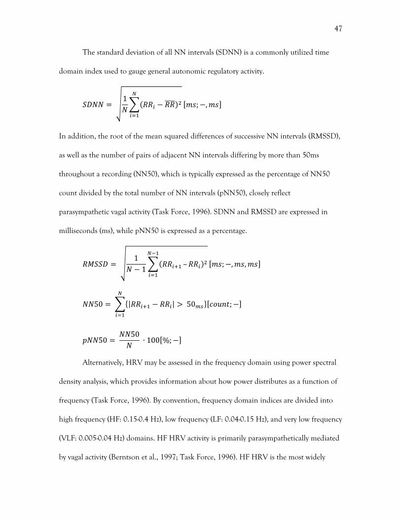
47
The standard deviation of all NN intervals (SDNN) is a commonly utilized time
domain index used to gauge general autonomic regulatory activity.
𝑆𝐷𝑁𝑁 = 1𝑁 𝑅𝑅! − 𝑅𝑅 !
!
!!!
𝑚𝑠;−,𝑚𝑠
In addition, the root of the mean squared differences of successive NN intervals (RMSSD),
as well as the number of pairs of adjacent NN intervals differing by more than 50ms
throughout a recording (NN50), which is typically expressed as the percentage of NN50
count divided by the total number of NN intervals (pNN50), closely reflect
parasympathetic vagal activity (Task Force, 1996). SDNN and RMSSD are expressed in
milliseconds (ms), while pNN50 is expressed as a percentage.
𝑅𝑀𝑆𝑆𝐷 = 1
𝑁 − 1 𝑅𝑅!!! –𝑅𝑅! !
!!!
!!!
𝑚𝑠;−,𝑚𝑠,𝑚𝑠
𝑁𝑁50 = 𝑅𝑅!!! − 𝑅𝑅! > 50!" 𝑐𝑜𝑢𝑛𝑡;−!
!!!
𝑝𝑁𝑁50 = 𝑁𝑁50𝑁 ∙ 100 %;−
Alternatively, HRV may be assessed in the frequency domain using power spectral
density analysis, which provides information about how power distributes as a function of
frequency (Task Force, 1996). By convention, frequency domain indices are divided into
high frequency (HF: 0.15-0.4 Hz), low frequency (LF: 0.04-0.15 Hz), and very low frequency
(VLF: 0.005-0.04 Hz) domains. HF HRV activity is primarily parasympathetically mediated
by vagal activity (Berntson et al., 1997; Task Force, 1996). HF HRV is the most widely
48
studied aspect of HRV because it captures fine-grained changes in parasympathetic
activation. Of the spectral HRV indices, it is most indicative of a well-functioning ANS and
general health, and is commonly affected in cases of pathology (e.g., Appelhans &
Luecken, 2006; Eddie, 2012; Hughes & Stoney, 2000). LF HRV reflects both
parasympathetic and sympathetic influences, such that it captures the dual action of the
baroreflex system as it affects both HRV and vascular tone (Cevese, Gulli, Polati, Gottin, &
Grasso, 2001; Vaschillo et al., 2011). Finally, though not well understood, VLF HRV is
thought to reflect fluctuations in the ANS (Berntson et al., 1997) that mediate baroreflex
control of vascular tone (Vaschillo et al., 2011). Spectral indices are expressed as ms^2.
Blood pressure characterizes pressure exerted on arterial walls by ventricular
ejection of blood from the heart, and is commonly expressed as systolic arterial blood
pressure (SAP; arterial pressure during systolic contraction of the heart) and diastolic
arterial blood pressure (DAP; arterial pressure during ventricular relaxation between heart
contractions). Derived from the vascular pressure waveform, measures of BPV characterize
baroreflex mediated moment-to-moment changes in SAP and DAP that are indicative of
autonomic flexibility and adaptability (Vaschillo et al., 2011; Voss, Boettger, Schulz, Gross,
& Bär, 2011). Blood pressure variability is expressed in the present investigation as the
standard deviation of systolic blood pressure (SAPD), and the standard deviation diastolic
blood pressure (DAPD). Overall blood pressure may be expressed by mean arterial pressure
(MAP; the average blood pressure of an individual that considers both SAP and DAP), and
its standard deviation (MAPD; Parati et al., 2001). All BP indices are expressed in
millimeters of mercury (mmHg).
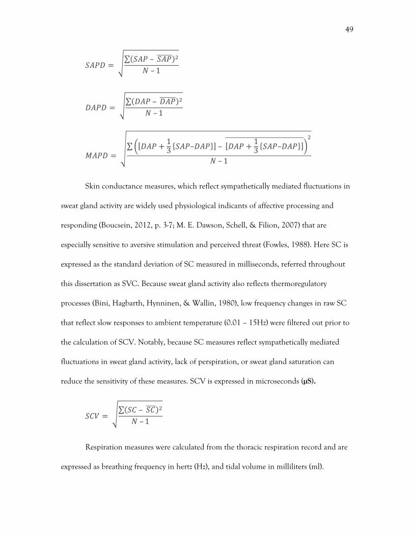
49
𝑆𝐴𝑃𝐷 = 𝑆𝐴𝑃 – 𝑆𝐴𝑃 !
𝑁 – 1
𝐷𝐴𝑃𝐷 = 𝐷𝐴𝑃 – 𝐷𝐴𝑃 !
𝑁 – 1
𝑀𝐴𝑃𝐷 = 𝐷𝐴𝑃 + 13 𝑆𝐴𝑃–𝐷𝐴𝑃 – 𝐷𝐴𝑃 + 13 𝑆𝐴𝑃–𝐷𝐴𝑃
!
𝑁 – 1
Skin conductance measures, which reflect sympathetically mediated fluctuations in
sweat gland activity are widely used physiological indicants of affective processing and
responding (Boucsein, 2012, p. 3-7; M. E. Dawson, Schell, & Filion, 2007) that are
especially sensitive to aversive stimulation and perceived threat (Fowles, 1988). Here SC is
expressed as the standard deviation of SC measured in milliseconds, referred throughout
this dissertation as SVC. Because sweat gland activity also reflects thermoregulatory
processes (Bini, Hagbarth, Hynninen, & Wallin, 1980), low frequency changes in raw SC
that reflect slow responses to ambient temperature (0.01 – 15Hz) were filtered out prior to
the calculation of SCV. Notably, because SC measures reflect sympathetically mediated
fluctuations in sweat gland activity, lack of perspiration, or sweat gland saturation can
reduce the sensitivity of these measures. SCV is expressed in microseconds (µS).
𝑆𝐶𝑉 = 𝑆𝐶 – 𝑆𝐶 !
𝑁 – 1
Respiration measures were calculated from the thoracic respiration record and are
expressed as breathing frequency in hertz (Hz), and tidal volume in milliliters (ml).
50
Analysis
Data Distribution and Outlier Analyses
Preliminary visual inspection and univariate analysis were used to check for
normality for each psychological measure. Variables that with skew or kurtosis >1 were
subjected to logarithmic transformation (i.e., logarithm/reflect and logarithm).
Mahalanobis distance was used to detect multivariate outliers (de Maesschalck, Jouan-
Rimbaud, & Massart, 2000).
Associations Between Anxiety and Depression, and Baseline Physiological Measures
Anticipated associations between trait anxiety and depression, and baseline
physiological indices within the BPD group were examined using Pearson’s correlation
coefficients.
A Priori Hypothesis Testing
A priori hypotheses were tested using repeated measures mixed models with
unstructured covariance matrix (Wolfinger & Chang, 1995) to examine the effect of group
(BPD, control), experimental task (baseline, cue exposure, recovery period), and their
interaction on target physiological indices including HR, SDNN, HF HRV, LF HRV,
SAPD, and SCV, with model level Bonferroni correction (6 tests, a’= .008) to control for
alpha level inflation. To check for possible respiration effects on HRV, these models were
also tested with respiration frequency included as a covariate.
Main effects of group are indicative of differences between the BPD and control
groups on target physiological measures across experimental tasks. Main effects of time are
indicative of change from baseline to cue reactivity, from cue reactivity to the recovery
51
period, and from baseline to the recovery period on each target index, in the combined
groups. The group × time interaction effects were indicative of whether the groups differed
in the way they changed from baseline to cue reactivity, and from cue reactivity to the
recovery period, on each of the target physiological indices. When indicated by significant
main or interactions effects, post hoc least square means difference tests were conducted.
Tests for Effects of Substance Use in Participants with Borderline Personality Disorder
Within the BPD group, the effects of chronic substance use on baseline physiology,
change in physiology in response to cue exposure, and change in physiology from cue
exposure to the recovery period were assessed. To test this, two sets of residual change
scores were derived by regressing HR, SDNN, HF HRV, LF HRV, SAPD, and SCV scores
during cue exposure onto their respective scores during baseline, and by regressing post-cue
exposure recovery period scores onto their respective measure during cue exposure.
Residuals represent the error in the regression model and are calculated by subtracting the
model’s predicted values from the observed values (i.e., e = y – ŷ), and are preferable to
utilizing raw change scores because they account for effects of regression to the mean.
Residual scores were subsequently tested for association with substance use measures using
general linear models.
Exploratory Analyses
Post Cue Exposure Recovery Latency
A more fine-grained analysis of latency of recovery from cue exposure was
accomplished by separating the recovery period into a first and a second 3-minute interval.
Mixed models for HR, SDNN, HF HRV, LF HRV, SAPD, and SCV were used to examine
52
between group differences in change in from cue exposure, to the first and second halves
of the recovery period, with model level Bonferroni correction (6 tests, a’= .008) to control
for alpha level inflation.
Dissociative Tendencies as a Moderator of Psychophysiological Cue Reactivity
Previous work has shown individuals with BPD who report high levels of state
dissociative tendencies tend to be less psychophysiologically responsive to startle (Ebner-
Priemer et al., 2005; Ebner-Priemer et al., 2009). As such, in participants with BPD,
dissociative tendencies measured by the DES-II, were posited to moderate the relationship
between psychophysiological indices at baseline and during cue exposure. Partial least
squares path modeling (Henseler & Fassott, 2010) was employed to parse out the possible
effects of trait dissociation on cue reactivity, by regressing covaried baseline HR, SDNN,
pNN50, HF HRV, LF HRV, SAP, SAPD, or SCV (exogenous variable), continuous DES-II
score (moderator variable), and HR, SDNN, pNN50, HF HRV, LF HRV, SAP, SAPD, or
SCV × DES-II score (interaction term), onto HR, SDNN, pNN50, HF HRV, LF HRV,
SAP, SAPD, or SCV during cue exposure (endogenous variable).
Expression of Effect Sizes
For consistency, effect sizes were expressed as Cohen’s d throughout, with d = .20
interpreted as a small effect, d = .30 interpreted as a medium effect, and d = .50 interpreted
as a large effect (J. Cohen, 1988). Effect sizes for means tests were calculated using Cohen’s
standard formula (J. Cohen, 1988, p. 274). For regression analyses, eta-squared effect sizes
were converted to d per Cohen’s algorithm (1988, p. 281), while for correlation analyses,
53
Pearson’s r was converted to d using Rosenthal’s formula (R. Rosenthal, 1994, p. 239). For
chi-square analyses, χ2 was converted to d per Ellis’ formula (Ellis, 2015).
Results
Data Distribution and Outlier Analyses
Variables including SDNN, RMSSD, HF HRV, LF HRV, VLF HRV, and SCV
were excessively skewed or kurtotic and were thus logarithmically transformed. The
remaining physiological indices, HR and pNN50, did not require transformation.
The Mahalanobis distance test detected one multivariate outlier in the baseline task
(a BPD group participant), and one outlier in the recovery period task (a control group
participant). These participants were subsequently excluded from analyses involving
baseline and/or recovery period physiological measures, respectively. A separate test for
multivariate outliers using the change scores from baseline to cue exposure, and cue
exposure to recovery period did not reveal any outliers.
Sample Characteristics
Groups did not differ significantly on sex, age, and body mass index (all p > .05; see
Table 1). In the BPD group, 16 participants identified as White/European American, 2 as
Asian, and 4 as other/mixed race. In the control group, 16 participants identified as
White/European American, 5 as Asian, and 1 as other/mixed race.
Participants with BPD reported significantly more lifetime months of therapy (see
Table 1). Modal income for participants with BPD was < $15,000, while the modal income
for controls was bifurcated between two income brackets, < $15,000, and > $90,000. In the
BPD group, 4 participants were married, 17 had never been married, 1 was divorced, and 1
54
was separated, while in the control group, 5 participants were married, 16 had never
married, and 1 was widowed.
Characterization of BPD Group Participant Psychiatric Comorbidity
As anticipated, participants with BPD exhibited a high degree of psychiatric
comorbidity. Nineteen of 22 participants had a co-occurring psychological diagnosis or
diagnoses. The count of various DSM-IV-TR Axis I diagnoses were as follows: 11 major
depressive disorder, 10 generalized anxiety disorder, 7 panic disorder, 7 social anxiety
disorder, 5 bipolar disorder (4 type II, and 1 type I), 4 post-traumatic stress disorder, 4
obsessive compulsive disorder, 4 eating disorder not otherwise specified, 2 anxiety disorder
not otherwise specified, 2 specific phobia, 2 body dysmorphic disorder, 1 bulimia nervosa,
1 hypochondriasis, 1 somatization disorder, and 1 agoraphobia.
There was also considerable personality disorder comorbidity. The count was as
follows: 4 avoidant personality disorder, 2 obsessive compulsive personality disorder, 2
dependent personality disorder, 2 paranoid personality disorder, 1 antisocial personality
disorder, and 1 histrionic personality disorder.
Participant Medications
Fourteen of 22 participants in the BPD reported current use of prescribed
medications that could potentially affect psychophysiological measures, including 7
participants taking an antipsychotic or mood stabilizers (i.e., aripiprazole, lithium,
lurasidone, olanzapine, risperidone), 4 taking a benzodiazepines (i.e., alprazolam,
clonazepam), 3 taking a non-benzodiazepine anxiolytic or sleep aid (i.e., buspirone,
zolpidem), 6 taking a selective serotonin reuptake inhibitor (i.e., citalopram, escitalopram,
55
fluoxetine, sertraline), 3 taking a serotonin agonist / reuptake inhibitors (i.e., trazodone), 1
taking bupropion, and 2 taking a stimulant for ADHD (i.e., lisdexamfetamine,
methylphenidate).
Control group participants reported no medications that are known to affect
physiological measures.
Preliminary Between-group Psychosocial Comparisons
Between groups comparisons of self-reported psychosocial measures are reported in
Table 2. As predicted, participants with BPD, compared to healthy controls, reported
greater state and trait anxiety (STAI-Y scores), greater depression (BDI scores), more
present moment negative affect (PANAS scores), greater emotion dysregulation (DERS and
AAQ-II scores), and greater dissociative symptomology (DES-II scores). Contrary to
hypotheses though, groups were not different in terms of present moment positive affect as
measured by the PANAS.
Groups did not differ in terms of frequency of past year exercise, or average length
of exercise sessions (p > .05). Groups did differ, however, on sleep measures. Participants
with BPD reported significantly more trouble initiating sleep and falling back to sleep
compared to controls, χ2 (1, N = 44) = 11.2, p < .05, d = 1.17, although ultimately the
groups did not differ on average hours of sleep per night (p > .05).
Substance Use
As predicted, the BPD group had higher rates of lifetime alcohol and other drug
dependence (see Table 3 & 4), however, contrary to predictions, groups were not different
56
in terms of past month and past year drinking behaviors (see Table 3), or drug use
behaviors aside from nicotine (see Table 4).
One BPD participant was in recovery from alcohol dependence and was abstinent
from alcohol in the past year, and 3 control participants endorsed trying alcohol, but never
becoming drinkers. For individuals in each group that endorsed drinking in the past year,
results showed that BPD and control group participants initiated drinking at a similar age,
had a similar number of drinking days in a typical week in the past month, drank similar
quantities in the past month, and were similar in terms of drinking during their heaviest
drinking week in the past month. Additionally, a non-significant chi-square test (p > .05)
indicated groups drank at similar frequencies in the past year.
Participants in the BPD group used nicotine more frequently in the past month
and past year compared to participants in the control group (see Table 4).
The groups, however, were similar in terms of their use of drugs other than alcohol
or nicotine. There were no between group differences in frequency of drug use in the past
month (measured as number of days of use in the past month) or frequency of drug use
over the past year (measured as average times using per week during the past year) for
amphetamines, cannabis, cocaine, or opiates (see Table 4). Notably, for these drug classes,
both groups reported very little past month and past year use.
Subjective Arousal to Emotionally Evocative Pictures
As predicted, participants with BPD reported significantly more subjective arousal
to emotionally evocative pictures during the cue exposure component of the experimental
session, t(42) = 2.03, p <.05, d = .63, scoring an average arousal rating of 5.02 (SD = 1.33),
57
with a total arousal rating of 180.8 (SD = 47.9), while control group participants scored an
average arousal rating of 4.21 (SD = 1.33), and a total arousal rating of 151.5 (SD = 47.9).
Within the BPD group, BPD severity, as measured by the combined standardized
scores of the Borderline Symptom List and Difficulties in Emotion Regulation Scale, was
not significantly associated with mean subjective arousal, p > .05.
Associations Between Anxiety and Depression, and Baseline Physiological Measures
The hypothesis that trait anxiety and depression would be negatively associated
with parasympathetically mediated HRV within the BPD group was partially supported.
Trait anxiety (total STAI-Y trait score) was negatively associated with pNN50, r(19)
= –.46, p < .05, d = 1.04, while directionally concordant large effect sizes that did not reach
statistical significance were observed for SDNN, r(19) = –.43, p = .05, d = .95, RMSSD,
r(19) = –.43, p = .05, d = .95, and HF HRV, r(19) = –.38, p > .05, d = .82. Depression (total
BDI score), however, was not associated with HRV.
A Priori Hypotheses Tests
Hypothesis 1, that relative to controls, participants with BPD would show lower
baseline levels of parasympathetically mediated neurocardiac flexibility and greater
sympathetic activation was partially supported. Participants with BPD did exhibit greater
HR and SCV at resting baseline, however, groups were not significantly different on other
measures reflecting neurocardiac flexibility including SDNN, RMSSD, pNN50, HF HRV,
LF HRV, and SAPD (Table 5). Further, groups were similar on measures of respiration
(Table 5).
58
Hypothesis 2, that participants with BPD would report greater subjective arousal
than controls, but that both groups would exhibit similar parasympathetic withdrawal and
increased sympathetic activation in response to cue exposure, was partially supported.
Participants with BPD reported greater subjective arousal to the picture stimuli. In
addition, as shown by a main effect of time (Table 6) and subsequent post hoc least square
means tests (Table 7), the combined groups demonstrated increased sympathetic activation
from baseline to cue exposure as evinced by an increase in SCV. Similarly, both groups also
demonstrated an increase in HR in response to cue exposure, which may be indicative of
increased sympathetic activation, decreased parasympathetic activation, or a combination
of both. The combined groups also demonstrated an increase in SDNN from cue exposure
to cue recovery period, although after respiration frequency was included as model
covariate, this effect was no longer statistically significant. Overall, however, there was no
significant change in parasympathetic activation in response to cue exposure, as indicated
by a null main effect for HF HRV.
Hypothesis 3, that during the post-cue exposure recovery period participants with
BPD would show continued parasympathetic withdrawal and sympathetic activation, while
the control group would return to their pre-challenge physiological baseline, was not
supported. Contrary to predictions, no interaction effects were observed on any target
physiological index, suggesting groups did not differ in their physiological change between
these tasks. A main effect of time, however, indicated that the combined groups
demonstrated a decrease in SCV from cue exposure to the post-cue exposure recovery
period (Table 7).
59
A Posteriori Hypothesis Tests
Because of BPD’s complex polythetic presentation, and the varying degrees of
severity among individuals meeting diagnostic criteria for BPD (Crowell, Beauchaine, &
Linehan, 2009; Gunderson & Links, 2009), BPD severity had been highlighted a priori as
a potential factor to consider in the present analyses, though no specific hypotheses were
made. With this in mind, it was thus tentatively hypothesized that individual differences in
BPD severity may be driving the observed null effects for cue exposure recovery on target
physiological measures. To more fully characterize the potential effects of BPD severity on
responses to emotional stimuli, the potential moderating effects of BPD severity on cue
reactivity were also tested.
To do this, first a measure of BPD severity was calculated by combining individuals’
standardized total scores (z-scores) on the BSL-23 and DERS, two correlated measures (r =
.77, p < .05, d = 2.41) reflecting BPD symptomology (standardized total scores were used to
give the two questionnaires equal weighting). Then, BPD severity scores were regressed
onto residualized physiological change scores (previously derived by regressing HR, SDNN,
HF HRV, LF HRV, SAPD, and SCV scores during cue exposure onto their respective
measure during baseline, and by regressing recovery period scores onto their respective
measure during cue exposure). To more fully characterize the potential role of
parasympathetic factors, pNN50 and RMSSD scores were also included in these a
posteriori analyses because, like HF HRV, they are understood to reflect parasympathetic
tone.
60
The a posteriori hypothesis was supported. In terms of physiological change from
cue exposure to recovery period, lower BPD severity within the BPD group predicted
significant decreases in RMSSD, pNN50, and HF HRV from cue exposure to the recovery
period, while higher BPD severity scores predicted significant increases in RMSSD,
pNN50, and HF HRV (Table 8 & Figure 2). In addition, SDNN evinced a large effect size
trend that was directionally concordant with results for RMSSD, pNN50, and HF HRV.
With regard to physiological change from baseline to cue exposure, within the BPD
group, BPD severity was negatively associated with pNN50, such that lower BPD severity
predicted increases in pNN50 from baseline to cue exposure, while greater BPD severity
predicted decreases in pNN50 from baseline to cue exposure (Table 8 & Figure 1). Large
effect sizes following the same directional pattern were observed for SDNN, RMSSD, HF
HRV, and LF HRV, though these results did not reach statistical significance (Table 8 &
Figure 1).
Mean HR also evinced a large, but non-significant effect size, trending in the
opposite direction (Table 8 & Figure 1). Specifically, lower BPD severity predicted
decreases in HR from baseline to cue exposure, while greater BPD severity predicted
increases in HR from baseline to cue exposure.
Similarly, a large, but non-significant effect size was observed for SCV, such that
greater BPD severity was associated with increases in SCV from baseline to cue exposure
(Table 8). Careful slope inspection, however, indicated the majority of participants (16 of
22) showed slight to moderate reductions in SCV from baseline to cue exposure.
61
Respiration frequency was not correlated with BPD severity, or any physiological
measure change score, suggesting participants’ respiration did not influence these results.
Tests for Effects of Substance Use in Participants with Borderline Personality Disorder
The low numbers of participants with BPD endorsing past month, and past year
amphetamine, cannabis, cocaine, opiate, and nicotine use, as well as lifetime histories of
alcohol and other drug dependence, precluded analysis for these measures. As such, tests
for the effects of substance use in participants with BPD focused on past month and past
year alcohol use.
The hypothesis that frequency of past month and past year alcohol use, as well as
quantity of alcohol consumed in a typical week in the past month would be negatively
associated with basal levels of target HRV measures in the BPD sample, was not supported,
all p > .05.
The hypothesis that substance use would predict cue exposure response, specifically
that greater past month and past year substance use would predict greater autonomic
responses to emotionally evocative pictures, was not supported. All analyses for associations
between alcohol use and change in physiology from baseline to cue exposure were non-
significant, p > .05.
Hypothesis 4, that greater substance use would predict less autonomic recovery
after perturbation was partially supported. Quantity of alcohol consumed in a typical week
in the past month was negatively associated with change in SAPD, F(1, 20) = 5.53, p < .05,
d = 1.05. Slope inspection revealed that greater alcohol use predicted either decreases, or
very small increases in SAPD from cue exposure to the recovery period, while those who
62
drank less or not at all were more likely to experience larger increases in SAPD. All other
analyses for associations between past month and past year alcohol use and change in
physiology from cue exposure to the recovery period were non-significant, p > .05.
Post Cue Exposure Recovery Latency Analysis
An exploratory analysis of latency of recovery from cue exposure was carried out by
separating the recovery period into a first and a second 3-minute interval. Mixed models
for HR, SDNN, HF HRV, LF HRV, SAPD, and SCV were used to examine between group
differences in change in from cue exposure, to the first and second halves of the recovery
period. Analyses did not produce any significant interaction effects, all p > .05, suggesting
groups did not differ in terms of their temporal patterns of recovery.
Dissociative Tendencies as a Moderator of Psychophysiological Cue Reactivity
Partial least squares path modeling was used to test if dissociative tendencies—
measured by participants’ total score on the DES-II—moderate psychophysiological cue
reactivity. Results indicated a significant moderating effect of dissociative tendencies on
pNN50, β = –0.4, SE = 0.0, z = –4.3, p <.05, and SAPD, β = 1.3, SE = 0.0, z = 3.2, p <.05.
Slope inspection showed that greater trait dissociation reflected by total DES-II scores
predicted decreases in pNN50 from baseline to cue exposure, while lower levels of trait
dissociation predicted increases in this HRV index. Slope inspection also revealed that
greater trait dissociation was associated with increases or little change in SAPD from
baseline to cue exposure, while lower trait dissociation predicted decreases in SAPD.
Moderation effects for HR, SDNN, HF HRV, LF HRV, and SAP did not reach statistical
63
significance. Including respiration frequency as a covariate in the models did not affect
these results.
Checks for Effects of Medication, Time in Treatment, and Age on Physiological Measures
T-tests showed participants in the BPD group taking medication were not different
from those not taking medication on physiological measures at baseline, during cue
exposure, or during the recovery period, all p > .05. Further, initial results for between
group differences in baseline physiology were not changed by removing BPD participants
on medication, or not on medication, from the analysis. Similarly, the results of the a
priori hypothesis tests were not affected by excluding BPD participants on medication, or
not on medication, from the analysis.
For participants with BPD, general linear models showed that time in Dialectical
Behavior Therapy and total lifetime psychological treatment were not associated with basal
levels of physiology, nor were they predicted by BPD severity, all p > .05.
In the combined sample, participant age predicted SDNN, F(1, 41) = 7.47, p < .05,
d = 0.85, however, all other associations between age and physiological indices at baseline
were non-significant, p > .05. Adding age to the a priori hypothesis tests models did not
change their results.
Discussion
BPD is a complex psychiatric condition characterized by intense and rapidly
shifting emotional states, impulsivity, and instability in self-image (Glenn & Klonsky,
2009). Its sequelae include intense feelings of emptiness, shame, loneliness, panic, and
rage, which lead to behaviors such as substance misuse, non-suicidal self-injury, and suicide
64
(Gunderson & Links, 2009; Leichsenring et al., 2011). To date, the cognitive components
of emotion dysregulation in BPD have received much research attention. The collateral
psychophysiological processes, however, remain poorly understood. Because emotion
regulation is mediated by both cognitive and physiological processes (Benarroch, 1997;
Hagemann et al., 2003), this knowledge gap may be limiting progress in the treatment of
BPD. As such, this investigation sought to comprehensively assess psychophysiological
differences between individuals with BPD and controls, and examine whether a loss of
flexibility in fundamental ANS processes may contribute to the emotion dysregulation
observed in BPD. To accomplish this, psychophysiological differences between individuals
with BPD and healthy controls were assessed at rest, during exposure to emotionally
evocative images, and during a post cue exposure recovery period. Additionally, because
substance use disorders are highly comorbid with BPD, and may interact with and
exacerbate BPD pathology (Axelrod et al., 2011; Dimeff et al., 2000; Kruedelbach et al.,
1993), this study also investigated the effects of co-occurring substance use on
psychophysiological processes in individuals with BPD.
As hypothesized, between group psychophysiological differences were observed at
baseline showing greater HR and SCV in participants with BPD, compared to controls,
however, contrary to predictions, there were no significant between group differences in
HRV at baseline. Main effects of group and time were found in response to, as well as in
recovery from exposure to emotionally evocative images on HR and SCV, while a main
effect of time was observed for SDNN. Specifically, the main effects of group indicated
participants with BPD had greater HR and SCV when all three tasks were considered
65
together. A main effect of time for HR, SDNN, and SCV, and subsequent post hoc
analyses showed the combined groups demonstrated increases in HR and SCV in response
to exposure to emotionally evocative images, a decrease in SCV during recovery from image
exposure, and an increase in SDNN from baseline to the recovery period. No group × time
interaction effects were observed, suggesting the groups were not different in terms of how
they responded to or recovered from exposure to emotionally evocative stimuli. This was in
spite of the fact that participants with BPD rated the images as subjectively more arousing
than controls. The absence of observed interaction effects led to speculation that BPD
symptom severity may be influencing results. This postulate was supported by a posteriori
analyses, which found that within the BPD group, BPD severity moderated physiological
response to, as well as recovery from exposure to emotionally evocative images.
Associations between physiological measures and substance use behaviors were also
investigated within the BPD group. Contrary to expectations, however, participants with
BPD generally reported relatively low levels of substance use, and fewer than expected
participants had lifetime histories of substance dependence. This precluded the planned,
in-depth analyses, although associations between alcohol use and physiology were
investigated. Hypotheses about the effects of alcohol on physiology in participants with
BPD, however, were largely unsupported.
Preliminary Between-group Psychosocial Comparisons
As anticipated, participants with BPD, compared to controls, reported greater state
and trait anxiety, greater depression, more present moment negative affect, greater emotion
dysregulation, greater dissociative symptomology, and more difficulty initiating sleep and
66
falling back to sleep. This is in line with a large literature showing individuals with BPD
experience higher rates of problems in these areas than non-clinical samples (see
Leichsenring et al., 2011 for review).
Contrary to hypotheses, groups were not different in terms of present moment
positive affect, as measured by the PANAS. Though this finding ran contrary to what was
hypothesized, it is not without precedent. Levine et al. (1997) also found that individuals
with BPD experience greater negative affect to non-psychiatric controls, but similar levels of
positive affect. Although negative affect, per se, is not a diagnostic marker of BPD in either
the DSM-IV-TR (American Psychiatric Association, 2000) or ICD-10 (World Health
Organisation, 1992) classifications, heightened negative affect, and not reduced positive
affect, may be an important marker of BPD pathology, especially considering negative
affect is positively associated with BPD traits and features (Cheavens et al., 2005; M. Z.
Rosenthal, Cheavens, Lejuez, & Lynch, 2005).
Substance Use
Participants with BPD were more likely to have a lifetime diagnosis of alcohol and
other drug dependence, however, contrary to predictions, groups were not different in
terms of past month and past year alcohol or other drug use behaviors, aside from nicotine.
The low rates of substance use in the present sample was surprising given the usually high
rates of substance use in individuals with BPD (Trull et al., 2000). This may be explained
by the fact that all study participants with BPD were receiving Dialectical Behavior Therapy
at the time of their participation in the present investigation. In most instances, patients
engaged in Dialectical Behavior Therapy for BPD who also have co-occurring substance use
67
problems are encouraged or required to stop substance use while in treatment, as this
behavior typically interferes with treatment goals and precludes reliance on more effective,
skill-based coping strategies (Linehan, 1993, 2014). It is therefore possible that in the
present sample, the relatively low rates of substance use in participants with BPD may be
accounted for by their active participation in treatment.
The fact, however, that participants with BPD reported similar substance use to
controls in the past year runs contrary to this postulate. Though it is possible they were
engaged in other therapies prior to beginning Dialectical Behavior Therapy, on average,
participants with BPD had only received an average of approximately three months of
lifetime Dialectical Behavior Therapy at the time they participated in the study.
Subjective Arousal to Emotionally Evocative Pictures
As anticipated, participants with BPD reported significantly more subjective arousal
to emotionally evocative pictures during the cue exposure component of the study. The
difference between the BPD and control groups was, however, not especially robust, as
evinced by a small effect size difference in arousal ratings. This relatively small effect size
difference and the lack of a relationship between subjective arousal and BPD severity
within the BPD group, suggests images depicting interpersonal and social content taken
from the IAPS may not sufficiently target BPD sensitivities at a level of intensity that is
optimal for BPD cue reactivity studies. For instance, images depicting abandonment, non-
suicidal self-injury, and suicide do not feature heavily in the IAPS and thus were not well
represented in the set formed in Sloan et al.’s (2010) study. This suggests that the
development of an effective BPD specific picture set may require the collection and
68
validation of a corps of novel images that closely focus on key BPD themes such as frantic
efforts to avoid abandonment, unstable interpersonal relationships, transient dissociative
episodes, non-suicidal self-injury, and suicide.
Associations Between Anxiety and Depression, and Baseline Physiological Measures
Previous investigations have shown trait anxiety and depression are inversely related
to indices of HRV characterizing resting parasympathetic activation (e.g., Chang et al.,
2012; Eddie, 2012; Miu et al., 2009). As such, within the BPD group, greater levels of trait
anxiety and depression were expected to predict lower basal levels of pNN50, RMSSD, and
HF HRV. This postulate was partially supported.
In participants with BPD, trait anxiety was significantly negatively associated with
pNN50. Parallel large effect size associations were observed between anxiety and SDNN,
RMSSD, and HF HRV, although these did not reach statistical significance. This is in line
with psychophysiological perspectives on anxiety, which hold that chronic anxiety is related
to a loss of biobehavioral flexibility reflected in, or resulting from a loss of vagal tone
(Friedman & Thayer, 1998; Thayer et al., 1996; Thayer & Lane, 2000).
Contrary to predictions, however, depression did not predict HRV in the present
BPD sample. Notably, the majority of studies investigating the relationship between
depression and HRV have studied individuals with primary depressive disorders, with little
or no psychiatric comorbidity. These populations are different from the present sample of
individuals who have a primary diagnosis of BPD. It is possible that there are differences in
autonomic system involvement in primary depressive disorders and depression occurring
secondary to BPD or other mental disorders. Future studies should investigate these
69
potential differences, by comparing individuals with primary depressive disorders and
individuals with secondary depressive disorders on psychophysiological measures.
A Priori Hypotheses Tests
Hypothesis 1 predicted that relative to controls, participants with BPD would show
lower baseline levels of HRV, and greater sympathetic activation. This was partially
supported. Participants with BPD did exhibit greater HR and SCV at resting baseline,
however, groups were not significantly different at resting baseline on measures of HRV, or
SAPD.
This finding is concordant with Ebner-Priemer et al.’s (2007) observation of greater
resting HR in individuals with BPD compared to healthy controls, as well as Ebner-Priemer
et al. (2007), Austin et al.’s (2007), and Gratz et al.’s (2011) null finding for between group
differences in resting HRV. It is also consistent with Kuo and Linehan’s (2009) observation
of greater resting levels of skin conductance response in individuals with BPD. The current
findings are, however, discordant with results published by Weinberg et al. (2009), and
Kuo and Linehan (2009), who observed lower resting levels of parasympathetically
mediated HRV in individuals with BPD, compared to healthy controls.
The heterogeneity of the samples utilized in these investigations may explain some
of the divergent findings across studies. Further, though this investigation recruited a
naturalistic sample that minimized and/or controlled for potential confounds associated
with participant age, time in treatment, diagnosis of BPD, medication, and psychiatric
comorbidity, some of these factors may have nevertheless influenced results.
70
For instance, psychiatric comorbidity is typical in BPD samples, and may influence
psychophysiology in unpredicted ways. While excluding study volunteers with comorbid
disorders may seem prudent, omitting such individuals can reduce ecological validity by
inadvertently excluding people with more severe BPD who commonly have affective
disorders secondary to BPD (Comtois, Cowley, Dunner, & Roy-Byrne, 1999). Thus,
whether including or excluding co-occurring mental disorders, comorbidity remains a key
issue that bears consideration whenever interpreting psychophysiological results in studies
of BPD.
As previously noted, medications can also affect physiology, and may in part
account for the equivocal findings in differences in basal HRV between individuals with
BPD and controls across the studies reviewed here, and the present investigation. Notably
however, checks for effects of medication in the present study showed no differences in
psychophysiology between BPD participants taking medication and those not taking
medication. Further, results for between group differences in baseline psychophysiology
and results of the a priori hypotheses tests were not affected by including or excluding BPD
participants on medication, or not on medication.
While ideally studies such as this would utilize samples free of medication,
excluding volunteers because of medication also threatens ecological validity as participants
with greater BPD severity are also most likely to be on medication (Sansone et al., 2003).
The present investigation anticipated this problem and included individuals on
medications, while making marked efforts to reduce potential effects of medication by
selectively excluding volunteers taking certain pharmaceuticals known to directly affect the
71
cardiovascular system, and strategically scheduling session times to reduce acute medication
effects. This factor, nevertheless, could have influenced results.
Another possible explanation for these equivocal findings is participant BPD
severity, both within samples and between studies. As discussed below, BPD severity had a
moderating effect on response to, and recovery from emotionally evocative images in the
present sample. This finding speaks to the possibility that within sample differences in
BPD severity may have undermined tests for between group differences in basal physiology,
although null associations between BPD severity and basal physiology within the BPD
group suggest otherwise.
Future studies will ideally utilize large samples so that possible moderating effects of
psychiatric comorbidity, medication, and BPD severity on ANS processes can be parsed
out. Larger samples will afford strategies such as within group tests for moderation, as well
as assessment of physiological differences between individuals with psychiatric comorbidity
versus no psychiatric comorbidity, more versus less severe BPD, and those on medication
versus those not on medication.
Hypothesis 2, predicted that compared to controls, participants with BPD would
report greater subjective arousal to the emotionally evocative images, while both groups
would exhibit similar reductions in parasympathetic tone, and increased sympathetic
activation in response to the images. This hypothesis was partially supported. Participants
with BPD reported greater subjective arousal to the picture stimuli. Further, consistent
with findings by Weinberg and colleagues (2009) who challenged volunteers with a social
stressor, both groups demonstrated increased sympathetic activation from baseline to cue
72
exposure as evinced by an increase in SCV. Similarly, both BPD and control groups also
demonstrated an increase in HR in response to cue exposure, which may be an effect of
increased sympathetic activation, decreased parasympathetic activation, or a combination
of both. Overall, however, consistent with Weinberg et al.’s findings, there was no
significant change in parasympathetic activation in response to cue exposure in either
group. These results suggest groups were similar in their response to emotionally evocative
images in that they both demonstrated increases in sympathetic activation, but no
significant changes in parasympathetic tone.
A posteriori analyses, however, highlighted an important factor not considered in
previous studies. Within the BPD group, BPD severity moderated cue reactivity, such that
participants with greater BPD severity tended to show decreases in HRV and SCV in
response to cue exposure, while individuals with lower BPD severity tended to show
increases in HRV. This is a key finding as it demonstrates for the first time differing
patterns of psychophysiological responding to emotional stimuli within a group of people
with BPD. Specifically, individuals with lower BPD severity were shown to exhibit
physiological responses similar to controls, whereas individuals with higher BPD severity
exhibited the opposite response.
This finding shares important commonalities with Gratz et al. (2013) who observed
that participants with co-occurring BPD and AVPD demonstrated decreases in HRV in
response a distress tolerance task, while participants with BPD but not AVPD, and non-
BPD controls showed increases in HRV. Their findings for between group differences in
emotion regulation difficulties, as measured by DERS subscales, suggest the BPD with co-

73
occurring AVPD group represented a more severely disordered sub-group of individuals
than the group of participants with BPD but not AVPD. It is possible that Gratz et al.
(2013) and the present study both captured a moderating effect of BPD severity on HRV,
though the two studies captured BPD severity in different ways.
The present finding for a moderating effect of BPD severity on cue reactivity is also
consistent with Austin et al.’s (2007) observation that participants with BPD showed
decreases in HRV during exposure to emotionally evocative stimuli, while controls showed
increases in HRV. Austin and colleagues interpreted their results in terms of Porges’
polyvagal theory. They inferred that the BPD group was exhibiting a physiological state of
preparedness for defensive behaviors characterized by vagal withdrawal during exposure to
emotionally evocative stimuli, (i.e., a fight or flight response). Controls, on the other hand,
demonstrated increased vagal activation in response to emotionally evocative stimuli,
which the authors posited would support spontaneous social engagement behaviors.
Taken together, these findings suggest individuals with more severe BPD mount a
physiological response that goes above and beyond what is called for in the moment. This
supports work that suggests individuals with BPD are more sensitive to perceived threat
(Dixon-Gordon et al., 2013; Donegan et al., 2003; Lynch et al., 2006), and may help
explain why once emotionally dysregulated, people with BPD have difficulty returning to
an affective baseline (Glenn & Klonsky, 2009; Selby et al., 2009; Selby & Joiner Jr, 2013).
Additionally, these findings may begin to explain the differing results across studies
investigating psychophysiological cue reactivity in BPD. Future psychophysiological BPD
studies should report, and control for BPD severity. Perhaps most importantly, this finding
74
highlights the necessity of considering multiple domains of functioning when attempting
to understand BPD pathology.
These findings also speak to the discourse on dimensional versus categorical
diagnostic systems for BPD. Many have argued that dimensional classification of BPD is
more reliable and valid than the current categorical classification systems used in DSM-5
and ICD-10 (e.g., Conway, Hammen, & Brennan, 2012; Trull, Widiger, & Guthrie, 1990).
The present findings support the use of a dimensional diagnostic system because they show
that there are important autonomic differences in how individuals with varying degrees of
BPD severity respond to emotionally evocative stimuli.
Notably, there was a non-significant, large effect size relationship between BPD
severity and BPD participants’ subjective arousal to the emotionally evocative images. This
finding makes it difficult to determine whether the observed physiological responses to the
images are indicative of underlying neurological differences in the CAN in individuals with
BPD, or a corollary of consciously perceived threat. Future studies will need to disentangle
this relationship. A first step toward this end might be to test whether physiological
reactivity in individuals with severe BPD changes through the course of treatments such as
Dialectical Behavior Therapy to become more similar to healthy controls, or whether it
stays the same even as BPD severity ameliorates with treatment.
Hypothesis 3, that during the post-cue exposure recovery period participants with
BPD would show continued reductions in parasympathetic tone, and sustained
sympathetic activation, while the control group would return to their pre-challenge
75
physiological baseline, was not supported. Groups appeared to be similar in terms of how
they recovered from cue exposure.
The aforementioned a posteriori analyses, however, also highlighted a key
consideration in terms of understanding recovery from emotional stimuli in individuals
with BPD. It was evident that within the BPD group, BPD severity moderated recovery
from exposure to emotionally evocative stimuli, such that participants with greater BPD
severity tended to show increases in HRV during cue exposure recovery, while individuals
with lower BPD severity tended to show the opposite recovery response, that is, decreases
in HRV. It is possible that this reflects natural rebound effects from autonomic
perturbation during cue exposure. The large effect sizes for the relationships between BPD
severity and parasympathetically mediated HRV from cue exposure to the recovery period,
coupled with the small effect size, negative relationship between BPD severity and change
in SCV during cue exposure, and during cue exposure recovery, suggests that
parasympathetically mediated processes may be more strongly implicated in BPD affective
reactivity than sympathetic processes.
Tests for Effects of Substance Use in Participants with Borderline Personality Disorder
The low numbers of participants with BPD endorsing past month, and past year
amphetamine, cannabis, cocaine, opiate, and nicotine use, as well as lifetime histories of
alcohol and other drug dependence meant planned analysis for these measures could not
be carried out. As a result, tests for the effects of substance use in participants with BPD
focused on measures for which there was sufficient data to conduct analyses, that is, past
month and past year alcohol use.
76
The hypothesis that frequency of past month and past year alcohol use, as well as
quantity of alcohol consumed in a typical week in the past month would be negatively
associated with basal levels of target HRV measures in the BPD sample, was not supported.
This is perhaps not surprising given the relatively low levels of drinking in the BPD sample,
and that moderate alcohol consumption is not known to appreciably affect HRV (Janszky
et al., 2005).
The hypothesis that substance use would predict cue exposure response, specifically
that greater past month and past year substance use would predict greater autonomic
responses to emotionally evocative pictures, was not supported. All analyses for associations
between alcohol use and change in physiology from baseline to cue exposure were non-
significant. Again, the relatively low levels of alcohol consumption in participants with
BPD may account for these null effects.
Hypothesis 4, that greater substance use would predict less autonomic recovery
after perturbation was partially supported. Quantity of alcohol consumed in a typical week
in the past month was negatively associated with change in SAPD. Slope inspection
revealed that greater alcohol use predicted either decreases, or very small increases in SAPD
from cue exposure to the recovery period, while those who drank less or not at all were
more likely to experience larger increases in SAPD. This finding suggests even at relatively
low levels, past month alcohol use may affect dynamic recovery of SAP in response to
stressors.
77
Dissociative Tendencies as a Moderator of Psychophysiological Cue Reactivity
Path modeling was used to test if dissociative tendencies moderate
psychophysiological cue reactivity. Dissociative tendencies were found to moderate change
in pNN50, and SAPD from baseline to cue exposure. Careful slope inspection indicated
that greater trait dissociation was associated with decreases in pNN50 from baseline to cue
exposure, while lower levels of trait dissociation predicted increases in pNN50. Further,
greater trait dissociation predicted increases or little change in SAPD from baseline to cue
exposure, while lower trait dissociation predicted decreases in SAPD.
Previously, Ebner-Priemer et al. (2005) showed that individuals with BPD reporting
high levels of state dissociation demonstrated lower orbicularis oculi electromyographic
response to acoustic startle compared to individuals reporting low levels of state
dissociation, although notably they found no response differences in terms of HR or mean
SC. More recently, in a classical conditioning paradigm, Ebner-Priemer et al. (2009) found
differences in mean SC response between individuals expressing high versus low levels of
state dissociation during exposure to a conditioned stimulus that had been paired with an
aversive sound. That is, participants with BPD who had low levels of state dissociation
demonstrated significant increases in mean SC in response to the conditioned stimulus
during the extinction phase of the study, while participants with high levels of state
dissociation showed no change in mean SC.
Although the current investigation assessed the effects of trait and not state
dissociation, and utilized emotionally evocative images rather than auditory stressors, the
present findings thematically extend Ebner-Priemer and colleagues’ previous work by
78
showing for the first time that greater dissociative tendencies predict lower HRV, and
greater BPV in response to an emotionally evocative stressor. It is possible that these results
reflect dampening of emotional responding through the ventral vagal complex, a
parasympathetic CAN structure that plays a central role in the coordination of sensory
perception, affective expression, and cardiac modulation through innervation of the
sensory organs, facial and vocalization muscles, and heart (Porges, 2001). So, while the
present study showed individuals with BPD reporting low trait dissociation responded to
the emotionally evocative stimuli with increased vagal activation in the same way as
controls—a response that would support social engagement and behaviors—participants
high in dissociative tendencies responded with decreased vagal tone, which would
ultimately undermine adaptive responses.
It is noteworthy that this moderation pattern for dissociative tendencies closely
reflects the moderation pattern seen in the a posteriori analyses reported above for BPD
severity. That is, change in pNN50 from baseline to cue exposure was shown to be
negatively associated with BPD severity, just as with dissociative tendencies. This is perhaps
not surprising, given dissociation is a diagnostic marker for BPD, and is generally greater in
individuals with more severe BPD. Moreover, in the present sample there was a trend
toward a positive association between the BPD severity measure (which did not contain any
direct measures of dissociation) and DES-II scores used to characterize dissociation (r = .41,
p = .06, d = .90). Nevertheless, this finding supports the notion that dissociation is a key
feature of BPD, such that it is intertwined with BPD pathology at an autonomic level.
79
Notably, Ebner-Priemer et al. (2005) and the present investigation both found null
effects for mean HR, which at face value may suggest that HR is not especially influenced
by dissociative tendencies. This conclusion, however, would be premature. It is possible
that the stressors used in Ebner-Priemer et al. (2005) and the present investigation were not
of sufficient magnitude to elicit a cardiac response from the sympathetic nervous system, or
dorsal vagal complex. So, while the stimuli may have been sufficient to engage the ventral
vagal complex with its fast acting, myelinated, parasympathetic fibers that predominantly
influence HRV, it may not have been sufficient to engage the sympathetic pathway
innervating the heart, with its attendant capacity to significantly increase HR. Moreover,
the stimuli used in these studies certainly was not of the magnitude necessary to engage the
dorsal ventral complex, which is capable of effecting a freeze response, characterized by
dramatic drop in HR, and is typically only activated in situations of extreme perceived
threat, or hypoxic crisis (Porges, 2001).
Understanding the moderating effect of dissociation on SAPD cue reactivity is
more challenging given the present study is the first to investigate the relationship between
dissociation and dynamic BP, and little is understood about the role of dynamic blood
pressure in emotion regulation. As such, there is little precedent to draw upon. This
finding, however, suggests future research on the psychophysiological processes attendant
with dissociation will do well to consider indices of BPV.
Limitations
While the present investigation had many methodological strengths including
recruiting an ecologically valid sample, matching groups based on sex and age, checking for
80
effects of medication, time in treatment, and dissociation, and ensuring BPD participants
met DSM-IV-TR diagnostic criteria for BPD, some limitations should be noted.
While the majority of BPD participants (14 of 22) were recruited from DBT-RU
where they had been comprehensively assessed for Axis I and Axis II disorders, eight BPD
participants not recruited from DBT-RU were asked to self-report current and previous
psychiatric diagnoses (other than BPD) to reduce participant burden. It is possible that
some Axis I and II diagnoses may have been missed in these participants.
In addition, while a priori power analyses (not reported) suggested groups of 18
would be sufficient to find extant group differences on physiological measures, many large
effect size results were observed that did not reach statistical significance, suggesting a larger
sample size was called for.
Another factor to consider is that while the IAPS images used to evoke an
emotional response in the present investigation had been selected by expert consensus, and
characterized a range of BPD sensitivities, the image set had not previously been validated
with individuals with BPD, and did not evoke an especially powerful subjective response in
the present sample.
It should also be noted that while allowing for psychiatric medications within the
BPD group reduced the risk of excluding participants with more severe BPD, it may also
have affected results in unpredictable ways. It is not clear to what extent differing
medication regimes across participants with BPD who were on medication may have
affected their physiology at baseline, or in response to and recovery from cue exposure.
81
Further, it is not clear to what extent medication may have affected individuals’ subjective
experience of the images.
Similarly, while allowing for comorbidity in the BPD group reduced the likelihood
of excluding individuals with more severe BPD, who are more likely to meet criteria for
other psychological disorders, this may have also affected results in unpredictable ways.
Though the BPD severity measure investigated may have captured some of this variance,
and to some extent assuages these concerns, it is not clear to what extent co-occurring
disorders may have affected results.
Finally, the rate if substance use in the BPD group was markedly lower than
anticipated. This largely precluded planned hypothesis tests concerned with the effects of
substance use on ANS processes in individuals with BPD.
Conclusions and Future Directions
The present investigation sought to comprehensively assess psychophysiological
differences between individuals with BPD and controls, and examine whether a loss of
flexibility in fundamental ANS processes may contribute to the emotion dysregulation
observed in BPD. Distinct between group differences were observed at baseline on
measures influenced by the sympathetic branch of the ANS, while resting differences in
parasympathetic activation were not observed. A priori analyses did not find predicted
between group differences, however a posteriori analyses revealed an important moderating
effect of BPD severity. The importance of this finding cannot be stressed enough. It speaks
to the heterogeneity in BPD pathology observed by clinicians, and suggests that within
BPD populations there are fundamental differences in how individuals respond
82
physiologically to emotionally valenced stimuli, and that this difference is related to BPD
severity. This finding also supports the case for moving away from categorical diagnosis of
BPD towards dimensional diagnostic systems. Additionally, this finding may begin to
explain why individuals with BPD, especially severe BPD, often become dysregulated when
confronted with relatively low level stressors, and commonly have great difficulty returning
to affective baseline once dysregulated.
Further, no previous study investigating psychophysiological processes in BPD has
actively controlled for BPD severity. It is possible that equivocal results in previous studies
may have been driven in part by this factor. If this is in fact the case, studying BPD
pathology at the level of the mean is inappropriate as it fails to appreciate important inter-
individual differences. Future studies should consider and report measures of BPD severity
and will benefit from utilizing person-centered, rather than variable-centered analytic
strategies (e.g., Bates, Buckman, Eddie, Voelbel, & Freeman, 2013).
Although low levels of substance use in the present BPD sample precluded planned
analyses to test for effects of co-occurring substance use on target physiological indices at
resting baseline, during cue exposure, and during recovery from cue exposure, recent
alcohol use was implicated in changes in SAP variability during recovery from exposure to
emotionally evocative images. Future studies using samples of people with BPD and co-
occurring, active substance use disorders should attend especially to the effects of alcohol
on blood pressure dynamics and the systems that regulate these processes, including the
baroreflex.
83
The picture stimulus set was rated as significantly more arousing by participants
with BPD, however, the mean between groups difference was not especially robust. Though
the IAPS picture set offers advantages, including being well validated in a variety of clinical,
and non-clinical samples, the present findings suggest a more targeted BPD specific image
set may be indicated for cue-reactivity studies with people with BPD.
In sum, this investigation supports the postulate that BPD pathology manifests in,
or is affected by dysregulation of ANS processes, both at rest, and in response to stimuli.
Further, BPD severity appears to be a key factor associated with individuals’ responses to
emotionally evocative stimuli, and may represent an important, hitherto unrecognized
psychophysiological component implicit in BPD heterogeneity.
84
References
Agelink, M. W., Boz, C., Ullrich, H., & Andrich, J. (2002). Relationship between major depression and heart rate variability: Clinical consequences and implications for antidepressive treatment. Psychiatry Research, 113, 139-149.
Agelink, M. W., Malessa, R., Baumann, B., Majewski, T., Akila, F., Zeit, T., & Ziegler, D. (2001). Standardized tests of heart rate variability: Normal ranges obtained from 309 healthy humans, and effects of age, gender, and heart rate. Clinical Autonomic Research, 11(2), 99-108.
American Psychiatric Association. (2000). Diagnostic and statistical manual of mental disorders: DSM-IV-TR (4th ed., text revision). Washington, DC: American Psychiatric Publishing.
Andersen, M. C., & Kunze, D. L. (1994). Nucleus tractus solitarius-gateway to neural circulatory control. Annual Review of Physiology, 56(1), 93-116.
Appelhans, B. M., & Luecken, L. J. (2006). Heart rate variability as an index of regulated emotional responding. Review of General Psychology, 10, 229-240.
Austin, M. A., Riniolo, T. C., & Porges, S. W. (2007). Borderline personality disorder and emotion regulation: Insights from the polyvagal theory. Brain and Cognition, 65(1), 69-76.
Axelrod, S. R., Perepletchikova, F., Holtzman, K., & Sinha, R. (2011). Emotion regulation and substance use frequency in women with substance dependence and borderline personality disorder receiving dialectical behavior therapy. The American Journal of Drug and Alcohol Abuse, 37(1), 37-42.
Ayduk, Ö., Zayas, V., Downey, G., Cole, A. B., Shoda, Y., & Mischel, W. (2008). Rejection sensitivity and executive control: Joint predictors of borderline personality features. Journal of Research in Personality, 42(1), 151-168.
Barnes, L. L. B., Harp, D., & Jung, W. S. (2002). Reliability generalization of scores on the Spielberger state-trait anxiety inventory. Educational and Psychological Measurement, 62, 603-618.
Bates, M. E., Bowden, S. C., & Barry, D. (2002). Neurocognitive impairment associated with alcohol use disorders: Implications for treatment. Experimental and Clinical Psychopharmacology, 10(3), 193-212.
Bates, M. E., & Buckman, J. F. (2011). Emotional dysregulation in the moment: Why some college students may not mature out of hazardous alcohol and drug use. In H. R. White & D. L. Rabiner (Eds.), College drinking and drug use. New York, NY: The Guilford Press.
Bates, M. E., & Buckman, J. F. (2013). Integrating body and brain systems in addiction neuroscience. In P. Miller (Ed.), Comprehensive addictive behaviors and disorders (Vol. 2, pp. 187-196). New York, NY: Academic Press.
Bates, M. E., Buckman, J. F., Eddie, D., Voelbel, G. T., & Freeman, J. (2013). The mean and the individual: A person-centered approach to neuropsychological recovery in men and women with substance use disorders. Frontiers in Addictive Disorders and Behavioral Dyscontrol, 4, 77.
85
Bates, M. E., Buckman, J. F., Vaschillo, E. G., Fonoberov, V. A., Fonoberova, M., Vaschillo, B., . . . Mezić, I. (2011). The redistribution of power: Neurocardiac signaling, alcohol and gender. PLoS One, 6(12), e28281. doi:10.1371/journal.pone.0028281
Beck, A. T., Steer, R. A., & Brown, G. K. (1986). Beck depression inventory (2nd ed.). San Antonio, TX: The Psychological Corporation. Harcourt, Brace and Company.
Beck, A. T., Steer, R. A., & Carbin, M. G. (1988). Psychometric properties of the Beck Depression Inventory: Twenty-five years of evaluation. Clinical Psychology Review, 8, 77-100.
Benarroch, E. E. (1993). The central autonomic network: Functional organization, dysfunction, and perspective. Mayo Clinic Proceedings, 68, 988-1001.
Benarroch, E. E. (1997). The central autonomic network. In P. A. Low (Ed.), Clinical autonomic disorders (2nd ed., pp. 17-23). Philadelphia, PA: Lippincott-Raven.
Bender, D. S., & Skodol, A. E. (2007). Borderline personality as a self-other representational disturbance. Journal of Personality Disorders, 21(5), 500-517.
Bennett, A. J., Sponberg, A. C., Graham, T., Suomi, S. J., Higley, J. D., & DePetrillo, P. B. (2001). Initial ethanol exposure results in decreased heart rate variability in ethanol-naive rhesus monkeys. European Journal of Pharmacology, 433, 169-172.
Berntson, G. G., Bigger, J. T. J., Eckberg, D. L., Grossman, P., Kaufmann, P. G., Malik, M., . . . van der Molen, M. W. (1997). Heart rate variability: Origins, methods, and interpretive caveats. Psychophysiology, 34, 623-648.
Bini, G., Hagbarth, K. E., Hynninen, P., & Wallin, B. G. (1980). Thermoregulatory and rhythm-generating mechanisms governing the sudomotor and vasoconstrictor outflow in human cutaneous nerves. The Journal of Physiology, 306, 537-552.
Bohus, M., Kleindienst, N., Limberger, M. F., Stieglitz, R. D., Domsalla, M., Chapman, A. L., . . . Wolf, M. (2008). The short version of the Borderline Symptom List (BSL-23): Development and initial data on psychometric properties. Psychopathology, 42(1), 32-39.
Bond, F. W., Hayes, S. C., Baer, R. A., Carpenter, K. M., Guenole, N., Orcutt, H. K., . . . Zettle, R. D. (2011). Preliminary psychometric properties of the Acceptance and Action Questionnaire–II: A revised measure of psychological inflexibility and experiential avoidance. Behavior Therapy, 42, 676-688.
Boucsein, W. (2012). Electrodermal activity (2nd ed.). New York: Springer Science & Business Media.
Bradley, S. J. (2003). Affect regulation and the development of psychopathology. New York, NY: The Guilford Press.
Buckman, J. F., White, H. R., & Bates, M. E. (2010). Psychophysiological reactivity to emotional picture cues two years after college students were mandated for alcohol interventions. Addictive Behaviors, 35, 786-790.
Calhoun, P. S., Sampson, W. S., Bosworth, H. B., Feldman, M. E., Kirby, A. C., Hertzberg, M. A., . . . Beckham, J. C. (2000). Drug use and validity of substance use self-reports in veterans seeking help for posttraumatic stress disorder. Journal of Consulting and Clinical Psychology, 68(5), 923-927.
86
Card, J. P., & Sved, A. F. (2011). Central autonomic pathways. In I. J. Llewellyn-Smith & a. J. M. Verberne (Eds.), Central regulation of autonomic functions (pp. 3-17). New York, NY: Oxford University Press.
Carlson, E. B., & Putnam, F. W. (1993). An update on the dissociative experiences scale. Dissociation: Progress in the Dissociative Disorders, 6, 16-27.
Cevese, A., Gulli, G., Polati, E., Gottin, L., & Grasso, R. (2001). Baroreflex and oscillation of heart period at 0.1 Hz studied by alpha-blockade and cross-spectral analysis in healthy humans. Journal of Physiology, 531(Pt 1), 235-244.
Chambers, A. S., & Allen, J. J. B. (2002). Vagal tone as an indicator of treatment response in major depression. Psychophysiology, 39, 861-864.
Chang, J. S., Yoo, C. S., Yi, S. H., Her, J. Y., Choi, H. M., Ha, T. H., . . . Ha, K. (2012). An Integrative Assessment of the Psychophysiologic Alterations in Young Women With Recurrent Major Depressive Disorder. Psychosomatic Medicine, 74, 495-500.
Cheavens, J. S., Rosenthal, M. Z., Daughters, S. B., Nowak, J., Kosson, D. S., Lynch, T. R., & Lejuez, C. W. (2005). An analogue investigation of the relationships among perceived parental criticism, negative affect, and borderline personality disorder features: The role of thought suppression. Behaviour Research and Therapy, 43(2), 257-268.
Cohen, H., Benjamin, J., Geva, A. B., Matar, M. A., Kaplan, Z., & Kotler, M. (2000). Autonomic dysregulation in panic disorder and in post-traumatic stress disorder: Application of power spectrum analysis of heart rate variability at rest and in response to recollection of trauma or panic attacks. Psychiatry Research, 96, 1-13.
Cohen, J. (1988). Statistical power analysis for the behavioral sciences (2nd ed.). Hillsdale, NJ: Lawrence Erlbaum Associates.
Comtois, K. A., Cowley, D. S., Dunner, D. L., & Roy-Byrne, P. P. (1999). Relationship between borderline personality disorder and Axis I diagnosis in severity of depression and anxiety. The Journal of Clinical Psychiatry, 60(11), 752-758.
Conway, C., Hammen, C., & Brennan, P. A. (2012). A comparison of latent class, latent trait, and factor mixture models of DSM-IV borderline personality disorder criteria in a community setting: implications for DSM-5. Journal of Personality Disorders, 26(5), 793-803. doi:10.1521/pedi.2012.26.5.793
Cooke, W. H., Hoag, J. B., Crossman, A. A., Kuusela, T. A., Tahvanainen, K. U. O., & Eckberg, D. L. (1999). Human responses to upright tilt: A window on central autonomic integration. The Journal of Physiology, 517, 617-628.
Corrigan, F. M., Davidson, A., & Heard, H. (2000). The role of dysregulated amygdalic emotion in borderline personality disorder. Medical Hypotheses, 54, 574-579.
Cox, W. M., & Klinger, E. (2011). A motivational model of alcohol use: Determinants of use and change. In W. M. Cox & E. Klinger (Eds.), Handbook of motivational counseling (pp. 131-158).
Crowell, S. E., Beauchaine, T. P., & Linehan, M. M. (2009). A biosocial developmental model of borderline personality: Elaborating and extending linehan’s theory. Psychological Bulletin, 135(3), 495-510.
Damasio, A. R. (2001). Emotion and the human brain. Annals of the New York Academy of Sciences, 935, 101-106.
87
Damasio, A. R. (2003). Feelings of emotion and the self. Annals of the New York Academy of Sciences, 1001(1), 253-261.
Dawson, D. A., Grant, B. F., Stinson, F. S., Chou, P. S., Huang, B., & Ruan, W. (2005). Recovery from DSM IV alcohol dependence: United States, 2001-2002. Addiction, 100(3), 281-292.
Dawson, M. E., Schell, A. M., & Filion, D. L. (2007). The electrodermal system (3rd ed.). New York, NY: Cambridge University Press.
de Maesschalck, R., Jouan-Rimbaud, D., & Massart, D. L. (2000). The mahalanobis distance. Chemometrics and Intelligent Laboratory Systems, 50, 1-18.
Del Boca, F. K., & Darkes, J. (2003). The validity of self‐reports of alcohol consumption: State of the science and challenges for research. Addiction, 98(s2), 1-12.
Dimeff, L. A., Rizvi, S. L., Brown, M., & Linehan, M. M. (2000). Dialectical behavior therapy for substance abuse: A pilot application to methamphetamine-dependent women with borderline personality disorder. Cognitive and Behavioral Practice, 7, 457-468.
Dixon-Gordon, K. L., Chapman, A. L., Lovasz, N., & Walters, K. (2011). Too upset to think: The interplay of borderline personality features, negative emotions, and social problem solving in the laboratory. Personality Disorders: Theory, Research, and Treatment, 2(4), 243-260.
Dixon-Gordon, K. L., Gratz, K. L., Breetz, A., & Tull, M. T. (2013). A laboratory-based examination of responses to social rejection in borderline personality disorder: The mediating role of emotion dysregulation. Journal of Personality Disorders, 27, 157-171.
Donegan, N. H., Sanislow, C. A., Blumberg, H. P., Fulbright, R. K., Lacadie, C., Skudlarski, P., . . . Wexler, B. E. (2003). Amygdala hyperreactivity in borderline personality disorder: Implications for emotional dysregulation. Biological Psychiatry, 54, 1284-1293.
Ebner-Priemer, U. W., Badeck, S., Beckmann, C., Wagner, A., Feige, B., Weiss, I., . . . Bohus, M. (2005). Affective dysregulation and dissociative experience in female patients with borderline personality disorder: A startle response study. Journal of Psychiatric Research, 39, 85-92.
Ebner-Priemer, U. W., Kuo, J., Kleindienst, N., Welch, S. S., Reisch, T., Reinhard, I., . . . Bohus, M. (2007). State affective instability in borderline personality disorder assessed by ambulatory monitoring. Psychological Medicine, 37, 961-970.
Ebner-Priemer, U. W., Kuo, J., Schlotz, W., Kleindienst, N., Rosenthal, M. Z., Detterer, L., . . . Bohus, M. (2008). Distress and affective dysregulation in patients with borderline personality disorder: A psychophysiological ambulatory monitoring study. The Journal of Nervous and Mental Disease, 196, 314-320.
Ebner-Priemer, U. W., Mauchnik, J., Kleindienst, N., Schmahl, C., Peper, M., Rosenthal, M. Z., . . . Bohus, M. (2009). Emotional learning during dissociative states in borderline personality disorder. Journal of Psychiatry & Neuroscience: JPN, 34(3), 214-222.
Eddie, D. (2012). Heart rate variability, negative affect, and personality disorder symptomology in women receiving treatment for alcohol dependence. (M.S.), Rutgers, The State University of New Jersey, New Brunswick, NJ.
88
Eddie, D., Buckman, J. F., Mun, E. Y., Vaschillo, B., Vaschillo, E. G., Udo, T., . . . Bates, M. E. (2013). Different associations of alcohol cue reactivity with negative alcohol expectancies in mandated and inpatient samples of young adults. Addictive Behaviors, 38, 2040–2043.
Eddie, D., Hunter–Reel, D., Epstein, E., & Cohn, A. (2015). Pathways to vulnerability for alcohol problem severity in a treatment–seeking sample. Addictive Disorders & Their Treatment, 14(2), 82-94.
Eippert, F., Finsterbusch, J., Bingel, U., & Büchel, C. (2009). Direct evidence for spinal cord involvement in placebo analgesia. Science, 326(5951), 404-404.
Ellis, P. D. (2015). Effect size calculators. Retrieved from http://www.polyu.edu.hk/mm/effectsizefaqs/calculator/calculator.html
Esler, M., & Kaye, D. (2000). Sympathetic nervous system activation in essential hypertension, cardiac failure and psychosomatic heart disease. Journal of Cardiovascular Pharmacology, 35, S1-S7.
Etkin, A., Egner, T., & Kalisch, R. (2011). Emotional processing in anterior cingulate and medial prefrontal cortex. Trends in Cognitive Sciences, 15(2), 85-93.
Fabes, R. A., & Eisenberg, N. (1997). Regulatory control and adults' stress-related responses to daily life events. Journal of Personality and Social Psychology, 73(5), 1107-1117.
First, M. B. (1997). SCID-II Personality Questionnaire (to be Used with SCID-II Interview). Washington, DC: American Psychiatric Press.
First, M. B., Gibbon, M., Spitzer, R. L., Williams, J. B. W., & Benjamin, L. S. (1997). Structured Clinical Interview for DSM-IV Axis II Personality Disorders, (SCID-II). Washington, DC: American Psychiatric Press.
First, M. B., Spitzer, R. L., Gibbon, M., & Williams, J. B. W. (2002). Structured Clinical Interview for DSM-IV-TR Axis I Disorders: Research Version, Patient Edition (SCID-I/P) New York, NY: Biometrics Research, New York State Psychiatric Institute.
Fowles, D. C. (1988). Psychophysiology and psychopathology: A motivational approach. Psychophysiology, 25(4), 373-391.
Friedman, B. H., & Thayer, J. F. (1998). Anxiety and autonomic flexibility: A cardiovascular approach. Biological Psychology, 49(3), 303-323.
Frischholz, E. J., Braun, B. G., Sachs, R. G., Schwartz, D. R., Lewis, J., Shaeffer, D., . . . Pasquotto, J. (1992). Construct validity of the dissociative experiences scale: II. Its relationship to hypnotizability. American Journal of Clinical Hypnosis, 35(2), 145-152.
Fuller, B. F. (1992). The effects of stress-anxiety and coping styles on heart rate variability. International Journal of Psychophysiology, 12(1), 81-86.
Glenn, C. R., & Klonsky, E. D. (2009). Emotion dysregulation as a core feature of borderline personality disorder. Journal of Personality Disorders, 23, 20-28.
Grant, B. F., Chou, S. P., Goldstein, R. B., Huang, B., Stinson, F. S., Saha, T. D., . . . Pickering, R. P. (2008). Prevalence, correlates, disability, and comorbidity of DSM-IV borderline personality disorder: Results from the Wave 2 National Epidemiologic Survey on Alcohol and Related Conditions. The Journal of Clinical Psychiatry, 69, 533-545.
89
Gratz, K. L., & Roemer, L. (2004). Multidimensional assessment of emotion regulation and dysregulation: Development, factor structure, and initial validation of the Difficulties in Emotion Regulation Scale. Journal of Psychopathology and Behavioral Assessment, 26, 41-54.
Gratz, K. L., Rosenthal, M. Z., Tull, M. T., Lejuez, C. W., & Gunderson, J. G. (2006). An experimental investigation of emotion dysregulation in borderline personality disorder. Journal of Abnormal Psychology, 115, 850-855.
Gratz, K. L., Rosenthal, M. Z., Tull, M. T., Lejuez, C. W., & Gunderson, J. G. (2010). An experimental investigation of emotional reactivity and delayed emotional recovery in borderline personality disorder: The role of shame. Comprehensive Psychiatry, 51(3), 275-285.
Gratz, K. L., & Tull, M. T. (2010). The Relationship Between Emotion Dysregulation and Deliberate Self-Harm Among Inpatients with Substance Use Disorders. Cognitive Therapy and Research, 34, 544-553.
Gratz, K. L., Tull, M. T., Matusiewicz, A. M., Breetz, A. A., & Lejuez, C. W. (2013). Multimodal examination of emotion regulation difficulties as a function of co-occurring avoidant personality disorder among women with borderline personality disorder. Personality Disorders: Theory, Research, and Treatment, 4(4), 304-314.
Gunderson, J. G., & Links, P. S. (2009). Borderline personality disorder: A clinical guide (2nd ed.). Arlington, VA: American Psychiatric Publishing.
Hagemann, D., Waldstein, S. R., & Thayer, J. F. (2003). Central and autonomic nervous system integration in emotion. Brain & Cognition, 52(1), 79-87.
Haines, J., Williams, C. L., Brain, K. L., & Wilson, G. V. (1995). The psychophysiology of self-mutilation. Journal of Abnormal Psychology, 104(3), 471-489.
Hansen, A. L., Johnsen, B. H., & Thayer, J. F. (2003). Vagal influence on working memory and attention. International Journal of Psychophysiology, 48(3), 263-274.
Hazlett, E. A., New, A. S., Newmark, R., Haznedar, M. M., Lo, J. N., Speiser, L. J., . . . Siever, L. J. (2005). Reduced anterior and posterior cingulate gray matter in borderline personality disorder. Biological Psychiatry, 58(8), 614-623.
Henseler, J., & Fassott, G. (2010). Testing moderating effects in PLS path models: An illustration of available procedures. In V. E. Vinzi, W. W. Chin, J. Henseler, & H. Wang (Eds.), Handbook of partial least squares (pp. 713-735). Berlin: Springer.
Herpertz, S. C., Dietrich, T., Werth, U., Qunaibi, M., Lukas, G., Schuerkens, A., . . . Mueller‐Isberner, R. (2002). Affect regulation in borderline personality disorder: Experimental findings from psychophysiology and functional neuroimaging. Acta Neuropsychiatrica, 14, 71-75.
Herpertz, S. C., Gretzer, A., Steinmeyer, E. M., Muehlbauer, V., Schuerkens, A., & Sass, H. (1997). Affective instability and impulsivity in personality disorder: Results of an experimental study. Journal of Affective Disorders, 44(1), 31-37.
Herpertz, S. C., Kunert, H. J., Schwenger, U. B., & Sass, H. (1999). Affective responsiveness in borderline personality disorder: A psychophysiological approach. American Journal of Psychiatry, 156, 1550-1556.
90
Herpertz, S. C., Werth, U., Lukas, G., Qunaibi, M., Schuerkens, A., Kunert, H. J., . . . Osterheider, M. (2001). Emotion in criminal offenders with psychopathy and borderline personality disorder. Archives of General Psychiatry, 58, 737-745.
Hilsenroth, M. J., Holdwick Jr, D. J., Castlebury, F. D., & Blais, M. A. (1998). The effects of DSM-IV cluster B personality disorder symptoms on the termination and continuation of psychotherapy. Psychotherapy: Theory, Research, Practice, Training, 35, 163-176.
Hughes, J. W., & Stoney, C. M. (2000). Depressed mood is related to high-frequency heart rate variability during stressors. Psychosomatic Medicine, 62, 796-803.
Hunter-Reel, D., Epstein, E., McCrady, B., & Eddie, D. (2014). Personality disorders and the prediction of alcohol use outcomes for women: Dimensional versus categorical classification. Addiction Research & Theory, 22, 176-180. doi:10.3109/16066359.2013.793314
Ingjaldsson, J. T., Laberg, J. C., & Thayer, J. F. (2003). Reduced heart rate variability in chronic alcohol abuse: Relationship with negative mood, chronic thought suppression, and compulsive drinking. Biological Psychiatry, 54, 1427-1436.
Ingjaldsson, J. T., Thayer, J. F., & Laberg, J. C. (2003). Preattentive processing of alcohol stimuli. Scandinavian Journal of Psychology, 44, 161-165.
Iversen, S., Iversen, L., & Saper, C. B. (2000). The autonomic nervous system and the hypothalamus. In E. R. Kandel, C. E. Schwartz, & T. M. Jessell (Eds.), Principle of neural science (Vol. 4th ed., pp. 960-981). New York, NY: McGraw-Hill.
Izard, C. E. (2009). Emotion theory and research: Highlights, unanswered questions, and emerging issues. Annual Review of Psychology, 60, 1-25.
Jacobsberg, L., Perry, S., & Frances, A. (1995). Diagnostic agreement between the SCID-II screening questionnaire and the Personality Disorder Examination. Journal of Personality Assessment, 65(3), 428-433.
Jane, J. S., Pagan, J. L., Turkheimer, E., Fiedler, E. R., & Oltmanns, T. F. (2006). The interrater reliability of the Structured Interview for DSM-IV Personality. Comprehensive Psychiatry, 47, 368-375.
Janszky, I., Ericson, M., Blom, M., Georgiades, A., Magnusson, J. O., Alinagizadeh, H., & Ahnve, S. (2005). Wine drinking is associated with increased heart rate variability in women with coronary heart disease. Heart, 91(3), 314-318.
Jennings, J. R., Kamarck, T., Stewart, C., Eddy, M., & Johnson, P. (1992). Alternate cardiovascular baseline assessment techniques: Vanilla or resting baseline. Psychophysiology, 29, 742-750.
Juengling, F. D., Schmahl, C., Hesslinger, B., Ebert, D., Bremner, J. D., Gostomzyk, J., . . . Lieb, K. (2003). Positron emission tomography in female patients with borderline personality disorder. Journal of Psychiatric Research, 37(2), 109-115.
Juster, R. P., McEwen, B. S., & Lupien, S. J. (2010). Allostatic load biomarkers of chronic stress and impact on health and cognition. Neuroscience & Biobehavioral Reviews, 35(1), 2-16.
Kc, P., & Dick, T. E. (2010). Modulation of cardiorespiratory function mediated by the paraventricular nucleus. Respiratory Physiology & Neurobiology, 174, 55-64.
91
Kemeny, M. E. (2003). The psychobiology of stress. Current Directions in Psychological Science, 12(4), 124-129.
Kemp, A. H., & Quintana, D. S. (2013). The relationship between mental and physical health: Insights from the study of heart rate variability. International Journal of Psychophysiology, 89(3), 288–296.
Kemp, A. H., Quintana, D. S., Felmingham, K. L., Matthews, S., & Jelinek, H. F. (2012). Depression, comorbid anxiety disorders, and heart rate variability in physically healthy, unmedicated patients: implications for cardiovascular risk. PLoS One, 7(2), e30777.
Kernberg, O. F. (1967). Borderline personality organization. Journal of the American Psychoanalytic Association, 15(3), 641-685.
Kernberg, O. F. (1975). Borderline conditions and pathological narcissism. Lanham, MD: Rowman & Littlefield.
Kikusui, T., Winslow, J. T., & Mori, Y. (2006). Social buffering: Relief from stress and anxiety. Philosophical Transactions of the Royal Society, Biological Sciences, 361, 2215-2228.
Koenig, J., Kemp, A. H., Feeling, N., Thayer, J. F., & Kaess, M. (In press). Resting state vagal tone in borderline personality disorder: A meta-analysis. Progress in Neuro-Psychopharmacology & Biological Psychiatry.
Koenigsberg, H. W., Harvey, P. D., Mitropoulou, V., Schmeidler, J., New, A. S., Goodman, M., . . . Siever, L. J. (2002). Characterizing affective instability in borderline personality disorder. American Journal of Psychiatry, 159(5), 784-788.
Koerner, K. (2007). Case formulation in dialectical behavior therpapy for borderlne personality disorder. In T. D. Eells (Ed.), Handbook of psychotherapy case formulation. New York, NY: Guilford.
Koob, G. F., & Le Moal, M. (2001). Drug addiction, dysregulation of reward, and allostasis. Neuropsychopharmacology, 24(2), 97-129.
Koskinen, P., Virolainen, J., & Kupari, M. (1994). Acute alcohol intake decreases short-term heart rate variability in healthy subjects. Clinical Science, 87, 225-230.
Kreek, M. J., & Koob, G. F. (1998). Drug dependence: Stress and dysregulation of brain reward pathways. Drug and Alcohol Dependence, 51(1-2), 23-47.
Kruedelbach, N., McCormick, R. A., Schulz, S. C., & Grueneich, R. (1993). Impulsivity, coping styles, and triggers for craving in substance abusers with borderline personality disorder. Journal of Personality Disorders, 7(3), 214-222.
Kuo, J. R., & Linehan, M. M. (2009). Disentangling emotion processes in borderline personality disorder: Physiological and self-reported assessment of biological vulnerability, baseline intensity, and reactivity to emotionally evocative stimuli. Journal of Abnormal Psychology, 118, 531-544.
Lang, P. J., Bradley, M. M., & Cuthbert, B. N. (2001). International Affective Picture System (IAPS): Instruction manual and affective ratings (Technical Report A-4). Gainesville, FL: The Center for Research in Psychophysiology, University of Florida.
Lang, P. J., Bradley, M. M., & Cuthbert, B. N. (2005). International affective picture system (IAPS): Affective ratings of pictures and instruction manual. Gainesville, FL: NIMH, Center for the Study of Emotion & Attention.
92
Lehrer, P., & Eddie, D. (2013). Dynamic processes in regulation and some implications for biofeedback and biobehavioral interventions. Applied Psychophysiology and Biofeedback, 38(2), 143-155.
Lehrer, P., Vaschillo, E. G., Lu, S.-E., Eckberg, D., Vaschillo, B., Scardella, A., & Habib, R. (2006). Heart rate variability biofeedback: Effects of age on heart rate variability, baroreflex gain, and asthma. Chest, 129, 278-284.
Leichsenring, F., Leibing, E., Kruse, J., New, A. S., & Leweke, F. (2011). Borderline personality disorder. The Lancet, 377(9759), 74-84.
Lejuez, C. W., Kahler, C. W., & Brown, R. A. (2003). A modified computer version of the Paced Auditory Serial Addition Task (PASAT) as a laboratory-based stressor. The Behavior Therapist, 26, 290–293.
Levine, D., Marziali, E., & Hood, J. (1997). Emotion processing in borderline personality disorders. The Journal of Nervous and Mental Disease, 185(4), 240-246.
Li, S., & Kirouac, G. J. (2012). Sources of inputs to the anterior and posterior aspects of the paraventricular nucleus of the thalamus. Brain Structure and Function, 217, 257-273.
Lieb, K., Zanarini, M. C., Schmahl, C., Linehan, M. M., & Bohus, M. (2004). Borderline personality disorder. The Lancet, 364(9432), 453-461.
Linehan, M. M. (1993). Cognitive-behavioral treatment of borderline personality disorder. New York, NY: Guilford Press.
Linehan, M. M. (2014). Skills training manual for treating borderline personality disorder (2nd ed.). New York, NY: Guilford Press.
Links, P. S., Heslegrave, R. J., Mitton, J. E., van Reekum, R., & Patrick, J. (1995). Borderline psychopathology and recurrences of clinical disorders. The Journal of Nervous and Mental Disease, 183, 582-586.
Links, P. S., Heslegrave, R. J., & van Reekum, R. (1999). Impulsivity: Core aspect of borderline personality disorder. Journal of Personality Disorders, 13(1), 1-9.
Lobbestael, J., Leurgans, M., & Arntz, A. (2011). Inter-rater reliability of the Structured Clinical Interview for DSM-IV Axis I Disorders (SCID I) and Axis II Disorders (SCID II). Clinical Psychology & Psychotherapy, 18(1), 75-79.
Lynch, T. R., Rosenthal, M. Z., Kosson, D. S., Cheavens, J. S., Lejuez, C. W., & Blair, R. J. R. (2006). Heightened sensitivity to facial expressions of emotion in borderline personality disorder. Emotion, 6(4), 647-655. doi:10.1037/1528-3542.6.4.647
Maffei, C., Fossati, A., Agostoni, I., Barraco, A., Bagnato, M., Deborah, D., . . . Petrachi, M. (1997). Interrater reliability and internal consistency of the structured clinical interview for DSM-IV axis II personality disorders (SCID-II), version 2.0. Journal of Personality Disorders, 11, 279-284.
Malpas, S. C., Whiteside, E. A., & Maling, T. J. (1991). Heart rate variability and cardiac autonomic function in men with chronic alcohol dependence. British Heart Journal, 65, 84-88.
McCraty, R., Atkinson, M., Tomasino, D., & Stuppy, W. P. (2001). Analysis of twenty-four hour heart rate variability in patients with panic disorder. Biological Psychology, 56, 131-150.
93
McEwen, B. S. (2000). Allostasis and allostatic load: Implications for neuropsychopharmacology. Neuropsychopharmacology, 22(2), 108-124.
McEwen, B. S., & Gianaros, P. J. (2010). Central role of the brain in stress and adaptation: links to socioeconomic status, health, and disease. Annals of the New York Academy of Sciences, 1186(1), 190-222.
McEwen, B. S., & Wingfield, J. C. (2003). The concept of allostasis in biology and biomedicine. Hormones and Behavior, 43(1), 2-15.
Mehta, S. K., Super, D. M., Salvator, A., Singer, L., Connuck, D., Fradley, L. G., . . . Kaufman, E. S. (2001). Heart rate variability in cocaine-exposed newborn infants. American Heart Journal, 142(5), 828-832.
Meissner, W. W. (1978). Theoretical assumptions of concepts of the borderline personality. Journal of the American Psychoanalytic Association, 26, 559-598.
Minami, J., Todoroki, M., Ishimitsu, T., Yamamoto, H., Abe, S., Fukunaga, T., & Matsuoka, H. (2002). Effects of alcohol intake on ambulatory blood pressure, heart rate, and heart rate variability in Japanese men with different ALDH2 genotypes. Journal of Human Hypertension, 16, 345-351.
Miu, A. C., Heilman, R. M., & Miclea, M. (2009). Reduced heart rate variability and vagal tone in anxiety: Trait versus state, and the effects of autogenic training. Autonomic Neuroscience, 145(1), 99-103.
Morey, L. C. (1991). Personality Assessment Inventory: Professional Manual. Odessa, FL: Psychological Assessment Resources.
Nahshoni, E., Aravot, D., Aizenberg, D., Sigler, M., Zalsman, G., Strasberg, B., . . . Weizman, A. (2004). Heart rate variability in patients with major depression. Psychosomatics, 45, 129-134.
Parati, G., Tortorici, E., Glavina, F., Zaniboni, D., Gritti, S., Groppelli, A., . . . Mancia, G. (2001). Blood pressure variability. Italian Heart Journal, 2(5), 455-471.
Pauls, C. A., & Stemmler, G. (2003). Repressive and defensive coping during fear and anger. Emotion, 3(3), 284-302.
Platt, J. J., Spivack, G., & Bloom, W. (1975). Manual for the means–end problem-solving procedure (MEPS): A measure of interpersonal problemsolving skill. Retrieved from Philadelphia, PA:
Porges, S. W. (2001). The polyvagal theory: Phylogenetic substrates of a social nervous system. International Journal of Psychophysiology, 42, 123-146.
Porges, S. W. (2003). The polyvagal theory: Phylogenetic contributions to social behavior. Physiology & Behavior, 79, 503-513.
Porges, S. W. (2009). The polyvagal theory: New insights into adaptive reactions of the autonomic nervous system. Cleveland Clinic Journal of Medicine, 76 Supplement 2, S86-S90.
Pumprla, J., Howorka, K., Groves, D., Chester, M., & Nolan, J. (2002). Functional assessment of heart rate variability: Physiological basis and practical applications. International Journal of Cardiology, 84(1), 1-14.
Quintana, D. S., McGregor, I. S., Guastella, A. J., Malhi, G. S., & Kemp, A. H. (2013). A meta‐analysis on the impact of alcohol dependence on short‐term resting‐state
94
heart rate variability: Implications for cardiovascular risk. Alcoholism: Clinical and Experimental Research, 37(s1), E23-E29.
Regier, D. A., Farmer, M. E., Rae, D. S., Locke, B. Z., Keith, S. J., Judd, L. L., & Goodwin, F. K. (1990). Comorbidity of mental disorders with alcohol and other drug abuse. JAMA: the journal of the American Medical Association, 264, 2511-2518.
Reiman, E. M. (1997). The application of positron emission tomography to the study of normal and pathologic emotions. Journal of Clinical Psychiatry, 58, 4-12.
Richter, P., Werner, J., Heerlein, A., Kraus, A., & Sauer, H. (2000). On the validity of the Beck Depression Inventory. Psychopathology, 31(3), 160-168.
Rizvi, S. L., Brown, M. Z., Bohus, M., & Linehan, M. M. (2011). The role of shame in the development and treatment of borderline personality disorder. In R. L. Dearing & J. P. Tangney (Eds.), Shame in the therapy hour (pp. 237-260). Washington, DC: American Psychological Association.
Rizvi, S. L., & Salters-Pedneault, K. (2013). Borderline Personality Disorder. In W. O. O'Donohue & S. O. Lilienfeld (Eds.), Case studies in clinical psychological science: Bridging the gap from science to practice (pp. 301-322). New York, NY: Oxford University Press.
Rosenthal, M. Z., Cheavens, J. S., Lejuez, C. W., & Lynch, T. R. (2005). Thought suppression mediates the relationship between negative affect and borderline personality disorder symptoms. Behaviour Research and Therapy, 43(9), 1173-1185.
Rosenthal, M. Z., Gratz, K. L., Kosson, D. S., Cheavens, J. S., Lejuez, C., & Lynch, T. R. (2008). Borderline personality disorder and emotional responding: A review of the research literature. Clinical psychology review, 28(1), 75-91.
Rosenthal, R. (1994). Parametric measures of effect size. In H. Cooper & L. V. Hedges (Eds.), The handbook of research synthesis. New York, NY: Sage.
Ruiz-Padial, E., Sollers, J. J., 3rd, Vila, J., & Thayer, J. F. (2003). The rhythm of the heart in the blink of an eye: Emotion-modulated startle magnitude covaries with heart rate variability. Psychophysiology, 40(2), 306-313.
Sansone, R. A., Rytwinski, D., & Gaither, G. A. (2003). Borderline personality and psychotropic medication prescription in an outpatient psychiatry clinic. Comprehensive Psychiatry, 44(6), 454-458.
Saper, C. B. (2002). The central autonomic nervous system: Conscious visceral perception and autonomic pattern generation. Annual Review of Neuroscience, 25(1), 433-469.
Schneider, B., Maurer, K., Sargk, D., Heiskel, H., Weber, B., Frölich, L., . . . Seidler, A. (2004). Concordance of DSM-IV Axis I and II diagnoses by personal and informant's interview. Psychiatry Research, 127, 121-136.
Selby, E. A., Anestis, M. D., Bender, T. W., & Joiner Jr, T. E. (2009). An exploration of the emotional cascade model in borderline personality disorder. Journal of Abnormal Psychology, 118, 375-387.
Selby, E. A., & Joiner Jr, T. E. (2009). Cascades of emotion: The emergence of borderline personality disorder from emotional and behavioral dysregulation. Review of General Psychology, 13(3), 219-229.
95
Selby, E. A., & Joiner Jr, T. E. (2013). Emotional cascades as prospective predictors of dysregulated behaviors in borderline personality disorder. Personality Disorders: Theory, Research, and Treatment, 4, 168-174.
Settlage, C. F. (1977). The psychoanalytic understanding of narcissistic and borderline personality disorders: Advances in developmental theory. Journal of the American Psychoanalytic Association, 25, 805-833.
Sgoifo, A., Braglia, F., Costoli, T., Musso, E., Meerlo, P., Ceresini, G., & Troisi, A. (2003). Cardiac autonomic reactivity and salivary cortisol in men and women exposed to social stressors: relationship with individual ethological profile. Neuroscience & Biobehavioral Reviews, 27(1), 179-188.
Sloan, D. M., Sege, C. T., McSweeney, L. B., Suvak, M. K., Shea, M. T., & Litz, B. T. (2010). Development of a borderline personality disorder-relevant picture stimulus set. Journal of Personality Disorders, 24, 664-675.
Spielberger, C. D., Gorsuch, R. L., Lushene, R., Vagg, P. R., & Jacobs, G. A. (1983a). Manual for the State-Trait Anxiety Inventory (STAI Form Y). Palo Alto, CA: Consulting Psychologists Press, Inc.
Spielberger, C. D., Gorsuch, R. L., Lushene, R., Vagg, P. R., & Jacobs, G. A. (1983b). State-trait Anxiety Inventory for adults. California: Mind Garden.
Spielberger, C. D., Jacobs, G., Russell, S., & Crane, R. S. (1983). Assessment of anger: The state-trait anger scale (Vol. 2). Hillsdale, NJ: Lawrence Erlbaum Associates.
Sprinkle, S. D., Lurie, D., Insko, S. L., Atkinson, G., Jones, G. L., Logan, A. R., & Bissada, N. N. (2002). Criterion validity, severity cut scores, and test-retest reliability of the Beck Depression Inventory-II in a university counseling center sample. Journal of Counseling Psychology, 49, 381-385.
Standring, S. (2008). Gray's anatomy. London, UK: Churchill Livingstone, Elsevier. Sterling, P., & Eyer, J. (1988). Allostasis: A new paradigm to explain arousal pathology. In
J. Fisher & J. Reason (Eds.), Handbook of life stress, cognition, and health (pp. 629-649). London: John Wiley & Sons.
Stiglmayr, C. E., Grathwol, T., Linehan, M. M., Ihorst, G., Fahrenberg, J., & Bohus, M. (2005). Aversive tension in patients with borderline personality disorder: A computer‐based controlled field study. Acta Psychiatrica Scandinavica, 111(5), 372-379.
Strauman, T. J. (2002). Self-regulation and depression. Self and Identity, 1(2), 151-157. Task Force. (1996). Heart rate variability: Standards of measurement, physiological
interpretation, and clinical use. Circulation, 93, 1043–1065. Taylor, J. A., Carr, D. L., Myers, C. W., & Eckberg, D. L. (1998). Mechanisms underlying
very-low-frequency RR-interval oscillations in humans. Circulation, 98, 547-555. Thayer, J. F., & Brosschot, J. (2005). Psychosomatics and psychopathology: Looking up and
down from the brain. Psychoneuroendocrinology, 30, 1050-1058. Thayer, J. F., Friedman, B. H., & Borkovec, T. D. (1996). Autonomic characteristics of
generalized anxiety disorder and worry. Biological Psychiatry, 39, 255-266. Thayer, J. F., Hansen, A. L., Saus-Rose, E., & Johnsen, B. H. (2009). Heart rate variability,
prefrontal neural function, and cognitive performance: the neurovisceral
96
integration perspective on self-regulation, adaptation, and health. Annals of Behavioral Medicine, 37, 141-153.
Thayer, J. F., & Lane, R. D. (2000). A model of neurovisceral integration in emotion regulation and dysregulation. Journal of Affective Disorders, 61, 201-216.
Thayer, J. F., & Lane, R. D. (2009). Claude Bernard and the heart-brain connection: Further elaboration of a model of neurovisceral integration. Neuroscience & Biobehavioral Reviews, 33(2), 81-88.
Thayer, J. F., Yamamoto, S. S., & Brosschot, J. F. (2010). The relationship of autonomic imbalance, heart rate variability and cardiovascular disease risk factors. International Journal of Cardiology, 141(2), 122-131.
Toichi, M., Sugiura, T., Murai, T., & Sengoku, A. (1997). A new method of assessing cardiac autonomic function and its comparison with spectral analysis and coefficient of variation of RR interval. Journal of the Autonomic Nervous System, 62(1-2), 79-84.
Tourangeau, R., & Yan, T. (2007). Sensitive questions in surveys. Psychological Bulletin, 133(5), 859-883.
Trull, T. J., Sher, K. J., Minks-Brown, C., Durbin, J., & Burr, R. (2000). Borderline personality disorder and substance use disorders: A review and integration. Clinical Psychology Review, 20, 235-253.
Trull, T. J., Widiger, T. A., & Guthrie, P. (1990). Categorical versus dimensional status of borderline personality disorder. Journal of Abnormal Psychology, 99(1), 40-48. doi:10.1037/0021-843X.99.1.40
Udo, T., Bates, M. E., Mun, E. Y., Vaschillo, E. G., Vaschillo, B., Lehrer, P., & Ray, S. (2009). Gender differences in acute alcohol effects on self-regulation of arousal in response to emotional and alcohol-related picture cues. Psychology of Addictive Behaviors, 23, 196-204.
Udo, T., Mun, E.-Y., Buckman, J. F., Vaschillo, E. G., Vaschillo, B., & Bates, M. E. (2013). Potential side effects of unhealthy lifestyle choices and health risks on basal and reactive heart rate variability in college drinkers. Journal of Studies on Alcohol and Drugs, 74, 787–796.
Udupa, K., Sathyaprabha, T. N., Thirthalli, J., Kishore, K. R., Lavekar, G. S., Raju, T. R., & Gangadhar, B. N. (2007). Alteration of cardiac autonomic functions in patients with major depression: A study using heart rate variability measures. Journal of Affective Disorders, 100, 137-141.
Vaschillo, E. G., Bates, M. E., Vaschillo, B., Lehrer, P., Udo, T., Mun, E. Y., & Ray, S. (2008). Heart rate variability response to alcohol, placebo, and emotional picture cue challenges: Effects of 0.1-Hz stimulation. Psychophysiology, 45, 847-858.
Vaschillo, E. G., Vaschillo, B., Buckman, J. F., Pandina, R. J., & Bates, M. E. (2011). The investigation and clinical significance of resonance in the heart rate and vascular tone baroreflexes. In A. Fred, J. Filipe, & H. Gamboa (Eds.), Biomedical engineering systems and technologies: Communications in computer and information science (Vol. 127, pp. 224-237). Heidelberg, Germany: Springer.
97
Voss, A., Boettger, M. K., Schulz, S., Gross, K., & Bär, K.-J. (2011). Gender-dependent impact of major depression on autonomic cardiovascular modulation. Progress in Neuro-Psychopharmacology and Biological Psychiatry, 35(4), 1131-1138.
Watson, D., Clark, L. A., & Tellegen, A. (1988). Development and validation of brief measures of positive and negative affect: The PANAS scales. Journal of Personality and Social Psychology, 54, 1063-1070.
Weinberg, A., Klonsky, E. D., & Hajcak, G. (2009). Autonomic impairment in Borderline Personality Disorder: A laboratory investigation. Brain and Cognition, 71, 279-286.
Weise, F., Müller, D., Krell, D., Kielstein, V., & Koch, R. D. (1986). Heart rate variability in chronic alcoholics: a follow-up study. Drug and Alcohol Dependence, 17, 365-368.
Williams, J. B. W., Gibbon, M., First, M. B., Spitzer, R. L., Davies, M., Borus, J., . . . Rounsaville, B. (1992). The structured clinical interview for DSM III-R (SCID II). Multisite test-retest reliability. Archives of General Psychiatry, 49, 630-636.
Winters, K. C., Stinchfield, R. D., Henly, G. A., & Schwartz, R. H. (1990). Validity of adolescent self-report of alcohol and other drug involvement. Substance Use & Misuse, 25(S11), 1379-1395.
Wise, R. A., & Koob, G. F. (2014). The development and maintenance of drug addiction. Neuropsychopharmacology, 39(2), 254-262.
Wolf, M., Limberger, M. F., Kleindienst, N., Stieglitz, R.-D., Domsalla, M., Philipsen, A., . . . Bohus, M. (2009). Short version of the borderline symptom list (BSL-23): Development and psychometric evaluation. Psychotherapie, Psychosomatik, Medizinische Psychologie, 59, 321-324. doi:10.1055/s-0028-1104598
Wolfinger, R., & Chang, M. (1995). Comparing the SAS GLM and MIXED procedures for repeated measures. Cary, NC: SAS Institute Inc.
World Health Organisation. (1992). International statistical classification of diseases and related health problems (10th ed.). Geneva, Switzerland: WHO.
Yen, S., Zlotnick, C., & Costello, E. (2002). Affect regulation in women with borderline personality disorder traits. The Journal of Nervous and Mental Disease, 190, 693-696.
Yeragani, V. K., Pohl, R., Berger, R. D., Balon, R., Ramesh, C., Glitz, D., . . . Weinberg, P. (1993). Decreased heart rate variability in panic disorder patients: a study of power-spectral analysis of heart rate. Psychiatry Research, 46(1), 89-103.
Zanarini, M. C., & Frankenburg, F. R. (2001). Attainment and maintenance of reliability of axis I and II disorders over the course of a longitudinal study. Comprehensive Psychiatry, 42, 369-374.
Zanarini, M. C., Frankenburg, F. R., Hennen, J., & Silk, K. R. (2004). Mental health service utilization by borderline personality disorder patients and Axis II comparison subjects followed prospectively for 6 years. The Journal of Clinical Psychiatry, 65(1), 28-36.
Zanarini, M. C., Skodol, A. E., Bender, D., Dolan, R., Sanislow, C., Schaefer, E., . . . McGlashan, T. H. (2000). The collaborative longitudinal personality disorders study: Reliability of axis I and II diagnoses. Journal of Personality Disorders, 14, 291-299.
Zanarini, M. C., Vujanovic, A. A., Parachini, E. A., Boulanger, J. L., Frankenburg, F. R., & Hennen, J. (2003). A screening measure for BPD: The McLean screening

98
instrument for borderline personality disorder (MSI-BPD). Journal of Personality Disorders, 17, 568-573.
Zingrone, N. L., & Alvarado, C. S. (2001). The Dissociative Experiences Scale-II: Descriptive statistics, factor analysis, and frequency of experiences. Imagination, Cognition and Personality, 21(2), 145-157.
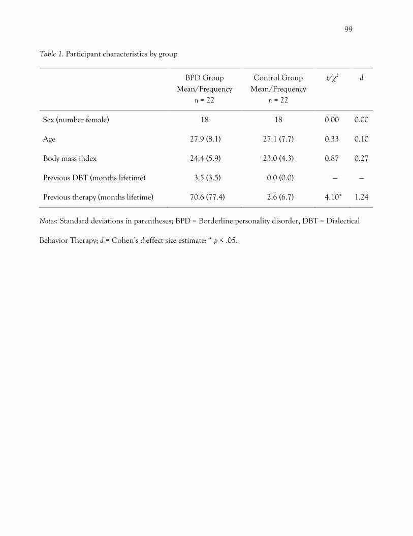
99
Table 1. Participant characteristics by group
BPD Group Mean/Frequency
n = 22
Control Group Mean/Frequency
n = 22
t/χ2 d
Sex (number female) 18 18 0.00 0.00
Age 27.9 (8.1) 27.1 (7.7) 0.33 0.10
Body mass index 24.4 (5.9) 23.0 (4.3) 0.87 0.27
Previous DBT (months lifetime) 3.5 (3.5) 0.0 (0.0) — —
Previous therapy (months lifetime) 70.6 (77.4) 2.6 (6.7) 4.10* 1.24
Notes: Standard deviations in parentheses; BPD = Borderline personality disorder, DBT = Dialectical
Behavior Therapy; d = Cohen’s d effect size estimate; * p < .05.
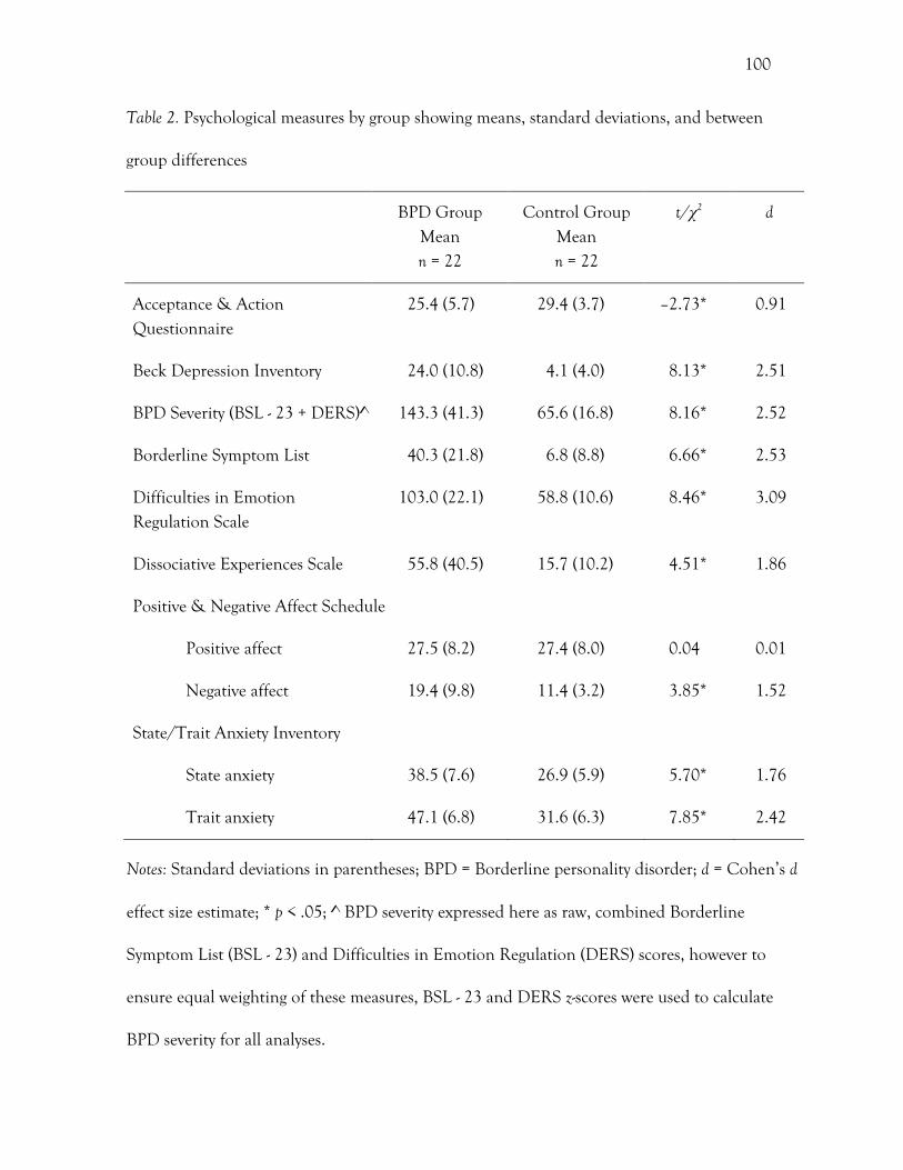
100
Table 2. Psychological measures by group showing means, standard deviations, and between
group differences
BPD Group Mean n = 22
Control Group Mean n = 22
t/χ2 d
Acceptance & Action Questionnaire
25.4 (5.7) 29.4 (3.7) –2.73* 0.91
Beck Depression Inventory 24.0 (10.8) 4.1 (4.0) 8.13* 2.51
BPD Severity (BSL - 23 + DERS)^ 143.3 (41.3) 65.6 (16.8) 8.16* 2.52
Borderline Symptom List 40.3 (21.8) 6.8 (8.8) 6.66* 2.53
Difficulties in Emotion Regulation Scale
103.0 (22.1) 58.8 (10.6) 8.46* 3.09
Dissociative Experiences Scale 55.8 (40.5) 15.7 (10.2) 4.51* 1.86
Positive & Negative Affect Schedule
Positive affect 27.5 (8.2) 27.4 (8.0) 0.04 0.01
Negative affect 19.4 (9.8) 11.4 (3.2) 3.85* 1.52
State/Trait Anxiety Inventory
State anxiety 38.5 (7.6) 26.9 (5.9) 5.70* 1.76
Trait anxiety 47.1 (6.8) 31.6 (6.3) 7.85* 2.42
Notes: Standard deviations in parentheses; BPD = Borderline personality disorder; d = Cohen’s d
effect size estimate; * p < .05; ^ BPD severity expressed here as raw, combined Borderline
Symptom List (BSL - 23) and Difficulties in Emotion Regulation (DERS) scores, however to
ensure equal weighting of these measures, BSL - 23 and DERS z-scores were used to calculate
BPD severity for all analyses.
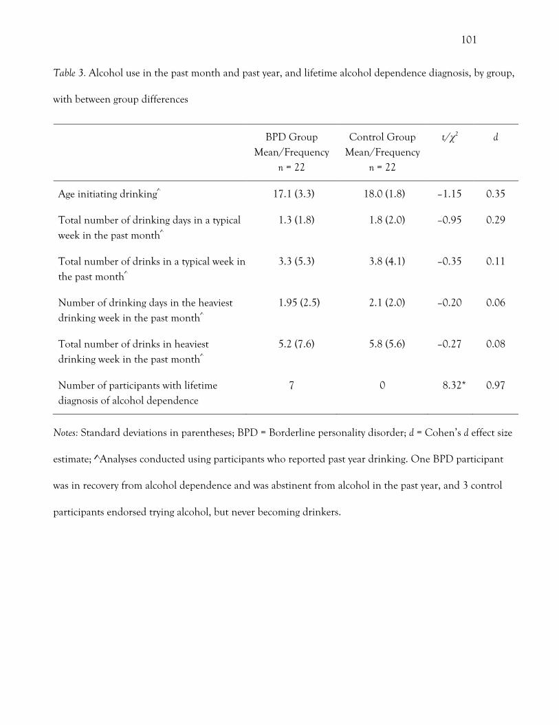
101
Table 3. Alcohol use in the past month and past year, and lifetime alcohol dependence diagnosis, by group,
with between group differences
BPD Group Mean/Frequency
n = 22
Control Group Mean/Frequency
n = 22
t/χ2 d
Age initiating drinking^ 17.1 (3.3) 18.0 (1.8) –1.15 0.35
Total number of drinking days in a typical week in the past month^
1.3 (1.8) 1.8 (2.0) –0.95 0.29
Total number of drinks in a typical week in the past month^
3.3 (5.3) 3.8 (4.1) –0.35 0.11
Number of drinking days in the heaviest drinking week in the past month^
1.95 (2.5) 2.1 (2.0) –0.20 0.06
Total number of drinks in heaviest drinking week in the past month^
5.2 (7.6) 5.8 (5.6) –0.27 0.08
Number of participants with lifetime diagnosis of alcohol dependence
7 0 8.32* 0.97
Notes: Standard deviations in parentheses; BPD = Borderline personality disorder; d = Cohen’s d effect size
estimate; ^Analyses conducted using participants who reported past year drinking. One BPD participant
was in recovery from alcohol dependence and was abstinent from alcohol in the past year, and 3 control
participants endorsed trying alcohol, but never becoming drinkers.
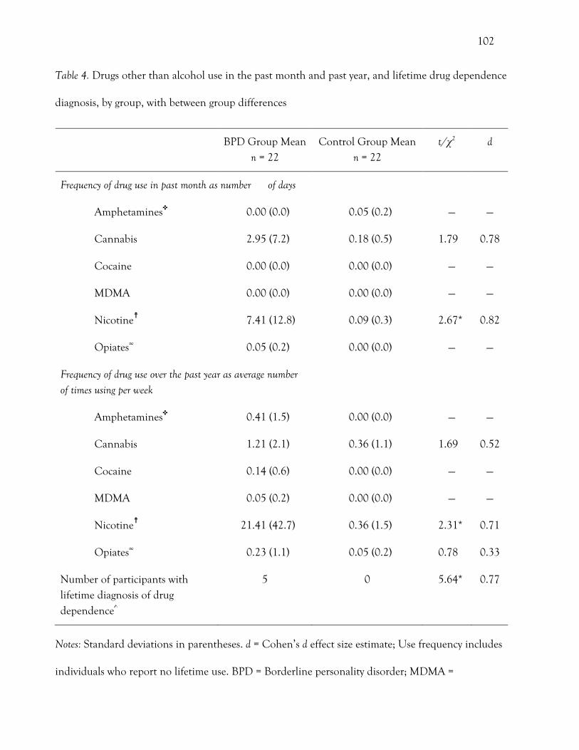
102
Table 4. Drugs other than alcohol use in the past month and past year, and lifetime drug dependence
diagnosis, by group, with between group differences
BPD Group Mean n = 22
Control Group Mean n = 22
t/χ2 d
Frequency of drug use in past month as number of days
Amphetamines✜ 0.00 (0.0) 0.05 (0.2) — —
Cannabis 2.95 (7.2) 0.18 (0.5) 1.79 0.78
Cocaine 0.00 (0.0) 0.00 (0.0) — —
MDMA 0.00 (0.0) 0.00 (0.0) — —
Nicotine☨ 7.41 (12.8) 0.09 (0.3) 2.67* 0.82
Opiates∞ 0.05 (0.2) 0.00 (0.0) — —
Frequency of drug use over the past year as average number of times using per week
Amphetamines✜ 0.41 (1.5) 0.00 (0.0) — —
Cannabis 1.21 (2.1) 0.36 (1.1) 1.69 0.52
Cocaine 0.14 (0.6) 0.00 (0.0) — —
MDMA 0.05 (0.2) 0.00 (0.0) — —
Nicotine☨ 21.41 (42.7) 0.36 (1.5) 2.31* 0.71
Opiates∞ 0.23 (1.1) 0.05 (0.2) 0.78 0.33
Number of participants with lifetime diagnosis of drug dependence^
5 0 5.64* 0.77
Notes: Standard deviations in parentheses. d = Cohen’s d effect size estimate; Use frequency includes
individuals who report no lifetime use. BPD = Borderline personality disorder; MDMA =

103
Methylenedioxymethamphetamine; * p < .05; ✜ Methamphetamine & prescription amphetamines
combined, ☨Cigarettes, e-cigarettes, and smokeless tobacco combined, ∞ Heroin, methadone, and
prescription opioids combined, ^ Reflects dependence to drugs other than alcohol and nicotine.
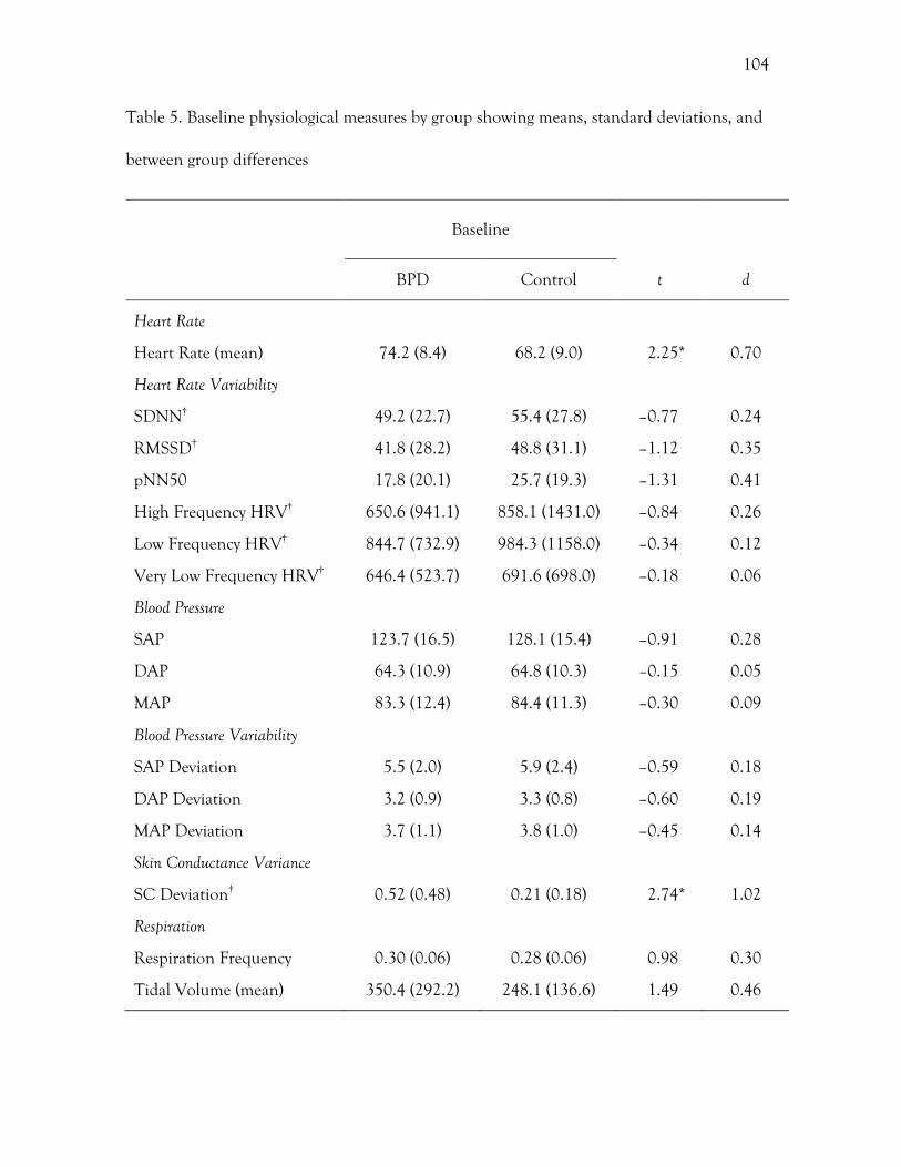
104
Table 5. Baseline physiological measures by group showing means, standard deviations, and
between group differences
Baseline
BPD Control t d
Heart Rate
Heart Rate (mean) 74.2 (8.4) 68.2 (9.0) 2.25* 0.70
Heart Rate Variability
SDNN† 49.2 (22.7) 55.4 (27.8) –0.77 0.24
RMSSD† 41.8 (28.2) 48.8 (31.1) –1.12 0.35
pNN50 17.8 (20.1) 25.7 (19.3) –1.31 0.41
High Frequency HRV† 650.6 (941.1) 858.1 (1431.0) –0.84 0.26
Low Frequency HRV† 844.7 (732.9) 984.3 (1158.0) –0.34 0.12
Very Low Frequency HRV† 646.4 (523.7) 691.6 (698.0) –0.18 0.06
Blood Pressure
SAP 123.7 (16.5) 128.1 (15.4) –0.91 0.28
DAP 64.3 (10.9) 64.8 (10.3) –0.15 0.05
MAP 83.3 (12.4) 84.4 (11.3) –0.30 0.09
Blood Pressure Variability
SAP Deviation 5.5 (2.0) 5.9 (2.4) –0.59 0.18
DAP Deviation 3.2 (0.9) 3.3 (0.8) –0.60 0.19
MAP Deviation 3.7 (1.1) 3.8 (1.0) –0.45 0.14
Skin Conductance Variance
SC Deviation† 0.52 (0.48) 0.21 (0.18) 2.74* 1.02
Respiration
Respiration Frequency 0.30 (0.06) 0.28 (0.06) 0.98 0.30
Tidal Volume (mean) 350.4 (292.2) 248.1 (136.6) 1.49 0.46
105
Notes. Standard deviations in parentheses; d = Cohen’s d effect size estimate; SDNN = Standard
deviation of all normal-to-normal intervals, RMSSD = Root of the mean squared differences of
successive normal-to-normal intervals, pNN50 = percent of normal-to-normal adjacent intervals
greater than 50ms, HRV = Heart rate variability, SAP = Systolic arterial pressure, DAP =
Diastolic arterial pressure, MAP = Mean arterial pressure, SC = Skin conductance; * p < .05;
† Logarithmically transformed data used for t-tests. BPD group n = 21 (after one influential
outlier was excluded), control group n = 22.
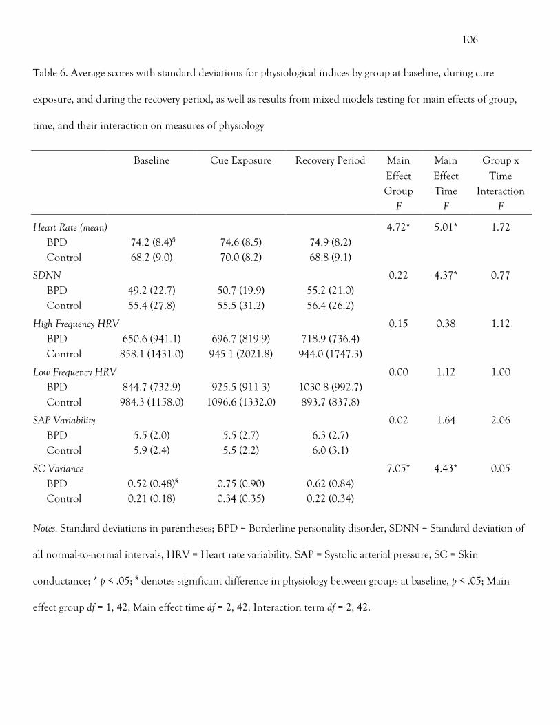
106
Table 6. Average scores with standard deviations for physiological indices by group at baseline, during cure
exposure, and during the recovery period, as well as results from mixed models testing for main effects of group,
time, and their interaction on measures of physiology
Notes. Standard deviations in parentheses; BPD = Borderline personality disorder, SDNN = Standard deviation of
all normal-to-normal intervals, HRV = Heart rate variability, SAP = Systolic arterial pressure, SC = Skin
conductance; * p < .05; § denotes significant difference in physiology between groups at baseline, p < .05; Main
effect group df = 1, 42, Main effect time df = 2, 42, Interaction term df = 2, 42.
Baseline Cue Exposure Recovery Period Main Effect Group
F
Main Effect Time
F
Group x Time
Interaction F
Heart Rate (mean) 4.72* 5.01* 1.72 BPD 74.2 (8.4)§ 74.6 (8.5) 74.9 (8.2) Control 68.2 (9.0) 70.0 (8.2) 68.8 (9.1)
SDNN 0.22 4.37* 0.77 BPD 49.2 (22.7) 50.7 (19.9) 55.2 (21.0) Control 55.4 (27.8) 55.5 (31.2) 56.4 (26.2)
High Frequency HRV 0.15 0.38 1.12 BPD 650.6 (941.1) 696.7 (819.9) 718.9 (736.4) Control 858.1 (1431.0) 945.1 (2021.8) 944.0 (1747.3)
Low Frequency HRV 0.00 1.12 1.00 BPD 844.7 (732.9) 925.5 (911.3) 1030.8 (992.7) Control 984.3 (1158.0) 1096.6 (1332.0) 893.7 (837.8)
SAP Variability 0.02 1.64 2.06 BPD 5.5 (2.0) 5.5 (2.7) 6.3 (2.7) Control 5.9 (2.4) 5.5 (2.2) 6.0 (3.1)
SC Variance 7.05* 4.43* 0.05 BPD 0.52 (0.48)§ 0.75 (0.90) 0.62 (0.84) Control 0.21 (0.18) 0.34 (0.35) 0.22 (0.34)
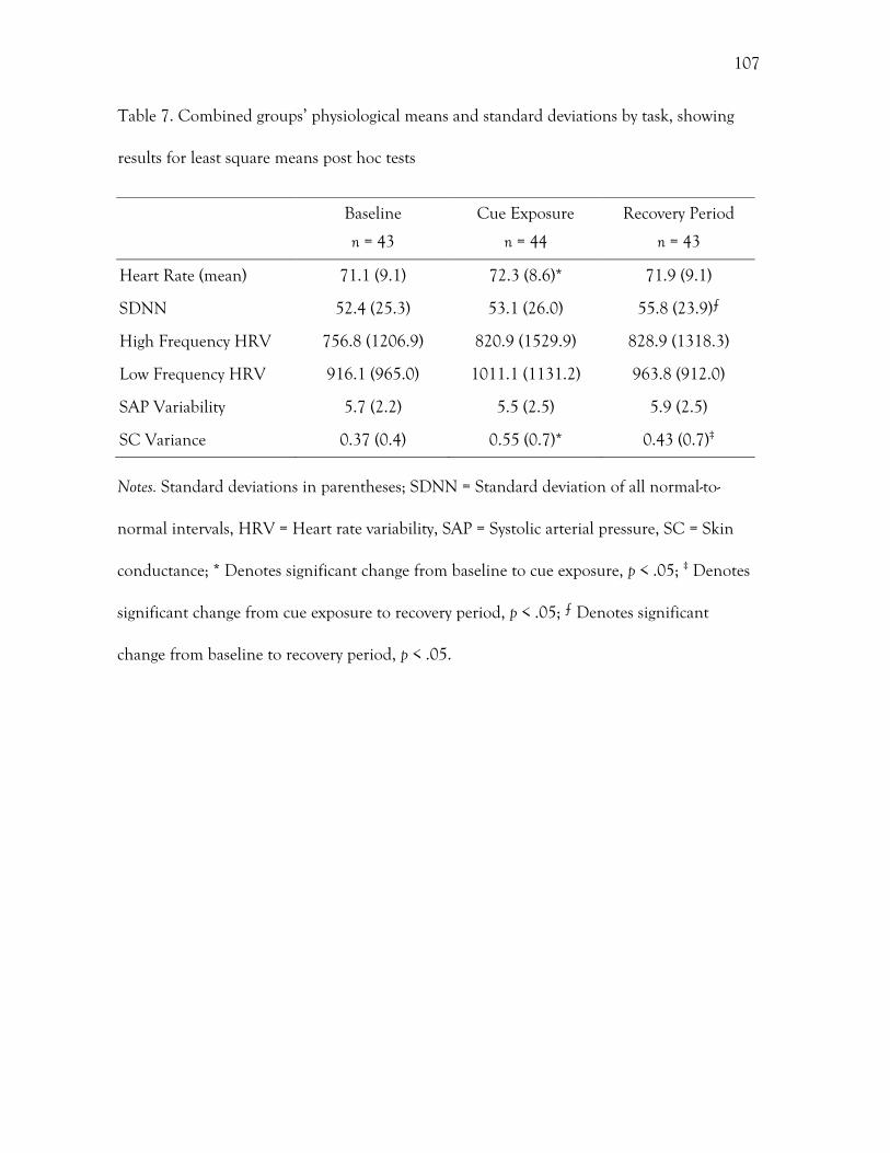
107
Table 7. Combined groups’ physiological means and standard deviations by task, showing
results for least square means post hoc tests
Notes. Standard deviations in parentheses; SDNN = Standard deviation of all normal-to-
normal intervals, HRV = Heart rate variability, SAP = Systolic arterial pressure, SC = Skin
conductance; * Denotes significant change from baseline to cue exposure, p < .05; ‡ Denotes
significant change from cue exposure to recovery period, p < .05; ⨍ Denotes significant
change from baseline to recovery period, p < .05.
Baseline
n = 43
Cue Exposure
n = 44
Recovery Period
n = 43
Heart Rate (mean) 71.1 (9.1) 72.3 (8.6)* 71.9 (9.1)
SDNN 52.4 (25.3) 53.1 (26.0) 55.8 (23.9)⨍
High Frequency HRV 756.8 (1206.9) 820.9 (1529.9) 828.9 (1318.3)
Low Frequency HRV 916.1 (965.0) 1011.1 (1131.2) 963.8 (912.0)
SAP Variability 5.7 (2.2) 5.5 (2.5) 5.9 (2.5)
SC Variance 0.37 (0.4) 0.55 (0.7)* 0.43 (0.7)‡
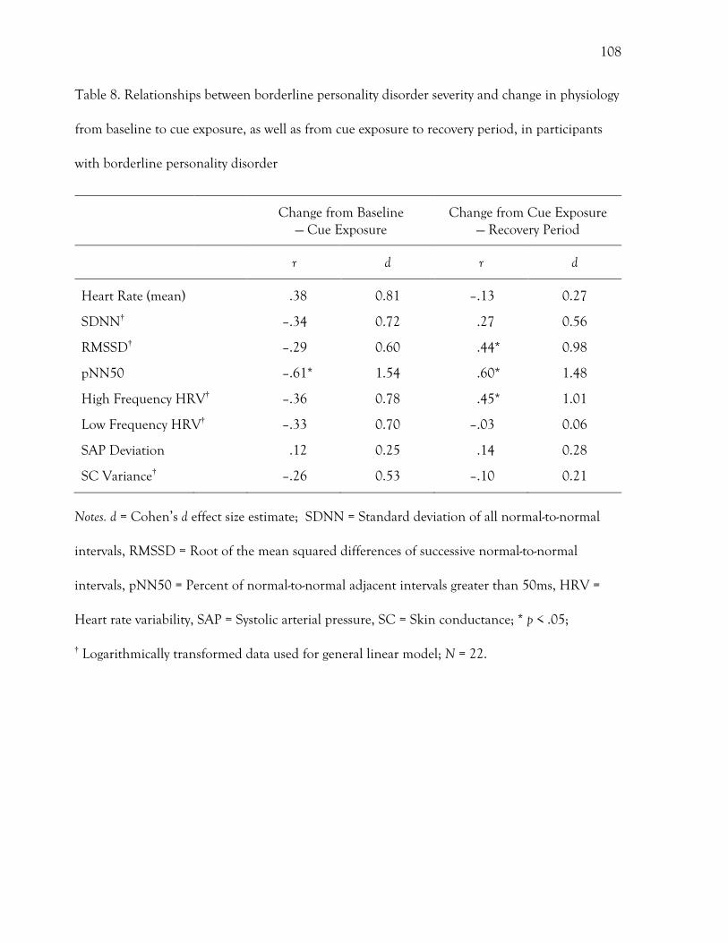
108
Table 8. Relationships between borderline personality disorder severity and change in physiology
from baseline to cue exposure, as well as from cue exposure to recovery period, in participants
with borderline personality disorder
Change from Baseline — Cue Exposure
Change from Cue Exposure — Recovery Period
r d r d
Heart Rate (mean) .38 0.81 –.13 0.27
SDNN† –.34 0.72 .27 0.56
RMSSD† –.29 0.60 .44* 0.98
pNN50 –.61* 1.54 .60* 1.48
High Frequency HRV† –.36 0.78 .45* 1.01
Low Frequency HRV† –.33 0.70 –.03 0.06
SAP Deviation .12 0.25 .14 0.28
SC Variance† –.26 0.53 –.10 0.21
Notes. d = Cohen’s d effect size estimate; SDNN = Standard deviation of all normal-to-normal
intervals, RMSSD = Root of the mean squared differences of successive normal-to-normal
intervals, pNN50 = Percent of normal-to-normal adjacent intervals greater than 50ms, HRV =
Heart rate variability, SAP = Systolic arterial pressure, SC = Skin conductance; * p < .05;
† Logarithmically transformed data used for general linear model; N = 22.
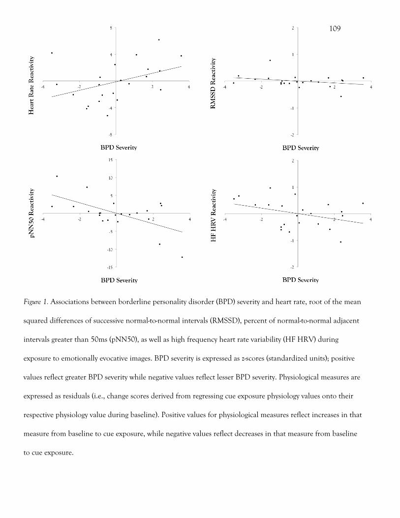
109
Figure 1. Associations between borderline personality disorder (BPD) severity and heart rate, root of the mean
squared differences of successive normal-to-normal intervals (RMSSD), percent of normal-to-normal adjacent
intervals greater than 50ms (pNN50), as well as high frequency heart rate variability (HF HRV) during
exposure to emotionally evocative images. BPD severity is expressed as z-scores (standardized units); positive
values reflect greater BPD severity while negative values reflect lesser BPD severity. Physiological measures are
expressed as residuals (i.e., change scores derived from regressing cue exposure physiology values onto their
respective physiology value during baseline). Positive values for physiological measures reflect increases in that
measure from baseline to cue exposure, while negative values reflect decreases in that measure from baseline
to cue exposure.
109
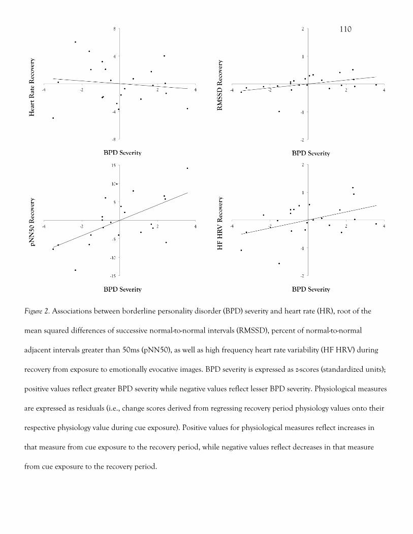
110
Figure 2. Associations between borderline personality disorder (BPD) severity and heart rate (HR), root of the
mean squared differences of successive normal-to-normal intervals (RMSSD), percent of normal-to-normal
adjacent intervals greater than 50ms (pNN50), as well as high frequency heart rate variability (HF HRV) during
recovery from exposure to emotionally evocative images. BPD severity is expressed as z-scores (standardized units);
positive values reflect greater BPD severity while negative values reflect lesser BPD severity. Physiological measures
are expressed as residuals (i.e., change scores derived from regressing recovery period physiology values onto their
respective physiology value during cue exposure). Positive values for physiological measures reflect increases in
that measure from cue exposure to the recovery period, while negative values reflect decreases in that measure
from cue exposure to the recovery period.
110
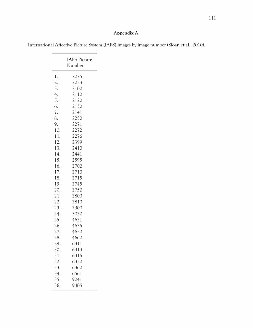
111
Appendix A.
International Affective Picture System (IAPS) images by image number (Sloan et al., 2010).
IAPS Picture Number
1. 20252. 20533. 21004. 21105. 21206. 21307. 21418. 22509. 227110. 227211. 227612. 239913. 241014. 244115. 259516. 270217. 271018. 271519. 274520. 275221. 280022. 281023. 290024. 302225. 462126. 463527. 465028. 466029. 631130. 631331. 631532. 635033. 636034. 656135. 904136. 9405