
Dissection of the Topology, Structure and Function of the INO80
113
Dissertation zur Erlangung des Doktorgrades der Fakultät für Chemie und Pharmazie der Ludwig-Maximilians-Universität München Dissection of the Topology, Structure and Function of the INO80 Chromatin Remodeler Alessandro Tosi aus München, Deutschland 2013
Transcript of Dissection of the Topology, Structure and Function of the INO80

Dissertation zur Erlangung des Doktorgrades
der Fakultät für Chemie und Pharmazie
der Ludwig-Maximilians-Universität München
Dissection of the Topology, Structure and Function of the INO80 Chromatin
Remodeler
Alessandro Tosi
aus
München, Deutschland
2013

Erklärung
Diese Dissertation wurde im Sinne von §7 der Promotionsordnung vom 28. November 2011 von
Herrn Prof. Dr. Karl-Peter Hopfner betreut.
Eidesstattliche Versicherung
Diese Dissertation wurde eigenständig und ohne unerlaubte Hilfe erarbeitet.
München, am 07.10.2013
______________________
Alessandro Tosi
Dissertation eingereicht am 07.10.2013
1. Gutachter: Herr Prof. Dr. Karl-Peter Hopfner
2. Gutachter: Herr Prof. Dr. Roland Beckmann
Mündliche Prüfung am 22.11.2013

My PhD thesis has been prepared from March 2010 to Oktober 2013 in the laboratory of Prof.
Dr. Karl-Peter Hopfner at the Gene Center of the Ludwig-Maximilians-Univeristy of Munich
(LMU).
Parts of this thesis have been published in scientific journals:
Alessandro Tosi*, Caroline Haas*, Franz Herzog*, Andrea Gilmozzi, Otto Berninghausen,
Charlotte Ungewickell, Christian B. Gerhold, Kristina Lakomek, Ruedi Aebersold, Roland
Beckmann and Karl-Peter Hopfner: Structure and subunit topology of the INO80 chromatin
remodeler and its nucleosome complex. Cell, Volume 154, Issue 6, 1207-1219, 12 September
2013.
*These authors contributed equally
Parts of this thesis have been presented at international conferences:
Poster presentation at the EMBO practical course “Protein-protein and protein-nucleic acid
cross-linking and mass spectrometry”, Göttingen, Germany, 23-29. October 2011.
Poster presentation at the “Epigenetics & Chromatin: Interactions and processes” conference,
Boston, USA, 11-13. March 2013.
Oral and poster presentation at the “Helicases and nucleic acid translocases” EMBO, Harden
conference, Cambridge, UK, 04-08. August 2013.

Table of Contents 4
1 Table of Contents
1 TABLE OF CONTENTS 4
2 SUMMARY 7
3 INTRODUCTION 9
3.1 DYNAMIC CHROMATIN ENVIRONMENT 9
3.2 SWI2/SNF2 REMODELERS 11
3.3 CHROMATIN REMODELERS 13
3.4 THE INO80/SWR1 FAMILY 14
3.5 INO80 COMPLEX 16
3.5.1 THE COMPONENTS OF THE INO80 COMPLEX 16
3.5.2 THE CHROMATIN REMODELING COMPLEX INO80 IS INVOLVED IN DNA PROCESSING AND METABOLISM 20
3.5.3 INO80 MEDIATES CHECKPOINT PATHWAYS 21
3.6 HYBRID APPROACHES HELP TO DISSECT THE MOLECULAR ARCHITECTURE OF LARGE COMPLEXES 22
4 RESULTS 25
4.1 RECONSTITUTION OF A NUCLEOSOME 25
4.2 A NOVEL PURIFICATION PROCEDURE OF INO80 IMPROVES COMPLEX HOMOGENEITY 27
4.3 NANOBODIES AGAINST THE INO80 COMPLEX 31
4.4 ASSESSMENT OF THE ACTIVITY OF THE PURIFIED INO80 33
4.5 CHEMICAL CROSS-LINKING AND MASS SPECTROMETRY ANALYSIS OF THE INO80 COMPLEX 35
4.5.1 MAPPING OF SUBUNIT INTERACTIONS BY CROSS-LINKING AND MASS SPECTROMETRY 35
4.5.2 SUBUNIT TOPOLOGY AND STRUCTURAL MODULES OF INO80 38
4.6 VALIDATION OF INO80’S MODULES IN VIVO 40
4.7 STRUCTURE OF THE INO80 COMPLEX 41
4.7.1 ELECTRON MICROSCOPY OF INO80 41
4.7.2 TOWARDS A CRYSTAL STRUCTURE OF INO80 43
4.8 RVB1/2 FORM A HETERO-DODECAMER 43
4.8.1 RVB1/2 IS COMPOSED OF TWO HEXAMERIC RINGS IN INO80 43
4.8.2 RVB1/2 ASSEMBLE AS HETERO-HEXAMERS INTERACTING VIA THE DOMAIN 2 WITHIN INO80 44

Table of Contents 5
4.9 THE CATALYTIC CORE OF INO80 47
4.9.1 THE SWI2/SNF2 DOMAIN OF INO80 47
4.9.2 EXPRESSION AND PURIFICATION STUDIES OF THE ATPASE DOMAIN OF INO80 WITH IES2 AND RVB1 48
4.10 LOCALIZATION OF THE ARP8-, ARP5- AND NHP10-MODULE 48
4.11 THE NHP10-MODULE 50
4.11.1 RECONSTITUTION OF THE NHP10 MODULE: NHP10-IES1-IES3-IES5-INO80 (N-TERMINUS) 50
4.11.2 THE NHP10 SUB-COMPLEX FORMS A STABLE DNA COMPLEX 53
4.12 DISSECTING THE FUNCTION AND ACTIVITY OF INO80-MODULES 55
4.12.1 FUNCTIONAL CHARACTERIZATION OF INO80-MODULES 55
4.12.2 INTERACTION AND VISUALIZATION OF AN INO80-NUCLESOME COMPLEX 57
4.13 INO80 FORMS A STABLE COMPLEX WITH THE MEC1 COMPLEX 61
5 DISCUSSION 63
5.1 HYBRID VIEW ON INO80 63
5.2 THE CHROMATIN REMODELER INO80 64
5.3 STRUCTURE OF RVB1/2 IN THE INO80 COMPLEX 65
5.4 THE NHP10-MODULE 68
5.5 THE ARP8-MODULE 69
5.6 THE NUCLEOSOME REMODELER INO80 70
5.7 CHROMATIN REGULATORS FACILITATE TRANSCRIPTION 75
6 MATERIAL AND METHODS 77
6.1 MATERIALS 77
6.2 YEAST STRAINS 78
6.3 PLASMID LISTS 78
6.4 OLIGONUCLEOTIDE LIST 79
6.5 BUFFER LIST 81
6.6 MOLECULAR BIOLOGY METHODS 82
6.6.1 CLONING IN BACTERIA 82
6.6.2 CLONING IN YEAST 83
6.7 PROTEIN EXPRESSION AND PURIFICATION 83
6.7.1 PROTEIN EXPRESSION IN E. COLI 83

Table of Contents 6
6.7.2 PROTEIN EXPRESSION IN INSECT CELLS 84
6.7.3 CULTURING OF YEAST 84
6.7.4 SDS-PAGE AND WESTERN BLOT ANALYSIS 85
6.7.5 SILVER STAINING 86
6.7.6 PURIFICATION OF HISTONES AND RECONSTITUTION OF NUCLEOSOMES 87
6.7.7 ESTABLISHMENT OF AN INO80 PURIFICATION PROTOCOL 89
6.7.8 PURIFICATION OF THE SWI2/SNF2 SUB-COMPLEX 92
6.7.9 PURIFICATION OF THE NHP10 SUBCOMPLEX 92
6.8 GENERATION OF INO80 BINDING NANOBODIES 93
6.9 CROSS-LINKING AND MASS SPECTROMETRY 94
6.9.1 TITRATION OF CROSS-LINKER 94
6.9.2 SAMPLE PREPARATION FOR MASS SPECTROMETRY ANALYSIS 94
6.9.3 SAMPLE ANALYSIS 95
6.10 ANALYSIS OF SUBUNIT COMPOSITION OF INO80’S KNOCK-OUT MUTANTS 96
6.11 ANALYSIS OF DISTANT RESTRAINTS 96
6.12 FUNCTIONAL ASSAYS 97
6.12.1 ELECTROPHORETIC MOBILITY SHIFT ASSAYS 97
6.12.2 REMODELING ASSAY 97
6.12.3 ATP HYDROLYSIS ASSAYS 98
7 REFERENCES 99
8 CURRICULUM VITAE 111
9 ACKNOWLEDGMENTS 113

Summary 7
2 Summary
Eukaryotic genomes are organized into highly condensed chromatin. This packaging
obviously impedes essential DNA mediated processes. ATP-dependent chromatin remodelers
are therefore required to establish a dynamic chromatin environment. The chromatin
remodeler INO80 is involved in various fundamental nuclear processes such as DNA repair,
DNA replication and transcription. INO80 is thought to contribute to these processes by
controlling genome wide levels of the histone variant H2A.Z. The INO80 chromatin remodeler
is a macro-molecular complex composed of >15 subunits and a molecular mass of ~1.3 MDa.
INO80 is found in human, fly and yeast. INO80 contains core subunits, which are conserved
across species, as well as species-specific proteins. Not much was known about the
organization of the INO80 subunits and their contribution to chromatin remodeling.
Therefore, a hybrid approach was applied on yeast INO80 combining chemical cross-linking
and mass spectrometry (XL-MS) (in collaboration with Franz Herzog, Ruedi Aebersold’s group,
ETH, Zurich), electron microscopy (EM) (in collaboration with Caroline Haas, Roland
Beckmann’s group, Gene Center, Munich) and biochemical analysis. For this, firstly the
purification of INO80 was established. In order to yield sufficient amounts of highly purified
and monodisperse complex, INO80 was purified endogenously from yeast by a combination
of affinity and chromatography methods. In addition, nanobodies targeting the INO80
complex were generated that could yield even larger amounts of INO80 in the future. EM
analysis revealed that INO80 is an embryo-shaped particle with a dynamic head-neck-body-
foot architecture that can undergo large conformational changes. XL-MS unraveled the
interaction map of the INO80 complex. The analysis of INO80 deletion mutants verified the
observed interactions in vivo and proved the modular architecture of INO80. Additionally, the
gained knowledge allowed the design and purification of stable and novel sub-complexes that
could improve crystallization behavior. An integration of the results from different techniques
deepened our understanding of the molecular architecture of INO80. The enigmatic subunits
Rvb1 and 2 assemble as a dodecamer composed of two hetero-hexameric rings within the
head of the INO80 complex. Rvb1/2 is flanked by the Swi2/Snf2 ATPase of Ino80 and the actin

Summary 8
related protein (Arp) 5 in the neck. The Nhp10-module localizes to the body and the Arp8-
module to the foot. Biochemical analysis showed that the Nhp10-module is a high affinity
DNA/nucleosome binder. The Nhp10-module might together with the Arp8-module target
INO80 to chromatin. The Arp5-module is catalytically crucial for nucleosome remodeling and
senses the histone entity in chromatin. In order to map interaction sites to the substrate,
INO80-nucleosome complexes were analyzed by XL-MS and were visualized by EM. Two-
dimensional class averages showed that the nucleosome bound to the central groove of
INO80 and was flanked by the head and foot module. The nucleosome was oriented in
respect to INO80 as the H2A/H2B dimer- the moiety to be exchanged- was in contact with
subunits situated in the neck. All INO80 modules contribute to nucleosome binding and the
observed flexibility proposes a mechanism of how INO80 may remodel its substrate. This
study established a structural and functional framework of these large remodelers. The
investigation of the interaction with the checkpoint kinase Mec1 will contribute to the
understanding of the obscure signaling of INO80.

Introduction 9
3 Introduction
3.1 Dynamic chromatin environment
Eukaryotic genomes are organized into chromatin to compact DNA. The basic unit of
chromatin is the nucleosome that consists of 147 base pairs of DNA wrapped in approximately
two superhelical turns around a histone octamer composed of two heterodimers of histone
H2A-H2B and a histone hetero-tetramer (H3-H4)2 (Luger et al., 1997). Histones in principal
consist of two functional and structural distinct folds, the flexible N-terminal tail and the
histone fold. The histone fold is composed of three α-helices, which are connected by two loops
(α1-L1-α2-L2-α3) (Luger et al., 1997). The nucleosome core particle is based on protein-protein
and protein-DNA interactions. The histones form dimer pairs that assemble via an interaction of
the histone folds in a head-to-tail-arrangement in the characteristic handshake manner (Arents
et al., 1991). The assembled octamer contacts the nucleosomal DNA at its entire length. The
histone folds interact with the DNA minor groove at the inner site of the supercoil (Luger et al.,
1997). The DNA entry and exit sites are exclusively organized by the N-terminus of H3.
Figure 1. The basic unit of chromatin, the nucleosome. A) Structure of the fly nucleosome core particle (PDB:2pyo, (Clapier et al., 2008)). Histones are colored: H2A, yellow; H2B, red; H3, blue and H4, green and depicted as pipes. The dyad axis (φ) and DNA entry/exit sited are indicated. B) The nucleosomal (blue circles) distribution at yeast promoters and genes. Peaks represent consensus distribution of nucleosomes relative to transcription start site (TSS). Green indicates high degree of H2A.Z, acetylation, H3K4 methylation and phasing. Figure adapted from (Pugh, 2013).

Introduction 10
The nucleosome core particle is the repetitive unit of nucleosomal arrays defining the
11 nm structure (“bead on a string”). The linker histone H1 further compacts the arrays in the
condensed 30 nm chromatin fiber (Robinson et al., 2006), which is then further compacted in
the highest order chromosome (Felsenfeld and Groudine, 2003). The packaging into chromatin
obviously impedes fundamental DNA-dependent nuclear processes that require free access to
genomic information as DNA replication, repair and transcription. A dynamic chromatin
environment is generated by (i) remodeling of nucleosomes, (ii) chemical modification of
histones or incorporation of variants, (iii) non-histone DNA binding proteins and (iv) non-coding
RNAs.
Chromatin remodelers are versatile tools that catalyze a broad range of chromatin
changing reactions including sliding of an octamer across the DNA (nucleosome sliding),
changing the conformation of nucleosomal DNA and changing the composition of the octamers
(histone variant exchange). Histone variants differ from the canonical histones in their primary
sequence. They show different physiochemical properties compared to canonical histones and
can alter protein-protein and protein-DNA interactions thereby changing the chromatin
structure (Billon and Cote, 2012). Two major variants of histone H3 have been studied
extensively, the centromer specific CenH3 and H3.3. Variants of H2A are much more abundant
in number, including H2A.X and H2A.Z (Zlatanova and Thakar, 2008), which are required for cell
stability and viability (Redon et al., 2002). H2A.X is phosphorylated upon DNA damage by DNA-
activated protein kinases from the phosphatidylinositol 3-kinase-related kinases (PIKKs) family
that mediate DNA damage response. H2A.Z associates with actively transcribed chromatin
(Stargell et al., 1993). H2A.Z shares only 60% sequence identity with the canonical H2A, but is
conserved within higher eukaryotes (Jackson et al., 1996). Major differences within H2A.Z are in
the C-terminal docking domain that organizes the penultimate 10 bp of the DNA (Shukla et al.,
2011) and in the acidic patch that is involved in the interaction with interacting proteins as for
instance the viral latency-associated nuclear antigen (LANA) peptide (Barbera et al., 2006; Luger
et al., 2012). The overall crystal structure of a H2A.Z containing nucleosome is similar to the
crystal structure of a nucleosome containing canonical histones, but the interaction between
the histone pairs is subtly destabilized (Suto et al., 2000). However, further studies could not

Introduction 11
unambiguously clarify, if H2A.Z promotes destabilization or stabilization of chromatin (Abbott
et al., 2001; Fan et al., 2002; Placek et al., 2005; Thambirajah et al., 2006; Zhang et al., 2005).
The interaction between the acidic patch of H2A.Z and the N-terminus of H4 increases intra-
molecular folding of nucleosomal arrays (Fan et al., 2002). Nucleosome arrays are interrupted
by nucleosome free regions (NFR), which normally contain the promoter sequences. The
nucleosomes flanking the array are referred as -1 and +1 nucleosomes (Bernstein et al., 2004;
Jiang and Pugh, 2009; Yuan et al., 2005). The -1 nucleosome is followed by a NFR, the 5’ NFR
and then the transcriptions start site (TSS). Among all genome wide distributed nucleosomes,
the +1 is the tightest positioned or phased nucleosome (Mavrich et al., 2008). The combination
of histone variants H2A.Z and H3.3 in one nucleosome lead to the most unstable state of
chromatin (Jin and Felsenfeld, 2007) and H2A.Z incorporated in the +1 nucleosome was
suggested to destabilize this region to accelerate gene activation (Guillemette et al., 2005; Jin
and Felsenfeld, 2007; Li et al., 2005; Meneghini et al., 2003; Zhang et al., 2005). The
stabilization could also be influenced by post-translational modifications on H2A.Z (Billon and
Cote, 2012) and by the number of H2A.Z incorporated per nucleosome. A nucleosome that
contains two copies of H2A.Z-H2B dimers was more unstable that one with only one copy (Luk
et al., 2010; Weber et al., 2010).
To establish a dynamic chromatin environment specific variants are incorporated into
the nucleosome by specialized ATP-dependent remodeling complexes.
3.2 Swi2/Snf2 remodelers
In general, chromatin remodelers are versatile molecular machines that use the energy of
ATP hydrolysis in order to disrupt protein-protein or protein-nucleic acid contacts. The motor
domain that creates this chemo-mechanical force is a Swi2/Snf2 ATPase. The Snf2 protein was
originally discovered to regulate mating type switching (SWI) and sucrose fermentation
(Sucrose Non Fermenting) (SNF) by a genetic screen and was subsequently identified as the
catalytic subunit of the SWI/SNF complex (Sudarsanam and Winston, 2000). Members of the
Introduction 12
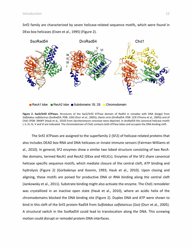
Snf2 family are characterized by seven helicase-related sequence motifs, which were found in
DExx box helicases (Eisen et al., 1995) (Figure 2).
Figure 2. Swi2/Snf2 ATPases. Structures of the Swi2/Snf2 ATPase domain of Rad54 in complex with DNA (beige) from Solfolubus solfataricus (SsoRad54; PDB: 1Z63 (Durr et al., 2005)), Danio rerio (DroRad54; PDB: 1Z3I (Thoma et al., 2005)) and of Chd1 (PDB: 3MWY (Hauk et al., 2010) from Saccharomyces cerevisae were depicted. In droRad54 the canonical helicase motifs I, II, III, IV, V and VI are indicated. The chromodomain of Chd1 contacts both ATPase lobes and occupies the DNA binding cleft.
The Snf2 ATPases are assigned to the superfamily 2 (SF2) of helicase-related proteins that
also includes DEAD box RNA and DNA helicases or innate immune sensors (Fairman-Williams et
al., 2010). In general, SF2 enzymes show a similar two lobed structure consisting of two RecA-
like domains, termed RecA1 and RecA2 (DExx and HELICc). Enzymes of the SF2 share canonical
helicase specific sequence motifs, which mediate closure of the central cleft, ATP binding and
hydrolysis (Figure 2) (Gorbalenya and Koonin, 1993; Hauk et al., 2010). Upon closing and
aligning, these motifs are poised for productive DNA or RNA binding along the central cleft
(Jankowsky et al., 2011). Substrate binding might also activate the enzyme. The Chd1 remodeler
was crystallized in an inactive open state (Hauk et al., 2010), where an acidic helix of the
chromodomains blocked the DNA binding site (Figure 2). Duplex DNA and ATP were shown to
bind in this cleft of the Snf2 protein Rad54 from Sulfolobus solfataricus (Sso) (Durr et al., 2005).
A structural switch in the SsoRad54 could lead to translocation along the DNA. This screwing
motion could disrupt or remodel protein-DNA interfaces.

Introduction 13
The Snf2 ATPase folds have been shown to mediate the remodeling reaction of multi-
subunit chromatin remodelers. The ATPase binds to nucleosomal DNA and provides the major
ATP hydrolysis activity (Cote et al., 1994; Shen et al., 2000) and thus delivers energy to the core
remodeling reaction.
3.3 Chromatin remodelers
Chromatin remodelers are classically divided into 4 families: SWI/SNF, ISWI (imitation switch),
Mi-2/CHD (chromodomain-helicase-DNA-binding) and INO80 (inositol auxotroph mutant 80).
The SWI/SNF family is the best structural characterized family of large remodelers. They
catalyze several remodeling events depending of the chromatin context including sliding and
eviction of the octamers (Clapier and Cairns, 2009, 2012; Gangaraju and Bartholomew, 2007).
Electron microscopy (EM) studies on members of the SWI/SNF as RSC (remodel the structure of
chromatin), the human homolog PBAF and the SWI/SNF complex itself are available (Asturias et
al., 2002; Chaban et al., 2008; Dechassa et al., 2008; Leschziner, 2011; Leschziner et al., 2005;
Leschziner et al., 2007; Skiniotis et al., 2007; Smith et al., 2003). The RSC and PBAF complex are
globular complexes with a C-shaped architecture and an obvious binding pocket that could
accommodate a nucleosome (Chaban et al., 2008; Leschziner, 2011; Leschziner et al., 2005). In
the structure of a RSC-nucleosome complex the density could not account for the complete
nucleosome and the authors suggested that the remodeling activity of the complex could have
partially disrupted the nucleosome particle (Chaban et al., 2008). Despite the presence of
several conserved subunits including actin related proteins 7 and 9 (Arp) the structure of the
RSC-related SWI/SNF remodeler is somewhat different and has no obvious nucleosome binding
groove, but it was proposed that it could occur at a large depression (Dechassa et al., 2008;
Smith et al., 2003).
The ISWI/ACF (ATP dependent chromatin-assembly factor) remodelers contain two to
four subunits and are thus smaller than the SWI/SNF family complexes (Clapier and Cairns,
2009; Gangaraju and Bartholomew, 2007). In contrast to the SWI/SNF family that randomizes

Introduction 14
arrays, ISWI remodelers evenly space nucleosomes and are implicated in gene silencing and
condensation. In addition to their catalytic Swi2/Snf2 ATPase, ISWI remodelers also contain
auxiliary domains and subunits. The HAND-SANT-SLIDE domain is located C-terminally of the
ATPase in flies ISWI (Grune et al., 2003). SANT-SLIDE domains recognize linker DNA and
nucleosomes and target the complex to the substrate. How ISWI remodelers space
nucleosomes is still under debate. One model suggests that the ISWI is bound to two
nucleosomes simultaneously and pulls them together until its helical linker-DNA-binding
domain-SLIDE-SANT prevents further movement and thus works as a molecular ruler (Yamada
et al., 2011). In the other scenario each of the two ISWI protomers take turns in moving the
nucleosome on either side with the protomer at the longer linker DNA translocating more
efficiently and frequently (Blosser et al., 2009; Racki et al., 2009).
CHD and Mi-2 remodelers have characteristic N-terminal tandem chromodomains
reviewed in Seeber et al., 2012. Interestingly, in Chd1 the chromodomains contact the
Swi2/Snf2 ATPase lobes and thereby disrupt the DNA engagement (Hauk et al., 2010). This
keeps Chd1 in an auto-inhibited state that could be released by nucleosomal DNA binding. The
chromodomains target Chd1 to lysine 4-methylated H3 tails, which is a hallmark of actively
transcribed chromatin (Flanagan et al., 2005). In contrast, Chd3 or Chd4 are members of the
Mi-2/NURD complex (nucleosome remodeling deacetylase) that deacetylates chromatin and
thus represses transcription (Seeber et al., 2013b). CHD1 complex has various functions and
was shown to assemble, slide and space nucleosomes (Lusser et al., 2005; Stockdale et al.,
2006). It can even incorporate the histone variant H3.3 in vivo (Konev et al., 2007).
3.4 The INO80/SWR1 family
The INO80 family includes following complexes: INO80 and SWR1 (sick with rat8 or SWI/SNF
related) in Saccharomyces cerevisiae (S. c.); INO80, SRCAP (Snf2-related CBP activator protein)
and p400 in mammals and INO80 and p400 in Drosophila melanogaster (D. melanogaster)
(Table 1) (reviewed in (Bao and Shen, 2011; Billon and Cote, 2012; Morrison and Shen, 2009)).
INO80/SWR1 are involved in various chromatin related processes (see 3.5) and contribute to
the genome wide distribution of the histone variant H2A.Z (Kobor et al., 2004; Mizuguchi et al.,

Introduction 15
2004; Papamichos-Chronakis et al., 2011). According to the dogma, SWR1 incorporates H2A.Z
while INO80 evicts H2A.Z in a unidirectional and stepwise manner. Both complexes show a
strong preference for the -1 and +1 nucleosome flanking the NFR (Yen et al., 2012; Yen et al.,
2013). The NFR is sufficient to target SWR1 and histone acetylation has a positive effect on this
recruitment. The cooperative relationship was shown to be a hierarchical one (Ranjan et al.,
2013). Higher eukaryotic SRCAP and p400 have been shown to harbor H2A exchange functions,
too (Kusch et al., 2004; Ruhl et al., 2006). The p400 subunit is associated with the Tip60
complex, which is an acetyltransferase. This relationship physically merges the yeast SWR1 with
the NuA4 (nucleosomal acetyltransferase of H4) histone acetyltransferase complex (Auger et
al., 2008; Billon and Cote, 2012; Doyon et al., 2004).
Table 1 Homologous INO80, SWR1 and SRCAP complexes. Subunits of the INO80 and SWR1 complex were assigned in homologous features from S. cerevisiae and Homo sapiens. Conserved domains were identified by pFAM search. Used abbrevations: BAF53A, BRG1-associated factor 53A; CCDC95, coiled-coil domain-containing 95; DMAP1, DNA methyltransferase 1-associated protein 1; GAS41, glioma amplified sequence 41; MCRS1, microspherule protein 1; NFRKB, nuclear factor related to κB-binding protein; SRCAP, SNF2-related CBP activator protein; Swc, SWR1 complex; UCH37, ubiquitin C-terminal hydrolase 37; XPG, xeroderma pigmentosa group G; Yaf9, yeast AF9; YEATS, Yaf9, ENL, AF9, Taf14, Sas5; YY1, yin yang 1; Znf-HIT1, zinc finger-His triad protein 1. The table was adapted from (Morrison and Shen, 2009).
INO80 complex SWR1 complex
Subunit type S. cerevisiae Human S. cerevisiae Human
Swi2/Snf2 ATPase Ino80 INO80 Swr1 SRCAP
RuvB-like Rvb1 and Rvb2 RUVBL1 and RUVBL2 Rvb1 and Rvb2 RUVBL1 and RUVBL2
Actin Act β-Actin Act β-Actin
Actin related proteins Arp4, Arp5 and
Arp8
BAF53, Arp5 and Arp8 Arp4 and Arp6 BAF53 and Arp6
YEATS Taf14 - Yaf9 GAS41
YL-1 Ies6 IES6 Swc2 YL1
PAPA-1 Ies2 IES2 - -
DNA binding subunit
(domain)
Nhp10 (HMG-box) YY1 (Zn-finger C2H2) Swc3
(SANT/myb)
XPG (H3TH)
Non conserved Ies1, Ies3, Ies4,
Ies5
Amida, CCDC95,
FLJ20309,
MCRS1, NFRKB, UCH37
Bdf1, Swc3 - 7 DMAP1, GAS41, tubulin, ZnF-
HIT1
The INO80 family is evolutionary conserved owing to the high degree of homology in the
Swi2/Snf2 ATPase containing subunits, which share the unique insertion loop between the

Introduction 16
RecA1 and RecA2 domains. The INO80 and SWR1 remodelers are both large multi-subunit
complexes with at least 14 components. This class of chromatin remodelers has been the
structurally most obscure.
Figure 3. Composition of INO80 and SWR1. Subunit organization of the budding yeast INO80 and SWR1 is depicted with the state of knowledge before this study. The Ino80 and Swr1 subunits are the assembly platforms for the specific sub-complexes. The figure was adapted from (Bao and Shen, 2011).
The N- and C-terminal regions of yeast Swr1 recruit the Bdf1-Arp4-Act-Swc4-Yaf9-Swc7
module (N-module) and Swc3-Swc2-Arp6-Swc6-Rvb1/2 (C-module), respectively (Wu et al.,
2005; Wu et al., 2009) (Figure 3). The composition of INO80 is described in detail below.
Deletion of the insertion of the split ATPase of Swr1 lead to a loss of Rvb1/2 (RuvB-like) (Wu et
al., 2005).In addition to the Rvb1/2, the SWR1 and INO80 complexes share Arp4, Act (Actin1)
and some domains (Table 1).
3.5 INO80 complex
3.5.1 The components of the INO80 complex
The INO80 complex is involved in various DNA mediated processes and has been identified in
yeast, flies, plants and mammals (Ebbert et al., 1999; Fritsch et al., 2004; Jin et al., 2005;
Klymenko et al., 2006; Shen et al., 2000). INO80 was initially identified as the transcriptional
regulator of inositol-responsive gene expression (Ebbert et al., 1999). Further characterizations
revealed that INO80 also plays central roles apart from transcription specifically in DNA repair,
DNA damage checkpoint response and chromosomal DNA replication (Bao and Shen, 2007).

Introduction 17
The budding yeast S. c., INO80 complex has a molecular mass of 1.3 MDa and consists of
15 subunits: the Swi2/Snf2 subunit Ino80, Rvb1 and Rvb2, Act, Arp4, Arp5 and Arp8 (actin
related protein), Taf14 (TBP associated factor 14), Nhp10 (non-histone protein 10), Ies1-Ies6
(Ino eighty subunits) (Shen et al., 2000; Shen et al., 2003).
The Ino80 subunit not only harbors the DNA translocase activity, but also provides a
recruiting platform for its additional subunits. The HSA (helicase SANT associated) domain in
the N-terminus of Ino80 is essential for forming a complex with Arp4, Arp8 and Act (Shen et al.,
2003; Szerlong et al., 2008). In general, remodelers that contain Act and/or Arps include a HSA
domain in the core ATPase subunit. The HSA domain selectively binds to the specific Arps that
are part of the respective complex (Szerlong et al., 2008). Arp4 and Arp8 are involved in histone
interactions and Act has been associated with binding to extranucleosomal linker DNA (Gerhold
et al., 2012; Harata et al., 1999; Kapoor et al., 2013; Saravanan et al., 2012). The HSAIno80-Arp4-
Arp8-Act subcomplex and its components, Arp4 and Arp8 prefer binding to the (H3–H4)2
tetramer over the H2A-H2B dimer (Gerhold et al., 2012). Apart from the function as chromatin
binding modules, Arp4 and Arp8 have been shown to impair Actin filament growth and to
depolymerize F-Actin sequestering monomeric Actin for incorporation into INO80 (Fenn et al.,
2011). Once incorporated in INO80, HSAIno80-Arp4-Arp8-Act can nucleate Actin filaments. Thus,
Arps regulate Actin dynamics in the context of chromatin remodeling.
Taf14 was identified to negatively influence Actin organization, thus it was previously
named actin non-complementing 1 (ANC1) (Welch and Drubin, 1994). Taf14 is a member of
various multi-subnunit complexes, as TFIID, TFIIF, Mediator, NuA3, SWI/SNF, RSC and INO80
(Schulze et al., 2010). It comprises a YEATS domain at the N-terminus and a C-terminal domain,
which is responsible for binding to transcription and remodeler complexes (Schulze et al.,
2010). Although the precise role in those complexes is unknown, the YEATS domain of Yaf9, a
subunit of the SWR1 complex is similar to that of the histone chaperone Asf1 (Wang et al.,
2009). In addition, Yaf9 interacts with histones H3 and H4 that is in agreement with a histone
chaperone function.
Introduction 18
Nhp10 is a member of the HMG (High Mobility Group) family (Ray and Grove, 2009,
2012). Nhp10 consists of two HMG-boxes, which is followed by an acidic patch. In general,
HMG-boxes are composed of three α-helices that form an L-shaped fold and bind primarily in
the minor groove of DNA bending it towards the major groove (Allain et al., 1999; Klass et al.,
2003; Love et al., 1995; Masse et al., 2002; Stott et al., 2006; Stros, 2010). HMG-box proteins
are DNA binders that show a strong affinity for non-canonical DNA substrates (Stros, 2010). The
in vivo DNA binding sites are still mostly unknown and are likely to represent DNA structures.
Nhp10 has been recently observed to bind to distorted DNA and DNA ends in vitro (Ray and
Grove, 2009, 2012) and it binds to a cognate motif (RCCGGGGA) situated in the NFR (Badis et
al., 2008). Reb1 that is found at promoters and mediates gene activation or repression through
transcription factors mirrors the genome wide distribution of Nhp10 and Ies5 (Badis et al.,
2008; Yen et al., 2013).
The Ies1, 3, 4 and 5 subunits are yeast specific subunits and are not sequence conserved
in other eukaryotes. Instead, metazoan INO80 contains specific subunits as the deubiquitinating
enzyme Uch37 or the less characterized Amida (Chen et al., 2011; Yao et al., 2008) (Table 1).
Human, fly and fission yeast (Saccharomyces pombe) INO80 share a GLI-Kruppel zing finger
containing subunit, named YY1 (Ying-Yang 1), Pleiohomeotic and Iec1 (Ino eighty complex),
repsecetively (Cai et al., 2007; Hogan et al., 2010; Klymenko et al., 2006; Wu et al., 2007). Ies2
and Ies6 are conserved in eukaryotes. Ies6 contains an YL-1 domain that is also found in Swc2 of
SWR1. Swc2 is enriched in charged amino acids. A feature that is typically found in histone
chaperones and indeed Swc2 preferentially binds to the histone variant H2A.Z over H2A (Wu et
al., 2005). Furthermore, loss of Ies6 resulted in increased ploidy and chromosome
missegregation (Chambers et al., 2012). Ies2 contains a less well characterized PAPA-1 domain
(Pim-1-associated protein-1 (PAP-1)-associated protein-1) that seems be important for protein
interactions (Kuroda et al., 2004).
Rvb1 and 2 are AAA+ ATPases (ATPase associated with diverse cellular activities) and are
eukaryotic homologues of the bacterial DNA dependent helicase RuvB (Putnam et al., 2001;
Yamada et al., 2001). AAA+ ATPases form oligomeric complexes, often hexamers, therefore the

Introduction 19
complex will be referred as Rvb1/2 (reviewed in (Jha and Dutta, 2009)). Rvb1 and 2 are highly
conserved across species and have a unique molecular architecture among AAA+ ATPases:
domains 1 and 3 fold back to form the ATPase core and domain 2 is attached via a long flexible
hairpin-shaped linker composed of two β-sheets (Matias et al., 2006). Parts of domain 2
resemble the single strand binding protein RPA (replication protein A), which is thus referred as
oligonucleotide binding domains (OB) (Matias et al., 2006). Rvb1/2 is involved in various
processes and a component of several large nucleic acid metabolic complexes including INO80,
SWR1 and TIP60/NuA4 (Jha and Dutta, 2009). Furthermore, Rvb1/2 represses transcription via
cMyc/Miz-1 (Wanzel et al., 2005) and Polycomb, β-catenin, and nuclear factor (NF)-κB (Bauer et
al., 2000; Diop et al., 2008; Kim et al., 2005). In addition, Rvb1/2 is involved in small nucleolar
ribonucleolar protein (snoRNPs) assembly (Jha and Dutta, 2009). Rvb1/2 is associated with a
multiplicity of processes and complexes and structure function analysis could not clarify their
molecular role in these so far. They have been extensively studied in an isolated state, though
the organization of the protomers is controversially discussed. It is not clear, if Rvb1/2 form
hetero- or homo-hexamers and if they are associated in hexameric or dodecameric complexes
within the respective protein assemblies (Cheung et al., 2010; Gorynia et al., 2011; Gribun et
al., 2008; Lopez-Perrote et al., 2012; Matias et al., 2006; Niewiarowski et al., 2010; Puri et al.,
2007; Torreira et al., 2008).
Deletion of Rvb1 and Rvb2 from INO80 resulted in the loss of Arp5 and indicating that
Arp5 forms a complex with Rvb1/2 (Jonsson et al., 2004). The deletion of Arp5 prevented H2A.Z
exchange and resulted in increased levels of this histone variant (Yen et al., 2013). The
conserved subunits, Ies2, Ies6, Arp5 and Rvb1/2 bind to the C-terminus of human Ino80
including the Swi2/Snf2 ATPase and metazoan specific components were associated with the N-
terminal part (Chen et al., 2011). A detailed topology of the subunits was however missing.
Introduction 20
3.5.2 The chromatin remodeling complex INO80 is involved in DNA processing and
metabolism
Faithfull repair of DNA lesions is essential for genome integrity and the survival of a cell.
Therefore, DNA repair pathways and cell cycle checkpoints are crucial. In eukaryotes, double
strand breaks (DSBs) are repaired mainly by two pathways: Non-homologous end-joining (NHEJ)
and homologous recombination (HR) (Harper and Elledge, 2007). In NHEJ the DNA strands are
tethered and directly religated after processing of DNA ends resulting in potentially mutagenic
changes. In contrast, HR is error-free as the sister chromatids are used as templates. Both
pathways are dependent on the central repair machinery, the Mre11:Rad50:Nbs1 (MRN)
complex. MRN together with other factors creates resection to single-stranded DNA (Mimitou
and Symington, 2008) and activates ATM (ataxia telangiectasia mutated) kinase. Rad50 can
bridge other MR complexes via dimerization and thereby promote homology search and strand
invasion (de Jager et al., 2001; Hopfner et al., 2002).
In response to DNA damage the histone variant H2A.X is rapidly phosphorylated on its C-
terminus (referred as γ-H2A.X) at places surrounding the damage by the PIKK family kinases
ATM and ATR (ATM- and Rad3-related) (Burma et al., 2001; Ward and Chen, 2001). Yeast has no
identical histone variant but show analogous modification of histone H2A. γ-H2A.X serves as
docking sites for several DNA damage response proteins including INO80 and SWR1 complexes
(Downs et al., 2004; Fernandez-Capetillo et al., 2004; Morrison et al., 2004).
The yeast INO80 complex has already been implicated early in DNA repair (Shen et al.,
2000). Indeed, the INO80 complex is recruited to HO endonuclease-induced DSB at the mating
type locus in yeast (Morrison et al., 2004; van Attikum et al., 2007; van Attikum et al., 2004).
The specific interaction between Ino80 and γ-H2AX in turn is dependent on Nhp10 as the
recruitment of the INO80 complex to DSB site was compromised in a nhp10 deletion strain
(Morrison et al., 2004). A sub-complex comprising Nhp10 and Ies3 was indicated as the INO80
complex from strains lacking Nhp10 showed not only reduced γ-H2AX but also decreased Ies3
binding (Morrison et al., 2004). Arp4 has been shown to physically interact with γ-H2AX (Downs
et al., 2004). At the DSB INO80 is involved in the nucleosome eviction and thereby supports

Introduction 21
association of DNA repair factors and downstream events (Tsukuda et al., 2005; van Attikum et
al., 2007). SWR1 conversely is not evicting nucleosomes surrounding the DSB (van Attikum et
al., 2007). A likely model is that the DSB and its DNA overhang mimic a NFR and thus INO80 is
recruited through this common structural motif.
The DNA repair pathways function cooperatively with the S-phase DNA damage
response checkpoint that orchestrates DNA replication and allow re-entry into the cell cycle
when lesions are repaired. DNA replication machinery is stalled when encountering a DNA
lesion. A stalled replication fork can collapse and cause DNA damage (Branzei and Foiani, 2008).
The INO80 complex associates with stalled replication forks induced by DNA damaging agents
and regulates its efficient progression (Papamichos-Chronakis and Peterson, 2008; Vincent et
al., 2008). The SWR1 complex is not enriched at replication origins and complex mutations have
no influence on viability (Mizuguchi et al., 2004). The exact role of INO80 at replication forks is
not understood so far. It seems, however that INO80 together with the ISW2 remodelers and
γH2A.X cooperatively mediate replisome integrity (Vincent et al., 2008).
3.5.3 INO80 mediates checkpoint pathways
Cell cycle checkpoints coordinate stalling and progression of DNA mediated processes and are
predominately controlled by three PIKK family kinases: ATM, ATR and DNA-PK (DNA-dependent
protein kinase). ATM and DNA-PK respond primarily to DNA double strand brakes, whereas ATR
reacts to resplisome stability and origin firing (Cimprich and Cortez, 2008). In the canonical
signaling ATR is recruited to RPA covered ssDNA via ATRIP (ATR-interacting protein) or LCD1 in
yeast. ATR and ATRIP form a stoichiometric complex also without a DNA damage signal (Ball et
al., 2005; Unsal-Kacmaz and Sancar, 2004). The recruitment of ATR-ATRIP to stalled replication
forks or DNA lesions alone is not sufficient for activation but requires TOPBP1 (topoisomerase-
binding protein 1) (Kumagai et al., 2006). TOPBP1 is recruited via modified 9-1-1 (Rad9-Hus1-
Rad1) complex (Delacroix et al., 2007). Once activated, ATR phosphorylates serine and
threonine residues followed by a glutamine residue (S/TQ) of hundreds of proteins, but one of

Introduction 22
the key players is Chk1 (checkpoint kinase 1), which modulates entry into mitosis (Liu et al.,
2000).
The Ies4 subunit of the INO80 complex is also a target of ATR and its phosphorylation
modulates DNA replication checkpoint response (Morrison et al., 2007). INO80 is therefore
acting downstream of checkpoint activation and is needed for increased global chromatin
mobility, which can be advantageous for the cell in promoting homology search in HR
(Neumann et al., 2012; Seeber et al., 2013a). Mutations of Ies4 residues mimicking constitutive
phosphorylation showed elevated S phase checkpoint activation resulting in decreased viability
when treated with DNA damaging agents. The viability of yeast H2A.Z mutants was decreased
when nucleotide levels were diminished indicating a role of H2A.Z in DNA damage response
(Mizuguchi et al., 2004). As a direct target of ATM and ATR, γ-H2A.X is enriched at DNA lesions.
Arp5 promotes the accumulation of γ-H2AX in human cells and in addition Arabidopsis Arp5 is
required to acquire resistance to DNA damaging agents (Kandasamy et al., 2009; Kitayama et
al., 2009). Nhp10 and Arp4 also contribute to recruitment of INO80 to γ-H2A.X (Downs et al.,
2004; Morrison et al., 2004). Histone variants including γ-H2A.X and H2A.Z could thereby form a
platform for INO80 recruitment, which then could directly function at the hazardous DNA site.
Various aspects of INO80’s function have been elucidated; however the structural
framework remains mainly unclear. Large and low abundant complexes are difficult to
crystallize, thus an integrative structural approach contributes to the understanding of their
structure and function relationship.
3.6 Hybrid approaches help to dissect the molecular architecture of
large complexes
Hybrid methods refer to a combination of structural techniques to determine the molecular
structure of complexes. Low resolution data is thereby typically complemented with additional
low or high resolution information of larger assemblies. NMR (nuclear magnetic resonance) and
X-ray crystallography are used to produce high resolution data. In traditional NMR, the size is

Introduction 23
limited to approximately 40 kDa covering either only domains or small protein complexes. The
major obstacle for X-ray crystallography is that diffracting protein crystals are required. If
atomic structures are available, they can be docked into low resolution SAXS (small angle X-ray
scattering) or EM (electron microscopy) shapes allowing a pseudo-atomic interpretation. SAXS
allows to study the molecule in solution in a native environment (Petoukhov and Svergun,
2013). Cryo EM structures are also derived from molecules in a quasi native vitreous ice
environment. EM is not limited by size, but rather the bigger the complex the better it is
suitable for EM (Lander et al., 2012). The newest add-on into the hybrid toolbox is the
combined approach of chemical cross-linking and mass spectrometry (XL-MS) analysis.
XL-MS was already developed more than 10 years ago (Young et al., 2000). Further
advances in high end mass spectrometers and modified cross-linkers improved this technique
and enabled the assessment of macro molecular complexes (Leitner et al., 2012b). The aim of
this technique is to identify two sites that are in spatial proximity and thereby infer structural
information from the molecule. For this, a covalent bond is formed by a chemical reactive
compound that connects either two proximate residues from a single or between two
polypeptide chains. The cross-linked peptides are then analyzed and identified by a mass
spectrometer. The covalent bond between two polypeptide chains, termed inter-link or
between one chain, termed intra-link is not the only reaction product. The bi-functional cross-
linker (two reactive groups) can be bound to only one site in the protein and the second group
is hydrolyzed. Such a link is referred as mono-link. Typically cross-linkers with two reactive sites
and good leaving groups connected via a linker are used. Commonly cross-linkers react with the
primary amino group of lysines. This amino acid is a good target due to its high prevalence in
proteins. Active esters as N-hydroxysuccinimidyl or sulfosuccinimidyl are good reagents with
high reaction rates for coupling. To facilitate the analysis, the cross-linker includes features as
stable isotope labels, affinity tags or distinct fragmentation patterns (Leitner et al., 2012b). The
isotopic feature facilitates identification of cross-linked peptides among the large majority of
unmodified fragments and thereby reduces the search space and helps with the interpretation
of the data (Rinner et al., 2008). Identification of cross-linked sites by MS allows the
identification of novel binding partners, of protein-protein interaction sites or even enables to

Introduction 24
build complete interaction maps. Beyond, XL-MS reveals the position of spatial proximity
between polypeptide chains. Therefore, this technique provides intermediate resolution
structural data, which is perfectly suited to build larger macro molecular assemblies, which are
not amenable for crystallization from single protein atomic coordinates. For instance, the
initiation factor, TFIIF could be oriented on the RNA polymerase II core complex (Chen et al.,
2010) and the register of the coiled-coils and the organization of the tetramerization domain of
Ndc80 could be determined (Ciferri et al., 2007; Ciferri et al., 2008). The structural restrains can
also be used to complement moderate resolved EM shapes (Rossmann et al., 2005) and thus
further restrain the fitting of X-ray structures. For example, building of a complete model of the
molecular architecture of the chromatin modifier, PRC2 (polycomb repressive complex 2) was
assisted by protein-protein cross-links that refined the fitting of available high resolution crystal
structures into a low resolution EM structure (Ciferri et al., 2012).
Cross-linking data thus provides a bridge in space between high resolution and low
resolution coordinates. In addition, the cross-linker catches conformational heterogeneity in a
native environment and therefore expands the snapshots gained by X-ray structures. The
structural constraints also help to design optimized constructs for improved crystallization of
proteins and protein sub-complexes. Furthermore, the cross-linking data can be integrated in
molecular modeling approaches to further constrain the conformational space of atomic
models (Alber et al., 2008).
Single structural techniques are strong by themselves; however the complete big picture
can only be tackled by a combination of them. In this study, a hybrid approach was used to
elucidate the molecular architecture of INO80 (Tosi et al., 2013). EM and XL-MS were combined
to zoom in for a close-up picture gaining molecular contact points.

Results 25
4 Results
4.1 Reconstitution of a nucleosome
Nucleosomes consist of nucleosomal DNA wrapped around a histone octamer core particle
containing histones H2A, H2B, H3 and H4 (Luger et al., 1997). The histone variant H2A.Z is
highly conserved. In D. melanogaster the homologue of H2A.Z is H2A.v (van Daal and Elgin,
1992), which is a hybrid combining features of H2A.X and H2A.Z. H2A.v was cloned for
reconstitution of an H2A.v containing nucleosome. Genes of canonical histones were codon-
optimized. Canonical histones and H2A.v from D. melanogaster were expressed in E. coli BL21
Star (DE3) cells and purified under denaturing conditions. Histones were enriched by SP cation
exchange and DNA was removed by Q anion exchange chromatography (Figure 4A). All four
canonical histones were purified successfully; however, the H2A.v variant could not be
sufficiently enriched (Figure 4B). In order to improve expression level and ultimately the purity
of H2A.v a codon-optimized gene will be used in future studies. Octamers composed of
canonical histones (Figure 4C) as well as of the histone variant H2A.v (Figure 4D) were
reconstituted and octamers were separated from smaller molecular weight species by size
exclusion chromatography. The canonical octamer showed stoichiometric presence of all
histones (Figure 4E), but the H2A.v containing octamer failed to reconstitute properly (Figure
45D and F).
Nucleosomes were reconstituted with diverse DNAs using salt-gradient dialysis (Figure
4G). A DNA sequence covering the TSS to 359 bp downstream of the INO1 gene was used to
reconstitute INO1 nucleosomes. These nucleosomes were shown to have alternative
positioning sites and INO80 locally re-mobilize nucleosomes along this DNA (Ford et al., 2007).
In addition, core nucleosome as well as off-centered and centered nucleosomes were
reconstituted with DNA overhangs of 40 and 20 bp or none (Figure 4G). To correctly position
the octamer the 601 positioning sequence was included (Huynh et al., 2005; Lowary and
Widom, 1998).

Results 26
Figure 4 Reconstitution of nucleosomes. A) Histones were purified by cation- and anion-exchange chromatography. Depicted is chromatogram of the cation-exchange chromatography of H2AB. B) All canonical histones were sufficiently enriched despite the variant H2A.v showed a high degree of impurities. C and D) Size exclusion chromatography of the canonical octamer (C) and the octamer composed of the histone variant H2A.v. E and F) SDS-PAGE showing the size exclusion chromatography of the canonical octamer (E) and the H2A.v containing octamer (F). G) Reconstituted nucleosomes were analyzed by native gel electrophoresis.

Results 27
4.2 A novel purification procedure of INO80 improves complex
homogeneity
The previously described purification of the INO80 complex (Shen, 2004; Shen et al., 2000)
yielded not sufficiently enriched and homogenous INO80 for structural analysis. This protocol
only included one immunopurification step via a FLAG tag. In our hands INO80 purified
according to this protocol was contaminated with over 300 proteins. The most identified
proteins were heat shock proteins and DNA associated factors as the RSC remodeler. Indeed,
the preparation was contaminated with DNA and DNA could be a scaffold for contaminations.
To reduce the DNA associated with INO80, we included polytron shearing and sonication to
fragment the DNA. During optimization of buffer conditions and other modifications in the
purification protocol, a planetary ball mill was used to crack the yeast cells under freezing
conditions. To up-scale and increase the yield of INO80, bead-beaters were used allowing cell
lysis of up to 500 g yeast cells simultaneously. For both cell lysis methods the chromatin
fragmentation was assessed and DNA was fragmented to a length of 500 – 2,000 bp. (Figure
5A).
Subsequently, the cell lysate was cleared by centrifugation and sticky proteins were
removed by pre-clearing the lysate with unspecific protein G beads. The INO80 complex was
immunopurified with M2 FLAG-beads (Sigma-Aldrich) and eluted from beads by FLAG-peptide
(Figure 5B). As INO80 was not quantitatively pulled-out of the lysate, the beads were re-
incubated with the lysate over night. This step increased the yield of up to 100% (Figure 5B).

Results 28
Figure 5 Chromatin assessment and optimization of elution of INO80-FLAG. A) Chromatin was fragmented using shearing by polytron and sonication for up to 8 rounds of sonifying for 30 s. Total DNA was isolated and the degree of fragmentation was analyzed on a native PAGE. B) INO80 was pulled-out of the lysate using the FLAG-tag and eluted by FLAG-peptide. Re-incubation of affinity beads after elution increased the yield of purified INO80.
To remove DNA and contaminations from the crude INO80 purification, it was required to
further purify INO80. It was not possible to concentrate INO80 using conventional Amicon
centrifugal filters, since INO80 aggregated on the membrane. In order to concentrate INO80,
stringent elution from different chromatography materials (Heparin, cation- (S) and anion- (Q)
exchange chromatography) was assessed. INO80 bound to the Heparin material quantitatively
eluted at 360 mM KCl (Figure 6A and B). However, the elution peak was broad and stringent
washing with salt was not possible due to early elution of the complex. INO80 did not
quantitatively bind to cation exchange chromatography material, but was detected in the flow
through (Figure 6 C and D). In contrast, INO80 was binding quantitatively to anion exchange
chromatography material. The appropriate salt concentrations were tested by the stepwise
increase of the KCl concentration in 10% steps (80 mM). INO80 eluted at 520 mM (40% of high-
salt buffer) from the Q-material in sharp peaks and thus high protein concentration (Figure 6E -
G). In addition, the elution at high salt concentrations allowed stringent washing conditions
with lower salt concentrations.

Results 29
Figure 6 Testing Heparin, cation- and anion-exchange chromatography to improve purity of INO80. A and B) INO80 was applied (L = load) onto a Heparin column and was eluted by stepwise increasing the KCl concentrations in 10% steps (10%=280 mM KCl; 20%=360 mM KCl; 30%=440 mM KCl; 40%=520 mM KCl; 50%=600 mM KCl and 60%=680 mM KCl;). INO80 eluted from the Heparin column at 360 mM KCl (20%). The early elution prevented stringent washing to remove contaminants, hence INO80 showed a heterogeneous composition. Fractions were analyzed by SDS-PAGE and silver-staining. C and D) INO80 did not bind quantitatively to S-material. Consistently, INO80 was detected in the flow-through (FT). Fractions were analzed by Western-blot and an antibody was used against the FLAG-tag of the Ino80 subunit. E- G) INO80 bound to the Q material and eluted from it at 520 mM KCl (40%). DNA contaminated INO80 eluted at 680 mM KCl (60%, fractionsC5-9). Thus DNA free INO80 could be separated from chromatin bound INO80. Smeary bands are an indication for DNA contaminations. (G). Fractions were analyzed by SDS-PAGE and Western-blot.
The final protocol included a washing step with 400 mM KCl before elution of INO80
with 600 mM KCl from the anion exchange material (Figure 7A). In order to optimize the

Results 30
concentration of INO80, the elution peak was separated in smaller 60 µl fractions. The 680 mM
salt concentration step (60%) contained chromatin associated INO80 and consequentially a
heterogeneous INO80 sample (Figure 6G). In conclusion, this stepwise gradient not only
removed contaminations and separated INO80 from INO80 bound to DNA, but also
concomitantly yielded highly concentrated INO80 without using centrifugal concentrators.
Figure 7 Optimized purification protocol of the INO80 complex. A) Typical elution profile of INO80 from a MonoQ column. Prior to elution INO80 was washed with 25% high salt buffer containing 400 mM KCl. INO80 eluted at 600 mM KCl (50%) in a sharp peak. Fractionation in 60 µl steps allowed collection of all INO80 containing fractions without losing the concentration effect. B) Workflow of novel purification protocol: INO80 was purified by FLAG immunopurification, anion-exchange (Q) and size exclusion (SEC) chromatography. C) INO80 was directly applied to a Superose 6 column and eluted in a symmetric and monodisperse peak. D and E) INO80 purified by FLAG, Q and SEC were analyzed by SDS-PAGE and silver- and colloidal Coomassie staining. All subunits of INO80 were present and could be assigned to the respective bands.

Results 31
For cross-linking and mass spectrometry analysis it is a prerequisite to have a
monodisperse sample. Otherwise it is impossible to differentiate between cross-links between
two complexes that were linked due to aggregation or cross-links found within one complex.
Therefore, INO80 was further purified by size exclusion chromatography (Figure 7B and C).
INO80 eluted from the anion exchange column was directly applied to a small size exclusion
column with a bed volume of 2.4 ml (Figure 7C). INO80 eluted in a single symmetric peak and
did not contain any aggregated INO80.
INO80 was highly enriched by FLAG, anion-exchange and size exclusion chromatography
(Figure 7D). All INO80 subunits were present with the reported stoichiometry (Shen et al.,
2000). However, a quantification of subunits was not possible due to the different sizes of
INO80 members (13 -171 kDa) and Coomassie staining of protein is dependent on the size and
amino acid composition.
To stabilize INO80 for EM, INO80 was mildly cross-linked with glutaraldehyde. The cross-
linked INO80 was then again applied on a size exclusion chromatography and eluted once more
in a monodisperse peak with no sign of aggregation. Covalent linking of all subunits was
accomplished as INO80 did not separate in a SDS-PAGE.
In summary, this novel purification enabled a preparation of INO80 to near homogeneity
with high concentrations within two days.
4.3 Nanobodies against the INO80 complex
Antibodies from Camelidae are composed of only one heavy chain and they recognize the
antigen via the variable domain known as, nanobody or VHH (Hamers-Casterman et al., 1993).
The lack of the light chain marks them as the smallest integer antigen-binding-fragment
(Muyldermans et al., 2001). The heavy-chain-only antibody is easy to clone, can be expressed in
Escherichia coli and has similar antigen binding affinities as conventional antibodies (Arbabi
Ghahroudi et al., 1997). The aim was to generate a nanobody against INO80 to purify INO80
Results 32
without any tag from wild-type yeast, which is accessible in large amounts for low costs. In
addition, the nanobodies are planned to be used to assist crystallization of sub-complexes or
even the whole complex (Rasmussen et al., 2011).
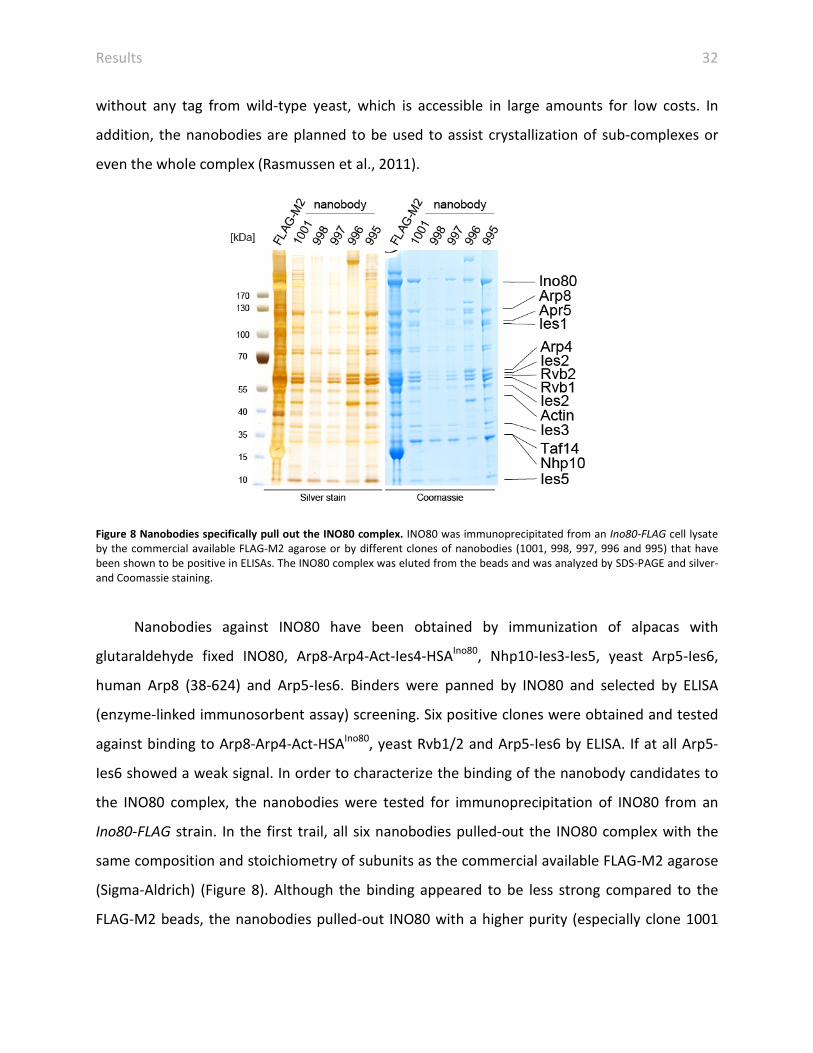
Figure 8 Nanobodies specifically pull out the INO80 complex. INO80 was immunoprecipitated from an Ino80-FLAG cell lysate by the commercial available FLAG-M2 agarose or by different clones of nanobodies (1001, 998, 997, 996 and 995) that have been shown to be positive in ELISAs. The INO80 complex was eluted from the beads and was analyzed by SDS-PAGE and silver- and Coomassie staining.
Nanobodies against INO80 have been obtained by immunization of alpacas with
glutaraldehyde fixed INO80, Arp8-Arp4-Act-Ies4-HSAIno80, Nhp10-Ies3-Ies5, yeast Arp5-Ies6,
human Arp8 (38-624) and Arp5-Ies6. Binders were panned by INO80 and selected by ELISA
(enzyme-linked immunosorbent assay) screening. Six positive clones were obtained and tested
against binding to Arp8-Arp4-Act-HSAIno80, yeast Rvb1/2 and Arp5-Ies6 by ELISA. If at all Arp5-
Ies6 showed a weak signal. In order to characterize the binding of the nanobody candidates to
the INO80 complex, the nanobodies were tested for immunoprecipitation of INO80 from an
Ino80-FLAG strain. In the first trail, all six nanobodies pulled-out the INO80 complex with the
same composition and stoichiometry of subunits as the commercial available FLAG-M2 agarose
(Sigma-Aldrich) (Figure 8). Although the binding appeared to be less strong compared to the
FLAG-M2 beads, the nanobodies pulled-out INO80 with a higher purity (especially clone 1001

Results 33
and 995). Due to different coupling efficiency of the nanobodies to the beads, the
immunoprecipitated quantity of INO80 is not really comparable between the nanobodies and
the FLAG-M2 agarose. But decreased amounts pulled out with nanobodies can easily be
compensated by simply using more nanobody coupled beads, because of the low production
price.
In summary, nanobodies have been obtained that specifically immunoprecipitate an
entire and stoichiometric INO80 complex. In future this can be used to adapt the purification
protocol and circumvent the use of commercial FALG-M2 agarose beads and genetically
modified yeast. This brings several advantages including cheaper purification, usage of large
wild-type cell amounts and they might promote crystallization of sub-complexes and of INO80.
Furthermore, INO80 purified by nanobodies even shows a higher degree of purity. Future
studies will show, if INO80 could be purified in large amounts from an endogenous source by
nanobodies to have enough material to thoroughly screen for proper crystallization conditions.
4.4 Assessment of the activity of the purified INO80
To test, whether the novel purification preserved the activity of INO80, remodeling assays and
ATPase were performed (Tosi et al., 2013). INO80 was shown to mobilize and equally space
nucleosomes (Shen et al., 2003; Udugama et al., 2011). INO80 acts at INO genes; therefore we
used nucleosomes reconstituted with a DNA sequence based on INO1 (Ford et al., 2007).
Indeed, INO80 could re-distribute octamers along the INO1 DNA with increasing concentrations
of INO80 (Figure 9A). This reaction was ATP-hydrolysis dependent as INO80 failed to mobilize
nucleosomes in the presence of the non-hydrolysable ATP analog AMP-PCP or the transition
state analog ADP-BeFx.
INO80 was reported to have DNA and nucleosome induced ATPase activity and the
nucleosome stimulated the ATPase activity two-fold more than DNA (Shen et al., 2000;
Udugama et al., 2011). However, Shen et al. had to treat their samples with DNase before DNA
stimulation was observed, as their prepared INO80 contained contaminating DNA. Our purified

Results 34
INO80 showed basal ATPase activity (Figure 9B). A 356 bp long DNA fragment that was used to
reconstitute INO1 nucleosomes stimulated the ATP hydrolysis rate about 4-fold more. INO1
nucleosomes resembling native chromatin even increased the ATPase activity 2-fold more than
DNA stimulated complex (Tosi et al., 2013).
Figure 9 Purified INO80 exhibited ATPase and nucleosome remodeling activity. A) INO80 was able to mobilize nucleosomes of INO1 chromatin in the presence of ATP. Remodeling efficiency was concentration dependent. Non-hydrolysable ATP (AMP-PCP) or transition state (ADP-BeFx) analogs prevented nucleosome re-distribution. Remodeling reactions were analyzed by native PAGE. B) ATPase assay showed that INO80 had basal ATP hydrolysis activity, which was stimulated by DNA and INO1 nucleosomes (NCP). ATPase reactions were quantified and presented relative to ATPase hydrolysis rates of alkaline phosphatase. Data are represented as mean standard deviation. C and D) Fixation with glutaraldehyde reduced but not abolished ATPase and remodeling activity by INO80.

Results 35
To test if cross-linking of INO80 with glutaraldehyde influenced native activity of INO80,
we tested remodeling and ATPase activity of fixed INO80. Unexpectedly, cross-linking was not
completely abolishing ATPase and remodeling activity (Figure 9 C and D). Glutaraldehyde reacts
majorly with lysines and therefore potentially might distort the active site of enzymes
(Migneault et al., 2004). The residual ATPase activity might originate from any of INO80’s
ATPase.
The novel purification procedure of INO80 yielded highly active and DNA free INO80 that
is suitable for structural and biochemical characterization.
4.5 Chemical cross-linking and mass spectrometry analysis of the INO80
complex
4.5.1 Mapping of subunit interactions by cross-linking and mass spectrometry
The architecture of INO80-type remodelers was only based on genetic studies and was not
complete. In order to increase the resolution and unravel the entire topology of INO80, we
used the XL-MS analysis (Figure 10) (Tosi et al., 2013). The appropriate concentration of the
isotopically labeled cross-linker DSS was assessed by a titration of the cross-linker to INO80
(Figure 11A). We analyzed four experiments and cross-linked INO80 with 1.5x, 3x, 3.5x DSS. This
resulted in 534 intra-links and 217 inter-links, whereas 212 and 116 unique intra- or inter-links
could be assigned (Tosi et al., 2013).

Results 36
Figure 10 Interaction map of the INO80 complex. XL-MS revealed the topology of INO80. Intra-links with a minimum of 30 amino acids are depicted in grey. Ino80 (HSA (dark yellow), RecA1 (orange), insertion and RecA2 (light and dark green)) and Ies2 (pink) were a scaffold for Nhp10-Ies1-Ies3-Ies5 (blue), Arp8-Arp4-Act-Ies4-Taf14 (yellow), Rvb1/2 (grey and coppery) and Arp5-Ies6 (red) sub-complexes. The Figure was adapted from (Tosi et al., 2013).
The cross-linker can be understood as a molecular ruler and the Euclidean distance of a
cross-link pair is measured between Cα- Cα. The distance restraint for DSS was reported to be
≤ 30 Å (Herzog et al., 2012; Jennebach et al., 2012; Leitner et al., 2012b; Leitner et al., 2010). To
validate the cross-linking approach, distances were estimated in available crystal structures,
however structural information on INO80 subunits is limited. Atomic coordinates of yeast Actin
(Vorobiev et al., 2003), Arp4 (Fenn et al., 2011) and human and yeast Arp8 (Gerhold et al.,
2012; Saravanan et al., 2012) were accessible. The crystal structure of human full-length Rvb1
was available (Matias et al., 2006). Yeast and human Rvb1 share a sequence identity of almost
70%. The atomic coordinates of yeast Rvb1 and Rvb2 were modeled based on the crystal
structure of human Rvb1. The sequence coverage of paralogous yeast Rvb1 and Rvb2 was only
about 40%, nevertheless, the modeled yeast Rvb2 matches almost perfectly the OB-fold
deleted structure of human Rvb2 (3UK6) (Petukhov et al., 2012) with a root mean square
deviation (rmsd) of 0.791 Å. In general, the ATPase motor domains of Snf2 enzymes are highly
conserved. To estimate the cross-links in the Ino80 Swi2/Snf2 domain crystal structures of
Danio rerio (Dro) (Thoma et al., 2005) and Sulfolobus solfataricus (Durr et al., 2005) Rad54 were
compared with each other. Dro and Sso Rad54 share a sequence identity of only ~28% to each
other and also to the Snf2 domain of Ino80. However, the corresponding lobes of Dro and Sso
individually matched with good rmsds of ~1.05 Å (Tosi et al., 2013).

Results 37
Figure 11 Titration of the cross-linker and assignment of intra-links. A) INO80 was incubated with increasing molar excess of DSS over concentration of lysines. Cross-linked and untreated complexes were separated by SDS-PAGE and visualized by silver staining. INO80 was cross-linked and analyzed by MS with DSS 1.5x - 3.5x over lysines. B) Euclidean distances of intra-links were measured in modeled yeast Rvb1/2 (C), homolgous DroRad54 (Durr et al., 2005) (D) and available crystal structures: yeast Actin (Vorobiev et al., 2003) (E), Arp4 (Fenn et al., 2011) (F) and Arp8 (Saravanan et al., 2012) (G). Non-redundant cross-links were categorized in distance ranges. C-G) Intra-links were depicted in black and interface residues and corresponding interaction partners are colored in yellow. The Figure was adapted from (Tosi et al., 2013).

Results 38
In general, the intra-links measured in available crystal and modeled structures were
fulfilled (Figure 11B - G) (Tosi et al., 2013). Both crystal structures of Dro and Sso Rad54 fulfilled
the distant restraints of the intra-links. Most cross-links averaged between 15 - 18 Å. A similar
distance was observed before to be suitable for linking formation (Leitner et al., 2012b).
However, one link in Arp4 was over the introduced distance cut-off of 30 Å (Herzog et al., 2012;
Leitner et al., 2012b) with 32.1 Å, but corresponding residues were situated in a loop just
before a crystallographic unresolved region.
The majority of the intra-links satisfied distance constraints of the cross-linker validating
the XL-MS approach.
4.5.2 Subunit topology and structural modules of INO80
All subunits of INO80 were assigned by XL-MS. Interestingly, cross-links clustered within four
sub-complexes: Rvb1/2, Arp5-Ies6, Nhp10-Ies1-Ies3-Ies5 and Arp8-Arp4-Act-Ies4-Taf14
assembled at the scaffolds Ies2 and Ino80 (Figure 10) (Tosi et al., 2013).
It was well established that Rvb1/2 form a stable complex (Cheung et al., 2010; Gorynia et
al., 2011; Gribun et al., 2008; Lopez-Perrote et al., 2012; Niewiarowski et al., 2010; Petukhov et
al., 2012; Puri et al., 2007; Torreira et al., 2008) and indeed they were highly interconnected.
The C-terminus of Arp5 exclusively cross-linked to the YL-1 domain of Ies6, which in turn cross-
linked to the OB-fold of Rvb2. Deletion of Rvb2 consistently resulted in the loss of Arp5 in
purified INO80 deletion mutants (Jonsson et al., 2004). Rvb1/2 cross-linked to the RecA2 and to
the insertion loop of Ino80 as well as to the uncharacterized PAPA-1 domain. Linkages were
mostly found in the domain 2 of Rvb1/2. In agreement with this, complexes of Rvb1/2 and
Arp5-Ies6 could be recombinantly expressed and purified (Figure 12 A).

Results 39
Figure 12 XL-MS completed sub-complex assignment. Recombinantly expressed and purified sub-complexes were analyzed by SDS-PAGE and Western-Blot analysis. A stable sub-complex consisting of Arp5-Ies6 (A) and Nhp10-Ies3-Ies5-Ino8014-450 could be purified and visualized by Coomassie staining. C) Nhp10-Ies3-Ies5-Ino8014-450 recruited Ies1 and vice versa shown by Western-blot analysis. D) Arp8, Arp4, Act, HSA and Ies4 formed a stable complex. Figure was adapted from (Tosi et al., 2013).
The Nhp10 sub-complex consists of Nhp10, Ies1, Ies3 and Ies5, which in turn cross-linked
to the N-terminus of Ino80 (Figure 10). In agreement, Ies1 formed a complex with the N-
terminus of Ino80 (Ino8014-450) and Nhp10-Ies3-Ies5 (Figure 12B and C (also see section 4.11.1).
Nhp10, Ies1, Ies3 and Ies5 are yeast specific subunits, but the N-terminus of metazoan Ino80
was shown to interact analogously with non conserved, metazoan specific subunits (Chen et al.,
2011).
The Arp8 sub-complex contains Arp8, Arp4, Act, Taf14 and Ies4 (Figure 10). Subunits of
the Arp8 sub-complex cross-linked to the N-terminal part of the previously defined HSA patch
(Szerlong et al., 2008). Indeed, the complex of Arp8-Arp4-Act was only formed stably when the
HSAIno80 was included (Figure 12D). Cross-link data indicated that Ies4 is a novel member of the
Arp8 sub-complex. Consistently, Ies4 was recruited to an Arp8-Arp4-Act-HSAIno80 complex
(Figure 12D). Cross-links are indicative for interfaces and most of inter-links were found in the
insertion domains apart from the Actin cores in Arp4 and Arp8. Especially the HSAIno80 domain,
Ies4 and Arp4 cross-linked to the N-terminus of Arp8 that is so far not structurally described.

Results 40
Ies2 cross-linked to the N-terminus, HSA and RecA1 and RecA2 folds of INO80. The cross-
links of Ies2 along the Ino80 polypeptide clustered especially in the PAPA-1 domain. Ies2 is very
small, indicating that Ino80 is not extended within INO80 but rather adopts a bent
conformation.
For the first time the XL-MS analysis of INO80 provided interaction studies with motif
resolution of a large chromatin remodeler. This extended prior interaction studies as was
performed on human INO80 (Chen et al., 2011) and now allows for a detailed structural
interpretation.
4.6 Validation of INO80’s modules in vivo
XL-MS indicated that INO80 has a modular organization, as cross-links clustered within sub-
complexes. However, a lack of information is no gain of knowledge. Therefore, strains were
created with Δarp5, Δarp8 and Δnhp10 in an Ino80-FLAG background. INO80 was purified from
those deletion strains and the composition was analyzed by SDS-PAGE and MS (Figure 13A and
B) (Tosi et al., 2013).
Figure 13 INO80 is organized in modules. A) SDS-PAGE of purified wild-type (WT), INO80(Δarp5), INO80(Δarp8) and INO80(Δnhp10) complex. Asterisks indicate lost or reduced subunits and circles indicate degraded Ino80. B) Summary of composition of INO80 deletion mutants. Loss of subunits (X) and reduced levels (x) were indicated. This figure was adapted from (Tosi et al., 2013).

Results 41
INO80 purified form Δnhp10 strains lacked Nhp10 and additionally Ies1, Ies3 and Ies5.
INO80(Δarp8) was omitted of Arp8, Arp4 and Ies4 and showed reduced levels of Act and Taf14.
INO80(Δarp5) exclusively lacked Ies6.
Reduction or loss of subunits has already been indicated by XL-MS analysis that strongly
validated our approach, but further showed that INO80 is built up by four modules also in vivo:
Rvb1/2, Arp5-Ies6, Nhp10-Ies1-Ies3-Ies5 and Arp8-Arp4-Act-Ies4-Taf14 next to an Ino80-Ies2
scaffold (Tosi et al., 2013).
4.7 Structure of the INO80 complex
4.7.1 Electron microscopy of INO80
In order to provide structural information of an INO80-type remodeler, we determined the
structure of INO80 (Tosi et al., 2013). Electron micrographs of negatively stained and cryo
preserved INO80 were recorded. Particles were manually selected and classified by reference-
free class averaging using EMAN2 and ISAC (iterative and stable alignment and clustering) (Tang
et al., 2007; Yang et al., 2012). Common line reconstruction and refinement resulted in 3D
negative stain and cryo structure of INO80 with 22 Å and 17 Å, respectively (Figure 14A and B)
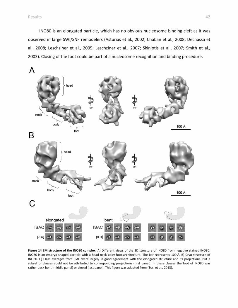
(Tosi et al., 2013). Negative stain and cryo EM revealed that INO80 is an embryo-shaped
particle with a head-neck-body architecture. The globular head has a diameter of ~120 Å and is
connected to the residual neck-body-foot cone.
Class averages of negatively stained INO80 were in good agreement with projections.
However, a small subset of classes could not be assigned and showed rather back bent or
closed conformations (Figure 14C). In agreement, the cryo structure of INO80 showed a lower
resolution in the foot strongly suggesting conformational flexibility in the foot (Tosi et al.,
2013). However, we were not able to visualize stable bent intermediate or end states arguing
for a continuum of conformations of INO80 (Tosi et al., 2013).
Results 42
INO80 is an elongated particle, which has no obvious nucleosome binding cleft as it was
observed in large SWI/SNF remodelers (Asturias et al., 2002; Chaban et al., 2008; Dechassa et
al., 2008; Leschziner et al., 2005; Leschziner et al., 2007; Skiniotis et al., 2007; Smith et al.,
2003). Closing of the foot could be part of a nucleosome recognition and binding procedure.
Figure 14 EM structure of the INO80 complex. A) Different views of the 3D structure of INO80 from negative stained INO80. INO80 is an embryo-shaped particle with a head-neck-body-foot architecture. The bar represents 100 Å. B) Cryo structure of INO80. C) Class averages from ISAC were largely in good agreement with the elongated structure and its projections. But a subset of classes could not be attributed to corresponding projections (first panel). In these classes the foot of INO80 was rather back bent (middle panel) or closed (last panel). This figure was adapted from (Tosi et al., 2013).

Results 43
4.7.2 Towards a crystal structure of INO80
The novel purification protocol yielded INO80 at concentrations high enough (up to 6 mg/ml)
for crystallization without an extra concentration step being required. Commercial screens
were set up in 96-well format and nanoscale free interface diffusion. Promising spherulites and
micro crystals were yielded that need to be analyzed further.
4.8 Rvb1/2 form a hetero-dodecamer
4.8.1 Rvb1/2 is composed of two hexameric rings in INO80
Rvb1/2 were extensively characterized in the isolated state by EM and crystallography (Cheung
et al., 2010; Gorynia et al., 2011; Lopez-Perrote et al., 2012; Matias et al., 2006; Petukhov et al.,
2012; Puri et al., 2007; Torreira et al., 2008), however, a structure in the native context was
missing. The structure of INO80 was one of the first structures of Rvb1/2 within a native
complex (Nguyen et al., 2013; Tosi et al., 2013). Human Rvb1 and domain II deleted Rvb2 were
crystallized as homo-hexamers (Matias et al., 2006; Petukhov et al., 2012), but Rvb1/2 with a
truncated domain II was crystallized in a hetero-hexameric state (Gorynia et al., 2011). EM
structures could also not clarify the question, if Rvb1/2 is a homo- or hetero-hexamer as the
resolution of available structures were too low to differentiate between the two similar
proteins (Cheung et al., 2010; Gorynia et al., 2011; Lopez-Perrote et al., 2012; Puri et al., 2007;
Torreira et al., 2008). Further biochemical analysis on isolated Rvb1/2 could also not clarify the
oligomeric state. EM structures of human and yeast Rvb1/2 suit the volume of two hexamers
(Cheung et al., 2010; Lopez-Perrote et al., 2012; Puri et al., 2007; Torreira et al., 2008), but
biochemical analysis and structural data also suggest the formation of hexamers (Gribun et al.,
2008) or even other oligomeric states as dimers, trimers or higher molecular species than
dodecamers (Niewiarowski et al., 2010). The first step to analyze the oligomeric state and
composition of Rvb1/2 in INO80 was to localize the hexameric rings. Since AAA+-ATPases in
general and Rvb1/2 in particular have been shown to form hexameric complexes; a six-fold
symmetry was searched in INO80 (Tosi et al., 2013). Indeed, a six-fold symmetry axis was found
in the head of the negative stain and cryo structure of INO80 that was quasi parallel to the

Results 44
residual cone (Figure 15A and B). The symmetry axis proved that Rvb1/2 was situated in the
head. The averaged volume of head is composed of two rings indicating that Rvb1/2 is
composed of two hexamers in INO80 (Figure 15C). Indeed, the volume and shape is suitable to
accommodate two rings of Rvb1/2 (Figure 15D). The reported dodecameric structures of
Rvb1/2 had a height of either 130 Å (Torreira et al., 2008) and 160 Å (Puri et al., 2007) and the
Llorca group could resolve a closed and a stretched conformation of Rvb1/2 with a height of
130 Å and 145 Å (Lopez-Perrote et al., 2012). The height of the averaged head volume of INO80
is around 120 Å (Figure 15C) and is thus the most compact version of Rvb1/2 reported so far.
Figure 15 The head of INO80 contains a Rvb1/2 dodecamer. A and B) Six-fold symmetry axes were located in the head that is quasi parallel to the cone of INO80 in the negative stain (A) and cryo (B) EM structures. Cross-correlation plots rotated along the axis showed six maxima spaced by 60°. C) Averaged volumes of the head of the cryo and negative stain EM structure of INO80 showed two ring like structures stacked on top of each other. D) The volume and the shape of INO80’s head was suitable to accommodate two human Rvb1 rings (2C9O (Matias et al., 2006)). This figure was adapted from (Tosi et al., 2013).
4.8.2 Rvb1/2 assemble as hetero-hexamers interacting via the domain 2 within INO80
In order to analyze, if the double-ring is composed of two hetero-hexamers of Rvb1 and Rvb2 or
of two homo-hexamers, inter-links of Rvb1/2 were evaluated (Tosi et al., 2013). In principal, the
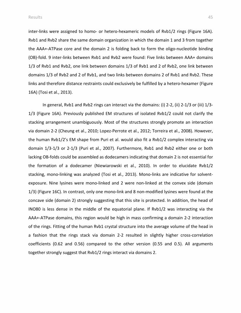
Results 45
inter-links were assigned to homo- or hetero-hexameric models of Rvb1/2 rings (Figure 16A).
Rvb1 and Rvb2 share the same domain organization in which the domain 1 and 3 from together
the AAA+-ATPase core and the domain 2 is folding back to form the oligo-nucleotide binding
(OB)-fold. 9 inter-links between Rvb1 and Rvb2 were found: Five links between AAA+ domains
1/3 of Rvb1 and Rvb2, one link between domains 1/3 of Rvb1 and 2 of Rvb2, one link between
domains 1/3 of Rvb2 and 2 of Rvb1, and two links between domains 2 of Rvb1 and Rvb2. These
links and therefore distance restraints could exclusively be fulfilled by a hetero-hexamer (Figure
16A) (Tosi et al., 2013).
In general, Rvb1 and Rvb2 rings can interact via the domains: (i) 2-2, (ii) 2-1/3 or (iii) 1/3-
1/3 (Figure 16A). Previously published EM structures of isolated Rvb1/2 could not clarify the
stacking arrangement unambiguously. Most of the structures strongly promote an interaction
via domain 2-2 (Cheung et al., 2010; Lopez-Perrote et al., 2012; Torreira et al., 2008). However,
the human Rvb1/2’s EM shape from Puri et al. would also fit a Rvb1/2 complex interacting via
domain 1/3-1/3 or 2-1/3 (Puri et al., 2007). Furthermore, Rvb1 and Rvb2 either one or both
lacking OB-folds could be assembled as dodecamers indicating that domain 2 is not essential for
the formation of a dodecamer (Niewiarowski et al., 2010). In order to elucidate Rvb1/2
stacking, mono-linking was analyzed (Tosi et al., 2013). Mono-links are indicative for solvent-
exposure. Nine lysines were mono-linked and 2 were non-linked at the convex side (domain
1/3) (Figure 16C). In contrast, only one mono-link and 8 non-modified lysines were found at the
concave side (domain 2) strongly suggesting that this site is protected. In addition, the head of
INO80 is less dense in the middle of the equatorial plane. If Rvb1/2 was interacting via the
AAA+-ATPase domains, this region would be high in mass confirming a domain 2-2 interaction
of the rings. Fitting of the human Rvb1 crystal structure into the average volume of the head in
a fashion that the rings stack via domain 2-2 resulted in slightly higher cross-correlation
coefficients (0.62 and 0.56) compared to the other version (0.55 and 0.5). All arguments
together strongly suggest that Rvb1/2 rings interact via domains 2.
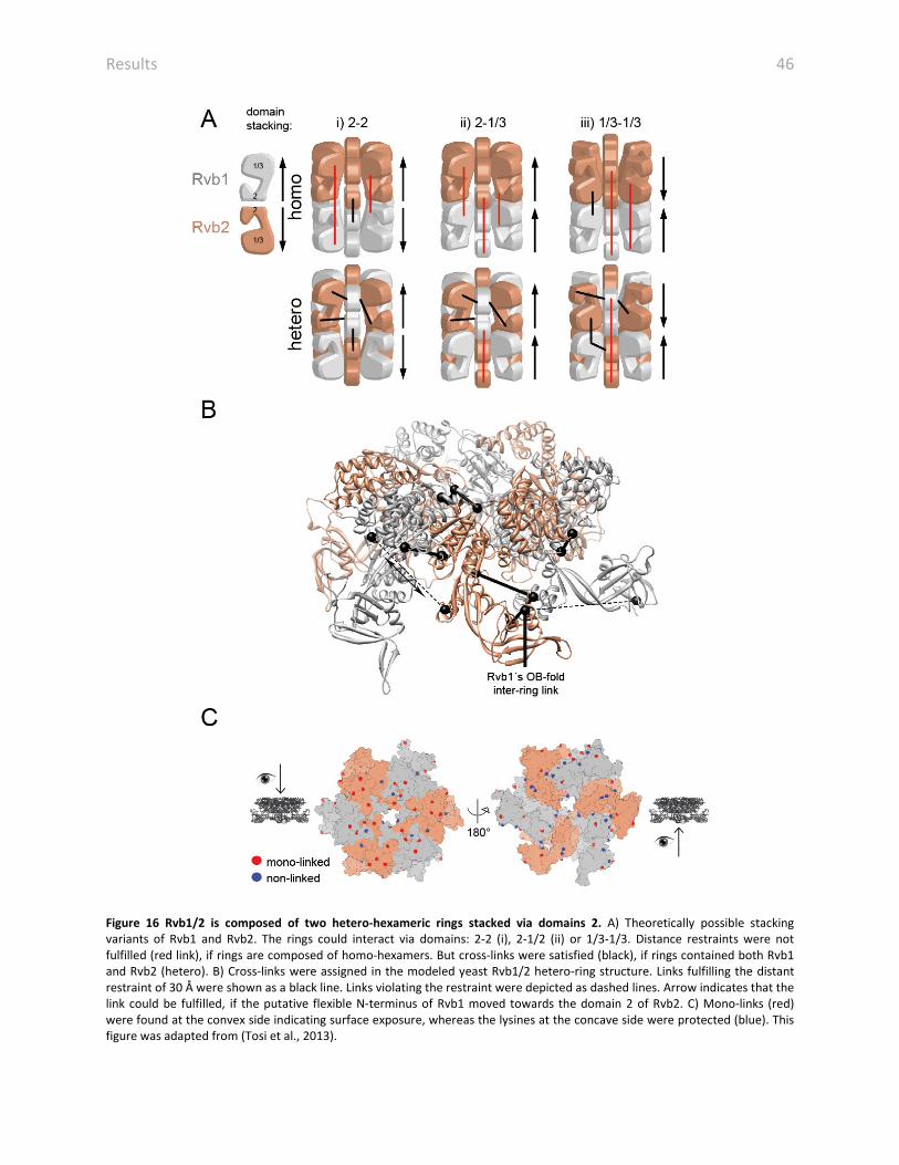
Results 46
Figure 16 Rvb1/2 is composed of two hetero-hexameric rings stacked via domains 2. A) Theoretically possible stacking variants of Rvb1 and Rvb2. The rings could interact via domains: 2-2 (i), 2-1/2 (ii) or 1/3-1/3. Distance restraints were not fulfilled (red link), if rings are composed of homo-hexamers. But cross-links were satisfied (black), if rings contained both Rvb1 and Rvb2 (hetero). B) Cross-links were assigned in the modeled yeast Rvb1/2 hetero-ring structure. Links fulfilling the distant restraint of 30 Å were shown as a black line. Links violating the restraint were depicted as dashed lines. Arrow indicates that the link could be fulfilled, if the putative flexible N-terminus of Rvb1 moved towards the domain 2 of Rvb2. C) Mono-links (red) were found at the convex side indicating surface exposure, whereas the lysines at the concave side were protected (blue). This figure was adapted from (Tosi et al., 2013).

Results 47
4.9 The catalytic core of INO80
4.9.1 The Swi2/Snf2 domain of INO80
The Swi2/Snf2 ATPase of Ino80 is the active core of the INO80 complex as a mutation in the
Walker A motif results in a decrease of the ATPase activity to only 5% compared to WT complex
(Shen et al., 2000). The Swi2/Snf2 fold of Ino80, especially the RecA2 domain was highly inter-
linked with Rvb1 and Rvb2 (Tosi et al., 2013). Rvb1/2 is situated in the head of INO80. Thus, the
Swi2/Snf2 fold has to be in spatial proximity to the head. Swi2/Snf2 helicases have a
characteristic two-lobed structure (Durr et al., 2006). A density patch in the neck of INO80
resembles this typical shape (Tosi et al., 2013). The docking of the homologous DroRad54
(Thoma et al., 2005) resulted in a cross-correlation coefficient of over 0.8 (Figure 17) (Tosi et al.,
2013). The orientation was further constrained as the RecA2 domain exclusively cross-linked to
Rvb1/2. The density connecting the head and the neck could accommodate the insertion loop
of the Ino80 ATPase domain that would protrude out of the top of the RecA2 domain. The
insertion cross-linked to the domain 2 of Rvb1/2. In this position all cross-links would be full-
filled.
Figure 17 Docking of the catalytic core of INO80. The crystal structure of the homologous Swi2/Snf2 (Thoma et al., 2005) was docked into the neck of INO80 and further oriented by cross-links between Rvb1/2 and the RecA2 fold of the Swi2/Snf2 domain (black). Cross-linked lysines are depicted as small spheres. The insertion loop protrudes out at the top of the RecA2 fold (asterisk) and can be accommodated in the density patch connecting the head and the neck (large light green sphere). Cross-links of Rvb1/2 to the insertion would support this localization. Ies2 (large pink sphere) could be placed at the top of the Swi2/Snf2 domain. The cross-links track the way of cross-linked lysines along the C-terminus of Ies2 and from the top of the RecA1 to the RecA2 fold. This figure was adapted from (Tosi et al., 2013).

Results 48
Remaining density at the top of the Swi2/Snf2 domain is sufficient to accommodate Ies2
(Figure 17). Interestingly, cross-linked lysines along the top of the Swi2/Snf2 domain towards
the RecA2 fold follow cross-linked residues of Ies2 in the C-terminal direction indicating that
Ies2 lies on top of the RecA folds (see discussion part 5.6).
In summary, we could localize the Swi2/Snf2 domain with its insertion loop and Ies2 in
the neck region of INO80.
4.9.2 Expression and purification studies of the ATPase domain of Ino80 with Ies2 and
Rvb1
XL-MS indicated that Ies2 and the OB-folds of Rvb1/2 are interacting with the Swi2/Snf2 ATPase
of Ino80 (Tosi et al., 2013). Since expression of any Ino80 constructs including the ATPase was
not successful so far, constructs of Ino80 were tried to be co-expressed with Ies2 and the OB-
folds of Rvb1. Therefore, coding sequences of Ino80, Ies2 and Rvb1 were cloned from S. c. and
Chaetomium thermophilum (C. t.) into vectors for expression in bacteria and insect cells. The
Ino80 ATPase including the insertion loop as well as only the RecA1 and RecA2 domains were
tried to express. Unfortunately, reasonable expression of soluble Ino80 could not be achieved.
Certainly, the expression should be optimized in future studies.
4.10 Localization of the Arp8-, Arp5- and Nhp10-module
Localization of other INO80-modules based on inference was not possible. Therefore, DID-
tagging (Flemming et al., 2010) of representative subunits of each module was performed. DID-
labeled INO80 complexes were purified and visualized by negative stain EM (Tosi et al., 2013).
In order to localize the Arp8-module, INO80 complexes with DID-labeled Arp4 were
visualized and the DID-tag was found to protrude out of the foot of INO80 (Figure 18A). Arp4 is
a member of the Arp8-module indicating that the Arp8-module is localized in the foot. In

Results 49
addition, a distal position agrees with that the Arp8-module is not cross-linking to any other
module.
To position the Nhp10-module, Nhp10 DID-labeled INO80 complexes were purified and
visualized. The DID-tag at Nhp10 protruded out of the body close to the neck of INO80
suggesting that Nhp10 is localized in the body (Figure 18A). This position is in agreement with a
cross-link between Ies2 and Ies3 (Figure 10).
To validate the position of Ies2 in the neck, which was inferred from XL-MS and EM
results, Ies2 DID-tagged INO80 was visualized. Indeed, the DID-tag protruded out of the neck
close to the head (Figure 18A).
Figure 18 Assignment of INO80-modules. A) Purification and visualization of DID-labeled INO80 complexes by EM allowed localization of INO80-modules. Gallery of single particles of purified DID-Arp4 (first panel), DID-Nhp10 (2nd panel), DID-Ies2 (3rd panel) and DID-Ies6 (last panel). The DID-tags were colored in the color of the respective module: Arp4 yellow, Nhp10 blue, Ies2 pink and Ies6 red. B) Modules were assigned in the INO80 density. Rvb1/2 is situated in the head, Snf2-Ies2 and the Arp5-module in the neck, the Nhp10-module in the body and the Arp8-module in the foot. This figure was adapted from (Tosi et al., 2013).
Arp5-Ies6 exclusively cross-linked to Rvb1/2 in the head of INO80. The DID-tag at Ies6
protruded also out of the neck (Figure 18A), which is consistent with the cross-links. Moreover,

Results 50
at the back of the localized Snf2 domain in the neck there is a density patch that resembles an
Actin fold, which could accommodate Arp5 (Tosi et al., 2013).
In summary, all INO80-modules could be localized. Rvb1/2 is situated in the head
connected to the neck, which contains the Swi2/Snf2-Ies2 and backed by Arp5-Ies6 (Figure 18B)
(Tosi et al., 2013). The cone comprises the Nhp10-module in the body and the Arp8-module in
the foot. We could not exclude that parts of Arp8 reach over to the body. Still, this is the first
positioning of domains and modules into a large remodeler.
4.11 The Nhp10-module
4.11.1 Reconstitution of the Nhp10 module: Nhp10-Ies1-Ies3-Ies5-Ino80 (N-terminus)
An interaction between Nhp10 and Ies3 was already indicated in the literature (Morrison et al.,
2004; Shen et al., 2003) and a stable complex formation between Nhp10, Ies3 and Ies5 was
established in the lab (unpublished data from Sebastian Fenn, AG Hopfner). Nhp10-Ies3-Ies5
was expressed in insect cells and purified via an N-terminal His-tag on Nhp10 and anion
exchange and size exclusion chromatography (Figure 19A and B). Nhp10-Ies3-Ies5 formed a
stable complex on gel filtration, but run with a much higher molecular weight than expected.
Crystallization screens of Nhp10-Ies3-Ies5 did not yield any promising hits and treatment with
proteases (Trypsin and Chymotrypsin) in the drop did not promote crystallization.
Nhp10 was degraded in the Nhp10-Ies3-Ies5 complex to a stable degradation product
Nhp10 1-164 (Nhp10-ISNI), which was identified by MS analysis (Figure 19B). This heterogeneity
in the preparation could have prevented crystallization. Therefore, a construct of Nhp10 ISNI-
Ies3-Ies5 was expressed and purified (Figure 20A). However, this Nhp10 construct was unstable,
the yield was significantly reduced compared to full-length Nhp10 and did not improve
crystallization.

Results 51
Figure 19 Purification of Nhp10-Ies3-Ies5. A) Nhp10-Ies3-Ies5 was expressed in insect cells and purified by Ni-NTA agarose. DNA free Nhp10-Ies3-Ies5 eluted from anion exchange chromatography at approximately 250 mM KCl and was analyzed by SDS-PAGE. The UV absorption at 260 or 280 nm is indicated to track protein-DNA ratios. B) Nhp10-Ies3-Ies5 was further purified via size exclusion chromatography. Nhp10-Ies3-Ies5 eluted at a higher molecular weight than expected (66 kDa) corresponding to about 250 kDa.
To facilitate crystallization of the Nhp10 sub-complex binding partners were searched
that stabilize the complex. XL-MS analysis showed that the Nhp10-module consists of Nhp10,
Ies1, Ies3 and Ies5 interacting with the N-terminus of Ino80 (Figure 10). Therefore, N-terminal
constructs of Ino80 containing amino acids: 1-152, 1-213, 1-258, 14-450 were cloned into the
Nhp10-Ies3-Ies5 containing vector and were purified (Figure 20B). All constructs were
expressed, soluble and could be co-purified together with Nhp10-Ies3-Ies5 or Nhp10 ISNI-Ies3-

Results 52
Ies5. Nhp10-Ies3-Ies5 formed a stable complex with Ino80 14-450 that could be purified by size-
exclusion (Figure 20C). This complex additionally interacted with Ies1 (Figure 12C).
Figure 20 Nhp10-Ies3-Ies5 form a stable complex with the N-terminus of Ino80. A) Nhp10 ISNI (1-164) was purified by Ni-NTA, anion exchange and size exclusion chromatography. The gel filtration profile and the corresponding SDS-PAGE are shown. B) N-terminal constructs of Ino80 were expressed with Nhp10-Ies3-Ies5 and purified via Ni-NTA. Ino80 containing the amino acids 1-152, 14-450, 1-213 and 1-258 interacted with Nhp10-Ies3-Ies5. C) Nhp1-Ies3-Ies5-Ino80 14-450 could be purified by IMAC, anion exchange and size exclusion chromatography. The UV absorption at 280 nm is indicated to track protein.

Results 53
A complex consisting of Nhp10-Ies1-Ies3-Ies5-Ino80 (N-terminus) was identified that
could facilitate crystallization and proves once more the modular organization of INO80
inferred from XL-MS.
4.11.2 The Nhp10 sub-complex forms a stable DNA complex
Nhp10 is composed of two DNA binding domains (HMG boxes) and Nhp10 has been shown to
interact favorably with structured DNA (Ray and Grove, 2009, 2012). Therefore, DNA binding of
Nhp10-Ies3-Ies5 to different structured DNAs was assessed. Mammalian HMGB and Nhp10
were shown to prefer a distorted 37 bp long DNA with loops introduced by tandem mismatches
separated by 9 bp (Grove et al., 1996). Nhp10 alone showed increased affinity to a DNA that is
50 bp long, but with the same features of mismatches (Ray and Grove, 2009). Binding to both
types of DNA duplexes was tested by EMSAS (Figure 21A). Nhp10 in complex with Ies3 and Ies5
bound preferentially to the longer 50 bp tandem mismatch substrate.
INO80 is involved in the proper restart of stalled replication forks (Morrison et al., 2007;
Papamichos-Chronakis and Peterson, 2008; Shimada et al., 2008), thus DNA duplexes
resembling replication forks (Guy and Bolt, 2005) were also tested for binding. The INO80
complex binds and acts on nucleosomes therefore a DNA was designed that shows a curvature
(Goodsell and Dickerson, 1994). Indeed, Nhp10-Ies3-Ies5 showed high binding affinity to fork
and curved DNA substrates (Figure 21B). In addition, Nhp10-Ies3-Ies5 formed stable complexes
with all tested DNA substrates on a gel filtration column (Figure 21D - F).
Nhp10-Ies3-Ies5 in complex with curved DNA formed a stable complex that could be
separated from unbound DNA by size exclusion chromatography. Therefore, this complex was
screened for crystallization and promising crystalline precipitation or micro crystals were
among initial hits, which should be refined in future.
Results 54
Figure 21 Nhp10-Ies3-Ies5-DNA complexes. A and B) Binding of 50 bp and 37 bp long duplex DNAs with tandem mismatches (Ray50 or Ray37) (Ray and Grove, 2009, 2012), fork DNA (Guy and Bolt, 2005) and curved DNA created by phased AGGAGAG sequence to Nhp10-Ies3-Ies5 was tested by EMSA. Increasing amounts of Nhp10-Ies3-Ies5 were incubated with structured DNAs. C) Nhp10-Ies3-Ies5 bound also to a 187 bp long DNA oligo containing the 601 positioning sequence (Lowary and Widom, 1998). D - F) Nhp10-Ies3-Ies5 in complex with fork DNA (D), Ray50 (E) and curved DNA (F) were analyzed on a size exclusion column. DNA containing complexes were shifted towards a higher molecular weight compared to Nhp10-Ies3-Ies5 or DNA alone. The UV absorption at 260 or 280 nm is indicated to track protein-DNA ratios.

Results 55
4.12 Dissecting the function and activity of INO80-modules
4.12.1 Functional characterization of INO80-modules
INO80 binds preferentially to nucleosomes with long extranucleosomal DNA (Udugama et al.,
2011). In order to functionally characterize INO80-modules EMSAs with DNA, nucleosomes and
remodeling assays as well as ATPase assays were performed (Tosi et al., 2013). Deletion of the
Arp8-, Nhp10- or Arp5-modules from INO80 did not completely abolish binding to DNA (Figure
22A and B). In principal, DNA binding, remodeling and ATPase activity of these mutants were
tested earlier (Shen et al., 2003). However, we could expand the analysis by for instance testing
for ATPase stimulation by DNA in addition to nucleosomal stimulation.
Figure 22 The Nhp10- and Arp8-sub-complex are responsible for DNA binding. A - C) Increasing amounts of WT INO80, INO80(Δarp8), INO80(Δnhp10) and INO80(Δarp5) (A) and recombinantly expressed and purified Nhp10-Ies3-Ies5 (Nhp10.com), Arp8-Arp4-Act-HSAIno80 (Arp8.com) (B), Rvb1/2 or Arp5-Ies6 (Arp5.com) (C) were incubated with 187 bp DNA including the 601 nucleosome positioning sequence and were analyzed by native PAGE. This figure was adapted from (Tosi et al., 2013).

Results 56
INO80(Δnhp10) showed decreased DNA binding affinity compared to wild-type (WT)
complex (Figure 22A and B). Reciprocal experiments with recombinantly expressed sub-
complexes showed that Nhp10-Ies3-Ies5 bound strongly to DNA (Figure 21 and 22A and B). In
agreement with Shen et al., 2003, deletion of the Nhp10-module did if at all slightly negatively
affect remodeling rates, but had no negative influence on the ATPase activity (Figure 23A and B)
(Tosi et al., 2013).
Figure 23 Deletion of INO80-modules influenced remodeling and ATPase activity. A and B) The ability to re-distribute nucleosomes along a 359 bp long INO1 DNA by WT INO80, INO80(Δarp8), INO80(Δnhp10) or INO80(Δarp5) was analyzed by native PAGE. D) The ATPase activity of WT and mutants INO80 complexes was analyzed by radioactive ATP hydrolysis assays. The ATPases was either measured with no stimuli (-) or was stimulated with 359 bp long INO1 DNA (DNA) or INO1 nucleosomes (chromatin). ATP hydrolysis was quantified and presented relative to ATPase hydrolysis rates of alkaline phosphatase. Data was represented as mean standard deviation. This figure was adapted from (Tosi et al., 2013).
INO80(Δarp8) bound less efficiently to DNA and remodeling was decreased compared to
WT complex (Figure 22A and 23A). However, the binding was not completely abolished as
published earlier (Shen et al., 2003). Arp8-Arp4-Act-HSAIno80 bound DNA, but less efficiently
compared to Nhp10-Ies3-Ise5 (Figure 22B). Both Nhp10-Ies3-Ies5 and Arp8-Arp4-Act-HSAIno80
oligomerized on DNA (Figure 21 and 22B). DNA failed to stimulate ATPase hydrolysis rates of
INO80(Δarp8) (Figure 23B). Nucleosomes stimulated the ATPase activity of INO80(Δarp8) to
DNA stimulated WT levels (Tosi et al., 2013).
Deletion of the Arp5-module did not affect DNA binding capabilities of INO80(Δarp5)
(Figure 22A). Arp5-Ies6 did not bind the nucleosome, but DNA was shifted at mM
concentrations (Figure 22C). Consistent with previous work (Shen et al., 2003) INO80(Δarp5)

Results 57
failed to mobilize nucleosomes on INO1 chromatin (Figure 23A). In agreement, the ATPase
activity of INO80(Δarp5) failed to be simulated further by the histone entity of INO1
nucleosomes (Figure 23B). However, DNA activated the ATPases activity to similar rates as the
WT complex (Tosi et al., 2013).
In summary, the Arp containing modules (Arp8 and Arp5) are essential for providing the
catalytic activity, whereas the Nhp10- and the Arp8-module are responsible for binding and
recruitment to the substrate.
4.12.2 Interaction and visualization of an INO80-nuclesome complex
The Nhp10- and Arp8-module are nucleosome and DNA binders (Shen et al., 2003; Tosi et al.,
2013). To map directly the interaction sites of the INO80 subunits on the histones, INO80-
nucleosome complexes were reconstituted and analyzed by XL-MS (Tosi et al., 2013). The
appropriate DSS concentration was titrated (Figure 24A). Ten cross-link experiments with
different molar ratios of the nucleosome to INO80 and different ATP analogs identified 52 inter-
links between histones and INO80 subunits (Figure 25A - D) (Tosi et al., 2013). 35 cross-links
were formed to histone tails that are unstructured (Luger et al., 1997) and thus only offer loose
distant constraints (Figure 25B). 17 inter-links were to structured histone core folds and thus
provide tight distant restraints (Figure 25A).
Inter-links between histones and INO80 modules were equally distributed: 12 cross-links
were found to the Rvb1/2 head, 14 to the Ino80 ATPase, Ies2 and Arp5-Ies6 neck, 10 to the
Ino80 N-terminus and Nhp10-Ies1-Ies3-Ies5 body and 16 to the tail containing the Arp8-Arp4-
Act-Ies4-Taf14-HSAIno80 (Figure 25A and B).

Results 58
Figure 24 Titration of cross-linker of an INO80-nucleome complex. INO80 was incubated with nucleosomes and then was cross-linked with increasing molar excess of DSS over concentration of lysines. Cross-linked and untreated complexes were separated by SDS-PAGE and visualized by silver staining. INO80-nucleosome complexes were cross-linked and analyzed by MS with DSS 1.5x - 3.5x over lysines. This figure was adapted from (Tosi et al., 2013).
Rvb1/2 cross-linked to the N- and C-termini of H2A (Figure 25C and D). The OB-folds of
Rvb2 cross-linked to the α2 helix of H2B and to the N-termini of H3 and H4. The AAA+ ATPase of
Rvb2 moreover cross-linked to the L1 of H4. Rvb1 cross-linked to the N-termini of H2B and H3.
Interestingly, residues in the AAA+ ATPase domain (Rvb131) and in the domain 2 of Rvb2
(Rvb2154 and Rvb2233) and Rvb1290, which cross-linked to the N- and C-terminus of H2A are 70 Å
apart from each other. A similar distance (74 Å) is the cross-linking N- and C-terminus of H2A
(H2A13, H2A122) apart. This makes it perfectly suitable to dock the Rvb1/2 dodecamer on one
side of the nucleosome.
The Ino80 ATPase sitting in the neck region of INO80 cross-linked to the loop L2 of H2A.
All cross-links in Ies2 to histones clustered in the short PAPA-1 domain (Figure 25 C). Ies2 cross-
linked to the α1 of H3 and to αC of H2B as well as to the N-termini of H2A and H2B (Figure
25D). Cross-linking sites of Arp5 and Ies6 were in the insertion between the actin folds and in
the YL-1 domain, respectively (Figure 25D). Arp5-Ies6 cross-linked to αC of H2B, to L1 of H4 and
to the N-terminus of H3.

Results 59
Figure 25 INO80-nucleosome interaction map. A-D) Different depictions of inter-links formed between nucleosome (2pyo,(Clapier et al., 2008)) and INO80 subunits. Inter-links to structured histone folds provide tight restraints (A) and inter-links to flexible histone tails offer loose restraints (B). (C) All INO80 modules are involved in nucleosome cross-links. Interfaces already represented in the apo-INO80 and were also found in INO80-nucleosome complexes were indicated with dashed lines. D) Histones were depicted as secondary structures.

Results 60
The subunits in the head and neck of INO80 clustered at the H2A/H2B dimer and
therefore blocked one complete side of the nucleosome (Figure 25C). This is in particular
interesting as the H2A/H2B dimer is the part to be exchanged by INO80 and accordingly the
product state.
The Nhp10-module cross-linked to the α1 and α2 helices of H4, αC of H2B and to all the
N-termini of histones (Figure 25D). The Arp8-module was cross-linking to the αC helix of H2B, to
the L2 loop of H2A and to the N-termini of all histones. Cross-linking sites between histones and
the body and foot module were also covering one the side of the nucleosome (Figure 25C).
The head-neck and the body-foot modules occupied each one side of the nucleosome
strongly arguing for a central position of the nucleosome (Figure 25C). However, it could not be
excluded that more than one nucleosome was bound to INO80. The distant restraint of DSS
(30 Å) could not be fulfilled by only a rigid body fitting of the nucleosome into a central position
of INO80. A closure of the foot would lead to a compact packing of the nucleosome (Figure 14)
and could be part of the nucleosome recognition (Tosi et al., 2013).
Some of the lysines in histones situated near the DNA entry/exit site that cross-linked to
Ies6, Ies2 and Rvb2 were not accessible in an intact nucleosome and would require a partial
unwrapping of the DNA. Exactly at this site the Swi2/Snf2 fold and catalytic essential Arp5 were
located that could provide the mechanical force to generate access (Tosi et al., 2013).
Figure 26 Visualization of INO80-nucleosome complexes. Class averages of INO80-nucleosome gained by ISAC (first and middle panel). Extra density in the central groove is stained blue. Extra density could not be assigned in the respective projections of apo-INO80 (last panel).
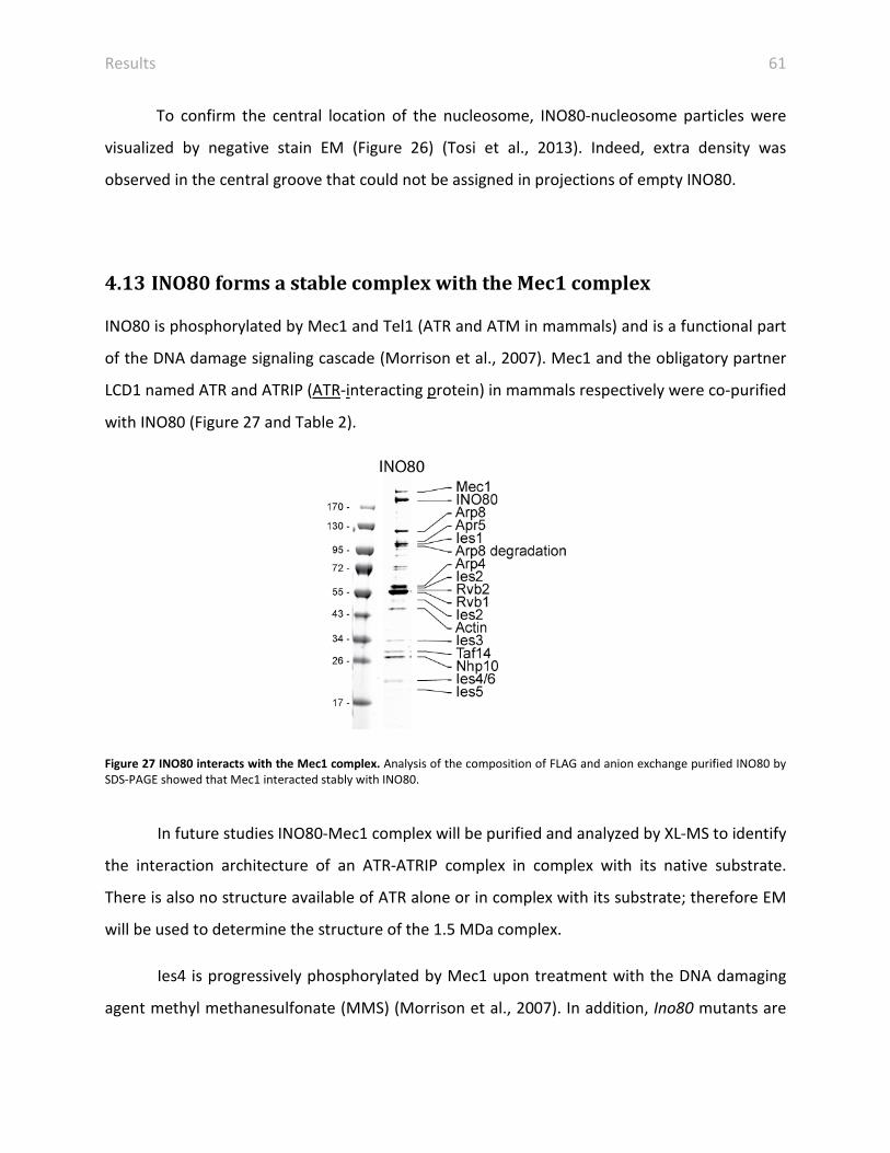
Results 61
To confirm the central location of the nucleosome, INO80-nucleosome particles were
visualized by negative stain EM (Figure 26) (Tosi et al., 2013). Indeed, extra density was
observed in the central groove that could not be assigned in projections of empty INO80.
4.13 INO80 forms a stable complex with the Mec1 complex
INO80 is phosphorylated by Mec1 and Tel1 (ATR and ATM in mammals) and is a functional part
of the DNA damage signaling cascade (Morrison et al., 2007). Mec1 and the obligatory partner
LCD1 named ATR and ATRIP (ATR-interacting protein) in mammals respectively were co-purified
with INO80 (Figure 27 and Table 2).
Figure 27 INO80 interacts with the Mec1 complex. Analysis of the composition of FLAG and anion exchange purified INO80 by SDS-PAGE showed that Mec1 interacted stably with INO80.
In future studies INO80-Mec1 complex will be purified and analyzed by XL-MS to identify
the interaction architecture of an ATR-ATRIP complex in complex with its native substrate.
There is also no structure available of ATR alone or in complex with its substrate; therefore EM
will be used to determine the structure of the 1.5 MDa complex.
Ies4 is progressively phosphorylated by Mec1 upon treatment with the DNA damaging
agent methyl methanesulfonate (MMS) (Morrison et al., 2007). In addition, Ino80 mutants are

Results 62
sensitive to hydroxyurea (HU) that inhibits ribonuleotide reductase and causes thus stalling of
replication forks (Papamichos-Chronakis and Peterson, 2008). Thus, it will be tested, if MMS
and HU increase the association between Mec1 and INO80.
Table 2 Identified subunits of the INO80 complex. The composition of the FLAG-purified INO80 complex and the interacting proteins were analyzed by LC-MS/MS. Hit Nb: Number of found protein; Protein Id: abbreviation of protein; Protein description: name of protein; Score: Mascot score; Mass: molecular weight of protein in Da; Coverage %: calculated form length and the set of peptides assigned to the protein. Proteins identified with a score below 100, where manually validated. INO80 core subunits are highlighted in grey and novel indentified subunits of the Mec1 complex in orange.
Hit Nb Protein Id Protein description Score Mass [Da] Coverage %
1 Ino80 Putative DNA helicase INO80 3688 171863 29.5
2 Rvb2 RuvB-like protein 2 2949 51694 43.3
3 Rvb1 RuvB-like protein 1 2549 50593 39.7
4 Arp8 Actin-like protein ARP8 1472 100604 26.6
5 Mec1 Serine/threonine-protein kinase MEC1/ATR 1168 275108 13.3
6 Ies1 Ino eighty subunit 1 1084 78932 25.7
7 Arp4 Actin-related protein 4 1040 55197 34.6
8 Act Actin 754 41891 28
9 Nhp10 Non-histone protein 10 694 23956 38.4
10 Hsp71 Heat shock protein SSA1 545 69786 24.3
10 Hsp72 Heat shock protein SSA2 462 69599 24.3
11 Arp5 Actin-related protein 5 532 87677 14.3
12 Ies2 Ino eighty subunit 2 513 36189 22.2
13 Ies4 Ino eighty subunit 4 509 13084 50.9
14 Hsp75 Heat shock protein SSB1 499 66732 17
14 Hsp76 Heat shock protein SSB2 495 66668 17
15 6p21 6-phosphofructo-2-kinase 1 422 93587 13.5
16 Taf14 Transcription initiation factor TFIID subunit 14 370 27423 16.8
17 Lcd1 DNA damage checkpoint protein LCD1/ATRIP 334 86712 15.5
18 Ef1a Elongation factor 1-alpha 316 50400 14.8
19 Ies3 Ino eighty subunit 3 295 28225 18.8
20 Ksp1 Serine/threonine-protein kinase KSP1 286 117351 6.7
21 Rpa34 DNA-directed RNA polymerase I subunit RPA34 264 26916 18.5
22 Ies6 Chromatin-remodeling complex subunit IES6 258 18579 30.1
23 Rs3 40S ribosomal protein S3 249 26543 18.8
24 Ltn1 E3 ubiquitin-protein ligase listerin 224 181270 6.3
… … ... … … …
57 Ies5 Ino eighty subunit 5 95 14303 32

Discussion 63
5 Discussion
5.1 Hybrid view on INO80
Large chromatin remodelers are multi subunit complexes. The remodeling mechanism
especially of macromolecular complexes is scarcely understood. Overall shapes have been
reported of all families of remodelers expect the INO80/SWR1 family (Asturias et al., 2002;
Chaban et al., 2008; Leschziner et al., 2005; Leschziner et al., 2007; Skiniotis et al., 2007; Smith
et al., 2003). Molecular topologies of large remodelers are known partially and most of the data
was based on genetic studies. For INO80 it was indicated that Nhp10-Ies3, Rvb1/2-Arp5 and
Arp8-Arp4-Act-HSAIno80 form sub-complexes with each other. Using XL-MS the interaction map
of the INO80 complex could be unraveled and all INO80 subunits could be associated to the
respective modules. So far interaction data was used to indentify networks, to map additional
interacting proteins on homology models or to clarify the topology of complexes (Ciferri et al.,
2008; Herzog et al., 2012; Jennebach et al., 2012; Leitner et al., 2012a). The XL-MS approach
indicates interfaces between proteins and thus dissects with motif resolution. This is in
particular strong in combination with low resolution EM structures that do not go under
subnanometer resolution. In the case of INO80 this allowed building up a partial atomic fitting
of available crystal structures in the head and neck region of INO80. Large complexes as INO80
are often dynamic and thus exhibit conformational heterogeneity. Crystal and EM structures
only provide us with snapshots and therefore flexible under-represented conformational states
or bound ligands are lost. The XL-MS technique allowed identifying the histone interaction sites
in respect to INO80 subunits. A separation of for instance INO80-nucleosome or INO80-Mec1
complexes from apo-INO80 would allow analyzing the effect on the INO80 architecture in
response to ligand binding.

Discussion 64
5.2 The chromatin remodeler INO80
EM revealed that INO80 is an elongated particle (Tosi et al., 2013) and thus has complete
difference appearance compared to the globular shaped, large RSC or SWI/SNF remodelers
(Asturias et al., 2002; Chaban et al., 2008; Dechassa et al., 2008; Leschziner, 2011; Leschziner et
al., 2005; Leschziner et al., 2007; Skiniotis et al., 2007; Smith et al., 2003). The foot of INO80
could undergo large conformational rearrangements that could be part of nucleosome
recognition (Tosi et al., 2013). INO80 also looks remarkably different from SWR1 (Nguyen et al.,
2013). The most striking molecular difference is probably that SWR1 contains only a hetero-
hexamer compared to INO80’s dodecamer (see below).
We solved the molecular architecture of INO80 by an integrated approach combining XL-
MS, EM, subunit tagging and localization (Tosi et al., 2013). Four modules could be assigned
within the INO80 complex next to the Ino80-Ies2 scaffold: Rvb1/2, Arp5-Ies6, Nhp10-Ies1-Ies3-
Ies5 and Arp8-Arp4-Act-Ies4-Taf14. Rvb1/2 was localized in the head, the Arp5-module in the
neck with the Swi2/Snf2 ATPase and Ies2, the Nhp10-module in the body and the Arp8-module
in the foot (Figure 28). The modules will be separately discussed in more detail below.
Figure 28 Model of the subunit architecture of INO80. Rvb1/2 was localized in the head of INO80 with a hetero-dodecameric organization. The Snf2 ATPase and Ies2 sit in the neck with Arp5-Ies6 in the back. Nhp10-Ies1-Ies3-Ies5 was localized in the body and Arp8-Arp4-Act-Ies4-Taf14 in the foot.

Discussion 65
5.3 Structure of Rvb1/2 in the INO80 complex
The Rvb1/2 complex is a prominent feature of INO80 and there was no structure of an Rvb1/2
within its native complex. The structures of isolated Rvb1/2 could not clarify (i) if Rvb1/2 is a
hexamer or a dodecamer; (ii) if the rings are homo- or hetero-oligomeric and (iii) how the rings
interact with each other.
The INO80 complex is among the first structure containing the Rvb1/2 in a native context.
However, conclusions gained about INO80’s Rvb1/2 are not necessarily applicable to Rvb1/2 in
other complexes or in an isolated state, if there is one. In the majorities of isolated structures,
Rvb1/2 is dodecameric (Lopez-Perrote et al., 2012; Puri et al., 2007; Torreira et al., 2008), but
Rvb1/2 was even be found in other oligomeric states (Cheung et al., 2010; Gorynia et al., 2011;
Niewiarowski et al., 2010). Endogenously purified yeast Rvb1/2 in complex with the chaperone
Hsp90 has been shown to be hetero-hexameric (Cheung et al., 2010). Rvb1/2 in the native
INO80 environment adopts a dodecameric state, comprised of two hexameric rings (Tosi et al.,
2013).
The resolution of the isolated Rvb1/2 EM structures was too low to differentiate Rvb1 from
Rvb2; consequently the composition of the rings could not directly be abbreviated (Lopez-
Perrote et al., 2012; Puri et al., 2007; Torreira et al., 2008). However, antibody labeling
suggested that the rings could be composed of one species (Torreira et al., 2008). In addition,
there are crystal structures of homo-rings composed of either Rvb1 (Matias et al., 2006) or
Rvb2 (Petukhov et al., 2012), and hetero-hexameric rings (Gorynia et al., 2011), but this
structures has a deleted domain 2. The distance restraints determined by XL-MS exclusively
allow a hetero-hexameric composition of Rvb1/2 in INO80 (Tosi et al., 2013).
The stacking of the rings has been under debate as well. Yeast and human structures from
the Llorca lab could easily interpreted as a domain 2-2 stacking (Lopez-Perrote et al., 2012;
Torreira et al., 2008), but the stacking could not be unambiguously determined in the structure
from the Tsaneva lab (Puri et al., 2007). A following work from the same lab showed that the
deletion of domain 2 of Rvb1 destabilizes the dodecamer and they thus concluded that this
argues against a domain 2-2 interaction of the rings (Niewiarowski et al., 2010). However, a

Discussion 66
complete deletion of the domain 2 resulted in insoluble protein. Therefore Niewiaroski et al.,
deleted only the external part, the OB-folds and thus leaving the internal part of domain 2,
which then can mediate the interaction (Niewiarowski et al., 2010). The volume and shape of
the head of INO80 were sufficient to accommodate two rings of Rvb1 (Matias et al., 2006; Tosi
et al., 2013). However, the diameter of the head was only ~120 Å. Thus, it was the most
compact version compared to any isolated Rvb1/2 structure. Movements in the domain 2 were
shown to mediate between the stretched (145 Å) and compact (130 Å) form of human isolated
Rvb1/2 (Lopez-Perrote et al., 2012). Our dodecameric model of Rvb1/2 was based on the crystal
structure of the hetero-dodecameric Rvb1/2 (Gorynia et al., 2011). In this structure, it was
indicated that the two rings form a complex via the internal domain. But the electron densities
of Gorynia et al. as well as of Matias et al. showed poor coverage in this region impeding a
detailed analysis of interaction interphases (Gorynia et al., 2011; Matias et al., 2006). The
compact packing of Rvb1/2 in INO80 would bring the internal domains 2 of the two opposing
rings in such a close proximity principally enabling an interaction between them (Tosi et al.,
2013). This would also explain why the deletion of the external OB-folds did not split the
dodecameric version of Rvb1/2 (Niewiarowski et al., 2010).
In our model, the domain 2 of the same sort (Rvb1 or Rvb2) would interact with each
other. This makes it likely that the hetero-hexameric composition promotes a dodecameric
complex formation.
The six-fold axis in the head of INO80 enabled a placement of Rvb1/2 (Tosi et al., 2013).
However, a closer inspection revealed that it is not a strict six-fold symmetry axis. The main
deviation from a stringent six-fold symmetry was found in the region where the neck-body-foot
cone is inserted. Such an insertion could be mediated by the insertion loop of Ino80 that was
shown to recruit Rvb1/2 to SWR1 (Wu et al., 2005). In theory, if every Rvb1/2 pair in the
dodecamer could bind an Ino80 insertion loop, three Ino80s could be inserted resulting in a
tripod like structure. However, insertion at one site could influence the symmetry and the EM
density indicated that the insertion of Ino80 resulted in an opening of the rings (Tosi et al.,
2013). This would generate tension in the rings that may prevent further insertions of Ino80.

Discussion 67
A similar hybrid approach as we performed on INO80 was applied on SWR1. SWR1 is more
compact and the authors inferred that it contains a single hetero-hexameric ring (Nguyen et al.,
2013). Both remodelers belong to the same family of remodelers, but SWR1 is composed of a
different sub-set of subunits as INO80. The insertion in the split ATPase is a common structural
feature with Ino80, albeit is not conserved on a sequence level. The insertion was shown to
recruit the Rvb1/2 complex in SWR1 (Wu et al., 2005). Consistently, different insertion loops
could assemble a different kind of Rvb1/2 species: a hexamer in the case of Swr1 and a
dodecamer in the case of Ino80. However, non-GraFix treated samples from the study on SWR1
contained particles resembling a two layered Rvb1/2 particle (Nguyen et al., 2013). The author
also quantified the intensity of the Rvb1 and 2 from a SDS-PAGE of SWR1 and INO80. The
observed stoichiometry was consistent with a hexamer in both complexes. However, a similar
quantifications of our highly purified and structural integer INO80 preparation resulted in ratios
of 1:5.4:5.5 (Ino80:Rvb1:Rvb2), which is in agreement with the dodecamer observed in the EM
map (Tosi et al., 2013). Remarkably, Nguyen et al. also found fractions containing Rvb1/2 and
Swr1 with a stoichiometry of 6:6:1 in their glycerol gradient strongly indicating that the purified
SWR1 was composed of hexamers and dodecamers (Nguyen et al., 2013). In general, protein
staining methods are highly dependent on the amino acid composition, which is why a simple
quantification of bands of a SDS-PAGE is not suitable for proper analysis of stoichiometry.
Instead structural methods as analytical ultracentrifugation or native MS should be applied.
In Archaea only one form of Rvb1/2 exists indicating that two sorts of Rvb have developed
during evolution, maybe by gene duplication. Rvb1 has approximately 12,000 copies per cell,
whereas Rvb2 was found with 3,000 molecules (Ghaemmaghami et al., 2003). The higher copy
number of Rvb1 suggests that it is also associated in other complexes independent of Rvb2.
In summary, we could show that in the INO80 complex Rvb1/2 form a hetero-dodecamer
that is stacked via domains 2.

Discussion 68
5.4 The Nhp10-module
XL-MS and biochemical analysis of INO80 deletion mutants showed that the Nhp10-module
consists of Nhp10-Ies1-Ies3-Ies5 and that this module assembles at the N-terminal part of Ino80
(Tosi et al., 2013). Metazoan complexes also contain species-specific subunits that also
assemble at the N-terminal part of Ino80 (Chen et al., 2011). Indeed, the Nhp10 sub-complex
could be recombinantly expressed and purified (Tosi et al., 2013). Association with Ies1 was not
stable and the yields were low, when Ies1 was co-expressed from a separate vector. To increase
expression yields, Ies1 coding sequence will be integrated into one vector containing Nhp10-
Ies1-Ies3-Ies5-Ino80N-term. The complex of Nhp10-Ies3-Ies5 alone did not yield any promising
crystallization hits. However, stabilization with DNA improved the crystallization properties. The
full Nhp10-module may stabilize the complex sufficiently to yield diffracting crystals.
The Nhp10-module was assigned to the body of INO80, adjacent to the neck containing the
Swi2/Snf2 ATPase of Ino80 (Tosi et al., 2013). The Nhp10-module does not belong to the
conserved subunits. In general, one might expect that the conserved subunits evolved together
and therefore are localized in a similar sub-compartment. In contrast, the conserved Rvb1/2,
the Swi2/Snf2 domain, Ies2 and the Arp5-module localize in the head and neck of INO80 (Tosi
et al., 2013), whereas the conserved Arp8-module is positioned in the foot of INO80. Cross-links
and inference (see 5.5) indicated that the Ino80 polypeptide folds back thereby placing the
Nhp10-module next to the Swi2/Snf2 module (Tosi et al., 2013). Human, fly and fission yeast
INO80 complexes include GLI-Kruppel family zinc finger containing subunits that are involved in
DNA binding (Cai et al., 2007; Hogan et al., 2010; Klymenko et al., 2006; Wu et al., 2007).
Conceivably, INO80 only requires a strong DNA binder that facilitates recruitment and
remodeling and is thus integrated close to the neck region.
Nhp10 was shown to preferentially bind long distorted DNA (Ray and Grove, 2009, 2012).
In general, one HMG box interacts with ~10 bp of duplex DNA (Love et al., 1995; Ohndorf et al.,
1999; Wong et al., 2002; Yen et al., 1998). Since Nhp10 consists of two HMG boxes, it was
predicted to bind to a 20 bp long DNA (Ray and Grove, 2009). But Nhp10 preferred far longer
constructs and a 50 bp long duplex was even favored over 37 bp. On those substrates Nhp10-
Discussion 69
Ies3-Ies5 was forming oligomers. The DNA could either serve as binding platform for one
molecule or oligomerization could increase the binding affinity.
Nhp10-Ies3-Ies5 was found to bind structured and curved DNA and to be a high affinity
nucleosome binder (Tosi et al., 2013). This renders the Nhp10-module a perfect candidate to
stabilize reaction intermediates in the remodeling reaction or target the INO80 complex to sites
of action.
5.5 The Arp8-module
The Arp8-module consists of Arp8-Arp4-Act-Ies4-Taf14-HSAIno80 (Tosi et al., 2013). Arp4 and
Arp8 were shown to bind to histones and to DNA (Gerhold et al., 2012; Harata et al., 1999; Shen
et al., 2003). Indeed, we found that INO80(Δarp8) was compromised in DNA and nucleosome
binding and remodeling. However, binding and remodeling of nucleosomes were not
completely abolished in the case of INO80(Δarp8) as was observed previously (Shen et al.,
2003). We found that the structural integrity of INO80(Δarp8) was diminished. Thus all assays
had to be performed with freshly purified INO80(Δarp8) as otherwise the complex was
aggregated and failed to bind to its substrate.
The distant restraints gained by XL-MS analysis could be used to structurally model
complexes by minimizing the constraint. Although crystal structures of yeast Arp4, Arp8 and
Actin (Fenn et al., 2011; Gerhold et al., 2012; Saravanan et al., 2012; Schubert et al., 2013;
Vorobiev et al., 2003), and the structure of the related complex of Arp7-Arp9-Rtt102-HSASwi/Snf
were available an unambiguous modeling could not be achieved. Arp4 more closely resembles
Arp7 on a sequence basis (Muller et al., 2005). Therefore, Arp4 could be placed in the position
where Arp7 resides and Act could be modeled in the respective position of Arp9 in the Arp7-
Arp9-Rtt102-HSASwi/Snf complex without significant steric clashes (Schubert et al., 2013). The
inter-links in principally agreed with that fitting. However, the majority of the inter-links were
found in loops inserted into the core actin folds. These insertions were mostly unstructured in
the crystal structures indicating flexibility and thus prohibited reliable fitting. These loop

Discussion 70
regions might be stabilized when the interface is formed within the Arp8-complex. Rtt102 was
shown to bind in a extended form to a conserved region of the Arp7-Arp9 interface suggesting a
stabilizing role (Schubert et al., 2013). This function could be fulfilled by for instance either Ies4
or Taf14 within INO80’s HSA module. The C-terminal part of Taf14 was highly inter-linked within
the Arp8-module (Tosi et al., 2013) and could thus stabilize the interface. The N-terminal part
containing the YEATS-domain was not necessary for the association with chromatin remodelers,
but rather may provide a regulatory platform outside of INO80 (Schulze et al., 2010). In
summary, insufficient distant restraints in ordered parts and observed flexibility in the foot
region of INO80 (Tosi et al., 2013) prohibited modeling of the Arp8-module with an
unambiguous solution.
The HSA domain in the Arp7-Arp9-Rtt102-HSASwi/Snf complex adopts a largely extended α-
helical structure (Schubert et al., 2013). This extended conformation of the HSA-domain could
explain how Ino80 could span across the neck and the foot. This elongated form is also perfect
suitable to provide an interaction platform and structural rearrangements of the foot of INO80
could thereby mediate the complete mechanistic framework of INO80.
5.6 The nucleosome remodeler INO80
INO80 is an ATP-dependent nucleosome remodeler that catalyzes various DNA mediated
processes such as exchange of histone variants or even evicting complete nucleosomes or
sliding (Morrison and Shen, 2009; Shen et al., 2000). Therefore, it is of importance to study the
holo-INO80 complex bound to a nucleosome. The interactions sites of INO80 to histones were
determined by XL-MS and the nucleosome bound to INO80 was visualized by 2D EM. XL-MS of
INO80-nucleosome complexes oriented the H2A/H2B dimer at the head and neck modules of
INO80 (Tosi et al., 2013). EM revealed extra density corresponding to the nucleosome in the
central groove of INO80. This central position of the nucleosome is also consistent with the
placement of the INO80-modules. The nucleosome could be sandwiched between the head and

Discussion 71
the foot (Figure 29). The different modules could thus approach the nucleosome from opposing
sites imposing an asymmetry on the pseudo-symmetric nucleosome.
Figure 29 Model of an INO80-nucleosome complex. The nucleosome is sandwiched between the Rvb1/2 head module and the Arp8-module containing foot of INO80. The DNA could be contacted by the Nhp10-module in the body. The histones are flanked by the Snf2 domain, Ies2 and the Arp5-module in the neck and the Arp8-module. The observed conformational flexibility of the foot could explain how INO80 engulfs the nucleosome.
Both the Swi2/Snf2 domain of Ino80 and Arp5-Ies6 in the neck, which are both required for
INO80’s catalytic activity, are in close proximity to the H2A/H2B dimer (Tosi et al., 2013). It is of
particular interest how INO80 interacts with the H2A/H2B dimer in the nucleosome, since this is
the product state of an exchanged H2A.Z/H2B dimer. Intriguingly, the DNA binding OB-folds of
Rvb1/2 are in proximity to the H2A/H2B dimer close to the DNA entry/exit site and to the
RecA2 fold and Ies2 allowing a coordinated action. Rvb1/2 marginally bound to DNA and
nucleosomes in shift assays (Tosi et al., 2013). However, Rvb1/2 was highly inter-linked with
histones. A deletion of Rvb1/2 from INO80 results in a remodeling deficient complex (Jonsson
et al., 2004). But the loss of Rvb1/2 is not necessarily the cause for remodeling deficiency as the
Arp5-module, which is critical for remodeling was also lost together with Rvb1/2 (Jonsson et al.,
2004; Shen et al., 2003; Tosi et al., 2013). Rvb1/2 showed no nucleosome or DNA binding, albeit
was highly inter-linked with it. It is thus likely that Rvb1/2 plays a critical role in the catalytic
mechanism of remodeling.
Ies2 covers the central cleft of the two Swi2/Snf2 ATPase lobes (Figure 30). Work on other
Swi2/Snf2 ATPases, as the Chd1 and ISWI remodeler has shown that accessory domains
influence the activity and provide negative autoregulation (Hauk et al., 2010; Narlikar et al.,
2013). In general, the helicase motifs of Swi2/Snf2 ATPases have to align to be compatible with

Discussion 72
ATP hydrolysis. In the case of Chd1, the chromodomains block the DNA binding site on the
ATPase lobe 2 and prevent proper alignment of conserved helicase motifs thereby reducing the
discrimination of naked DNA over nucleosomes (Hauk et al., 2010). ISWI’s NegC lies across the
two ATPase lobes bridging the cleft and inhibits coupling of the ATPase activity to DNA
translocation (Clapier and Cairns, 2012).
Figure 30 The PAPA-1 domain of Ies2 covers the Swi2/Snf2 ATPase lobes. Ies2 (pink) cross-linked to the Swi2/Snf2 fold of Ino80 (used homology model DroRad54 (PDB: 1Z3I (Thoma et al., 2005)) in a way that it would cover both RecA lobes. Residues that were cross-linking to Ies2 were marked as pink small spheres. Numbers indicate the cross-linking amino acid of Ies2.
Cross-links indicated that Ies2 is localized at the Swi2/Snf2 ATPase of Ino80 in a manner
that it is crossing the two lobes from N- to C-terminus (Tosi et al., 2013). This involves only a
short patch in Ies2, the PAPA-1 domain suggesting that this region is extended. In this position,
Ies2 is perfectly situated to regulate the ATPase of Ino80 and therefore it could be an accessory
subunit helping to discriminate between substrates and to regulate the Ino80 ATPase.
The Arp5-module sits together with the Swi2/Snf2 and Ies2 in the neck of INO80 (Tosi et
al., 2013). Arp5-Ies6 was crucial for remodeling, but only marginally bound to DNA and
nucleosomes (Shen et al., 2003; Tosi et al., 2013). ATP hydrolysis of INO80(Δarp5) was
stimulated by DNA, but the histone component in INO1 nucleosomes did not further increase
ATPase activity. Ies6 has an YL-1 domain that is also found in Swc2, a subunit in the SWR1
remodeler, which has been shown to directly bind to H2A.Z-H2B (Wu et al., 2005). Swc2
recruits, positions and locks SWR1 on the +1 nucleosome (Ranjan et al., 2013; Watanabe et al.,
2013; Yen et al., 2013). Deletion of Swc2 resulted in low and irregular recruitment of SWR1 and

Discussion 73
substantial loss of H2A.Z (Yen et al., 2013). The counterpart of Swc2 in INO80, Ies6 could fulfill
similar roles. Loss of Arp5 led to the retainment of H2A.Z at the +1 nucleosome and even led to
increased occupancy of the variant. This suggests that Arp5-Ies6 could act as a histone
chaperone that may bind to either the substrate or product state H2A or H2A.Z, respectively.
This notion is additionally supported by the fact that the ATPase activity of INO80(Δarp5) is not
reacting anymore to the histone moiety. Moreover, human Arp5 has been shown to shuttle
between the nucleus and cytoplasm (Kitayama et al., 2009) and yeast Arp5 fulfills functions
outside from INO80 (Yen et al., 2012).
The Nhp10-module is situated in the body close to the neck of INO80. The central upright
orientation of the nucleosome would thus allow the high affinity DNA binder, Nhp10-Ies3-Ies5
to fixate the substrate in the remodeling reaction (Tosi et al., 2013). Apart from just merely
binding to the substrate, HMG-box proteins have been implicated in stimulating the remodeling
activity (Stros, 2010; Ugrinova et al., 2009). It is plausible that Nhp10 can facilitate the reaction
by binding to intermediate states or by bending DNA to distort histone-DNA contacts.
The structure of the INO80 complex is remarkably different from all other chromatin
remodelers and the proposed clamping mechanism of how INO80 interacts with the
nucleosome is unique for remodelers (Figure 29) (Tosi et al., 2013). INO80 embraces the
nucleosome and has a large interface with its substrate, which is completely different to the
limited contacts provided by the related SWR1 (Nguyen et al., 2013). All modules of INO80
cross-linked to the histones suggesting that they are all involved in the interaction with the
nucleosome. Simple docking of the nucleosome particle into the concave groove of INO80 could
not explain how all the subunits are able to contact it. Indeed large conformational changes
with a flexible foot have been observed (Tosi et al., 2013). Such structural rearrangements
could bring especially the subunits of the foot in touch with the nucleosome (Figure 30).
The proposed enclosure could explain how INO80 acts as a steric ruler in the
nucleosome spacing reaction. In fact, INO80 spaces nucleosomes with extreme long linker DNA
in vitro (168 bp) far more precisely than the hallmarked spacer, ISWI that rather randomize
arrays with such a linker length (Gangaraju and Bartholomew, 2007; Kagalwala et al., 2004;

Discussion 74
Udugama et al., 2011; Vary et al., 2003). One simple reason could be the bigger size of INO80
that enables a complete engulfment of the nucleosome including long linker DNA. In fact, SWR1
is not able to slide nucleosomes and showed to have limited contacts to its substrate (Nguyen
et al., 2013). Remodelers that have large interfaces with its substrate have been shown to slide
octamers (Chaban et al., 2008; Dechassa et al., 2008; Tosi et al., 2013). Thus, it could be
speculated that extensive nucleosome contacts are required to compensate for unwound DNA
states.
The closing and opening rearrangements of INO80 around a nucleosome could mediate
partial disassembly of the nucleosome particle (Figure 29). Indeed, open nucleosome states
have been identified ((Bohm et al., 2011), reviewed in (Andrews and Luger, 2011)). Those open
intermediates exist in a minority of 0.2-3% under physiological conditions and could be
promoted by remodelers such as INO80. INO80 regulates the high nucleosome turnover at +1
that could conceivably promote a partial disassembly of this nucleosome and the created
accessible H3/H4 histones could then be removed by histone chaperones (Yen et al., 2013).
Many cross-linking sites between subunits of INO80 and histones were found, which were not
accessible in an intact nucleosome as observed in crystal structures (Clapier et al., 2008; Luger
et al., 1997; Tosi et al., 2013). In fact, those cross-linked and buried residues were located at the
DNA entry/exit site, which was contacted by the Swi2/Snf2 ATPase of Ino80 and further
catalytically crucial subunits of INO80 that could in principal distort contacts between the DNA
and histones. The chemo-mechanical force could be provided by the Swi2/Snf2 ATPase to
create more accessible or partially unfolded states, and accessory subunits could stabilize those
intermediates. Interestingly, at this position the C-terminal H2A.Z docking domain is located,
which shows the highest sequence divergence to canonical H2A. It is conceivably that Ies2 or
the putative histone chaperone Ies6 senses their substrate at this site. Strikingly, the location of
the RecA2 cross-link at the H2A/H2B dimer coincides with the binding site of ISW2 on the
nucleosome (Dang and Bartholomew, 2007). ISWI has been shown to contact DNA at an
internal location of ~20 bp away from the dyad (SHL -2/+2). The Swr1 subunit containing the
Swi2/Snf2 ATPase likewise cross-linked to a ~20 bp region that is 70 bp away from the dyad in
ChIP-exo data (Yen et al., 2013). Ino80 was broadly distributed over the +1 nucleosome

Discussion 75
consistent with a complete embracement of the nucleosome by INO80 (Tosi et al., 2013; Yen et
al., 2013). Nonetheless, it is tempting to speculate that it is a common feature among Swi2/Snf2
remodeler that they act close to the SHL2 of nucleosomes.
Still, the overall mechanism of H2A.Z histone exchange is not clear. A plausible scenario
might be that SWR1 is recruited to the distal H2A/H2B dimer at the +1 nucleosome via the NFR,
histone acetylation and the Swc2 subunit (Luk et al., 2010; Ranjan et al., 2013; Yen et al., 2013).
Then the exchange with a H2A.Z/H2B dimer takes place. To produce a homo-typic ZZ
nucleosome (two copies of H2A.Z), the second H2A/H2B containing dimer must be exchanged.
The structure of SWR1 with its limited nucleosome contact points provides though no clue how
it could move to the other side of the nucleosome. Given the small interface SWR1 may bind
very transiently and thus just hops randomly to the other side. INO80 could be recruited to the
NFR and the sequence specific DNA binder Reb1, which is known to organize arrays could
promote the binding (Badis et al., 2008; Hartley and Madhani, 2009; Yen et al., 2013). Reb1
typically resides approximately 70 bp upstream of the +1 nucleosome in the NFR (Badis et al.,
2008; Yen et al., 2013). A nucleosome with a 70 bp long extranucleosomal DNA is the preferred
substrate of INO80 (Udugama et al., 2011). Nhp10 and Ies5 mirror the occupancy of Reb1 and
Arp8 might be the bridge to the +1 nucleosome core (Yen et al., 2013). It is therefore likely that
the proximal H2A.Z/H2B dimer is evicted first. Consistently, the distal H2A.Z/H2B dimer is
enriched at +1 nucleosomes. However, the genome wide occupancies of INO80 subunits do not
correlate with a simply docking of the nucleosome into the structure of INO80 (Tosi et al., 2013;
Yen et al., 2013). INO80 in contrast to SWR1 is able to remodel the nucleosome and thus could
partially unfold or dismantle the nucleosomes (Tosi et al., 2013; Udugama et al., 2011).
5.7 Chromatin regulators facilitate transcription
The transcription machinery is probably assembled at the 5’ NFR upstream of the +1
nucleosome (Pugh, 2013; Venters and Pugh, 2013). But how do all these involved factors access
the DNA? Transcription factors and the polymerase need to contact and read the underlying

Discussion 76
DNA sequence. Besides the accessible NFR and exposed major groove in the nucleosome,
thermal fluctuations and DNA breathing might create accessibility of certain DNA regulatory
elements (Polach and Widom, 1995). Access to DNA is also actively mediated by chromatin
remodeling. Nucleosome sliding by RSC and SWI/SNF could distort histone-DNA contacts
thereby creating unwrapped regions (Dechassa et al., 2008; Engeholm et al., 2009; Ulyanova
and Schnitzler, 2005). Furthermore, nucleosome spacing by ISWI1a, Chd1 or INO80 could either
open and close DNA sites or ISW1 and CHD1 could space nucleosomes after a partial
disassembly and reassembling following transcription, which could be assisted by chaperones
as FACT (facilitates chromatin transcription) or Spt6 (Bortvin and Winston, 1996; Narlikar et al.,
2013; Orphanides et al., 1999). The +1 nucleosome can be additionally modified by histone
variants, such as the incorporation of H2A.Z mediated by SWR1 (Kobor et al., 2004; Krogan et
al., 2003; Mizuguchi et al., 2004). At activated genes, the -1 and +1 nucleosomes are modified
by methylation and acetylation. Acetylated histones are recognized by bromodomain
containing chromatin regulators including the SAGA histone acetyltransferase complex and the
general transcription factor TFIID (Hassan et al., 2002; Jacobson et al., 2000; Pugh, 2013). TFIID
and SWR1 share the bromodomain-containing factor 1, Bdf1 that recognizes acetyl histone
marks (Ladurner et al., 2003; Pugh, 2013). INO80 contains Taf14 that is also found in TFIID.
However, whether Taf14 in the context of INO80 could function together with TFIID remains to
be elucidated (Schulze et al., 2010). SAGA and TFIID deliver TBP (TATA-binding protein) to
promoters (Pugh, 2013; Sermwittayawong and Tan, 2006). TBP recruits TFIIB, which positions
the polymerase II at the promoters (Bushnell et al., 2004; Hausner et al., 1996; Nikolov et al.,
1995). Chromatin remodelers engage the same NFR and +1 nucleosome interface as the
transcription machinery (Yen et al., 2013). Those sites are thus frequented crossroads for
factors of chromatin regulation and transcription. How this tight traffic is controlled remains to
be elucidated in future.

Material and Methods 77
6 Material and Methods
6.1 Materials
For all methods described, deionized water, sterile solutions and sterile flasks were used.
Unless otherwise mentioned, chemicals and reagents were obtained from Amersham-
Pharmacia, Applied BioSystems, BioMol, BioNEER, BioRad, Difco, Fluka, Invitrogen, Kodak,
Merck, New England BioLabs, Promega, Roth, Roche, Riedel de Haen, Serva, Sigma or
Stratagene.
Oligonucleotides and enzymes were obtained from Fermentas (Thermo Scientific),
Metabion and Eurofins MWG.
E. coli strains were cultivated in Luria broth (LB) medium containing 10 g bacto tryptone,
5 g yeast extract, 5 g NaCl and 1.3 ml 2 M NaOH per 1 L and the respective antibiotic
(100 mg/ml ampicillin; 50 mg/ml kanamycin; 35 mg/ml chloramphenicol; 12.5 mg/ml
tetracycline; 7.5 mg/ml gentamycin). Agar plates contained 15 g agar.
S. cerevisiae strains were cultivated in YPD medium containing 20 g bacto peptone, 10 g
yeast extract and 20 g glucose per 1 L.
All purifications were performed on Äkta systems with appropriate chromatographic
columns (GE Healthcare). Crystallization screens and tools were purchased from Jena
Bioscience, Hampton Research or Nextal Bioctechnologies (Qiagen).
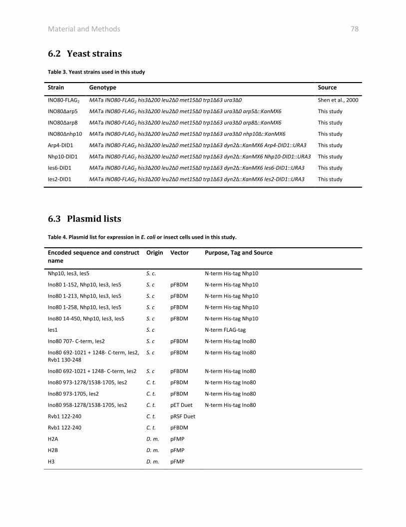
Material and Methods 78
6.2 Yeast strains
Table 3. Yeast strains used in this study
Strain Genotype Source
INO80-FLAG2 MATa INO80-FLAG2 his3Δ200 leu2Δ0 met15Δ0 trp1Δ63 ura3Δ0 Shen et al., 2000
INO80Δarp5 MATa INO80-FLAG2 his3Δ200 leu2Δ0 met15Δ0 trp1Δ63 ura3Δ0 arp5Δ::KanMX6 This study
INO80Δarp8 MATa INO80-FLAG2 his3Δ200 leu2Δ0 met15Δ0 trp1Δ63 ura3Δ0 arp8Δ::KanMX6 This study
INO80Δnhp10 MATa INO80-FLAG2 his3Δ200 leu2Δ0 met15Δ0 trp1Δ63 ura3Δ0 nhp10Δ::KanMX6 This study
Arp4-DID1 MATa INO80-FLAG2 his3Δ200 leu2Δ0 met15Δ0 trp1Δ63 dyn2Δ::KanMX6 Arp4-DID1::URA3 This study
Nhp10-DID1 MATa INO80-FLAG2 his3Δ200 leu2Δ0 met15Δ0 trp1Δ63 dyn2Δ::KanMX6 Nhp10-DID1::URA3 This study
Ies6-DID1 MATa INO80-FLAG2 his3Δ200 leu2Δ0 met15Δ0 trp1Δ63 dyn2Δ::KanMX6 Ies6-DID1::URA3 This study
Ies2-DID1 MATa INO80-FLAG2 his3Δ200 leu2Δ0 met15Δ0 trp1Δ63 dyn2Δ::KanMX6 Ies2-DID1::URA3 This study
6.3 Plasmid lists
Table 4. Plasmid list for expression in E. coli or insect cells used in this study.
Encoded sequence and construct name
Origin Vector Purpose, Tag and Source
Nhp10, Ies3, Ies5 S. c. N-term His-tag Nhp10
Ino80 1-152, Nhp10, Ies3, Ies5 S. c pFBDM N-term His-tag Nhp10
Ino80 1-213, Nhp10, Ies3, Ies5 S. c pFBDM N-term His-tag Nhp10
Ino80 1-258, Nhp10, Ies3, Ies5 S. c pFBDM N-term His-tag Nhp10
Ino80 14-450, Nhp10, Ies3, Ies5 S. c pFBDM N-term His-tag Nhp10
Ies1 S. c N-term FLAG-tag
Ino80 707- C-term, Ies2 S. c pFBDM N-term His-tag Ino80
Ino80 692-1021 + 1248- C-term, Ies2, Rvb1 130-248
S. c pFBDM N-term His-tag Ino80
Ino80 692-1021 + 1248- C-term, Ies2 S. c pFBDM N-term His-tag Ino80
Ino80 973-1278/1538-1705, Ies2 C. t. pFBDM N-term His-tag Ino80
Ino80 973-1705, Ies2 C. t. pFBDM N-term His-tag Ino80
Ino80 958-1278/1538-1705, Ies2 C. t. pET Duet N-term His-tag Ino80
Rvb1 122-240 C. t. pRSF Duet
Rvb1 122-240 C. t. pFBDM
H2A D. m. pFMP
H2B D. m. pFMP
H3 D. m. pFMP

Material and Methods 79 H4 D. m. pFMP
H2A.v D. m pET21
Gentamicin - pFA6a Kanamycin resistance marker for G418 selection (P. Wendler)
URA3 promoter and gene - pBluescript URA3 auxotrophy marker for ura selection (K. Strässer)
DID1-Nup120 - pProEx DID tagging in yeast; fusion of DID1 to gene of interest (Flemming from E. Hurt)
PrA-TEV-DID1 - pProEx DID tagging in yeast; fusion of DID1 to gene of interest (Flemming from E. Hurt)
GST-FLAG-DID2 - pProEx DID tagging in yeast; DID2 is expressed in Rosetta(DE3) (Flemming from E. Hurt)
His6-Dyn2 - pET DID tagging in yeast; Dynein2 (Dyn2) is expressed in Rosetta(DE3) (Flemming from E. Hurt)
6.4 Oligonucleotide list
Table 5. Oligonucleotide list used in this study.
Primer name 5’ – 3’ Sequence Vector/Purpose
Nhp10_SalI _for AAAAAGTCGACATG AAA CATCATCATCATCATCATAAAATGTCAGTTGAA GAAAAAAAGCGCAGAC
pFBDM polh; N-terminal His-tag TAGzyme
Nhp10_NotI_bac AAAAA GCGGCCGCTTAGTTAGAGGAAACTAAACTTCCATC pFBDM polh
yIes3_XhoI_fw AAAAACTCGAGATGAAGTTCGAAGACCTCTTGGC PFBDM p10
yIes3_NheI_rv AAAAAGCTAGCTTAAAGAAT GTTTTCCAAAAG GCC G PFBDM p10
yIes5_XhoI_fw AAAAACTCGAGATGCCTAGTAAA GATCCA GAG PFBDM p10
yIes5_NheI_rv AAAAA GCTAGCTTATGGTGTGTGCGTATC PFBDM p10
yIo8-1-x_SalI_f AAAAAGTCGACATGTCACTGGCAGTTCTAC pFBDM polh
yIo8-1-152Not_r AAAAAGCGGCCGCTCAAAGCTCGTCTTCATCATC pFBDM polh
yIo8-1-213Not_r AAAAAGCGGCCGCTCATCCATTAACAGTAACAACAG pFBDM polh
yIo8-1-258Not_r AAAAAGCGGCCGCTCATCCATTTTCTTCCTCG pFBDM polh
yIo8-1-450Not_r AAAAAGCGGCCGCTCACAACCCGTGTCTAGTG pFBDM polh
yIo8_14-xSal_f AAAAAGTCGACATGATATCTGATTTTTCAAAG pFBDM polh
KanB rev CTGCAGCGAGGAGCCGTAAT check G418 cassette in Δ strain
KanC for TGATTTTGATGACGAGCGTAAT check G418 cassette in Δ strains
Arp8 up for GAACAACTCTCGAACAAGG amplify arp8 Δ box
Arp8 down rev GCTACGGGGTATTGTCTC amplify arp8 Δ box
Arp8 up check for AGAGTGGTAAGTAACAAACAGAAC check for right locus of deletion
Nhp10 up for GTGATAGTCGGCAAAGG amplify nhp10 Δ box
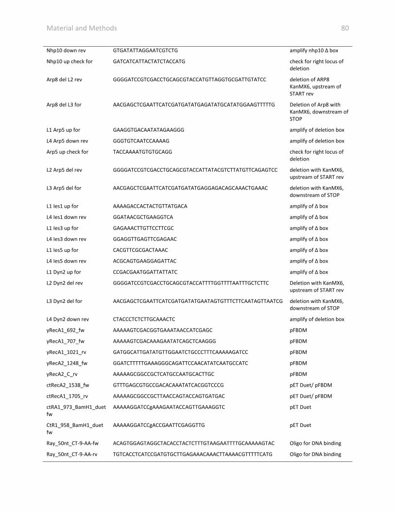
Material and Methods 80 Nhp10 down rev GTGATATTAGGAATCGTCTG amplify nhp10 Δ box
Nhp10 up check for GATCATCATTACTATCTACCATG check for right locus of deletion
Arp8 del L2 rev GGGGATCCGTCGACCTGCAGCGTACCATGTTAGGTGCGATTGTATCC deletion of ARP8 KanMX6, upstream of START rev
Arp8 del L3 for AACGAGCTCGAATTCATCGATGATATGAGATATGCATATGGAAGTTTTTG Deletion of Arp8 with KanMX6, downstream of STOP
L1 Arp5 up for GAAGGTGACAATATAGAAGGG amplify of deletion box
L4 Arp5 down rev GGGTGTCAATCCAAAAG amplify of deletion box
Arp5 up check for TACCAAAATGTGTGCAGG check for right locus of deletion
L2 Arp5 del rev GGGGATCCGTCGACCTGCAGCGTACCATTATACGTCTTATGTTCAGAGTCC deletion with KanMX6, upstream of START rev
L3 Arp5 del for AACGAGCTCGAATTCATCGATGATATGAGGAGACAGCAAACTGAAAC deletion with KanMX6, downstream of STOP
L1 Ies1 up for AAAAGACCACTACTGTTATGACA amplify of Δ box
L4 Ies1 down rev GGATAACGCTGAAGGTCA amplify of Δ box
L1 Ies3 up for GAGAAACTTGTTCCTTCGC amplify of Δ box
L4 Ies3 down rev GGAGGTTGAGTTCGAGAAC amplify of Δ box
L1 Ies5 up for CACGTTCGCGACTAAAC amplify of Δ box
L4 Ies5 down rev ACGCAGTGAAGGAGATTAC amplify of Δ box
L1 Dyn2 up for CCGACGAATGGATTATTATC amplify of Δ box
L2 Dyn2 del rev GGGGATCCGTCGACCTGCAGCGTACCATTTTGGTTTTAATTTGCTCTTC Deletion with KanMX6, upstream of START rev
L3 Dyn2 del for AACGAGCTCGAATTCATCGATGATATGAATAGTGTTTCTTCAATAGTTAATCG deletion with KanMX6, downstream of STOP
L4 Dyn2 down rev CTACCCTCTCTTGCAAACTC amplify of deletion box
yRecA1_692_fw AAAAAGTCGACGGTGAAATAACCATCGAGC pFBDM
yRecA1_707_fw AAAAAGTCGACAAAGAATATCAGCTCAAGGG pFBDM
yRecA1_1021_rv GATGGCATTGATATGTTGGAATCTGCCCTTTCAAAAAGATCC pFBDM
yRecA2_1248_fw GGATCTTTTTGAAAGGGCAGATTCCAACATATCAATGCCATC pFBDM
yRecA2_C_rv AAAAAGCGGCCGCTCATGCCAATGCACTTGC pFBDM
ctRecA2_1538_fw GTTTGAGCGTGCCGACACAAATATCACGGTCCCG pET Duet/ pFBDM
ctRecA1_1705_rv AAAAAGCGGCCGCTTAACCAGTACCAGTGATGAC pET Duet/ pFBDM
ctRA1_973_BamH1_duet fw
AAAAAGGATCCgAAAGAATACCAGTTGAAAGGTC pET Duet
CtR1_958_BamH1_duet fw
AAAAAGGATCCgACCGAATTCGAGGTTG pET Duet
Ray_50nt_CT-9-AA-fw ACAGTGGAGTAGGCTACACCTACTCTTTGTAAGAATTTTGCAAAAAGTAC Oligo for DNA binding
Ray_50nt_CT-9-AA-rv TGTCACCTCATCCGATGTGCTTGAGAAACAAACTTAAAACGTTTTTCATG Oligo for DNA binding

Material and Methods 81 Ray_37nt_CT-9-AA-fw TGGAGTAGGCTACACCTACTCTTTGTAAGAATTTTGC Oligo for DNA binding
Ray_37nt_CT-9-AA-rv ACCTCATCCGATGTGCTTGAGAAACAAACTTAAAACG Oligo for DNA binding
Guy_fork_3_1 CAACGTCATAGACGATTACATTGCTACATGGAGCTGTCTAGAGGATCCGA Oligo for DNA binding
Guy_fork_3_2 GTCGGATCCTCTAGACAGCTCCATGATCACTGGCACTGGTAGAATTCGGC Oligo for DNA binding
Guy_fork_3_3 TGCCGAATTCTACCAGTGCCAGTGAT Oligo for DNA binding
Guy_fork_3_4 TAGCAATGTAATCGTCTATGACGTT Oligo for DNA binding
6.5 Buffer list
Table 6. Buffers used for protein purification used in this study.
Buffers
Buffer name Ingredients
Protease Inhibitor 500 µM pepstatin A, 100 µM phenylmethanesulfonylfluoride (PMSF), 66 µM leupeptin, 270 mM
benzamidine in ethanol
Resuspension buffer 20 mM HEPES KOH pH 8.0, 5% glycerol (v/v), 1.2% PVP-40, 1x protease inhibitor
Lysis buffer 25 mM HEPES KOH pH 8.0, 500 mM KCl, 10% glycerol (v/v), 0.05% NP40 (v/v), 1 mM EDTA, 1 mM
DTT, 4 mM MgCl2, 1x protease inhibitor
H-0.5 FLAG 25 mM HEPES KOH pH 8.0, 500 mM KCl, 10% glycerol (v/v), 0.05% NP40 (v/v), 1 mM EDTA, 1 mM
DTT, 4 mM MgCl2
H-0.2 FLAG 30 mM HEPES KOH pH 8.0, 200 mM KCl, 10% glycerol (v/v), 0.05% NP40 (v/v), 1 mM DTT, 4 mM
MgCl2
Elution buffer 30 mM HEPES KOH pH 8.0, 200 mM KCl, 10% glycerol (v/v), 0.05% NP40 (v/v), 1 mM DTT, 4 mM
MgCl2, FLAG peptide
H-0.2 Q 25 mM HEPES KOH pH 8.0, 200 mM KCl, 7% glycerol (v/v), 0.05% NP40 (v/v), 1 mM DTT, 4 mM MgCl2
H-1 Q 25 mM HEPES KOH pH 8.0, 1000 mM KCl, 7% glycerol (v/v), 0.05% NP40 (v/v), 1 mM DTT, 4 mM
MgCl2
H-1 SEC 25 mM HEPES KOH pH 8.0, 200 mM KCl, 3% glycerol (v/v), 0.05% NP40 (v/v), 1 mM DTT, 4 mM MgCl2
H-0.1 Nhp10 25 mM HEPES KOH pH 8.0, 100 mM KCl, 10% glycerol (v/v), 2 mM β-mercaptoethanol, 4 mM MgCl2
H-0.1 Q Nhp10 25 mM HEPES KOH pH 8.0, 100 mM KCl, 7% glycerol (v/v), 2 mM β-mercaptoethanol, 4 mM MgCl2
H-1 Q Nhp10 25 mM HEPES KOH pH 8.0, 1000 mM KCl, 7% glycerol (v/v), 2 mM β-mercaptoethanol, 4 mM MgCl2
H-0.1 SEC Nhp10 25 mM HEPES KOH pH 8.0, 100 mM KCl, 3% glycerol (v/v), 2 mM β-mercaptoethanol, 1 mM MgCl2

Material and Methods 82 Sau-0 40 mM NaOAc pH 8.0, 8 M Urea, 5 mM β-mercaptoethanol, 5 mM lysine
Sau-1000 40 mM NaOAc pH 8.0, 8 M Urea, 5 mM β-mercaptoethanol, 5 mM lysine, 1000 mM NaCl
Sau-200 40 mM NaOAc pH 8.0, 8 M Urea, 5 mM β-mercaptoethanol, 5 mM lysine, 200 mM NaCl
Histones-0 Q 15 mM Tris HCl pH 8.0
Histones-2 Q 15 mM Tris HCl pH 8.0, 2 M NaCl
Unfolding buffer 20 mM Tris HCl pH 7.5, 7 M guanidine hydrochloride, 10 mM DTT
Refolding buffer 10 mM Tris HCl pH 7.5, 2 M NaCl, 5 mM β-mercaptoethanol, 1 mM EDTA
NCP buffer-0 20 mM Tris HCl pH 7.5, 10 mM DTT
NCP buffer-2000 20 mM Tris HCl pH 7.5, 10 mM DTT, 2000 mM KCl
Remodeling buffer 25 mM Hepes KOH pH 7.6, 50 mM NaCl, 10% glycerol, 0.05% NP-40, 0.1 mM EDTA, 4 mM MgCl2,
1 mM DTT
6.6 Molecular biology methods
6.6.1 Cloning in bacteria
In principal, cloning was performed with standard protocols. Polymerase chain reactions (PCR)
were performed using a premixed reaction and a primer concentration of 0.5 µM to generate
constructs listed in table 4. The annealing temperatures were adapted to primers listed in table
5. PCR reactions were analyzed by a 1% agaraose gel electrophoresis. The insert and vector
DNA were digested with appropriate restriction enzymes and according to instructions from the
manufacturer. The vector was subsequently dephosphorylated by alkaline phosphatase.
Cleavage reactions were purified by agarose gel electrophoresis. The digested insert was ligated
into the linearized vector using DNA ligase. Multiple genes were cloned in one vector using the
pFBDM vector system (Berger et al., 2004). The pFBDM vector has two cloning cassettes that
can be inserted multiple times enabling the expression of several subunits from one vector.
Ligated products were transformed into chemical competent E. coli XL1 blue cells by heat
shock. After generation of antibiotic resistance mediated by the plasmid, cells were selected on
LB agar plates containing the appropriate antibiotic. Amplified plasmids from an overnight

Material and Methods 83
culture were purified using plasmid extraction kits. Positive clones were verified by test
restriction cleavage or sequencing.
6.6.2 Cloning in yeast
In order to clone deletion mutants and tag subunits, the respective open reading frame was
replaced by a KanMX6 resistance cassette (pFA6a KanMX6, Ampr, AG Wendler) created by PCR
(Reid et al., 2002). DID1-tags (Flemming et al., 2010) were C-terminally inserted at the
endogenous locus together with URA3 marker (pRS316, Amp, Strässer) for selection. Deletions
strains were generated, either by amplification of the deletion box from gene disruption yeast
library strain by PCR (Table 5) or by the amplification of the KanMX6 cassette with primers
flanking regions homologous to regions of targeted gene. The integration of the G418 (Sigma-
Aldrich) marker occurred by transformation of the purified PCR product into the INO80-FLAG
strain leading to a deletion of the respective ORF. For DID1-tagging, a PCR product was
amplified such that the STOP codon was inserted behind the DID-tag, URA3 was inserted in the
terminator of the gene of interest.
Yeast transformation was performed according to (Gietz and Woods, 2002). Cells grown
to an OD600 of 0.5 to 0.8 were resuspended in a buffer containing 0.1 M LiAc, 40% PEG 4000,
salmon sperm carrier DNA and DNA to be transformed. Cells were heat shocked at 42°C for
10 min and after a rescue step selected on the appropriate antibtiotic or minimal media plate.
6.7 Protein expression and purification
6.7.1 Protein expression in E. coli
For overexpression of proteins in E. coli, BL21(DE3), BL21(DE3)-star or Rosetta (DE3) strains
were transformed with the plasmid containing the gene of interest. In general, protein
expression in E. coli was perfomed as described before. Protein expression was induced by
supplementing the media with the galactose analogous IPTG (Isopropyl-β-D-
Thiogalactopyranoside) at late logarithmic phase. Protein expression was allowed for either 4 h
at 37°C or over night at 18°C. Cells were harvested by centrifugation.

Material and Methods 84
6.7.2 Protein expression in insect cells
The use of E. coli as an expression host is limited as many eukaryotic large polypeptides fail to
express in bacteria. Expression in insect cells offer several advantages (Berger et al., 2004): (i)
the multiplication module in the pFBDM vector allows the insertion of several genes encoding
components of multiprotein complexes in one vector under two different promoters. (ii)
Recombinant baculoviruses produce high expression levels. (iii) The expression in a eukaryotic
organism facilitates the production of difficult gene products and allows a similar post-
translational modification state as in the original host.
Genes of interest were cloned into the pFBDM vector. The insertion into the MultiBac
baculoviral DNA is mediated via the Tn7 transposition sequence upon transformation in
DH10MultiBac E. coli cells (Berger et al., 2004; Fitzgerald et al., 2006). The antibiotic resistance
propagated by the pFBDM vector and the adjacent LacZ gene on the MultiBac DNA that enables
blue/white screening allow selection of positive clones on agar plates containing (X-α-galactose;
100 µg/ml, tetracycline and kanamycine). Recombinant bacmid DNA was isolated with standard
Mini and Midi plamid isolation kits. 2 ml of Sf21 (Spodoptera frugiperda) (Gibco, Invitrogen)
insect cells with 0.4 mio/ml were transfected with 2 µg DNA with 3 µl transfection reagent
(FuGene, Promega) in serum-free medium (Gibco). Baculoviral particles were amplified at
27.5°C for four days and the supernatant of the attached cells was used to infect 10 ml of SF21
cells with 1 mio/ml in a suspension culture for four days. The Cells were harvested and the
supernatant containing the virus were steril-filtered. The virus was amplified for another
generation before it was used for infection of High Five (Trichopulsia ni) (Gibco, Invitrogen).
High Five cells with 1 mio/ml were infected with appropriate virus titer for recombinant protein
expression.
6.7.3 Culturing of yeast
To prepare large-scale yeast fermentation, yeast cultures were cultivated stepwise. First, 10 ml
of YPD media supplemented with 50 mg/ml ampicillin were inoculated with colonies from a
freshly grown plate and cultivated at 30°C for 8 h. A 200 ml culture was inoculated with the

Material and Methods 85
10 ml pre-culture and was cultivated over night. This culture was used to inoculate a 2 L culture
with 50 ml and subsequently cultivated for 8 h. The media for either a 100 or 200 L fermenter
was supplemented with tetracycline (12.5 mg/ml), ampicillin (100 mg/ml) and 200 ml antifoam
per 200 L and the pH was titrated to 6.8 – 6.9. The fermenter was inoculated with a starting OD
of 0.0115. The fermentation was performed at 30°C with 250 rpm and 85 NL/h air-flow for 13 h.
At a final OD600 of 5 – 7 the cells were harvested.
Mutant strains were grown in 10 L buckets heated to 30°C in a water bath. The
ventilation was accomplished by pumping air through an aquarium air stone. YPD media was
supplemented equivalent to the large scale fermentation.
Cells were harvested by centrifugation, washed first with water, then with lysis buffer
(Table 6) and subsequently were aliquoted and frozen in liquid nitrogen.
6.7.4 SDS-PAGE and Western blot analysis
Proteins were separated according to their size by SDS polyacrylamide gel electrophoresis (SDS-
PAGE), as described earlier (Laemmli, 1970). INO80 complex was analyzed by pre-casted
gradient gels either 4-12% (Invitrogen) in MOPS buffer or 10-20% (BioRad). Recombinant sub-
complexes and histones were denatured with 4xLaemmli buffer (50% v/v glycerol, 250 mM Tris-
HCl pH 6.8, 7.5% w/v SDS, 5 mM EDTA, 10 mM DTT, 0.5% w/v bromphenol blue) and analyzed
on 15% or 18% polyacrylamide gels with 4% stacking gels (Table 7), respectively. After
separation of the proteins by electrophoresis, the gels were stained with colloidal Coomassie
staining solution (Invitrogen) or silver staining.

Material and Methods 86 Table 7 Gel components for 2 gels with 15% or 18% resolving gel and 4% stacking gel.
6.7.5 Silver staining
Silver staining was performed as in principal described before (Sasse and Gallagher,
2009). Gels were washed shortly with water and were then fixed (10% acetic acid, 45%
methanol) twice for 10 min. Gels were then treated twice with 50% ethanol for 10 min and
once with 30% ethanol. Gels were sensitized in 0.8 mM Na2S2O3 for 60 sec and then washed in
water for 3x 20 sec. Bands were then stained with 2 g/l AgNO3, 0.026% formaldehyde for
20 min. Silver solution was washed off with water 3x 20 sec. Bands were visualized with 6%
Na2CO3, 0.0185% formaldehyde and 16µM Na2S2O3. Developing was then quenched with 10%
acetic acid.
6.7.5.1 Immunoblotting – Western Blotting
In order to detect proteins with specific antibodies after SDS-PAGE, they were transferred onto
a polyvinylidine difluoride (PVDF) membrane in a wet (tank) blot system. To equilibrate and
activate the PVDF membrane, it was plunged briefly in methanol. The blot was assembled as
follows: two layers of 5 MM Whatman soaked in Transfer buffer (50 mM Tris/HCl pH 8.3, 0.19
M glycine, 0.1 (w/v) % SDS and 20 % methanol) and enclosed the gel lying over the PVDF
membrane. Blotting was performed for 1 h at a constant voltage of 100 V and 400 mA. Prior to
incubation with the first antibody, unspecific binding sites were blocked by incubating the
membrane for 20 min in 5% (w/v) milk in 1× TBS-T. The first antibody incubation of the
membrane was performed with 1× TBS-T, 5% (w/v) milk and the required antibody in the
Resolving gel Stacking gel 15% 18% 4% Reagent Volume
[ml] Volume [ml]
Reagent Volume [ml]
Water 5.7 3.7 Water 12 Acrylamide/Bis Solution 37,5:1 (30 % w/v)
9.3 11.3 Acrylamide/Bis Solution 37,5:1 (30 % w/v)
6
Lower 3 M Tris, pH 8.5 5 5 Upper 0.5 M Tris, pH 6.8 30 10% APS 100 µl 100 µl 10% APS 0.048 TEMED 10 µl 10 µl TEMED 0.45

Material and Methods 87
indicated dilution by shaking over night at 4°C. The blot was subsequently washed by shaking
three times for 10 min in 1× TBS-T buffer. The membrane was incubated with the secondary,
horse radish peroxidase coupled antibody in 1× TBS-T, 5% (w/v) milk for 1 h at room
temperature. The blot was again washed as described above. To visualize the immunodetected
proteins, 10 ml ECL solution was freshly supplemented with 3 µl H2O2, 25 µl cumaric acid and
50 µl luminol and incubated for 1 min at room temperature with the membrane by shaking. The
signal was then detected by exposure to film (Biomax Light, Maximum Resolution or Maximum
Sensitivity film, Kodak) and subsequent developing by Kodak X-Omat 2000 processor in the dark
room.
6.7.6 Purification of histones and reconstitution of nucleosomes
In principal, histones were purified as described before (Dyer et al., 2004; Huynh et al., 2005).
Coding sequences of D. m. histones H2A, H2B, H3 and H4 are codon optimized in expression
vectors (Peter Becker’s group). Coding sequence of D.m. H2A.v was cloned into pET21 vector.
Histones were expressed in BL12 Star (DE3) cells. Cells were resuspended in Sau-200 buffer
(Table 6) and subsequently sonicated with an output control of 5 and a duty cycle of 40%. The
lysate was cleared by centrifugation at 20,000 rpm for 20 min at 4°C. The supernatant was
filtered through Mira-cloth and 0.45 µm filter. The filtrate was loaded online onto a HiTrap SP
HP 5 ml column (GE Healthcare) using a 50 ml injection super loop. The histones were eluted
from the column as follows: (i) wash with 20% Sau-2000 for 3 column volumes, (ii) wash with
25% Sau-2000 for 5 column volumes and (iii) elution with a gradient of 20-60% and 60-100% of
Sau-2000 for 6 and 3 column volumes, respectively. Proper fractions were pooled and dialyzed
against 3 L of ddH2O for 30 min, over night and again for 1 h at 4°C. Subsequently, histones
were loaded on a HiTrap Q FF 5 ml column (GE Healthcare) in Histones-0 Q buffer (Table 6). The
histone containing flow through were collected and concentration was determined by
NanoDrop photospetrometer (Thermo Scientific) and SDS-PAGE. Histones were aliquoted,
frozen and lyophilized overnight.

Material and Methods 88
Histones were dissolved yielding a concentration of 4 mg/ml in Unfolding buffer (Table 6).
Histones were unfolded for 30 min – 3 h. The octamer was reconstituted by mixing the four
histones in 120% H2A and H2B to 100% H3 and H4 equimolar ratio. The octamer was dialyzed
against 3x 2 L of refolding buffer (Table 6). The precipitation was removed by centrifugation and
the octamer was concentrated using Amicon centrifugal filters (Merck Millipore). The octamer
was finally purified on size exclusion chromatography column S200 26/60 (GE Healthcare). The
stoichiometry and purity was controlled by SDS-PAGE and the octamer was stored in 50%
glycerol (w/v).
Various DNAs were used to reconstitute nucleosomes. INO1 nucleosomes were
reconstituted using genomic sequence covering +1 - +359 of the INO1 gene. 147 bp, 187 long
(with 20 bp on each side or 40 bp overhang) DNAs were based on the Widom 601 positioning
sequence (Huynh et al., 2005).
In brief, 147 bp and 187 bp long DNAs were cloned into a pFMP vector. Plasmids were
amplified in bacteria and isolated. Purified plasmids were digested with respective restriction
enzymes. Digested DNA was purified on native 8% acrylamide gels. DNA was isolated by
electroelution from the gel. DNA was purified by phenol/chloroform-isoamyl alcohol and
ethanol precipitation.
For INO1 nucleosomes, the DNA was amplified by PCR (Table 5). 2 ng of template DNA
was amplified in 50 µl PCR reactions containing 5 µl 10x Taq buffer, 5 µl dNTPs (FIRMA), 2.5 µl
10 µM forward and reverse primer and 1 µl Taq polymerase with following settings: 2 min at
94°C; 30 cycles with 15 sec 94°C, 15 sec 56°C, 30 sec 72°C; 5 min at 72°C. DNA was purified on a
1.2% agarose gel with a Gel Extraction Kit (QIAquick®) and by ethanol precipitation. 1/10 (v/v)
NaOAc and 2.5 volume 100% ethanol were added and incubated at -20°C over night. The DNA
was pelleted at 4°C with 15.000 g for 30 min and washed with 70% ethanol. The purified DNA
was dissolved in Tris EDTA buffer (10 mM Tris and 1 mM EDTA) to a final concentration of 5-
7 µg/µl. The DNA concentration was determined on a NanoDrop photospetrometer (Thermo
Scientific).

Material and Methods 89
In order to avoid high concentrations of free DNA in the nucleosome preparations, the
proper DNA concentration had to be titrated. Various molar ratios of DNA to octamer were
mixed together and transferred to small dialysis chambers. The salt concentration was slowly
reduced by linear salt gradient dialysis with following steps: (i) 7h 0.2 ml/min, (ii) 16h
0.4 ml/min and (iii) 7h 1ml/min. the final salt concentration was 50 mM KCl in the nucleosome
reconstitution. The quality of nucleosomes was assessed by 5% native PAGE and measuring
DNA absorption at 260 nm.
6.7.7 Establishment of an INO80 purification protocol
The endogenous purification of INO80 from S. cerevisiae was based on earlier purification
protocols (Shen, 2004; Shen et al., 2000). The modified protocol improved quality and
homogeneity of INO80 and allowed a comprehensive structural analysis.
6.7.7.1 Cell lysis
6.7.7.1.1 Cryogenic disruption of yeast cells
For small-scale purifications cells were disrupted using a planetary ball mill on frozen cells
(Alber et al., 2007). Yeast strains were grown to an OD600 of 0.5 – 1.5. Cells were harvested by
centrifugation and were washed with water and resuspension buffer (Table 6). Cell paste was
pressed through a syringe into liquid nitrogen producing frozen noodles. These noodles were
then cryogenically disrupted using a planetary ball mill (PM 100, Retsch). Disrupted frozen cells
were then resuspended in lysis buffer (Table 6).
6.7.7.1.2 Bead-beater
Large-scale yeast disruption was accomplished with bead-beaters (BioSpec). The bead-beaters
were used after instructions of the manufacturer. The chamber was filled with 200 ml glass
beads (0.5 mm, Roth) and 150 ml cell pellet resupended Lysis buffer (Table 6). Cells were lysed

Material and Methods 90
for 1.15 h with 1 min intervals. Residual cell lysate was washed from the beads with 200 ml lysis
buffer.
6.7.7.2 Chromatin fragmentation
In the course of assessing appropriate chromatin fragmentation, MNase (Roche) treatment and
sonication was tested. For MNase digestion, 400 µl cell lysate was incubated with 45 U of
MNase in lysis buffer containing 10 mM CaCl2 for either 1 h at 4°C or 10 min at 37°C. Digestion
was stopped by adding EDTA. In the standard protocol, chromatin was fragmented with a
polytron homogenizer (Kinematica; Fisher Scientific) for four rounds for 30 s at 16.000 rpm/min
and then by sonication (Branson) for seven round for 30 s with a duty cycle of 4, an output
control of 5 and output yield of 60% on ice. For DNA isolation 500 µl of diluted lysate were
mixed with 500 µl phenol and phases were separated by centrifugation at 15.000 g for 5 min.
The aqueous phase was recovered and mixed with 460 µl chloroform-isoamylalcohol. 400 µl
were recovered and DNA was precipitated by 2.5 volumes -20°C 96% ethanol and 1/10 (v/v)
3 M NaAc pH 5.2 for at least 3 h at -80°C. The precipitated DNA was washed twice with -20°C
cold 70% ethanol. The pelleted DNA was dried at room temperature and dissolved in 10 µl H2O.
RNA was digested with 1 µl RNase (Fermentas) for 30 min at 37°C. DNA length was analyzed by
gel electrophoresis using 1% agarose gels.
6.7.7.3 FLAG purification of the INO80 complex
In order to remove debris and unbroken cells, the lysate was centrifuged with 14,000 rpm for
30 min at 4°C (rotor SLA 1500; Thermo Scientific). The supernatant was removed and futher?
centrifuged in an ultra centrifuge at 40,000 rpm for 90 min at 4°C (Ti45; Beckmann and Coulter).
The fatty layer on top was removed and supernatant was gently collected. To remove sticky
proteins, a pre-clearing step was performed with 200 µl protein G beads (Invitrogen)
equilibrated in lysis buffer for 30 min on a nutator. Beads were removed by centrifugation at
3,000 g (Hettich centrifuge) for 10 min. Immunopurification was performed with 2 ml of M2
FLAG-beads (Sigma-Aldrich) for 1.5 h on a nutator at 4°C. Beads were collected by

Material and Methods 91
centrifugation at 1,000 g (Hettich centrifuge) for 10 min. The supernatant was not
quantitatively removed and the beads were resuspended in residual liquid and transformed to
gravity flow column. Unbound proteins were removed by washing with 10 column volumes of
H-0.5 buffer and 5 column volumes of H-0.2 buffer (Table 6). INO80 was eluted from FLAG-
beads by adding subsequently elution buffer (Table 6) either containing 0.1 mg/ml or 0.2 mg/ml
FLAG peptide. FLAG peptide (Sigma Aldrich) was dissolved in buffer containing 10 mM HEPES
pH 8.0, 100 mM KCl and 2 mM MgCl2 to yield a final concentration of 5 mg/ml. Elution was
performed as follows: (i) 15 min incubation with 2 ml of 0.1 mg/ml FLAG peptide containing
buffer and collection of elution by gravity flow, (ii) 10 min incubation with 1 ml of 0.1 mg/ml
FLAG peptide containing buffer and collection of elution by gravity flow, (iii) 15 min incubation
with 2 ml of 0.2 mg/ml FLAG peptide containing buffer and collection of elution by gravity flow,
(iiii) 10 min incubation with 1 ml of 0.2 mg/ml FLAG peptide containing buffer and collection of
elution by gravity flow and (iiiii) FLAG beads were washed with 2 ml of H-0.2 buffer. After
elution FLAG-beads were re-incubated with the lysate over night on nutator. FLAG beads were
collected and treated as described. Last washing step was complemented with 0.1 mg/ml FLAG
peptide and beads were incubated for 15 min for elution. INO80 was further eluted by 2 ml of
0.2 mg/ml FLAG peptide containing buffer and elution was collected by gravity flow. Residual
INO80 was collected by washing the beads with 1 ml of H-0.2 buffer (Table 6). INO80
purification was analyzed by SDS-PAGE and Western blot analysis.
6.7.7.4 Ion exchange chromatography
In the course of establishing purification protocol for INO80 diverse columns have been
assessed: HiTrap Heparin HP 1 ml, Mini S 4.6/50 PE 0.8 ml and a Mono Q 5/50 1 ml (GE
Healthcare) connected to a Micro Aekta system (GE Healthcare).
In principal, the pooled elution fractions from FLAG purification were loaded online onto
the columns with a 10 ml super-loop (GE Healthcare) and H-01 Q buffer (Table 6). In the
standard protocol the Mono Q column was used. For this, the sample was loaded with
0.125 ml/min onto the column. The washing and elution protocol was run with 0.4 ml/min.

Material and Methods 92
Unbound proteins were washed away with 25% (400 mM KCl) of buffer H-1 Q (Table 6). INO80
complexes were eluted form resin at 50% (600 mM KCl) of buffer H-1 Q and elution was
fractionated in 60 µl. DNA containing proteins eluted at higher salt-concentrations.
6.7.7.5 Size exclusion chromatography
In order to exchange buffer and achieve a monodisperse sample, the elution fractions were
pooled and directly applied to a Superose 6 PC 3.2/30 (2.4 ml) column (GE Healthcare)
connected to Ettan Aekta system (GE Healthcare). The column was run with 0.04 ml/min of H-
0.2 SEC buffer (Table 6) and was fractionated in 25 µl.
To stabilize INO80 for the EM analysis, INO80 was cross-linked with 0.05%
glutaraldehyde (EM grade, Sigma-Aldrich) at 20°C for 30 min. The reaction was quenched by
adding 100 mM Tris pH 8.2 and incubation at 20°C for 15 min. Cross-linked samples were
applied to another round of size exclusion chromatography to asses homogeneity.
6.7.8 Purification of the Swi2/Snf2 sub-complex
In brief, either coding sequences from S. c. or C. t. of the full Snf2 ATPase of Ino80 including the
insertion fold or constructs coding only for the RecA1 and RecA2 folds without the insertion
domain were cloned in either pFBDM vectors for expression in insect cells or pET duet vector
for expression in E. coli. Ino80 constructs were tried to express with Ies2 alone or together with
the OB-folds of Rvb1. All the Ino80 constructs contained an N-terminal His-tag that allowed
immunopurification.
6.7.9 Purification of the Nhp10 subcomplex
S. cerevisiae Nhp10, Ies3 and Ies5 were cloned into pFBDM vector (Table 4). A coding sequence
for a His6-tag was fused N-terminal to Nhp10. Additional to Nhp10, Ies3 and Ies5, diverse N-
terminal constructs of Ino80 were cloned into this vector. Furthermore, a vector containing the

Material and Methods 93
coding sequence of Ies1 fused to N-terminal FLAG tag was used to assess interaction with the
Nhp10 sub-complex. Proteins were expressed accordingly (Fitzgerald et al., 2006). In brief, High
five cells were infected with 1:100 Nhp10-Ies3-Ies5 baculovirus or were co-infected with each
1:20 Nhp10-Ies3-Ies5-Ino80 (NTD) and Ies1-FLAG viruses for 3 days. Cells were lysed by
sonication on ice for 8x 40 bursts with a duty cycle of 4, an output control of 5 and output yield
of 60%. The lysate was cleared by twice centrifugation at 18.000 rpm (SS-34, Thermo Scientific)
for 30 min. Immunopurification of complex was performed with Ni-NTA beads. Unbound
proteins and contaminations were washed off with buffers containing 20 mM, 40 mM or 1 M
NaCl. Complexes were eluted with a buffer containing 300 mM imidazole. Ni2+-elution fractions
were pooled and directly loaded onto HiTrap Q HP (GE Healthcare) column. Complexes were
eluted by a linear gradient. Proper fractions were pooled and the complex was concentrated
using Amicon centrifugal filters (Merck Millipore). Complexes were then applied on a size
exclusion chromatography column S200 (GE Healthcare). Proteins were subsequently
concentrated and analyzed by SDS-PAGE.
6.8 Generation of INO80 binding nanobodies
To generate nanobodies that target the INO80 complex an alpaca was immunized with 500 µg
freshly purified and glutaraldehyde fixed INO80, 700 µg Arp8-Arp4-Act-HSA-Ies4, 700 µg yeast
Arp5-Ies6, 700 µg Nhp10-Ies3-Ies5, 500 µg human Arp8 (38-624) and human 700 µg Arp5-Ies6.
Peripheral blood lymphocytes were isolated; total RNA was extracted and written into cDNA.
The phagemid library was generated by cloning the VHH repertoire from the heavy-chain
repertoire. Binders were selected after panning with ~400 µg INO80 followed by solid-phase
ELISA screening. Positive clones were tested to bind to Arp8-Arp4-Act-HSA, yeast Rvb1/2 and
Arp5-Ies6.
To validate the ability to immunoprecipitate INO80, binders covalently linked to agarose
beads were incubated with cell lysate from an Ino80-FLAG strain. The same amount of FLAG-M2
beads (Sigma-Aldrich) was used as a reference. After the normal INO80 purification procedure,

Material and Methods 94
the bound proteins were eluted from the beads with conventional Laemmli-buffer and were
analyzed by SDS-PAGE visualized by silver and Coomassie staining. Nanobodies have been
generated as described before (Rothbauer et al., 2006) and in collaboration with ChromoTek
(Munich).
6.9 Cross-linking and mass spectrometry
6.9.1 Titration of cross-linker
A titration of the cross-linker, DSS (Creative Molecules) was performed to find appropriate
cross-linking concentration. As lysines are the exclusive target for NHS esters, the cross-linker
concentration was determined in respect to the lysine amount. It was assumed that 1 µg of
protein contains 500 pmol of lysines. Based on this assumption the concentration was
expressed and calculated as cross-linker over lysines. In general, 1 mg of an equimolar mixture
of light and heavy labeled DSS was dissolved in dimethylformamide (DMF, Thermo Scientific) to
a concentration of 25 mM. DSS was then further diluted to cross-linking concentration with
H2O. The samples were warmed up to room temperature and cross-linked by adding the diluted
cross-linker and mixing. The samples were then incubated for 35 min at 30°C with 1000 rpm.
After spinning the samples down, the reaction was quenched by adding 100 mM Tris pH 8.0 or
ammonium bicarbonate. The samples were then incubated for 15 min at 30°C with 1000 rpm.
To evaluate cross-linking efficiency 1 µg of complex was analyzed on pre-cast SDS-PAGE
gradient gel 4-12% (Invitrogen). For the titration, 2 µg of freshly purified and monodisperse
INO80 were cross-linked with 0.4x, 1x, 2.5x or 5x DSS over lysines and 1.5 µg of INO80-
nucleosome complex were cross-linked with 0.5x, 1.5x, 3x and 5x DSS over lysines.
6.9.2 Sample preparation for mass spectrometry analysis
Cross-linking of samples was in principal performed as the titration, but with the difference that
the dissolved cross-linker was directly added to the complex. We cross-linked 30, 40, 50, 84 µg
of INO80 from different preparations with 3x, 3.5x, 1.5x and 3x DSS, respectively. For INO80-

Material and Methods 95
nucleosome complexes, ten preparations of INO80 between 100 and 200 µg were incubated
with a 1 to 5 fold molar excess of nucleosomes with either 187 bp off-centered or 147 bp DNA
in absence or presence of ADP BeFx or AMP-PCP on ice for 30 min. INO80-nucleosome
complexes were cross-linked with 1.5x or 3.5x DSS over lysines. Cross-linking and peptide
preparations were in principal performed as described before (Herzog et al., 2012; Jennebach
et al., 2012; Leitner et al., 2012b).
After quenching the cross-linking reaction, proteins were denatured with twice the volume of
8 M urea. Cysteines were reduced with 5 mM tris(2-carboxyethyl)phosphine (TCEP,Thermo
Scientific) for 15 min at 37°C. Cysteines were further alkylated with 10 mM iodoacetamide
(Sigma-Aldrich). Proteins were digested with 1:50 (w/w) of lysyl endopeptidase C (Wako) for 1 h
at 37°C at 950 rpm. After diluting the sample to a urea concentration of 1 M, 1/50 (w/w) trypsin
(Promega) was added and incubated at 37°C at 950 rpm over night. Samples were acetified
with 1% (v/v) trifluoroacetic acid (TFA, Sigma-Aldrich) and peptides were enriched by solid-
phase extraction using C18 cartridges (Sep-Pak, Waters). Elution was dried by vacuum
centrifugation and dissolved in 18.5 µl of (3% ACN; 0.2% formic acid (FA)) at 37°C 1400 rpm.
Cross-linked peptides were enriched by peptide size exclusion chromatography Superdex
peptide PC 3.2/30 (GE Healthcare) with a mobile phase (70% H2O, 30% ACN, 0.1% TFA). A
peptide standard, insulin B oxidized (Sigma-Aldrich) helped to identify fractions containing
cross-linked peptides. Fractions were dried and dissolved in 3% ACN; 0.2% FA for 1 h at
1400 rpm at 37°C.
Samples were analyzed on a liquid chromatography coupled to tandem mass
spectrometry (LC-MS/MS), LTQ Orbitrap (Thermo) equipped with a standard nanoelectrospray
source.
6.9.3 Sample analysis
In general, analysis and validation of cross-links was performed as described before (Herzog et
al., 2012; Leitner et al., 2010; Walzthoeni et al., 2012). For the cross-link peptide search xQuest
was used and FDR were determined by xProphet. Parameters were set as follows: maximum

Material and Methods 96
number of missed cleavages = 2, peptide length = 4-45 amino acids, fixed modifications =
carbamidomethyl-Cys (mass shift = 57.021460 Da), variable modifications = oxidation-Met
(mass shift = 15.99491), mass shift of the light cross-linker = 138.068080 Da, mass shift of
mono-links = 156.078644 and 155.096428 Da, MS1 tolerance = 15 ppm, MS2 tolerance = 0.2 Da
for common ions and 0.3 Da for cross-link ions, search in ion-tag mode.
The experimental spectrum was assigned to theoretical candidate spectra and was
scored according to quality of the match. The cross-link candidates were filtered by the mass
error (-4 to +7 ppm) and the Δscore (≥ 15%) that indicated relative scores between the next
ranked match. All cross-links were manually validated. Manual validation included that each
peptide had at least four bond cleavages in total or three in a series and a minimum length of
six amino acids. Cross-links composed of one peptide with five amino acids that fulfilled filtering
and manual criteria were included.
6.10 Analysis of subunit composition of INO80’s knock-out mutants
INO80 complexes from subunit knock-out mutants were purified according to the WT. The
composition and quantity of subunits of the INO80 mutants was determined by analysis of the
FLAG-elutions using SDS-PAGE. The omission of subunits was verified by MS. To remove agents
interfering with MS-analysis, the FLAG-elutions were shortly run on a SDS-PAGE. Entire lanes
were cut out and subjected to in-gel trypsinization. The obtained mixtures of tryptic peptides
were analyzed by LC-MS/MS using an OrbitrapXL instrument. In agreement with the genetic
subunit deletions, corresponding INO80 protein subunits could not be identified in single band
LC-MS/MS analysis. MS/MS spectra were searched with MASCOT using the yeast subset of the
Swiss-Prot protein database. Proteins identified with a Protein score below 100, where
manually validated.
6.11 Analysis of distant restraints
We assessed Cα-Cα distances of intra-links using available crystal structures from the RCSB
protein data bank: Dro Snf2 (1Z3I, (Thoma et al., 2005)), yeast Actin (1YAG; (Vorobiev et al.,

Material and Methods 97
2003)), yeast Arp4 (3QB0; (Fenn et al., 2011)), human and yeast Arp8 (4FO0 and 4AM6;
(Gerhold et al., 2012; Saravanan et al., 2012)). Distant constraints in atomic coordinates were
measured using the UCSF chimera package (Pettersen et al., 2004). All illustrations involving EM
structures as well as crystal structures were acquired from UCSF chimera.
To determine whether hexameric Rvb1/2 rings are composed of hetero- or homo-
hexamers, we modeled the 3D structure of yeast Rvb1/2 using SWISS MODEL (Kiefer et al.,
2009). The model of the yeast Rvb1 and Rvb2 is based on the crystal structure of hexameric
human Rvb1 (Matias et al., 2006; 2C9O). Human and yeast sequences were acquired from NCBI
and were aligned using ClustalW (Larkin et al., 2007).
For analysis of INO80-nucleosome cross-links we used sequences acquired from NCBI and
the atomic coordinates of the D. melanogaster nucleosome (2PYO) (Clapier et al., 2008).
6.12 Functional assays
6.12.1 Electrophoretic mobility shift assays
For electrophoretic mobility assays, increasing amounts of proteins were incubated with
diverse structured DNA oligonucleotides. Each 50 nM of 50 bp and 37 bp long distorted DNA
duplexes (Ray and Grove, 2009, 2012), 50 bp long fork structure (Guy and Bolt, 2005), 50 bp
long curved DNA and 187 bp long 601 sequence (Huynh et al., 2005) were assessed for binding.
The reaction buffer contained 5% glycerol (v/v) and total volume was 10 µl. The reactions were
loaded on a 5% native polyacrylamide gel, which was pre-run before. After electrophoresis in
0.5x Tris-borate-EDTA (TBE) for 1.5-2 h at 100V at 4°C, the gel was stained with Sybr Green I
(1:5000 in 0.5x TBE) for 30 min and scanned on a Typhoon scanner (GE Healthcare).
6.12.2 Remodeling assay
Remodeling assays contained 50 nM nucleosomes on a 359 bp long DNA (INO1 gene bp +1 to
+359) were incubated with increasing concentrations of INO80 in a remodeling buffer (Table 6)

Material and Methods 98
in absence or presence of 2 mM ATP or non-hydrolysable ATP analogs, AMP∙PCP or ADP∙BeFx
for 30 min at 30°C. Subsequently, 10 µl of the reaction were loaded onto 4% native
polyacrylamide gels. After electrophoresis in 0.5x TBE buffer for 1.5-2 h at 100 V at 4°C, the
DNA was visualized by Sybr Green I (1:5000 in 0.5x TBE) for 30 min.
6.12.3 ATP hydrolysis assays
An ATPase reaction (5 µL) was performed in remodeling buffer and contained 30 µM ATP and
20 nCi [γ-32P]ATP. 20 nM INO80 complexes were either stimulated with 50 nM 359bp long DNA
(INO1 gene bp +1 until +359) or nucleosomes or none. Reactions were incubated for 30 min at
30°C in a PCR machine. Reactions were separated by thin-layer chromatography with 1 M
formic acid containing 0.5 M LiCl. TLC plates were subsequently incubated on storage
phosphate screens for 30 min. Signals were scanned on a STORM scanner (GE Healthcare). In
general, two experiments were quantified by Image J. ATP hydrolysis rates were calculated
relative to complete ATP hydrolysis achieved by calf intestinal alkaline phosphatase treatment.

References 99
7 References
Abbott, D.W., Ivanova, V.S., Wang, X., Bonner, W.M., and Ausio, J. (2001). Characterization of the stability and folding of H2A.Z chromatin particles: implications for transcriptional activation. J Biol Chem 276, 41945-41949.
Alber, F., Dokudovskaya, S., Veenhoff, L.M., Zhang, W., Kipper, J., Devos, D., Suprapto, A., Karni-Schmidt, O., Williams, R., Chait, B.T., et al. (2007). Determining the architectures of macromolecular assemblies. Nature 450, 683-694.
Alber, F., Forster, F., Korkin, D., Topf, M., and Sali, A. (2008). Integrating diverse data for structure determination of macromolecular assemblies. Annu Rev Biochem 77, 443-477.
Allain, F.H., Yen, Y.M., Masse, J.E., Schultze, P., Dieckmann, T., Johnson, R.C., and Feigon, J. (1999). Solution structure of the HMG protein NHP6A and its interaction with DNA reveals the structural determinants for non-sequence-specific binding. EMBO J 18, 2563-2579.
Andrews, A.J., and Luger, K. (2011). Nucleosome structure(s) and stability: variations on a theme. Annu Rev Biophys 40, 99-117.
Arbabi Ghahroudi, M., Desmyter, A., Wyns, L., Hamers, R., and Muyldermans, S. (1997). Selection and identification of single domain antibody fragments from camel heavy-chain antibodies. FEBS Lett 414, 521-526.
Arents, G., Burlingame, R.W., Wang, B.C., Love, W.E., and Moudrianakis, E.N. (1991). The nucleosomal core histone octamer at 3.1 A resolution: a tripartite protein assembly and a left-handed superhelix. Proc Natl Acad Sci U S A 88, 10148-10152.
Asturias, F.J., Chung, W.H., Kornberg, R.D., and Lorch, Y. (2002). Structural analysis of the RSC chromatin-remodeling complex. Proc Natl Acad Sci U S A 99, 13477-13480.
Auger, A., Galarneau, L., Altaf, M., Nourani, A., Doyon, Y., Utley, R.T., Cronier, D., Allard, S., and Cote, J. (2008). Eaf1 is the platform for NuA4 molecular assembly that evolutionarily links chromatin acetylation to ATP-dependent exchange of histone H2A variants. Mol Cell Biol 28, 2257-2270.
Badis, G., Chan, E.T., van Bakel, H., Pena-Castillo, L., Tillo, D., Tsui, K., Carlson, C.D., Gossett, A.J., Hasinoff, M.J., Warren, C.L., et al. (2008). A library of yeast transcription factor motifs reveals a widespread function for Rsc3 in targeting nucleosome exclusion at promoters. Mol Cell 32, 878-887.
Ball, H.L., Myers, J.S., and Cortez, D. (2005). ATRIP binding to replication protein A-single-stranded DNA promotes ATR-ATRIP localization but is dispensable for Chk1 phosphorylation. Mol Biol Cell 16, 2372-2381.
Bao, Y., and Shen, X. (2007). INO80 subfamily of chromatin remodeling complexes. Mutat Res 618, 18-29.
Bao, Y., and Shen, X. (2011). SnapShot: Chromatin remodeling: INO80 and SWR1. Cell 144, 158-158 e152.
Barbera, A.J., Chodaparambil, J.V., Kelley-Clarke, B., Joukov, V., Walter, J.C., Luger, K., and Kaye, K.M. (2006). The nucleosomal surface as a docking station for Kaposi's sarcoma herpesvirus LANA. Science 311, 856-861.
Bauer, A., Chauvet, S., Huber, O., Usseglio, F., Rothbacher, U., Aragnol, D., Kemler, R., and Pradel, J. (2000). Pontin52 and reptin52 function as antagonistic regulators of beta-catenin signalling activity. EMBO J 19, 6121-6130.
Berger, I., Fitzgerald, D.J., and Richmond, T.J. (2004). Baculovirus expression system for heterologous multiprotein complexes. Nat Biotechnol 22, 1583-1587.
Bernstein, B.E., Liu, C.L., Humphrey, E.L., Perlstein, E.O., and Schreiber, S.L. (2004). Global nucleosome occupancy in yeast. Genome Biol 5, R62.

References 100
Billon, P., and Cote, J. (2012). Precise deposition of histone H2A.Z in chromatin for genome expression and maintenance. Biochim Biophys Acta 1819, 290-302.
Blosser, T.R., Yang, J.G., Stone, M.D., Narlikar, G.J., and Zhuang, X. (2009). Dynamics of nucleosome remodelling by individual ACF complexes. Nature 462, 1022-1027.
Bohm, V., Hieb, A.R., Andrews, A.J., Gansen, A., Rocker, A., Toth, K., Luger, K., and Langowski, J. (2011). Nucleosome accessibility governed by the dimer/tetramer interface. Nucleic Acids Res 39, 3093-3102.
Bortvin, A., and Winston, F. (1996). Evidence that Spt6p controls chromatin structure by a direct interaction with histones. Science 272, 1473-1476.
Branzei, D., and Foiani, M. (2008). Regulation of DNA repair throughout the cell cycle. Nat Rev Mol Cell Biol 9, 297-308.
Burma, S., Chen, B.P., Murphy, M., Kurimasa, A., and Chen, D.J. (2001). ATM phosphorylates histone H2AX in response to DNA double-strand breaks. J Biol Chem 276, 42462-42467.
Bushnell, D.A., Westover, K.D., Davis, R.E., and Kornberg, R.D. (2004). Structural basis of transcription: an RNA polymerase II-TFIIB cocrystal at 4.5 Angstroms. Science 303, 983-988.
Cai, Y., Jin, J., Yao, T., Gottschalk, A.J., Swanson, S.K., Wu, S., Shi, Y., Washburn, M.P., Florens, L., Conaway, R.C., et al. (2007). YY1 functions with INO80 to activate transcription. Nat Struct Mol Biol 14, 872-874.
Chaban, Y., Ezeokonkwo, C., Chung, W.H., Zhang, F., Kornberg, R.D., Maier-Davis, B., Lorch, Y., and Asturias, F.J. (2008). Structure of a RSC-nucleosome complex and insights into chromatin remodeling. Nat Struct Mol Biol 15, 1272-1277.
Chambers, A.L., Ormerod, G., Durley, S.C., Sing, T.L., Brown, G.W., Kent, N.A., and Downs, J.A. (2012). The INO80 chromatin remodeling complex prevents polyploidy and maintains normal chromatin structure at centromeres. Genes Dev 26, 2590-2603.
Chen, L., Cai, Y., Jin, J., Florens, L., Swanson, S.K., Washburn, M.P., Conaway, J.W., and Conaway, R.C. (2011). Subunit organization of the human INO80 chromatin remodeling complex: an evolutionarily conserved core complex catalyzes ATP-dependent nucleosome remodeling. J Biol Chem 286, 11283-11289.
Chen, Z.A., Jawhari, A., Fischer, L., Buchen, C., Tahir, S., Kamenski, T., Rasmussen, M., Lariviere, L., Bukowski-Wills, J.C., Nilges, M., et al. (2010). Architecture of the RNA polymerase II-TFIIF complex revealed by cross-linking and mass spectrometry. EMBO J 29, 717-726.
Cheung, K.L., Huen, J., Kakihara, Y., Houry, W.A., and Ortega, J. (2010). Alternative oligomeric states of the yeast Rvb1/Rvb2 complex induced by histidine tags. J Mol Biol 404, 478-492.
Ciferri, C., Lander, G.C., Maiolica, A., Herzog, F., Aebersold, R., and Nogales, E. (2012). Molecular architecture of human polycomb repressive complex 2. Elife 1, e00005.
Ciferri, C., Musacchio, A., and Petrovic, A. (2007). The Ndc80 complex: hub of kinetochore activity. FEBS Lett 581, 2862-2869.
Ciferri, C., Pasqualato, S., Screpanti, E., Varetti, G., Santaguida, S., Dos Reis, G., Maiolica, A., Polka, J., De Luca, J.G., De Wulf, P., et al. (2008). Implications for kinetochore-microtubule attachment from the structure of an engineered Ndc80 complex. Cell 133, 427-439.
Cimprich, K.A., and Cortez, D. (2008). ATR: an essential regulator of genome integrity. Nat Rev Mol Cell Biol 9, 616-627.
Clapier, C.R., and Cairns, B.R. (2009). The biology of chromatin remodeling complexes. Annu Rev Biochem 78, 273-304.
Clapier, C.R., and Cairns, B.R. (2012). Regulation of ISWI involves inhibitory modules antagonized by nucleosomal epitopes. Nature 492, 280-284.
Clapier, C.R., Chakravarthy, S., Petosa, C., Fernandez-Tornero, C., Luger, K., and Muller, C.W. (2008). Structure of the Drosophila nucleosome core particle highlights evolutionary constraints on the H2A-H2B histone dimer. Proteins 71, 1-7.

References 101
Cote, J., Quinn, J., Workman, J.L., and Peterson, C.L. (1994). Stimulation of GAL4 derivative binding to nucleosomal DNA by the yeast SWI/SNF complex. Science 265, 53-60.
Dang, W., and Bartholomew, B. (2007). Domain architecture of the catalytic subunit in the ISW2-nucleosome complex. Mol Cell Biol 27, 8306-8317.
de Jager, M., van Noort, J., van Gent, D.C., Dekker, C., Kanaar, R., and Wyman, C. (2001). Human Rad50/Mre11 is a flexible complex that can tether DNA ends. Mol Cell 8, 1129-1135.
Dechassa, M.L., Zhang, B., Horowitz-Scherer, R., Persinger, J., Woodcock, C.L., Peterson, C.L., and Bartholomew, B. (2008). Architecture of the SWI/SNF-nucleosome complex. Mol Cell Biol 28, 6010-6021.
Delacroix, S., Wagner, J.M., Kobayashi, M., Yamamoto, K., and Karnitz, L.M. (2007). The Rad9-Hus1-Rad1 (9-1-1) clamp activates checkpoint signaling via TopBP1. Genes Dev 21, 1472-1477.
Diop, S.B., Bertaux, K., Vasanthi, D., Sarkeshik, A., Goirand, B., Aragnol, D., Tolwinski, N.S., Cole, M.D., Pradel, J., Yates, J.R., 3rd, et al. (2008). Reptin and Pontin function antagonistically with PcG and TrxG complexes to mediate Hox gene control. EMBO Rep 9, 260-266.
Downs, J.A., Allard, S., Jobin-Robitaille, O., Javaheri, A., Auger, A., Bouchard, N., Kron, S.J., Jackson, S.P., and Cote, J. (2004). Binding of chromatin-modifying activities to phosphorylated histone H2A at DNA damage sites. Mol Cell 16, 979-990.
Doyon, Y., Selleck, W., Lane, W.S., Tan, S., and Cote, J. (2004). Structural and functional conservation of the NuA4 histone acetyltransferase complex from yeast to humans. Mol Cell Biol 24, 1884-1896.
Durr, H., Flaus, A., Owen-Hughes, T., and Hopfner, K.P. (2006). Snf2 family ATPases and DExx box helicases: differences and unifying concepts from high-resolution crystal structures. Nucleic Acids Res 34, 4160-4167.
Durr, H., Korner, C., Muller, M., Hickmann, V., and Hopfner, K.P. (2005). X-ray structures of the Sulfolobus solfataricus SWI2/SNF2 ATPase core and its complex with DNA. Cell 121, 363-373.
Dyer, P.N., Edayathumangalam, R.S., White, C.L., Bao, Y., Chakravarthy, S., Muthurajan, U.M., and Luger, K. (2004). Reconstitution of nucleosome core particles from recombinant histones and DNA. Methods Enzymol 375, 23-44.
Ebbert, R., Birkmann, A., and Schuller, H.J. (1999). The product of the SNF2/SWI2 paralogue INO80 of Saccharomyces cerevisiae required for efficient expression of various yeast structural genes is part of a high-molecular-weight protein complex. Mol Microbiol 32, 741-751.
Eisen, J.A., Sweder, K.S., and Hanawalt, P.C. (1995). Evolution of the SNF2 family of proteins: subfamilies with distinct sequences and functions. Nucleic Acids Res 23, 2715-2723.
Engeholm, M., de Jager, M., Flaus, A., Brenk, R., van Noort, J., and Owen-Hughes, T. (2009). Nucleosomes can invade DNA territories occupied by their neighbors. Nat Struct Mol Biol 16, 151-158.
Fairman-Williams, M.E., Guenther, U.P., and Jankowsky, E. (2010). SF1 and SF2 helicases: family matters. Curr Opin Struct Biol 20, 313-324.
Fan, J.Y., Gordon, F., Luger, K., Hansen, J.C., and Tremethick, D.J. (2002). The essential histone variant H2A.Z regulates the equilibrium between different chromatin conformational states. Nat Struct Biol 9, 172-176.
Felsenfeld, G., and Groudine, M. (2003). Controlling the double helix. Nature 421, 448-453. Fenn, S., Breitsprecher, D., Gerhold, C.B., Witte, G., Faix, J., and Hopfner, K.P. (2011). Structural
biochemistry of nuclear actin-related proteins 4 and 8 reveals their interaction with actin. EMBO J 30, 2153-2166.
Fernandez-Capetillo, O., Lee, A., Nussenzweig, M., and Nussenzweig, A. (2004). H2AX: the histone guardian of the genome. DNA Repair (Amst) 3, 959-967.
Fitzgerald, D.J., Berger, P., Schaffitzel, C., Yamada, K., Richmond, T.J., and Berger, I. (2006). Protein complex expression by using multigene baculoviral vectors. Nat Methods 3, 1021-1032.

References 102
Flanagan, J.F., Mi, L.Z., Chruszcz, M., Cymborowski, M., Clines, K.L., Kim, Y., Minor, W., Rastinejad, F., and Khorasanizadeh, S. (2005). Double chromodomains cooperate to recognize the methylated histone H3 tail. Nature 438, 1181-1185.
Flemming, D., Thierbach, K., Stelter, P., Bottcher, B., and Hurt, E. (2010). Precise mapping of subunits in multiprotein complexes by a versatile electron microscopy label. Nat Struct Mol Biol 17, 775-778.
Ford, J., Odeyale, O., Eskandar, A., Kouba, N., and Shen, C.H. (2007). A SWI/SNF- and INO80-dependent nucleosome movement at the INO1 promoter. Biochem Biophys Res Commun 361, 974-979.
Fritsch, O., Benvenuto, G., Bowler, C., Molinier, J., and Hohn, B. (2004). The INO80 protein controls homologous recombination in Arabidopsis thaliana. Mol Cell 16, 479-485.
Gangaraju, V.K., and Bartholomew, B. (2007). Dependency of ISW1a chromatin remodeling on extranucleosomal DNA. Mol Cell Biol 27, 3217-3225.
Gerhold, C.B., Winkler, D.D., Lakomek, K., Seifert, F.U., Fenn, S., Kessler, B., Witte, G., Luger, K., and Hopfner, K.P. (2012). Structure of Actin-related protein 8 and its contribution to nucleosome binding. Nucleic Acids Res 40, 11036-11046.
Ghaemmaghami, S., Huh, W.K., Bower, K., Howson, R.W., Belle, A., Dephoure, N., O'Shea, E.K., and Weissman, J.S. (2003). Global analysis of protein expression in yeast. Nature 425, 737-741.
Gietz, R.D., and Woods, R.A. (2002). Transformation of yeast by lithium acetate/single-stranded carrier DNA/polyethylene glycol method. Methods Enzymol 350, 87-96.
Goodsell, D.S., and Dickerson, R.E. (1994). Bending and curvature calculations in B-DNA. Nucleic Acids Res 22, 5497-5503.
Gorbalenya, A.E., and Koonin, E.V. (1993). Helicases - Amino-Acid-Sequence Comparisons and Structure-Function-Relationships. Curr Opin Struc Biol 3, 419-429.
Gorynia, S., Bandeiras, T.M., Pinho, F.G., McVey, C.E., Vonrhein, C., Round, A., Svergun, D.I., Donner, P., Matias, P.M., and Carrondo, M.A. (2011). Structural and functional insights into a dodecameric molecular machine - the RuvBL1/RuvBL2 complex. J Struct Biol 176, 279-291.
Gribun, A., Cheung, K.L., Huen, J., Ortega, J., and Houry, W.A. (2008). Yeast Rvb1 and Rvb2 are ATP-dependent DNA helicases that form a heterohexameric complex. Journal of molecular biology 376, 1320-1333.
Grove, A., Galeone, A., Mayol, L., and Geiduschek, E.P. (1996). Localized DNA flexibility contributes to target site selection by DNA-bending proteins. J Mol Biol 260, 120-125.
Grune, T., Brzeski, J., Eberharter, A., Clapier, C.R., Corona, D.F., Becker, P.B., and Muller, C.W. (2003). Crystal structure and functional analysis of a nucleosome recognition module of the remodeling factor ISWI. Mol Cell 12, 449-460.
Guillemette, B., Bataille, A.R., Gevry, N., Adam, M., Blanchette, M., Robert, F., and Gaudreau, L. (2005). Variant histone H2A.Z is globally localized to the promoters of inactive yeast genes and regulates nucleosome positioning. PLoS Biol 3, e384.
Guy, C.P., and Bolt, E.L. (2005). Archaeal Hel308 helicase targets replication forks in vivo and in vitro and unwinds lagging strands. Nucleic Acids Res 33, 3678-3690.
Hamers-Casterman, C., Atarhouch, T., Muyldermans, S., Robinson, G., Hamers, C., Songa, E.B., Bendahman, N., and Hamers, R. (1993). Naturally occurring antibodies devoid of light chains. Nature 363, 446-448.
Harata, M., Oma, Y., Mizuno, S., Jiang, Y.W., Stillman, D.J., and Wintersberger, U. (1999). The nuclear actin-related protein of Saccharomyces cerevisiae, Act3p/Arp4, interacts with core histones. Mol Biol Cell 10, 2595-2605.
Harper, J.W., and Elledge, S.J. (2007). The DNA damage response: ten years after. Mol Cell 28, 739-745. Hartley, P.D., and Madhani, H.D. (2009). Mechanisms that specify promoter nucleosome location and
identity. Cell 137, 445-458.

References 103
Hassan, A.H., Prochasson, P., Neely, K.E., Galasinski, S.C., Chandy, M., Carrozza, M.J., and Workman, J.L. (2002). Function and selectivity of bromodomains in anchoring chromatin-modifying complexes to promoter nucleosomes. Cell 111, 369-379.
Hauk, G., McKnight, J.N., Nodelman, I.M., and Bowman, G.D. (2010). The chromodomains of the Chd1 chromatin remodeler regulate DNA access to the ATPase motor. Mol Cell 39, 711-723.
Hausner, W., Wettach, J., Hethke, C., and Thomm, M. (1996). Two transcription factors related with the eucaryal transcription factors TATA-binding protein and transcription factor IIB direct promoter recognition by an archaeal RNA polymerase. J Biol Chem 271, 30144-30148.
Herzog, F., Kahraman, A., Boehringer, D., Mak, R., Bracher, A., Walzthoeni, T., Leitner, A., Beck, M., Hartl, F.U., Ban, N., et al. (2012). Structural probing of a protein phosphatase 2A network by chemical cross-linking and mass spectrometry. Science 337, 1348-1352.
Hogan, C.J., Aligianni, S., Durand-Dubief, M., Persson, J., Will, W.R., Webster, J., Wheeler, L., Mathews, C.K., Elderkin, S., Oxley, D., et al. (2010). Fission yeast Iec1-ino80-mediated nucleosome eviction regulates nucleotide and phosphate metabolism. Mol Cell Biol 30, 657-674.
Hopfner, K.P., Craig, L., Moncalian, G., Zinkel, R.A., Usui, T., Owen, B.A., Karcher, A., Henderson, B., Bodmer, J.L., McMurray, C.T., et al. (2002). The Rad50 zinc-hook is a structure joining Mre11 complexes in DNA recombination and repair. Nature 418, 562-566.
Huynh, V.A., Robinson, P.J., and Rhodes, D. (2005). A method for the in vitro reconstitution of a defined "30 nm" chromatin fibre containing stoichiometric amounts of the linker histone. J Mol Biol 345, 957-968.
Jackson, J.D., Falciano, V.T., and Gorovsky, M.A. (1996). A likely histone H2A.F/Z variant in Saccharomyces cerevisiae. Trends Biochem Sci 21, 466-467.
Jacobson, R.H., Ladurner, A.G., King, D.S., and Tjian, R. (2000). Structure and function of a human TAFII250 double bromodomain module. Science 288, 1422-1425.
Jankowsky, A., Guenther, U.P., and Jankowsky, E. (2011). The RNA helicase database. Nucleic Acids Res 39, D338-341.
Jennebach, S., Herzog, F., Aebersold, R., and Cramer, P. (2012). Crosslinking-MS analysis reveals RNA polymerase I domain architecture and basis of rRNA cleavage. Nucleic Acids Res 40, 5591-5601.
Jha, S., and Dutta, A. (2009). RVB1/RVB2: running rings around molecular biology. Mol Cell 34, 521-533. Jiang, C., and Pugh, B.F. (2009). Nucleosome positioning and gene regulation: advances through
genomics. Nat Rev Genet 10, 161-172. Jin, C., and Felsenfeld, G. (2007). Nucleosome stability mediated by histone variants H3.3 and H2A.Z.
Genes Dev 21, 1519-1529. Jin, J., Cai, Y., Yao, T., Gottschalk, A.J., Florens, L., Swanson, S.K., Gutierrez, J.L., Coleman, M.K.,
Workman, J.L., Mushegian, A., et al. (2005). A mammalian chromatin remodeling complex with similarities to the yeast INO80 complex. J Biol Chem 280, 41207-41212.
Jonsson, Z.O., Jha, S., Wohlschlegel, J.A., and Dutta, A. (2004). Rvb1p/Rvb2p recruit Arp5p and assemble a functional Ino80 chromatin remodeling complex. Mol Cell 16, 465-477.
Kagalwala, M.N., Glaus, B.J., Dang, W., Zofall, M., and Bartholomew, B. (2004). Topography of the ISW2-nucleosome complex: insights into nucleosome spacing and chromatin remodeling. EMBO J 23, 2092-2104.
Kandasamy, M.K., McKinney, E.C., Deal, R.B., Smith, A.P., and Meagher, R.B. (2009). Arabidopsis actin-related protein ARP5 in multicellular development and DNA repair. Dev Biol 335, 22-32.
Kapoor, P., Chen, M., Winkler, D.D., Luger, K., and Shen, X. (2013). Evidence for monomeric actin function in INO80 chromatin remodeling. Nat Struct Mol Biol 20, 426-432.
Kiefer, F., Arnold, K., Kunzli, M., Bordoli, L., and Schwede, T. (2009). The SWISS-MODEL Repository and associated resources. Nucleic acids research 37, D387-392.

References 104
Kim, J.H., Kim, B., Cai, L., Choi, H.J., Ohgi, K.A., Tran, C., Chen, C., Chung, C.H., Huber, O., Rose, D.W., et al. (2005). Transcriptional regulation of a metastasis suppressor gene by Tip60 and beta-catenin complexes. Nature 434, 921-926.
Kitayama, K., Kamo, M., Oma, Y., Matsuda, R., Uchida, T., Ikura, T., Tashiro, S., Ohyama, T., Winsor, B., and Harata, M. (2009). The human actin-related protein hArp5: nucleo-cytoplasmic shuttling and involvement in DNA repair. Exp Cell Res 315, 206-217.
Klass, J., Murphy, F.V.t., Fouts, S., Serenil, M., Changela, A., Siple, J., and Churchill, M.E. (2003). The role of intercalating residues in chromosomal high-mobility-group protein DNA binding, bending and specificity. Nucleic Acids Res 31, 2852-2864.
Klymenko, T., Papp, B., Fischle, W., Kocher, T., Schelder, M., Fritsch, C., Wild, B., Wilm, M., and Muller, J. (2006). A Polycomb group protein complex with sequence-specific DNA-binding and selective methyl-lysine-binding activities. Genes Dev 20, 1110-1122.
Kobor, M.S., Venkatasubrahmanyam, S., Meneghini, M.D., Gin, J.W., Jennings, J.L., Link, A.J., Madhani, H.D., and Rine, J. (2004). A protein complex containing the conserved Swi2/Snf2-related ATPase Swr1p deposits histone variant H2A.Z into euchromatin. PLoS Biol 2, E131.
Konev, A.Y., Tribus, M., Park, S.Y., Podhraski, V., Lim, C.Y., Emelyanov, A.V., Vershilova, E., Pirrotta, V., Kadonaga, J.T., Lusser, A., et al. (2007). CHD1 motor protein is required for deposition of histone variant H3.3 into chromatin in vivo. Science 317, 1087-1090.
Krogan, N.J., Keogh, M.C., Datta, N., Sawa, C., Ryan, O.W., Ding, H., Haw, R.A., Pootoolal, J., Tong, A., Canadien, V., et al. (2003). A Snf2 family ATPase complex required for recruitment of the histone H2A variant Htz1. Mol Cell 12, 1565-1576.
Kumagai, A., Lee, J., Yoo, H.Y., and Dunphy, W.G. (2006). TopBP1 activates the ATR-ATRIP complex. Cell 124, 943-955.
Kuroda, T.S., Maita, H., Tabata, T., Taira, T., Kitaura, H., Ariga, H., and Iguchi-Ariga, S.M. (2004). A novel nucleolar protein, PAPA-1, induces growth arrest as a result of cell cycle arrest at the G1 phase. Gene 340, 83-98.
Kusch, T., Florens, L., Macdonald, W.H., Swanson, S.K., Glaser, R.L., Yates, J.R., 3rd, Abmayr, S.M., Washburn, M.P., and Workman, J.L. (2004). Acetylation by Tip60 is required for selective histone variant exchange at DNA lesions. Science 306, 2084-2087.
Ladurner, A.G., Inouye, C., Jain, R., and Tjian, R. (2003). Bromodomains mediate an acetyl-histone encoded antisilencing function at heterochromatin boundaries. Mol Cell 11, 365-376.
Lander, G.C., Saibil, H.R., and Nogales, E. (2012). Go hybrid: EM, crystallography, and beyond. Curr Opin Struct Biol 22, 627-635.
Larkin, M.A., Blackshields, G., Brown, N.P., Chenna, R., McGettigan, P.A., McWilliam, H., Valentin, F., Wallace, I.M., Wilm, A., Lopez, R., et al. (2007). Clustal W and Clustal X version 2.0. Bioinformatics 23, 2947-2948.
Leitner, A., Joachimiak, L.A., Bracher, A., Monkemeyer, L., Walzthoeni, T., Chen, B., Pechmann, S., Holmes, S., Cong, Y., Ma, B., et al. (2012a). The molecular architecture of the eukaryotic chaperonin TRiC/CCT. Structure 20, 814-825.
Leitner, A., Reischl, R., Walzthoeni, T., Herzog, F., Bohn, S., Forster, F., and Aebersold, R. (2012b). Expanding the chemical cross-linking toolbox by the use of multiple proteases and enrichment by size exclusion chromatography. Mol Cell Proteomics 11, M111 014126.
Leitner, A., Walzthoeni, T., Kahraman, A., Herzog, F., Rinner, O., Beck, M., and Aebersold, R. (2010). Probing native protein structures by chemical cross-linking, mass spectrometry, and bioinformatics. Mol Cell Proteomics 9, 1634-1649.
Leschziner, A.E. (2011). Electron microscopy studies of nucleosome remodelers. Curr Opin Struct Biol 21, 709-718.

References 105
Leschziner, A.E., Lemon, B., Tjian, R., and Nogales, E. (2005). Structural studies of the human PBAF chromatin-remodeling complex. Structure 13, 267-275.
Leschziner, A.E., Saha, A., Wittmeyer, J., Zhang, Y., Bustamante, C., Cairns, B.R., and Nogales, E. (2007). Conformational flexibility in the chromatin remodeler RSC observed by electron microscopy and the orthogonal tilt reconstruction method. Proc Natl Acad Sci U S A 104, 4913-4918.
Li, B., Pattenden, S.G., Lee, D., Gutierrez, J., Chen, J., Seidel, C., Gerton, J., and Workman, J.L. (2005). Preferential occupancy of histone variant H2AZ at inactive promoters influences local histone modifications and chromatin remodeling. Proc Natl Acad Sci U S A 102, 18385-18390.
Liu, Q., Guntuku, S., Cui, X.S., Matsuoka, S., Cortez, D., Tamai, K., Luo, G., Carattini-Rivera, S., DeMayo, F., Bradley, A., et al. (2000). Chk1 is an essential kinase that is regulated by Atr and required for the G(2)/M DNA damage checkpoint. Genes Dev 14, 1448-1459.
Lopez-Perrote, A., Munoz-Hernandez, H., Gil, D., and Llorca, O. (2012). Conformational transitions regulate the exposure of a DNA-binding domain in the RuvBL1-RuvBL2 complex. Nucleic Acids Res 40, 11086-11099.
Love, J.J., Li, X., Case, D.A., Giese, K., Grosschedl, R., and Wright, P.E. (1995). Structural basis for DNA bending by the architectural transcription factor LEF-1. Nature 376, 791-795.
Lowary, P.T., and Widom, J. (1998). New DNA sequence rules for high affinity binding to histone octamer and sequence-directed nucleosome positioning. J Mol Biol 276, 19-42.
Luger, K., Dechassa, M.L., and Tremethick, D.J. (2012). New insights into nucleosome and chromatin structure: an ordered state or a disordered affair? Nat Rev Mol Cell Biol 13, 436-447.
Luger, K., Mader, A.W., Richmond, R.K., Sargent, D.F., and Richmond, T.J. (1997). Crystal structure of the nucleosome core particle at 2.8 A resolution. Nature 389, 251-260.
Luk, E., Ranjan, A., Fitzgerald, P.C., Mizuguchi, G., Huang, Y., Wei, D., and Wu, C. (2010). Stepwise histone replacement by SWR1 requires dual activation with histone H2A.Z and canonical nucleosome. Cell 143, 725-736.
Lusser, A., Urwin, D.L., and Kadonaga, J.T. (2005). Distinct activities of CHD1 and ACF in ATP-dependent chromatin assembly. Nat Struct Mol Biol 12, 160-166.
Masse, J.E., Wong, B., Yen, Y.M., Allain, F.H., Johnson, R.C., and Feigon, J. (2002). The S. cerevisiae architectural HMGB protein NHP6A complexed with DNA: DNA and protein conformational changes upon binding. J Mol Biol 323, 263-284.
Matias, P.M., Gorynia, S., Donner, P., and Carrondo, M.A. (2006). Crystal structure of the human AAA+ protein RuvBL1. J Biol Chem 281, 38918-38929.
Mavrich, T.N., Ioshikhes, I.P., Venters, B.J., Jiang, C., Tomsho, L.P., Qi, J., Schuster, S.C., Albert, I., and Pugh, B.F. (2008). A barrier nucleosome model for statistical positioning of nucleosomes throughout the yeast genome. Genome Res 18, 1073-1083.
Meneghini, M.D., Wu, M., and Madhani, H.D. (2003). Conserved histone variant H2A.Z protects euchromatin from the ectopic spread of silent heterochromatin. Cell 112, 725-736.
Migneault, I., Dartiguenave, C., Bertrand, M.J., and Waldron, K.C. (2004). Glutaraldehyde: behavior in aqueous solution, reaction with proteins, and application to enzyme crosslinking. Biotechniques 37, 790-796, 798-802.
Mimitou, E.P., and Symington, L.S. (2008). Sae2, Exo1 and Sgs1 collaborate in DNA double-strand break processing. Nature 455, 770-774.
Mizuguchi, G., Shen, X., Landry, J., Wu, W.H., Sen, S., and Wu, C. (2004). ATP-driven exchange of histone H2AZ variant catalyzed by SWR1 chromatin remodeling complex. Science 303, 343-348.
Morrison, A.J., Highland, J., Krogan, N.J., Arbel-Eden, A., Greenblatt, J.F., Haber, J.E., and Shen, X. (2004). INO80 and gamma-H2AX interaction links ATP-dependent chromatin remodeling to DNA damage repair. Cell 119, 767-775.

References 106
Morrison, A.J., Kim, J.A., Person, M.D., Highland, J., Xiao, J., Wehr, T.S., Hensley, S., Bao, Y., Shen, J., Collins, S.R., et al. (2007). Mec1/Tel1 phosphorylation of the INO80 chromatin remodeling complex influences DNA damage checkpoint responses. Cell 130, 499-511.
Morrison, A.J., and Shen, X. (2009). Chromatin remodelling beyond transcription: the INO80 and SWR1 complexes. Nat Rev Mol Cell Biol 10, 373-384.
Muller, J., Oma, Y., Vallar, L., Friederich, E., Poch, O., and Winsor, B. (2005). Sequence and comparative genomic analysis of actin-related proteins. Mol Biol Cell 16, 5736-5748.
Muyldermans, S., Cambillau, C., and Wyns, L. (2001). Recognition of antigens by single-domain antibody fragments: the superfluous luxury of paired domains. Trends Biochem Sci 26, 230-235.
Narlikar, G.J., Sundaramoorthy, R., and Owen-Hughes, T. (2013). Mechanisms and Functions of ATP-Dependent Chromatin-Remodeling Enzymes. Cell 154, 490-503.
Neumann, F.R., Dion, V., Gehlen, L.R., Tsai-Pflugfelder, M., Schmid, R., Taddei, A., and Gasser, S.M. (2012). Targeted INO80 enhances subnuclear chromatin movement and ectopic homologous recombination. Genes Dev 26, 369-383.
Nguyen, V.Q., Ranjan, A., Stengel, F., Wei, D., Aebersold, R., Wu, C., and Leschziner, A.E. (2013). Molecular Architecture of the ATP-Dependent Chromatin-Remodeling Complex SWR1. Cell 154, 1220-1231.
Niewiarowski, A., Bradley, A.S., Gor, J., McKay, A.R., Perkins, S.J., and Tsaneva, I.R. (2010). Oligomeric assembly and interactions within the human RuvB-like RuvBL1 and RuvBL2 complexes. The Biochemical journal 429, 113-125.
Nikolov, D.B., Chen, H., Halay, E.D., Usheva, A.A., Hisatake, K., Lee, D.K., Roeder, R.G., and Burley, S.K. (1995). Crystal structure of a TFIIB-TBP-TATA-element ternary complex. Nature 377, 119-128.
Ohndorf, U.M., Rould, M.A., He, Q., Pabo, C.O., and Lippard, S.J. (1999). Basis for recognition of cisplatin-modified DNA by high-mobility-group proteins. Nature 399, 708-712.
Orphanides, G., Wu, W.H., Lane, W.S., Hampsey, M., and Reinberg, D. (1999). The chromatin-specific transcription elongation factor FACT comprises human SPT16 and SSRP1 proteins. Nature 400, 284-288.
Papamichos-Chronakis, M., and Peterson, C.L. (2008). The Ino80 chromatin-remodeling enzyme regulates replisome function and stability. Nat Struct Mol Biol 15, 338-345.
Papamichos-Chronakis, M., Watanabe, S., Rando, O.J., and Peterson, C.L. (2011). Global Regulation of H2A.Z Localization by the INO80 Chromatin-Remodeling Enzyme Is Essential for Genome Integrity. Cell 144, 200-213.
Petoukhov, M.V., and Svergun, D.I. (2013). Applications of small-angle X-ray scattering to biomacromolecular solutions. Int J Biochem Cell Biol 45, 429-437.
Pettersen, E.F., Goddard, T.D., Huang, C.C., Couch, G.S., Greenblatt, D.M., Meng, E.C., and Ferrin, T.E. (2004). UCSF Chimera--a visualization system for exploratory research and analysis. J Comput Chem 25, 1605-1612.
Petukhov, M., Dagkessamanskaja, A., Bommer, M., Barrett, T., Tsaneva, I., Yakimov, A., Queval, R., Shvetsov, A., Khodorkovskiy, M., Kas, E., et al. (2012). Large-scale conformational flexibility determines the properties of AAA+ TIP49 ATPases. Structure 20, 1321-1331.
Placek, B.J., Harrison, L.N., Villers, B.M., and Gloss, L.M. (2005). The H2A.Z/H2B dimer is unstable compared to the dimer containing the major H2A isoform. Protein Sci 14, 514-522.
Polach, K.J., and Widom, J. (1995). Mechanism of protein access to specific DNA sequences in chromatin: a dynamic equilibrium model for gene regulation. J Mol Biol 254, 130-149.
Pugh, B.F. (2013). Molecular biology: The ends justify the means. Nature 497, 48-49. Puri, T., Wendler, P., Sigala, B., Saibil, H., and Tsaneva, I.R. (2007). Dodecameric structure and ATPase
activity of the human TIP48/TIP49 complex. Journal of molecular biology 366, 179-192.

References 107
Putnam, C.D., Clancy, S.B., Tsuruta, H., Gonzalez, S., Wetmur, J.G., and Tainer, J.A. (2001). Structure and mechanism of the RuvB Holliday junction branch migration motor. J Mol Biol 311, 297-310.
Racki, L.R., Yang, J.G., Naber, N., Partensky, P.D., Acevedo, A., Purcell, T.J., Cooke, R., Cheng, Y., and Narlikar, G.J. (2009). The chromatin remodeller ACF acts as a dimeric motor to space nucleosomes. Nature 462, 1016-1021.
Ranjan, A., Mizuguchi, G., Fitzgerald, P.C., Wei, D., Wang, F., Huang, Y., Luk, E., Woodcock, C.L., and Wu, C. (2013). Nucleosome-free Region Dominates Histone Acetylation in Targeting SWR1 to Promoters for H2A.Z Replacement. Cell 154, 1232-1245.
Rasmussen, S.G., DeVree, B.T., Zou, Y., Kruse, A.C., Chung, K.Y., Kobilka, T.S., Thian, F.S., Chae, P.S., Pardon, E., Calinski, D., et al. (2011). Crystal structure of the beta2 adrenergic receptor-Gs protein complex. Nature 477, 549-555.
Ray, S., and Grove, A. (2009). The yeast high mobility group protein HMO2, a subunit of the chromatin-remodeling complex INO80, binds DNA ends. Nucleic Acids Res 37, 6389-6399.
Ray, S., and Grove, A. (2012). Interaction of Saccharomyces cerevisiae HMO2 domains with distorted DNA. Biochemistry 51, 1825-1835.
Redon, C., Pilch, D., Rogakou, E., Sedelnikova, O., Newrock, K., and Bonner, W. (2002). Histone H2A variants H2AX and H2AZ. Curr Opin Genet Dev 12, 162-169.
Reid, R.J., Lisby, M., and Rothstein, R. (2002). Cloning-free genome alterations in Saccharomyces cerevisiae using adaptamer-mediated PCR. Methods Enzymol 350, 258-277.
Rinner, O., Seebacher, J., Walzthoeni, T., Mueller, L.N., Beck, M., Schmidt, A., Mueller, M., and Aebersold, R. (2008). Identification of cross-linked peptides from large sequence databases. Nat Methods 5, 315-318.
Robinson, P.J., Fairall, L., Huynh, V.A., and Rhodes, D. (2006). EM measurements define the dimensions of the "30-nm" chromatin fiber: evidence for a compact, interdigitated structure. Proc Natl Acad Sci U S A 103, 6506-6511.
Rossmann, M.G., Morais, M.C., Leiman, P.G., and Zhang, W. (2005). Combining X-ray crystallography and electron microscopy. Structure 13, 355-362.
Rothbauer, U., Zolghadr, K., Tillib, S., Nowak, D., Schermelleh, L., Gahl, A., Backmann, N., Conrath, K., Muyldermans, S., Cardoso, M.C., et al. (2006). Targeting and tracing antigens in live cells with fluorescent nanobodies. Nat Methods 3, 887-889.
Ruhl, D.D., Jin, J., Cai, Y., Swanson, S., Florens, L., Washburn, M.P., Conaway, R.C., Conaway, J.W., and Chrivia, J.C. (2006). Purification of a human SRCAP complex that remodels chromatin by incorporating the histone variant H2A.Z into nucleosomes. Biochemistry 45, 5671-5677.
Saravanan, M., Wuerges, J., Bose, D., McCormack, E.A., Cook, N.J., Zhang, X., and Wigley, D.B. (2012). Interactions between the nucleosome histone core and Arp8 in the INO80 chromatin remodeling complex. Proc Natl Acad Sci U S A 109, 20883-20888.
Sasse, J., and Gallagher, S.R. (2009). Staining proteins in gels. Curr Protoc Mol Biol Chapter 10, Unit 10 16.
Schubert, H.L., Wittmeyer, J., Kasten, M.M., Hinata, K., Rawling, D.C., Heroux, A., Cairns, B.R., and Hill, C.P. (2013). Structure of an actin-related subcomplex of the SWI/SNF chromatin remodeler. Proc Natl Acad Sci U S A.
Schulze, J.M., Kane, C.M., and Ruiz-Manzano, A. (2010). The YEATS domain of Taf14 in Saccharomyces cerevisiae has a negative impact on cell growth. Mol Genet Genomics 283, 365-380.
Seeber, A., Dion, V., and Gasser, S.M. (2013a). Checkpoint kinases and the INO80 nucleosome remodeling complex enhance global chromatin mobility in response to DNA damage. Genes Dev 27, 1999-2008.
Seeber, A., Hauer, M., and Gasser, S.M. (2013b). Nucleosome remodelers in double-strand break repair. Curr Opin Genet Dev.

References 108
Sermwittayawong, D., and Tan, S. (2006). SAGA binds TBP via its Spt8 subunit in competition with DNA: implications for TBP recruitment. EMBO J 25, 3791-3800.
Shen, X. (2004). Preparation and analysis of the INO80 complex. Methods Enzymol 377, 401-412. Shen, X., Mizuguchi, G., Hamiche, A., and Wu, C. (2000). A chromatin remodelling complex involved in
transcription and DNA processing. Nature 406, 541-544. Shen, X., Ranallo, R., Choi, E., and Wu, C. (2003). Involvement of actin-related proteins in ATP-dependent
chromatin remodeling. Mol Cell 12, 147-155. Shimada, K., Oma, Y., Schleker, T., Kugou, K., Ohta, K., Harata, M., and Gasser, S.M. (2008). Ino80
chromatin remodeling complex promotes recovery of stalled replication forks. Curr Biol 18, 566-575. Shukla, M.S., Syed, S.H., Goutte-Gattat, D., Richard, J.L., Montel, F., Hamiche, A., Travers, A., Faivre-
Moskalenko, C., Bednar, J., Hayes, J.J., et al. (2011). The docking domain of histone H2A is required for H1 binding and RSC-mediated nucleosome remodeling. Nucleic Acids Res 39, 2559-2570.
Skiniotis, G., Moazed, D., and Walz, T. (2007). Acetylated histone tail peptides induce structural rearrangements in the RSC chromatin remodeling complex. J Biol Chem 282, 20804-20808.
Smith, C.L., Horowitz-Scherer, R., Flanagan, J.F., Woodcock, C.L., and Peterson, C.L. (2003). Structural analysis of the yeast SWI/SNF chromatin remodeling complex. Nat Struct Biol 10, 141-145.
Stargell, L.A., Bowen, J., Dadd, C.A., Dedon, P.C., Davis, M., Cook, R.G., Allis, C.D., and Gorovsky, M.A. (1993). Temporal and spatial association of histone H2A variant hv1 with transcriptionally competent chromatin during nuclear development in Tetrahymena thermophila. Genes Dev 7, 2641-2651.
Stockdale, C., Flaus, A., Ferreira, H., and Owen-Hughes, T. (2006). Analysis of nucleosome repositioning by yeast ISWI and Chd1 chromatin remodeling complexes. J Biol Chem 281, 16279-16288.
Stott, K., Tang, G.S., Lee, K.B., and Thomas, J.O. (2006). Structure of a complex of tandem HMG boxes and DNA. J Mol Biol 360, 90-104.
Stros, M. (2010). HMGB proteins: interactions with DNA and chromatin. Biochim Biophys Acta 1799, 101-113.
Sudarsanam, P., and Winston, F. (2000). The Swi/Snf family nucleosome-remodeling complexes and transcriptional control. Trends Genet 16, 345-351.
Suto, R.K., Clarkson, M.J., Tremethick, D.J., and Luger, K. (2000). Crystal structure of a nucleosome core particle containing the variant histone H2A.Z. Nat Struct Biol 7, 1121-1124.
Szerlong, H., Hinata, K., Viswanathan, R., Erdjument-Bromage, H., Tempst, P., and Cairns, B.R. (2008). The HSA domain binds nuclear actin-related proteins to regulate chromatin-remodeling ATPases. Nat Struct Mol Biol 15, 469-476.
Tang, G., Peng, L., Baldwin, P.R., Mann, D.S., Jiang, W., Rees, I., and Ludtke, S.J. (2007). EMAN2: an extensible image processing suite for electron microscopy. J Struct Biol 157, 38-46.
Thambirajah, A.A., Dryhurst, D., Ishibashi, T., Li, A., Maffey, A.H., and Ausio, J. (2006). H2A.Z stabilizes chromatin in a way that is dependent on core histone acetylation. J Biol Chem 281, 20036-20044.
Thoma, N.H., Czyzewski, B.K., Alexeev, A.A., Mazin, A.V., Kowalczykowski, S.C., and Pavletich, N.P. (2005). Structure of the SWI2/SNF2 chromatin-remodeling domain of eukaryotic Rad54. Nat Struct Mol Biol 12, 350-356.
Torreira, E., Jha, S., Lopez-Blanco, J.R., Arias-Palomo, E., Chacon, P., Canas, C., Ayora, S., Dutta, A., and Llorca, O. (2008). Architecture of the pontin/reptin complex, essential in the assembly of several macromolecular complexes. Structure 16, 1511-1520.
Tosi, A., Haas, C., Herzog, F., Gilmozzi, A., Berninghausen, O., Ungewickell, C., Gerhold, C.B., Lakomek, K., Aebersold, R., Beckmann, R., et al. (2013). Structure and Subunit Topology of the INO80 Chromatin Remodeler and Its Nucleosome Complex. Cell 154, 1207-1219.
Tsukuda, T., Fleming, A.B., Nickoloff, J.A., and Osley, M.A. (2005). Chromatin remodelling at a DNA double-strand break site in Saccharomyces cerevisiae. Nature 438, 379-383.
References 109
Udugama, M., Sabri, A., and Bartholomew, B. (2011). The INO80 ATP-dependent chromatin remodeling complex is a nucleosome spacing factor. Molecular and cellular biology 31, 662-673.
Ugrinova, I., Pashev, I.G., and Pasheva, E.A. (2009). Nucleosome binding properties and Co-remodeling activities of native and in vivo acetylated HMGB-1 and HMGB-2 proteins. Biochemistry 48, 6502-6507.
Ulyanova, N.P., and Schnitzler, G.R. (2005). Human SWI/SNF generates abundant, structurally altered dinucleosomes on polynucleosomal templates. Mol Cell Biol 25, 11156-11170.
Unsal-Kacmaz, K., and Sancar, A. (2004). Quaternary structure of ATR and effects of ATRIP and replication protein A on its DNA binding and kinase activities. Mol Cell Biol 24, 1292-1300.
van Attikum, H., Fritsch, O., and Gasser, S.M. (2007). Distinct roles for SWR1 and INO80 chromatin remodeling complexes at chromosomal double-strand breaks. EMBO J 26, 4113-4125.
van Attikum, H., Fritsch, O., Hohn, B., and Gasser, S.M. (2004). Recruitment of the INO80 complex by H2A phosphorylation links ATP-dependent chromatin remodeling with DNA double-strand break repair. Cell 119, 777-788.
van Daal, A., and Elgin, S.C. (1992). A histone variant, H2AvD, is essential in Drosophila melanogaster. Mol Biol Cell 3, 593-602.
Vary, J.C., Jr., Gangaraju, V.K., Qin, J., Landel, C.C., Kooperberg, C., Bartholomew, B., and Tsukiyama, T. (2003). Yeast Isw1p forms two separable complexes in vivo. Mol Cell Biol 23, 80-91.
Venters, B.J., and Pugh, B.F. (2013). Genomic organization of human transcription initiation complexes. Nature.
Vincent, J.A., Kwong, T.J., and Tsukiyama, T. (2008). ATP-dependent chromatin remodeling shapes the DNA replication landscape. Nat Struct Mol Biol 15, 477-484.
Vorobiev, S., Strokopytov, B., Drubin, D.G., Frieden, C., Ono, S., Condeelis, J., Rubenstein, P.A., and Almo, S.C. (2003). The structure of nonvertebrate actin: implications for the ATP hydrolytic mechanism. Proc Natl Acad Sci U S A 100, 5760-5765.
Walzthoeni, T., Claassen, M., Leitner, A., Herzog, F., Bohn, S., Forster, F., Beck, M., and Aebersold, R. (2012). False discovery rate estimation for cross-linked peptides identified by mass spectrometry. Nat Methods 9, 901-903.
Wang, A.Y., Schulze, J.M., Skordalakes, E., Gin, J.W., Berger, J.M., Rine, J., and Kobor, M.S. (2009). Asf1-like structure of the conserved Yaf9 YEATS domain and role in H2A.Z deposition and acetylation. Proc Natl Acad Sci U S A 106, 21573-21578.
Wanzel, M., Kleine-Kohlbrecher, D., Herold, S., Hock, A., Berns, K., Park, J., Hemmings, B., and Eilers, M. (2005). Akt and 14-3-3eta regulate Miz1 to control cell-cycle arrest after DNA damage. Nat Cell Biol 7, 30-41.
Ward, I.M., and Chen, J. (2001). Histone H2AX is phosphorylated in an ATR-dependent manner in response to replicational stress. J Biol Chem 276, 47759-47762.
Watanabe, S., Radman-Livaja, M., Rando, O.J., and Peterson, C.L. (2013). A histone acetylation switch regulates H2A.Z deposition by the SWR-C remodeling enzyme. Science 340, 195-199.
Weber, C.M., Henikoff, J.G., and Henikoff, S. (2010). H2A.Z nucleosomes enriched over active genes are homotypic. Nat Struct Mol Biol 17, 1500-1507.
Welch, M.D., and Drubin, D.G. (1994). A nuclear protein with sequence similarity to proteins implicated in human acute leukemias is important for cellular morphogenesis and actin cytoskeletal function in Saccharomyces cerevisiae. Mol Biol Cell 5, 617-632.
Wong, B., Masse, J.E., Yen, Y.M., Giannikopoulos, P., Feigon, J., and Johnson, R.C. (2002). Binding to cisplatin-modified DNA by the Saccharomyces cerevisiae HMGB protein Nhp6A. Biochemistry 41, 5404-5414.

References 110
Wu, S., Shi, Y., Mulligan, P., Gay, F., Landry, J., Liu, H., Lu, J., Qi, H.H., Wang, W., Nickoloff, J.A., et al. (2007). A YY1-INO80 complex regulates genomic stability through homologous recombination-based repair. Nat Struct Mol Biol 14, 1165-1172.
Wu, W.H., Alami, S., Luk, E., Wu, C.H., Sen, S., Mizuguchi, G., Wei, D., and Wu, C. (2005). Swc2 is a widely conserved H2AZ-binding module essential for ATP-dependent histone exchange. Nat Struct Mol Biol 12, 1064-1071.
Wu, W.H., Wu, C.H., Ladurner, A., Mizuguchi, G., Wei, D., Xiao, H., Luk, E., Ranjan, A., and Wu, C. (2009). N terminus of Swr1 binds to histone H2AZ and provides a platform for subunit assembly in the chromatin remodeling complex. J Biol Chem 284, 6200-6207.
Yamada, K., Frouws, T.D., Angst, B., Fitzgerald, D.J., DeLuca, C., Schimmele, K., Sargent, D.F., and Richmond, T.J. (2011). Structure and mechanism of the chromatin remodelling factor ISW1a. Nature 472, 448-453.
Yamada, K., Kunishima, N., Mayanagi, K., Ohnishi, T., Nishino, T., Iwasaki, H., Shinagawa, H., and Morikawa, K. (2001). Crystal structure of the Holliday junction migration motor protein RuvB from Thermus thermophilus HB8. Proc Natl Acad Sci U S A 98, 1442-1447.
Yang, Z., Fang, J., Chittuluru, J., Asturias, F.J., and Penczek, P.A. (2012). Iterative stable alignment and clustering of 2D transmission electron microscope images. Structure 20, 237-247.
Yao, T., Song, L., Jin, J., Cai, Y., Takahashi, H., Swanson, S.K., Washburn, M.P., Florens, L., Conaway, R.C., Cohen, R.E., et al. (2008). Distinct modes of regulation of the Uch37 deubiquitinating enzyme in the proteasome and in the Ino80 chromatin-remodeling complex. Mol Cell 31, 909-917.
Yen, K., Vinayachandran, V., Batta, K., Koerber, R.T., and Pugh, B.F. (2012). Genome-wide nucleosome specificity and directionality of chromatin remodelers. Cell 149, 1461-1473.
Yen, K., Vinayachandran, V., and Pugh, B.F. (2013). SWR-C and INO80 Chromatin Remodelers Recognize Nucleosome-free Regions Near +1 Nucleosomes. Cell 154, 1246-1256.
Yen, Y.M., Wong, B., and Johnson, R.C. (1998). Determinants of DNA binding and bending by the Saccharomyces cerevisiae high mobility group protein NHP6A that are important for its biological activities. Role of the unique N terminus and putative intercalating methionine. J Biol Chem 273, 4424-4435.
Young, M.M., Tang, N., Hempel, J.C., Oshiro, C.M., Taylor, E.W., Kuntz, I.D., Gibson, B.W., and Dollinger, G. (2000). High throughput protein fold identification by using experimental constraints derived from intramolecular cross-links and mass spectrometry. Proc Natl Acad Sci U S A 97, 5802-5806.
Yuan, G.C., Liu, Y.J., Dion, M.F., Slack, M.D., Wu, L.F., Altschuler, S.J., and Rando, O.J. (2005). Genome-scale identification of nucleosome positions in S. cerevisiae. Science 309, 626-630.
Zhang, H., Roberts, D.N., and Cairns, B.R. (2005). Genome-wide dynamics of Htz1, a histone H2A variant that poises repressed/basal promoters for activation through histone loss. Cell 123, 219-231.
Zlatanova, J., and Thakar, A. (2008). H2A.Z: view from the top. Structure 16, 166-179.

Curriculum Vitae 111
8 Curriculum Vitae
Curriculum Vitae of Alessandro Tosi
Education PhD thesis in Karl-Peter Hopfner’s group, Gene Center, Ludwig-Maximilians-University Munich (LMU), Germany In collaboration with Roland Beckmann´s group, Gene Center and Ruedi Aebersold´s group, ETH Zurich
Since 03/2010
Master in Biochemistry at the Ludwig-Maximilians-University Munich ø 1.1, Master Thesis 1.0
03/2008 - 01/2010
Bachelor in Chemistry and Biochemistry at the Ludwig-Maximilians-University and Technical University, Munich
10/2004 - 02/2008
Abitur at St.-Anna Gymnasium, Munich 09/1994 - 05/2003 Experience
PhD thesis in Karl-Peter Hopfner’s group, Gene Center Munich “Structure and subunit topology of the chromatin remodeler INO80 and its interaction with the nucleosome”
Since 03/2010
Master thesis in Arne Klungland’s group, Center for Molecular Biology and Neuroscience, Oslo, Norway “A modified chromatin immunoprecipitation (ChIP) protocol allows streamlined epigenetic analysis of spermatogenesis to implicate a role for the testis specific histone variant TH2B”
06/2009 - 01/2010
Internship in Heinrich Leonhardt’s group, LMU Biocenter, Munich “Analysis of the Dnmt1 activity and DNA specificity using an in vitro fluorescent binding and trapping assay”
08/2008 - 10/2008
Internship in Erich Nigg’s group, Max-Planck-Institute, Munich “Functional analysis of BubR1 by time-lapse fluorescence video microscopy”
03/2008 - 05/2008
Bachelor Thesis in Karl-Peter Hopfner’s group, Gene Center, Munich “Cloning, purification and structural characterization of S.pombe Nse2 protein”
10/2007 - 02/2008
Student assistant in Karl-Peter Hopfner’s group, Gene Center, Munich 12/2006 - 09/2007 Internship in an environmental trace analysis lab, Obermeyer, Munich 08/2005
Publication
Tosi A, Haas C, Herzog F, Gilmozzi A, Berninghausen O, Ungewickell O, Gerhold CB, Lakomek K, Aebersold R, Beckmann R and Hopfner KP: Structure and subunit topology of the INO80 chromatin remodeler and its nucleosome complex. Cell in press

Curriculum Vitae 112
Research Skills Structural Biology Cross-linking and mass spectrometry, bioinformatics (homology modelling and molecular docking), and protein crystallization screening using robotic systems Protein purification Establishment of purification protocol for a large multi-subunit complex using chromatography techniques (Ni- and FLAG-affinity, ion exchange, size exclusion) and handling of standard and analytical Äkta systems Protein characterisation SDS and native gel electrophoresis, Western blotting, EMSA, radioactive ATPase assays, immunoflourescence microscopy, ChIP assays Protein Expression Recombinant expression of proteins in E.coli and insect cells (baculovirus system), endogenous expression in S.cerevisiae and large scale yeast fermentation Molecular Biology Molecular cloning in bacteria and yeast, working with human cell cultures
Scholarship and Presentations Oral presentation and poster presentation at the ”Helicases and nucleic acid transolcases”, EMBO, Harden conference, Cambridge, United Kingdom, 04-08. August 2013 Poster presentation at the “Epigenetics & Chromatin: Interactions and processes”, conference, Boston, USA, 11-13. March 2013 Poster presentation at the “Protein-protein and protein-nucleic acid cross-linking and mass spectrometry”, EMBO practical course, Göttingen, Germany, 23-29. October 2011 Scholarship for Master Thesis in Oslo, Norway given by “Deutscher Akademischer Austauschdienst” (DAAD), June 2009 – October 2009
Social Commitment Gap year (“Freiwillig Soziales Jahr”) at the “Malteser Hilfsdienst” in emergency medical service and patient transport with education as emergency medical technician. This replaced the compulsory community service (“Zivildienst”).
09/2003 - 08/2004
Miscellaneous • Course in business studies at the LMU (“Student und Arbeitsmarkt”) • Personality training with video feedback (LMU Center for Leadership and Peoplemanagement) • Communication training (LMU Center for Leadership and Peoplemanagement)
Languages • German – native language • English – speak fluently, write and read with proficiency • French – basic competence
Software Skills MS Office, Adobe Creative Suite (Photoshop and Illustrator), LINUX (basic competence)

Acknowledgments 113
9 Acknowledgments
First of all I would like to thank my supervisor Prof. Dr. Karl-Peter Hopfner for giving me the
opportunity to work on this fantastic project in his group. I always admired Karl-Peter ingenious
ideas. I am also very grateful for his constant advice and leaving me the freedom to design my
own experiments und projects. Thank you for the great and really fruitful time.
One of the highlights during my PhD was certainly that Karl-Peter gave me the opportunity to
collaborate with Dr. Franz Herzog from the Ruedi Aebersold lab. It was not only a productive
collaboration, but Franz was also a great mentor.
I also would like to thank Prof. Dr. Roland Beckmann not only for his razor sharp advice in
biochemical and visual related questions, but also for giving us “private” lessons in scientific
writing.
I am also thankful to all the members of the “Happy Hopfner” laboratory for always being
supportive not only lab and data wise, but also just for having a nice chat on the floor and
helping me through my PhD. Further acknowledgments go especially to the INO80-gang
(Christian B. Gerhold, Kristina Lakomek, Sebastian Fenn, Marianne Schwarz, Sebastian
Eustermann, Sandra Schneller, Brigitte Keßler and Manuela Moldt) for the great teamwork. I
am also thankful for the helping hands of my students Maurine Rothe and Isabel Blunck.
I am also very grateful to the members of the Cramer and Halic groups for helping me with
fermentation, cell lysis methods, sharing equipment and advice.
In addition, I would like to thank Karl-Peter and Roland for paving the way for the fruitful
collaboration with Caroline Haas from the Roland Beckmann lab. This also showed us that we
not only work as a private team, but can also apply this successfully to work.
Zuletzt möchte ich mich noch bei meinen Eltern bedanken, ohne die ich es bestimmt nicht
soweit geschafft hätte.