Dissection of the Superficial Part of the Back
22
of the Superficial Part of the Back I. Surface Anatomy II. Skin II. Skin Incision Incision III.Superficial III.Superficial Fascia Fascia IV.Muscle IV.Muscle layer layer Contents as following :
-
Upload
suelita-ypina -
Category
Documents
-
view
50 -
download
2
description
Dissection of the Superficial Part of the Back. Contents as following :. I. Surface Anatomy. II. Skin Incision. III.Superficial Fascia. IV.Muscle layer. Place the cadaver in the prone position. - PowerPoint PPT Presentation
Transcript of Dissection of the Superficial Part of the Back

Dissection of the Superficial Part of the Back
I. Surface Anatomy
II. Skin IncisionII. Skin Incision
III.Superficial FasciaIII.Superficial Fascia
IV.Muscle layerIV.Muscle layer
Contents as following :

Place the cadaver in the in the prone position.
I. Surface Anatomy:
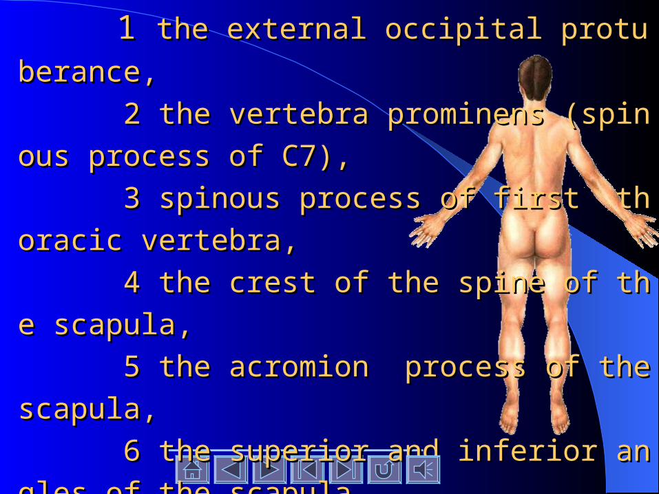
Palpate the Palpate the following bony landmarks: bony landmarks:
1 1 the external occipital protuberance, the external occipital protuberance,
2 the vertebra prominens (spinous process of C7), 2 the vertebra prominens (spinous process of C7),
3 spinous process of first thoracic vertebra, 3 spinous process of first thoracic vertebra,
4 the crest of the spine of the scapula, 4 the crest of the spine of the scapula,
5 the acromion process of the scapula, 5 the acromion process of the scapula,
6 the superior and inferior angles of the scapula, 6 the superior and inferior angles of the scapula,
7 the iliac crest. 7 the iliac crest.

II. Skin IncisionII. Skin Incision:: 1. Make a vertical midline skin incision from the exter 1. Make a vertical midline skin incision from the external occipital protuberance downward to the level of the ilnal occipital protuberance downward to the level of the iliac crest. iac crest.
2. At the lower end,extend the incision transversely to t 2. At the lower end,extend the incision transversely to t
he right and left as far as thehe right and left as far as the midaxillary line.midaxillary line.
3. At the upper end, extend the incision from the exter 3. At the upper end, extend the incision from the external occipital protuberance to the acromion process.nal occipital protuberance to the acromion process. 4. On the right and left sides, reflect the skin laterally f 4. On the right and left sides, reflect the skin laterally from the back of the neck, the shoulders, and the back to rom the back of the neck, the shoulders, and the back to the midaxillary lines.the midaxillary lines.

III. Superficial Fascia III. Superficial Fascia (subcutaneous tissue) (subcutaneous tissue) : :
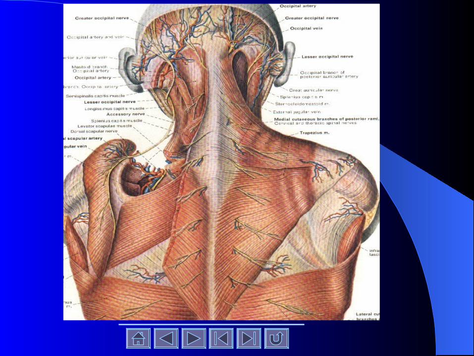
Identify the cutaneous branches of the Identify the cutaneous branches of the posterior rami of the cervical,thoracic,and posterior rami of the cervical,thoracic,and lumbar spinal nerves in the underlying sulumbar spinal nerves in the underlying subcutaneous tissue.bcutaneous tissue.

1. Locate the greater occipital nerve, which is the posterior ramus of the second cervical nerve. It emerges from the trapezius about 2.5cm below and lateral to the
external occipital protuberance.
2. Clean and study one cutaneous branch of a posterior ramus of a thoracic spinal nerve. 3. To assist you in finding such a nerve, carefully loo
k for a blood vessel that follows each of these nerves. Do not waste time tracing out the cutaneous branches of all the posterior rami
Now reflect the superficial fascia of the
back in line with the skin incisions, being car
efully not to damage the underlying muscles,
the trapezius and the latissimus dorsi.
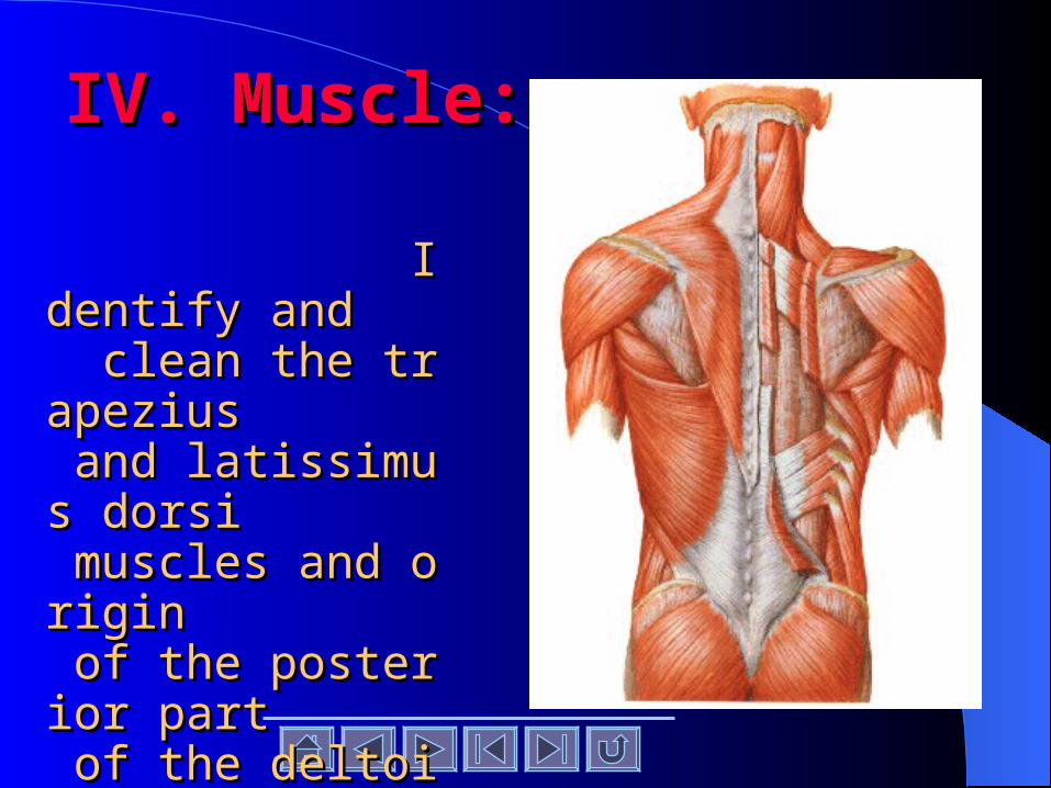
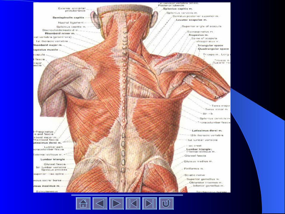
IV. Muscle:IV. Muscle:
Identify and Identify and clean the trapezius clean the trapezius and latissimus dorsi and latissimus dorsi muscles and origin muscles and origin of the posterior part of the posterior part of the deltoid muscleof the deltoid muscle
Trapezius muscle.Trapezius muscle.
Confirm that this muscle has an extensive or Confirm that this muscle has an extensive or
igin from the medial one-third of the superior nuigin from the medial one-third of the superior nu
chal line of the occipitl bone, the occipital protubchal line of the occipitl bone, the occipital protub
erance, the ligamentum nuchae, the spinous proceerance, the ligamentum nuchae, the spinous proce
sses of the seventh cervical vertebra and the spinosses of the seventh cervical vertebra and the spino
us processes and supraspinous ligaments of thoraus processes and supraspinous ligaments of thora
cic vertebrae one to twelve.cic vertebrae one to twelve.
Trace the superior muscle fibers to theirTrace the superior muscle fibers to their ii
nsertion on the lateral third of the claviclnsertion on the lateral third of the clavicl
e, the middle fibers to their insertion on te, the middle fibers to their insertion on t
he acromion and the spine of the scapula, he acromion and the spine of the scapula,
and the inferior fibers to their insertion oand the inferior fibers to their insertion o
n the base of the spine of the scapula.n the base of the spine of the scapula.
Cut free the origin of the trapezius from the skCut free the origin of the trapezius from the sk
ull, the ligamentumull, the ligamentum nuchae, and the spinous nuchae, and the spinous
processes of the vertebrae. Carefully reflect thprocesses of the vertebrae. Carefully reflect th
e trapezius laterally, taking care not to damage trapezius laterally, taking care not to damag
e the underlying levator scapulae and rhomboie the underlying levator scapulae and rhomboi
d muscles.d muscles.

Identify and preserve the spinal pIdentify and preserve the spinal p
art of the art of the accessory accessory nerve and the nerve and the
posterior rami of the posterior rami of the third and fothird and fo
urth cervical nervesurth cervical nerves, which suppl, which suppl
y the trapezius muscle.y the trapezius muscle.

Latissimus dorsi muscleLatissimus dorsi muscle.. Confirm that this muscle arises from the posterior Confirm that this muscle arises from the posterior
part of the iliac crest, the lumbar fascia, the spinopart of the iliac crest, the lumbar fascia, the spino
us processes of the lower six thoracic vertebrae, aus processes of the lower six thoracic vertebrae, a
nd from the lower three or four ribs; sometimes a nd from the lower three or four ribs; sometimes a
few fibers arise from the inferior angle of the scafew fibers arise from the inferior angle of the sca
pula. Note that the fibers sweep upward and laterpula. Note that the fibers sweep upward and later
ally, converging on a tendon that passes into the ally, converging on a tendon that passes into the
axilla to be inserted into the humerus.axilla to be inserted into the humerus.

Transect the origin of the latissimTransect the origin of the latissim
us dorsi and reflect the muscle latus dorsi and reflect the muscle lat
erally, thus exposing the underlyierally, thus exposing the underlyi
ng serratus posterior inferior musng serratus posterior inferior mus
cle.cle.
Identify and clean the Identify and clean the levator scapullevator scapulaeae and and rhomboid minorrhomboid minor and and rhomborhomboid major musclesid major muscles. Leave the levator . Leave the levator scapulae intact and trace its fibers to scapulae intact and trace its fibers to their insertion on the medial border their insertion on the medial border of the scapula. of the scapula.
Cut free the origins of the rhomboid minor anCut free the origins of the rhomboid minor an
d major muscles from the vertebrae and carefd major muscles from the vertebrae and caref
ullyully reflect them laterally to their insertions oreflect them laterally to their insertions o
n the medial border of the scapula. Be careful n the medial border of the scapula. Be careful
to preserve the underlying to preserve the underlying serratus posterior sserratus posterior s
uperior muscleuperior muscle, which is sometimes stuck to t, which is sometimes stuck to t
he rhomboid muscles.he rhomboid muscles.

Identify and clean the dorsal
scapular nerve and the deep branches
of the superficial cervical vessels that
lie deep to the rhomboid muscles,
close to their insertion.
Identify the Identify the serratus anterioserratus anterio
r muscler muscle at its insertion along the at its insertion along the
anterior surface of the medial bordanterior surface of the medial bord
er of the scapula.er of the scapula.

Define the posterior border of the deltoid muscle anDefine the posterior border of the deltoid muscle an
d confirm the origin of this muscle from the spine and acrod confirm the origin of this muscle from the spine and acro
mion process of the scapula. With a scalpel, cut free the orimion process of the scapula. With a scalpel, cut free the ori
gin of the deltoid muscle from the spin and acromion procesgin of the deltoid muscle from the spin and acromion proces
s but leave its attachment to the clavicle intact. Note that the s but leave its attachment to the clavicle intact. Note that the
deltoid fibers pass downward to their insertion on the deltoideltoid fibers pass downward to their insertion on the deltoi
d tuberosity of the humerus. To assist you with the later stud tuberosity of the humerus. To assist you with the later stu
dy, now remove the skin and superficial fascia from the bacdy, now remove the skin and superficial fascia from the bac
k of the shoulder and the back of the arm, down as far as the k of the shoulder and the back of the arm, down as far as the
olecranon process of the ulna.olecranon process of the ulna.
Identify and clean the supraspinatus, infraspinatus,teres minor and teres major, and the long head of the triceps muscles.Expose the boundaries and contents of the quadrilateral and triangular spaces.