Metabolic Diseases of the Bone Osteoporosis Osteomalacia Paget’s Gout Bone Tumors.
1
2
Disorders of Bone Mass
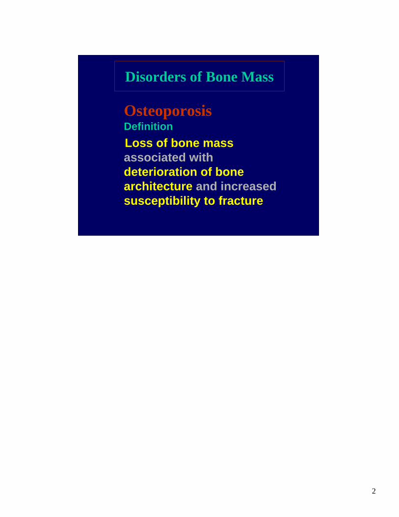
OsteoporosisDefinitionLoss of bone mass associated with deterioration of bone architecture and increasedsusceptibility to fracture

3
Compare these two scanning electron microscopic pictures of the cancellousbone architecture of two women, one 25 years old and the other sixty five or so years old. The former shows a normal system of interconnected curved plates and columns surrounding holes which contain the hematopoietic bone marrow. By contrast, the latter is a depleted structure made up of walls perforated with large holes partially bridged by thin, pincil-like rods. This illustrates the loss of bone mass and deterioration of bone architecture resulting from perforation and removal of entire trabecular plates by increased osteoclastic resorption, a mechanism typical of postmenopausal bone loss.

4
Epidemiology
75 million in US, Europe and Japan300,000 new hip fractures/ year500,000 vertebral fractures/ year1 of every 4 postmenopausal women
5
Osteoporosis is a this “silent epidemic” which manifests itself by fractures vertebral and hip fractures, is becoming a public health problem worldwide. Vertebral fractures, typical of postmenopausal osteoporosis, occur predominantly in women, while hip fractures, a consequent of age-relatedosteoposis, occurs both in elder women and men. Of these two osteoporoticfractures, hip fracture is the more serious and is associated with a higher morbidity and mortality. Serious life-threatening complications including deep femoral vein thrombosis and pulmonary embolism, fat embolism, occur early and contribute to the mortality rate which can be as high as 25% in the first year following fracture. 20 to 30 years ago, these fractures were treated by insertion of pins through the greater trochanter across the femoral neck fracture to adequately fix the fracture (your left). This procedure was followed months later by the late complication called avascular necrosis, due to disruption of theretinacular artery, branch of the circumflex artery at the upper border of the femoral neck by the fracture. This disruption causes an interruption to the blood flow to the largest part of the dome despite adequate healing of the fracture, and results into necrosis and late collapse of the femoral dome, a very painful complication which contribute further to the post-operative morbidity. For the past 20 years, to avoid this complication, fracture is treated by total hip replacement (your right) whereby the femoral head is resected and replaced by a metallic femoral head which articulates with an acetabular component made up of a hard plastic cup, realizing a low-friction arthroplasty system.
6
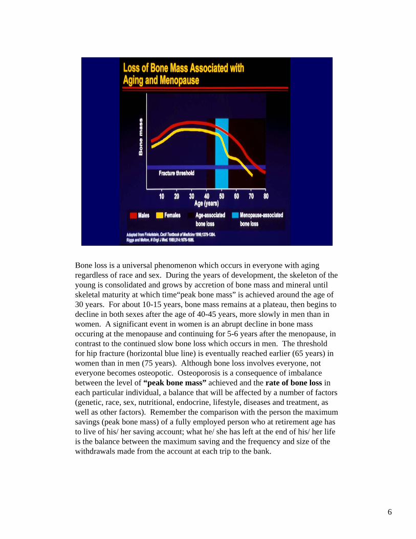
Bone loss is a universal phenomenon which occurs in everyone with aging regardless of race and sex. During the years of development, the skeleton of the young is consolidated and grows by accretion of bone mass and mineral until skeletal maturity at which time“peak bone mass” is achieved around the age of 30 years. For about 10-15 years, bone mass remains at a plateau, then begins to decline in both sexes after the age of 40-45 years, more slowly in men than in women. A significant event in women is an abrupt decline in bone mass occuring at the menopause and continuing for 5-6 years after the menopause, in contrast to the continued slow bone loss which occurs in men. The threshold for hip fracture (horizontal blue line) is eventually reached earlier (65 years) in women than in men (75 years). Although bone loss involves everyone, not everyone becomes osteopotic. Osteoporosis is a consequence of imbalance between the level of “peak bone mass” achieved and the rate of bone loss in each particular individual, a balance that will be affected by a number of factors (genetic, race, sex, nutritional, endocrine, lifestyle, diseases and treatment, as well as other factors). Remember the comparison with the person the maximum savings (peak bone mass) of a fully employed person who at retirement age has to live of his/ her saving account; what he/ she has left at the end of his/ her life is the balance between the maximum saving and the frequency and size of the withdrawals made from the account at each trip to the bank.

7
Osteoporosis Risks Factors
Genetic• Race • Familial
prevalenceAge/ Sex
Endocrine• Menopause • Ovarian/
testicular dysfunction
See Syllabus notes. The major risk factors are highlighted in red.

8
Osteoporosis Risks Factors
Nutritional• Low calcium • High alcohol• High caffeine/
sodium• High anim. protein
Lifestyle• Low physical
activity• Cigarette
smoking
See Syllabus notes. The major risk factors are highlighted in red.
9
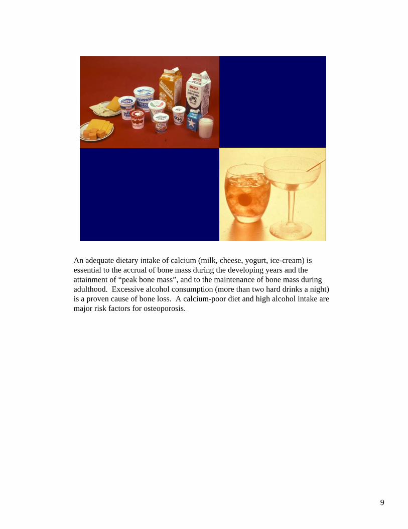
An adequate dietary intake of calcium (milk, cheese, yogurt, ice-cream) is essential to the accrual of bone mass during the developing years and the attainment of “peak bone mass”, and to the maintenance of bone mass during adulthood. Excessive alcohol consumption (more than two hard drinks a night) is a proven cause of bone loss. A calcium-poor diet and high alcohol intake are major risk factors for osteoporosis.

10
Weight-bearing physical exercise (involved in most athletic activities such as walking, running, swimming, bicycle-riding etc…) during adolescence and early adulthood are essential to the achievement of an adequate “peak bone mass. Physical exercise, although not proven to increase bone mass in older individuals, is associated with a lower rate of fractures in this age group. Therefore, a sedentary lifestyle, prolonged immobilization (wheelchair-bound patients or bedridden paraplegics), weightlessness experienced in spaceflights are major risk factors for osteoporosis.

11
• Postmenopausal• Age-related• Idiopathic
OsteoporosisPrimary
A broad classification of osteoporosis includes primary and secondary osteoporosis. Postmenopausal and age-related osteoporosis are the most common types of primary osteoporosis, the idiopathic variety including rare forms occuring in young or middle-aged men without apparent causes.
12
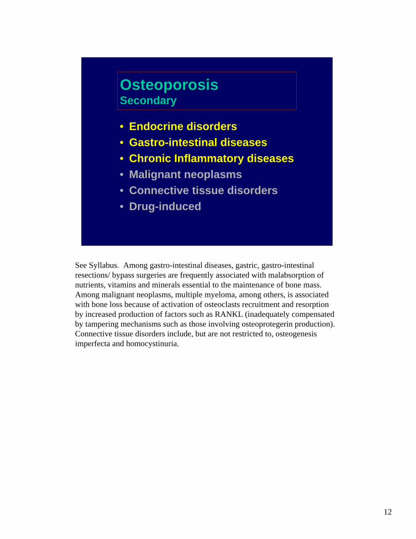
• Endocrine disorders• Gastro-intestinal diseases• Chronic Inflammatory diseases• Malignant neoplasms• Connective tissue disorders• Drug-induced
OsteoporosisSecondary
See Syllabus. Among gastro-intestinal diseases, gastric, gastro-intestinal resections/ bypass surgeries are frequently associated with malabsorption of nutrients, vitamins and minerals essential to the maintenance of bone mass. Among malignant neoplasms, multiple myeloma, among others, is associated with bone loss because of activation of osteoclasts recruitment and resorption by increased production of factors such as RANKL (inadequately compensated by tampering mechanisms such as those involving osteoprotegerin production). Connective tissue disorders include, but are not restricted to, osteogenesis imperfecta and homocystinuria.

13
Endocrine• Ovarian/ testicular dysfunction
early menopause• Cushing syndrome• Hyperthyroidism• Hyperparathyroidism (severe)
OsteoporosisRisk Factors
See Syllabus. Cushing syndrome (hypercortisolism) causes a severe loss of bone most particularly at the spine. As you will hear or have heard in Dr Bilezikian’s lecture, primary hyperparathyroidism in its mild form (its mostcommon presentation nowadays in the US) is not associated with bone loss. However, primary hyperparathyroidism in its severe form (still seen in Europe and other countries), can be a cause of osteoporosis.
14
Fracture sites• Spine• Hips• Distal radius (Colle’s fracture)• Proximal humerus
OsteoporosisClinical
15
Unsuitable for early detectionIndicate advanced disease
RadiologyRoutine Xrays
See Syllabus notes.

16
• “Fish-mouth” deformity• Thin cortices• Diffuse osteopenia• Old or current fractures
RadiologyRoutine Xrays
The “fish-mouth” deformity and diffuse osteopenia will be illustrated in the following slide. The thin cortices which you will also be able to appreciate will be important in the pathogenesis of the vertebral collapse. While the posterior cortex of the vertebra is supported by the posterior elements (laminae, facet joints), the anterior cortex is subject to collapse, leading to anterior wedging of the vertebra with decreased height. Wedging of several subsequent vertebrae leads to a convex deformity or gibbus of the spine which translates clinically in the dorsal kyphosis or Dowager’s hump, typical of postmenopausal osteoporosis.Routine X-rays are insensitive as a tool of early detection of osteoporosis. Early detection requires bone densitometry through energy X-ray absorptiometry or DXA (See Syllabus notes).

17
A sign of advanced osteoporosis is the “fish-mouth” deformity of intervertebral spaces. This deformity is so named because a biconcave shape of the intervertebral spaces due to the collapse of the bony end plates of the vertebrae above and below each space. This collapse is, in turn, due to the deterioration of the cancellous architecture of the vertebral bodies, resulting in the loss ot the columns supporting the bony end plates.

18
Contrary to other sites of cancellous bone in which bone trabeculae have are anisotropic, those of the vertebral bodies are characterized by vertical columns extending from one verbebral end plate to the other and horizontal columns that link the vertical columns. In the display in color (on your left), there are four trabecular systems seen under polarized light from four women approximatelyaged 25 and 45 years (above) and 65 and 85 years (below). As you can see, the loss of bone involves the disparition of the horizontal bars first, leaving vertical columns that have become mechanically weak (think of an old ladder which has lost its horizontal bars and will have become shaky and unstable). Finally, unable to sustain the weight of the plates, the vertical columns break, causing the collapse of the upper and lower end plates of the vertebra. The radiographs of two women (on your right), a 40 years old (above) and an 80 years old (below) illustrates the pathogenesis of the vertebral collapse fracture.
19
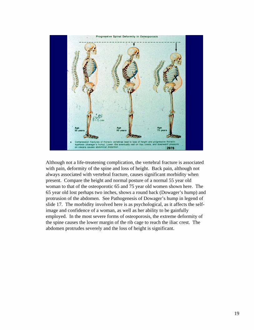
Although not a life-treatening complication, the vertebral fracture is associated with pain, deformity of the spine and loss of height. Back pain, although not always associated with vertebral fracture, causes significant morbidity when present. Compare the height and normal posture of a normal 55 year old woman to that of the osteoporotic 65 and 75 year old women shown here. The 65 year old lost perhaps two inches, shows a round back (Dowager’s hump) and protrusion of the abdomen. See Pathogenesis of Dowager’s hump in legend of slide 17. The morbidity involved here is as psychological, as it affects the self-image and confidence of a woman, as well as her ability to be gainfully employed. In the most severe forms of osteoporosis, the extreme deformity of the spine causes the lower margin of the rib cage to reach the iliac crest. The abdomen protrudes severely and the loss of height is significant.

20
Iliac crest needle biopsy is not necessary to establish a diagnosis of osteoporosis. This procedure is performed in the diagnosis of special types of osteoporosis only (idiopathic osteoporosis to rule out mastocytosis or Gaucher disease, osteoporosis associated with bone pain, osteoporosis of men, high bone turnover osteoporosis) and is, otherwise, a research tool. Compared to the biopsy of a normal individual, this low-power photomicrograph of the biopsy of an osteoporotic biopsy demonstrates severe bone loss represented by thin cortices and thinning and loss of connectivity of cancellous bone plates. The latter are reduced to isolated, dot-like profiles separated by increased spacing between trabeculae. This is most likely the result of increased osteoclastic resorption causing perforation and removal of entire trabecular plates, a mechanism typically associated with post-menopausal osteoporosis.
21
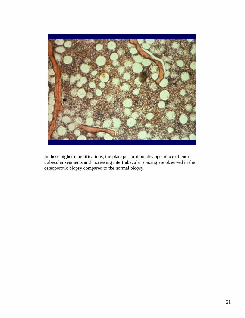
In these higher magnifications, the plate perforation, disappearence of entire trabecular segments and increasing intertrabecular spacing are observed in the osteoporotic biopsy compared to the normal biopsy.
22
Diseases of Mineralization
RicketsOsteomalacia
See Syllabus notes.

23
This group of diseases result from a deficiency of calcium and phosphorus. Because these minerals are highly regulated by vitamin D, it is important that you have a good knowledge of vitamin D metabolism (Dr Bilezikian’s lecture and your recommended reading sources). In a nutshell, (diagram of your left), vitamin D comes from endogenous and exogenous sources. The largest source of vitamin D comes from the transformation of the pro-vitamin D, 7-dehydrocholesterol into cholecalciferol under the influence of UV light from the sun. The endogenous cholecalciferol accesses the blood pool of vit D. Cholecalciferol will also join the blood pool from exogenous, dietary sources (oily fish and vitamin D added to milk or other food products). Cholecalciferol then travels to the liver where it undergoes its first hydroxylation (under the action of 25-hydroxylase in the liver), becoming 25-hydroxycholecalciferol (25-OH D, liver metabolite). Vitamin D then reaches the kidney where it undergoes a second hydroxylation (action of 1-alpha hydroxylase, produced by the renal tubules under the stimulation of parathyroid hormone), becoming 1,25-dihydroxycholecalciferol (1,25-(OH)2 D or renal metabolite, the active metabolite of vitamin D which is essentially a hormone. Vit D must, at all cost, maintain the levels of calcium and phosphorus in the extracellular fluid between physiologic levels and has 3 target organs: the intestines (direct action, independent of PTH), kidneys (together with PTH), and bones (together with PTH). In the intestines, vit D stimulates the absorption of calcium and phosphorus. In the kidney, low levels of calcium will summon PTH secretion which stimulate increased production of 1-alpha hydroxylase, greater conversion of 25 (OH) D into 1,25 (OH)2 D, causing increased calcium and phosphorus absorption by the intestines. Furthermore, these two hormones will cause decreased calcium excretion, while increasing phosphorus excretion by th l t b l I th b PTH d 1 25(OH)2 D ill l l i d
24
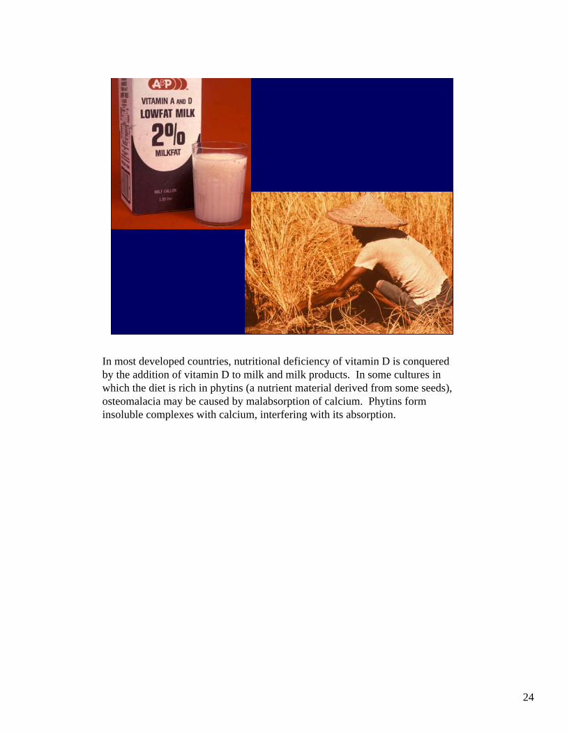
In most developed countries, nutritional deficiency of vitamin D is conquered by the addition of vitamin D to milk and milk products. In some cultures in which the diet is rich in phytins (a nutrient material derived from some seeds), osteomalacia may be caused by malabsorption of calcium. Phytins form insoluble complexes with calcium, interfering with its absorption.

25
RicketsPathogenesis
Defective mineralization/ turnover
• Bone matrix• Growth cartilage
See Syllabus notes.

26
In both osteomalacia and rickets, unmineralized bone matrix which is inefficiently resorbed by osteoclasts is retained on bone surfaces. Thick layers of osteoid (red) accumulate on bone surfaces (hyperosteoidosis) (cores of previously calcified bone stain black). This retention interferes with the turnover of bone. See Syllabus notes on pathogenesis.

27
Normal growth plate at medium (your left) and high magnification (your right) showing the alignment in columns of chondrocytes at the junction of the zones of hypertrophy and provisional calcification. The chondrocyte walls are resorbed osteoclasts/ chondroclasts, the cells die and the cartilage matrix (blue cores between chondrocytic columns) becomes calcified (your left). On your right, these cores of calcified cartilage serve as a template for rows of osteoblasts (lines of basophylic cells) to deposit the osteoid (red matrix).

28
Costo-chondral junction in rickets. Because of failure to calcify the columns of cartilage cells at the zone of hypertrophy, disorderly accumulations of light-staining, unmineralized cartilage matrix occur in nodules at the zone of provisional calcification and extend into the primary metaphysis of the rib (below imaginary horizontal midline of the picture. Note the expansion of the metaphysis caused by this accumulation (and by the capillary invasion of the growth plate due to microfractures).

29
• Cranio-tabes/ frontal bossing• Widely opened fontanels• Rachitic rosary• Harrison groove/pigeon breast• Swelling of wrists and ankles• Genu valgum/ varum
RicketsClinical
See Syllabus notes. In the older (weight-bearing) child, deformities of the spine (lumbar lordosis) occur, together with swelling of the wrists and ankles, a clinical signs explained by the expansion of the metaphyses near the growth zones(see legend of picture 29).

30
Photograph of a toddler with osteomalacia. Note the bowing deformity of the legs (genu varum) to due soft, deformable long bones. On your right is a photograph of a deformed, largely unmineralized cartilaginous humerus in another child with rickets.

31
Gross photograph of a ribcage (your left). Note the vertical display of consecutive nodules (beadings) seen at the costochondral junctions (rachitic rosary). In the photomicrograph (your right), the beading is explained by the expansion of the growth zone by the accumulation of unmineralized cartilage, plus the buckling or plication of the soft, uncalcified bone of the rib. These phenomena are responsible for the beaded appearance of the bone and, collectively in the entire rib cage, for the rachitic rosary.

32
Long bonesDeformitiesFractures
Growth platesBlurred/WidenedEpiphyses cup-
shaped
RicketsRadiology
The radiologic appearances listed in this slide are the direct result of the changes occuring at the tissue level. Note the widening (think uncalcified cartilage/ osteoid retention), blurring and loss of demarcation of the growth plate (think extensions of retained cartilage into metaphysis) and expansion of the metaphyses with cup-shape deformities.

33
Compared to the sharply defined growth plates and well mineralized bones of a normal child, observed the widened, blurred growth plates, cup-shaped epiphyses and diffuse osteopenia (the bones resemble a gauze-like fabric) of a rachitic child. The appearance of diffuse osteopenia is false and due to the limitations of the X-ray which can only “read” the mineralized part of the bone matrix. This explains why rachitic/ osteomalacic bones often appear as osteoporotic bone. In reality, these patients have similar or greater volumetric measurements of bone (which measure calcified and non-calcified bone matrix) as normal individuals.
34
Defective mineralization/ turnover
• Bone matrix
OsteomalaciaPathogenesis
See Syllabus notes.

35
• Bone pain• Fractures• Proximal myopathy• Muscle weakness• Waddling gait
OsteomalaciaClinical
Pain dominates the symptomatology of osteomalacia and is due to microfractures. The proximal myopathy involves the shoulder and hip girdles, causes muscle weakness and a waddling gait. This gait (similar to that of a duck) is due to the fact that the patient, because of weakness in the normal muscles which control the gait, is forced to use alternative muscle groups.
36
Bowing of long bones (genuvarum)
Pseudo-fractures “Looser’slines”
Fractures (Milkman)
OsteomalaciaRadiology
Unlike rickets, osteomalacia causes bowing deformity of long bones without enlargement of the ends of the bones.
37
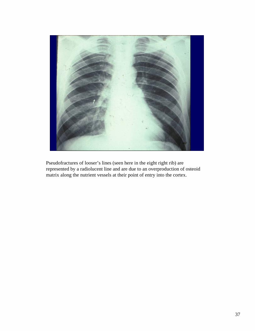
Pseudofractures of looser’s lines (seen here in the eight right rib) are represented by a radiolucent line and are due to an overproduction of osteoid matrix along the nutrient vessels at their point of entry into the cortex.

38
Looser’s lines are usually bilateral and symetrical and frequently found in the cortex of long bones particularly the femoral neck, and in other bones (ischium, rib, pubis).
39
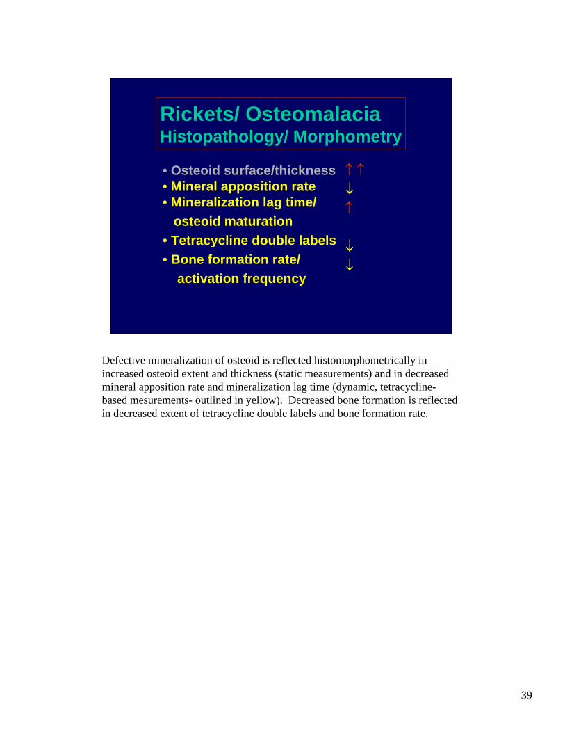
Rickets/ OsteomalaciaHistopathology/ Morphometry
↑ ↑↓↑
↓↓
• Osteoid surface/thickness • Mineral apposition rate• Mineralization lag time/
osteoid maturation• Tetracycline double labels• Bone formation rate/
activation frequency
Defective mineralization of osteoid is reflected histomorphometrically in increased osteoid extent and thickness (static measurements) and in decreased mineral apposition rate and mineralization lag time (dynamic, tetracycline-based mesurements- outlined in yellow). Decreased bone formation is reflected in decreased extent of tetracycline double labels and bone formation rate.

40
Increased extent and thickness of osteoid on bone surfaces. Note the number of lamellae comprised in the osteoid thickness (10-12) compared to the normal number of 3-4 lamellae in each seam. Also observe the absence of osteoclastic activity on osteoid surfaces.

41
At the upper and left corner, normal extent of tetracycline double labels (showing two sharp lines of normally apposed mineral – reflecting the two doses of tetracycline – separated by a dark band – reflecting the days off tetracycline. In contrast, at the lower, right corner, the uptake of tetracycline is very slow, causing very blurred labels poorly separated, reflecting the long time taken by the osteoid matrix to calcify. The extent of tetracycline double labels is markedly decreased.

42
Renal OsteodystrophyDefinition
A spectrum of skeletal abnormalities resulting from renal failure

43
See Syllabus notes.

44
See Syllabus notes. Briefly, decreased phosphate excretion by the renal tubules causes hyperphosphatemia. The increased calcium x phosphate molar product being high, calcium phosphates deposits are precipitated into soft tissues where they form large “tumor-like” masses principally on the extensor surfaces of the joints. These masses are white, nodular and often ulcerate through the skin.

45
Renal OsteodystrophySubtypes
• Secondary hyperparathyroidism• Mixed bone disease• Low bone turnover
Pure osteomalaciaAplastic/ adynamic bone disease

46
The most frequent clinical and histologic subtype of renal osteodystrophy is secondary hyperparathyroidism (in morphological terms osteitis fibrosa). It is characterized by high turnover shown by marked osteoclastic resorption of bone associated with bone marrow fibrosis. Typically, the osteoclasts penetrate and tunnel the trabeculae in a pattern called “Tunneling or dissecting erosion”.

47
Renal osteodystrophy, high turnover or osteitis fibrosa. Note the increased number of osteoclasts, resorption bays on trabecular surfaces (mid-right field) and marked fibrosis of the bone marrow.
48
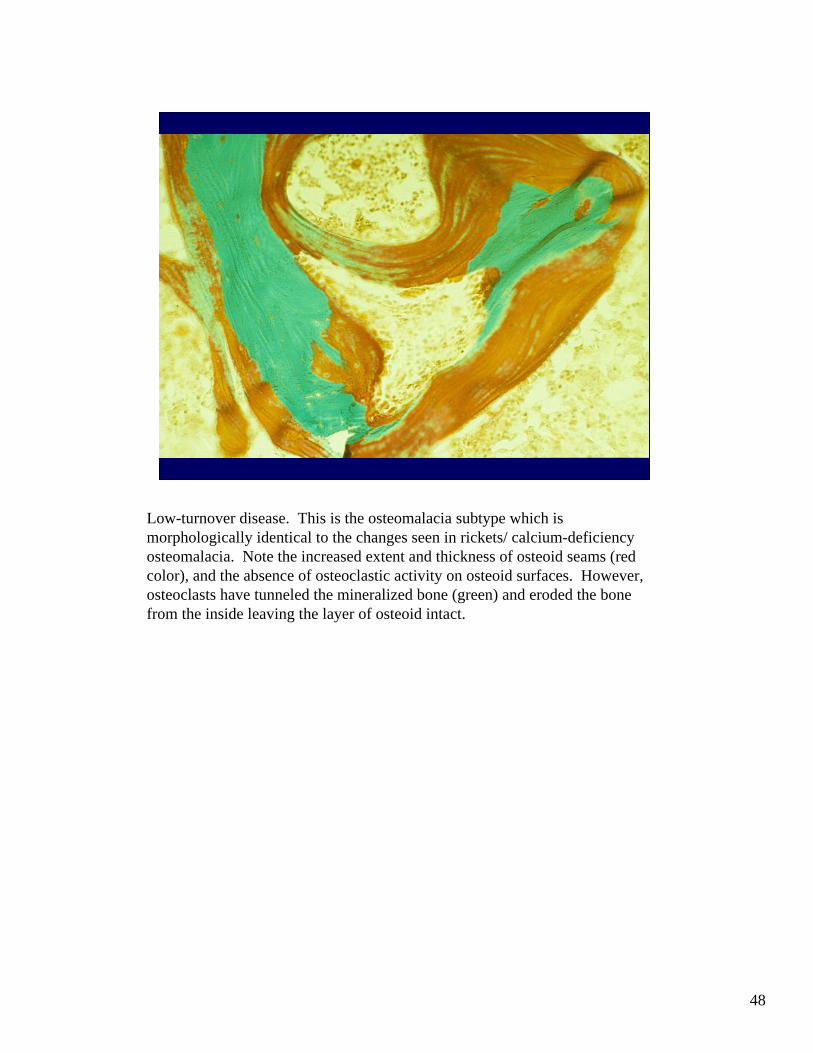
Low-turnover disease. This is the osteomalacia subtype which is morphologically identical to the changes seen in rickets/ calcium-deficiency osteomalacia. Note the increased extent and thickness of osteoid seams (red color), and the absence of osteoclastic activity on osteoid surfaces. However, osteoclasts have tunneled the mineralized bone (green) and eroded the bone from the inside leaving the layer of osteoid intact.
49
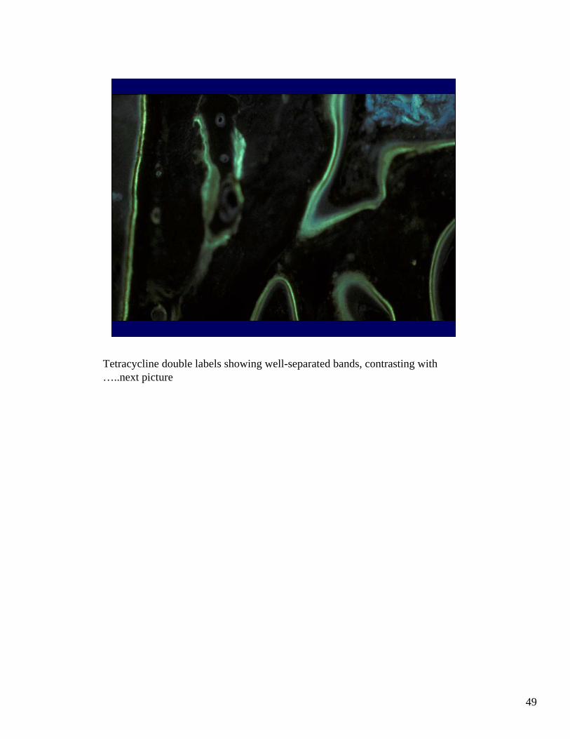
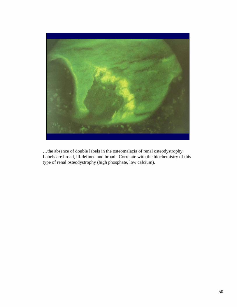
Tetracycline double labels showing well-separated bands, contrasting with …..next picture
50
…the absence of double labels in the osteomalacia of renal osteodystrophy. Labels are broad, ill-defined and broad. Correlate with the biochemistry of this type of renal osteodystrophy (high phosphate, low calcium).

51
CLASS OF 2008
Thank you!