diseases/Retinal Artery Occlusion/Retina… · Web viewThese pictures show the location of the...
5
Retinal Artery Occlusion Fundus photo of patient with Central Retinal Artery OCT image showing of macula area Occlusion What are Retinal Artery Occlusions? Similar to other tissues in the body, the retina can suffer from an interruption of blood flow involving the arteries (blood vessels that carry oxygenated blood from the heart to tissues such as the retina). Blood enters the retina through a single large artery (the central artery) and moves to progressively smaller arteries (branch arteries), allowing blood to reach the whole retina. There are two main types of retinal artery occlusions: Central Retinal Artery Occlusion (CRAO) and Branch Retinal Artery Occlusions (BRAO). The names correspond to where the blockage is located. The major cause of both CRAO and BRAO is a blockage of the artery from an embolism (usually a cholesterol or calcific plaque) that has migrated from another part of the body. Thrombosis (a blood clot that forms at the location)
Transcript of diseases/Retinal Artery Occlusion/Retina… · Web viewThese pictures show the location of the...
Retinal Artery Occlusion
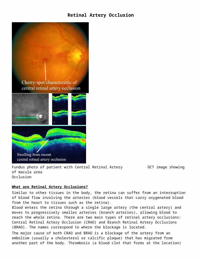
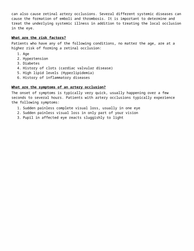
Fundus photo of patient with Central Retinal Artery OCT image showing of macula areaOcclusion
What are Retinal Artery Occlusions?Similar to other tissues in the body, the retina can suffer from an interruption of blood flow involving the arteries (blood vessels that carry oxygenated blood from the heart to tissues such as the retina). Blood enters the retina through a single large artery (the central artery) and moves to progressively smaller arteries (branch arteries), allowing blood to reach the whole retina. There are two main types of retinal artery occlusions: Central Retinal Artery Occlusion (CRAO) and Branch Retinal Artery Occlusions (BRAO). The names correspond to where the blockage is located. The major cause of both CRAO and BRAO is a blockage of the artery from an embolism (usually a cholesterol or calcific plaque) that has migrated from another part of the body. Thrombosis (a blood clot that forms at the location) can also cause retinal artery occlusions. Several different systemic diseases can cause the formation of emboli and thrombosis. It is important to determine and treat the underlying systemic illness in addition to treating the local occlusion in the eye.
What are the risk factors?Patients who have any of the following conditions, no matter the age, are at a higher risk of forming a retinal occlusion:
1. Age2. Hypertension3. Diabetes4. History of clots (cardiac valvular disease)5. High lipid levels (Hyperlipidemia)6. History of inflammatory diseases
What are the symptoms of an artery occlusion?The onset of symptoms is typically very quick, usually happening over a few seconds to several hours. Patients with artery occlusions typically experience the following symptoms:
1. Sudden painless complete visual loss, usually in one eye2. Sudden painless visual loss in only part of your vision3. Pupil in affected eye reacts sluggishly to light
Early-frame Fluroescein Angiography (FA) showing no Mid-frame FA showing dye in cilioretinal arteries, yet blood within the arteries. Patient has cilioretinal no dye within the main arteriesarteries, allowing some profusion of blood.
How can the doctor determine the extent of my artery occlusion?The doctor will perform a dilated exam with a slit lamp to determine the extent of the occlusion and what effect it has on the center of your retina (the macula). To check the extent of the occlusion on the outer retina, the doctor will use an indirect ophthalmoscope. To determine the exact location of the blockage and if ischemia (damage from lack of oxygen) of the retina is present, the doctor may order several tests.
What tests are performed? Testing is important because it helps the doctor to precisely document the retinal occlusion, to determine the extent of ischemia and presence of swelling and/or edema and measure changes that occur over time. The three types of tests described below are performed in our clinic.
Optical Coherence Tomography (OCT) is a high definition image of the retina taken by a scanning ophthalmoscope with a resolution of 5 microns. These images can determine the presence and extent of ischemia within the retina. The doctor will use OCT images to objectively document the progress of the disease throughout the course of your treatment. Fundus Photography is an image taken by a digital fundus camera to document the retinal occlusion in the retina.Fluorescein Angiography is a test that documents blood circulation in the retina using fluorescein dye which luminesces under blue light. Fluorescein is injected into a vein in your arm and digital fundus pictures are taken afterwards for 10 minutes. These pictures show the location of the embolism or thrombosis and extent of blockage by measuring the time it takes for blood to enter the eye.

Mid-frame FA showing location of embolism causing Late-frame FA showing staining of vessel walls indicatingthe central retinal artery occlusion damage to the vessels
What treatments are available?If the onest of symptoms occurred less than 24 hours prior to the visit, an anterior chamber paracentesis can be performed in attempt to dislodge the occlusion. If the occlusion is long-standing (onest of symptoms occurred more than 24 hours prior to the visit), nothing has been shown effective in treating the occlusion. To help manage local effects of the occlusion on the retina, injections of Anti-VEGF and/or anti-inflammatory medicines into the eye can be performed in our office. Sometimes focal laser to the retina is necessary. The doctor may recommend the treatments below:
1. Paracentesis if the occlusion occurred less than 24 hours prior to the visit 2. Intravitreal injection of an anti-Vascular Endothelial Growth Factor (Anti-VEGF) such as Avastin or
Lucentis3. Intravitreal injection of a corticosteroid (Dexamethasone or Triamcinolone)4. Focal Laser therapy5. Pan-retinal photocoagulation therapy
It is also very important that any underlying systemic conditions (hypertension, diabetes, cardiac diseases, hyperlipidemia, etc.) be evaluated and treated by your primary care doctor.
What is my follow up care?Return visits with us are recommended to monitor your disease progress. To determine the underlying cause of the occlusion, the doctor may order several studies to be performed outside our clinic. Once the systemic condition causing the occlusion is determined, it is very important that it be treated aggressively.