Diseases of aging II A&S300-002 Jim Lund In a man's middle years there is scarcely a part of the...
42
Diseases of aging II A&S300-002 Jim Lund In a man's middle years there is scarcely a part of the body he would hesitate to turn over to the proper authorities. -E.B. White
-
Upload
janel-spencer -
Category
Documents
-
view
213 -
download
0
Transcript of Diseases of aging II A&S300-002 Jim Lund In a man's middle years there is scarcely a part of the...

Diseases of aging II
A&S300-002 Jim Lund
In a man's middle years there is scarcely a part of the body he would hesitate to turn over to the proper authorities.
-E.B. White

Diseases of Aging
• Cancer
• Heart disease
• Cerebrovascular disease
• Arthritis
• Osteoporosis
• Neurodegenerative disease
• Diabetes (Type II)

• Size and number of cardiac muscle cells decreases, replaced by fibrous tissue.
• Increase in fat deposits on the surface of the heart.
• Endocardium thickens.
• Calcification of heart valves (30% of people over 75).
• Characteristic electrocardiogram (EKG) changes.• Perhaps due to fibroses in conductive fibers.
• Systolic/diastolic blood pressures tend to increase: 120/80mmHg -> 130/90mmHg.
Age-related changes in the heart

• Reduced maximum oxygen consumption.• Decreases by 30, 40% reduction by 65 yrs.
• Resting and maximum heart rate decrease.
• Cardiac output (blood pumped per minute) declines 1% per year after age 20, down 50% by age 80.
• Cardiac reserve declines with age.
Age-related changes in the heart

• Complex diseases with a common origin:
Blood vessel disfunction
Heart and cerebrovascular disease

• Reduction of elasticity in vessel walls (20->70yrs, 50% decrease).
• Reduction in eleastin protein content, replaced by collagen.
• Elastin calcifies.• These changes can narrow arteries
and increase peripheral resistance.
• Arteriosclerosis
Blood vessel changes
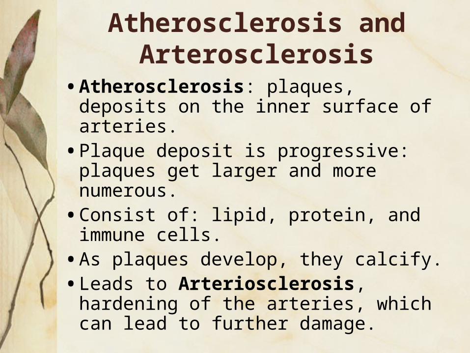
• Atherosclerosis: plaques, deposits on the inner surface of arteries.
• Plaque deposit is progressive: plaques get larger and more numerous.
• Consist of: lipid, protein, and immune cells.
• As plaques develop, they calcify.
• Leads to Arteriosclerosis, hardening of the arteries, which can lead to further damage.
Atherosclerosis and Arterosclerosis

Fatty Arteries
Normal CoronaryArtery
AtheroscleroticArtery
Photos: Klatt, Edward C., WebPath.com

Pathogenesis of Atherosclerosis
• Endothelial Dysfunction• Injury to the endothelium is the
primary event
• Mechanical, tissue hypoxia, aging, etc.
• Impair endothelial protection• Decrease in plasminogen activators,
heparan sulphate, prostacyclin

• If the endothelium is damaged it no longer serves as a barrier.
• LDL cholesterol passes into the intima (internal layer of the vessel) and accumulates and modified (oxidized) by free radicals • Attracts monocytes and is ingested by
macrophages• Key step is attraction of monocytes
and T lymphocytes by TNF and MCP released by injured endothelium.
Pathogenesis of Atherosclerosis

• Monocytes migrate to subendothelial space where they become macrophages.
• Foam cells secrete PDGF, IL-1, TGF, TNF which activate SM cells to migrate and proliferate and deposit connective tissue.
• Foam cells also release TNF which is highly thrombogenic.
• Gives rise to overlying thrombus formation
Pathogenesis of Atherosclerosis


Atherosclerosis

• Caused by: aging changes of the vessels, atherosclerosis, arteriosclerosis, high sodium.
• Effects: heart attack, heart failure, kidney damage, blood vessel rupture (hemorrhage stroke).
Hypertension

Ischemic heart disease:
• Occluded arteries->insufficient blood flow->ischemic heart attack.
• Plaques can trap blood platelets, cause a blood clot (thrombus).
• Heart disease is progressive and has positive feedback cycle.
Coronary artery disease

Diseases of Aging
• Cancer
• Heart disease
• Cerebrovascular disease
• Arthritis
• Osteoporosis
• Neurodegenerative disease
• Diabetes (Type II)

Aging of the Central Nervous System
• Cell loss• Brain weight increases to age 30, declines by
10% by 90 yrs of age.
• Due to this• Ventricles enlarge.• Gyri become smaller, sulci between them
enlarge.• Grey and white matter reduced.

The Neuron

The Neuron: the brain’s basic functional unit
Axon
DendritesSoma
Axon Terminals
Myelin Sheath

Aging of the Central Nervous System
• Neuronal function decline• Rate of conduction along axons declines, due to loss of
myelin.• Synapses time increases.
• Reduced levels of synapse enzymes, receptors, etc.
• Reduced numbers of dendrites and dendritic spines (in some areas o the brain).
• Cellular changes:• Lipofuscin deposits.• Decrease in dark staining cytoplasmic Nissl bodies.
• Glia: 10 times more glial cells than neurons• In some areas, glial numbers increase, in other areas
they decrease.

Neurodegeneration
• Involved in disorders like Alzheimer’s, Huntington’s, Parkinson’s.
• Also involved in neuromuscular diseases like ALS or Lou Gehrig’s disease.

Alzheimer’s Disease
• Neurodegenerative disease causing progressive memory & language loss
• Associated with deposition of amyloid protein (APP) in CNS and neurofibrillary tangles (NFTs). NFTs associated with mutations to Tau proteins that stabilize microtubules.
• Mutations to PS-1 and PS-2 (presenelin genes) give rise to early onset disease.
• Mutation to apolipoprotein E gives rise to late onset.

Neurofibrillary Tangles in Alzheimer’s Disease
From http://www.rnw.nl/health/html/brain.html

Neuronal Plaques in Alzheimer’s Disease
From http://www.rnw.nl/health/html/brain.html

Plaques and neurofibrillary tangles
From Department of Pathology, Virginia Commonwealth University

http://abdellab.sunderland.ac.uk/lectures/Neurodegeneration/ References/Brain_Neurons_AD_Normal.html
Alzheimer’s Disease

Amyloid precursor protein (APP) is membrane protein that sits in the membrane and extends outward. It is though to be important for neuronal growth, survival, and repair.

Enzymes cut the APP into fragments, the most important of which for AD is called b-amyloid (beta-amyloid) or Aß.

Beta-amyloid is “sticky” so the fragments cling together along with other material outside of the cell, forming the plaques seen in the AD brain.

Alzheimer’s pathogenesis
• Rate of Aß accumulation and aggregation determined by:• Genotype, production of amyloid peptide, tau,
presenilin proteins.
• Efficiency of degradation of Aß.
• Levels of plasmin (cleavage product of plasminogen).

Amyloid Hypothesis
• The trigger for alzheimer’s disease is the A-beta peptide, and the accumulation of this peptide in the form of plaques is the initiating molecular event.
• The plaques trigger an inflammatory response, neuronal cell death, and gradual cognitive decline.
• The rest of the disease process, including formation of neurofibrillary tangles containing tau protein, is caused by an imbalance between A-beta production and A-beta clearance.

The History of Parkinson’s Disease
• Parkinson’s Disease (PD) was first described by James Parkinson in 1817(1)
• He noted• ‘Involuntary tremulous motion’• ‘A propensity to bend forwards’• ‘The senses and intellect are intact’
• 40 years later Charcot named Parkinson’s Disease

Parkinson’s disease
• Progressive neurodegenerative disease
• Incidence: 1 in 200 over the age of 55.• Clinical descriptions:
• Useless contractions of the skeletal muscles causing muscle rigidity and tremors.
• Resting tremor, muscular rigidity, bradykinesia, and postural instability.
• 20% of patients develop Alzheimer’s disease.

Parkinson’s disease
Pathologic features:
• Loss of dopaminergic neurons in the substantia nigra (SN).
• Presence of Lewy bodies, intracellular inclusions, in surviving neurons in various areas of the brain, particularly the SN.
• Leads to reduced production of dopamine
• Reduced dopamine levels leads to striatal dopamine deficiency and development of PD symptoms.

The role of dopamine
• Dopamine acts to oppose acetylcholine
• Dopamine inhibitory
• Acetylchloline excitatory
• Depletion in dopamine results in hypokinetic disorders such as PD

-synuclein pathology: abnormal neuronal and glial inclusions and processes

Lewy body disease
• Mutations in -synuclein can lead to either mendelian Parkinson’s or Lewy body dementia.
• Triplication of -synuclein leads to disease onset in the 30’s.
• Normal genetic variability: people with higher expressing alleles have a higher risk of sporadic disease.

Models of Parkinson’s disease
• 6-OHDA: neurotransmitter analogue • depletes noradrenergic stores in nerve endings ->
reduces dopamine levels.• produces free-radicals -> apoptosis.
• MPTP: a contaminant that can result from sloppy synthesis of MPPP, a street analog of the opioid meperidine (Demerol).• Taken up by domaminergic neurons -> free
radicals -> apoptosis.
• -synuclein overexpression -> inhibits MAPK signaling -> induces apoptosis.

Models of Parkinson’s disease
• Transgenic mice that expressed wildtype -synuclein w/ platelet-derived growth factor-beta gene promoter (pan-neuronal)
• Progressive accumulation of -synuclein and ubiquitin-immunoreactive inclusions in neurons in the neocortex, hippocampus, and substantia nigra.
• Ultrastructural analysis shows electron-dense intranuclear deposits and cytoplasmic inclusions. These alterations were associated with loss of dopaminergic terminals in the basal ganglia and with motor impairments.
• Masliah et al., 2000

Models of Parkinson’s disease
• Transgenic flys that expressed wildtype and pathogenic a-synuclein (pan-neuronal).
• Observed: adult-onset loss of dopaminergic neurons, filamentous intraneuronal inclusions containing alpha-synuclein reminiscent of Lewy bodies, and locomotor dysfunction.
• One pathogenic mutation esp. bad.
• All produced premature loss of climbing ability.• Feany and Bender, 2000
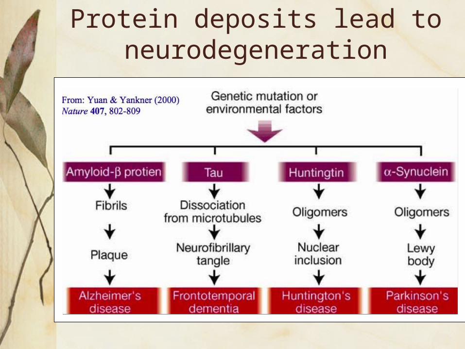
Protein deposits lead to neurodegeneration

Alzheimer’s disease
Relationship between age, Amyloid Beta (Αβ)42 accumulation, normal aging, Mild cognitive impairment (MCI), and Alzheimer’s disease (AD). Typically, the Αβ42 levels in the brains of AD patients are 1,000-10,000-fold higher than in the brains of normal controls.