Allosteric interactions between agonists and antagonists ...

1521-0103/354/3/340–349$25.00 http://dx.doi.org/10.1124/jpet.115.224071THE JOURNAL OF PHARMACOLOGY AND EXPERIMENTAL THERAPEUTICS J Pharmacol Exp Ther 354:340–349, September 2015Copyright ª 2015 by The American Society for Pharmacology and Experimental Therapeutics
Discovery of D1 Dopamine Receptor Positive AllostericModulators: Characterization of Pharmacology andIdentification of Residues that Regulate Species Selectivity s
Martin A. Lewis, Lisa Hunihan, John Watson, Robert G. Gentles, Shuanghua Hu,Yazhong Huang, Joanne Bronson, John E. Macor, Brett R. Beno, Meredith Ferrante,Adam Hendricson, Ronald J. Knox, Thaddeus F. Molski, Yan Kong, Mary Ellen Cvijic,Kristin L. Rockwell, Michael R. Weed, Angela M. Cacace, Ryan S. Westphal, Andrew Alt,and Jeffrey M. BrownBristol-Myers Squibb, Wallingford, Connecticut (M.A.L., L.H., J.W., R.G.G., S.H.,Y.H., J.B., B.R.B., M.F., A.H., R.J.K., T.F.M.,K.L.R., M.R.W., A.M.C., A.A., J.M.B.); Bristol-Myers Squibb, Hopewell, New Jersey (Y.K., M.E.C.); Bristol-Myers Squibb,Lawrence Township, New Jersey (J.E.M.); and Lilly Corporate Center, Eli Lilly and Company, Indianapolis, Indiana (R.S.W.)
Received March 5, 2015; accepted June 22, 2015
ABSTRACTThe present studies represent the first published report ofa dopamine D1 positive allosteric modulator (PAM). D1 recep-tors have been proposed as a therapeutic target for thetreatment of cognitive deficits associated with schizophrenia.However, the clinical utility of orthosteric agonist compounds islimited by cardiovascular side effects, poor pharmacokinetics,lack of D1 selectivity, and an inverted dose response. A numberof these challengesmay be overcome by utilization of a selectiveD1 PAM. The current studies describe two chemically distinctD1 PAMs: Compound A [1-((rel-1S,3R,6R)-6-(benzo[d][1,3]dioxol-5-yl)bicyclo[4.1.0]heptan-3-yl)-4-(2-bromo-5-chlorobenzyl)piperazine] and Compound B [rel-(9R,10R,12S)-N-(2,6-dichloro-3-
methylphenyl)-12-methyl-9,10-dihydro-9,10-ethanoanthracene-12-carboxamide]. Compound A shows pure PAM activity, with anEC50 of 230 nM and agonist activity at the D2 receptor inD2-expressing human embryonic kidney cells. Compound B showssuperior potency (EC50 of 43 nM) and selectivity for D1 versusD2 dopamine receptors. Unlike Compound A, Compound B isselective for human and nonhuman primate D1 receptors, but lacksactivity at the rodent (rat and mouse) D1 receptors. Using molecularbiology techniques, a single amino acid was identified at position130, which mediates the species selectivity of Compound B. Thesedata represent the first described D1-selective PAMs and definecritical amino acids that regulate species selectivity.
IntroductionSchizophrenia is a neuropsychiatric disorder characterized
by positive symptoms, negative symptoms, and cognitivedeficits. Working memory, a critical component of cognition,has been associated with functional outcomes in schizo-phrenic patients (Green et al., 2000). Because currentantipsychotic medications do not address cognitive deficitsin schizophrenia, a large unmet clinical need for treatingthese symptoms remains.Dopamine is a catecholamine neurotransmitter found in
both the brain and the periphery. Dopamine receptors can becategorized into two groups: D1-like (D1 and D5 receptors)and D2-like (D2–D4 receptors) (Sibley and Monsma, 1992).
Within the brain, particularly in the prefrontal cortex,dopamine plays an important role in regulating cognition(Puig et al., 2014). Brozoski et al. (1979) first demonstratedthe critical role of dopamine in regulating working memory innonhuman primates by showing that depletion of dopamine inthe cortex impaired performance on a delayed alternationperformance task. Moreover, this effect was reversed withlevodopa treatment (Brozoski et al., 1979). Subsequent studieshave demonstrated that administration of a full or partialagonist of D1-like receptors reversed working memory deficitsinduced by ketamine administration or in aged monkeys (Caiand Arnsten, 1997; Castner and Goldman-Rakic, 2004; Nakakoet al., 2013). Data from these studies support the hypothesisthat the development of a subtype-selective D1 agonist orpartial agonist may be beneficial for the treatment of cognitivedeficits associated with schizophrenia.
dx.doi.org/10.1124/jpet.115.224071.s This article has supplemental material available at jpet.aspetjournals.org.
ABBREVIATIONS: b2AR, b2-adrenergic receptor; Compound A, 1-((rel-1S,3R,6R)-6-(benzo[d][1,3]dioxol-5-yl)bicyclo[4.1.0]heptan-3-yl)-4-(2-bromo-5-chlorobenzyl)piperazine; Compound B, rel-(9R,10R,12S)-N-(2,6-dichloro-3-methylphenyl)-12-methyl-9,10-dihydro-9,10-ethanoanthracene-12-carboxamide;D, dopamine; GPCR, G protein–coupled receptor; HEK, human embryonic kidney; IBMX, 3-isobutyl-1-methylxanthine; ICL2, intracellular loop 2; PAM,positive allosteric modulator; RMSD, root-mean-square deviation; SCH23390, (R)-(1)-7-chloro-8-hydroxy-3-methyl-1-phenyl-2,3,4,5-tetrahydro-1H-3-benzazepine; SKF-38393, (6)-1-phenyl-2,3,4,5-tetrahydro-(1H)-3-benzazepine-7,8-diol; TM, transmembrane; SKF-81297, (6)-6-chloro-2,3,4,5-tetrahydro-1-phenyl-1H-3-benzazepine; SKF-83959, 6-chloro-2,3,4,5-tetrahydro-3-methyl-1-(3-methylphenyl)-1H-3-benzazepine-7,8-diol; WT, wild type.
340
http://jpet.aspetjournals.org/content/suppl/2015/06/24/jpet.115.224071.DC1Supplemental material to this article can be found at:
at ASPE
T Journals on M
ay 13, 2018jpet.aspetjournals.org
Dow
nloaded from

To date, a number of D1-like agonists have been developedand tested preclinically, but clinical success of theseagonists has been hindered by a number of issues. Theseinclude lack of D1 selectivity (against both D2-like and D5receptors), poor pharmacokinetics, and adverse cardiovas-cular effects. Specifically, dihydrexidine is a D1-like fullagonist with similar affinity for D1 and D5 and roughly10-fold selectivity over D2-like receptors (Mottola et al., 2002).Dihydrexidine showed improvement in a working memorytask in patients with schizotypical personality disorder(Rosell et al., 2015). The D1 agonist prodrug ABT-431 alsocaused hypotension at doses that demonstrated clinicalefficacy versus negative symptoms (Rascol et al., 1999). D1and D5 receptors are expressed in vascular and renaltissues and both modulate blood pressure, although theirindividual roles are still being investigated (Zeng et al.,2007). Indeed, the only clinically approved D1 agonist,fenoldopam, is approved for the treatment of hypertensionand hypertensive crisis.Positive allosteric modulators (PAMs) represent an alter-
native approach to orthosteric agonists (compounds thatinteract with the native ligand-binding site). PAMs canincrease the affinity and/or efficacy of the orthosteric agonistfor its target receptor by acting at a site other that the nativeligand-binding site (allosteric) and represent a novel approachfor the development of antipsychotic agents. The PAMapproach to central nervous system pharmacology hasalready had clinical validation. In GABAergic systems, PAMs(such as the prototype diazepam) are equally effective toorthosteric agonists, with significantly better safety andtolerability profiles (Costa, 1991). Importantly, so-called pureG protein–coupled receptor (GPCR) PAMs, which lack in-trinsic agonist activity within a specific signaling pathway,have been described. These compounds modulate the basaltone of the endogenous ligand in a manner that conservesspatial and temporal elements of native neurotransmission(Christopoulos and Kenakin, 2002; Wootten et al., 2013).Indeed, multiple PAMs have been identified for GPCRs,which may circumvent the challenges of orthosteric agonists(Conn et al., 2009; Keov et al., 2011). The present studiesherein describe two novel D1 PAM chemotypes representedby Compound A [1-((rel-1S,3R,6R)-6-(benzo[d][1,3]dioxol-5-yl)bicyclo[4.1.0]heptan-3-yl)-4-(2-bromo-5-chlorobenzyl)piperazine](piperazine series) and Compound B [rel-(9R,10R,12S)-N-(2,6-dichloro-3-methylphenyl)-12-methyl-9,10-dihydro-9,10-ethanoanthracene-12-carboxamide] (ethanoanthracene series),which show selectivity for D1 and lack intrinsic agonist activity.Of these two PAMs, Compound B was selective for the humanD1 versus rat D1 receptor, and this selectivity was mediated bya single amino acid residue within the human sequence. Thesestudies are the first to describe a D1 PAMand provide structuralinformation regarding key amino acids that regulate the PAMactivity, thus providing a path forward for the development of D1PAMs as potential therapeutics.
Materials and MethodsCell culture and transfection reagents were purchased from
Invitrogen (Carlsbad, CA). cAMP homogeneous time-resolved fluo-rescence kits were purchased from Cisbio (Bedford, MA). Dopamine,dopamine agonists, and antagonists were purchased from Sigma-Aldrich (St. Louis, MO).
Heterologous Expression. Human embryonic kidney (HEK) andChinese hamster ovary cells were transfected with cDNA containingthe human, rat, or mouse dopamine D1 receptor, and stable cell lineswere developed using the following sequence information: humanDRD-1 NM_000794.3, human DRD-2 NM_000795.3, human DRD-4NM_000797, humanDRD-5 NM_000798, rat DRD-1 NM_012546, andmouse DRD-1 NM_010076. Transient transfections were performedusing Lipofectamine and Lipofectamine Plus reagent (Invitrogen)following the manufacturer’s instructions.
Primary Neuronal Cultures. Cortical and striatal tissue wasremoved from embryonic day 19 rat embryos. Tissue was dissociatedusing the Papain dissociation system (Worthington, Lakewood, NJ).Following dissociation, cells were cultured in neurobasal mediumplusB27 supplement. Primary neurons were used in functional assaysbetween days 12 and 14 in culture.
cAMP Accumulation Assay. The cAMP accumulation assay wasperformed according to the manufacturer’s instructions. Briefly, cellswere plated into polylysine-coated 384-well plates. The medium wasremoved and replaced with 20 ml of Hanks’ balanced salt solution plus0.3 ml of serial diluted PAM compounds or control diluent for15 minutes. An EC20 of dopamine (∼EC20 for the PAM mode) or buffer(for the agonist mode) was added to the cells and incubated at roomtemperature for 30 minutes. The dopamine EC20 was determined foreach experiment and ranged from 2 to 10 nM for transfected HEKcells and 300 to 600 nM for primary neuronal cultures. Anti-cAMPcryptate and D2-labeled cAMPwere added to the wells, and the plateswere incubated for 1 hour. The plates were read on an Envision platereader (PerkinElmer, Waltham, MA).
D1 Receptor Mutagenesis. D1 alanine mutants in a PCXP-nFlag-DRD1/PC DNA3 expression vector were created by site-directed mutagenesis. Two hundred nanograms per 200 nl wild-type(WT) or mutant DNA was added per well of a 384-well amine plate(BD Biosciences, San Jose, CA) using the Labcyte ECHO550(Sunnyvale, CA). A 1:20 dilution of TransFast transfection reagent(Promega, Madison, WI) in serum-free Dulbecco’s modified Eagle’smedium high glucose medium without additive (Life Technologies,Grand Island, NY) was added to the DNA plate following themanufacturer’s instructions and mixed by shaking for 15 minutes atroom temperature. Ten thousand HEK293T cells in assay medium(Dulbecco’s modified Eagle’s medium with 10% fetal bovine serum;Gibco, Madison, WI) were then dispensed per well in the assay plateand incubated for 18–24 hours at 37°C and 5% CO. The cell mediumwas aspirated, and cells were washed once with phosphate-bufferedserum (Life Technologies). For the PAMmode cAMP assay, 8 ml IBMX(3-isobutyl-1-methylxanthine) buffer (Sigma-Aldrich) was added toeach well, followed by the addition of 100 nl in an 11-point, 3-foldserial dilution (final concentrations ranged from 80 mM to 1.3 nM).Two microliters of dopamine (Sigma-Aldrich) at a final concentrationof 2.5 nM (∼EC20) was then added. Cells were incubated for30 minutes at room temperature, and the homogeneous time-resolvedfluorescence cAMP assay was conducted according to the manufac-turer’s specifications. Briefly, 5 ml anti-cAMP cryptate and 5 mlD2-labeled cAMP, both in lysis buffer, were added to the wells of the cellplate, 20 ml were transferred to a proxi plate (PerkinElmer), and theplates were incubated for 1 hour at room temperature. Fluorescencewas quantitated (fluorescence ratio of 665:620 nm) by PerkinElmerEnvision. Activation data for the test compound over a range ofconcentrations were plotted as the percentage activation of the testcompound (100% 5 maximum response). After correcting forbackground, the EC50 values were determined. The EC50 is defined asthe concentration of the test compound that produces 50% of themaximal response and was quantified using the four-parameter logisticequation to fit the data.
Generation of D1 Chimeras. All human/rat chimera constructsand the human R to Q 130-point mutant were provided by the LifeTechnologies Gene Art custom de novo synthesis service. Start codonswere preceded by kozak consensus sequences CCACC. All sequencesincluded 59 and 39 Gateway adapters, and final sequences were
Discovery of Novel D1 Dopamine Receptor PAMs 341
at ASPE
T Journals on M
ay 13, 2018jpet.aspetjournals.org
Dow
nloaded from
expressed in a Gateway-modified pIRES neo2 vector (Clontech,Mountain View, CA). The rat Q130 R mutant and rat/human 1Q130R [rat/human chimera (depicted in Fig. 5), with a single aminoacid Q130 converted back to the corresponding amino acid R] pointmutants were generated using the Stratagene QuikChange Lightningsite-directed mutagenesis kit (La Jolla, CA) and expressed in pIRESneo2.
Molecular Modeling. The Structure Prediction Wizard withinPrime (version 3.1; Schrödinger, LLC, New York, NY) in the Maestro(version 9.3; Schrödinger, LLC) software package was used togenerate a homology model starting from a hand-edited alignmentof a human D1R sequence, with the b2-adrenergic receptor (b2AR)protein sequence from the X-ray crystal structure of the nanobody-stabilized agonist-bound b2AR (PDB ID 3P0G) (Rasmussen et al.,2011). D1R residues 23–232 and 267–346 were included, andintracellular loop 3 (residues 233–266) was omitted. The initial modelwas generated using default model-building parameters, and loopswere refined with Prime. Following energy minimization of the modelusing the OPLS2-2005 force field (Jorgensen et al., 1996; Jorgensenand Tirado-Rives, 1998; Shivakumar et al., 2010), the D1 agonistdinapsoline (Ghosh et al., 1996) was modeled into the orthosteric site.In the modeled binding pose, the protonated amine of dinapsolineforms a salt bridge with conserved D1033.32, and each hydroxyl groupis hydrogen bonded to either S1985.42 or S2025.46 [for residues predictedto lie within transmembrane helices, Ballesteros-Weinstein indices(Ballesteros and Weinstein, 1995) are provided as superscripts]. Muta-genesis studies (Pollock et al., 1992; Tomic et al., 1993; Kong et al., 2006)have shown that these residues are important for dopamine binding toD1R, and in the crystal structure of the b2AR bound to carazolol (PDB ID3P0G) (Rasmussen et al., 2011), the agonist forms similar interactionswith the corresponding residues. D1R residues 11–22 were omittedduring the initial homology model generation, and when it wasdetermined that inclusion of these residues in the model was desirable,they were appended to the N terminus of the initial model (trans-membrane [TM] 1) in a canonical a-helical conformation, as observed inthe b2AR structural template, and the N terminus of this modifiedstructural model was acetylated. Following energy minimization of therevised D1R/dinapsoline model using the OPLS2-2005 force field(Jorgensen et al., 1996; Jorgensen and Tirado-Rives, 1998; Shivakumar
et al., 2010), a single 100-nanosecond molecular dynamics simulation(isothermal-isobaric ensemble) in a 1-palmitoyl-2-oleoyl-sn-glycero-3-phosphocholine lipid bilayer with full explicit solvation (transferableintermolecular potential 3 point water) (Jorgensen et al., 1983) at300 K was performed on a Linux cluster using the NAMD version 2.8program (Phillips et al., 2005) and the AMBER/GAFF force field(Weiner et al., 1984; Cornell et al., 1995; Wang et al., 2004; Hornaket al., 2006; Lindorff-Larsen et al., 2010). The simulation time of100 nanoseconds is adequate for alleviating high-energy unfavorableinteractions in the initial model and refolding modeled loops into low-energy conformations, but is unlikely to yield the native structure ofthe D1R, especially if the initial model was inaccurate. In-housescripts were used to monitor the protein and ligand root-mean-squaredeviation (RMSD) values, with respect to the initial structure as wellas the total energy of the system and other parameters. Afterapproximately 25 nanoseconds, large changes in protein and ligandRMSD were completed and the mean of all the heavy atom proteinand ligand RMSD values averaged over the entire simulation was3.2 and 1.7 Å, respectively (Supplemental Fig. 1). Key hydrogen bondinginteractions between the ligand and D1033.32, S1985.42, and S2025.46 weremaintained during the simulation (Supplemental Figs. 2 and 3). Themeanoid structure from the 100th nanosecond of the simulation wassubjected to limited energy minimization to yield a final model.
Data Analysis. Data were analyzed using GraphPad Prism(GraphPad Software, Inc., La Jolla, CA). All values are shown asthe mean 6 standard deviation unless otherwise indicated. Multiplecomparisons were made using an analysis of variance, followed byNewman-Keuls post hoc analysis. In all cases, P , 0.05 wasconsidered to be statistically significant.
ResultsFollowing a high-throughput screen of the Bristol-Myers
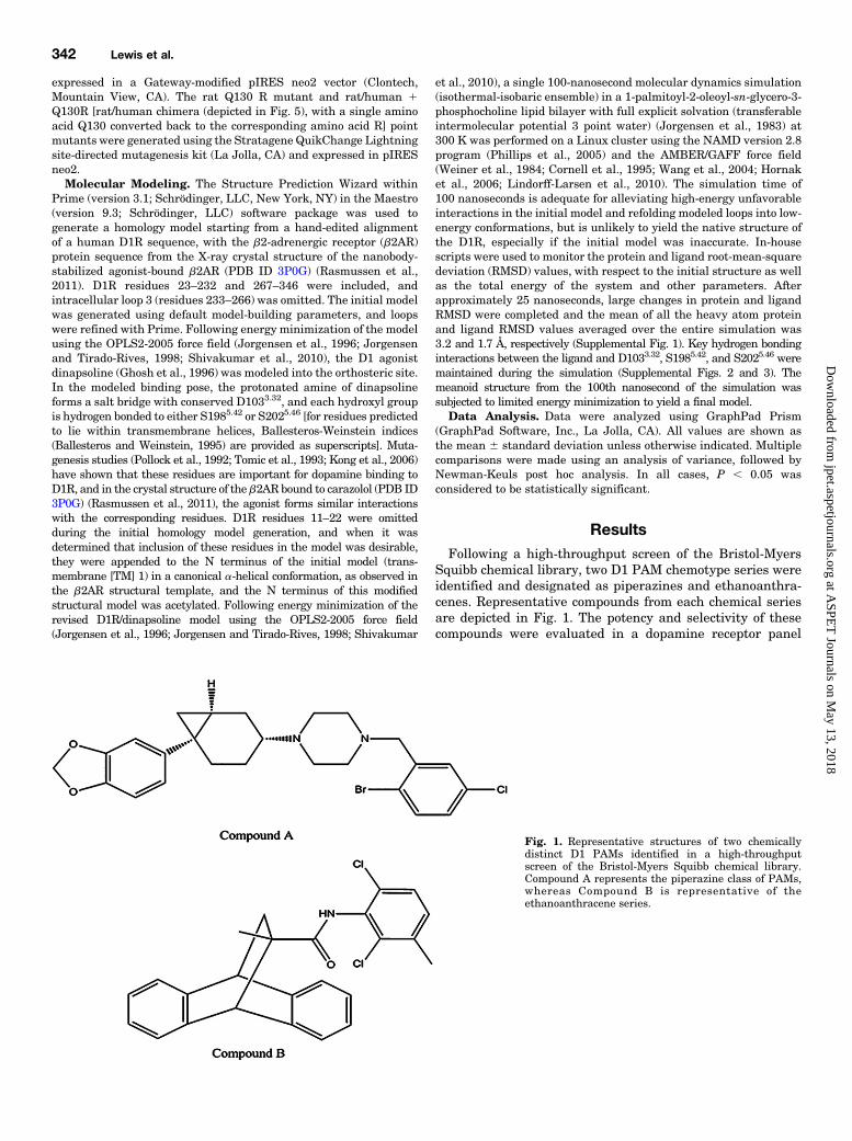
Squibb chemical library, two D1 PAM chemotype series wereidentified and designated as piperazines and ethanoanthra-cenes. Representative compounds from each chemical seriesare depicted in Fig. 1. The potency and selectivity of thesecompounds were evaluated in a dopamine receptor panel
Fig. 1. Representative structures of two chemicallydistinct D1 PAMs identified in a high-throughputscreen of the Bristol-Myers Squibb chemical library.Compound A represents the piperazine class of PAMs,whereas Compound B is representative of theethanoanthracene series.
342 Lewis et al.
at ASPE
T Journals on M
ay 13, 2018jpet.aspetjournals.org
Dow
nloaded from

using both Chinese hamster ovary and HEK cell backgroundsin the absence (agonist mode) or presence of an EC20 ofdopamine (PAM mode). Data presented in Table 1 show thatmembers of the ethanoanthracene series, represented byCompound B, were selective D1 PAMs with no agonistactivity. Because we were unable to generate a D3-expressing cell line sufficient for evaluating compoundactivity, selectivity data at this receptor remain to bedetermined. Compounds from the piperazine series, repre-sented by Compound A, showed D1 PAM activity but alsoagonist activity at the D2 receptor (Table 1). As agonism at D2receptors would worsen positive symptoms of schizophrenia,we chose to focus efforts on the ethanoanthracene series.Compound B was tested in a fold-shift assay to determine themaximal shift in dopamine potency seen with this compound.Data presented in Fig. 2 show that Compound B is a high fold-shift compound (produces amaximal 18-fold shift in dopaminepotency in a cAMP accumulation assay), with a potency(shift50) value of 0.4 mM (0.2–0.6 mM). This shift50 valuecorresponds to the affinity (KB) of the PAM for D1 in theabsence of the agonist according to the allosteric ternarycomplex model (Christopoulos and Kenakin, 2002). Basedupon this model, the affinity of the PAM in the presence of theagonist can be calculated to be approximately 0.02 mM,although this could not be measured directly in the currentstudy. To confirm the activity of this series in a nativeD1 cellular background, we generated rat primary neuronalcultures and characterized the activity of D1 receptors in thisneuronal system. Table 2 summarizes the potency ofdopamine and dopamine agonists in primary cortical neurons.Dopamine increased cAMP production in primary cortical
cultures, with a potency of 354 nM (6167 nM; n5 8; Table 2).The effect of dopamine in cAMP production was completelyblocked by the D1 antagonist SCH23390 [(R)-(1)-7-chloro-8-hydroxy-3-methyl-1-phenyl-2,3,4,5-tetrahydro-1H-3-benzazepine],but was unaffected by the D2 antagonist sulpiride (Fig. 3).The D1 partial agonists SKF-38393 [(6)-1-phenyl-2,3,4,5-tetrahydro-(1H)-3-benzazepine-7,8-diol] and SKF-83959[6-chloro-2,3,4,5-tetrahydro-3-methyl-1-(3-methylphenyl)-1H-3-benzazepine-7,8-diol] showed nanomolar potency, with sub-maximal cAMP activation (relative to dopamine control),whereas the D1 full agonist SKF-81297 [(6)-6-chloro-2,3,4,5-tetrahydro-1-phenyl-1H-3-benzazepine] increased cAMP levelsto a level comparable to that of dopamine (Table 2). Expressionprofiling of cortical neurons also confirmed D1 receptors as thehighest expressing dopamine receptors in these neuronalpreparations (data not shown). Collectively, these data confirmthat this rat primary neuronal culture system has endogenousD1 receptors and validate the use of this endogenous cellularsystem for evaluating D1 PAM activity.Activity of Compound B was evaluated in rat primary
neurons in both the agonist and PAM mode. Compound Bshowed minimal PAM activity (Ymax , 20% of dopaminecontrol), with no agonist activity in rat primary corticalneurons (Fig. 4A). For comparison, Compound A was alsotested in rat primary neurons. Consistent with data intransformed cell lines, Compound A showed no agonistactivity, but showed increased cAMP production (Ymax 550% of dopamine control) in the presence of an EC20 ofdopamine (Fig. 4A). These data were unexpected as Com-pound B was five times more potent than Compound A, andboth showed similar Ymax values (63.6 and 64.3, respectively)in D1-expressing HEK cells. To explore this apparentdisconnect between cells transfected with human D1 recep-tors and rat primary neurons, Compound B and Compound Awere evaluated in HEK cells expressing the rat D1 receptor.Consistent with data in rat primary neurons, this experimentshowed that Compound A exhibited PAM activity at rat D1receptors, whereas Compound B was inactive at rat D1receptors (Fig. 4B). To determine if this reduced activity at ratD1 receptors was consistent for other compounds from theethanoanthracene series and to evaluate activity at non-human primate D1 receptors, an additional set of ethanoan-thracene compounds was evaluated in HEK cells expressing,rat, cynomolgus (cyno), and human D1 receptors (Supple-mental Fig. 4; Table 3). Consistent with the decreased activityat rat D1 receptors for Compound B, all structurally similarcompounds showed reduced activity (less potent and
TABLE 1Selectivity of D1 PAMs in heterologous expression systemsPotency of D1 PAMs in Chinese hamster ovary and HEK cells expressing the humanisoform of D1. Activity of D1 PAMs at other dopamine receptors was evaluated inChinese hamster ovary cells expressing D2, D4, or D5 receptors. Values represent themean IC50 values (61 S.D.). To determine agonist and PAM activity, compounds weretested in the absence of dopamine (agonist mode) or in the presence of an EC20 ofdopamine (PAM mode).
D1 PAM EC50
Compound A Compound B
nM nM
Chinese hamster ovary 210 6 30 (n = 2) 30 6 13 (n = 4)HEK 250 6 60 (n = 2) 56 6 64 (n = 2)D1 agonist .30,000 (n = 2) .30,000 (n = 2)D2 PAM 330 6 100 (agonist) .30,000 (n = 2)D4 PAM .30,000 (n = 2) .30,000 (n = 2)D5 PAM .30,000 (n = 3) .30,000 (n = 4)
Fig. 2. Fold-shift analysis of Compound B. HEK cellsexpressing the human D1 receptor were treated withincreasing concentrations of dopamine in the presenceor absence of increasing concentrations of Compound B.The maximal potency shift (shiftmax) obtained withCompound B was 18-fold (13- to 24-fold), with a shift50(KB) value of 0.4 mM (0.2–0.6 mM).
Discovery of Novel D1 Dopamine Receptor PAMs 343
at ASPE
T Journals on M
ay 13, 2018jpet.aspetjournals.org
Dow
nloaded from

decreased Ymax) at the rat D1 receptor, whereas activity wascomparable between human and cyno D1 receptors.To identify the amino acid(s) involved in this species
selectivity, two parallel work streams were initiated. First,an alanine scan was conducted to replace key amino acidswithin the human D1 receptor. For these studies, select pointmutants were generated based on their position within thereceptor protein, their ability to retain dopamine signaling(potency within 10� of the wild-type receptor), and a level ofoverall receptor expression comparable to the wild-typereceptor. For each of these mutants, a PAM concentrationresponse curve was run in the presence of an EC20 ofdopamine and PAM EC50 values were generated. Becausewe were interested in amino acids that contributed to loss ofefficacy of Compound B at the rat D1 receptor, we focused onmutants that showed the largest difference in potencybetween Compound B and Compound A. Of the point mutantsevaluated, five showed a shift in potency greater than 4-foldfor Compound B versus Compound A (Table 4).In parallel with mutagenesis efforts, the potency of each
compound was evaluated at a series of human-rat D1 chimericreceptors in an effort to identify the amino acid(s) thatcontributed to the species selectivity of Compound B (Fig. 5).In all of these chimeras, the potency of dopamine wasunchanged, suggesting that orthosteric binding and effectorcoupling was unchanged by the chimera (Table 5). Among thisseries of chimeric transpositions, only the replacement of thehuman sequence with the rat sequence from the N terminusup to the start of TM5 showed loss of activity of Compound B
while sparing Compound A. No loss of potency was seen whenthe N terminus or extracellular loop two of the human D1 wasexchanged with the analogous rat sequence. Collectively,these data suggest that the binding site for Compound Bmight lie between the start of TM1 and the end of TM4.Further analysis of the human and rat D1 sequences in thisregion identified only three amino acids that differ betweenthe human and rat sequence: F92→ L, S943.23 → P, and R130→Q (human→ rat changes). Of these, F92, which is located inextracellular loop 1, and S943.23 are conserved in the mouseand did not affect the potency of Compound B in the alaninemutagenesis studies. To further test the role of R130, locatedin intracellular loop 2 (ICL2), in contributing to the speciesselectivity of Compound B, two point mutants were generatedby substituting the rat glutamine at position 130 with thehuman amino acid arginine. In the rat N terminus throughTM5 chimera, substitution of Q130 with the human aminoacid R130 completely restored the activity of Compound B(Table 5). Moreover, in the full-length rat receptor, conversionof this single amino acid to the human amino acid (R130)restored activity of Compound B. To confirm this observation,full fold-shift cAMP accumulation assays were performed inparallel utilizing HEK293 cells transiently expressing eitherwild-type human D1, wild-type rat D1, or the rat Q130R D1mutant. In these experiments, Compound B produceda maximal 11-fold (6- to 19-fold) increase in dopaminepotency, with a measured shift50 value of 0.4 mM (0.1–2 mM)(Fig. 6), in agreement with the results obtained from similarexperiments using cells stably expressing human D1 (Fig. 2).Compound B produced no observable effect on dopamineresponses in HEK293 cells transiently expressing wild-typerat D1. However, in cells transiently expressing the rat
TABLE 2Characterization of D1 full and partial agonists in rat primary neuronsPotency of dopamine and D1 agonists in primary rat cortical cultures. Valuesrepresent the calculated nanomolar potency (61 S.D.). Ymax values represent themaximal increase in cAMP relative to dopamine control.
EC50 Ymax N
nM % dopamine
Dopamine 353 6 167 100 8SFK-83959 4 6 5.2 22 6 6 4SKF-38393 10 6 9 67 6 19 8SKF-81297 3 6 2 107 6 13 6
Fig. 3. Effects of D1 and D2 antagonists on dopamine-mediated increasesin cAMP production in rat cortical neuronal cultures. Neurons weretreated with 1 mM antagonist or vehicle for 10 minutes and then withincreasing concentrations of dopamine. cAMP levels were quantified asdescribed in Materials and Methods. The points represent the meanchange in cAMP levels relative to dopamine 6 1 S.D.
Fig. 4. Compounds A and B were tested in (A) primary cultures or (B)HEK cells transfected with the rat D1 receptor in the absence (agonistmode) or presence (PAM mode) of an EC20 of dopamine, and cAMP levelswere quantified as described in Materials and Methods. The pointsrepresent the mean change in cAMP levels relative to dopamine 6 1 S.D.
344 Lewis et al.
at ASPE
T Journals on M
ay 13, 2018jpet.aspetjournals.org
Dow
nloaded from

Q130R mutant D1 receptor, both the cooperativity andpotency of Compound B were fully restored, with shiftmax 511-fold (7- to 18-fold) and shift50 5 0.5 mM (0.2–2 mM), valuesthat were indistinguishable from those observed at human D1(Fig. 6). Collectively, these data suggested a critical role ofarginine 130 in the human D1 receptor in the activity/speciesselectivity of Compound B.In Fig. 7, the locations of R130 and the residues where
alanine mutation more negatively impacts the EC50 ofCompound B than Compound A (Table 4) are highlightedon a molecular dynamics-refined homology model of theD1R/dinapsoline complex. This model was generated usingthe X-ray crystal structure of the nanobody-stabilized agonist-bound 2AR as a template (Rasmussen et al., 2011). Based onthe model (and as expected from sequence proximity), R130 inICL2 is close to V1193.48 and W1233.52, which are located onthe membrane-exposed face of a-helix transmembrane regionTM3. The distances from the Ca atom of R130 to those ofV1193.48 andW1233.52 are 16 and 10 Å, respectively. The EC50
values of Compound B against these mutants are more than900-fold larger than the EC50 measured in WT HEK cells. Inthe model, residue V582.38 is at the intracellular end of thetransmambrane region TM2 helix. The inter-Ca distance forR130 and V582.38 is 15 Å, and the V582.38Amutation increasesthe EC50 of Compound B by 220-fold relative to WT. M135 islocated in ICL2 about 9 Å from R130, and the EC50 ofCompound B against the mutant is about 42-fold larger thanthe WT EC50. Proximal to the extracellular (C-terminal) endof the TM3 a-helix, F953.24 is distant from the aforementionedresidues, and its role in PAM binding is not apparent. Sinceresidues V582.38, V1193.48, W1233.52, and M135 are predictedto be proximal to each other and to R130 and substantialnegative impacts on the PAM activity of Compound B resultedfrommutation of these residues to alanine, it is plausible thatin addition to R130, residues V582.38, V1193.48, W1233.52, andM135 comprise part of the binding site for Compound B in theD1R.Alignment of human dopamine receptor sequences reveals
that none of the residues found to impact Compound Bactivity in D1R are identical in D2R or D3R (Table 6). In D4R,arginine and valine are present at the positions correspondingto R130 and V1193.48 in D1R, whereas there are differences atthe other four positions. D1R and D5R differ only at theposition corresponding to V582.38 in D1R. If R130, V582.38,V1193.48, W1233.52, and M135 do indeed form part of theCompound B–binding site in D1R, the observed selectivity forthis receptor relative to D2R and D4R is not surprising. Thelack of D5R activity is less easily rationalized given only one
difference in the set of residues that impact Compound Bactivity in D1R, although there are other differences betweenD1R and D5R at positions proximal to these residues in theD1R model.As shown in Fig. 7, the residues discussed above are also
close to the D1R-conserved D1203.49, R1213.50, and Y1223.51
motif, which is important in modulating the constitutiveactivity of GPCRs (Scheer et al., 1996; Capra et al., 2004;Rovati et al., 2007). ICL2 is hypothesized to form part of theCompound B binding site, and this loop has been proposed tostabilize the inactive state of GPCRs. Binding of Compound Bproximal to these two D1R structural elements couldpotentially impact their function, contributing to the observedPAM activity of the compound.Compound B is primarily hydrophobic/aromatic in nature,
and the amide is the only polar moiety in the molecule. Withthe exception of R130, all of the residues discussed above havehydrophobic side chains that could make favorable van derWaals contacts with this PAM. A p-cation interactionbetween the side chain of R130 and one of the aromatic ringsin the compound is also plausible. In the rat D1R, this residueis a glutamine that cannot form an analogous interaction withthe ligand. Absence of this putative p-cation interaction in therat D1R could contribute to the observed species selectivity ofCompound B.As discussed above, the PAM-binding site may include
parts of ICL2 and the N- and C-terminal ends of TM a-helices2 and 3, respectively. However, it is also possible that themutations that diminished the PAM activity of CompoundB impact the binding of Compound B at some other site inD1R through an allosteric mechanism. Alternatively, the
TABLE 3Characterization of additional ethanoanthracene compounds in HEK cells expressing rat, human, or cynoD1 receptorsPotency of additional ethanoanthracene (ETC) compounds in HEK cells expressing human, cyno, or rat D1 receptors.Potency of compounds was determined in the presence of an EC20 of dopamine. Values represent the calculatednanomolar potency (6 S.D.). Ymax values represent the maximal increase in cAMP relative to dopamine control.
CompoundHuman D1 Cyno D1 Rat D1
EC50 Dopamine Ymax n EC50 Dopamine Ymax n EC50 Dopamine Ymax n
nM % nM % nM %
ETC-1 38 6 8 82 6 12.3 5 48 6 16 95 6 12.6 3 1216 6 269 51 6 14.1 7ETC-2 77 6 10 72 6 19 3 65 6 20 85 6 9 3 332 6 55 38 6 11.4 5ETC-3 254 6 122 57 6 9.7 4 423 6 53 60 6 14.5 4 1630 6 993 14 6 13.1 4
TABLE 4Top amino acid mutants showing loss of activity for Compound BPotency of Compounds B and A was determined in HEK cells transiently expressingvarious human D1 point mutants as described in Materials and Methods. For eachmutant, the endogenous amino acid and the predicted position with the human D1protein sequence are given. Values represent the PAM EC50 and Ymax (percentage ofdopamine response) for each mutant.
D1 VariantCompound A Compound B
EC50 Ymax EC50 Ymax
nM % dopamine response nM % dopamine response
WT 250 100 60 100W123A 600 20 .50,000 ,5V119A 1800 35 .50,000 ,5V58A 730 22 12,000 36F95A 380 37 2200 60M135A 650 21 2300 24
Discovery of Novel D1 Dopamine Receptor PAMs 345
at ASPE
T Journals on M
ay 13, 2018jpet.aspetjournals.org
Dow
nloaded from

mutations could mitigate the positive allosteric modulationeffects of Compound B by altering the interactions of themutant D1R with the G protein and/or modifying the levelof constitutive activity of the receptor while not directly
impacting the binding of Compound B. Since two of theresidues where mutations significantly diminished thePAM activity of Compound B (V1193.48 and W1233.52)immediately flank the D1203.49, R1213.50, and Y1223.51
Fig. 5. Schematic representation of generated human rat chimeras. Blue regions represent human sequences, with black regions indicating the ratsequence. The circles represent the approximate location of amino acids that differed between rat and human sequences.
TABLE 5Potency of dopamine, Compound A, and Compound B across rat-human D1 receptor chimerasPotency of Compound B and Compound A across various D1 human/rat chimeras (described in Fig. 5). Receptor chimeraswere transiently expressed in HEK cells, and EC50 values were determined. Rat/human + Q130R represent the rat/humanchimera (depicted in Fig. 5), with a single amino acid Q130 converted back to the corresponding amino acid R. EC50 valuesrepresent nanomolar potency from three to six independent experiments. For the experiment (n = 2), individual EC50 andYmax values are given. Ymax values represent the percentage of dopamine response set at 100%.
ChimeraDopamine Compound A Compound B
EC50 Ymax n EC50 Ymax n EC50 Ymax n
nM % nM % nM %
Human 33 6 11 100 6 245 6 115 50 6 12 6 172 6 196 39 6 6 6Rat 48 6 17 100 6 270 6 168 62 6 19 6 .10 mM ,10 6Rat N terminus 16 6 4 100 4 662 6 213 61 6 19 4 378, 321 62, 52 2Rat ECL2 21 6 3 100 4 712 6 168 67 6 14 4 222, 287 53, 69 2Rat TM6/ECL3 34 6 9 100 4 960 6 197 61 6 19 4 33, 258 53, 48 2Rat ICL3 49 6 38 100 3 296 6 175 58 6 28 3 230 6 219 40 6 10 3Rat C terminus 22 6 17 100 3 237 6 243 57 6 32 3 1059 6 1681 63 6 15 3Human/rat 37 6 25 100 3 311 6 165 69 6 30 3 361 6 363 47 6 18 3Rat/human 54 6 31 100 6 210 6 110 47 6 14 6 .10 mM ,10 3Rat Q130R 74 6 47 100 3 273 6 125 59 6 9 3 110 6 40 31 6 3 3Rat/human + Q130R 80 6 12 100 3 345 6 50 46 6 14 3 102 6 5 28 6 8 3
ECL, extracellular loop.
346 Lewis et al.
at ASPE
T Journals on M
ay 13, 2018jpet.aspetjournals.org
Dow
nloaded from

motif in D1R, this latter possibility also seems likely. Atpresent, the available data are insufficient to discriminatebetween these possibilities.
DiscussionCompounds A and B were identified as D1 PAMs and
optimized following a high-throughput screen of the Bristol-Myers Squibb chemical library. These compounds representtwo distinct chemotypes and are the first reported D1 receptorPAMs. Both Compounds A and B showed nanomolar PAMpotency, with no agonist activity (Fig. 1; Table 1). Moreover,Compound B showed selectivity against other dopaminereceptors, including D2, D4, and D5 receptors. Although D3heterologous expression systems have been used as modelsto support D3 receptor-binding studies, we were unable togenerate a D3-expressing cell line with a functional readoutsufficient to support evaluation of PAM activity. Therefore,functional activity of these PAMs at the D3 receptor remainsto be determined. However, these D1 PAMs do provide theinitial steps toward the development of D1 PAMs for thepotential treatment of cognitive dysfunction associated withpsychiatric disorders, such as schizophrenia.The development of D1 agonists for the treatment of
cognitive dysfunction has been limited by two major factors.First, D1 agonists used in the clinic have been associated withhypotension, limiting the clinical dose (Blanchet et al., 1998;Rascol et al., 1999). To date, no D1 selective compounds havebeen identified to delineate the role of D1 versus D5 receptorsin regulating blood pressure. However, preclinical data maysupport a greater role of the D5 receptor in regulating bloodpressure, relative to the D1 receptor. For example, D5receptors were shown to play an important role in regulating
Fig. 6. Fold-shift analysis of Compound B at (A) wild-type human D1, (B) Q130R mutant rat D1, or (C) wild-type rat D1. HEK cells transientlyexpressing these various D1 receptors were treated with various concentrations of dopamine in the presence or absence of increasing concentrations ofCompound B. The cooperativity (a or shiftmax) and shift50 values reported represent the mean and 95% confidence intervals from three experiments.
Fig. 7. Homology model of the D1R/dinapsoline complex. TMs, extracellu-lar loops (ECL), and ICL are labeled. Key residues identified via alaninemutagenesis and human/rat chimera studies are depicted in a cyan ball andstick representation. The D1R D1203.49, R1213.50, and Y1223.51 conservedmotif and the agonist dinapsoline are shown in a magenta and orange balland stick representation, respectively. The image was created with thePyMOL Molecular Graphics System (version 1.6.0.0; Schrödinger, LLC).
TABLE 6Differences between human dopamine receptors at residues wherealanine mutation impacts Compound B PAM activity in D1RIdentities of amino acids in the human dopamine receptors at positions in the D1Rwhere mutation to alanine negatively impacted the PAM activity of Compound 2were determined via multiple sequence alignment with CLUSTAL W version 1.83(Thompson et al., 1994) using default parameters and sequences NM_000794.3,NM_000795.3, NM_000796.5, NM_000797.3, and NM_000798.4 for the humanD1–D5 receptors, respectively.
Residue D1R D2R D3R D4R D5R
582.38 V T T P M953.24 F H C L F1193.48 V I I V V1233.52 W T T V W130 (ICL2) R L H R R135 (ICL2) M S S G M
Discovery of Novel D1 Dopamine Receptor PAMs 347
at ASPE
T Journals on M
ay 13, 2018jpet.aspetjournals.org
Dow
nloaded from

hypertension in D3 knockoutmice (Wang et al., 2013). Geneticdeletion of D5 receptors in mice results in elevated bloodpressure (Hollon et al., 2002; Yang et al., 2004), whereas therole of D1 receptors in regulating blood pressure is stillunclear (Albrecht et al., 1996; Wang et al., 1999). A selectiveD1 PAM may also have a reduced hypotensive liabilitycompared with an agonist at both the D1 and D5 receptorssimply because a D1 PAM would increase activity of fewerrenal or vascular dopamine receptors. Although it was beyondthe scope of this study, the lack of activity at D5 receptors maytherefore reduce the hypotensive liability of the identifiedD1 PAMs relative to D1 agonists that lack D1 versus D5receptor selectivity.A second challenge with D1 receptor agonists is that
preclinically, agonists have shown an inverted U doseresponse in that too much or too little D1 receptor activationcan have adverse effects on cognition (for a review, seeWilliams and Castner, 2006). Because cognitive deficitsassociated with schizophrenia are hypothesized to result froma hypofunction of the prefrontal dopaminergic tone (Daviset al., 1991), a D1 PAM would be beneficial as it wouldenhance the effect of endogenous dopamine without directactivation of the D1 receptor.One unexpected finding in the current study was the lack of
activity of Compound B in rat primary neuronal cultures. Thislack of activity in a rat endogenous expression system wasconfirmed in HEK cells overexpressing the D1 receptor (Fig.4). The human and rat D1 sequence share a .90% sequencehomology, with the majority of this sequence divergenceoccurring at the N and C termini. Based on these findings, wesought to determine the critical amino acid(s) that mediatespecies selectivity of Compound B. Alanine mutagenesis isa rapid screening method for identifying key amino acidsimportant for binding of radioligands (Gregory et al., 2013).We adapted this approach to identify amino acids thatcontribute to the species selectivity of our D1 PAMs. Ina parallel approach, a series of D1 human/rat chimeras weregenerated to further evaluate regions critical for PAMactivity. Collectively, these data identified a critical aminoacid, R130, as directly mediating the species selectivity ofCompound B. Moreover, alanine mutagenesis studies suggestthat other amino acids in ICL2 as well as proximal regions ofTM helix 2 and TM helix 3 were critical in mediatingthe activity of Compound B (Table 4). These data suggest thebinding region for the Compound B chemotype includes thesecond intracellular loop and possibly the second and thirdtransmembrane helices of the D1 receptor. However, it is alsopossible that Compound B binds elsewhere in the D1R, withits activity negatively impacted by the mutations discussedabove via an allosteric mechanism, modulation of receptor/Gprotein interactions, or mutation-induced changes in the levelof constitutive activity of D1R. There are insufficient data todistinguish between these possibilities at present.Little information has been published as to the contribution
of this region in the function or expression of the D1 receptor.However, the amino acids identified from these studies are inclose proximity to the glutamic acid/aspartic acid-arginine-tyrosinemotif, which has been rigorously studied across manyGPCRs (for a review, see Rovati et al., 2007). For example,changes in the glutamic acid or aspartic acid motif canincrease the constitutive activity of the GPCR or increaseagonist affinity and efficacy (Scheer et al., 1996; Capra et al.,
2004). Chung et al. (2002) proposed that the ICL2 of someGPCRs takes on a helical structure and stabilizes the inactivestate of the receptor. It is possible that our current D1 PAM(Compound B) modified the tertiary structure of the ICL2 ina manner that enhances efficacy of the orthosteric ligandwithout increasing the constitutive activity.In summary, the current studies provide the first de-
scription of PAMs selective for the D1 receptor: Compound Band Compound A (Fig. 1). These compounds may be usefultools for understanding the contribution of increases in D1versus D5 receptor activity across multiple physiologicsystems, from the kidneys to the brain. Moreover, usingmolecular biology approaches and species differences, wewere able to map the amino acids critical in the activity of ourD1 PAM (Compound B), thus providing a structural scaffoldfor the development of future D1 PAMs. Collectively, thesefindings represent the initial steps toward the development ofD1 PAMs for the potential treatment of neuropsychiatricdiseases.
Authorship Contributions
Participated in research design: Lewis, Hunihan, Watson, Gentles,Hu, Huang, Bronson, Beno, Macor, Hendricson, Knox, Weed, Cacace,Westphal, Alt, Brown.
Conducted experiments: Lewis, Hunihan, Watson, Beno, Ferrante,Molski, Kong, Cvijic, Rockwell, Alt, Brown.
Contributed new reagents or analytic tools: Lewis, Hunihan,Gentles, Hu, Huang, Bronson, Beno, Macor, Hendricson, Molski,Kong, Cvijic, Alt, Brown.
Performed data analysis: Lewis, Hunihan, Watson, Beno, Ferrante,Hendricson, Kong, Cvijic, Alt, Brown.
Wrote or contributed to the writing of the manuscript: Lewis,Hunihan, Beno, Alt, Brown.
References
Albrecht FE, Drago J, Felder RA, Printz MP, Eisner GM, Robillard JE, Sibley DR,Westphal HJ, and Jose PA (1996) Role of the D1A dopamine receptor in thepathogenesis of genetic hypertension. J Clin Invest 97:2283–2288.
Ballesteros JA and Weinstein H (1995) Integrated methods for the construction ofthree-dimensional models and computational probing of structure-function rela-tions in G protein-coupled receptors. Methods Neurosci 25:366–428.
Blanchet PJ, Fang J, Gillespie M, Sabounjian L, Locke KW, Gammans R, MouradianMM, and Chase TN (1998) Effects of the full dopamine D1 receptor agonist dihy-drexidine in Parkinson’s disease. Clin Neuropharmacol 21:339–343.
Brozoski TJ, Brown RM, Rosvold HE, and Goldman PS (1979) Cognitive deficitcaused by regional depletion of dopamine in prefrontal cortex of rhesus monkey.Science 205:929–932.
Cai JX and Arnsten AF (1997) Dose-dependent effects of the dopamine D1 receptoragonists A77636 or SKF81297 on spatial working memory in aged monkeys.J Pharmacol Exp Ther 283:183–189.
Capra V, Veltri A, Foglia C, Crimaldi L, Habib A, Parenti M, and Rovati GE (2004)Mutational analysis of the highly conserved ERY motif of the thromboxane A2receptor: alternative role in G protein-coupled receptor signaling. Mol Pharmacol66:880–889.
Castner SA and Goldman-Rakic PS (2004) Enhancement of working memory in agedmonkeys by a sensitizing regimen of dopamine D1 receptor stimulation. J Neurosci24:1446–1450.
Christopoulos A and Kenakin T (2002) G protein-coupled receptor allosterism andcomplexing. Pharmacol Rev 54:323–374.
Chung DA, Zuiderweg ER, Fowler CB, Soyer OS, Mosberg HI, and Neubig RR (2002)NMR structure of the second intracellular loop of the alpha 2A adrenergic receptor:evidence for a novel cytoplasmic helix. Biochemistry 41:3596–3604.
Conn PJ, Christopoulos A, and Lindsley CW (2009) Allosteric modulators of GPCRs:a novel approach for the treatment of CNS disorders.Nat Rev Drug Discov 8:41–54.
Cornell WD, Cieplak P, Bayly CL, Gould IR, Merz KM, Jr, Ferguson DM, SpellmeyerDC, Fox T, Caldwell JW, and Kollman PA (1995) A second generation force field forthe simulation of proteins, nucleic acids, and organic molecules. J Am Chem Soc117:5179–5197.
Costa E (1991) The Di Mascio Lecture. The allosteric modulation of GABAA recep-tors. Seventeen years of research. Neuropsychopharmacology 4:225–235.
Davis KL, Kahn RS, Ko G, and Davidson M (1991) Dopamine in schizophrenia:a review and reconceptualization. Am J Psychiatry 148:1474–1486.
Ghosh D, Snyder SE,Watts VJ, Mailman RB, and Nichols DE (1996) 9-Dihydroxy-2,3,7,11b-tetrahydro-1H-naph[1,2,3-de]isoquinoline: a potent full dopamine D1 ago-nist containing a rigid-b-phenyldopamine pharmacophore. J Med Chem 39:549–555.
348 Lewis et al.
at ASPE
T Journals on M
ay 13, 2018jpet.aspetjournals.org
Dow
nloaded from
Gregory KJ, Nguyen ED, Reiff SD, Squire EF, Stauffer SR, Lindsley CW, Meiler J,and Conn PJ (2013) Probing the metabotropic glutamate receptor 5 (mGlu₅) posi-tive allosteric modulator (PAM) binding pocket: discovery of point mutations thatengender a “molecular switch” in PAM pharmacology. Mol Pharmacol 83:991–1006.
Green MF, Kern RS, Braff DL, and Mintz J (2000) Neurocognitive deficits andfunctional outcome in schizophrenia: are we measuring the “right stuff”? SchizophrBull 26:119–136.
Hollon TR, Bek MJ, Lachowicz JE, Ariano MA, Mezey E, Ramachandran R, Wersinger SR,Soares-da-Silva P, Liu ZF, Grinberg A, et al. (2002) Mice lacking D5 dopaminereceptors have increased sympathetic tone and are hypertensive. J Neurosci 22:10801–10810.
Hornak V, Abel R, Okur A, Strockbine B, Roitberg A, and Simmerling C (2006)Comparison of multiple Amber force fields and development of improved proteinbackbone parameters. Proteins 65:712–725.
Jorgensen WL, Chandrasekhar J, Madura JD, Impey RW, and Klein ML (1983)Comparison of simple potential functions for simulating liquid water. J Chem Phys79:926–935.
Jorgensen WL, Maxwell DS, and Tirado-Rives J (1996) Development and testing ofthe OPLS all-atom force field on conformational energetics and properties of or-ganic liquids. J Am Chem Soc 118:11225–11236.
Jorgensen WL and Tirado-Rives J (1998) The OPLS [optimized potentials for liquidsimulations] potential functions for proteins, energy minimizations for crystals ofcyclic peptides and crambin. J Am Chem Soc 110:1657–1666.
Keov P, Sexton PM, and Christopoulos A (2011) Allosteric modulation of G protein-coupled receptors: a pharmacological perspective. Neuropharmacology 60:24–35.
Kong MMC, Fan T, Varghese G, O’dowd BF, and George SR (2006) Agonist-inducedcell surface trafficking of an intracellularly sequestered D1 dopamine receptorhomo-oligomer. Mol Pharmacol 70:78–89.
Lindorff-Larsen K, Piana S, Palmo K, Maragakis P, Klepeis JL, Dror RO, and ShawDE (2010) Improved side-chain torsion potentials for the Amber ff99SB proteinforce field. Proteins 78:1950–1958.
Mottola DM, Kilts JD, Lewis MM, Connery HS, Walker QD, Jones SR, Booth RG,Hyslop DK, Piercey M, Wightman RM, et al. (2002) Functional selectivity ofdopamine receptor agonists. I. Selective activation of postsynaptic dopamine D2receptors linked to adenylate cyclase. J Pharmacol Exp Ther 301:1166–1178.
Nakako T, Murai T, Ikejiri M, Ishiyama T, Taiji M, and Ikeda K (2013) Effects ofa dopamine D1 agonist on ketamine-induced spatial working memory dysfunctionin common marmosets. Behav Brain Res 249:109–115.
Phillips JC, Braun R, Wang W, Gumbart J, Tajkhorshid E, Villa E, Chipot C, SkeelRD, Kalé L, and Schulten K (2005) Scalable molecular dynamics with NAMD.J Comput Chem 26:1781–1802.
Pollock NJ, Manelli AM, Hutchins CW, Steffey ME, MacKenzie RG, and Frail DE(1992) Serine mutations in transmembrane V of the dopamine D1 receptor affectligand interactions and receptor activation. J Biol Chem 267:17780–17786.
Puig MV, Antzoulatos EG, and Miller EK (2014) Prefrontal dopamine in associativelearning and memory. Neuroscience 282C:217–229.
Rascol O, Blin O, Thalamas C, Descombes S, Soubrouillard C, Azulay P, Fabre N,Viallet F, Lafnitzegger K, Wright S, et al. (1999) ABT-431, a D1 receptor agonistprodrug, has efficacy in Parkinson’s disease. Ann Neurol 45:736–741.
Rasmussen SGF, Choi H-J, Fung JJ, Pardon E, Casarosa P, Chae PS, Devree BT,Rosenbaum DM, Thian FS, Kobilka TS, et al. (2011) Structure of a nanobody-stabilized active state of the b(2) adrenoceptor. Nature 469:175–180.
Rosell DR, Zaluda LC, McClure MM, Perez-Rodriguez MM, Strike KS, Barch DM,Harvey PD, Girgis RR, Hazlett EA, Mailman RB, et al. (2015) Effects of the D1dopamine receptor agonist dihydrexidine (DAR-0100A) on working memory inschizotypal personality disorder. Neuropsychopharmacology 40:446–453.
Rovati GE, Capra V, and Neubig RR (2007) The highly conserved DRY motif of classA G protein-coupled receptors: beyond the ground state. Mol Pharmacol 71:959–964.
Scheer A, Fanelli F, Costa T, De Benedetti PG, and Cotecchia S (1996) Constitutivelyactive mutants of the alpha 1B-adrenergic receptor: role of highly conserved polaramino acids in receptor activation. EMBO J 15:3566–3578.
Shivakumar D, Williams J, Wu Y, Damm W, Shelley J, and Sherman W (2010)Prediction of absolute solvation free energies using molecular dynamics free energyperturbation and the OPLS force field. J Chem Theory Comput 6:1509–1519.
Sibley DR and Monsma FJ, Jr (1992) Molecular biology of dopamine receptors.Trends Pharmacol Sci 13:61–69.
Thompson JD, Higgins DG, and Gibson TJ (1994) CLUSTAL W: improving thesensitivity of progressive multiple sequence alignment through sequence weight-ing, position-specific gap penalties and weight matrix choice. Nucleic Acids Res 22:4673–4680.
Tomic M, Seeman P, George SR, and O’Dowd BF (1993) Dopamine D1 receptormutagenesis: role of amino acids in agonist and antagonist binding. BiochemBiophys Res Commun 191:1020–1027.
Wang J, Wolf RM, Caldwell JW, Kollman PA, and Case DA (2004) Development andtesting of a general amber force field. J Comput Chem 25:1157–1174.
Wang X, Escano CS, Asico L, Jones JE, Barte A, Lau YS, Jose PA, and Armando I(2013) Upregulation of renal D5 dopamine receptor ameliorates the hypertensionin D3 dopamine receptor-deficient mice. Hypertension 62:295–301.
Wang ZQ, Felder RA, and Carey RM (1999) Selective inhibition of the renal dopa-mine subtype D1A receptor induces antinatriuresis in conscious rats. Hypertension33:504–510.
Weiner SJ, Kollman PA, Case DA, Singh UC, Ghio C, Alagona G, Profeta S, Jr,and Weiner P (1984) A new force field for molecular mechanical simulation ofnucleic acids and proteins. J Am Chem Soc 106:765–784.
Williams GV and Castner SA (2006) Under the curve: critical issues for elucidatingD1 receptor function in working memory. Neuroscience 139:263–276.
Wootten D, Christopoulos A, and Sexton PM (2013) Emerging paradigms in GPCRallostery: implications for drug discovery. Nat Rev Drug Discov 12:630–644.
Yang Z, Sibley DR, and Jose PA (2004) D5 dopamine receptor knockout mice andhypertension. J Recept Signal Transduct Res 24:149–164.
Zeng C, Zhang M, Asico LD, Eisner GM, and Jose PA (2007) The dopaminergicsystem in hypertension. Clin Sci (Lond) 112:583–597.
Address correspondence to: Jeffrey M. Brown, Genetically Defines Diseaseand Genomics, Bristol-Myers Squibb, 5 Research Parkway, Wallingford, CT06492. E-mail: [email protected]
Discovery of Novel D1 Dopamine Receptor PAMs 349
at ASPE
T Journals on M
ay 13, 2018jpet.aspetjournals.org
Dow
nloaded from