Direct Detection and Sequencing of Damaged DNA Bases
9
RESEARCH Open Access Direct Detection and Sequencing of Damaged DNA Bases Tyson A Clark, Kristi E Spittle, Stephen W Turner and Jonas Korlach * Abstract Products of various forms of DNA damage have been implicated in a variety of important biological processes, such as aging, neurodegenerative diseases, and cancer. Therefore, there exists great interest to develop methods for interrogating damaged DNA in the context of sequencing. Here, we demonstrate that single-molecule, real- time (SMRT ® ) DNA sequencing can directly detect damaged DNA bases in the DNA template - as a by-product of the sequencing method - through an analysis of the DNA polymerase kinetics that are altered by the presence of a modified base. We demonstrate the sequencing of several DNA templates containing products of DNA damage, including 8-oxoguanine, 8-oxoadenine, O6-methylguanine, 1-methyladenine, O4-methylthymine, 5-hydroxycytosine, 5-hydroxyuracil, 5-hydroxymethyluracil, or thymine dimers, and show that these base modifications can be readily detected with single-modification resolution and DNA strand specificity. We characterize the distinct kinetic signatures generated by these DNA base modifications. Keywords: DNA Damage, Modified Bases, Sequencing Background DNA is under constant stress from both endogenous and exogenous sources. As the carrier of genetic infor- mation, DNA relies on the maintenance and repair of existing molecules and is the only biological molecule to do so. The bases exhibit limited chemical stability and are vulnerable to chemical modifications through differ- ent types of damage, including oxidation, alkylation, radiation damage, and hydrolysis. DNA base modifica- tions resulting from these types of DNA damage are wide-spread and play important roles in affecting phy- siological states and disease phenotypes (reviewed in [1-3]). Examples include 8-oxoguanine, 8-oxoadenine (oxidative damage; aging, Alzheimer’s, Parkinson’ s), 1- methyladenine, 6-O-methylguanine (alkylation; gliomas and colorectal carcinomas), benzo[a]pyrene diol epoxide (BPDE), pyrimidine dimers (adduct formation; smoking, industrial chemical exposure, UV light exposure; lung and skin cancer), and 5-hydroxycytosine, 5-hydroxyura- cil, 5-hydroxymethyluracil, and thymine glycol (ionizing radiation damage; chronic inflammatory diseases, pros- tate, breast and colorectal cancer). Currently, methods for detecting these and other pro- ducts of DNA damage are limited to bulk measurements including chromatographic techniques, polymerase chain reaction assays, the Comet assay, mass spectrometry, electrochemistry, radioactive labeling and immunochem- ical methods (reviewed in [4]). To our knowledge, the integration of DNA damage detection into a high- throughput DNA sequencing technique has not been reported. Because base damage can occur at random DNA template positions, sequencing capabilities reach- ing the level of individual DNA molecules are highly desirable. Recently, single-molecule, real-time (SMRT) DNA sequencing has been described for the direct detection of methylated and hydroxymethylated DNA bases [5]. In SMRT sequencing, the progression of single molecules of DNA polymerase is monitored in real time during base incorporations using fluorescent phospholinked nucleotides [6,7]. The dynamics of DNA polymerization is thereby recorded in the form of a train of fluorescent pulses. The length of time that the polymerase retains a nucleotide bound in its active site (pulse width, PW), and the time interval between successive nucleotide- bound states (interpulse duration, IPD) are the principal pulse metrics used in the analysis to ascertain that the * Correspondence: [email protected] Pacific Biosciences, 1380 Willow Rd, Menlo Park, California, USA Clark et al. Genome Integrity 2011, 2:10 http://www.genomeintegrity.com/content/2/1/10 GENOME INTEGRITY © 2011 Clark et al; licensee BioMed Central Ltd. This is an Open Access article distributed under the terms of the Creative Commons Attribution License (http://creativecommons.org/licenses/by/2.0), which permits unrestricted use, distribution, and reproduction in any medium, provided the original work is properly cited.
Transcript of Direct Detection and Sequencing of Damaged DNA Bases

RESEARCH Open Access
Direct Detection and Sequencing of DamagedDNA BasesTyson A Clark, Kristi E Spittle, Stephen W Turner and Jonas Korlach*
Abstract
Products of various forms of DNA damage have been implicated in a variety of important biological processes,such as aging, neurodegenerative diseases, and cancer. Therefore, there exists great interest to develop methodsfor interrogating damaged DNA in the context of sequencing. Here, we demonstrate that single-molecule, real-time (SMRT®) DNA sequencing can directly detect damaged DNA bases in the DNA template - as a by-product ofthe sequencing method - through an analysis of the DNA polymerase kinetics that are altered by the presence ofa modified base. We demonstrate the sequencing of several DNA templates containing products of DNA damage,including 8-oxoguanine, 8-oxoadenine, O6-methylguanine, 1-methyladenine, O4-methylthymine, 5-hydroxycytosine,5-hydroxyuracil, 5-hydroxymethyluracil, or thymine dimers, and show that these base modifications can be readilydetected with single-modification resolution and DNA strand specificity. We characterize the distinct kineticsignatures generated by these DNA base modifications.
Keywords: DNA Damage, Modified Bases, Sequencing
BackgroundDNA is under constant stress from both endogenousand exogenous sources. As the carrier of genetic infor-mation, DNA relies on the maintenance and repair ofexisting molecules and is the only biological molecule todo so. The bases exhibit limited chemical stability andare vulnerable to chemical modifications through differ-ent types of damage, including oxidation, alkylation,radiation damage, and hydrolysis. DNA base modifica-tions resulting from these types of DNA damage arewide-spread and play important roles in affecting phy-siological states and disease phenotypes (reviewed in[1-3]). Examples include 8-oxoguanine, 8-oxoadenine(oxidative damage; aging, Alzheimer’s, Parkinson’s), 1-methyladenine, 6-O-methylguanine (alkylation; gliomasand colorectal carcinomas), benzo[a]pyrene diol epoxide(BPDE), pyrimidine dimers (adduct formation; smoking,industrial chemical exposure, UV light exposure; lungand skin cancer), and 5-hydroxycytosine, 5-hydroxyura-cil, 5-hydroxymethyluracil, and thymine glycol (ionizingradiation damage; chronic inflammatory diseases, pros-tate, breast and colorectal cancer).
Currently, methods for detecting these and other pro-ducts of DNA damage are limited to bulk measurementsincluding chromatographic techniques, polymerase chainreaction assays, the Comet assay, mass spectrometry,electrochemistry, radioactive labeling and immunochem-ical methods (reviewed in [4]). To our knowledge, theintegration of DNA damage detection into a high-throughput DNA sequencing technique has not beenreported. Because base damage can occur at randomDNA template positions, sequencing capabilities reach-ing the level of individual DNA molecules are highlydesirable.Recently, single-molecule, real-time (SMRT) DNA
sequencing has been described for the direct detectionof methylated and hydroxymethylated DNA bases [5]. InSMRT sequencing, the progression of single moleculesof DNA polymerase is monitored in real time duringbase incorporations using fluorescent phospholinkednucleotides [6,7]. The dynamics of DNA polymerizationis thereby recorded in the form of a train of fluorescentpulses. The length of time that the polymerase retains anucleotide bound in its active site (pulse width, PW),and the time interval between successive nucleotide-bound states (interpulse duration, IPD) are the principalpulse metrics used in the analysis to ascertain that the* Correspondence: [email protected]
Pacific Biosciences, 1380 Willow Rd, Menlo Park, California, USA
Clark et al. Genome Integrity 2011, 2:10http://www.genomeintegrity.com/content/2/1/10 GENOME INTEGRITY
© 2011 Clark et al; licensee BioMed Central Ltd. This is an Open Access article distributed under the terms of the Creative CommonsAttribution License (http://creativecommons.org/licenses/by/2.0), which permits unrestricted use, distribution, and reproduction inany medium, provided the original work is properly cited.

polymerase kinetics was altered in the modification-con-taining template when compared to an unmodified con-trol template [5]. Because the DNA polymerase is incontact with the modified base over a region of ~11bases [8], the kinetic effects are not necessarily restrictedto the nucleotide incorporation opposite of the modifiedbase. The magnitude and extent of the kinetic signatureis dependent on the type of the base modification andthe local sequence context. This results in distinct, morecomplex kinetic signatures that can be used to discrimi-nate between different chemical base modifications [5].Here, we apply SMRT DNA sequencing towards the
direct detection of damaged DNA bases. Using syntheticDNA templates, we survey several common products ofDNA damage for their kinetic effects on the polymerasekinetics in SMRT sequencing. We show that thismethod can readily detect damaged DNA bases withsingle-base resolution and discriminate between differ-ent types of DNA damage products through distinctkinetic signatures observed for different damaged bases.
Results and DiscussionTo investigate the effect of products of DNA damage inSMRT sequencing, we designed synthetic SMRTbell™templates [9] carrying two instances of a particular che-mical base modification that can occur as a result ofDNA damage (Additional File 1). The DNA templateswere subjected to standard SMRT DNA sequencing [7]and analyzed for their effects on the polymerase kinetics,as compared to a control DNA template of identicalsequence but lacking the two instances of chemical basemodifications and containing the unmodified canonicalbases at those positions. In the simplest form of analysis,the ratio of averaged kinetic values between the modi-fied and the control template is calculated for each tem-plate position. A deviation of IPD or PW from a ratio ofunity indicates the presence of a modified nucleotide[5]. For the characterization of products of DNAdamage in the synthetic templates studied here, weemployed this ratio analysis.
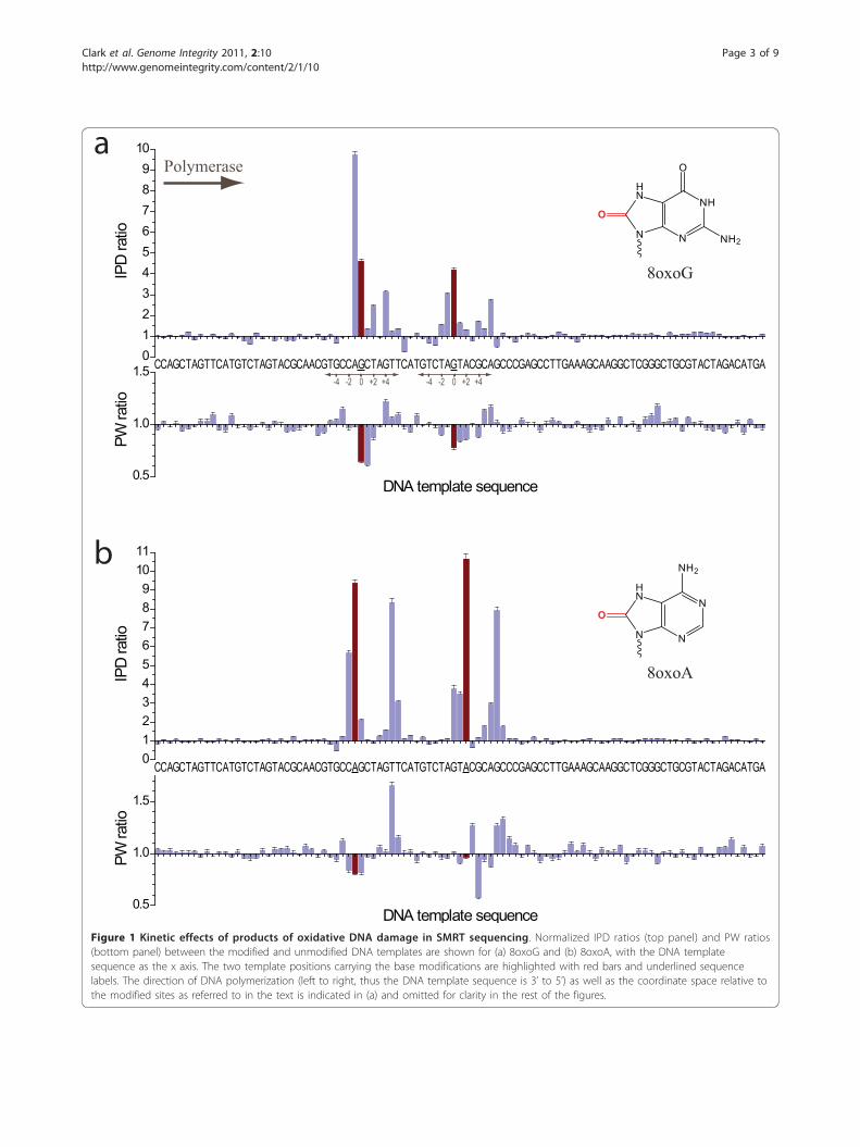
Oxidative DamageProducts of oxidative DNA damage are very common,especially in mitochondrial DNA, and can accumulatewith age, or in conjunction with diseases such as Alzhei-mer’s, Parkinson’s, and cancer [10-12]. We investigatedthe effects of 8-oxoguanine (8oxoG) and 8-oxoadenine(8oxoA) in SMRT DNA sequencing, and both resultedin readily detectable kinetic signatures (Figure 1). Thepresence of 8oxoG caused a ~4fold increase of the IPDat the sites of the base modification (position zero),compared to the unmodified control (Figure 1a). Thiscorresponds to a ~4fold delay of the binding of thephospholinked dCTP nucleotide into the active site
across the 8oxoG modification in the template. Addi-tional characteristic kinetic signals surrounding the siteof the base modification were observed, with a relativelystrong IPD ratio increase for nucleotide incorporationsjust prior to the site of the modification (position -1),and smaller signals up to 6 bases past the modificationsite (positions +1 to +6). For both modification sites, adecrease in the IPD ratio for the +7 position wasobserved, i.e. the polymerase bound the nucleotide fastercompared to the unmodified control. Pulse widths werealso affected by the 8oxoG modifications albeit withsmaller magnitude, with decreased PW values spanningpositions zero to +2 compared to the control. In addi-tion to the kinetic signature, we observed an increasedfrequency of misincorporation of phospholinked dATPopposite the 8oxoG template position, ~5fold highercompared to the control template (data not shown).This is not unexpected since 8oxoG is known to mispairwith adenine [13].The kinetic signature of 8oxoA (Figure 1b) was overall
similar to 8oxoG, but with a stronger, ~10fold increasedIPD at the modification position, and additional signalsone to two bases prior to the modification position. Inthe region five to seven nucleotide incorporations afterthe 8oxoA position, strong additional kinetic signalswere observed, exhibiting similar magnitudes comparedto the modification position IPD ratio. Pulse widthswere slightly increased in this region. The observedkinetic signature for 8oxoA differs from the previouslydescribed kinetic signature for adenine methylation [5];N6-methyladenine (6mA) exhibited kinetic signals at thezero position of ~5fold, i.e. half the signal observed for8oxoA, and a weak signal at the +5 position of ~2fold.These differences can be used to discriminate betweendifferent chemical base modifications that occur on thesame base type.
AlkylationThe transfer of an alkyl group onto a DNA base canoccur naturally for epigenetic modifications, but alsodetrimentally as part of DNA damage, and it is alsoused in chemotherapy to damage the DNA of cancercells (reviewed in [2]). We studied the kinetic effects ofthree examples of alkylated bases in SMRT sequencing:O6-methylguanine (O6mG), 1-methyladenine (1mA),and O4-methylthymine (O4mT) (Figure 2). The pre-sence of O6mG resulted in a very strong kinetic signalat the modification position, with IPD ratios of ~20-40,accompanied by a weaker signal at the -1 position (Fig-ure 2a). Pulse widths were also markedly affected, withdecreased PW ratios at the -1 position and morestrongly at position zero, and an increased PW of ~2-3fold at the +1 position. DNA templates containing1mA displayed IPD ratios of ~5-6 at position zero, a
Clark et al. Genome Integrity 2011, 2:10http://www.genomeintegrity.com/content/2/1/10
Page 2 of 9
CCAGCTAGTTCATGTCTAGTACGCAACGTGCCAGCTAGTTCATGTCTAGTACGCAGCCCGAGCCTTGAAAGCAAGGCTCGGGCTGCGTACTAGACATGA0123456789
1011
IPD
ratio
a
bN
NN
HN
NH2
O
NH
NN
HN
O
NH2
O
8oxoG
Polymerase
8oxoA
0.5
1.0
1.5
PW ra
tio
DNA template sequence
CCAGCTAGTTCATGTCTAGTACGCAACGTGCCAGCTAGTTCAT0 +2 +4-4 -2
GTCTAGTACGCAGCCCGAGCCTTGAAAGCAAGGCTCGGGCTGCGTACTAGACATGA0123456789
10
IPD
ratio
0.5
1.0
1.5
PW ra
tio
DNA template sequence
0 +2 +4-4 -2
Figure 1 Kinetic effects of products of oxidative DNA damage in SMRT sequencing. Normalized IPD ratios (top panel) and PW ratios(bottom panel) between the modified and unmodified DNA templates are shown for (a) 8oxoG and (b) 8oxoA, with the DNA templatesequence as the x axis. The two template positions carrying the base modifications are highlighted with red bars and underlined sequencelabels. The direction of DNA polymerization (left to right, thus the DNA template sequence is 3’ to 5’) as well as the coordinate space relative tothe modified sites as referred to in the text is indicated in (a) and omitted for clarity in the rest of the figures.
Clark et al. Genome Integrity 2011, 2:10http://www.genomeintegrity.com/content/2/1/10
Page 3 of 9

0.5
1.0
1.5
PW ra
tio
DNA template sequence
0.5
1.0
1.5
PW ra
tio
DNA template sequence
CCAGCTAGTTCATGTCTAGTACGCAACGTGCCAGCTAGTTCATGTCTAGTACGCAGCCCGAGCCTTGAAAGCAAGGCTCGGGCTGCGTACTAGACATGA05
10152025303540
IPD
ratio
a
b
c
O6mG
1mA
O4mT
0
1
2
3
PW ra
tio
DNA template sequence
CCAGCTAGTTCATGTCTAGTACGCAACGTGCCAGCTAGTTCATGTCTAGTACGCAGCCCGAGCCTTGAAAGCAAGGCTCGGGCTGCGTACTAGACATGA0
5
10
15
20
25
30
IPD
ratio
CCAGCTAGTTCATGTCTAGTACGCAACGTGCCAGCTAGTTCATGTCTAGTACGCAGCCCGAGCCTTGAAAGCAAGGCTCGGGCTGCGTACTAGACATGA1
2
3
4
5
6
7
IPD
ratio N
NN
N
NH
CH3
N
NN
N
O
NH2
CH3
HN
N
O
O
CH3
Figure 2 Kinetic effects of products of alkylation DNA damage in SMRT sequencing. Normalized IPD ratios (top panel) and PW ratios(bottom panel) between the modified and unmodified DNA templates are shown for (a) O6 mG, (b) 1 mA, and (c) O4 mT, with the DNAtemplate sequence as the x axis. The two template positions carrying the base modifications are highlighted with red bars and underlinedsequence labels.
Clark et al. Genome Integrity 2011, 2:10http://www.genomeintegrity.com/content/2/1/10
Page 4 of 9

smaller signal at the -1 position, and no effects on thePW (Figure 2b). The kinetic effects of 1mA are there-fore similar to those of 6mA [5]. O4mT modifications(Figure 2c) resulted in ~10fold or greater IPD ratios atthe modification position, additional signals two basesup to two bases prior and after the modification posi-tion, and at positions +4 to +6. Pulse widths werereduced at position zero, and elevated at positions +2,+6 and +7.
Ionizing radiationIonizing radiation can confer damage to DNA baseseither through direct effects, or indirectly through thegeneration of free radicals that elicit damage [14]. Pro-ducts of DNA damage resulting from ionizing radiationinclude 5-hydroxycytosine (5hC), 5-hydroxyuracil (5hU),5-hydroxymethyluracil (5hmU). Kinetic effects fromthese base modifications (Figure 3) were also clearlydetectable albeit with smaller magnitudes, presumablydue to the smaller size of these modifications, theiraltered position relative to affecting base pairing duringnucleotide incorporation, smaller geometric distortionsof the nascent double-stranded DNA molecule, and lesscontact of the major groove by the polymerase enzyme[8]. For 5hmU, the magnitudes and phenomenology ofthe kinetic effects were similar to what has previouslybeen observed for the structurally related 5-hydroxy-methylcytosine modification [5,15], with characteristicIPD signals at positions +2 and +6. Interestingly, for5hC there are also two principal delayed locations, butin this case at positions +3 and +7. Thymine glycol,another principal DNA lesion induced by ionizing radia-tion and also oxidation, resulted in read termination, asthe polymerase was not able to advance beyond the siteof the modification (Additional File 2).
UV radiationCyclobutane pyrimidine dimers are the most commonform of DNA damage following UV radiation [1]. Weinvestigated the effect of a thymine dimer in the DNAtemplate during SMRT sequencing. We observed thatthe polymerase was able to bypass this lesion; however,the thymine dimer had dramatic effects on the polymer-ase kinetics with extremely long pauses around the siteof the lesion, in many instances lasting several minutes(Figure 4a). Such severe pausing translated to very largeIPD ratios (Figure 4b). It also affected the ability toaccurately map the sequencing reads in the vicinity ofthe thymine dimer, resulting in reduced sequencing foldcoverage around the position of the modification (Figure4b, inset), and translating to a higher uncertainty inmeasuring IPD ratios near the lesion. The sequencingkinetics were very strongly affected at many positionsalong the region of contact between the thymine dimer
and the polymerase (positions -1 to +8), likely due togeometric distortions imparted by the lesion. Upon pas-sing the lesion, sequencing resumed normally until thenext encounter on the same SMRTbell template mole-cule (Figure 4a).
ConclusionsReplicative DNA polymerases have evolved to movealong DNA templates, synthesizing a complementaryDNA strand, with remarkable efficiency, speed and fide-lity [16]. The presence of various forms of DNA damagein the template strand can lead to transient stalling, mis-incorporation, or even termination of DNA polymeriza-tion, depending on the type of damage [3,10,17]. Takingadvantage of this exquisite sensitivity of polymerasestowards changes in the DNA template, we have investi-gated the ability of SMRT DNA sequencing to detectproducts of DNA damage through an analysis of thepolymerase kinetics that is recorded in real time by thismethod. We have demonstrated the detection of severalcommon forms of DNA damage while performing theregular sequencing protocol, and without the need forspecial upfront sample preparation steps.Damaged DNA bases can impart effects on the poly-
merase kinetics in several ways. IPDs can be affected by(i) changes in the affinity of binding the incomingnucleotide, or (ii) altered DNA translocation rates fol-lowing the phospholinked nucleotide incorporation. Var-iations in PW can be caused from (i) effects on the ratesof conformational changes of the enzyme, as well as (ii)the rate of catalysis during the nucleotide incorporationcycle, as the damaged base can distort active site geome-tries. All of these effects are captured in SMRT sequen-cing through the real-time monitoring of eachnucleotide incorporation event, thereby making themethod sensitive to even extremely small changes torelatively subtle chemical modifications, such as 5hC,5hU or 5hmU. Because of the nature of the SMRTbellDNA template allowing the sequencing of both the for-ward and the reverse strand of the same DNA moleculeduring a SMRT sequencing reaction, products of DNAdamage can be detected in a strand-specific manner,allowing for the differentiation of hemi- and fully-modi-fied positions [15]).Kinetic signals were not limited to just the modified
base position, but were observed over a confined regionsurrounding the base modifications, encompassingapproximately three nucleotide incorporations prior andseven incorporations after the site of the modification,consistent with the ‘footprint’ of the polymerase onDNA [8], and with previously described results for basemethylation [5]. We attribute this effect to the extendedcontact the polymerase makes with the incoming DNAtemplate and the nascent double-stranded DNA over its
Clark et al. Genome Integrity 2011, 2:10http://www.genomeintegrity.com/content/2/1/10
Page 5 of 9

0.5
1.0
1.5
PW ra
tio
DNA template sequence
a
5hC
5hU
5hmU
b
c
GCCAGCTAGTTCATGTCTAGTACGCAACGTGCCAGCTAGTTCATGTCTAGTACGCAGCCCGAGCCTTGAAAGCAAGGCTCGGGCTGCGTACTAGACATGA0.0
0.5
1.0
1.5
2.0
IPD
ratio
0.5
1.0
1.5
PW ra
tio
DNA template sequence
0.5
1.0
1.5
PW ra
tio
DNA template sequence
CCAGCTAGTTCATGTCTAGTACGCAACGTGCCAGCTAGTTCATGTCTAGTACGCAGCCCGAGCCTTGAAAGCAAGGCTCGGGCTGCGTACTAGACATGA0
1
2
3
IPD
ratio
CCAGCTAGTTCATGTCTAGTACGCAACGTGCCAGCTAGTTCATGTCTAGTACGCAGCCCGAGCCTTGAAAGCAAGGCTCGGGCTGCGTACTAGACATGA0
1
2
3
IPD
ratio
N
N
NH2
O
OH
HN
N
O
O
OH
HN
N
O
O
OH
Figure 3 Kinetic effects of products of ionizing radiation DNA damage in SMRT sequencing. Normalized IPD ratios (top panel) and PWratios (bottom panel) between the modified and unmodified DNA templates are shown for (a) 5 hC, (b) 5 hU, and (c) 5 hmU, with the DNAtemplate sequence as the x axis. The two template positions carrying the base modifications are highlighted with red bars and underlinedsequence labels.
Clark et al. Genome Integrity 2011, 2:10http://www.genomeintegrity.com/content/2/1/10
Page 6 of 9

DNA binding region (Additional File 3). For misincor-poration events, the transmission of the presence of themismatch back to the enzyme active site through long-range distortions in the DNA has been described [18].Similarly here, distortions in the DNA geometry by amodified base throughout the extended DNA bindingregion affect the dynamics of nucleotide incorporationsat the polymerase active site, giving rise to additionalsignals that result in a distinct kinetic signature for agiven damaged DNA base. This allows the discrimina-tion of base modifications of different products of DNAdamage when they occur on the same base type -potentially even on the same DNA molecule. For exam-ple, the kinetic signature of 8oxoA was readily distin-guishable from methylated A, and likewise 8oxoG fromO6mG, and O4mT from 5hU and 5hmU. We are in theprocess of incorporating these characteristics into bioin-formatics algorithms for the automated calling ofdamaged DNA bases during SMRT sequencing, as wellas extending these studies to demonstrate sequencing of
modifications that are in very close proximity to eachother on the same DNA strand.Besides the sequencing application, the method opens
potential paths to increase our understanding abouthow different types of DNA polymerases are affected bycertain types of DNA lesions. The SMRT sequencingassay could potentially be adapted to study the detaileddynamics of lesion-specific polymerases at the single-molecule level, and to combine their activities withreplicative polymerases to capture products of DNAdamage which are currently resulting in read termina-tion. We anticipate that SMRT sequencing will becomea powerful, high-throughput tool for the detection andsequencing of DNA containing damaged bases toimprove our understanding of aging, DNA damage-related diseases, DNA polymerase enzymology, DNArepair mechanisms, and chemotherapeutic efficacies.The presented method should also be applicabletowards detecting previously unknown base changes inDNA.
CAACGTGCCAGCTAGTTCATGTCTAGTACGCAG0
20
40
60
80
100
Cove
rage
DNA template sequence
a
bHN
NH
N
NO
O
O
O
H3C
H3C
Thymine dimer
0 60 120 180 240 300 360 420 480 540 6000
20
40
60
80
100 A C G T
Fluo
resc
ence
inte
nsity
(a.u
.)
Time (s)
GCCAGCTAGTTCATGTCTAGTACGCAACGTGCCAGCTAGTTCATGTCTAGTACGCAGCCCGAGCCTTGAAAGCAAGGCTCGGGCTGCGTACTAGACATGA0
20
40
60
80
100
IPD
ratio
DNA template sequenceFigure 4 Kinetic effects of products of UV radiation DNA damage in SMRT sequencing. (a) Example sequencing trace, showing very longpausing by the polymerase upon repeated encounters with a thymine dimer (arrows) on the SMRTbell DNA template. (b) Normalized IPD ratiosbetween the thymine dimer containing and unmodified control DNA templates. The template sequence is given as the x axis. The two templatepositions constituting the thymine dimer are highlighted with red bars and underlined sequence labels. The inset shows the lack of sequencingcoverage after the thymine dimer position because of the extremely long pauses by the polymerase induced by the lesion.
Clark et al. Genome Integrity 2011, 2:10http://www.genomeintegrity.com/content/2/1/10
Page 7 of 9

MethodsCustom oligonucleotides containing modified bases werepurchased from Bio-synthesis (Lewisville, TX), TrilinkBioTechnologies (San Diego, CA), ChemGenes (Wil-mington, MA) and Integrated DNA Technologies (Cor-alville, IA). A list of the sequences can be found inAdditional File 4. All oligonucleotides contained 5’phosphate groups. SMRTbell templates were generatedby ligating several synthetic oligonucleotides, one ofwhich contained two instances of a chemical base modi-fication (Additional File 1). Complementary and hairpinoligonucleotides were annealed by heating to 80°C for 2minutes and slowly cooling to 25°C (0.1°C/sec) in 10mM Tris (pH 7.5), 100 mM NaCl. Annealed oligonu-cleotides were ligated using T4 DNA Ligase (NEB; Ips-wich, MA) for 60 minutes at 25°C followed by heat killfor 10 minutes at 65°C. Incompletely formed SMRTbelltemplates were degraded with a combination of Exonu-clease III (NEB; Ipswich, MA) and Exonuclease VII(USB; Cleveland, OH) at 37°C for 30 minutes. SMRTbelltemplates were purified using QIAquick PCR Purifica-tion columns (Qiagen; Valencia, CA).SMRTbell templates were subjected to standard
SMRT sequencing using an engineered phi29 DNApolymerase, as described [6,7]. All templates were run induplicate on different days, different sequencing instru-ments, and using different reagent lots to verify thereproducibility of the reported results. Reads were pro-cessed and mapped to the respective referencesequences using the BLASR mapper (http://www.pacbio-devnet.com/SMRT-Analysis/Algorithms/BLASR) and thePacific Biosciences SMRT Analysis pipeline (http://www.pacbiodevnet.com/SMRT-Analysis/Software/SMRT-Pipe) using the standard mapping protocol. IPDs weremeasured as previously described [5] for all pulsesaligned to each position in the reference sequence. Base-line correction was applied by dividing the IPD mean foreach position by the average of mean IPDs over all posi-tions in the template, excluding the positions of basemodifications and a window of six bases in each direc-tion around such positions. In addition, 5% of outliervalues were trimmed from both sides of the IPD distri-bution at each position before computing the mean.Thereafter, the ratio of mean IPDs was computedbetween the modified and control template samples foreach template position.The uncertainty in the IPD ratio was quantified by
calculating the standard error of the mean of the IPDratio using the delta method:
SE(µ1
µ2
)≈
√1n
√s12µ2
2
µ14
+s22
µ12
where μ1 and μ2 are the average IPD values of themodified and control, s1 and s2 are their standard devia-tions and n is the lower sequencing coverage of the twosamples.
Additional material
Additional File 1: DNA template constructs used in this study. Thecontrol template (top) contains annotations for the differentoligonucleotides (Additional File 4) that make up the SMRTbell DNAtemplate.
Additional File 2: Read termination induced by thymine glycol. Thefirst instance of thymine glycol in the DNA template at whichsequencing reads end is highlighted with a black bar and underlinedsequence label.
Additional File 3: Animation of the DNA polymerase catalytic cycleduring SMRT sequencing. The movie shows the binding of aphospholinked nucleotide, incorporation, and release of thepyrophosphate-linker-fluorophore reaction product. A hypotheticaldamaged DNA base is highlighted in red, and is moved into the activesite following the phospholinked nucleotide incorporation andpolymerase translocation. The animation highlights the close contact ofthe polymerase over an extended region with the nucleic acid: theincoming DNA template (positions -3 to zero) and nascent double-stranded DNA (positions +1 to approximately +7). The polymerizationdynamics can be altered by the presence of DNA base modificationsthroughout this region. The animation is based on an in-house crystalstructure and pdb structure 2PZS, followed by a brief energyminimization in PyMOL. The dye and linker are modeled solely forindicating their structures and do not reflect their real conformations andpositions.
Additional File 4: Oligonucleotides and primers used to generateSMRTbell DNA templates. All sequences are listed 5’ to 3’. Alloligonucleotides contain 5’ phosphate groups. The primer binding site inthe “Hairpin-Right” sequence is underlined.
List of Abbreviations Used5hC: 5-hydroxycytosine; 5hmU: 5-hydroxymethyluracil; 5hU: 5-hydroxyuracil;1mA: 1-methyladenine; 6mA: N6-methyladenine; 8oxoA: 8-oxoadenine;8oxoG: 8-oxoguanine; BPDE: benzo[a]pyrene diol epoxide; IPD: interpulseduration; O4mT: O4-methylthymine; O6mG: O6-methylguanine; PW: pulsewidth; SMRT: Single-Molecule, Real-Time;
AcknowledgementsThis work was supported in part by National Institutes of Health grant1RC2HG005618-01 (NHGRI). We thank M. Boitano, J. Bullard, K. Luong and S.Kamtekar for help with data acquisition and analysis.
Authors’ contributionsTC conceived of the study, made the SMRTbell templates, and assisted indrafting of the manuscript. KS carried out SMRT sequencing and dataacquisition. ST participated in the design of the study and manuscriptdrafting. JK participated in the design and coordination of the study, madethe figures, and drafted the manuscript. All authors read and approved thefinal manuscript.
Competing interestsThe authors are full-time employees at Pacific Biosciences, a companycommercializing single-molecule, real-time nucleic acid sequencingtechnologies.
Received: 10 November 2011 Accepted: 20 December 2011Published: 20 December 2011
Clark et al. Genome Integrity 2011, 2:10http://www.genomeintegrity.com/content/2/1/10
Page 8 of 9

References1. Geacintov NE, Broyde S: The Chemical Biology of DNA Damage Wiley-VCH
Verlag GmbH & Co. KGaA; 2010.2. Kelley MR: DNA Repair in Cancer Therapy: Molecular Targets and Clinical
Applications Elsevier Science; 2011.3. Preston BD, Albertson TM, Herr AJ: DNA replication fidelity and cancer.
Semin Cancer Biol 2011, 20:281-293.4. Kumari S, Rastogi RP, Singh KL, Singh SP, Sinha RP: DNA Damage:
Detection Strategies. EXCLI J 2008, 7:44-62.5. Flusberg BA, Webster DR, Lee JH, Travers KJ, Olivares EC, Clark TA, Korlach J,
Turner SW: Direct detection of DNA methylation during single-molecule,real-time sequencing. Nat Methods 2010, 7:461-465.
6. Eid J, Fehr A, Gray J, Luong K, Lyle J, Otto G, Peluso P, Rank D, Baybayan P,Bettman B, et al: Real-time DNA sequencing from single polymerasemolecules. Science 2009, 323:133-138.
7. Korlach J, Bjornson KP, Chaudhuri BP, Cicero RL, Flusberg BA, Gray JJ,Holden D, Saxena R, Wegener J, Turner SW: Real-time DNA sequencingfrom single polymerase molecules. Methods Enzymol 2010, 472:431-455.
8. Berman AJ, Kamtekar S, Goodman JL, Lazaro JM, de Vega M, Blanco L,Salas M, Steitz TA: Structures of phi29 DNA polymerase complexed withsubstrate: the mechanism of translocation in B-family polymerases.EMBO J 2007, 26:3494-3505.
9. Travers KJ, Chin CS, Rank DR, Eid JS, Turner SW: A flexible and efficienttemplate format for circular consensus sequencing and SNP detection.Nucleic Acids Res 2010, 38:e159.
10. De Bont R, van Larebeke N: Endogenous DNA damage in humans: areview of quantitative data. Mutagenesis 2004, 19:169-185.
11. Maynard S, Schurman SH, Harboe C, de Souza-Pinto NC, Bohr VA: Baseexcision repair of oxidative DNA damage and association with cancerand aging. Carcinogenesis 2009, 30:2-10.
12. Rao KS: Free radical induced oxidative damage to DNA: relation to brainaging and neurological disorders. Indian J Biochem Biophys 2009, 46:9-15.
13. Wood ML, Dizdaroglu M, Gajewski E, Essigmann JM: Mechanistic studies ofionizing radiation and oxidative mutagenesis: genetic effects of a single8-hydroxyguanine (7-hydro-8-oxoguanine) residue inserted at a uniquesite in a viral genome. Biochemistry 1990, 29:7024-7032.
14. Ward JF: DNA damage produced by ionizing radiation in mammaliancells: identities, mechanisms of formation, and reparability. Prog NucleicAcid Res Mol Biol 1988, 35:95-125.
15. Song XS, Clark TA, Lu XY, Kislyuk A, Dai Q, Turner SW, He C, Korlach J:Sensitive and specific single-molecule sequencing of 5-hydroxymethylcytosine. Nat Methods 2011.
16. Steitz TA: DNA polymerases: structural diversity and commonmechanisms. J Biol Chem 1999, 274:17395-17398.
17. Lindahl T, Barnes DE: Repair of endogenous DNA damage. Cold SpringHarb Symp Quant Biol 2000, 65:127-133.
18. Johnson SJ, Beese LS: Structures of mismatch replication errors observedin a DNA polymerase. Cell 2004, 116:803-816.
doi:10.1186/2041-9414-2-10Cite this article as: Clark et al.: Direct Detection and Sequencing ofDamaged DNA Bases. Genome Integrity 2011 2:10.
Submit your next manuscript to BioMed Centraland take full advantage of:
• Convenient online submission
• Thorough peer review
• No space constraints or color figure charges
• Immediate publication on acceptance
• Inclusion in PubMed, CAS, Scopus and Google Scholar
• Research which is freely available for redistribution
Submit your manuscript at www.biomedcentral.com/submit
Clark et al. Genome Integrity 2011, 2:10http://www.genomeintegrity.com/content/2/1/10
Page 9 of 9