Direct Access Community Ultrasound Service (Non-Obstetric) info/Referrer Information 16pp...
16
Providing services on behalf of the NHS Direct Access Community Ultrasound Service (Non-Obstetric) Referrer Information
-
Upload
dinhnguyet -
Category
Documents
-
view
217 -
download
0
Transcript of Direct Access Community Ultrasound Service (Non-Obstetric) info/Referrer Information 16pp...

Providing services on behalf of the NHS
Direct AccessCommunity Ultrasound Service
(Non-Obstetric)
Referrer Information

The ServicePhysiological Measurements Ltd work in partnership with the NHS to provide patients with access to a greater choice of quality services provided in the local community. Your patients can now benefit from our direct access non-obstetric ultrasound service which is operated in practices within the local area.
The PathwayThe below illustrates the typical patient pathway for our non-obstetric ultrasound service. Any variations to this will be provided to you with this booklet.
Appointment within 3 weeks
Urgent referralRoutine referralAdvice required
Hospital treatment requiredTreatment completeFollow up required
Management by GPGP carries out initial investigation and decides to refer
GP refers via: Choose & Book - Secure email, fax or post
Radiologist auditing Sonographer report written and sent to referring GP
Triage within 1 working day
*Suspected DVT will be seen in accordance with the pathway commissioned under this AQP Non Obstetric Ultrasound Service. Further information is available from our Patient Management Centre
Gynae inc.Trans Abdo
Vascularinc. DVT
Head & Neck Abdo MSK Scrotal inc.
Testes
Renal/Bladder/Prostate

ProtocolsReferrals may be submitted by email to [email protected], fax to 01691 676016 or via e-referrals (formerly Choose & Book). Referral forms must include all patient demographic information and details of the referring GP. Also, please always clearly state the clinical condition you wish to confirm or exclude when making a referral to our service.
It is essential that our ultrasound reports are accurate, concise, informative and provide clear advice to you in the form of a “conclusion”. To enable us to do this, it is essential that you provide as much relevant clinical history as possible.
Please noteFor the report containing patient results to be returned to you by email please supply a current nhs.net email address on the referral form - we are unable to send results to any other email address.
Inappropriate referralsPlease ask yourself the following before submitting a referral - inappropriate submissions will mean a longer pathway and negative experience for your patient:
• Will the result of the test affect clinical management?• Is the test being requested too quickly?• Is the test the most appropriate? • Has the test already been performed elsewhere or in the recent past? If so, the need for repetition should be questioned.
Acceptance and exclusion criteria is supplied as an appendix to this Guide.

How To ReferOur Patient Management Centre manages all referrals to our service. Referrals can be accepted via: • Secure Email to [email protected]• Fax to 01691 676016• E-referrals (formerly Choose & Book)
The Referral Form provided with this booklet should be completed for each patient. Electronic versions of the form can be found on our website (www.physiologicalmeasurements.com) or can be requested from our Patient Management Centre.
If you wish to telephone us before you submit a referral, you can reach the Patient Management Centre on 01691 676496.

The AppointmentThe Ultrasound scan is performed by an appropriately qualified and skilled Sonographer. A Health Care Assistant may work alongside the Sonographer - all staff will wear a Physiological Measurements uniform and will have an identity card.
ReportFollowing the ultrasound investigation, a written clinical report will be produced by the sonographer in accordance with the Red, Amber and Green reporting template (guidelines can be found overleaf) and sent to the referring clinician within two and a maximum of five working days of the investigation, depending on the RAGS result. Advice from a Consultant Radiologist will be available if clinically appropriate.
The report and the RAGS criteria will be produced in accordance with the document “Standards for the Reporting and Interpretation of Imaging investigations” as published by the Royal College of Radiologists.
Return of ResultsThe Patient Management Centre will transmit the report back to the registered practice via secure NHS mail (nhs.net). For those practices that do not have an NHS net address, the report will be sent via Royal Mail.
The patient is informed of the results by their registered practice and managed as appropriate.

Service Results
Mileage
0 No - Routine UltrasoundAQP - NHS General Ultrasound - Diagnostics - XXXXXXXXXX - XXXXX - PML - NXV Diagnostic Imaging
Service Name RestrictedDirectlyBookable
ReferrerAlert
NamedClinician
PrioritiesSupported
Service Type
NoneGroup By:
Specialty Service Provider Location
i
▼
E-Referrals
The service can be located by searching for a Specialty of Diagnostic Imaging and a Clinic Type of Ultrasound.
The service will only offer routine appointments so a Priority of Routine should be selected.
Click ‘Search Primary Care’ and the service will appear as shown in the image below.
Please NoteThe service descriptions of your local PML clinics will be provided to you with this booklet.
Service Results
Mileage
0 No - Routine UltrasoundAQP - NHS General Ultrasound - Diagnostics - XXXXXXXXXX - XXXXX - PML - NXV Diagnostic Imaging
Service Name RestrictedDirectlyBookable
ReferrerAlert
NamedClinician
PrioritiesSupported
Service Type
NoneGroup By:
Specialty Service Provider Location
i
▼

Service Results
Mileage
0 No - Routine UltrasoundAQP - NHS General Ultrasound - Diagnostics - XXXXXXXXXX - XXXXX - PML - NXV Diagnostic Imaging
Service Name RestrictedDirectlyBookable
ReferrerAlert
NamedClinician
PrioritiesSupported
Service Type
NoneGroup By:
Specialty Service Provider Location
i
▼
Service Results
Mileage
0 No - Routine UltrasoundAQP - NHS General Ultrasound - Diagnostics - XXXXXXXXXX - XXXXX - PML - NXV Diagnostic Imaging
Service Name RestrictedDirectlyBookable
ReferrerAlert
NamedClinician
PrioritiesSupported
Service Type
NoneGroup By:
Specialty Service Provider Location
i
▼
The process for referring into the direct access community ultrasound service will be:
1. Locate the service on E-referral’s Primary Care menu2. Create a ‘Request’ to this service by placing a tick in the box to
the left of the service and clicking Request3. Click Submit and then Close4. Print off E-Referral printout5. As per printout, the patient should contact our Patient
Management Centre who will arrange their appointment6. Within 3 days, electronically attach the referral letter within the
E-Referral system.

Clinic locationsWe operate from the following locations in your area. If you have a room available for rent and would be interested in hosting clinics at your own practice please contact us on 01691 676496.


Ultrasound ProtocolsUpper Abdomen Includes visualisation of the liver, biliary system, pancreas, spleen, kidneys, abdominal aorta, associated vasculature.
Clinical indications include: upper abdominal pain (not relieved by PPI), jaundice, radiating to back and right shoulder, possible abdominal aortic aneurysm, unexplained weight loss, fever, night sweats, nausea, persistent vomiting, dyspepsia, abnormal LFT’s, abnormal renal function tests, raised WCC, organomegaly, increased abdominal girth, abdominal free fluid.
Abdomen and PelvisIncludes visualisation of all of the above plus urinary bladder (including a post micturition volume), prostate gland, uterus, ovaries, adnexae.
Clinical indications include: UTI, nocturia/poor urinary stream, prostatism, dysuria/frequency/urgency, possible renal calculi, haematuria, proteinuria, raised serum creatinine, unexplained hypertension, flank pain/colic, FH polycystic kidneys.
ThyroidInvolves visualisation of both lobes, isthmus and parathyroid region including associated vasculature.
Clinical indications: palpable mass, neck swelling, abnormal TFT’s.
TestesClinical indications include: pain/discomfort, swelling/enlargement of scrotum or testis, trauma (resolving haematoma, suspected lipoma), undescended testis, suspected varicocele, suspected hydrocele.
Non-traumatic sudden onset of acute pain should be referred urgently to urology.

Any palpable lump within the testis, particularly in men between 20-50, must be referred urgently to Urology under the 2 week rule.
Soft tissueAny superficial soft tissue lump can be assessed using ultrasound in the first instance.
Musculoskeletal General: any superficial soft tissue masses. Suspected muscle or tendon tear.
Shoulder: pain, restricted movement, acromio-clavicular joint pain.
Hand/wrist: suspected tendon disease or synovitis, non opaque foreign body, suspected glomus tumour.
Elbow : suspected tennis or golfer’s elbow, synovitis, bursitis or ulnar nerve pathology.
Pelvis/hips: trochanteric pain, suspected hernia.
Knee: suspected collateral ligament tear, patellar and quadriceps tendon pathology, bursitis, and Baker’s cyst.
Ankle/foot: suspected tendinosis or synovitis, plantar fascitis, fibromatosis, Morton’s neuroma, foreign body.

Vascular UltrasoundClinical Indications Imaging Guidance
Cerebrovascular disease such as TIACarotid bruit or pulsation
Cardiovascular assessment for stroke risk
A Carotid Doppler study will show evidence of Vascular disease.
Intermittent claudication
Absent ankle or foot pulses
Discolouration and/or leg ulceration
Diabetic neuropathy
Peripheral Arterial Doppler ultrasound is useful to assess the arterial system and to determine the stage of peripheral arterial disease.
Varicose veins as a result of venous insufficiency
Pain in lower limbs
Ulceration or discolouration
Suspected Deep Vein Thrombosis (DVT)
Suspected recurrent DVT
Venous Doppler ultrasound is able to functionally assess the deep and the superficial system to evaluate the cause of venous disorders.
Venous Doppler is helpful to assess deep vein thrombosis (acute and chronic).

7
Transabdominal & Transvaginal Ultrasound
Clinical Indications Imaging Guidance
Palpable abdominal or pelvic mass
Any woman with a palpable abdominal or pelvic mass should have an ultrasound scan.
Transabdominal and transvaginal ultrasound is always available if necessary.
MRI is the best second-line investigation, and will be recommended if necessary.If the scan is suggestive of cancer, an urgent referral will be recommended.
Suspected endometriosis or pelvic inflammatory disease
Ultrasound is a helpful initial investigation.
Dysmenorrhoea/amenorrhoea Ultrasound is a helpful investigation.
Lost intrauterine contraceptive device (IUCD)
Transvaginal ultrasound is recommended to locate this device.
Symptoms of polycystic ovarian syndrome
Ultrasound is an adequate investigation.
Post menopausal bleeding Transvaginal ultrasound is indicated to exclude significant endometrial pathology in postmenopausal bleeding.
A gynaecological referral will be recommended for endometrial thickening > 5mm in postmenopausal women.
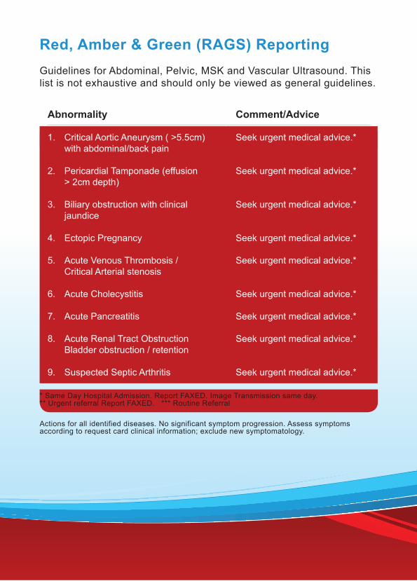
Red, Amber & Green (RAGS) ReportingGuidelines for Abdominal, Pelvic, MSK and Vascular Ultrasound. This list is not exhaustive and should only be viewed as general guidelines.
Abnormality Comment/Advice
1. Critical Aortic Aneurysm ( >5.5cm) with abdominal/back pain
2. Pericardial Tamponade (effusion > 2cm depth)
3. Biliary obstruction with clinical jaundice
4. Ectopic Pregnancy
5. Acute Venous Thrombosis / Critical Arterial stenosis
6. Acute Cholecystitis
7. Acute Pancreatitis
8. Acute Renal Tract Obstruction Bladder obstruction / retention
9. Suspected Septic Arthritis
Seek urgent medical advice.*
Seek urgent medical advice.*
Seek urgent medical advice.*
Seek urgent medical advice.*
Seek urgent medical advice.*
Seek urgent medical advice.*
Seek urgent medical advice.*
Seek urgent medical advice.*
Seek urgent medical advice.*
* Same Day Hospital Admission. Report FAXED. Image Transmission same day. ** Urgent referral Report FAXED. *** Routine Referral
Actions for all identified diseases. No significant symptom progression. Assess symptoms according to request card clinical information; exclude new symptomatology.

Abnormality Comment/Advice
10. Abdominal /Liver mass +/- ascites.
11. Pelvic mass(es) +/- ascites.
12. Chronic Liver Disease with ascites.
13. Aortic Aneurysm > 5.5cm , no evidence of leak.
14. Renal mass
15. Acute Epidydimo-orchitis
16. Abdominal /Pelvic Collection
17. Soft tissue mass/ lymphadenopathy
Seek same day medical advice. **
Seek same day medical advice.**
Seek same day medical advice.**
Seek same day medical advice.**
Seek same day medical advice. **
Seek same day medical advice.**
Seek same day medical advice.**
Seek same day medical advice.**
18. Gallstones/ Chronic Cholecystitis
19. Chronic Liver disease/ Chronic Pancreatitis
20. Renal calculi, no obstruction
21. Benign Uterine/ Pelvic abnormalities
22. Aortic Aneurysm < 5.5cm, no evidence of leak.
Normal Reporting.***
Normal Reporting.***
Normal Reporting.***
Normal Reporting.***
Normal Reporting.***

v.5 21/09/2015