Dimensional changes in the ... - NK Perio &...
123
Dimensional changes in the supporting tissues following immediate placement and restoration of dental implants in the aesthetic zone: a retrospective study Nabil Omar Khzam BDSc School of Dentistry and Oral Health Griffith Health Griffith University Submitted in fulfilment of the requirements of the degree of Master of Philosophy March, 2010
Transcript of Dimensional changes in the ... - NK Perio &...

Dimensional changes in the supporting tissues following
immediate placement and restoration of dental implants in the
aesthetic zone: a retrospective study
Nabil Omar Khzam
BDSc
School of Dentistry and Oral Health
Griffith Health
Griffith University
Submitted in fulfilment of the requirements of the degree of
Master of Philosophy
March, 2010

ABSTRACT
Aim: The objective of this retrospective study was to assess the survival rate and the hard and
soft tissue response following immediate placement and provisional restoration of single-
tooth implants in the aesthetic zone.
Materials and Methods: Thirty-four patients (13 male and 21 female) with 37 immediately
placed and restored implants (Astra-Tech® AB, Mölndal, Sweden) were identified as eligible
to participate in this retrospective study. Thirteen of these patients returned for the follow-up
examination. All participating patients underwent the same treatment strategy which was
removal of the failed tooth, flapless surgery, immediate implant placement and the
connection of a screw-retained temporary restoration. Reasons for tooth loss included failed
root canal treatment, trauma and tooth resorption. Three months following implant
placement, the temporary crowns were replaced by the definitive restorations. Implant
survival rates and hard and soft tissue changes were measured using photographs and peri-
apical x-rays. The range of observation period was between 12 to 27 months with a mean
period of 16 ± 5 months.
Results: At 16 ± 5 months, all implants were present at the time of follow-up with no
complications, resulting in an implant survival rate of 100%. Radiographic evaluation
revealed that there was no statistical difference in bone loss mesially and distally between
baseline and follow-up. Clinical evaluation of the soft tissue revealed no statistically
significant changes in mesial papilla, distal papilla and mid-facial tissue stability throughout
the observation period.

Conclusions: Within the limitation of this retrospective study, immediate implant placement
and provisional restoration in the aesthetic zone of the maxilla can result in acceptable
treatment outcomes as well as stable peri-implant tissues after a follow up period of 16 ± 5
months using the Astra Tech implant system.

This work has not previously been submitted for a degree or diploma in any university. To
the best of my knowledge and belief, the thesis contains no material previously published
or written by another person except where due reference is made in the thesis itself.

Table of contents
LIST OF ILLUSTRATIONS ………………………………………1
LIST OF TABLES…………………………………………………..2
ACKNOWLEDGEMENTS................................................................4
1.0 INTRODUCTION……………………………………………5
1.1 OVERVIEW……………………………………………….....5
1.2 RESEARCH QUESTION……………………………………6
1.3 SIGNIFICANCE OF THE CURRENT STUDY.…..……….6
1.4 OBJECTIVES OF THE CURRENT STUDY………..……..7
2.0 LITERATURE REVIEW…………………………………....8
2.1 TERMINOLOGY…………………….………………………8
2.2 HISTORICAL BACKGROUND OF IMMEDIATE
IMPLANT PLACEMENT AND RESTORATION…….....10
2.3 IMMEDIATELY PLACED AND RESTORED IMPLANT
COMPARED WITH DELAYED IMPLANT
PLACEMENT…......................................................................11
2.4 LIMITATION OF THE IMMEDIATE IMPLANT

PLACEMENT AND RESTORATION STRATEGY……...12
2.5 REVIEW OF THE LITERATURE AND SEARCH
STRATEGY……………………………………………….....14
2.5-1 SURVIVAL RATE OF IMMEDIATELY PLACED AND
RESTORED IMPLANTS IN THE AESTHETIC
ZONE…………………………………………………………18
2.5-2 SOFT TISSUE CHANGES FOLLOWING IMMEDIATE IMPLANT
PLACEMENT AND RESTORATION...................................29
2.5-3 HARD TISSUE CHANGES FOLLOWING IMMEDIATE
IMPLANT PLACEMENT AND RESTORATION……......39
3.0 MATERIALS AND METHODS …………………………...46
3.1 PATIENT SELECTION (THE INCLUSION
CRITERIA……………………………………………………46
3.2 SURGICAL PROTOCOL AND TECHNIQUES…………..47
3.3 TEMPORARY CROWN PLACEMENT…………….…......51
3.4 PERMANENT CROWN PLACEMENT...…………….........53
3.5 HARD TISSUE MEASUREMENTS…………………….….54
3.6 SOFT TISSUE MEASUREMENTS………………………...56
3.7 OTHER MEASUREMENTS………………………………..59
3.7-1 IMPLANT SURVIVAL RATE……………………………...59

3.7-2 ASSESSMENT OF INTERDENTAL PAPILLA…………..60
3.7-3 PLAQUE LEVELS ……………………………………….....60
3.7-4 GINGIVAL TISSUE BIOTYPE……………………………60
3.8 STATISTICS USED IN THE STUDY……………………...60
4.0 RESULTS…………………………………………………….62
4.1 PARTICIPANTS ENROLMENT AND
DEMOGRPHICS......................................................................62
4.2 HARD TISSUE PARAMETERS…………………………....65
4.3 SOFT TISSUE PARAMETERS…………………………….70
4.4 JEMT S’ INDEX……………………………………………..75
4.5 PRESENCE OF PLAQUE.......................................................75
5.0 DISCUSSION…………………………………………………76
5.1 PATIENT ENROLMENT………………………………...…76
5.2 IMPLANT SURVIVAL RATE…………………………...….77
5.3 HARD TISSUE MEASUREMENTS…………………….......79
5.4 SOFT TISSUE MEASUREMENTS……………………........82
5.5 PLAQUE LEVELS……………………………………….......91
5.6 GINGIVAL TISSUE BIOTYPE…………………………….92
5.7 FUTURE CONSIDERATIONS……………………………..92
6.0 CONCLUSIONS………………………………………….......94
7.0 LIST OF REFERENCES…………………………………….95


1
LIST OF ILLUSTRATIONS
Figure 1 Surgical techniques- failed tooth before extraction………………………49
Figure 2 Surgical techniques- tooth extraction…………………………………….50
Figure 3 Surgical techniques- implant secured in place…………………………....50
Figure 4 Temporary crown placement………………………………………………51
Figure 5 Temporary crown placement………………………………………………52
Figure 6 Temporary crown adjustment…………………………………………….52
Figure 7 Permanent crown placement- 3 months postoperatively.....................……53
Figure 8 Permanent crown placement- 6 months postoperatively …….…………..53
Figure 9 Illustration of hard tissue measurements on periapical x-ray…………….55
Figure 10 Illustration of the measurements of the soft tissue on photographs……..58
Figure 11 Level of mid-buccal gingiva at placement of temporary restoration……85
Figure 12 Level of mid-buccal gingival at the follow-up………………………….86
Figure 13 Level of mesial papilla at the placement of the temporary restoration….89
Figure 14 Level of mesial papilla at the follow-up……………………….………..89

2
LIST OF TABLES
Table 1 Selected studies reporting on immediately placed and provisionally restored single
maxillary implants in the aesthetic zone………………………………………………….17
Table 2 Implant survival rates for immediately placed and restored implants in the aesthetic
zone………………………………………………………………………………….........28
Table 3 Reported soft tissue dimensional changes following immediate implant placement
and restoration in the aesthetic zone………………………………………….…..............38
Table 4 Reported hard tissue dimensional changes following immediate implant placement
and restoration in the aesthetic zone……………………………………………..….........45
Table 5 Tooth types and reason for tooth extraction…………………….........................64
Table 6 Overview of clinical data………………………..................................................64
Table 7 Bone level changes (presented as percentage change)……………......................65
Table 8 Frequency analysis of hard tissue changes (mesial measurement in
millimetrs)...........................................................................................................................66
Table 9 Frequency analysis of hard tissue changes (mesial measurement in
percentage)..........................................................................................................................67
Table 10 Frequency analysis of hard tissue changes (distal measurement in
millimetrs)...........................................................................................................................68
Table 11 Frequency analysis of hard tissue changes (distal measurement in
percentage…………………………………………………………………………….......69

3
Table 12 Soft tissue level changes (presented as percentage
change)...............................................................................................................................70
Table 13 Frequency analysis for mesial papilla change in percentage..............................71
Table 14 Frequency analysis for distal papilla change in percentage................................72
Table 15 Frequency analysis for mid-facial change in percentage....................................73
Table 16 Changes in soft tissue levels in millimetres.........................................................74
Table 17 Changes in the interdental papilla.......................................................................75
Table 18 Implant survival rates, as reported in studies using similar treatment
protocol...............................................................................................................................78
Table 19 Changes in hard tissue (mesially and distally) as reported in published
studies.................................................................................................................................81
Table 20 Changes in soft tissue levels (mid-buccal aspect) as reported in published
studies………………………………………………………………………………….....87
Table 21 Changes in soft tissue (mesial and distal papillae) as reported in published studies.
………………………………………………………………………………….…………90

4
ACKNOWLEDGEMENTS
I wish to express my warm appreciation and thanks to:
Prof. Saso Ivanovski, for his continued guidance and commitment toward this
research project
A/P. Nikos Mattheos, for his kind assistance and professionalism in allowing
me to move forward with my research project
A sincere acknowledgment goes to my wife, Dr Sondus Abuoun for her
extraordinary understanding, selfless efforts and genuine friendship
My sponsor, the Libyan embassy in Canberra, with special thanks to Dr. Omran
Zoud and his staff for the enormous support
Sir Geoffrey Cresser for his help in the proof reading and editing of this thesis
Mrs Joy Robertson and Ms Leanne Pockley, for their daily help and assistance
Finally, I would like to dedicate this thesis to my parents

5
1.0 Introduction
1.1 Overview
Dental implants are metal structures inserted into alveolar bone in order to facilitate
the replacement of missing teeth. They are manufactured from titanium, which is highly
biocompatible and has a unique ability to integrate with bone, forming an irreversible bond.
This phenomenon is known as osseointegration. Following implant placement, a period of
time is required for Osseo integration and the implant is subsequently restored with a
prosthesis which has a similar appearance and functionality to natural teeth. The use of dental
prostheses supported by implants has become a widely utilised treatment option for tooth
replacement. This treatment modality has demonstrated favourable outcomes in terms of long
term prosthetic success as well as quality of life outcomes for patients.
Osseointegration was first described by Brånemark [1], who defined it as a direct
connection between living bone and a load-carrying endosseous implant, as observed at the
light microscopic level. Subsequently, osseointegration was defined as a direct structural and
functional connection between living bone and the surface of a load-bearing implant [2].
The original implant placement protocol involved a load free period after implant
placement of 3 and 6 months in the mandible and maxilla respectively [3]. It was postulated
that a load free period would assist in minimizing micromotion, thus reducing the possibility
of fibrous tissue formation or encapsulation and providing an environment conducive to
osseointegration [4].

6
The original ‘Branemark protocol’ also involved placement of the implant following
complete healing of the tooth extraction socket. The implant was then left to heal for six
months prior to prosthesis attachment in order to allow for osseointegration to occur [2].
During this healing period, the implant was submerged under the oral mucosa, necessitating a
second surgical procedure to expose it, which was followed by a period of soft tissue healing
prior to construction of the prosthesis. Although this treatment protocol demonstrated good
survival rates, it was also very lengthy.
With the increased popularity of dental implants, demand has grown for treatment
completion in a shorter period of time compared with the original ‘Branemark protocol’. This
has led to the introduction of new surgical and prosthetic protocols. One such technique
involves the immediate insertion of the implant at the same time as tooth extraction
(immediate implant placement) and subsequent restoration of the implant with a provisional
prosthesis within 24 hours (immediate restoration). This treatment protocol is most likely to
be utilised in the aesthetic zone of the patient’s dentition involving the anterior maxillary
teeth [5].
1.2 Research question
The research questions are:
1) What is the survival rate associated with this clinical procedure?
2) What are the soft and hard tissues dimensional changes associated with
immediately placed and restored implants in the aesthetic zone?
1.3 Significance of the current study

7
The assessment of clinical outcomes associated with immediately placed and restored
implants in the aesthetic zone will provide vital information to the clinical practitioner
regarding this increasingly popular treatment modality.
1.4 Objectives of the current study
The objectives of the current study were to retrospectively assess the survival rate, as
well as soft and hard tissue dimensional changes associated with immediately placed and
restored implants replacing single teeth in the aesthetic zone after a minimum follow up of 12
months.

8
2.0 LITERATURE REVIEW
2.1 Terminology
There has been inconsistency in the use of the key terms “immediate placement”,
“immediate loading” and “immediate restoration” in the literature. Various authors have
defined the terms differently leading to misinterpretation of results and miscommunication of
outcomes. For instance, a number of authors [4] define “immediate loading” as any
restoration that is visible in the oral cavity even if it does not have any occlusal contact with
the opposite dentition. They argue that lip and cheek pressure, as well as tongue movement
and food particles coming into contact with the implant will place a load on the implant, and
hence the definition of immediate implant loading should apply. Furthermore, they consider
that flexure stress and strain occurring in the jaw during opening and closing movement of
the mouth generates functional loads on implants even when they are not in direct occlusal
function [4]. Therefore, due to this discrepancy it is important to outline the terms and
definitions which clearly differentiate between different clinical implant loading and
placement protocols. Cochran et al [6] defined the various loading protocols as follows:
Conventional loading: Defined as the restoration of implants after a healing
period of 3 and 6 months in the mandible and maxilla respectively.
Immediate restorations: Defined as any restoration placed within 48 hours of
implant insertion but with no contact with the opposite dentition in both
centric and eccentric occlusion. The rationale behind the 48 hour window is
not based on any biological consideration but is purely related to the
practicality and logistics associated with the ability to perform the restorative
placement at the same setting as the surgical implant placement (which is not

9
always possible).
Immediate loading: Defined as the placement of the restoration in direct
contact with the occlusal plane of the opposing teeth within 48 hours of
implant insertion.
Early loading: Defined as the placement of a restoration in direct contact with
the opposite dentition more than 48 hours, but less than three months, after
implant insertion.
Delayed loading: Defined as the delivery of the prosthesis some time after the
conventional healing period of three and six months in the mandible and the
maxilla respectively.
Recently, a modification of the above definitions [6] has been recommended [7]:
Conventional loading of dental implants is defined as the restoration of the implant
more than 2 months after implant placement.
Early loading of dental implants is defined as loading between 1 week and 2 months
following implant placement.
Immediate loading is defined as being less than 1 week following implant placement.
A separate definition for delayed loading was no longer considered necessary.
In terms of implant placement protocols, the following classification can be utilised
[8]:
Type 1 or “Immediate implant placement”: The implant is placed immediately
following tooth extraction.

10
Type 2: Implant placement 4 to 8 weeks after tooth extraction to allow
complete soft tissue coverage of the tooth socket.
Type 3: Implant placement 3 to 4 months after tooth extraction to allow
substantial bone fill to occur within the tooth socket.
Type 4: Implant placement more than 4 months after tooth extraction to allow
complete healing of the tooth socket.
This retrospective study focuses on the clinical protocol of immediate implant
placement (type 1) into fresh extraction sockets immediately following tooth extraction, and
immediate provisional restoration of this implant. A follow up period of a minimum of 12
months was chosen as changes in soft and hard tissue dimensions can continue during the
period of wound healing and maturation, thus affecting the aesthetic outcome and level of
patient satisfaction.
2.2 Historical background of immediate implant placement and
restoration
Several investigators have investigated the clinical outcomes resulting from
immediate implant placement and/or restoration of dental implants. The concept of
immediate implant placement (without restoration) was introduced by Lazzara [9] in 1989,
with the rationale of reducing treatment time. Subsequently, Gomez et al [10] in 1997
reported a 98.84% five year success rate in eighty three implants placed immediately after
tooth extraction without immediate restoration.
In terms of immediate restoration of implants, Tarnow et al [11] in 1997 reported a

11
97.1% success rate using a protocol that involved the placement of implants in healed sockets
in both jaws and immediately restoring them without any contact in both centric and eccentric
occlusion. In 1998, Wohrle [12] was the first clinician who used immediate implantation in
fresh extraction sockets in the anterior area of the maxilla and placed a temporary crown
immediately after surgery. The outcome of this study was a 100% success rate in fourteen
patients and acceptable aesthetic outcome was reported.
2.3 Immediately placed and restored implant compared with
delayed implant placement
The traditional implant placement protocol, where the implants are inserted into the
completely healed socket, has shown a high clinical survival rate. However, efforts have now
focused on improving the aesthetic outcome of implant therapy, especially in more
demanding aesthetic circumstances. The immediate implant placement and immediate
restoration treatment protocol has been developed based on the rationale that it preserves both
soft and hard tissue architecture around the immediately installed implant. Hui at el [13]
compared implants placed according to the conventional placement protocol with those
placed into extraction sockets and immediately restored. His study concluded that both
groups showed promising initial results in terms of patient satisfaction and aesthetic outcome.
Another study carried out by Guirado at el [14] revealed that immediate implant placement in
fresh single tooth extraction sites followed by immediate restoration with provisional crowns
had high survival rates comparable to the conventional placement protocol. Conversely,
Chaushu et al [15] showed that immediate placement and restoration of single tooth implants
carried a 20% risk of failure. This outcome may be interpreted as being a result of using
press-fit rather than conventional screw type implants. Most recently, Paolantonio et al [16]

12
showed that immediate implant placement minimized post extraction bone resorption, this
maintaining the hard tissue topography close to its original contour before extraction and had
a positive influence on the soft tissue architecture around the restored implant.
In summary, there are several possible benefits of immediate placement compared
with conventional placement. Firstly, there are fewer surgical procedures resulting in less
patient morbidity. Secondly, soft tissue stability around the implant supported restoration is
improved. Thirdly, time is saved as both post-extraction healing and osseointegration events
take place at the same time.
2.4 Limitation of the immediate implant placement and
restoration strategy
Immediate placement and restoration of implants in the aesthetic regions appears to
offer a success rate that is equal to that associated with conventional treatment. However,
case selection is essential and there are several criteria which need to be considered. These
criteria include:
Morphology and configuration of the tooth socket, soft tissue contour is determined
by the underlying bone. Therefore any bone defects will potentially compromise the aesthetic
outcome of the treatment [17], [18], [19], [20], [21] and [22].
Gingival tissue configuration, the gingiva should be of healthy and symmetrical
harmony to allow an aesthetic outcome. Any gingival disease or defect would compromise
the final outcome

13
Gingival tissue biotype, in general, a thick gingival tissue biotype is preferable in the
aesthetic zone as it can mask the metallic hue of the implant neck, as well as being more
resistant to recession. In contrast, the thin gingival tissue biotype is more prone to recession
and tissue discoloration. In these situations, surgical and prosthetic planning before implant
placement is very critical, and the implant may be placed more palatally in order to have a
thicker buccal volume of hard and soft tissue in order to reduce the problem of metal showing
through the tissues [23], [24], [25] and [26].
Smile line, patients with a high smile line who routinely show their gingival margin
are a greater aesthetic risk than patients who don’t show their smile line during speech and
laughter.
Presence of pathology/infection, poor oral hygiene and microbial plaque are causative
factors that result in the occurrence of peri-implant infection and implant loss. Therefore,
evaluation of the oral hygiene of the patient is a mandatory step before commencing
immediate implant placement and restoration [27] and [28].
Smoking, studies show that smoking has a deleterious effect on implant integration in
the short term and on the peri-implant tissue health in the long term. These effects may be
exacerbated in an immediate implant placement and restoration treatment protocol [29], [30],
[31], [32], [33] and [34].

14
2.5 Review of the literature and search strategy
The following criteria were considered in the process of selecting studies of immediate
implant placement and restoration for this review:
1. Types of studies: all longitudinal studies were eligible for inclusion – randomized
controlled trials, controlled clinical trials, cohort studies, case control studies and
consecutive case report series. Only those studies that included 6 patients or more
were selected. A minimum follow up time of 1 year was set as an inclusion criterion.
Only studies published in English were included.
2. Type of treatment modality (intervention): immediate implantation into the fresh
extraction socket followed an immediate placement of a provisional restoration in the
aesthetic zone.
3. Outcomes that were measured:
Implant survival rate.
Patient satisfaction.
Peri implant soft tissue changes.
Peri implant hard tissue changes.
Gingival biotype and its relation to recession.
For this review, a detailed search strategy was used for each selected database in order to
identify all of the articles published in relation to the stated aims of this review. The search
strategy used was a combination of free text terms and MeSH* terms. The searched data
bases were:

15
# PUB MED, EBSCOhost and Ovid arms of MEDLINE.
# CENTRAL (The Cochrane Central Registar of Controlled Trials).
# Science Direct.
The terms used in this search were:
Dental Implants, Oral Implants, immediate placement, immediate restoration, Immediate
Provisionalization, aesthetic, single tooth replacement, single tooth in the maxilla and
extraction socket.
The search strategy was as follows:
(Single Tooth* OR teeth*) AND Maxilla* AND (Immediate OR Immediate placement, OR
Immediate Implantation, OR immediate restoration, OR extraction socket)
Single Tooth* AND Maxilla* AND
Single Tooth* AND maxilla * AND
Single Tooth* AND Maxilla* AND (OR provisionalization)
Single Tooth* AND Maxilla* AND
Furthermore, the search was complemented by checking the references of the selected
articles for additional useful publications. Also a manual search was carried out of the
following major journals in dental implantology: International Journal of Oral and
Maxillofacial Implants, Clinical Oral Implants Research, Clinical Implant Dentistry and
Related Research.
The search strategy initially yielded 80 articles. From these, 17 articles were considered

16
to fulfil the criteria for inclusion in this review. The other studies were excluded for the
following reasons:
1) Review articles (10 articles): [35], [36], [37], [38], [39], [40], [41], [42], [43] and
[44].
2) Implantation into healed sockets only (30 articles ): [45], [46], [47], [48], [49], [50],
[51], [52], [53], [54], [55], [56], [57], [58], [59], [60], [61], [62], [63], [64], [65] ,
[66], [67] , [68], [69], [70], [71], [72], [73] and [74].
3) Implantation in the mandible or maxilla without any differentiation (7 articles): [45],
[46], [52], [56], [59], [74] and [75].
4) Case reports of immediately placed and loaded implants (16 articles): [45], [47], [50],
[52], [53], [59], [76], [77], [78], [79], [80], [81], [82], [83], [84], and [85].
5) The 17 included articles, alongside a description of the study type, implant system
used and the numbers of included patients/implants are outlined in Table 1.

17
*Delayed implant placement.
RCT, randomized controlled trial; CS, case series.
Table 1 Selected studies reporting on immediately placed and provisionally restored
single maxillary implants in the aesthetic zone.
Study Design No. of
Patient/implant
Implant systems
Wohrle 12
(1998)
CS 14/ 14 Replace Select
Hui et al 13
(2001) CS 24/ 24 = 13 & 11* Brånemark
Guirado et al 14
(2002) CS 13/ 18 = 9& 9* Osseotite 3i
Chaushu et al 15
(2001) CS 26/28= 14 & 8* 21 Steri-Oss & 7 Alpha Bio
Groisman et al 86
(2003) CS 92/ 92 Replace select
Kan et al 87
(2003) CS 35/ 35 Replace Select
Lorenzoni et al 88
(2003) CS 9/ 12 =8 & 4* Frialit-2
Norton 89
(2004) CS 25/ 28 =16 & 12* Astra Tech
Tsirlis 90
(2005) CS 38/ 43 =28 & 15* Osseotite 3i
Cornelini et al 91
(2005) CS 22/ 22 ITI
Ferrarra et al 92
(2006) CS 33/ 33 Frialit-2
Kan et al 93
(2007) CS 29/ 38 = 23 &15* Replace Select
Canullo et al 94
(2007) CS 9/ 10 Defcon®
Hall et al 95
(2007) RCT 28/ 28 =14& 14* Southern Implants
Palattella et al 96
(2008) CS 16/ 18 = 9& 9* Tapered effect
De Rouck et al 97
(2008) CS 30/ 30 Replace select
Ribeiro et al 98
(2008) CS 64/82= 46 & 36* Connexao sistema de protese

18
2.5-1 Survival rate of immediately placed and restored implants
in the aesthetic zone
The term ‘implant survival rate’ is defined as the percentage of implants that present
at follow up [99], although it is important to note that the status of the implant not specified
[100]. Conversely, implant success is defined as the presence of the implant at the end of an
observation period, along with the absence of progressive bone loss, radiolucency, mobility
(clinically), pain, discomfort and/or neuro-sensory changes [101].
All studies selected for this review reported on the survival rates of immediate
implant placement and restoration in the aesthetic zone. A review of these studies in relation
to the implant survival outcome is presented in this part of the review.
The primary goal of Wohrle‘s (1988) investigation was to predictably maintain soft
tissue morphology in the aesthetic zone of the maxilla and avoid postextraction complications
related to hard tissue resorption and soft tissue recession. Fourteen implants were placed in
fourteen consecutive patients (14/14). Five of the implants were placed in the lateral incisor
position and the other nine were placed in the central incisor position. The implant system
used was Replace (Steri-Oss, Yorba Linda, CA, USA) and both screw-type cylindrical and
screw-type tapered implants were used. All surgical and prosthetic procedures were carried
out by the same clinician. None of the implants were lost with an overall survival rate of
100% in a follow-up period ranging from six to 36 months [12]. In 2001, Hui et al [13]
proposed immediate placement and restoration as a treatment modality aimed at provide an
immediate and cost effective solution for restoring a single missing tooth in the maxillary
aesthetic region. This prospective clinical investigation included twenty-four participants,

19
thirteen of which had immediate implant placement and restoration while eleven received
implants in healed sites. All of the implants were placed in the maxillary aesthetic region.
The overall follow-up period ranged from one to 15 months. According to the authors, the
desirable goals of patient satisfaction, good aesthetic outcome and reduced treatment cost
were achieved in this treatment protocol. The implant system used was Brånemark (Nobel
Biocare AB, Göteborg, Sweden), and the implant types were both screw-type tapered and
cylindrical. The implant survival rate of this study was 100% within the reported one to 15
months period of follow-up for both groups.
Guirado et al [14] conducted a prospective study involving eighteen implants in thirteen
patients using the Osseotite system (3i, Implant Innovation, USA). All of the implants were
placed in the maxillary aesthetic zone with nine placed into fresh extraction sockets and nine
into healed sites. The observation period was one year and a 100% implant survival rate was
reported. Advantages associated with the one stage protocol included immediate aesthetics,
comfort and no need for surgical re-entry. Furthermore, the interdental papilla adjacent to the
implants were preserved leading to optimal aesthetic results. The author concluded that the
placement of implants immediately into fresh extraction sockets was a viable and predictable
treatment alternative associated with a high survival rate.
Chaushu et al [15] hypothesised that the immediate restoration of implants replacing
single missing teeth could be successfully achieved following immediate implant placement
into fresh extraction sockets, as well as healed sockets. This study included twenty-six
consecutive patients who received twenty-eight implants. Nineteen implants were placed into
fresh extraction sites, and nine were placed into healed sites. The two implant systems used
in this study were Steri-Oss, (Yorba Linda, CA, USA), (21 implants) and Alpha Bio hydroxy-

20
apatite coated cylindrical implants, (7 implants). The follow-up period ranged from six to 18
months, with a mean of 13 months for the immediately placed implants and 16.4 months for
implants placed into healed sites. There were three failures among the immediately placed
and restored implants, all occurring during the first month following implantation, resulting
in an overall survival rate of 82.4% for this group. The patients who lost their implants were
over 50 years of age. In each of these patients there was initial discomfort, followed by
moderate pain and implant mobility. Another two patients experienced swelling with a
purulent exudate. On the other hand, all of the non-immediate implants survived the healing
period without any loss leading to a 100% short term survival rate for this group. All
surviving implants of both groups were free from any complications. The results of this study
revealed a 100% success rate in the healed sites but about a 20% failure rate associated with
immediate placement into extraction sites. The use of a press fit cylindrical implant type may
explain in part the low success rate in the immediate group.
In another prospective clinical study carried out by Kan et al [87] the implant survival
rate, peri-implant tissue response, aesthetic outcomes and patient satisfaction were evaluated.
This study included thirty-five patients with a mean age of 36.5 years, and each patient
received a single flat platform, screw type tapered implant (Replace, Nobel Biocare, Yorba
Linda, CA, USA). All of the implants were placed into fresh extraction sockets. The implant
survival rate was 100% after a follow-up period of one year. All of the patients were satisfied
with the aesthetic outcome of their restorations. The author concluded that a favourable
implant success rate, peri-implant tissue response and aesthetic outcome can be achieved with
immediately restored single implants placed in the maxillary aesthetic zone. In a more recent
investigation with similar aims, the same author conducted another study, but instead of using
flat platform implants, a scalloped implant platform design was used [93]. The introduction

21
of this new platform design aimed to replicate the irregular bony topography which results
after tooth extraction, thus preventing future bone loss. The implant system used was (Nobel
perfect; Nobel Biocare, USA). Thirty-eight implants were placed in twenty-nine patients with
a mean age of 45.1 years. Fifteen implants were placed into healed sites while the other
twenty-three were placed into fresh extraction sites. At the one year follow-up all implants
remained in function with an overall survival rate of 100%.
The stated purpose of Groisman‘s study [86] was to evaluate the survival rate of ninety-
two tapered implants which were immediately placed and restored in the maxillary aesthetic
region. The diameter of the inserted implants was selected based upon the size of the tooth
sockets (Nobel Biocare, Yorba Linda, CA, USA). The observation period was two years, but
only ten implants were followed up for the full 24 months. At the conclusion of the follow up
period, 6 implants had been lost resulting in an overall implant survival rate of 93.5%.
According to the author, one implant was lost due to trauma and two others due to overload
in patients with a deep overbite. The cause of failure for the other three implants was not
described. The study concluded that immediately placed and restored tapered implants did
not show any adverse effects with regards to osseointegration. Favourable aesthetic outcome
was achieved in eighty-two of 92 cases, representing 89% of the total number.
Lorenzoni et al [88] evaluated the clinical outcomes of immediately placed and restored
stepped-screw type grit-blasted, acid etched FRIALIT-2 Synchro implants one year after
placement in the maxillary anterior region. In the course of this study, nine patients received
12 implants; eight of which were placed into fresh extraction sites and four were placed into
healed sites. All implants were immediately restored with acrylic resin provisional crowns,

22
and patients provided with occlusal splints so that the restorations were free from any direct
loading. No implants failed in the 12 months after insertion, resulting in a 100% survival rate.
Norton [89] evaluated the clinical outcome of single-tooth immediately restored implants
placed in the upper arch. Twenty-five consecutive patients with a mean age of 48.2 years
received twenty-eight Astra Tech ST implants (Lexington, MA). Sixteen of the implants were
placed into fresh extraction sockets while the rest were placed into healed sites. The follow-
up period ranged from twelve to thirty months. All patients received friction-fit temporary
crowns instead of cemented crowns. After a mean duration of four and half months following
the surgical procedure, the permanent crowns were placed. Implant survival, along with hard
and soft tissue changes, was recorded at follow up with an overall survival rate of 96.4%.
One patient, a heavy smoker, lost one implant within one month of surgical placement.
Furthermore, unfavourable soft tissue recession was associated with one implant. However,
most of the restorations maintained an aesthetic gingival contour and architecture. Eleven of
the 28 provisional restorations needed further treatment; six required replacement during the
temporization period and five required re-cementation after becoming loose. The study
concluded that immediate temporization of maxillary single-tooth implants could be both safe
and predictable and the procedure appeared to yield favourable soft tissue aesthetic outcomes.
The authors concluded that this treatment protocol utilizing the Astra Tech ST implant system
resulted in predictable outcomes following immediate implant placement and restoration with
provisional acrylic resin crowns.
In a study using two different implant systems, NT Osseotite (3i Implant Innovations Inc)
implants and Frialit-2 (Friatec AG, Mannheim- Germany), forty-three single implants were
inserted in thirty-eight patients. A survival rate of 100% was reported following an

23
observation period of 24 months. The patients were divided into two groups, immediate and
delayed implant installation. The first group received twenty-eight immediately placed
implants while the rest of the patients received delayed implant placement [90].
In another study using the Straumann TE implant system (Institute Straumann AG,
Waldenburg, Switzerland), twenty-two single implants were immediately restored and
followed up for one year. Three of these implants were placed into healed sockets while the
rest were placed immediately into fresh extraction sockets [91]. The temporary crowns were
completely out of occlusion in both centric and eccentric positions. Screw-retained temporary
crowns were constructed to avoid the use of adhesive cements which may interfere with the
healing process during osseointegration [16]. Nineteen of the implants were placed in the
maxilla and three in the mandible. Premolars were the most common teeth to be replaced (13
teeth), followed by central incisors (6 teeth) and lateral incisors (3 teeth). The study reported
a survival rate of 100%. All the implants were successful according to the criteria of Smith
and Zarb [101]. Within the limits of this investigation, immediate restoration of single-tooth
implants placed in fresh extraction sockets was considered to be an acceptable option.
In a study conducted by Ferrara et al [92], thirty-three consecutive patients with a mean
age of 41 years received a single implant supported crown to replace a missing maxillary
tooth at the time of tooth extraction. The implant system used was Frialit-2 (Friatec AG,
Mannheim- Germany). Thirteen central incisors, nine lateral incisors, four canines and seven
first premolars were included. The follow-up period ranged from one to four years. There
were two implant failures resulting in an overall success rate of 93.9%. One implant did not
integrate while another one became unstable as a result of trauma.

24
Canullo and Rasperini [94] investigated immediately placed and restored implants using
the TSATM series 5 Defcon® Impladent (Barcelona, Spain) system. A platform switching
design, whereby the trans-mucosal abutment was narrower than the implant platform, was
used in order to maintain the surrounding peri-implant tissue dimensions. Ten 6 mm diameter
implants were immediately placed into fresh extraction sockets in the aesthetic zone of the
maxilla. A provisional 4 mm diameter trans-mucosal abutment was subsequently connected to
the implant body, and a provisional crown was adapted and adjusted for non-occlusal contact
in centric as well as eccentric positions. The definitive restoration was completed three
months following implant placement. Nine patients with 10 sites were treated and the follow-
up period was 22 months. All 10 implants were found to be clinically osseointegrated with a
100% survival rate.
Hall et al [95] evaluated the use of immediately placed and temporized tapered implants
(Southern Implants Ltd, Irene, South Africa) to replace single teeth in the anterior maxilla.
The participants’ mean ages were 43.25 years and the implants were followed up for one year.
The patients were randomly divided into conventional (control group = 14 patients) and
immediate restoration groups (test group = 14 patients). The test implants received
provisional screw-retained crowns within four hours of implant placement, while in the
conventional restoration group; temporary crowns were placed after 26 weeks. In the
immediate placement/restoration group, one implant was lost for an overall survival rate of
93%, while in the control group two implants were lost at the one year follow-up. This
investigation concluded that the immediate placement and restoration protocol used in this
study resulted in similar outcomes as conventionally restored implants.
The main aim of Palattella‘s study [96] was to compare the immediate restoration of

25
implants placed using an immediate and a delayed placement protocol. Sixteen patients with
a mean age of 35 years were treated for single-tooth substitution in the anterior maxilla. The
patient population was randomly divided into two groups. In the first group (test group), eight
patients received nine implants placed and restored at the time of tooth extraction. The second
group (control group) of eight patients received nine implants placed eight weeks after tooth
removal. All implants underwent immediate restoration. All patients received the same
implant system in the form of tapered effect (TE) Straumann implants (Institute Straumann
AG, Waldenburg, Switzerland). Marginal bone resorption, papilla index and the position of
mucosal margin were assessed at the time of provisional restoration fabrication (within 48
hours after implant placement) and at a two year follow-up visit. No implants were lost,
resulting in a 100% survival rate for both groups after twenty-four months. The results
suggest that immediate implant placement and restoration without functional loading may be
considered a valuable therapeutic option for selected cases of single-tooth replacement in the
aesthetic area.
A recent study conducted by De Rouck [97] also evaluated implant survival rates, soft
and hard tissue changes and patient satisfaction in relation to immediately placed and restored
implants in the anterior maxilla. Thirty consecutive patients underwent the same treatment
protocol which consisted of flap elevation followed by immediate implant placement and
connection of a screw-retained provisional restoration. The implant system used was Nobel
Replace Tapered (Nobel Biocare, Goteborg, Sweden). Clinical and radiographic assessments
were carried out at 1, 3, 6 and 12 monthly intervals. The results revealed that one implant
failed in the first month of follow up resulting in a survival rate of 97%. It was concluded that
this particular protocol can be considered to be a valuable treatment modality in carefully
selected patients.

26
Ribeiro et al [98] compared immediately placed implants with those placed into healed
sockets. Eighty-two implants were placed in the maxilla of forty-six patients, with forty-six
implants inserted using the immediate placement protocol while the other thirty-six were
inserted using the delayed placement protocol. The implant system used for this investigation
was Conexao Sistema (de Protese Ltda, Sao Paulo, SP, Brazil). Success of implant integration
was assessed according to the criteria described by Albrektsson [102]. The follow-up period
ranged from 18 to 39.7 months. Three of the implants from the immediate placement group
failed, resulting in a survival rate of a 93.5%. The delayed placement group had an overall
success rate of 100 %. The differences in survival rate between the two groups were not
statistically significant [98].
Summary and conclusion:
Table 2 shows the results of studies which investigated the survival rate of
immediately placed and restored implants in the aesthetic zone. An implant survival rate of
100% was described in all except five studies, namely, Chaushu et al [15] achieved
osseointegration in 78.6% of the cases while Groisman [86] achieved 93.5% and Ferrara et al
[92] reported 93.9%. In two other recent studies, the reported implant survival rates were
93% and 97% respectively [95, 97]. Therefore, the majority of the studies reported that the
implant survival rate following immediate placement and restoration is comparable to that
achieved using conventional therapy.
Based on the results of studies carried out over relatively short time periods, the
replacement of single teeth in the maxillary aesthetic region can be predictably achieved
using an immediate implant placement and restoration protocol. However, studies with a

27
longer follow up are needed to further document survival outcomes of this treatment
modality.

28
*Delayed implant placement.
RCT, randomized controlled trial; CS, case series.
Table 2 Implant survival rates for immediately placed and restored implants in the
aesthetic zone.
Study Design No. of
Patient/implant
Follow up
period
(months)
Implant type No. of implant
lost
Implant
survival rate %
WÖhrle 12
(1998)
CS 14/ 14 9- 36 Screw
Cylindrical
tapered
0 100
Hui et al 13
(2001)
CS 24/ 24 = 13 & 11* 1 – 15 Screw
Cylindrical
tapered
0 100
Guirado et al 14
(2002)
CS 13/ 18 = 9& 9* 12 Screw
cylindrical
0 100
Chaushu et al 15
(2001)
CS 26/28= 14 & 8* 6 -18 Press fit
cylindrical
3 78.6
Groisman et al 86
(2003)
CS 92/ 92 6 -24 Screw tapered 6 93.5
Kan et al 87
(2003)
CS 35/ 35 12 Screw tapered 0 100
Lorenzoni et al 88
(2003)
CS 9/ 12 =8 & 4* 12 – 14 Stepped screw-
tapered
0 100
Norton89
(2004) CS 25/ 28 =16 & 12* 13- 30 Screw
cylindrical
0 100
Tsirlis 90
(2005) CS 38/ 43 =28 & 15* 24 Screw tapered 0 100
Cornelini et al 91
(2005)
CS 22/ 22 12 Screw tapered 0 100
Ferrara et al 92
(2006)
CS 33/ 33 12 - 52 Stepped screw
tapered
2 93.9
Kan et al 93
(2007)
CS 29/ 38 = 23 &15* 12 Screw tapered
with scalloped
platform
0 100
Canullo et al 94
(2007)
CS 9/ 10 12 -36 Screw tapered
with 6 mm
platform
0 100
Hall et al 95
(2007)
RCT 28/ 28 =14& 14* 12 Screw tapered 1 93
Palattella et al 96
(2008)
CS 16/ 18 = 9& 9* 24 Screw tapered 0 100
De Rouck et al 97
(2008)
CS 30/ 30 12 Screw tapered 1 97
Ribeiro et al 98
CS 64/82 = 46 & 36* 18 – 39.7 Root form 3 96.3

29
2.5-2 Soft tissue changes following immediate implant placement
and restoration
Gingiva can be defined as that part of the oral mucosa immediately surrounding the
erupted tooth. It can also be defined as that part of the masticatory mucosa which covers the
alveolar process and surrounds the cervical portion of the teeth. It consists of an underlying
connective tissue layer (lamina propria) covered by an epithelium layer. It has several roles
including protection of the underlying periodontal structures which support the tooth,
attachment of the junctional epithelium to the tooth and clearance of food during mastication
[103].
The gingiva consists of two functional portions, the keratinized masticatory
component which faces the oral cavity and the non-keratinized area facing the tooth, which is
involved in the attachment of the gingiva to the tooth.
Anatomically, the normal mucosa surrounding the tooth is subdivided into three main
components:
The marginal gingiva or free gingiva is pink in colour and has a firm consistency. It
forms the unattached part of the gingiva. It extends from the top of the gingival margin to the
free gingival groove which is localized at the level of the cemento-enamel junction (CEJ).
The free gingival groove separates the free gingiva from the attached gingiva. The free
gingiva is separated from the tooth via gingival sulcus or crevice [103].

30
The interdental papilla is that part of the gingiva which occupies the region between
adjacent teeth or implants. It has different shapes and forms on anterior compared to posterior
teeth. Its shape is determined mainly by the contact point of the adjacent teeth, the width of
the proximal surface of the teeth and the position of the cemento-enamel junction. In the
anterior region of the dentition it is pyramidal in shape, while in the posterior region it is tent-
shaped with wider extension in the bucco-lingual direction. This is a result of the presence of
contact areas rather than contact points in the premolar-molar region [103].
The attached gingiva extends from the free gingival groove to the mucogingival line.
The mucogingival line separates the attached gingiva from the alveolar mucosa. The attached
gingiva is firmly attached to the underlying periostum of the alveolus [103]. It is firm in
texture, coral pink in colour and has small depressions on the surface called stippling. There
is no mucogingival line present on the palate, as the entire hard palate is covered by attached
masticatory mucosa.
The peri-implant mucosa is the soft tissue which immediately surrounds the dental
implant. Following the placement of a transmucosal healing abutment, a soft tissue seal
begins to form around the implant. This soft tissue seal will act as a barrier which protects the
underlying structures, thus supporting the establishment and maintenance of osteointegration
[103].
The appearance and dimensions of the buccal soft connective tissue depends mainly
on the anatomy of the underlying bony tissue. Indeed, the final architecture and form of
buccal tissue is determined by the position and inclination of the fully erupted teeth [104]. A
study by Ochenbein et al [105] showed that the anatomy of the gingiva is related mainly to

31
the shape of the alveolar bone crest. It was suggested that two types of gingival biotype exist,
namely a pronounced scalloped (thin) and a flat (thick) biotype. Individuals with the thin
biotype have delicate cervical convexity and a small interdental contact area which is located
very close to the incisal edge, while the crown form of the associated teeth is slender and
tapered. The incisor teeth surrounded by this type of biotype have a thin free gingiva and the
outline is highly scalloped [106]. Furthermore, the papillae associated with a thin biotype are
long and narrow. In contrast, the flat gingival biotype is associated with square crowns with
prominent cervical convexity. The contact area of the thick biotype is boarder and located
more apically resulting in short interdental papillae.
A study measuring the thickness of gingiva using bone sounding technique [87]
reported that the thickness of gingiva varied between subjects of different gingival biotypes.
This study concluded that the thick gingival biotype has more soft tissue volume than the thin
biotype on the buccal and interdental aspects.
The presence or absence of the interdental papilla may be assessed visually. If there is
a space apical to the contact area, which is characterised by a black triangle, the papillae are
considered incomplete. On the other hand, if there is no space apical to the contact area, the
papillae are considered complete. Tarnow et al [107] measured the distance between the
interdental contact point and the crest of the interdental alveolar bone in order to determine if
there is a relationship between this dimension and the height of the interdental papilla. The
results of this study showed that the papilla was always complete when the distance from the
contact area to the crest of interproximal bone was less than or equal to 5 mm. However,
when this distance was more than 5 mm, about half of the cases had incomplete papilla fill.

32
There must be gentle handling of the peri-implant soft tissues during surgical implant
placement due to their delicate nature. Any traumatic manipulation of these areas may
compromise the aesthetic outcome. The peri-implant soft tissue should be in complete
harmony with that of the surrounding tissues in terms of colour, form, shape and contour,
resulting in restorations that mimic the lost dentition. In recent years, the success of dental
implant restoration is no longer judged solely by successful osseointegration, with aesthetic
outcome become of increasingly importance. This is particularly the case when implants are
placed in the aesthetic zone, and this represents a challenge to both surgeons and restorative
dentists. The peri-implant soft tissue architecture is one of the most important factors in
determining the aesthetic outcome of implants placed in the aesthetic zone, especially in
patients with a high lip line.
This review identifies and discusses studies which measured the soft tissue outcomes of
immediately placed and restored implants in the anterior maxilla. The pioneering study of
Wohrle [12] assessed soft tissue changes in 14 consecutive cases involving immediate
implant placement and restoration during follow up period ranging from 9 to 36 months. The
outcome of this study revealed that only two cases showed facial recession of 2 mm or more,
with the remaining cases demonstrating stable soft tissue outcomes.
In the study by Cornelini et al [91] measurements of the soft tissue were taken at the time
of implant placement and after one year of follow up. The soft tissue parameters measured
included a mucositis score, mucosal margin level, variation of gingival level and variation of
papillary position. According to Jemt‘s index, no scores of 0, 1 or 4 were found [108].
Twenty-seven of the papillae received a score of two which meant that at least half of the
height of interdental papillae was present. The other seventeen papillae presented with a score

33
of three which meant that the interdental papillae filled the entire interproximal space. In
addition, the mid-facial soft tissue level was compared at baseline and at follow up using the
gingiva of the adjacent teeth as a reference line. An average recession of approximately 0.75
mm was noted at follow up. The soft tissue condition (mucositis score) was determined using
a modification of the criteria of the gingival index system described by Bengazi et al [109],
whereby a score of zero denoted no colour or texture alterations, a score of one revealed a
slight change in the colour and texture and a score of two indicated that there was a marked
change in the colour and texture plus mucosal bleeding was evident following probing. A
score of two was not observed in this study which indicated the absence of inflammation
[91].
Kan et al [87] also evaluated the soft tissue response and aesthetic outcomes of
immediately placed and restored single implants placed in the anterior maxilla. The surgical
phase was conducted without flap reflection in an attempt to make the procedure as
atraumatic as possible. The rationale for flapless surgery was to maintain continuous blood
flow to the labial soft tissues and hence prevent recession. However, other authors [97] have
argued that complete flap elevation results in similar soft tissue outcomes as those reported
by Kan et al [87], and therefore flapless access does not improve aesthetic outcomes. In Kan
et al’s study [87], the cervical gingival emergence of the extracted tooth was reproduced in
the temporary crown in order to optimise soft tissue adaptation. Soft tissue parameters,
namely the mid-facial gingival position and the height of the papillae, were measured at
baseline, 3, 6 and 12 months after implant placement. The soft tissue measurements were
obtained using 35 mm slides taken at 1:1 magnification, and the line connecting the mid-
facial level of the two adjacent teeth was used as a reference. Changes in the mid-facial tissue
level associated with the implant restorations were assessed by measuring the distance from

34
the reference line at each follow up visit. Similarly, changes in the mesial and distal papillae
were measured as the distance from the tip of the papillae to the fixed reference line. The
mean mid-facial gingival level changes after one year of follow up were 0.55 mm, which was
similar to the result reported by Cornelini at el [91]. The changes in mesial and distal papillae
levels were 0.53 mm and 0.30 mm respectively.
A second study by Kan [93], used the same protocol outlined earlier [87] but with a new
implant design. In this one year prospective study, the aim was to compare the peri-implant
tissue response between a scalloped (test) and a conventional (control) implant design
following immediate placement and restoration in the anterior maxilla. In relation to the soft
tissue data, only Jemt’s papilla index [108] was recorded. The mean papilla index scores at
the test sites at baseline, 3, 6 and 12 months follow up were 2.4, 2.6, 2.6 and 2.7 respectively.
Meanwhile, the mean follow up papilla index scores at the healed sites were 2.3, 2.4, and 2.4.
The study concluded that there were no differences between the two implant designs in
respect to papilla levels.
Groisman et al [86] evaluated 92 immediately placed and restored tapered implants in the
maxillary anterior region. The results of this two year study revealed that three implants had
more than 2 mm recession on the labial aspect and the shape of the papillae were completely
preserved in eighty-two of the 86 surviving implants.
Canullo and Rasperini [94] evaluated the soft tissue changes in relation to immediately
placed and restored implants with a platform switching design, whereby the temporary trans-
mucosal abutment was smaller in diameter than the implant platform. The study included ten

35
implants with a diameter of 6 mm which subsequently received a 4 mm diameter provisional
abutment. The soft tissue parameters, namely labial tissue levels and papilla height, were
measured at the time of prosthesis insertion (baseline) and every six months thereafter.
Measurements were taken from the incisal edges of the adjacent teeth and recorded to the
nearest 1 mm. No recession of the mid-facial tissues was found at the follow up visits and
indeed, a mean gain of about 0.2 mm was observed. Similar findings were noted in relation to
the mesial and distal interdental papillae which had a mean gain of 0.25 mm. The gingival
biotype was recorded as either thick or thin, but it did not seem to influence the final aesthetic
outcome.
Hall et al [95] conducted a randomized controlled clinical trial which involved the
placement of fourteens implants according to an immediate placement and restoration
protocol (test group) in which a screw-retained crown was placed within 4 hours of implant
placement and a definitive restoration inserted 8 weeks later. Another fourteen implants were
placed according to a conventional protocol (control group), with temporary crowns placed at
second-stage surgery after 26 weeks of healing. The study duration was one year. Recession
was measured at the mid-buccal aspect of each crown and the interdental papillae were
assessed according to Jemt s’ index [108]. At the one year follow up visit, there was no
change for 28.5%, and an improvement for 63% of papillae. However, in this study the
baseline measurements were taken 4 weeks after the placement of the definitive restorations.
At the completion of the study, only one adverse soft-tissue response was recorded in the
form of a mid-buccal recession of 2 mm in the conventional restoration group associated with
a buccal bone dehiscence. Two other participants had one papilla which received the lowest
papilla index rating.

36
Palattella et al [96] compared immediately placed and restored implants (test group) with
implants placed into healed sites (control group). The follow up period extended over two
years. The soft tissue parameters recorded were Jemt s’ papilla index [108] and the position
of the facial soft tissue margin. The mid-facial measurement was taken from the facial margin
to the implant shoulder directly in the patients’ mouth using a periodontal probe. The results
for the immediate implant placement group showed mean recession of 0.8 mm compared
with 0.5-0.6 mm for the control group. The results were statistically significant when the
baseline values were compared with follow-up measurements in both groups, but there was
no difference between the two groups. Similarly, the papilla index outcomes were not
statistically significant between the two groups.
De Rouck et al [97] also assessed soft tissue changes, as well as patient satisfaction, in
relation to immediately placed and restored implants in the anterior maxilla. The surgical
protocol involved minimal mucoperiosteal flap elevation following atraumatic tooth removal.
The reported mid-facial recession was 0.53 mm; while the mesial and distal papillae lost were
0.41 mm and 0.31 mm of height respectively. The measurements were recorded using an
occlusal stent to standardize the data.
Choquet et al [110] investigated interdental papilla dimensional changes adjacent to 27
immediately placed implants. The mean observation period of this study was 6 to 75 months
following permanent crown placement. This study concluded that the papilla height measured
from the apical aspect of the contact area between the crowns and the crest of alveolar bone
using radiographs, was consistently around 4 mm. It was shown that incomplete papilla fill
was more common when contact areas were located closer to the incisal edge of the crowns.
Chang et al [111] also studied the dimensions of papillae associated with single tooth

37
implants placed in the anterior maxillary zone, in this case comparing them with papillae at
the non-restored contra-lateral natural teeth. Their results revealed that the papilla height for
natural teeth was higher and showed more fill of the gingival embrasure than the implant
supported single restoration.
Summary and conclusion:
It is clear from table 3 that there are a limited number of studies which have
adequately assessed soft tissue dimensional change associated with immediately placed and
restored implants, with only a few studies reporting adequate one year follow up data on soft
tissue changes. Kan et al [87] reported a mean loss of interdental papilla which ranged
between 0.39 mm and 0.55 mm with mid-facial recession of 0.55 mm. Similarly, De Rouck et
al [97] reported a mean loss of papilla height ranging from 0.31 mm to 0.41 mm with an
average mid-facial recession of 0.53 mm and Cornelini et al [91] found 0.75 mm of mid-
facial recession, while 61% of interdental papilla received a score of 2 according to Jemts’
index (half of the papilla height present), and 39% presented a score of 3 (papilla fills all of
the proximal space). In a contrasting study, Canullo [94] showed gain of 0.2 mm in the mid-
facial margin and 0.25 mm mean gain in papilla height. Clearly, more studies with a longer
period of follow up are needed to assess the effect of this treatment protocol on soft tissues
changes.

38
Study Level of mid-
facial gum
Tip of
mesial
papilla
Tip of distal
papilla
Comments
WÖhlre 12
- - - More than 2 mm change in two cases
Groisman et al 86
- - - 2 mm soft tissue loss from buccal
aspect
Kan et al 87
0.55 ± 0.53
mm loss
0.53 ± 0.39
mm loss
0.39 ± 0.40
mmm loss
-
Cornelini et al 91
0.75 mm loss - - -
Canullo et al 94
0.2 mm gain - - 0.25 mm mean papilla gain
Hall et al 95
- - - No change in 28.5% of the papilla with
gaining in 63% of the sites.
Palattella et al 96
- - - 0.8 mm loss
De Rouck et al 97
0.53 mm loss 0.41 mm
loss
0.31 mm
loss
-
- Indicate no data.
Table 3 Reported soft tissue dimensional changes following immediate implant
placement and restoration in the aesthetic zone.

39
2.5-3 Hard tissue changes following immediate implant
placement and restoration
Alveolar bone is made up of an outer layer of cortical bone and an inner layer of coarse
bundle bone which lines the tooth socket. A central sponginoa zone is found between the
outer cortical and inner bundle layers. The outer cortical layer and inner bundle bone meet
together at the alveolar crest, about 1.5 to 2 mm below the level of the cemento-enamel
junction. The alveolar bone is also known as the cribriform plate because of the presence of
numerous openings for nerves, blood vessels and lymphatics (Volkmann s’ canals) supplying
the periodontal ligament [104]. The following structures make up the components of alveolar
bone:
The outer cortical plate which consists of lamellar bone composed of a
compact haversian system. These plates form the outer (labial or buccal) and the inner
(lingual or palatal) aspects of the alveolar process. The cortical plates are much
thinner in the maxilla than in the mandible and are also thinner in anterior compared
with posterior regions [104]. It is densest and thickest at the premolar-molar region of
the mandible, especially on the buccal aspect (external oblique ridge).
Cancellous (spongy) bone occupies the middle part of the alveolar process. It
does not exist at the alveolar crest in the canine-incisor region where the outer and
inner cortical plates fused together, leaving no room for cancellous bone. The
cancellous bone is made up of interlacing trabaculae with intervening bone marrow.
The volume of cancellous bone is largely dependent on the distribution of stress and
functional forces that are generated within the dental arch. For instance, in the canine-

40
incisor region, cancellous bone is often non-existent, while in the premolar-molar it is
thick and present in abundant amounts in response to functional demand [104].
The bundle bone is part of the alveolar bone which immediately lines the tooth
socket. It consists of lamellar bone running parallel to the socket wall. The
periodontal ligament fibres insert into bundle bone, thus providing attachment for the
tooth. Bundle bone is radiographically referred to as lamina dura due to its high radio-
opacity which is attributed to the presence of thick lamellar bone without any
trabercualtion [104].
In contrast to the limited literature available on the changes in soft tissue parameters
following immediate implant placement and restoration, the changes in hard tissue
dimensions are relatively well documented. However, the data shows a wide range of
variation, which may be attributed to the use of different implant systems, follow up periods,
as well as prosthetic and surgical techniques. Wohrle’s pioneering study [12] included only
patients with grade A (fully preserved) or grade B (minor atrophy) osseous ridge contours
surrounding the teeth. In terms of bone quality, none of the sites were determined to be type Ι,
nine sites were type Π and Ш, and five of the sites were type ΙV [112]. This study showed no
vertical bone loss greater than 1 mm following comparison between pre- and postoperative x-
rays. Furthermore, the harmony and continuity of hard and soft tissues was predictably
achieved in all cases.
Hui et al (2001) reported radiographical bone loss of less than one millimetre after
one year of follow up. Bone level measurements were carried out from the implant-abutment
junction to the first bone-implant contact mesially and distally [13]. In the study by Gurido et

41
al [14], the radiographs taken at one year post placement indicated crestal bone loss beyond
the implant collar and down to the first thread. The authors suggested that a larger implant
diameter works well for larger central incisor and canine sites because the coronal implant
diameter more closely mimics the cervical cross section of the lost tooth resulting in a
superior emergence profile.
In a somewhat different approach, Groisman et al [86] placed implants in the lingual
aspect of the extraction socket, resulting in approximately 2 mm of space between the socket
wall and the implant. A particulate autogenous bone graft was used to fill the space between
the neck of the implant and the tooth socket wall. Comparison of preoperative and
postoperative radiographs after a follow up of 6 to 24 months showed maximum bone loss of
2 mm. Kan et al [87] reported changes of mesial and distal bone levels of 0.26 ± 0.4 mm and
0.22 ± 0.28 mm from the time of implant placement up to one year of follow up. In this study,
the marginal bone change was measured using sequential peri-apical radiographs and the long
cone paralleling technique. Standardization of the peri-apical x-rays was achieved using an
occlusal jig, so that both the angulation and the position of the films were fixed. The
measurements were taken using the apical corner of the implant shoulder as a reference point.
The lower values of bone loss in this study were explained by the author as being the result of
bone gain in some cases which may be attributed to bone filling into the gap between the
implant and the socket wall following immediate implant placement.
Lorenzoni et al [88] reported bone resorption of 0.48 mm in the first six months and 0.75
mm after 12 to 14 month of follow up at both immediately and conventionally placed
implants. The maximum bone resorption reported at 6 and 12 months after implant placement
was 2 mm. In this study, intraoral radiographs (Sidexis-Intraoral, Sirona) with a digital sensor

42
positioning system (XCP-DS, Sirona) were taken monthly.
Norton’s 2004 study reported no bone loss or bone gain in 37.5% of cases treated with the
immediate implant placement and restoration protocol [89]. Standardized intraoral
radiographs were taken using a Rinn device (Dentsply-Rinn) prior to the cementation of the
definitive restoration and again at recall. The follow up period ranged from 12 to 30 months.
The mean marginal bone loss was 0.40 mm (range 0 to 1.53 mm) one year after placement of
implants, and a considerable number of implants (37.5%) had no observed bone loss.
Similarly, Ferrara et al [94] concluded that there was no apparent bone loss around the
immediate placed and restored implants after one to four years follow up period.
A study by Tsirlis et al [90] which had a follow up period of two years revealed average
bone loss of 0.75±1.05 mm around immediately placed and restored implants. Similarly,
Cornelini’s [91] investigation reported mean bone resorption on the mesial and distal aspects
of 0.5 mm at 12 months follow up. These measurements were relative to the baseline
readings taken at the time of implant placement. A manual technique was used to take the
measurements from x-rays. A transparent ruler and magnifying lens measured the distance
between the implant neck and the point of contact between bone and implant.
In the second study by Kan in 2007 [93] which utilised the scalloped platform implant
design, standardized x-rays were used to assess mesial and distal bone levels using the apical
corner of the implant collar as a reference. The readings with positive values indicated a level
coronal to the reference point and negative readings indicated a level apical to the reference
point. A mean bone gain of 1.0 mm was reported at the one year follow up. The bone gain
was attributed to the placement of bone graft into the gap between the implant and the wall of

43
the extraction socket. The mean marginal bone levels at baseline, 3, 6, and 12 months
following implant placement were 1.1 mm, 1.2 mm, 0.9 mm and 0.2 mm, respectively. The
corresponding mean marginal bone levels for healed sites were 1.8 mm, 0.3 mm, 0.3 mm and
0.1 mm, respectively. Bone quality [112], clinically evaluated at time of implant placement
was categorized as either type II (10 implants) or type III (28 implants).
Canullo and Rasperini [94] reported an overall bone loss of 0.78 ± 0.36 mm with more
loss in the distal aspect (1.01 mm) than the mesial aspect (0.57 mm) in their study which
utilized a platform switching implant design (TSATM series 5 Defcon® Impladent,
Barcelona, Spain). Peri-apical radiographs were obtained every 6 months to compare bone
changes relative to baseline measurements. The peri-implant marginal changes were
evaluated using a computerized measuring technique. The distances from both mesial and
distal margins of the implant collar to the point where bone appeared to be in contact with
implant were measured using image analysis software (Scion Image 4.02). This study
concluded that immediately placed and restored implants with platform switching could
preserve soft and hard tissue architecture and therefore may have the ability to provide better
aesthetic outcomes.
Palattella at el [96] assessed bone level changes using radiographs taken at the time of
implant placement and at two years of follow up. The distance between the most coronal
bone–implant contact and the implant shoulder was measured. Radiographs were not
standardized but care was taken to ensure a parallel position of the film. The mean bone loss
after 2 years was 0.54 ± 0.51 mm in the test group (immediate placement) and 0.46 ± 0.54
mm in the control group (late placement). The difference was not statistically significant.

44
The radiographic assessment carried out by De Rouck [97] showed 0.98 mm of mesial
bone loss and 0.78 mm of distal bone loss. The largest amount of bone loss happened in the
first three months (0.58 mm mesially and 0.47 mm distally). Thereafter, diminished bone loss
was observed. The x-rays were taken at the time of placement, 3, 6 and 12 months. An
occlusal jig was used in order to standardize angulations and film position.
Summary and conclusion:
Table 4 shows that most studies investigating the immediate placement and
restoration protocol report a mean bone loss ranging from 0.2 mm to 1 mm following one
year [13], [87], [88], [89], [91], [94], and [97]. However, for periods beyond one year the
average peri-implant bone loss increased [12], [86], [90], and [96]. Conversely, Ferrara [92]
et al showed no evidence of bone loss around immediately placed and restored implants.
Overall, the treatment concept of immediate implant placement and restoration seems at least
as favourable as the standard two stage protocol in terms of preserving the marginal peri-
implant hard tissues, at least in the short term (up to 3 years). More studies with a prolonged
follow up period are required to more precisely assess the long term dimensional bone tissue
changes in response to immediate implant placement and restoration.

45
- Indicate no data
Table 4 Reported hard tissue dimensional changes following immediate implant
placement and restoration in the aesthetic zone.
Study Bone level mesial Bone level distal Comments
Wohrle 12
- - 1 mm bone loss
Hui et al 13 - -
Maximum 0.6 mm bone loss
Guirado et al 14 - -
Bone loss to the first thread
Chaushu et al
15 - - Bone loss not extend beyond the junction
Groisman et al 86 - -
Maximum 2 mm of bone loss
Kan et al87
0.26 ± 0.40 mm 0.22 ± 0.28 mm -
Lorenzoni et al
88 - - 0.75 ± 0.50 mm bone loss
Norton 89 - -
0.5 mm bone loss
Tisirlis 90 - -
0.75 ± 1.05 bone loss
Cornelini et al 91 - -
0.5 mm bone loss
Ferrara et al
92 - - No bone loss
Canullo et al 94
0.57 mm bone
loss
1.01 mm bone
loss
0.78 ± 0.36 mm bone loss
Palattella et al 96 - -
0.54 ± 0.51 mm bone loss
De Rouck et al 97
0.98 mm bone
loss
0.78 mm bone
loss
-
46
3.0 Materials and Methods
This retrospective study aims to investigate the survival rate of immediately placed
and restored implants in the aesthetic zone, as well as the soft and hard tissues changes
associated with this treatment protocol. Thirty-four consecutive patients who received at least
one immediately placed and restored implant in the aesthetic zone were identified as eligible
to participate in this study. Six of these patients could not be contacted as they had relocated
and had not left a forwarding address. From the remaining twenty-eight patients, thirteen
returned for the clinical examination. The main reason for the refusal of the other fifteen
subjects to attend the follow-up examination appointments was related to their residence in
remote areas, in many cases hundreds or even thousands of kilometres away. All of the
patients that were unable to attend were contacted by telephone and indicated that their
implants were functioning well and that they were satisfied with the outcome.
3.1 Patient selection (the inclusion criteria)
All patients included in the study fulfilled the following criteria:
1. Minimum age of 20 years.
2. Received at least one immediate and immediately provisionalised implant in the
esthetic zone, with bilateral presence of neighbouring teeth
3. Primary implant stability was achieved.
4. There was adequate amount of bone to accommodate the implant without the need of
bone grafts and bone regeneration procedures other than use of bone graft to fill the

47
gap between the implant and the extraction socket wall or grafting of non-crestal bony
areas.
5. There was no contact of the temporary restoration in both centric and eccentric
relations prior to the final restoration.
6. All patients received screw retained temporary crowns.
7. All patients received screw type Astra tech implants.
8. Minimum insertion torque of 30 Ncm.
9. All implants were inserted by the same operator using the same treatment protocol, as
described in section 3.2.
The research protocol was reviewed and granted ethical approval by Griffith
University (DOH/09/09/HREC). All treatment was conducted in a periodontal private clinic
in Brisbane, QLD, Australia.
3.2 Surgical protocol and techniques
Contra-indications of the surgical protocol:
1. Presence of active infection around the tooth.
2. Medical history contraindicating elective surgery.
3. Dental history of parafunction (habits like bruxism, clenching), anterior deep bite and
unstable occlusion.

48
4. Heavy smoking (more than ten cigarettes/day).
5. Failure to achieve primary implant stability following placement.
All surgical procedures were conducted under local anesthesia. Preoperative
antibiotics were given to all patients. This prophylactic dose was 500 mg Amoxicillin three
times daily (20 caps) for 1 week. If the patients were allergic to penicillin, they were given
Clindamycin 150 mg qid (25 caps) for one week. Postoperative instructions included
chlorhexidine mouthwash (20 ml) for 2 minutes twice daily for two weeks. Patients were
instructed to not brush the implant site for at least two weeks and to avoid biting on the
provisional restoration. NSAIDs were recommended for pain control as needed, using either
1g of Paracetamol or 400 mg of Ibuprofen, according to the patient’s preference.
Implant treatment was undertaken according to the following protocol:
- Diagnostic evaluations of the site of placement including clinical examination,
radiographic analysis to assess the suitability for immediate implant placement, and
occlusal analysis with study models.
- Patient information and consent.
- Minimally traumatic extraction using a periotome without flap elevation.
- Surgical placement according to the instructions of the implant manufacturer (Astra
Tech®). Briefly, osteotomy was carried out in order to prepare the implant recipient
bed. Implant placement was carried out according to the manufacturer’s instructions.
- Primary stability was achieved to a minimum insertion torque of 30Ncm.

49
- The buccal space between the implant and the socket wall was filled using a
xenogenic bone graft (Bioss, Geistlich).
- Patients given post-operative instructions to avoid the surgical site during brushing
and eating, use a chlorhexidine mouthwash (20 ml) twice daily for a period of two
weeks and use post-operative medication (Paracetamol or Ibuprofen) as needed.
(Photos courtesy Prof. Saso Ivanovski, 2007).
Figure 1 Surgical techniques- failed tooth before extraction.

50
Figure 2 Tooth extraction.
Figure 3 Implant placed in final position.

51
3.3 Temporary crown placement
After abutment connection (figure 4), a prefabricated screw retained temporary crown
was adjusted and placed on the abutment (Figures 5 and 6). Appropriate adjustment of the
occlusal scheme was carried out in order to ensure that the placed restoration was free of any
contact in both centric and eccentric excursions. Final finishing of the provisional crown was
carried out with rubber cups and pumice.
Figure 4 Temporary crown placement.

52
Figure 5 Temporary crown placement.
Figure 6 Temporary crown adjustment.

53
3.4 Permanent crown placement
After 3 to 4 months the temporary restorations were replaced by permanent metal
ceramic restorations by a prosthodontist (Figure 7).
Figure 7 Permanent crown placement- 3 months postoperatively.
Figure 8 Permanent crown placement- 6 months postoperatively.

54
3.5 Hard tissue measurements
Periapical x-rays were used to measure the changes in alveolar bone height
surrounding the implant from the time of placement (baseline) to the follow up assessment
which was at least 1 year later. The parallel technique was used in order to obtain comparable
x-rays, and further standardization was carried out by using the known implant length to
calibrate the before and after measurements. The apical aspect of the implant was used as a
reference level from which mesial (Mbc) and distal (Dbc) lines were drawn in a coronal
direction towards to the first point of contact between bone and the implant (Figure 9).
Computer software (ImageJ) was used to calculate the length of these lines (Mbc and Dbc)
and express it as both percentage of the full implant length and as an absolute measurement in
millimetres.
The measurements were performed independently by both the principal investigator
(NK) and a second investigator (NM).

55
Figure 9 Illustration of hard tissue measurements on periapical x-ray.
Al= apical line, Mbc= mesial bone contact, Dbc= distal bone contact.
AL
Dbc
Mbc

56
3.6 Soft tissue measurements
The soft tissue data were collected from photographs taken with a fixed zoom ratio,
immediately after the placement of the temporary restoration (baseline) and at the follow up
visit (at least 1 year later). Study models, when available, confirmed the soft tissue
measurements at baseline. Two different protocols were used for assessment of tissue
conditions at the follow up examination:
1: For six of the subjects, standardised photographs were taken with a fixed angle and
zoom ratio and also with a periodontal probe within the frame. This allowed for measurement
of changes as percentages, but also direct measurement of soft tissue changes in millimetres.
2: For the other five subjects, standardised photographs were taken with a fixed angle
and zoom ratio but without the use of a periodontal probe. This allowed for relative
measurement expressed as percentage of a fixed length, without translating these
measurements in absolute mm values. The crown length of the tooth mesial to the implant
was accepted as the reference length, based on the assumption that no changes in the gingival
attachment had occurred between the baseline and the follow up examination.
This allowed comparisons of the changes that occurred before and after tooth removal
with regard to the position of the gingiva and the amount of recession.

57
An imaginary line extending from the incisal edges of the teeth adjacent to the
implant (Ocl) was the starting point for the measurements. From this line, perpendicular lines
were drawn extending to the tip of the mesial (Mp) and distal (Dp) papilla, as well as the
middle of the mid-buccal gingival margin (Bm). The length of these lines was calculated
based on the clinical photographs using a software program (ImageJ). The length was
expressed as percentage of the initial crown length for all patients and additionally in
millimeters for patients examined under protocol 3.6-1. The measurements were performed
by the principal investigator (NK) and also by a second investigator (NM).

58
Figure 10 Illustration of the measurements of soft tissue on photographs.
Ocl= occlusal line, Mp= distance to mesial papilla, Dp= distance to distal papilla
Bm= distance to middle of buccal gingival margin.
Ocl
Dp
Bm
Mp

59
3.7 Other measurements
3.7-1 Implant survival rate
The criteria for successful osseointegration according to Smith and Zarb [101] were
adopted. These criteria include:
Lack of continuous bone loss. It is widely accepted that continuous loss of
bone around osseointegrated implants over time can lead to implant failure.
The main factor that leads to bone loss around implants over time is plaque
induced inflammation, known as peri-implantitis [113]. Careful monitoring by
using standardized radiography, as well as probing around the peri-implant
tissues, is mandatory for the assessment of bone levels around implants. In
establishing valid criteria for success, a mean bone loss of no more than 0.2
mm per year following the first year of implant placement has historically
been considered to be acceptable [101].
Lack of mobility. Implant mobility remains the cardinal sign of implant
failure, and hence the detection of mobility is a vital diagnostic parameter.
Lack of peri-implant radiolucency. Healthy peri-implant bone is a prerequisite
for successful osseointegration and long term implant success.
Lack of discomfort and/or neurosensory changes. An implant cannot be
considered successful if its existence causes pain and discomfort to the patient
[101].

60
3.7-2 Assessment of interdental papilla
The triangular interdental papillae occupying the space between the implant retained
restoration and the adjacent teeth were assessed using Jemt s’ index [108]:
Score 0 (no papilla)
Score 1 (<½ of papilla)
Score 2 (½ of papilla)
Score 3 (papilla fills entire space)
Score 4 (hyperplasic papilla)
3.7-3 Plaque levels
Presence of plaque on the facial surface of the mesial neighbouring tooth was
dichotomously recorded from the photographic records at placement and follow up.
3.7-4 Gingival tissue biotype
Gingival tissue biotype was assessed as being thick or thin based on the Müller s’
criteria [117]:
1) Thick gingival tissue biotype- a periodontal probe placed into the labial gingival
crevice cannot be seen through the gingival tissue.
2) Thin gingival tissue biotype- a periodontal probe placed into the labial gingival
crevice can be seen through the gingival tissue.
3.8 Statistical analysis

61
The primary hypothesis of this study is that there is no significant change between
implant placement and follow-up with regards to the hard and soft tissues, Jemt‘s index and
plaque index. All these variables are paired (placement/follow-up). In case of parametric data
a paired t-test was used, while in non-parametric data a Wilcoxon signed rank test was used.
A frequency analysis was used to describe the distribution of hard and soft tissue changes
among the patients. All calculations were performed with the SPSS statistical software
program (version 16.0, SPSS Inc., Chicago, IL, USA).

62
4.0 RESULTS
4.1 Participant enrolment and demographics
Thirty-four patients (13 male and 21 female) with 37 immediately placed and restored
implants were identified as eligible to participate in this retrospective study. The patients’ age
ranged between 21 and 81 years, with a mean age of 50.8 ± 1.7 years. These patients were
then invited to a follow up examination through a letter and a follow up telephone call.
Thirteen patients accepted the invitation and appeared for a follow up examination by the
periodontist. Consequently, data from 13 patients (4 males and 9 females, mean age 44.67 ±
18.68 years) with 15 implants (thirteen incisors, one canine and one premolar), was made
available for analysis (table 5). No one in this sample was a smoker. The range of observation
period was between 12 to 27 months with a mean period of 16 ± 5 months (table 6). The
twenty-one participants could not return for a follow up appointment due to their remote
residency. However, all patients reported that their implants were still functioning and they
were satisfied with the treatment.

63
The patient recruitment process is summarized in the following flowchart:
Patient’s flow chart
34 patients eligible (13 male and 21
female)
21 patients were unable to
come for the follow up
13 patients available for follow up,
(4 males and 9 females)
11 Patients have photographs 13 patients have X-rays
5 patients photographed
with protocol B (see
M&M)
6 patients photographed with
protocol A (see M&M)

64
The reasons for tooth loss are outlined in Table 5. The patient demographics, implant
characteristics and follow up times are shown in Table 6.
Tooth type/reason for extraction Endodontic Fracture Root resorption Total
Incisors 6 6 1 13
Canines 0 1 0 1
Premolars 0 1 0 1
Total 6 8 1 15
Table 5 Tooth types and reason for tooth extraction.
Patient name
Sex Age (ys) Follow up (mths) Site of implant Length (mm) Diameter (mm)
AV1 1 37 12 11 15 5.0
AV2 1 37 12 21 13 4.0
DC 2 21 12 11 15 5.0
EC1 2 31 12 11 13 4.5
EC2 2 31 12 21 15 4.5
FR 2 60 14 21 13 5.0
HB 1 64 15 14 13 4.0
MC 1 62 21 12 13 3.5
MD 2 25 17 13 15 4.0
ML 2 42 27 21 13 4.5
PC 1 25 19 22 15 4.0
SM 2 81 12 22 15 4.0
TK1 2 60 18 11 13 4.0
TK2 2 30 24 21 13 5.0
WE 2 64 12 22 15 4.0
1= male, 2= female
Table 6 Overview of clinical data.

65
4.2 Hard tissue parameters
A paired t-test was conducted to investigate if there was a significant change in the
mesial bone level at the time of implant placement and at the follow-up assessment.
Furthermore, a separate test for the distal bone level change was also conducted. The results
in Table 7 showed that there was no significant change in either the mesial or distal bone
level between the time of implant placement and the follow-up assessment (P-value was
0.944 and 0.916 respectively for millimetres and 0.963 and 0.955 respectively for
percentage).
Parameters The change in
mm
P- value The change in % P- value
Mesial bone level -0.023 ± 1.253 0.944 0.106± 8.761 0.963
Distal bone level -0.040± 1.436 0.916 0.156± 10.628 0.955
Mean± SD
Table 7 Bone level changes (presented as percentage change).

66
Table 8 shows the distribution of bone gain and loss in millimetres at the mesial
aspect of implants for individual patients. The values range from 1.6 mm loss to 2.1 mm of
bone gain. The majority of the readings demonstrated either bone gain or bone loss < 0.5 mm
(11/15 cases) (Table 8). Similar findings were noted when percentage bone loss was used
(table 9).
Bone changes in mm/
Patients
2.5 & 2 mm 2.138 (EC1) 2.138 (FR) - -
2 & 1.5 mm 1.676 (SM) - - -
1.5 & 1 mm - - - -
1 & 0.5 mm 0.645 (MK) - - -
0.5 & 0 mm 0.309 (EC2) 0.115 (PC) - -
0 & -0.5 mm 0.194,0.372
(AV1,2)
0.411 (HB) 0.329 (MC) 0.448 (TK1)
-0.5 & -1 mm - - - -
-1 & -1.5 mm 1.475 (MD) 1.007 (TK2) - -
-1.5 & -2 mm 1.586 (DC) 1.647 (WE) - -
-2& -2.5 mm - - - -
- Indicate no data
Table 8 Frequency analysis of hard tissue changes (mesial measurement in
millimetres).

67
Bone changes in %/
Patients
25& 20% - - - -
20 & 15% 16.444(FR) - - -
15 & 10% 14.931(EC2) 11.175(CM) - -
10 & 5% - - - -
5 & 0% 2.373(EC1) 4.964(MK) 0.769(PK) -
0 & -5% 1.496,2.485
(AV1,2)
2.533(MC) 3.162(HB) 3.444(TK1)
-5 & -10% 9.836(MD) 7.74(TK2) - -
-10 & -15% 10.571(DC) 10.975(WE) - -
-15 & -20% - - - -
- Indicate no data
Table 9 Frequency analysis of hard tissue changes (mesial measurement in
percentage).

68
Table 10 shows the distribution of bone loss on the distal aspect of the implants which
reached a maximum of 1.9 mm, while maximum bone gain of 2.2 mm was noted. Most of
the cases (12/15) showed either bone gain or bone loss < 1 mm (Table 10). Similar findings
were noted with the percentage bone loss readings (Table 11).
Bone changes in mm/
Patients
2.5 & 2 mm 2.238 (FR) - - -
2 & 1.5 mm 1.807 (HB) 1.528 (SM) - -
1.5 & 1 mm - - - -
1 & 0.5 mm 0.621 (EC) 0.78 (EC) 0.605 (MK) -
0.5 & 0 mm - - - -
0 & -0.5 mm 0.173 (PC) - - -
-0.5 & -1 mm 0.904, 0.904
(AV1,2)
0.893(DC) 0.887(TK1) 0.636 (WE)
-1 & -1.5 mm 1.341 (MC) - - -
-1.5 & -2 mm 1.937(MD) 1.503 (TK2) - -
-2 & -2.5 mm - - - -
- Indicate no data
Table 10 Frequency analysis of hard tissue changes (distal measurement in millimetres).

69
Bone changes in %/
Patients
25& 20% 24.905(FR) - -
20 & 15% - - -
15 & 10% 13.902(HB) 10.189(SM) -
10 & 5% 5.195(EC2) - -
5 & 0% 4.772(EC1) 4.653(MK) -
0 & -5% 1.154(PC) 4.241(WE) -
-5 & -10% 6.973,6.026
(AV1,2)
5.953(DC) 6.823(TK1)
-10 & -15% 12.908(MD) 10.321(MC) 11.561(TK2)
-15 & -20% - - -
-20 &-25% - - -
- Indicate no data
Table 11 Frequency analysis of hard tissue changes (distal measurement in percentage).

70
4.3 Soft tissue parameters
A paired t-test was conducted to investigate the changes in the soft tissue architecture
of the interdental papillae and the mid-facial gingival level, as expressed in percentage of the
neighbouring tooth crown length. The mesial papilla showed the highest value of recession
which may be attributed to the fact that the sample included four implants replacing four
central incisors in two patients. In this case, the mesial papillae will be bordered by implants
on both sides, while for the rest of the sample the mesial papilla is bordered by a natural tooth
on one side. The results in Table 12 show that there was no statistically significant change in
either the mesial or distal interdental papillae, as well as mid-buccal gingival level between
the time of implant placement and the follow-up assessment (P-value was 0.107, 0.598 and
0.508 respectively).
Parameters The change in % P- value
Mesial papilla level in % -6.056±11.340 0.107
Distal papilla level in % -1.373±8.353 0.598
Mid-buccal gum level in % -1.461±7.068 0.508
Mean±SD
Table 12 Soft tissue level changes (presented as percentage change).

71
Table 13, 14 and 15 show the distribution of soft tissue changes on the mesial, distal
and mid-facial aspects of individual implants presented in percentages. Most of the readings
remain within 10% of the baseline value (Tables 14 and 15).
Soft tissue changes in %
(Patient initials)
25& 20% - - -
20 & 15% - - -
15 & 10% - - -
10 & 5% 6.507(AV1) 6.626(TK2) -
5 & 0% 1.54(FR) 2.937(MC) 2.104(PR)
0 & -5% - - -
-5 & -10% 9.462(AV2) 5.946(MK) 8.445(TK1)
-10 & -15% 14.182(EC1) - -
-15 & -20% - - -
-25 &-20% 20.679(SM) - -
-30 &-25% 27.618(EC2) - -
- Indicate no data
Table 13 Frequency analysis for mesial papilla change in percentage.

72
Soft tissue changes in %
(Patient initials)
25& 20% - - -
20 & 15% - - -
15 & 10% 13.881(TK2) - -
10 & 5% 5.001(AV2) 5.708(MC) -
5 & 0% 1.501(PC) 1.981(TK1) -
0 & -5% 1.456(AV1) 1.7(SM) -
-5 & -10% 6.908(EC1) 6.193(EC2) -
-10 & -15% 11.397(MK) - -
-15 & -20% 15.524(FR) - -
-25 &-20% - - -
-30 &-25% - - -
- Indicate no data
Table 14 Frequency analysis for distal papilla change in percentage.

73
Soft tissue changes in
% (Patient initials)
25& 20% - - - - -
20 & 15% - - - - -
15 & 10% 12.141(TK2) - - - -
10 & 5% - - - - -
5 & 0% 2.391(AV1) 2.927(EC1) 2.272(MK) 0.85(PC) 1.287(TK1)
0 & -5% 1.755(EC2) - - - -
-5 & -10% 8.026(AV2) 6.13(MC) 9.276(SM) - -
-10 & -15% 12.756(FR) - - - -
-15 & -20% - - - - -
-25 &-20% - - - - -
-30 &-25% - - - - -
- Indicate no data
Table 15 Frequency analysis for mid-facial change in percentage.

74
A paired t-test was conducted to investigate the changes in the soft tissue architecture
of the interdental papillae and the mid-facial gingival level for the six patients who had
measurements available in millimetres. These results also showed (Table 16) that there were
no statistically significant changes in either the mesial or distal interdental papillae, as well as
the mid-facial gingival level between the time of implant placement and the follow-up
assessment (P-values were 0.214, 0.138 and 0.124 respectively).
Parameters The change in mm P- value
Mesial papilla level in mm -0.708±1.219 0.214
Distal papilla level in mm -0.602±0.525 0.138
Mid-facial gum level in mm -0.385±0.510 0.124
Mean±SD
Table 16 Changes in soft tissue levels in millimetres.

75
4.4 Jemt s’ index
Wilcoxon signed rank test did not reveal any significant difference in the Jemt s’
index score for either mesial or distal papilla between implant placement and follow-up
observations (P-values 0.655 and 0.564 respectively).
Parameters The amount of change P- value
Mesial papilla level - 0.447* 0.655
Distal papilla level -0.564* 0.564
*Z value
Table 17 Changes in the interdental papilla.
4.5 Presence of plaque
Plaque was noted in 8 out of 11 patients at the time of implant placement, while
plaque was noted in only one case at the follow up.

76
5.0 DISCUSSION
Increased awareness of dental implants has resulted in more patients seeking
treatment with implant supported restorations instead of other types of conventional dental
therapy. The procedure of single tooth replacement by immediate and provisionally restored
implants in the aesthetic region seems to be a successful treatment option. This one stage
surgical procedure has advantages including immediate aesthetic outcomes, no need for
temporary fixed and/or removable partial denture, elimination of second stage surgical
intervention and finally, shorter treatment times as post extraction events and healing
coincide with osseointegration.
5.1 Patient enrolment
The objective of the current retrospective study was to assess changes in the hard and
soft tissues and implant survival rates associated with immediately placed and provisionally
restored implants replacing a single tooth in the aesthetic zone. Although thirty-four
consecutive patients were invited to participate in this clinical investigation, only thirteen of
patients (with fifteen implants) presented at the recall visits. The main reason for non-
attendance at the follow-up examination appointments was related to patients living in remote
areas. Indeed, this was the main reason that many of the patients included in this study chose
this mode of treatment. Another reason for non-attendance was the narrow time frame of the
data collection period, which did not suit many of the remote patients.

77
5.2 Implant survival rate
In this study, immediately placed and restored single-tooth implants in the maxillary
aesthetic zone were found to have an implant survival rate of 100 % after 16 ± 5 months of
follow up. This result may be attributed to the fact that only one implant system was used (15
Astra Tech® implants), all patients were not smokers and all implants included in the study
had achieved good primary stability after the surgical placement. This attainment of good
primary stability appears to be important, as a study carried out by Ottoni et al [56] in 2005
showed that there is a strong relationship between the placement torque and the survival of
single tooth-implants. The author concluded that appropriate insertion torque is crucial and
should be more than 20 Ncm. However the placement torque of all of the implants used in
our study was more than 30 Ncm.
Another reason contributing to the high survival rate observed in this study may be
the fact that the implant temporary crown was out of occlusion in both centric and eccentric
positions of the lower jaw. This strategy makes the implant free from any loading from the
opposite dentition during the period of osseointegration. This freedom of the temporary
crown is very important for the biology of osseointegration. Absence of occlusal contacts
minimises the possibility of micromotion at the implant-bone contact area, which jeopardises
osseointegration [118]. Furthermore, the use of a screw retained, instead of cemented,
prosthetic restorations prevents the ingress of cement in the area of early healing which may
lead to growth of granulation tissue between the implant and provisional crown resulting in
inflammation, formation of fistula and plaque retention [119].

78
The findings of the present study are consistent with other reported results using a similar
treatment protocol (Table 18).
Table 18 Implant survival rates, as reported in studies using similar treatment protocol.
Study
WÖhrle 12
(1998)
Hui et al 13
(2001)
Calvo et al 14
(2002)
Chaushu et al 15
(2001)
Groisman et al 86
(2003)
Kan et al 87
(2003)
Lorenzoni et al 88
(2003)
Norton 89
(2004)
Tsirlis 90
(2005)
Cornelini et al 91
(2005)
Ferrarra et al 92
(2006)
Kan et al 93
(2007)
Canullo et al 94
(2007)
Hall et al 95
(2007)
Palattella et al 96
(2008)
De Rouck et al 97
(2008)
Ribeiro et al 98
Implant survival rate %
100
100
100
78.6
93.5
100
100
100
100
100
93.9
100
100
93
100
97
96.3

79
5.3 Hard tissue measurements
A paired t-test revealed no statistically significant changes in the bone levels around
immediately placed and restored implants in this study. The assessment was carried out
radiographically, with the entire length of the implant being visible on the radiographs.
Measurements were conducted from the apical to the coronal part of the implant. Knowing
the exact implant length and diameter meant that precise measurement of the bone level in
millimetres was possible, compensating for any distortion of the radiograph. That was an
important element of the methodology of this study, in contrast to much of the literature,
where only the cervical portion of the radiograph is seen and measurements are made with
the use of the apical end [13], [14] or the coronal end [87] of the implant shoulder as a
reference line. Other studies used the contact area as a reference point. However, this point is
not going to be constant as provisional crowns will undergo replacement with permanent
restorations, while the radiographic location of the contact point is easily affected by image
distortion [90].
The results of the present study which showed no significant bone loss are consistent
with the findings of Ferrara et al [92]. Several other studies have reported changes in bone
levels following the immediate placement and restoration protocol. The study by Chaushu et
al [15], reported that marginal bone loss did not extend beyond the implant-abutment junction
(1 mm). Similarly, the study by Kan et al [87], with one year follow up showed a mean
marginal bone loss ranging from 0.26 to 0.40 mm on the mesial aspect of the implant and
from 0.22 to 0.28 mm on the distal aspect of the implant. These results which showed
relatively minor amounts of bone loss are in accordance with other investigations [13], [89],
[91] and [96].

80
Several other studies have shown larger amounts of bone loss. The study by De
Rouck et al [97] revealed a mean bone loss of 0.98 mm on the mesial aspect of the implant
and 0.78 mm distally. These results are in accordance with other studies which use the same
treatment strategy [88], [90] and [94].
On the other hand, Kan et al [93] reported that scalloped implants placed into
extraction sites showed a mean bone gain of 1.0 mm after one year follow up. The amount of
gain attributed in this study was the result of bone graft placement into the gap between the
implant and walls of the extraction socket that appeared to have led to rapid bone fill.
Our study used a ‘platform switching’ implant design, flapless surgery and applied a
particulate bone graft between the tooth socket and the implant, which may account for the
reported stability of the bone levels.

81
- Indicate no data
Table 19 Changes in hard tissue (mesially and distally) as reported in published studies.
Study Bone level mesial Bone level distal Comment
Wohrle 12 - - 1 mm bone loss
Hui et al 13 - - Maximum 0.6 mm bone loss
Guirado et al 14 - - Bone loss to the first thread
Chaushu et al 15 - - Bone loss not extend beyond the
junction
Groisman et al 86 - - Maximum 2 mm of bone loss
Kan et al 87 0.26 ± 0.40 mm 0.22 ± 0.28 mm -
Lorenzoni et al 88 - - 0.75 ± 0.50 mm bone loss
Norton 89 - - 0.5 mm bone loss
Tisirlis 90 - - 0.75± 1.05 bone loss
Cornelini et al 91 - - 0.5 mm bone loss
Ferrara et al 92 - - No bone loss
Kan et al 93 - - 1 mm bone loss
Canullo et al 94 0.57 mm bone loss 1.01 mm bone loss 0.78 ± 0.36 mm bone loss
Palattella et al 96 - - 0.54 ± 0.51 mm bone loss
De Rouck et al 97 0.98 mm bone
loss
0.78 mm bone loss -

82
5.4 Soft tissue measurements
Although there are a number of studies describing the outcomes of immediate implant
placement and provisional restoration in the maxillary aesthetic zone, only a few reports on
soft tissue changes. Consequently, it was one of the primary aims of this study to investigate
changes in the soft tissue topography.
A paired t-test revealed no statistically significant difference in the topography of the
soft tissues around immediately placed and provisionally restored single maxillary implants
during the period of observations. This indicates that there is short-medium term stability of
the soft tissue architecture around immediately placed and restored implants. This result is
based on eleven Astra implants. Although no significant different was found when comparing
the data taken at implant placement and at the follow up, there was apical drift of the mesial
papillae more than that of the distal papillae and mid-facial gum. However, the changes
outlined above were still within the range of a favourable aesthetic outcome. The greater
mesial papilla tissue loss attributed to the fact that the sample included four implants
replacing four central incisors in two patients. In this case, the mesial papillae will be
bordered by implants on both sides, and it is widely recognised that greater soft tissue loss
occurs between two implants compared with an implant and an adjacent natural tooth.
In six of the cases, a periodontal probe was included in the photograph as a point or
reference. This allowed for the measurements to be done with precision in millimetres.
Again, no statistically significant change was found. The results in millimetres are in
accordance with the soft tissue changes expressed in percentages.

83
The method being used for this study to record soft tissue measurements is different
from other studies [87], [91] and [92]. Other studies used a reference line connecting the mid-
facial gum level of the two teeth adjacent to the implant restoration. Furthermore, in these
studies flap elevation was used in order to have access to the implant site. This might lead to
variability of the position of the reference line connecting the mid-facial gingiva of the
adjacent teeth, as some soft tissue recession generally occurs following the elevation of a full
mucoperiosteal flap. Hence the imaginary reference line used in our study was different in
order to have more accurate readings. The reference line of our investigation extended from
the incisal edges of the teeth adjacent to the implant which acts as a fixed and non-
changeable reference.
In the current study, there was mean mid-facial recession at the implant site of 0.385
± 0.510 mm after a follow up of 16 ± 5 months. This apical drift may be attributed to the thin
buccal cortical plate underling the gum [120]. A previous investigation [87] assessed 35
patients with single immediately placed and restored maxillary implants. The reported results
for soft tissue loss from the facial aspect were close to our results with 0.55 mm loss at the
follow up period of one year. Similar results were found in another recent study by De Rouck
et al [97] with an average mid-buccal recession of 0.53 mm in the first year of function.
Another study by Cornelini et al [91] reported 0.75 mm mid-facial tissue loss after one year;
another investigation reported a gaining of the buccal gum of 0.2 mm instead of recession
[95]. Therefore, the results of these studies are broadly in line with the results of our study,
with differences in outcomes possibly attributable to different implant design (platform
switch, internal versus external attachment), different measurement protocols, and different

84
surgical protocols (Flap elevation versus flapless, augmentation of gap between tooth implant
and socket).
Some studies with data on the soft tissue changes following single tooth implant
placement in healed sockets reveal around 0.6 mm mid-facial recession within the first year
of placement [121], [122]. In a study using the conventional technique (two stages) with a
follow up period of three years, the recession on mid-facial aspect of the implant was found
to be 1 mm [110]. These finding indicate that soft tissue changes and remodelling is a
continuous process, the understanding of which requires long term observation. This has
implications for the interpretations of the results of our study, which is of a relatively short
duration.
The results of the present investigation demonstrate a very limited loss of soft tissue
on the mid-facial aspect. This fact can be attributed to atraumatic extraction of the failed
teeth, flapless surgical approach and the use of Bio Oss particles. Flapless surgery might
present increased risk for perforation, yet the experience of the surgeon might be a factor that
minimises that risk [123]. Insertion of bone grafting material (Bio Oss) into the gap between
the implant and the walls of the extraction socket might have helped to preserve a stable level
of hard tissues, as the Bio Oss particles do not get resorbed. Finally, the use of screw-retained
instead of cemented temporary restoration may contribute to the absence of complications
during the initial stages of wound healing and maturation. In contrast, fistula formation was
reported in study conducted by Kan and his associates using a cemented type of temporary
crowns [87].
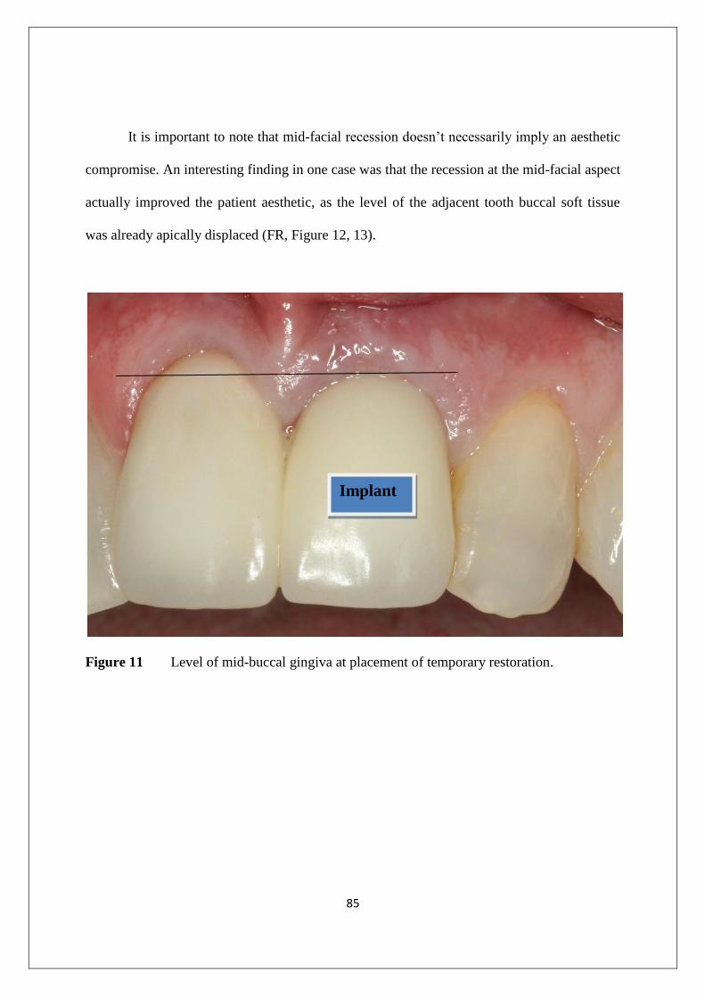
85
It is important to note that mid-facial recession doesn’t necessarily imply an aesthetic
compromise. An interesting finding in one case was that the recession at the mid-facial aspect
actually improved the patient aesthetic, as the level of the adjacent tooth buccal soft tissue
was already apically displaced (FR, Figure 12, 13).
Figure 11 Level of mid-buccal gingiva at placement of temporary restoration.
Implant

86
Figure 12 Level of mid-buccal gingival at the follow-up.
Implant

87
Study Level of mid- facial
gum
Comments
WÖhlre 12
- More than 2 mm change in Two cases
Groisman et al 86
- 2 mm soft tissue loss from buccal
Kan et al 87
0.55 ± 0.53 mm loss -
Cornelini et al 91
0.75 mm loss -
Canullo et al 94
0.2 mm gain 0.25 mm Mean papilla gain
Hall et al 95
- No change in 28.5% of the papilla with gaining in
63% of the sites.
Palattella et al 96
- -0.8 mm loss
De Rouck et al 97
0.53 mm loss -
- Indicate no data
Table 20 Changes in soft tissue levels (mid-buccal aspect) as reported in published
studies.

88
Although there was no statistical significance in the changes observed with regards to
soft tissues, there was a papilla recession of 0.708 mm (P-value 0.214) at the mesial papillae
and 0.602 mm (P-value 0.138) at the distal papillae. This observation might be attributed to
the fact that two patients (AV, EC) had both central incisors replaced with immediate
implants. This resulted in surrounding the mid-line papilla with two implants, instead of the
papillae being bordered by a tooth and an implant as was the case with the other implants
(Figure 13, 14).

89
Figure 13 Level of mesial papilla at the placement of the temporary restoration.
Figure 14 Level of mesial papilla at the follow-up.
Implant Implant
Implan
t
Implan
t

90
The papillary height observed in this study appears greater than that reported in other
studies. Kan et al [87] reported a mean loss of 0.50 mm for the mesial papillae and 0.30 mm
mean loss of the distal papillae. De Rouck and his associates [97] showed a reduction in the
papillae height of 0.41 mm on average for mesial papillae and 0.31 mm for distal papillae.
Study Tip of mesial papilla Tip of distal papilla
WÖhlre 12
- -
Groisman et al 86
- -
Kan et al 87
0.53 ± 0.39 mm loss 0.39 ± 0.40 mm loss
Cornelini et al 91
- -
Canullo et al 94
- -
Hall et al 95
- -
Palattella et al 96
- -
De Rouck et al 97
0.41 mm loss 0.31 mm loss
- Indicate no data
Table 21 Changes in soft tissue levels (mesial and distal papillae) as reported in
published studies.

91
Changes in Jemt s’ index were analysed using Wilcoxon signed rank test assessing
eleven patients with twenty two papillae. No statistically significant differences were found
between the papillae level at placement and at follow up during the observation period of 16
± 5 months. Although there was no significant difference statistically between the mesial and
distal papillae, at follow up 50% of the mesial papillae received score 3 which means that full
height of the interdental papillae was present (normal) while the same score was recorded in
60% of distal papillae. The rest of the results showed scores of 1, 2 and 4. No score of zero
was recorded.
A study by Cornelini et al [91], found no score of 0, 1 or 4 in their sample with 60%
of papillae receiving a score of 2, with the remainder scoring a 3. In another study by Kan
[93], the papilla index was measured at pre-treatment and 3, 6, and 12 months following
implant placement with no differences noted between baseline and any of the follow up
observations. In a study by Hall [95], the follow up was extended over one year and showed
no change for 28.5% of the papilla with an improvement or gaining for about 63% of the
sites. Therefore, the findings of our study in relation to Jemt s’ index are consistent with the
published literature.
5.5 Plaque levels
It is generally accepted that plaque retention and accumulation around the implant
may cause peri-implantitis [124], [125]. According to this study there was high improvement
in the plaque level (good oral hygiene) at the time of follow up in comparison to the time of
implant placement. This may be because the patient was aware that their natural tooth was

92
failing and were not able or not willing to brush them thoroughly. Generally, good oral
hygiene was a prerequisite for the provision of this implant treatment protocol.
5.6 Gingival tissue biotype
Few studies have investigated the impact of gingival biotype and its relation to gum
recession. In a study conducted by Canullo [94] it was revealed that there is no influence of
the gingival biotype (thin or thick) on the final aesthetic outcomes which is in accordance
with the findings of this study. However, this result may be due to the small number of
patients included in this study and the relatively short follow up period.
5.7 Future considerations
This pilot clinical investigation was limited by the retrospective nature, the small
number of samples, the inability to secure a fully standardised follow up examination
protocol and the relatively small length of observation period. All of these factors can have a
significant impact on the results obtained from this study and hence should be interpreted
with caution. Nevertheless, the study has provided some indications as to the nature and
extent of soft and hard tissue changes that may be expected with this protocol, although it
remains unclear if hard and soft tissue measures will remain stable over time. A longer
observation period and larger samples are needed to support more definite conclusions.
This pilot study has used a new set of reference lines to measure the changes in soft
and hard tissues surrounding the implants, which can provide more reliable observations in

93
comparison to previous studies. The methodology used in this study constitutes an
improvement over previous attempts to investigate similar changes. This investigation will be
continued and expanded both in terms of sample numbers but also in the length of the
observation period.

94
6.0 CONCLUSIONS
Within the limits of the current retrospective, pilot clinical investigation, the results
indicate that the immediate placement and provisional restoration of a single tooth implant in
the aesthetic zone of the maxilla can result in acceptable implant integration as well as stable
peri-implant tissues up to 16 ± 5 months using the Astra Tech implant system. In particular:
1- All implants were found to be clinical integrated leading to 100% survival rate
throughout the follow up period of this study.
2- Hard tissue data (from mesial and distal side) at the follow up visit showed there
were no significant differences throughout the entire follow up period (P > 0.05).
3- Soft tissue data (from mid-facial, mesial and distal papillae) at the follow up visit
showed there were no significant differences throughout the entire follow up
period (P > 0.05).
4- Plaque index measurements showed a statistically significant improvement of the
oral hygiene between the time of the implant placement and the follow up
examination (P < 0.05).
95
7.0 LIST OF REFERENCES
1. BRANEMARK, P. I., HANSSON, B. O., ADELL, R., BREINE, U., LINDSTROM,
J., HALLEN, O., et al. (1977). Osseointegrated implants in the treatment of the
edentulous jaw. Experience from a 10-year period. Scand J Plast Reconstr Surg
Suppl, 16, 1-132.
2. BECKER, W., BECKER, B. E., RICCI, A., BAHAT, O., ROSENBERG, E., ROSE,
L. F., et al. (2000). A Prospective Multicenter Clinical Trial Comparing One- and
Two-Stage Titanium Screw-Shaped Fixtures with One-Stage Plasma-Sprayed Solid-
Screw Fixtures. Clinical Implant Dentistry and Related Research, 2(3), 159-165.
3. BRANEMARK, P. I., ADELL, R., ALBREKTSSON, T., LEKHOLM, U.,
LUNDKVIST, S., & ROCKLER, B. (1983). Osseointegrated titanium fixtures in the
treatment of edentulousness. Biomaterials, 4(1), 25-28.
4. COCHRAN, D., MORTON, D., & WAEBER, H.-P. (2004). Consensus statements
and Recommended Clinical Procedures Regarding Loading Protocol for Endosseous
Dental Implants. The International Journal Of Oral & Maxillofacial Implants,
19(supplement), 109-113.
5. TOUATI, B., & GUEZ, G. (2002). Immediate implantation with provisionalization:
from literature to clinical implications. Pract Proced Aesthet Dent, 14(9), 699-707;
quiz 708.

96
6. COCHRAN, D. L. (2006). The evidence for immediate loading of implants. J Evid
Based Dent Pract, 6, 155-63.
7. WEBER, H. P., MORTON, D., GALLUCCI, G. O., ROCCUZZO, M., CORDARO,
L. & GRUTTER, L. (2009) Consensus statements and recommended clinical
procedures regarding loading protocols. Int J Oral Maxillofac Implants, 24 Suppl,
180-3.
8. HAMMERLE, C. H., CHEN, S. T. & WILSON, T. G., JR. (2004). Consensus
statements and recommended clinical procedures regarding the placement of implants
in extraction sockets. Int J Oral Maxillofac Implants, 19 Suppl, 26-8.
9. LAZZARA, R. J. (1989). Immediate implant placement into extraction sites: surgical
and restorative advantages. Int J Periodontics Restorative Dent, 9(5), 332-343.
10. GOMEZ-ROMAN, G., SCHULTE, W., D'HOEDT, B., & AXMAN-KRCMAR, D.
(1997). The Frialit-2 implant system: five-year clinical experience in single-tooth and
immediately postextraction applications. Int J Oral Maxillofac Implants, 12(3), 299-
309.
11. TARNOW, D. P., EMTIAZ, S., & CLASSI, A. (1997). Immediate loading of
threaded implants at stage 1 surgery in edentulous arches: ten consecutive case reports
with 1- to 5-year data. Int J Oral Maxillofac Implants, 12(3), 319-324.

97
12. WOHRLE, P. S. (1998). Single-tooth replacement in the aesthetic zone with
immediate provisionalization: fourteen consecutive case reports. Pract Periodontics
Aesthet Dent, 10(9), 1107-1114; quiz 1116.
13. HUI, E., CHOW, J., LI, D., LIU, J., WAT, P., & LAW, H. (2001). Immediate
provisional for single-tooth implant replacement with Branemark system: preliminary
report. Clin Implant Dent Relat Res, 3(2), 79-86.
14. CALVO GUIRADO, J. L., SAEZ YUGUERO, R., FERRER PEREZ, V., &
MORENO PELLUZ, A. (2002). Immediate anterior implant placement and early
loading by provisional acrylic crowns: a prospective study after a one-year follow-up
period. J Ir Dent Assoc, 48(2), 43-49.
15. CHAUSHU, G., CHAUSHU, S., TZOHAR, A., & DAYAN, D. (2001). Immediate
loading of single-tooth implants: immediate versus non-immediate implantation. A
clinical report. Int J Oral Maxillofac Implants, 16(2), 267-272.
16. PAOLANTONIO, M., DOLCI, M., SCARANO, A., D'ARCHIVIO, D., DI
PLACIDO, G., TUMINI, V., et al. (2001). Immediate implantation in fresh extraction
sockets. A controlled clinical and histological study in man. J Periodontol, 72(11),
1560-1571.
17. SALAMA, H., SALAMA, M., & KELLY, J. (1996). The orthodontic-periodontal
connection in implant site development. Pract Periodontics Aesthet Dent, 8(9), 923-
932; quiz 934.

98
18. PIETROKOVSKI, J. & MASSLER, M. (1967) Alveolar ridge resorption following
tooth extraction. J Prosthet Dent, 17, 21-7.
19. RICHARDSON, A. (1965) The pattern of alveolar bone resorption following
extraction of anterior teeth. Trans Br Soc Study Orthod, 47-50.
20. SCHROPP, L., WENZEL, A., KOSTOPOULOS, L. & KARRING, T. (2003) Bone
healing and soft tissue contour changes following single-tooth extraction: a clinical
and radiographic 12-month prospective study. Int J Periodontics Restorative Dent, 23,
313-23.
21. BOTTICELLI, D., BERGLUNDH, T. & LINDHE, J. (2004). Hard-tissue alterations
following immediate implant placement in extraction sites. J Clin Periodontol, 31,
820-8.
22. BUSER, D., MARTIN, W., & BELSER, U. C. (2004). Optimizing esthetics for
implant restorations in the anterior maxilla: anatomic and surgical considerations. Int
J Oral Maxillofac Implants, 19 Suppl, 43-61.
23. EVANS, C. D. & CHEN, S. T. (2008) Esthetic outcomes of immediate implant
placements. Clin Oral Implants Res, 19, 73-80.

99
24. OLSSON, M., LINDHE, J. & MARINELLO, C. P. (1993) On the relationship
between crown form and clinical features of the gingiva in adolescents. J Clin
Periodontol, 20, 570-7.
25. OLSSON, M. & LINDHE, J. (1991) Periodontal characteristics in individuals with
varying form of the upper central incisors. J Clin Periodontol, 18, 78-82.
26. LISTGARTEN, M. A. & LAI, C. H. (1999) Comparative microbiological
characteristics of failing implants and periodontally diseased teeth. J Periodontol, 70,
431-7.
27. MOMBELLI, A., VAN OOSTEN, M. A., SCHURCH, E., JR. & LAND, N. P. (1987)
The microbiota associated with successful or failing osseointegrated titanium
implants. Oral Microbiol Immunol, 2, 145-51.
28. VAN WINKELHOFF, A. J., GOENE, R. J., BENSCHOP, C. & FOLMER, T. (2000)
Early colonization of dental implants by putative periodontal pathogens in partially
edentulous patients. Clin Oral Implants Res, 11, 511-20.
29. BAIN, C. (1997) Influences of smoking on the periodontium and dental implants.
Dent Update, 24, 328-30.
30. BAIN, C. A. (1996) Smoking and implant failure--benefits of a smoking cessation
protocol. Int J Oral Maxillofac Implants, 11, 756-9.

100
31. BAIN, C. A. & MOY, P. K. (1993) The association between the failure of dental
implants and cigarette smoking. Int J Oral Maxillofac Implants, 8, 609-15.
32. CHUANG, S. K., WEI, L. J., DOUGLASS, C. W. & DODSON, T. B. (2002) Risk
factors for dental implant failure: a strategy for the analysis of clustered failure-time
observations. J Dent Res, 81, 572-7.
33. FELOUTZIS, A., LANG, N. P., TONETTI, M. S., BURGIN, W., BRAGGER, U.,
BUSER, D., DUFF, G. W. & KORNMAN, K. S. (2003) IL-1 gene polymorphism and
smoking as risk factors for peri-implant bone loss in a well-maintained population.
Clin Oral Implants Res, 14, 10-7.
34. BERGLUNDH, T., PERSSON, L. & KLINGE, B. (2002). A systematic review of the
incidence of biological and technical complications in implant dentistry reported in
prospective longitudinal studies of at least 5 years. J Clin Periodontol, 29 Suppl 3,
197-212; discussion 232-3.
35. BELSER, U. C., SCHMID, B., HIGGINBOTTOM, F. & BUSER, D. (2004).
Outcome analysis of implant restorations located in the anterior maxilla: a review of
the recent literature. Int J Oral Maxillofac Implants, 19 Suppl, 30-42.
36. CHEN, S. T., WILSON, T. G., JR. & HAMMERLE, C. H. (2004). Immediate or early
placement of implants following tooth extraction: review of biologic basis, clinical
procedures, and outcomes. Int J Oral Maxillofac Implants, 19 Suppl, 12-25.

101
37. ATTARD, N. J. & ZARB, G. A. (2005). Immediate and early implant loading
protocols: a literature review of clinical studies. J Prosthet Dent, 94, 242-58.
38. GLAUSER, R., ZEMBIC, A. & HAMMERLE, C. H. (2006). A systematic review of
marginal soft tissue at implants subjected to immediate loading or immediate
restoration. Clin Oral Implants Res, 17 Suppl 2, 82-92.
39. DE ROUCK, T., COLLYS, K. & COSYN, J. (2008). Single-tooth replacement in the
anterior maxilla by means of immediate implantation and provisionalization: a
review. Int J Oral Maxillofac Implants, 23, 897-904.
40. JUNG, R. E., PJETURSSON, B. E., GLAUSER, R., ZEMBIC, A., ZWAHLEN, M.
& LANG, N. P. (2008). A systematic review of the 5-year survival and complication
rates of implant-supported single crowns. Clin Oral Implants Res, 19, 119-30.
41. DEN HARTOG, L., SLATER, J. J., VISSINK, A., MEIJER, H. J. & RAGHOEBAR,
G. M. (2008). Treatment outcome of immediate, early and conventional single-tooth
implants in the aesthetic zone: a systematic review to survival, bone level, soft-tissue,
aesthetics and patient satisfaction. J Clin Periodontol, 35, 1073-86.
42. ESPOSITO, M. A., KOUKOULOPOULOU, A., COULTHARD, P. &
WORTHINGTON, H. V. (2006). Interventions for replacing missing teeth: Dental
implant in fresh extraction sockets (immediate, immediate-delayed and delayed
implants). Cochrane Database Syst Rev, CD005968.

102
43. DEL CASTILLO, R. & DRAGO, C. (2005). Indexing and provisional restoration of
single implants. J Oral Maxillofac Surg, 63, 11-21.
44. ANDERSEN, E., HAANAES, H. R. & KNUTSEN, B. M. (2002). Immediate loading
of single-tooth ITI implants in the anterior maxilla: a prospective 5-year pilot study.
Clin Oral Implants Res, 13, 281-7.
45. ABBOUD, M., KOECK, B., STARK, H., WAHL, G. & PAILLON, R. (2005).
Immediate loading of single-tooth implants in the posterior region. The International
Journal of Oral & Maxillofacial Implants, 20, 61-68.
46. CHAUSHU, G. & CHAUSHU, S. (2001). The use of orthodontic treatment and
immediate implant loading to restore the traumatic loss of a maxillary central incisor.
Int J Adult Orthodon Orthognath Surg, 16, 47-53.
47. DA CUNHA, H. A., FRANCISCHONE, C. E., FILHO, H. N. & DE OLIVEIRA, R.
C. (2004). A comparison between cutting torque and resonance frequency in the
assessment of primary stability and final torque capacity of standard and TiUnite
single-tooth implants under immediate loading. The International Journal Of Oral &
Maxillofacial Implants, 19, 578-585.
48. GOMES, A., LOZADA, J. L., CAPLANIS, N. & KLEINMAN, A. (1998). Immediate
loading of a single hydroxyapatite-coated threaded root form implant: a clinical
report. J Oral Implantol, 24, 159-66.

103
49. JO, H. Y., HOBO, P. K. & HOBO, S. (2001). Freestanding and multiunit immediate
loading of the expandable implant: an up-to-40-month prospective survival study. J
Prosthet Dent, 85, 148-55.
50. KUPEYAN, H. K. & MAY, K. B. (1998). Implant and provisional crown placement:
a one stage protocol. Implant Dent, 7, 213-9.
51. LINDEBOOM, J. A., FRENKEN, J. W., DUBOIS, L., FRANK, M., ABBINK, I. &
KROON, F. H. (2006). Immediate loading versus immediate provisionalization of
maxillary single-tooth replacements: a prospective randomized study with BioComp
implants. J Oral Maxillofac Surg, 64, 936-42.
52. LOCANTE, W. M. (2004). Single-tooth replacements in the esthetic zone with an
immediate function implant: a preliminary report. J Oral Implantol, 30, 369-75.
53. NUZZOLESE, E. (2005). Immediate loading of two single tooth implants in the
maxilla: preliminary results after one year. J Contemp Dent Pract, 6, 148-57.
54. PARK, Y. S., YI, K. Y., MOON, S. C. & JUNG, Y. C. (2005). Immediate loading of
an implant following implant site development using forced eruption: a case report.
Int J Oral Maxillofac Implants, 20, 621-6.
55. PROUSSAEFS, P., KAN, J., LOZADA, J., KLEINMAN, A. & FARNOS, A. (2002).
Effects of immediate loading with threaded hydroxyapatite-coated root-form implants

104
on single premolar replacements: a preliminary report. The International Journal of
Oral & Maxillofacial Implants, 17, 567-572.
56. OTTONI, J. M., OLIVEIRA, Z. F., MANSINI, R. & CABRAL, A. M. (2005).
Correlation between placement torque and survival of single-tooth implants. The
International Journal of Oral & Maxillofacial Implants, 20, 769-776.
57. PROUSSAEFS, P. & LOZADA, J. (2004). Immediate loading of hydroxyapatite-
coated implants in the maxillary premolar area: three-year results of a pilot study. J
Prosthet Dent, 91, 228-33.
58. ROCCI, A., MARTIGNONI, M. & GOTTLOW, J. (2003). Immediate loading in the
maxilla using flapless surgery, implants placed in predetermined positions, and
prefabricated provisional restorations: a retrospective 3-year clinical study. Clin
Implant Dent Relat Res, 5 Suppl 1, 29-36.
59. SCHWARTZ-ARAD, D. & LEVIN, L. (2004). Post-traumatic use of dental implants
to rehabilitate anterior maxillary teeth. Dent Traumatol, 20, 344-7.
60. SUDBRINK, S. D. (2005). Computer-guided implant placement with immediate
provisionalization: a case report. J Oral Maxillofac Surg, 63, 771-4.
61. CARDAROPOLI, G., LEKHOLM, U. & WENNSTROM, J. L. (2006). Tissue
alterations at implant-supported single-tooth replacements: a 1-year prospective
clinical study. Clin Oral Implants Res, 17, 165-71.

105
62. JEMT, T. & LEKHOLM, U. (2003). Measurements of buccal tissue volumes at
single-implant restorations after local bone grafting in maxillas: a 3-year clinical
prospective study case series. Clin Implant Dent Relat Res, 5, 63-70.
63. JEMT, T. & LEKHOLM, U. (2005). Single implants and buccal bone grafts in the
anterior maxilla: measurements of buccal crestal contours in a 6-year prospective
clinical study. Clin Implant Dent Relat Res, 7, 127-35.
64. MEIJNDERT, L., MEIJER, H. J., STELLINGSMA, K., STEGENGA, B. &
RAGHOEBAR, G. M. (2007). Evaluation of aesthetics of implant-supported single-
tooth replacements using different bone augmentation procedures: a prospective
randomized clinical study. Clin Oral Implants Res, 18, 715-9.
65. PALMER, R. M., PALMER, P. J. & SMITH, B. J. (2000). A 5-year prospective study
of Astra single tooth implants. Clin Oral Implants Res, 11, 179-82.
66. PALMER, R. M., SMITH, B. J., PALMER, P. J. & FLOYD, P. D. (1997). A
prospective study of Astra single tooth implants. Clin Oral Implants Res, 8, 173-9.
67. ZARONE, F., SORRENTINO, R., VACCARO, F. & RUSSO, S. (2006). Prosthetic
treatment of maxillary lateral incisor agenesis with osseointegrated implants: a 24-39-
month prospective clinical study. Clin Oral Implants Res, 17, 94-101.

106
68. ANDERSEN, E., HAANAES, H. R. & KNUTSEN, B. M. (2002). Immediate loading
of single-tooth ITI implants in the anterior maxilla: a prospective 5-year pilot study.
Clin Oral Implants Res, 13, 281-7.
69. COOPER, L., FELTON, D. A., KUGELBERG, C. F., ELLNER, S., CHAFFEE, N.,
MOLINA, A. L., MORIARTY, J. D., PAQUETTE, D. & PALMQVIST, U. (2001). A
multicenter 12-month evaluation of single-tooth implants restored 3 weeks after 1-
stage surgery. Int J Oral Maxillofacial Implants, 16, 182-92.
70. COOPER, L. F., ELLNER, S., MORIARTY, J., FELTON, D. A., PAQUETTE, D.,
MOLINA, A., CHAFFEE, N., ASPLUND, P., SMITH, R. & HOSTNER, C. (2007).
Three-year evaluation of single-tooth implants restored 3 weeks after 1-stage surgery.
Int J Oral Maxillofacial Implants, 22, 791-800.
71. GOTFREDSEN, K. (2004). A 5-year prospective study of single-tooth replacements
supported by the Astra Tech implant: a pilot study. Clin Implant Dent Relat Res, 6, 1-
8.
72. SCHROPP, L., ISIDOR, F., KOSTOPOULOS, L. & WENZEL, A. (2005).
Interproximal papilla levels following early versus delayed placement of single-tooth
implants: a controlled clinical trial. Int J Oral Maxillofacial Implants, 20, 753-61.
73. SCHROPP, L., KOSTOPOULOS, L., WENZEL, A. & ISIDOR, F. (2005). Clinical
and radiographic performance of delayed-immediate single-tooth implant placement

107
associated with peri-implant bone defects. A 2-year prospective, controlled,
randomized follow-up report. J Clin Periodontol, 32, 480-7.
74. DEGIDI, M., PIATTELLI, A. & CARINCI, F. (2006). Parallel screw cylinder
implants: comparative analysis between immediate loading and two-stage healing of
1,005 dental implants with a 2-year follow up. Clin Implant Dent Relat Res, 8, 151-
60.
75. TOLJANIC, J. A. & BAER, R. A. (2005). Same-day implant placement and
provisionalization for single-tooth implants: a technique update and review of clinical
experiences. Dent today, 24, 73-4, and 76-7; quiz 77.
76. FUGAZZOTTO, P. A. (2004). Guided Bone Regeneration at Immediate Implant
Insertion and Loading: A Case Report. Implant Dentistry, 13, 223-227.
77. GARBER, D. A., SALAMA, M. A. & SALAMA, H. (2001). Immediate total tooth
replacement. Compend Contin Educ Dent, 22, 210-6, 218.
78. GUIRADO, C., LUIS, J., YUGUERO, S., ROSARIO, M., PARDO ZAMORA, G. &
MUNOZ BARRIO, E. (2005). Immediate Osseotite implant placement and immediate
loading of a provisional restoration of maxillary lateral incisors. J Ir Dent Assoc, 51,
173-6.

108
79. KAN, J. Y. & RUNGCHARASSAENG, K. (2000). Immediate placement and
provisionalization of maxillary anterior single implants: a surgical and prosthodontic
rationale. Pract Periodontics Aesthet Dent, 2000; 12, 817-24.
80. LEARY, J. C. & HIRAYAMA, M. (2003). Extraction, immediate-load implants,
impressions and final restorations in two patient visits. J Am Dent Assoc, 134, 715-20.
81. LOCANTE, W. M. (2001). The non-functional immediate provisional in immediate
extraction sites: a technique to maximize esthetics. Implant Dent, 10, 254-8.
82. MANKOO, T. (2004). Contemporary implant concepts in aesthetic dentistry--Part 2:
Immediate single-tooth implants. Pract Proced Aesthet Dent, 16, 61-8; quiz 70.
83. SCHIROLI, G. (2003). Immediate tooth extraction, placement of a Tapered Screw-
Vent implant, and provisionalization in the esthetic zone: a case report. Implant Dent,
12, 123-31.
84. TOUATI, B. & GUEZ, G. (2002). Immediate implantation with provisionalization:
from literature to clinical implications. Pract Proced Aesthet Dent, 14, 699-707; quiz
708.
85. TSELIOS, N., PAREL, S. M. & JONES, J. D. (2006). Immediate placement and
immediate provisional abutment modeling in anterior single-tooth implant restorations
using a CAD/CAM application: a clinical report. J Prosthet Dent, 95, 181-5.

109
86. GROISMAN, M., FROSSARD, W. M., FERREIRA, H. M., DE MENEZES FILHO,
L. M., & TOUATI, B. (2003). Single-tooth implants in the maxillary incisor region
with immediate provisionalization: 2-year prospective study. Pract Proced Aesthet
Dent, 15(2), 115-122, 124; quiz 126.
87. KAN, J. Y., RUNGCHARASSAENG, K., UMEZU, K. & KOIS, J. C. (2003).
Dimensions of peri-implant mucosa: an evaluation of maxillary anterior single
implants in humans. J Periodontol, 74, 557-62.
88. LORENZONI, M., PERTL, C., ZHANG, K., WIMMER, G., & WEGSCHEIDER, W.
A. (2003). Immediate loading of single-tooth implants in the anterior maxilla.
Preliminary results after one year. Clin Oral Implants Res, 14(2), 180-187.
89. NORTON, M. R. (2004). A short-term clinical evaluation of immediately restored
maxillary TiOblast single-tooth implants. Int J Oral Maxillofac Implants, 19(2), 274-
281.
90. TSIRLIS, A. T. (2005). Clinical Evaluation of Immediate Loaded Upper Anterior
Single Implants. Implant Dentistry, 14(1), 94-103.
91. CORNELINI, R., CANGINI, F., COVANI, U., & WILSON, T. G., JR. (2005).
immediate restoration of implants placed into fresh extraction sockets for single-tooth
replacement: a prospective clinical study. Int J Periodontics Restorative Dent, 25(5),
439-447.

110
92. FERRARA, A., GALLI, C., MAURO, G., & MACALUSO, G. M. (2006).
Immediate provisional restoration of postextraction implants for maxillary single-
tooth replacement. Int J Periodontics Restorative Dent, 26(4), 371-377.
93. KAN, J. Y., RUNGCHARASSAENG, K., LIDDELOW, G., HENRY, P. &
GOODACRE, C. J. (2007). Periimplant tissue response following immediate
provisional restoration of scalloped implants in the esthetic zone: a one-year pilot
prospective multicenter study. The Journal Of Prosthetic Dentistry, 97, S109-18.
94. CANULLO, L., & RASPERINI, G. (2007). Preservation of peri-implant soft and
hard tissues using platform switching of implants placed in immediate extraction
sockets: a proof-of-concept study with 12- to 36-month follow-up. The International
Journal Of Oral & Maxillofacial Implants, 22(6), 995-1000.
95. HALL, J. A., PAYNE, A. G., PURTON, D. G., TORR, B., DUNCAN, W. J., & DE
SILVA, R. K. (2007). Immediately restored, single-tapered implants in the anterior
maxilla: prosthodontic and aesthetic outcomes after 1 year. Clin Implant Dent Relat
Res, 9(1), 34-45.
96. PALATTELLA, P., TORSELLO, F., & CORDARO, L. (2008). Two-year
prospective clinical comparison of immediate replacement vs. immediate restoration
of single tooth in the esthetic zone. Clin Oral Implants Res, 19(11), 1148-1153.

111
97. DE ROUCK, T., COLLYS, K., & COSYN, J. (2008). Immediate single-tooth
implants in the anterior maxilla: a 1-year case cohort study on hard and soft tissue
response. J Clin Periodontol, 35(7), 649-657.
98. RIBEIRO, F. S., PONTES, A. E., MARCANTONIO, E., PIATTELLI, A., NETO, R.
J. & MARCANTONIO, E., JR. (2008). Success rate of immediate non-functional
loaded single-tooth implants: immediate versus delayed implantation. Implant Dent,
17, 109-17. WEISGOLD, A. S. (1977) Contours of the full crown restoration. Alpha
Omegan, 70, 77-89.
99. DEN HARTOG, L., SLATER, J. J., VISSINK, A., MEIJER, H. J. & RAGHOEBAR,
G. M. (2008). Treatment outcome of immediate, early and conventional single-tooth
implants in the aesthetic zone: a systematic review to survival, bone level, soft-tissue,
aesthetics and patient satisfaction. J Clin Periodontol, 35, 1073-86.
100. LANG, N. P., BERGLUNDH, T., HEITZ-MAYFIELD, L. J., PJETURSSON, B.
E., SALVI, G. E. & SANZ, M. (2004). Consensus statements and recommended
clinical procedures regarding implant survival and complications. Int J Oral
Maxillofacial Implants, 19 Suppl, 150-4.
101. SMITH, D. E. & ZARB, G. A. (1989). Criteria for success of osseointegrated
endosseous implants. J Prosthet Dent, 62, 567-72.
102. ALBREKTSSON, T. & ZARB, G. A. (1993). Current interpretations of the
osseointegrated response: clinical significance. Int J Prosthodont, 6, 95-105.

112
103. LINDHE, J. & LANG, N. P. (2008). Clinical Periodontology and Implant Dentistry
Blackwell Munksgaard.
104. WEISGOLD, A. S. (1977). Contours of the full crown restoration. Alpha Omegan,
70, 77-89.
105. OCHSENBEIN, C. & ROSS, S. (1969). a re-evaluation of osseous surgery. Dent
Clin North Am, 13, 87-102.
106. OLSSON, M., LINDHE, J. & MARINELLO, C. P. (1993). On the relationship
between crown form and clinical features of the gingiva in adolescents. J Clin
Periodontol, 20, 570-7.
107. TARNOW, D. P., MAGNER, A. W. & FLETCHER, P. (1992). The effect of the
distance from the contact point to the crest of bone on the presence or absence of the
interproximal dental papilla. J Periodontol, 63, 995-6.
108. JEMT, T. (1997). Regeneration of gingival papillae after single-implant treatment.
Int J Periodontics Restorative Dent, 17, 326-33.
109. BENGAZI, F., WENNSTROM, J. L. & LEKHOLM, U. (1996). Recession of the
soft tissue margin at oral implants. A 2-year longitudinal prospective study. Clin Oral
Implants Res, 7, 303-10.

113
110. CHOQUET, V., HERMANS, M., ADRIAENSSENS, P., DAELEMANS, P.,
TARNOW, D. P. & MALEVEZ, C. (2001). Clinical and radiographic evaluation of
the papilla level adjacent to single-tooth dental implants. A retrospective study in the
maxillary anterior region. J Periodontol, 72, 1364-71.
111. CHANG, M., WENNSTROM, J. L., ODMAN, P. & ANDERSSON, B. (1999).
Implant supported single-tooth replacements compared to contralateral natural teeth.
Crown and soft tissue dimensions. Clin Oral Implants Res, 10, 185-94.
112. LEKHOLMU, ZARB GA. Patient selection and preparation. In: Tissue-integrated
prostheses. Branemark PI, Zarb GA, Albrektsson T, editors. Chicago: Quintessence;
(1985). p. 199-209.
113. MOMBELLI, A. & LANG, N. P. (1998). The diagnosis and treatment of peri-
implantitis. Periodontol 2000, 17, 63-76.
114. ESPOSITO, M., HIRSCH, J. M., LEKHOLM, U. & THOMSEN, P. (1998).
Biological factors contributing to failures of osseointegrated oral implants. (II).
Etiopathogenesis. Eur J Oral Sci, 106, 721-64.
115. NEWMAN, M. G. & CARRANZA, F. A. (2006). Clinical Periodontology, saunders
Elsvier.
116. KAN, J. Y., LOZADA, J. L., BOYNE, P. J., GOODACRE, C. J. &
RUNGCHARASSAENG, K. (1997). Mandibular fracture after endosseous implant

114
placement in conjunction with inferior alveolar nerve transposition: a patient treatment
report. Int J Oral Maxillofa Implants, 11, 19-47.
117. MULLER, H. P., HEINECKE, A., SCHALLER, N. & EGER, T. (2000).
Masticatory mucosa in subjects with different periodontal phenotypes. J Clin Periodontol,
27, 621-6.
118. BRUNSKI, J. B., PULEO, D. A. & NANCI, A. (2000). Biomaterials and
biomechanics of oral and maxillofacial implants: current status and future developments.
Int J Oral Maxillofacial Implants, 15, 15-46.
119. PAULETTO, N., LAHIFFE, B. J. & WALTON, J. N. (1999). Complications
associated with excess cement around crowns on osseointegrated implants: a clinical
report. Int J Oral Maxillofac Implants, 14, 865-8.
120. HERMANN, J. S., BUSER, D., SCHENK, R. K. & COCHRAN, D. L. (2000).
Crestal bone changes around titanium implants. A histometric evaluation of unloaded
non-submerged and submerged implants in the canine mandible. J Periodontol, 71, 1412-
24.
121. CARDAROPOLI, G., LEKHOLM, U. & WENNSTROM, J. L. (2006). Tissue
alterations at implant-supported single-tooth replacements: a 1-year prospective clinical
study. Clin Oral Implants Res, 17, 165-71.

115
122. GRUNDER, U. (2000). Stability of the mucosal topography around single-tooth
implants and adjacent teeth: 1-year results. Int J Periodontics Restorative Dent, 20, 11-7.
123. OH, T. J., SHOTWELL, J., BILLY, E., BYUN, H. Y. & WANG, H. L. (2007).
Flapless implant surgery in the esthetic region: advantages and precautions. Int J
Periodontics Restorative Dent, 27, 27-33.
124. ADELL, R., LEKHOLM, U., BRANEMARK, P. I., LINDHE, J., ROCKLER, B.,
ERIKSSON, B., LINDVALL, A. M., YONEYAMA, T. & SBORDONE, L. (1985).
Marginal tissue reactions at osseointegrated titanium fixtures. Swed Dent J Suppl, 28,
175-81.
125. ADELL, R., LEKHOLM, U., ROCKLER, B., BRANEMARK, P. I., LINDHE, J.,
ERIKSSON, B. & SBORDONE, L. (1986). Marginal tissue reactions at osseointegrated
titanium fixtures (I). A 3-year longitudinal prospective study. Int J Oral Maxillofac Surg,
15, 39-52.