Digital Microscopy with Versatile Illumination and … DVM6...DIGITAL MICROSCOPY WITH VERSATILE...
6
AUTHORS James DeRose Scientific Writer, Stereo & Digital Microscopy Marketing, Leica Microsystems AG, Switzerland DIGITAL MICROSCOPY WITH VERSATILE ILLUMINATION AND VARIOUS CONTRAST METHODS FOR MORE EFFICIENT INSPECTION AND QUALITY CONTROL Example applications using the Leica DVM6 with integrated ring light or coaxial illumination system Georg Schlaffer Product Manager, Digital Microscopy, Leica Microsystems AG, Switzerland From Eye to Insight
Transcript of Digital Microscopy with Versatile Illumination and … DVM6...DIGITAL MICROSCOPY WITH VERSATILE...

AUTHORS
James DeRose Scientific Writer, Stereo & Digital Microscopy Marketing,Leica Microsystems AG, Switzerland
DIGITAL MICROSCOPY WITH VERSATILE ILLUMINATION AND VARIOUS CONTRAST METHODS FOR MORE EFFICIENT INSPECTION AND QUALITY CONTROL
Example applications using the Leica DVM6 with integrated ring light or coaxial illumination system
Georg SchlafferProduct Manager,Digital Microscopy, Leica Microsystems AG, Switzerland
From Eye to Insight
DIGITAL MICROSCOPY WITH VERSATILE ILLUMINATION AND VARIOUS CONTRAST METHODS FOR MORE EFFICIENT INSPECTION AND QUALITY CONTROL
2
Introduction
State-of-the-art digital microscopes utilizing a versatile illumination system capable of achieving multiple contrast methods, such as the Leica
DVM6, are very useful for inspection, quality control, and failure analysis. These contrast methods allow flaws or defects on the surface of a
product or component to be more easily and rapidly detected. Some examples of how modern digital microscopes can help to make the workflow
more efficient for inspection, quality control, and failure analysis are discussed.
Background for digital microscopy applied to inspection, quality control, and failure analysis
Digital microscopes have no eyepieces, but use a digital camera as detector. They are used more and more often for diverse technical applications,
such as, fast and easy documentation of parts during manufacturing, assembly, inspection, quality control (QC), and failure analysis (FA)
To better visualize product or product component flaws, often different types of lighting contrast are exploited with optical microscopy [1–4].
Such illumination contrast methods are typically used in a variety of industries, such as, automotive, aerospace, railway, microelectronics and
electronics, semiconductors, precision engineering, metallurgy and metallography, glass and ceramics, petroleum, chemicals, pharmaceuticals,
and medical devices.
For industrial manufacturing, it is critical to speed up the inspection and QC process. If the feature of interest is detected or seen more easily
by enhancing its appearance with different types of illumination and contrast, then less time is needed for inspection and testing. Recent
developments in digital microscopy have led to more a practical and efficient way of using illumination contrast methods for inspection and QC
purposes.

DIGITAL MICROSCOPY WITH VERSATILE ILLUMINATION AND VARIOUS CONTRAST METHODS FOR MORE EFFICIENT INSPECTION AND QUALITY CONTROL
3
Examples of digital microscopy imaging with different illumination contrast methods
Leadframes
Leadframes are metal structures used inside microelectronic chip packages to connect the wiring from small electrical terminals on the semiconductor
surface to the larger scale circuitry on electronic devices and circuit boards. They are used in almost all microelectronic semiconductor packages.
For the case shown here (Fig. 1) of copper (Cu) leadframes plated with tin (Sn), the amount of Sn smeared over the cross section of the trimmed
leadframe is found to be a good indicator of wear for the trimming tool. When the smearing of the Sn reaches a critical level, then normally the
tool is replaced. Microscopy illumination contrast methods enable users to see the smearing of the Sn more easily.
Figure 1: Different images of the trimmed edge (cross section) of a Sn-plated Cu leadframe taken with the Leica DVM6 using different illumination contrast methods (schematic inset): A) full ring light; B) coaxial light with polarizer open; C) coaxial light with relief contrast and polarizer open; and D) coaxial light with polarizer closed. The images show with varying levels of contrast that the Sn is smeared over the most of the Cu surface, except at the bottom where the pin broke off and the Cu is visible.
DIGITAL MICROSCOPY WITH VERSATILE ILLUMINATION AND VARIOUS CONTRAST METHODS FOR MORE EFFICIENT INSPECTION AND QUALITY CONTROL
4
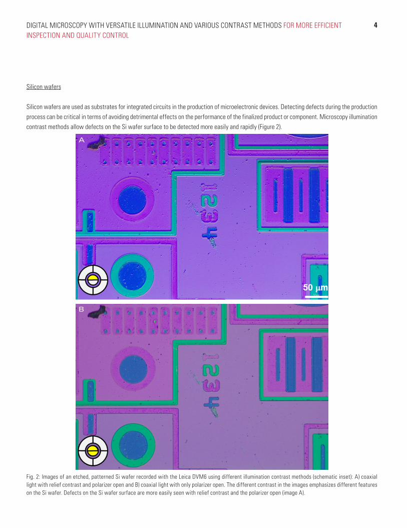
Silicon wafers
Silicon wafers are used as substrates for integrated circuits in the production of microelectronic devices. Detecting defects during the production
process can be critical in terms of avoiding detrimental effects on the performance of the finalized product or component. Microscopy illumination
contrast methods allow defects on the Si wafer surface to be detected more easily and rapidly (Figure 2).
Fig. 2: Images of an etched, patterned Si wafer recorded with the Leica DVM6 using different illumination contrast methods (schematic inset): A) coaxial light with relief contrast and polarizer open and B) coaxial light with only polarizer open. The different contrast in the images emphasizes different features on the Si wafer. Defects on the Si wafer surface are more easily seen with relief contrast and the polarizer open (image A).
A
B

DIGITAL MICROSCOPY WITH VERSATILE ILLUMINATION AND VARIOUS CONTRAST METHODS FOR MORE EFFICIENT INSPECTION AND QUALITY CONTROL
5
Embossed metal coated paper for food packaging
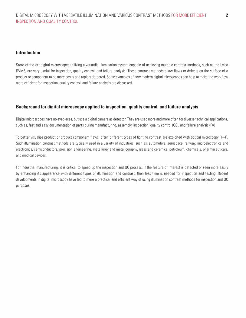
Embossing is a process which produces raised relief patterns, images, and designs in various materials. Here is shown an example of embossed
metal coated paper used for food packaging. Microscopy illumination contrast methods are helpful for better visualizing flaws in the embossed
metal coated paper or contamination present on its surface (Figure 3).
Fig. 3: Images of embossed metal coated paper acquired with the Leica DVM6 using different illumination contrast methods (schematic inset): A) full ring light; B) quarter of the ring light; C) coaxial light with polarizer open; and D) coaxial light with polarizer closed. The quarter ring (image B) and coaxial light with polarizer open (image C) enhance the embossed squares, while the coaxial light with polarizer closed (image D) enhance the imperfections or contamination.
Copy
right
© 2
016
Leic
a M
icro
syst
ems
(Sch
wei
z) A
G. A
ll rig
hts
rese
rved
. Sub
ject
to m
odifi
catio
ns.
LEIC
A an
d th
e Le
ica
Logo
are
regi
ster
ed tr
adem
arks
of L
eica
Mic
rosy
stem
s IR
Gm
bH. F
usio
nOpt
ics
is a
trad
emar
k of
Lei
ca M
icro
syst
ems
(Sch
wei
z) A
G re
gist
ered
in E
urop
e.
Leica Microsystems (Switzerland) Ltd. · Max-Schmidheiny-Strasse 201 · 9435 Heerbrugg, Schweiz
T +41 71 726 34 34 · F +41 71 726 34 44
www.leica-microsystems.com
CONNECT
WITH US!
Additional Reading
1. Diez D: Metallography – an Introduction: How to Reveal Microstructural Features of Metals and Alloys, Science Lab.
2. Christian U, and Jost N: Metallography with Color and Contrast: The Possibilities of Microstructural Contrasting, Science Lab.
3. Goeggel D, and Schlaffer G: 3D Visualization of Surface Structures, Vertical Resolution – Small Steps, Big Effect, Science Lab.
4. Goeggel D: Factors to Consider When Selecting a Stereo Microscope, Science Lab.
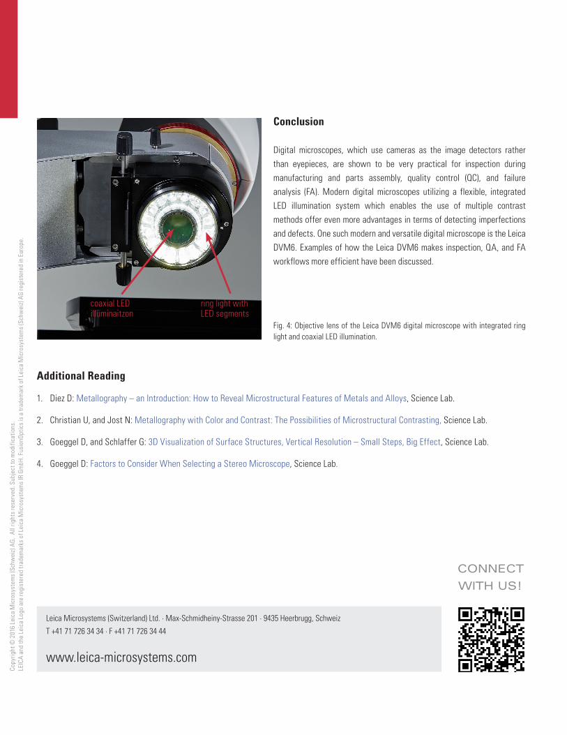
ring light with LED segments
coaxial LED illuminaitzon
Conclusion
Digital microscopes, which use cameras as the image detectors rather
than eyepieces, are shown to be very practical for inspection during
manufacturing and parts assembly, quality control (QC), and failure
analysis (FA). Modern digital microscopes utilizing a flexible, integrated
LED illumination system which enables the use of multiple contrast
methods offer even more advantages in terms of detecting imperfections
and defects. One such modern and versatile digital microscope is the Leica
DVM6. Examples of how the Leica DVM6 makes inspection, QA, and FA
workflows more efficient have been discussed.
Fig. 4: Objective lens of the Leica DVM6 digital microscope with integrated ring light and coaxial LED illumination.