Digital dental imaging - dentalplanet.com dental imaging ... PA TMJ lateral Ortho TMJ (alternative...
20
Digital dental imaging Orthopantomograph ® OP100 D Orthoceph ® OC100 D
Transcript of Digital dental imaging - dentalplanet.com dental imaging ... PA TMJ lateral Ortho TMJ (alternative...
Digital dental imaging
Orthopantomograph® OP100 DOrthoceph® OC100 D


Advanced direct digital panoramicand cephalometric imaging
Orthopantomograph OP100 Dand Orthoceph OC100 D
The name Orthopantomograph has always stoodfor consistent reliability and clinically excellentmaxillofacial imaging. Since the originalOrthopantomograph was first introduced in1961, over 45,000 installations have beencompleted worldwide, clearly illustrating theinternational success of the unit. And thissuccess continues with the all-digitalOrthopantomograph product family.
The Orthopantomograph OP100 D and the OrthocephOC100 D combine Instrumentarium Dental’s longexperience and know-how in x-ray generator design,digital image acquisition and information technology,in an advanced dental imaging system.
What’s more, existing owners of film-basedOrthopantomograph OP100’s can also enjoy thebenefits of digital imaging by choosing one of thedigital upgrade options.
The Orthopantomograph OP100 D
Revolutionary high tech through the decades
1946 Professor Y.V. Paatero published his first paper
on Panoramic Tomography
1951 “Pantomography” equipment was first presented
1961 Orthopantomograph, the first dental
panoramic X-ray, is developed
Orthopantomograph OP1
1964 Commercialization of Orthopantomograph begins
Orthopantomograph OP2
Orthopantomograph OP3
1978 Orthopantomograph is the leading name in the
dental panoramic imaging
Orthopantomograph OP5 / Orthoceph OC5
Orthopantomograph OP6 (Zonarc)
Orthopantomograph OP10 / Orthoceph OC10
1991 New innovations, such as lifting cassette head and linear
tomography were introduced along with the OP100 product
family
Orthopantomograph OP100 / Orthoceph OC100
Ortho Trans Linear Tomography option for OP100 / OC100
1999 Introduction of the direct digital Orthopantomograph
with the latest technology
Orthopantomograph OP2 (1965)
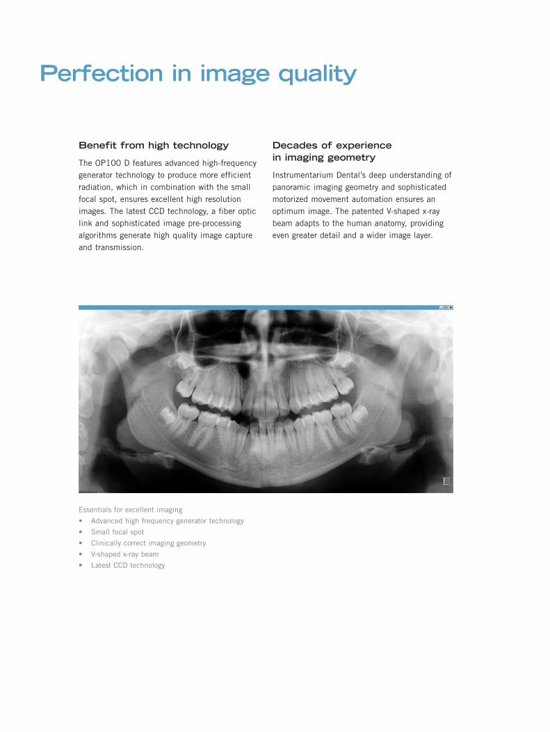
Perfection in image quality
Benefit from high technology
The OP100 D features advanced high-frequencygenerator technology to produce more efficientradiation, which in combination with the smallfocal spot, ensures excellent high resolutionimages. The latest CCD technology, a fiber opticlink and sophisticated image pre-processingalgorithms generate high quality image captureand transmission.
Decades of experiencein imaging geometry
Instrumentarium Dental’s deep understanding ofpanoramic imaging geometry and sophisticatedmotorized movement automation ensures anoptimum image. The patented V-shaped x-raybeam adapts to the human anatomy, providingeven greater detail and a wider image layer.
Essentials for excellent imaging
• Advanced high frequency generator technology
• Small focal spot
• Clinically correct imaging geometry
• V-shaped x-ray beam
• Latest CCD technology

Successful exposures — time after time
Correct imaging values —automatically
Automatic Exposure Control (AEC) andAutomatic Spine Compensation (ASC), utilizingInstrumentarium Dental’s unique and patentedsoftware algorithms, generate correct imagingvalues, exposure after exposure, with any sizepatient and with all users.
Accurate and stablepatient positioning
Incorporating the well-proven OP100 patientpositioning system, the OP100 D is just as easy,quick and accurate to use. Correct positioning isassured by three positioning light lines. Frankfurtand mid-sagittal lights aid correctangulation of the patient’s head and theOcclusion correction light guides occlusalcorrection. For easy adjustment, both the userand the patient can view the mid-sagittal planein the curved panoramic mirror. Anelectromagnetically locked rigid forehead supportis used together with a chin rest and bite fork toeliminate patient movement. Patient positioningis possible from the left or right side of theOP100 D.
Essentials for excellent imaging
• Automatic Exposure Control (AEC)
• Automatic Spine Compensation (ASC)
• Accurate patient positioning
• Open view of the patient
• Three clear positioning lights
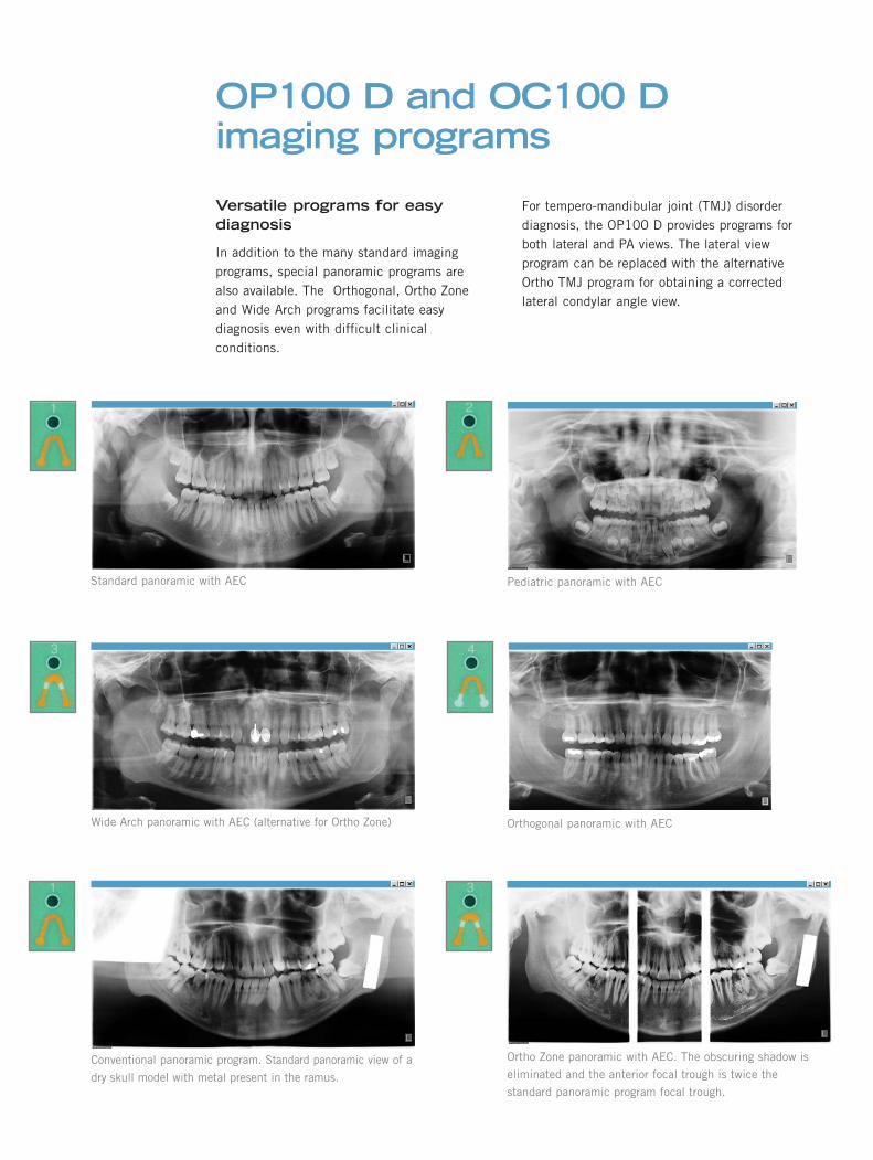
Versatile programs for easydiagnosis
In addition to the many standard imagingprograms, special panoramic programs arealso available. The Orthogonal, Ortho Zoneand Wide Arch programs facilitate easydiagnosis even with difficult clinicalconditions.
OP100 D and OC100 Dimaging programs
For tempero-mandibular joint (TMJ) disorderdiagnosis, the OP100 D provides programs forboth lateral and PA views. The lateral viewprogram can be replaced with the alternativeOrtho TMJ program for obtaining a correctedlateral condylar angle view.
Conventional panoramic program. Standard panoramic view of a
dry skull model with metal present in the ramus.
Ortho Zone panoramic with AEC. The obscuring shadow is
eliminated and the anterior focal trough is twice the
standard panoramic program focal trough.
Orthogonal panoramic with AEC
Pediatric panoramic with AEC
Wide Arch panoramic with AEC (alternative for Ortho Zone)
Standard panoramic with AEC
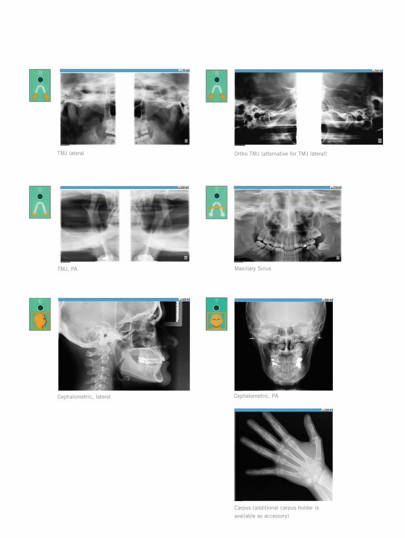
Carpus (additional carpus holder is
available as accessory)
Maxillary SinusTMJ, PA
Cephalometric, PACephalometric, lateral
TMJ lateral Ortho TMJ (alternative for TMJ lateral)
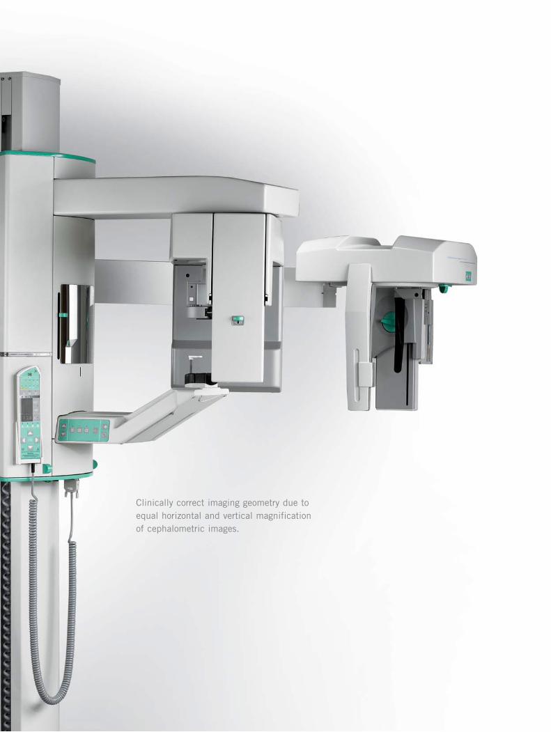
Clinically correct imaging geometry due toequal horizontal and vertical magnificationof cephalometric images.

Clinically correct geometry
In order to produce equal horizontal and verticalmagnification, the OC100 D uses a uniquemethod of synchronized tube head horizontalsweep and sensor movements while still keepingthe focal spot in the same position. Theclinically correct imaging geometry is the sameas traditional film imaging and allows easydetermination of orthodontic reference pointsand comparison with previous film images.
Variety of projections
The OC100 D provides a full range of imagingprojections for cephalometric radiography. It is acomprehensive diagnostic device that includeslateral, facial, posterioranterior and obliqueprojections as well as the possibility for handand wrist imaging.
Visible diagnostic details
The CliniView image post-processing tool kitadds value to the system by providing theoptimum viewing conditions. The selection of16-bit image format improves the visibility ofimportant orthodontic reference points.
Added diagnostic value
Essentials for excellent imaging
• Clinically correct imaging geometry
• 16-bit image format
A
B
OC100 D uses a unique horizontal sweep of the tube
head while keeping the focal spot steady.
This results in equal horizontal and vertical
magnification (A). Any focal spot movement during the
scan would result in an image magnified horizontally,
but not vertically (B).
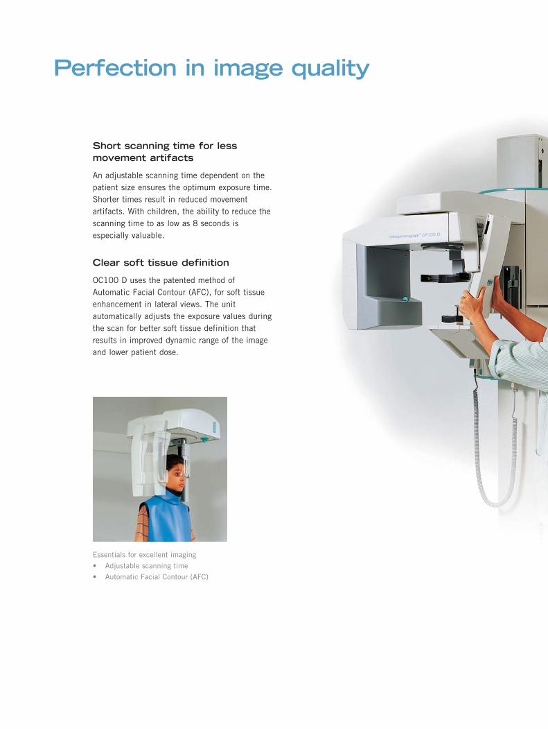
Short scanning time for lessmovement artifacts
An adjustable scanning time dependent on thepatient size ensures the optimum exposure time.Shorter times result in reduced movementartifacts. With children, the ability to reduce thescanning time to as low as 8 seconds isespecially valuable.
Clear soft tissue definition
OC100 D uses the patented method ofAutomatic Facial Contour (AFC), for soft tissueenhancement in lateral views. The unitautomatically adjusts the exposure values duringthe scan for better soft tissue definition thatresults in improved dynamic range of the imageand lower patient dose.
Perfection in image quality
Essentials for excellent imaging
• Adjustable scanning time
• Automatic Facial Contour (AFC)

Efficient work flow withtwo sensors
Two separate sensors (for panoramic andcephalometric imaging) allow easier and quickerchangeover between the two modalities withoutthe need to handle the sensor. A singleinterchangeable pan/ceph sensor is also anoption.
Stable patient positioning
The Frankfurt horizontal plane laser light,nasion support and rigid ear rods make patientpositioning easy and convenient. Motorizedvertical movement controls are convenientlylocated on the cephalostat head for easyaccess. Exposure values and correct program areautomatically selected depending on the imageprojection.
Optimum use of space
The OC100 D can be installed in your clinic forright or left-handed imaging and is “fieldchangeable”. A standard wall mount with swiveljoint allows the OC100 D to be installed at anangle for optimum use of room space andconvenient patient positioning.
Excellent usability
Essentials for excellent imaging• 1 or 2 sensor options• Frankfurt horizontal plane laser light• Position controls on the ceph head• Right or left handed versions
The Orthoceph OC100 D in a left-handed
configuration.

CliniView software is the completesolution for digital dental image handling.

For all modalities
CliniView is an easy-to-use software programwith powerful functions for digital imagecapturing, storage, and viewing, includinga wide range of dental specific imageenhancement tools.
CliniView supports image capturing fromInstrumentarium Dental’s Orthopantomographpanoramic, Orthoceph cephalometric x-rayunits, SIGMA intraoral sensors, and fromintraoral cameras. In addition, images canbe imported or scanned from a wide range ofother digital sources.
Fast and accurate diagnosis
CliniView’s advanced preprocessingalgorithms, which together with 16 bit imageformat, ensure excellent image quality. Noadditional adjustments are necessary. Fordifferent user and modality preferences andspecific details, CliniView offers a widerange of image enhancement tools, such asbrightness and contrast adjustment, noisereduction, zoom, edge enhancement, invert,region of interest tools, isodensitycolorization and grayscale filters.
Advanced tools
CliniView allows highlighting and explainingimportant areas on the image by providingdifferent tools for adding text and graphicalsymbols. With CliniView, it is possible toprint multiple images on the same page,design your own print layouts, resize andmove images freely on the paper. Imageinformation such as notes, tooth numbers,exposure values, and patient information canbe added to printouts as well as your practiceor clinic information with logos.
Versatile viewing with CliniViewsoftware
CliniView software provides advanced tools for easy
diagnosis and superior image quality.
Patient information and images are stored in CliniView SQL
database.
Advanced print editor enables multiple image printing with
desired image and clinic information.


Efficient clinical use
With OP100 D and OC100 D direct digitalimaging, there are no delays in imageacquisition. The image appears on the PC screenin real time during the exposure. Once taken, theimage can be transmitted wherever it is neededto another workstation, an imagearchive, a printer, or by e-mail.
Open connectivity
In addition to traditional ways to integrate topractice management and cephalometric analysissoftware, the new Instrumentarium Dental’sTWAIN module makes integration as easy asconnecting a digital camera to a personalcomputer. With the TWAIN software module,any approved dental or medical imaging softwarecan be used to acquire images from OP100 Dor OC100 D. The obtained images can be storedand retrieved in that very same program,without having to use several applications.The main benefit from this easy integrationmodule is that no integration work from thesoftware vendor is needed.
For hospital or other demanding environments,a DICOM 3.0 conformant version of CliniViewis available.
Expand according to your needsby upgrading yourOrthopantomograph
The Orthopantomograph has been made to last.Over the years, it has earned a reputation fordurability. The ability to upgrade has alwaysbeen important in the Orthopantomographfamily, allowing you to expand the functionalityof your unit as your diagnostic needs grow.The OP100 D can be upgraded with digitalcephalometric imaging (OC100 D) orwith optional imaging programs. A full digitalsystem can also be built on an existing filmbased Orthopantomograph OP100.
Build your total dental imagingsystem with InstrumentariumDental
Whenever needed, intraoral imaging can beadded as a part of your digital imaging solution,using the same CliniView diagnostic imagingsoftware.Instrumentarium Dental’s FOCUS highfrequency x-ray unit, SIGMA intraoral sensorsand unique, automated FocusLink systemcontinue the family of quality, durable and easy-to-use dental x-ray products.
A complete dental digitalimaging system fromInstrumentarium Dental

Technical specifications
X-ray generator
• Tube type– D-051S, stationary anode
• Nominal power– 1.2 kW
• Tube voltage and current– 57-85 kV, 2-16 mA
• High voltage– DC
• Frequency– 75-150 kHz
• Focus size– Modified 0.5 mm
• Minimum total filtration– 2.5 mm Al
Panoramic andcephalometric programsand technique factors
• Standard Adult (Program 1)– 57-85 kV / 2-16 mA / 17.6 s
• Pediatric (P2)– 57-85 kV / 2-16 mA / 16.8 s
• Ortho Zone orWide Arch (P3)– 57-85 kV / 2-16 mA / 17.0 s(Ortho Zone)– 57-85 kV / 2-16 mA / 17.4 s(Wide Arch)
• Orthogonal (P4)– 57-85 kV / 2-16 mA / 16.8 s
• Maxillary Sinus (P5)– 57-85 kV / 2-16 mA / 15.6 s
• Cephalometric:Lateral view (P6)– 60-85 kV / 3.2-16 mA / 8-20 s
• Cephalometric:PA/AP, facial and oblique views(P7)– 60-85 kV / 3.2-16 mA / 8-20 s
• Cephalometric:Carpus view (P7)– 60-85 kV / 3.2-16 mA / 8-20 s
• TMJ lateral, 2 views or OrthoTMJ (P8), 2 views– 57-85 kV / 2-16 mA / 10.8 s
• TMJ PA, 2 views (P9)– 57-85 kV / 2-16 mA / 8.0 s
• Quality Assurance QA (P0)– 57 kV / 2 mA - 85 kV / 8 mA, 12.7 s, 15 values
Panoramic patientpositioning
• Operation– Left or right side of the unit– Motorized carriage movement
• Positioning aids– Chin rest, bite fork, 3-point head support, curved mirror, 3 tungsten halogen positioning lights
– Occlusion correction– Edentulous bite positioner
Cephalometric patientpositioning
• Operation– Left or right side of the unit– Motorized carriage keys at cephalostat head assembly
– Lock for ear positioner rotation movement
• Positioning aids– Ear rods– Nasion support with vertical millimeter scale
– Frankfurt horizontal plane laser light (class II laser)
– Contact plate in Carpus view– Patient positioning mirror in left handed cephalostat
Exposure control
• Automatic Exposure Control (P1-P4)• Automatic Spine Compensation• Pre-Programmable icons for
all programs
• Automatic soft tissue adjustmentthrough nasion setting= Automatic Facial Contour (AFC)
• Manual Exposure Control
Cephalometric imagescanning
• Horizontal scan, synchronized CCDcamera and secondary slot motion,focus point stationary
• Scanning time– 8-20 s
• Image field width in lateral view– 260 mm / 10.2”, maximum
• Image field width in PA view– 200 mm / 7.9”
Image receptor
• Camera unit– Pan camera - for panoramic imaging
– Ceph camera, removable - for panoramic and cephalometric imaging
– 2 cameras - for panoramic and cephalometric imaging
• Technology– Charged Couple Device (CCD)
• Image pixel size– 90 x 90 μm2
• Resolution, panoramic image– 6 LP/mm
• Resolution, cephalometric image– 5 LP/mm
• Image field height, panoramic– 138 mm / 5.4” / 1538 pixels– Consists of 3 sensors, each 46 mm (1.8”) / sensor
• Image field height, cephalometric– 184 mm / 7.2” / 2052 pixels– Consists of 4 sensors, each 46 mm (1.8”) / sensor
• Data transmission– Fiberoptic cable– Transmission speed 160 Mbps
Physical measures, OC100 D
• Source-image distance (SID)– 1745 mm/ 68.7”
• Source-object distance (SOD)– 1524 mm/ 60.0”
• Nominal magnification: 1.14• Height x width x depth /mm/inches
– Max. 2225 x 1900 x 1000 mm87.6 x 74.8 x 39.4”(standard column)– Max. 2135 x 1900 x 1000 mm84.0 x 74.8 x 39.4”(short column)
• Weight– 210 kg / 465 lbs
Electrical connections
• Nominal line voltage– 110/230 VAC +/- 10% 50/60 Hz
• Nominal current– 10 A @ 230 VAC, 15 A @ 110 VAC
• Power consumption– 2.3 kVA @ 230 VAC, 1.65 kVA @ 110 VAC
Subject to change without notice.
Image storing and retrieving
• File formats– Enhanced 16-bit .png– Enhanced 16-bit .jpg2000
• Typical file size– 2-5 MB in png, 16-bit format
• Patient database -SQL database– Standalone workstation– Server on local area network (LAN)
• Communication standards– DICOM 3.0 print, storage,patient worklist, import / export
• Import / export file-formats– BMP, TIFF, JPG, PNG, D32
• TWAIN interfaces– scanners– digital cameras
Physical measures, OP100 D
• Source-image distance (SID)– 487 mm / 19.2”
• Nominal magnification– 1.3 in panoramic and Lateral TMJ, 1.8 in TMJ PA
• Installation– Standard wall mount with +/- 45° angled joint
– Optional base plate for free standing
• Height x width x depth /mm/inches– Max. 2225 x 830 x 1000 mm– 87.3 x 32.7 x 39.4” (standard column)
– Max. 2135 x 830 x 1000 mm– 84.0 x 32.7 x 39.4”(short column)
• Weight– 175 kg / 385 lbs
Minimum computer system
• Platform– Pentium® or equivalent
• Processor– 700MHz or higher
• Hard disk– 20 GB HDD minimum
• CD-ROM– 32X CD-ROM minimum
• Operating system– Windows® 2000– Windows® XP
• Main memory (RAM)– 256 MB minimum
• Display graphics– SVGA, 1024x768 minimum– 16.7M colors (24-bit), graphics card 8MB minimum
• Monitor size– 17” minimum
• PCI board connection– PCI slot
• Back-up– Magneto-optical disk drive, DAT or other available types of PC backup device
• Intraoral videocamera– Video for Windows® format
Check the latest requirementsfrom your distributor.

Orthopantomograph OP100 Ddimensions

Orthoceph OC100 D dimensions

www.instrumentariumdental.com
INSTRUMENTARIUM DENTAL develops and manufactures premium quality dental imaging solutions. Present models of legendary Orthopantomograph® - OP100, OC100, OP100 D and OC100 D - serve demanding panoramic and cephalometric diagnostic needs both in fi lm and digital environment.FOCUS™ x-ray and SIGMA™ sensors combine an intelligent solution for advanced intraoral imaging.
HeadquartersInstrumentarium DentalP.O.Box 20FI-04301 TuusulaFinlandTel. +358 10 394 6500Fax +358 10 394 6501
AmericasInstrumentarium Dental INC.300 West Edgerton AvenueMilwaukee, Wisconsin53207 U.S.ATel. +1 414 747 1030, 800 558 6120Fax +1 414 481 8665
GermanyInstrumentarium Dental GmbHSiemensstrasse 12 D-77694 Kehl, Germany
Tel. +49 7851 932 90Fax +49 7851 932 930
FranceInstrumentarium Dental S.A.R.L.P.A. des Petits Carreaux12 Avenue des Coquelicots94385 BONNEUILsur MARNE CedexFranceTel. +33 1 41 94 16 10Fax +33 1 43 77 24 90
ItalyInstrumentarium Dental S.R.L.Via Cassanese 100,20090 Segrate (Mi), ItalyTel. +39 02 213 0281Fax +39 02 2130 2860
Extraoral panoramic imaging
Extraoral cephalometric imaging
Intraoral imaging
© 2005 Instrumentarium Dental
Instrumentarium Dental reserves the right to make changes in specifi cations and features shown herein, or discontinue the product described at any time without notice or obligation. Contact your Instrumentarium Dental Representative for the most current information.
Orthopantomograph® and Orthoceph® are registered trademarks of PaloDEx Group Oy. U.S. patents 5,016,264; 5,425,065; 5,444,754; 6,731,717 and 6,829,326. German patent 4,344,745. FI patents 114383 and 112594. Orthopantomograph® OP100 and Orthoceph® OC100 comply with UL and C-UL (File E157261). CE marked according to Medical Device Directive (MDD) 93/42/EEC. Electrical safety according to IEC 60601-1. Operations comply with ISO 13485:2003, ISO 9001:2000, and ISO 14001:1996.
Printed in Finland
70527-10