Differential Sorting of Nerve Growth Factor and Brain
12
Differential Sorting of Nerve Growth Factor and Brain-Derived Neurotrophic Factor in Hippocampal Neurons S. Javad Mowla, 1 Sangeeta Pareek, 1 Hooman F. Farhadi, 1 Kevin Petrecca, 3 James P. Fawcett, 1 Nabil G. Seidah, 4 Stephen J. Morris, 1 Wayne S. Sossin, 2 and Richard A. Murphy 1 1 Centre for Neuronal Survival, 2 Cell Biology of Excitable Tissues Group, Department of Neurology and Neurosurgery, Montreal Neurological Institute, 3 Department of Physiology, McGill University, Montreal, Quebec, Canada H3A 2B4, and 4 Laboratory of Biochemical Neuroendocrinology, Clinical Research Institute of Montreal, Montreal, Quebec, Canada H2W 1R7 Nerve growth factor (NGF) is released through the constitutive secretory pathway from cells in peripheral tissues and nerves where it can act as a target-derived survival factor. In contrast, brain-derived neurotrophic factor (BDNF) appears to be pro- cessed in the regulated secretory pathway of brain neurons and secreted in an activity-dependent manner to play a role in synaptic plasticity. To determine whether sorting differences are intrinsic to the neurotrophins or reflect differences between cell types, we compared NGF and BDNF processing in cultured hippocampal neurons using a Vaccinia virus expression sys- tem. Three independent criteria (retention or release from cells after pulse–chase labeling, depolarization-dependent release, and immunocytochemical localization) suggest that the bulk of newly synthesized NGF is sorted into the constitutive pathway, whereas BDNF is primarily sorted into the regulated secretory pathway. Similar results occurred with AtT 20 cells, including those transfected with cDNAs encoding neurotrophin precur- sor–green fluorescent protein fusions. The NGF precursor, but not the BDNF precursor, is efficiently cleaved by the endopro- tease furin in the trans-Golgi network (TGN). Blocking furin activity in AtT 20 cells with a1-PDX as well as increasing the expression of NGF precursor partially directed NGF into the regulated secretory pathway. Therefore, neurotrophins can be sorted into either the constitutive or regulated secretory path- ways, and sorting may be regulated by the efficiency of furin cleavage in the TGN. This mechanism may explain how neuron- generated neurotrophins can act both as survival factors and as neuropeptides. Key words: NGF; BDNF; precursor; furin; constitutive secre- tion; regulated secretion; neurotrophin Numerous cell types secrete neurotrophins, including CNS and PNS neurons and non-neuronal cells in peripheral tissues. Once released, neurotrophins promote neuronal survival and plasticity by interacting with specific receptors on the membranes of target neurons (for review, see Thoenen, 1995; Snider and Lichtman, 1996). We know much about sites of neurotrophin production and utilization, but we know little about the mechanisms that regulate neurotrophin release from cells. Most secretory proteins are synthesized as high molecular weight precursors that translocate into the endoplasmic reticulum (ER) and then to the Golgi stacks. There they are post- translationally modified (L oh, 1993) and cleaved by endopro- teases that separate active peptides from inactive precursors. Many precursors are cleaved within the trans-Golgi network (TGN) by furin or furin-like enzymes that act on the COOH- terminal side of multibasic sites (generally Arg-X-Lys/Arg-Arg) (Hosaka et al., 1991) after which they can be constitutively re- leased (Dubois et al., 1995). In neurons, most neuropeptides are cleaved within the regulated secretory pathway not by furin-like enzymes but by prohormone convertases 1 and 2 (PC1 and PC2) (Rouille et al., 1995), which cleave precursors in immature secre- tory granules before or after granules bud from the TGN. Thus, proteolytic maturation of proteins destined for regulated secre- tion occurs at a later time point and in a different subcellular compartment than does proteolysis of constitutively secreted proteins. Neurotrophin processing can occur in either the constitutive or the regulated secretory pathways. Fibroblasts and Schwann cells contain the constitutive secretory pathway only. They also pro- duce furin, which cleaves neurotrophin precursors in vitro (Bresnahan et al., 1990; Seidah et al., 1996a,b), and bioactive NGF (Bunge, 1994; Singh et al., 1997), brain-derived neurotro- phic factor (BDN F) (Acheson et al., 1991), and neurotrophin-3 (NT-3) (Cartwright et al., 1994). NGF can also be processed in the regulated pathway of cells exposed to viruses or plasmids encoding the NGF precursor (Edwards et al., 1988; Heymach et al., 1996; Canossa et al., 1997; Kruttgen et al., 1998). BDNF processing appears to occur within the regulated path- way in cells that have both secretory mechanisms, including neurons. Depolarization releases BDNF from virus-infected hip- pocampal neurons (Goodman et al., 1996). BDN F has been detected in large dense-core vesicles of sensory neurons (Michael et al., 1997) and in brain synaptosomes (Fawcett et al., 1997). These data are consistent with a growing number of studies showing that BDNF, but not NGF, is anterogradely transported Received Nov. 17, 1998; revised Dec. 22, 1998; accepted Dec. 31, 1998. We thank Amgen for providing the antibody to brain-derived neurotrophic factor. This work was supported by grants from the Medical Research Council of Canada to R.A.M., W.S.S., and N.G.S., and by funding from the National Centres of Excellence Program in Neuroscience to R.A.M. and N.G.S. S.J.M. is supported by a studentship from the Iranian Ministry of Culture and Higher Education, W.S.S. is a Scholar of the EJLB foundation, and J.P.F. is supported by a studentship from the Rick Hansen Foundation. Correspondence should be addressed to Dr. Richard A. Murphy, Montreal Neurological Institute, McGill University, 3801 University Street, Montreal, Que- bec, Canada H3A 2B4. Copyright © 1999 Society for Neuroscience 0270-6474/99/192069-12$05.00/0 The Journal of Neuroscience, March 15, 1999, 19(6):2069–2080
Transcript of Differential Sorting of Nerve Growth Factor and Brain

Differential Sorting of Nerve Growth Factor and Brain-DerivedNeurotrophic Factor in Hippocampal Neurons
S. Javad Mowla,1 Sangeeta Pareek,1 Hooman F. Farhadi,1 Kevin Petrecca,3 James P. Fawcett,1Nabil G. Seidah,4 Stephen J. Morris,1 Wayne S. Sossin,2 and Richard A. Murphy1
1Centre for Neuronal Survival, 2Cell Biology of Excitable Tissues Group, Department of Neurology and Neurosurgery,Montreal Neurological Institute, 3Department of Physiology, McGill University, Montreal, Quebec, Canada H3A 2B4, and4Laboratory of Biochemical Neuroendocrinology, Clinical Research Institute of Montreal, Montreal, Quebec,Canada H2W 1R7
Nerve growth factor (NGF) is released through the constitutivesecretory pathway from cells in peripheral tissues and nerveswhere it can act as a target-derived survival factor. In contrast,brain-derived neurotrophic factor (BDNF) appears to be pro-cessed in the regulated secretory pathway of brain neurons andsecreted in an activity-dependent manner to play a role insynaptic plasticity. To determine whether sorting differences areintrinsic to the neurotrophins or reflect differences between celltypes, we compared NGF and BDNF processing in culturedhippocampal neurons using a Vaccinia virus expression sys-tem. Three independent criteria (retention or release from cellsafter pulse–chase labeling, depolarization-dependent release,and immunocytochemical localization) suggest that the bulk ofnewly synthesized NGF is sorted into the constitutive pathway,whereas BDNF is primarily sorted into the regulated secretory
pathway. Similar results occurred with AtT 20 cells, includingthose transfected with cDNAs encoding neurotrophin precur-sor–green fluorescent protein fusions. The NGF precursor, butnot the BDNF precursor, is efficiently cleaved by the endopro-tease furin in the trans-Golgi network (TGN). Blocking furinactivity in AtT 20 cells with a1-PDX as well as increasing theexpression of NGF precursor partially directed NGF into theregulated secretory pathway. Therefore, neurotrophins can besorted into either the constitutive or regulated secretory path-ways, and sorting may be regulated by the efficiency of furincleavage in the TGN. This mechanism may explain how neuron-generated neurotrophins can act both as survival factors and asneuropeptides.
Key words: NGF; BDNF; precursor; furin; constitutive secre-tion; regulated secretion; neurotrophin
Numerous cell types secrete neurotrophins, including CNS andPNS neurons and non-neuronal cells in peripheral tissues. Oncereleased, neurotrophins promote neuronal survival and plasticityby interacting with specific receptors on the membranes of targetneurons (for review, see Thoenen, 1995; Snider and Lichtman,1996). We know much about sites of neurotrophin production andutilization, but we know little about the mechanisms that regulateneurotrophin release from cells.
Most secretory proteins are synthesized as high molecularweight precursors that translocate into the endoplasmic reticulum(ER) and then to the Golgi stacks. There they are post-translationally modified (Loh, 1993) and cleaved by endopro-teases that separate active peptides from inactive precursors.Many precursors are cleaved within the trans-Golgi network(TGN) by furin or furin-like enzymes that act on the COOH-terminal side of multibasic sites (generally Arg-X-Lys/Arg-Arg)(Hosaka et al., 1991) after which they can be constitutively re-
leased (Dubois et al., 1995). In neurons, most neuropeptides arecleaved within the regulated secretory pathway not by furin-likeenzymes but by prohormone convertases 1 and 2 (PC1 and PC2)(Rouille et al., 1995), which cleave precursors in immature secre-tory granules before or after granules bud from the TGN. Thus,proteolytic maturation of proteins destined for regulated secre-tion occurs at a later time point and in a different subcellularcompartment than does proteolysis of constitutively secretedproteins.
Neurotrophin processing can occur in either the constitutive orthe regulated secretory pathways. Fibroblasts and Schwann cellscontain the constitutive secretory pathway only. They also pro-duce furin, which cleaves neurotrophin precursors in vitro(Bresnahan et al., 1990; Seidah et al., 1996a,b), and bioactiveNGF (Bunge, 1994; Singh et al., 1997), brain-derived neurotro-phic factor (BDNF) (Acheson et al., 1991), and neurotrophin-3(NT-3) (Cartwright et al., 1994). NGF can also be processed inthe regulated pathway of cells exposed to viruses or plasmidsencoding the NGF precursor (Edwards et al., 1988; Heymach etal., 1996; Canossa et al., 1997; Kruttgen et al., 1998).
BDNF processing appears to occur within the regulated path-way in cells that have both secretory mechanisms, includingneurons. Depolarization releases BDNF from virus-infected hip-pocampal neurons (Goodman et al., 1996). BDNF has beendetected in large dense-core vesicles of sensory neurons (Michaelet al., 1997) and in brain synaptosomes (Fawcett et al., 1997).These data are consistent with a growing number of studiesshowing that BDNF, but not NGF, is anterogradely transported
Received Nov. 17, 1998; revised Dec. 22, 1998; accepted Dec. 31, 1998.We thank Amgen for providing the antibody to brain-derived neurotrophic factor.
This work was supported by grants from the Medical Research Council of Canadato R.A.M., W.S.S., and N.G.S., and by funding from the National Centres ofExcellence Program in Neuroscience to R.A.M. and N.G.S. S.J.M. is supported bya studentship from the Iranian Ministry of Culture and Higher Education, W.S.S. isa Scholar of the EJLB foundation, and J.P.F. is supported by a studentship from theRick Hansen Foundation.
Correspondence should be addressed to Dr. Richard A. Murphy, MontrealNeurological Institute, McGill University, 3801 University Street, Montreal, Que-bec, Canada H3A 2B4.Copyright © 1999 Society for Neuroscience 0270-6474/99/192069-12$05.00/0
The Journal of Neuroscience, March 15, 1999, 19(6):2069–2080

in neurons [Altar et al. (1997); Fawcett et al. (1998); for review,see Altar and DiStefano (1998)].
In this study, we used a Vaccinia virus (VV) expression systemto directly compare the sorting of NGF and BDNF in hippocam-pal neurons and AtT 20 cells. Pulse–chase labeling, immunocy-tochemistry, and depolarization-dependent release studies sug-gest that under identical experimental conditions, NGF isprimarily sorted to the constitutive secretory pathway, and BDNFis sorted to the regulated secretory pathway. Inhibiting furin-likeenzymes alters the processing of pro-NGF but not pro-BDNF,and cold-block methods that inhibit protein exit from the TGNprevent cleavage of pro-BDNF but not pro-NGF. In addition,blocking furin activity directs some pro-NGF to the regulatedpathway, suggesting that sensitivity to furin-mediated cleavagemay be an important determinant in regulating neurotrophinsorting.
Some of these results have been published previously in ab-stract form (Mowla et al., 1997).
MATERIALS AND METHODSCell cultures. Hippocampal neurons were prepared according to themethod of Banker and Cowan (1977) as modified by Brewer et al. (1993).Briefly, the hippocampus was dissected from embryonic day 18 (E18)mice (Charles River, Montreal, Canada), exposed to trypsin, dissociatedmechanically, and grown in 60 mm collagen/poly-L-lysine-coated dishes.Cells from two litters of mice were plated into six dishes. Cultures weremaintained in serum-free Neurobasal medium (Life Technologies,Gaithersburg, MD) containing 0.5 mM glutamine and 13 B27 supple-ment (Life Technologies). Schwann cell cultures were prepared fromneonatal rat sciatic nerve as described previously (Pareek et al., 1993).AtT 20 cells and COS 7 cells were cultured as reported previously(Seidah et al., 1996a). We also used an AtT 20 cell line stably transfectedwith a1-PDX cDNA that has been described previously (Benjannet et al.,1997). Special care was taken to ensure that cells were distributed inequal numbers in dishes that were to be used for group comparisons.
VV recombinants and infections. Purified recombinant VVs containingthe full-length coding regions of mouse pro-NGF and human pro-BDNF(generously provided by Regeneron Pharmaceuticals, Tarrytown, NY)were constructed as described previously (Seidah et al., 1996a,b). VVscoding for a1-PDX were kindly provided by Dr. Gary Thomas (VollumInstitute, Portland, OR). Separate plates of cells were infected with VVencoding pro-NGF or pro-BDNF. In one series of studies, we coinfectedAtT 20 cells with VV encoding pro-NGF and a1-PDX. VV infectionswere performed as described previously (Seidah et al., 1996a), exceptthat we used a multiplicity of infection (MOI) of 1 followed by anincubation of 8–10 hr in virus-free medium before metabolic labeling.Under our experimental conditions, there was no evidence of cell deathin cells exposed to VVs for the times indicated in each experiment.
Green fluorescent protein–neurotrophin fusions. cDNAs coding for pro-BDNF and pro-NGF were amplified using primers that eliminated thestop codons and created restriction sites for inserting neurotrophincDNAs in frame with the coding sequence of green fluorescent protein(GFP) from EGFP-N1 (Clontech, Cambridge, UK). The GFP codingregion was inserted near the region coding for the C terminus of themature neurotrophin. Thus, the NGF–GFP construct coded for aminoacids 1–304 of pro-NGF, and the BDNF–GFP construct coded for aminoacids 1–250 of pro-BDNF. Clones were sequenced manually (Sequenase;United States Biochemical Corporation, Cleveland, OH). AtT 20 cellsgrowing on poly-L-lysine-coated coverslips were transfected with neuro-trophin–GFP constructs using lipofectamine (Life Technologies). Threedays later the cells were fixed in 4% paraformaldehyde in PBS andanalyzed by epifluorescence using a Zeiss Axioskop microscope with a403 objective.
To determine whether GFP-labeled neurotrophins were properly pro-cessed, we metabolically labeled the cells for 6 hr with [ 35S] cysteine-methionine (Cys-Met) Translabel 48 hr after cells were transfected withthe constructs, collected cell lysates and conditioned medium, exposedthem to neurotrophin antibodies, and analyzed the immunoprecipitatesby SDS-PAGE, as described below. We also analyzed the biological
activity of secreted GFP-tagged neurotrophins by testing conditionedmedium in a Trk autophosphorylation bioassay. Conditioned mediaobtained from nontransfected COS 7 cells or cells transfected with NGF,NGF–GFP, BDNF, or BDNF–GFP were incubated for 5 min with NIH3T3 cells engineered to express Trk A (for NGF) or Trk B (for BDNF).The cells were lysed and immunoprecipitated with anti-pan Trk 203antibody, fractionated by SDS-PAGE, and probed on Western blot rep-licas with a phosphotyrosine antibody, according to the methods ofHempstead et al. (1992).
Metabolic labeling and immunoprecipitation. For pulse–chase experi-ments, we incubated infected cells with 1.5 ml of Cys-Met-free DMEMcontaining 10% FCS and 0.5 mCi/ml [ 35S] Translabel (ICN Biochemi-cals, Montreal, Quebec, Canada) (70% methionine, 30% cysteine) for 30min. Pro-BDNF contains 10 methionines as compared with four inpro-NGF, and mature BDNF contains three methionines as comparedwith one in mature NGF. These differences, together with higher con-centrations of methionine in the Translabel, explain why pro-BDNF andmature BDNF tend to label more heavily than pro-NGF and NGF inmost figures showing metabolic labeling. Cells were washed, and themedium was replaced with an equal volume of DMEM containing 10%FCS plus twofold excess concentrations of nonradioactive cysteine andmethionine for the times indicated (chase periods). In some experiments,hippocampal neurons were incubated at 20°C for 3 hr in medium con-taining Translabel to monitor the effects of cold conditions on precursorprocessing.
In all experiments, conditioned media and cell lysates were brought tofinal volumes of 1.5 ml, 750 ml of which was subjected to immunopre-cipitation. Samples immunoprecipitated with nonimmune rabbit IgGshowed no bands corresponding to standards of neurotrophin precursorsor products.
Immunoprecipitations were performed as described previously (Sei-dah et al., 1996a). For NGF, we used an affinity-purified rabbit anti-NGFIgG described previously (Murphy et al., 1993; Seidah et al., 1996a).BDNF immunoprecipitations were performed using an antibody kindlysupplied by Amgen and characterized previously (Fawcett et al., 1997;Yan et al., 1997). Cell lysates and conditioned media were analyzed byelectrophoresis on a 13–22% SDS-PAGE. Gels were fixed in 40% meth-anol and 10% acetic acid, treated with ENHANCE (DuPont NEN,Boston, MA), and washed in 10% glycerol, all for 1 hr. Dried gels wereanalyzed by a phosphorimaging device (Molecular Dynamics, Sunnyvale,CA), and radioactivity in each band was quantitated using the Image-Quant program. Levels of radioactivity were within the linear range ofthe device. Statistical significance was determined using the Student’s ttest on a minimum of triplicate experiments.
To monitor the effects of depolarization on neurotrophin release, weinfected hippocampal neurons with recombinant viruses, metabolicallylabeled the cells for 30 min, and washed and incubated the cells inmedium containing excess nonradioactive methionine and cysteine for 4hr. The cells were exposed to tissue culture medium supplemented withor without KCl (56 mM) and CaCl2 (5.8 mM) for 15 min. Conditionedmedia and cell lysates were collected, immunoprecipitated, and fraction-ated by SDS-PAGE. Neurotrophin levels were estimated and comparedby phosphorimager analysis. In a control experiment, we examinedKCl-induced release of endogenous secretogranin II using immunopre-cipitation methods. VV/NGF-infected cultures of hippocampal neuronswere treated as above, and conditioned media and cell lysates wereimmunoprecipitated with an antibody to rat secretoneurin kindly pro-vided by Dr. Reiner Fischer-Colbrie (Department of Pharmacology,Innsbruck University, Austria).
Immunocytochemistry and confocal microscopy. VV/NGF–BDNF-infected AtT 20 cells and primary cultures of hippocampal neurons aswell as controls consisting of uninfected cells or cells infected withwild-type VVs were rinsed with PBS, fixed for 20 min in 4% parafor-maldehyde in 0.1 M phosphate buffer, pH 7.4, and permeabilized in 0.1%Triton X-100 for 10 min. The cells were preincubated for 20 min inHEPES-buffered saline (HBS) containing 10% FCS to reduce nonspe-cific antibody binding and exposed to 1:2000 dilutions of primary anti-bodies overnight at 4°C. The cells were washed three times with HBS(5 min each) and incubated 1 hr with CY3-conjugated goat anti-rabbit antibody (Jackson Laboratory, Bar Harbor, ME) diluted 1:2000in HBS containing 10% goat serum. Cells were washed three times inHBS and mounted in a Tris-buffered glycerol mounting medium (Sigma,St. Louis, MO).
2070 J. Neurosci., March 15, 1999, 19(6):2069–2080 Mowla et al. • Differential Sorting of NGF and BDNF

Cells were analyzed by confocal laser scanning microscopy with aZeiss LSM 410 inverted confocal microscope using a 633, 1.4 NAobjective. Cells were excited at 543 nm and imaged on a photomultiplierafter passage through FT 590 and LP 590 filter sets. The confocal imagesrepresent one confocal level (a depth of ;1 mm) that contains the cellnucleus along with as many cell processes as were possible to image, toevaluate the peripheral distribution of secretory vesicles. There were noperceptible differences in the distribution of NGF and BDNF immuno-reactivity when we scanned below and above the nucleus. All imageswere printed on a Kodak XLS 8300 high-resolution printer.
In some studies we used epifluorescence microscopy to compare inVV-infected AtT 20 cells the distribution of NGF and BDNF immuno-reactivity with TGN38, a marker of the trans-Golgi network (Luzio et al.,1990), and ACTH, which is packaged in secretory vesicles of AtT 20 cells.Antibody to TGN38 raised in guinea pig (kindly provided by Drs. G.Banting and W. Garten, University of Texas, Southwestern, Dallas, TX)was used at a 1:50 dilution and visualized using an FITC-conjugatedsecondary antibody raised in goat (Jackson Laboratory) diluted 1:50 inHBS containing 10% normal goat serum. ACTH localization was per-formed using a monoclonal antibody (Cortex Biochem) at a dilution of1:1000, visualized with a CY2-conjugated goat anti-mouse secondary
antibody (Jackson Laboratory) diluted 1:1000 in HBS containing normalgoat serum.
RESULTSDifferential retention of NGF and BDNF inhippocampal neuronsFigure 1 compares neurotrophin processing in hippocampal neu-rons infected with recombinant viruses encoding either pro-NGFor pro-BDNF. Figure 1A shows that over an 8 hr chase period,pro-NGF processing gives rise to mature NGF. The NGF pre-cursor (35 kDa) is evident in cell lysates at the start of the chaseperiod, and within 30 min, glycosylated higher molecular weightforms of the precursor (39–42 kDa) (Seidah et al., 1996a) areevident. Levels of the precursor remain steady in cell lysates forup to 2 hr but decrease thereafter. Small amounts of the precursorare also evident in conditioned medium sampled at 4 and 8 hr.Mature NGF (13.2 kDa) is visible in conditioned medium after 2
Figure 1. Pulse–chase metabolic labeling of pro-NGF (A) and pro-BDNF (B) in cultures of hippocampal neurons. Separate plates of cells were infectedwith VV encoding the NGF precursor or the BDNF precursor for 1 hr and postincubated in fresh medium without virus for 10 hr. Cells were exposedto medium containing [ 35S] Cys-Met for 30 min and chased for 0, 0.5, 1, 2, 4, and 8 hr. Identical volumes (750 ml) of cell lysates (CL) and conditionedmedia (CM ) were immunoprecipitated with antibodies to NGF or BDNF or with nonimmune serum (NI; a cell lysate sample) and electrophoresed on13–22% SDS gradient gels. Dried gels were exposed to a phosphorimaging screen.
Mowla et al. • Differential Sorting of NGF and BDNF J. Neurosci., March 15, 1999, 19(6):2069–2080 2071
hr, and at 4 hr, levels are higher than in the corresponding celllysates. Phosphorimager analysis revealed that in samples col-lected at 8 hr, 3.0 times (60.7 SEM) as much mature NGF isreleased into medium than is retained within cell lysates.
Figure 1B shows that pro-BDNF is also processed by hip-pocampal neurons. Pro-BDNF (32 kDa) is evident within celllysates in all time periods tested, with levels decreasing in samplescollected at 4 and 8 hr when levels of processed product increase.In contrast to pro-NGF, significant levels of the BDNF precursorare also evident in conditioned media in all samples collectedafter 1 hr, apparently because of constitutive release of theprotein. Mature BDNF (14.2 kDa) is evident within cell lysatesby 1 hr and remains detectable throughout the 8 hr period ofanalysis. The amount of BDNF retained in cell lysates exceedsthe amount released into conditioned medium by 4.0-fold (61.5SEM) in samples collected at 8 hr.
Figure 2 presents data obtained from triplicate experiments onhippocampal neurons performed as shown in Figure 1. The figurecompares the amount of processed NGF or BDNF in cell lysatesas a function of the total amount of processed neurotrophin incell lysates and conditioned media. Significantly higher levels ofBDNF are retained within cell lysates as compared with NGF asearly as 1 hr after chase, and the differences increase over the 8 hrchase period.
To determine whether hippocampal neurons are unique intheir ability to retain more BDNF than NGF, we repeated thepulse–chase experiments shown in Figure 1 in AtT 20 cells, a wellestablished cell line that contains both the regulated and consti-tutive secretory pathways (Burgess and Kelly, 1987). Figure 3shows that AtT 20 cells, like hippocampal neurons, release moreNGF into conditioned medium than they retain in cell lysates; theopposite occurs with BDNF. Therefore, in both neurons and AtT20 cells, most newly synthesized and processed NGF is releasedfrom cells, whereas most processed BDNF is retained in celllysates.
To determine whether retention of BDNF is only a character-istic of cells with the regulated secretory pathway, we repeated
the experiments with constitutively secreting rat Schwann cells.Figure 4 shows that pro-BDNF is processed by Schwann cells. By4 hr chase, slightly higher levels of processed BDNF are evidentin conditioned media than in cell lysates. By 8 hr, both matureBDNF and pro-BDNF are evident only in conditioned medium.Therefore, Schwann cells process pro-BDNF and release it, alongwith the BDNF precursor, into conditioned medium. Thus, theretention of processed BDNF by hippocampal neurons and AtT20 cells is likely caused by differences in the secretory pathways ofthese cells and Schwann cells.
Immunocytochemical localization of BDNF and NGF
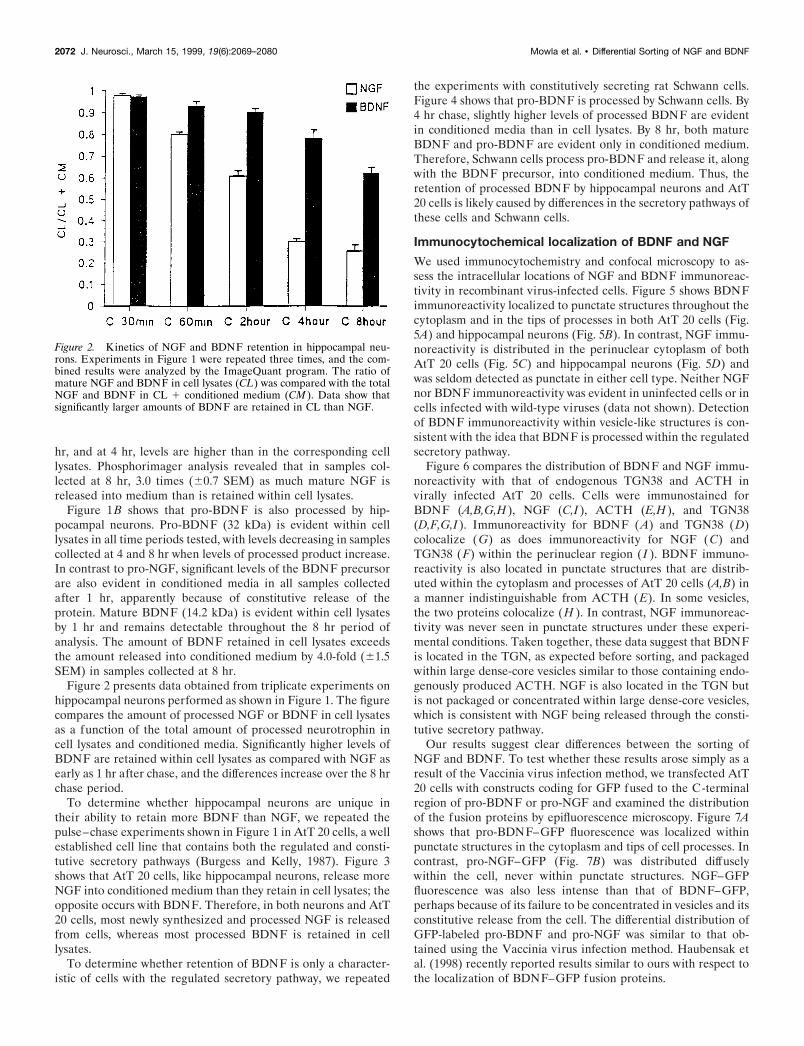
We used immunocytochemistry and confocal microscopy to as-sess the intracellular locations of NGF and BDNF immunoreac-tivity in recombinant virus-infected cells. Figure 5 shows BDNFimmunoreactivity localized to punctate structures throughout thecytoplasm and in the tips of processes in both AtT 20 cells (Fig.5A) and hippocampal neurons (Fig. 5B). In contrast, NGF immu-noreactivity is distributed in the perinuclear cytoplasm of bothAtT 20 cells (Fig. 5C) and hippocampal neurons (Fig. 5D) andwas seldom detected as punctate in either cell type. Neither NGFnor BDNF immunoreactivity was evident in uninfected cells or incells infected with wild-type viruses (data not shown). Detectionof BDNF immunoreactivity within vesicle-like structures is con-sistent with the idea that BDNF is processed within the regulatedsecretory pathway.
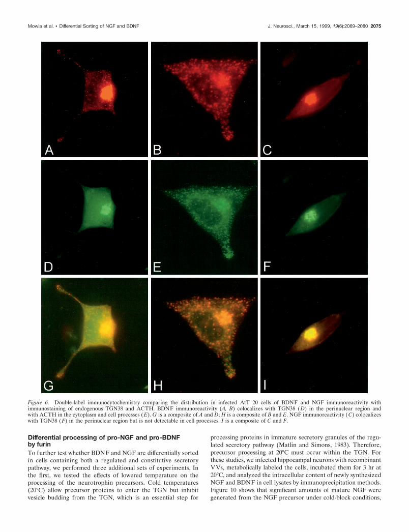
Figure 6 compares the distribution of BDNF and NGF immu-noreactivity with that of endogenous TGN38 and ACTH invirally infected AtT 20 cells. Cells were immunostained forBDNF (A,B,G,H), NGF (C,I), ACTH (E,H), and TGN38(D,F,G,I). Immunoreactivity for BDNF (A) and TGN38 (D)colocalize (G) as does immunoreactivity for NGF (C) andTGN38 (F) within the perinuclear region ( I ). BDNF immuno-reactivity is also located in punctate structures that are distrib-uted within the cytoplasm and processes of AtT 20 cells (A,B) ina manner indistinguishable from ACTH (E). In some vesicles,the two proteins colocalize (H ). In contrast, NGF immunoreac-tivity was never seen in punctate structures under these experi-mental conditions. Taken together, these data suggest that BDNFis located in the TGN, as expected before sorting, and packagedwithin large dense-core vesicles similar to those containing endo-genously produced ACTH. NGF is also located in the TGN butis not packaged or concentrated within large dense-core vesicles,which is consistent with NGF being released through the consti-tutive secretory pathway.
Our results suggest clear differences between the sorting ofNGF and BDNF. To test whether these results arose simply as aresult of the Vaccinia virus infection method, we transfected AtT20 cells with constructs coding for GFP fused to the C-terminalregion of pro-BDNF or pro-NGF and examined the distributionof the fusion proteins by epifluorescence microscopy. Figure 7Ashows that pro-BDNF–GFP fluorescence was localized withinpunctate structures in the cytoplasm and tips of cell processes. Incontrast, pro-NGF–GFP (Fig. 7B) was distributed diffuselywithin the cell, never within punctate structures. NGF–GFPfluorescence was also less intense than that of BDNF–GFP,perhaps because of its failure to be concentrated in vesicles and itsconstitutive release from the cell. The differential distribution ofGFP-labeled pro-BDNF and pro-NGF was similar to that ob-tained using the Vaccinia virus infection method. Haubensak etal. (1998) recently reported results similar to ours with respect tothe localization of BDNF–GFP fusion proteins.
Figure 2. Kinetics of NGF and BDNF retention in hippocampal neu-rons. Experiments in Figure 1 were repeated three times, and the com-bined results were analyzed by the ImageQuant program. The ratio ofmature NGF and BDNF in cell lysates (CL) was compared with the totalNGF and BDNF in CL 1 conditioned medium (CM ). Data show thatsignificantly larger amounts of BDNF are retained in CL than NGF.
2072 J. Neurosci., March 15, 1999, 19(6):2069–2080 Mowla et al. • Differential Sorting of NGF and BDNF

We performed metabolic labeling and SDS-PAGE analyses todetermine whether pro-BDNF- and pro-NGF–GFP-labeled con-structs were appropriately translated and processed in these ex-periments. However, these experiments were unsuccessful in AtT20 cells because of low transfection efficiency. We repeated theexperiments in COS 7 cells and found that both pro-NGF–GFPand pro-BDNF–GFP were processed appropriately, withoutcleavage of the GFP tag, as reported previously (Haubensak etal.,1998) (Fig. 7C). We also determined that medium conditionedby COS 7 cells that had been transfected with pro-NGF–GFP andpro-BDNF–GFP was fully active in inducing Trk A and Trk Bautophosphorylation, respectively, in NIH 3T3 cells engineeredto express the receptors (Fig. 7D). These data indicate that
GFP-tagged pro-BDNF and pro-NGF are processed appropri-ately and that conditioned media containing the precursor andmature forms of the proteins can activate their cognate receptors.Data monitoring the distribution of the neurotrophin–GFP fu-sion proteins further confirm our VV data indicating clear dif-ferences in the sorting and intracellular distribution of NGF andBDNF.
Depolarization-induced release of BDNF fromhippocampal neuronsIf BDNF is in the regulated secretory pathway, depolarizationshould promote its release. Figure 8 shows that BDNF levels inconditioned medium nearly doubled when hippocampal neurons
Figure 3. Pulse–chase metabolic labeling of pro-NGF (A) and pro-BDNF (B) production and processing in VV-infected AtT 20 cell cultures. Methodswere identical to those described in the legend to Figure 1. NGF and BDNF and their precursors were measured in conditioned medium (CM ) and incell lysates (CL). NI is a sample of CL precipitated with nonimmune serum.
Mowla et al. • Differential Sorting of NGF and BDNF J. Neurosci., March 15, 1999, 19(6):2069–2080 2073
were exposed to KCl; however, depolarization had no effect onNGF release. Depolarization did not promote the release ofpro-BDNF or pro-NGF under these experimental conditions(data not shown). To be certain that infecting hippocampal neu-rons with the NGF-coding virus had not altered the regulated
secretory pathway of hippocampal neurons, we monitored theeffects of KCl depolarization on the release of endogenously pro-duced secretogranin II, which is present in the regulated pathway.Figure 9 shows that KCl treatment effectively promoted secre-togranin II release in cells infected with pro-NGF encoding VV.
Figure 4. Pulse–chase metabolic labeling of primary rat Schwann cells infected with VV encoding pro-BDNF. Methods were identical to thosedescribed in the legend to Figure 1. BDNF and its precursor were measured in cell lysates (CL) and conditioned media (CM ). NI is a CL sampleprecipitated with nonimmune serum.
Figure 5. Confocal microscopy of AtT 20 cells (A, C) and hippocampal neurons (B, D) infected with VV encoding pro-NGF (C, D) or pro-BDNF (A,B). Cells were infected for 1 hr and postincubated in the absence of virus for another 8 hr. The cultures were fixed and treated with antibodies againstNGF or BDNF, followed by CY3-conjugated goat anti-rabbit IgG. Scale bar, 10 mm.
2074 J. Neurosci., March 15, 1999, 19(6):2069–2080 Mowla et al. • Differential Sorting of NGF and BDNF
Differential processing of pro-NGF and pro-BDNFby furinTo further test whether BDNF and NGF are differentially sortedin cells containing both a regulated and constitutive secretorypathway, we performed three additional sets of experiments. Inthe first, we tested the effects of lowered temperature on theprocessing of the neurotrophin precursors. Cold temperatures(20°C) allow precursor proteins to enter the TGN but inhibitvesicle budding from the TGN, which is an essential step for
processing proteins in immature secretory granules of the regu-lated secretory pathway (Matlin and Simons, 1983). Therefore,precursor processing at 20°C must occur within the TGN. Forthese studies, we infected hippocampal neurons with recombinantVVs, metabolically labeled the cells, incubated them for 3 hr at20°C, and analyzed the intracellular content of newly synthesizedNGF and BDNF in cell lysates by immunoprecipitation methods.Figure 10 shows that significant amounts of mature NGF weregenerated from the NGF precursor under cold-block conditions,
Figure 6. Double-label immunocytochemistry comparing the distribution in infected AtT 20 cells of BDNF and NGF immunoreactivity withimmunostaining of endogenous TGN38 and ACTH. BDNF immunoreactivity (A, B) colocalizes with TGN38 (D) in the perinuclear region andwith ACTH in the cytoplasm and cell processes (E). G is a composite of A and D; H is a composite of B and E. NGF immunoreactivity (C) colocalizeswith TGN38 (F) in the perinuclear region but is not detectable in cell processes. I is a composite of C and F.
Mowla et al. • Differential Sorting of NGF and BDNF J. Neurosci., March 15, 1999, 19(6):2069–2080 2075

whereas cold block totally inhibited the generation of matureBDNF from the BDNF precursor. Therefore pro-NGF, but notpro-BDNF, is cleaved within the TGN, probably by furin.
We also compared pro-NGF and pro-BDNF processing in thepresence of a1-PDX, an a1-anti-trypsin derivative that selectivelyinterferes with furin’s ability to process precursor proteins withinthe TGN (Anderson et al., 1993; Watanabe et al., 1995; Vollen-weider et al., 1996). In these studies, we monitored neurotrophinprocessing in AtT 20 cells coinfected with VVs encoding either
pro-NGF or pro-BDNF with or without VVs coding for a1-PDX.Figure 11 shows that a1-PDX had no detectable effect on pro-BDNF processing. However, a1-PDX did increase the amount ofpro-NGF released constitutively into conditioned medium, a re-sult that was similar to those obtained when we monitored pro-BDNF processing in hippocampal neurons and AtT 20 cells (Figs.1, 3). Identical results (data not shown) were obtained when weinfected neurotrophin-encoding viruses into a stably transfectedAtT 20 cell line overexpressing a1-PDX (Benjannet et al., 1997).
The finding that a1-PDX caused the constitutive release ofpro-NGF into conditioned medium suggested to us that a1-PDXmight be altering sorting of NGF within the cell, an idea con-firmed by immunocytochemistry. Figure 12B shows that in AtT20 cells that stably overexpress a1-PDX, pro-NGF encoding vi-ruses cause the accumulation of NGF immunoreactivity in punc-
Figure 7. Expression of pro-neurotrophin–GFP fusion proteins in AtT20 cells. Cells were plated on poly-L-lysine-coated coverslips and trans-fected using lipofectamine with cDNAs encoding either (A) pro-NGF–GFP or ( B) pro-BDNF–GFP. Three days later, the cells were analyzed byfluorescence microscopy. C, Immunoprecipitation and SDS-PAGE ofmetabolically labeled GFP fusion proteins from transfected COS 7 cells.D, Conditioned medium from pro-NGF–GFP or pro-BDNF–GFP ex-pressing COS 7 cells activate Trk A and Trk B phosphorylation, respec-tively, in NIH 3T3 cells engineered to overexpress either receptor.
Figure 8. KCl-induced release of BDNF but not NGF from hippocampalneurons. Hippocampal neurons from E18 mice were cultured for 7 d andinfected for 1 hr with VV encoding pro-NGF or pro-BDNF. After 10 hrin medium without virus, the cells were labeled for 30 min with [ 35S]Cys-Met, incubated in medium without radiolabel for 4 hr, and treatedwith medium with or without KCl and CaCl2 for 15 min. Conditionedmedia were immunoprecipitated with antibodies to NGF or BDNF andelectrophoresed on a SDS gel. Results were analyzed on a phosphorim-ager and are an average (6SEM) of three independent experiments.
Figure 9. Release of secretogranin II (sgII ) from hippocampal neuronsinfected with VV coding for pro-NGF. Eight hours after neurons wereexposed to VV, the cells were pulsed for 30 min with medium containing[ 35S] Cys-Met. The cells were chased for an additional 4 hr, after whichsamples of conditioned medium were analyzed (CM1), and again 30 minlater after the addition (1) or in the absence (2) of KCl (50 mM) addedto the culture medium (CM2).
2076 J. Neurosci., March 15, 1999, 19(6):2069–2080 Mowla et al. • Differential Sorting of NGF and BDNF
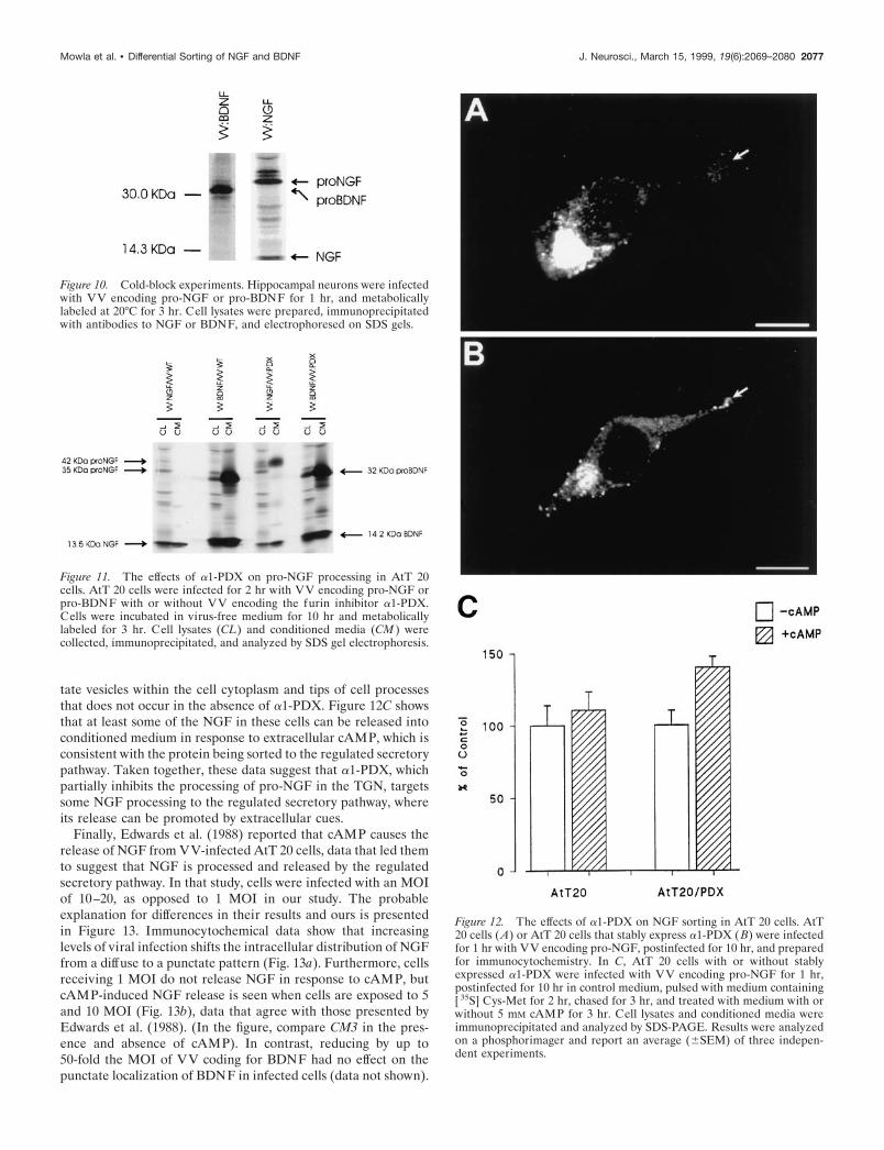
tate vesicles within the cell cytoplasm and tips of cell processesthat does not occur in the absence of a1-PDX. Figure 12C showsthat at least some of the NGF in these cells can be released intoconditioned medium in response to extracellular cAMP, which isconsistent with the protein being sorted to the regulated secretorypathway. Taken together, these data suggest that a1-PDX, whichpartially inhibits the processing of pro-NGF in the TGN, targetssome NGF processing to the regulated secretory pathway, whereits release can be promoted by extracellular cues.
Finally, Edwards et al. (1988) reported that cAMP causes therelease of NGF from VV-infected AtT 20 cells, data that led themto suggest that NGF is processed and released by the regulatedsecretory pathway. In that study, cells were infected with an MOIof 10–20, as opposed to 1 MOI in our study. The probableexplanation for differences in their results and ours is presentedin Figure 13. Immunocytochemical data show that increasinglevels of viral infection shifts the intracellular distribution of NGFfrom a diffuse to a punctate pattern (Fig. 13a). Furthermore, cellsreceiving 1 MOI do not release NGF in response to cAMP, butcAMP-induced NGF release is seen when cells are exposed to 5and 10 MOI (Fig. 13b), data that agree with those presented byEdwards et al. (1988). (In the figure, compare CM3 in the pres-ence and absence of cAMP). In contrast, reducing by up to50-fold the MOI of VV coding for BDNF had no effect on thepunctate localization of BDNF in infected cells (data not shown).
Figure 10. Cold-block experiments. Hippocampal neurons were infectedwith VV encoding pro-NGF or pro-BDNF for 1 hr, and metabolicallylabeled at 20°C for 3 hr. Cell lysates were prepared, immunoprecipitatedwith antibodies to NGF or BDNF, and electrophoresed on SDS gels.
Figure 11. The effects of a1-PDX on pro-NGF processing in AtT 20cells. AtT 20 cells were infected for 2 hr with VV encoding pro-NGF orpro-BDNF with or without VV encoding the furin inhibitor a1-PDX.Cells were incubated in virus-free medium for 10 hr and metabolicallylabeled for 3 hr. Cell lysates (CL) and conditioned media (CM ) werecollected, immunoprecipitated, and analyzed by SDS gel electrophoresis.
Figure 12. The effects of a1-PDX on NGF sorting in AtT 20 cells. AtT20 cells (A) or AtT 20 cells that stably express a1-PDX ( B) were infectedfor 1 hr with VV encoding pro-NGF, postinfected for 10 hr, and preparedfor immunocytochemistry. In C, AtT 20 cells with or without stablyexpressed a1-PDX were infected with VV encoding pro-NGF for 1 hr,postinfected for 10 hr in control medium, pulsed with medium containing[ 35S] Cys-Met for 2 hr, chased for 3 hr, and treated with medium with orwithout 5 mM cAMP for 3 hr. Cell lysates and conditioned media wereimmunoprecipitated and analyzed by SDS-PAGE. Results were analyzedon a phosphorimager and report an average (6SEM) of three indepen-dent experiments.
Mowla et al. • Differential Sorting of NGF and BDNF J. Neurosci., March 15, 1999, 19(6):2069–2080 2077

Therefore, sorting of BDNF to the regulated pathway is likely notattributable to concentration effects arising from the level of viralinfection.
DISCUSSIONPulse–chase studies show that hippocampal neurons and AtT 20cells retain more newly synthesized BDNF than they release.BDNF immunoreactivity is evident in punctate, vesicle-like struc-tures distributed throughout the cell cytoplasm, including in thetips of cell processes, and cell depolarization induces BDNFrelease. Thus, BDNF appears to sort primarily to the regulatedsecretory pathway. In contrast, hippocampal neurons and AtT 20cells release more NGF than they retain, NGF immunoreactivityis distributed diffusely within the perinuclear cytoplasm, presum-ably within the endoplasmic reticulum and Golgi apparatus, and
depolarization fails to promote NGF’s release into conditionedmedium. Thus, NGF appears to be processed and released in theconstitutive pathway. These results are not unique to the Vacciniavirus expression system because NGF and BDNF were differen-tially distributed as well within cells transfected with cDNAscoding for GFP–pro-neurotrophin fusion proteins.
Furin appears to cleave pro-NGF in the cells we tested. Pro-NGF processing is unaffected by cold-block conditions that in-hibit the exit of proteins from the TGN (Matlin and Simons,1983), suggesting that NGF processing occurs within the TGN. Incontrast, cold block totally inhibited the processing of pro-BDNF.Pro-BDNF processing likely occurs in immature secretory vesi-cles after they bud from the TGN, which is an early step in theregulated secretory pathway. Also, a1-PDX, a competitive inhib-itor of furin, increased levels of NGF precursor in conditionedmedium, which is consistent with its inhibiting pro-NGF process-ing within the TGN. a1-PDX did not affect pro-BDNF process-ing. In the presence of a1-PDX, NGF immunoreactivity ap-peared in punctate structures similar to those in cells infectedwith pro-BDNF encoding viruses, and some NGF precursor wasconstitutively secreted. Furthermore, depolarization releasedsmall amounts of NGF into conditioned medium. These resultsare similar to those obtained with pro-BDNF in the absence ofa1-PDX. Thus, inhibiting furin activity induces some pro-NGFto be sorted to the regulated secretory pathway. Similar effectshave been observed for pro-opiomelanocortin (Benjannet et al.,1997).
Cleavage by furin or furin-like enzymes within the TGN maybe one factor determining whether neurotrophins are sorted intothe constitutive or regulated secretory pathways. Studies with theprecursor for egg-laying hormone (pro-ELH) in mollusks mayexplain how this mechanism could work. Pro-ELH contains bio-active peptides on both the C- and amino-terminal sides of afurin cleavage site (Sossin et al., 1990). The C-terminal side ofthe precursor is sorted into the regulated secretory pathway afterfurin cleavage, and the amino-terminal side is released constitu-tively, degraded, or sorted into a separate regulated secretorypathway (Jung and Scheller, 1991). In cells with low furin levels,the precursor avoids cleavage, and both sides of the precursor aresorted into the same regulated secretory vesicles (Klumperman etal., 1996). Thus, sorting of the amino-terminal active peptidesinto dense-core secretory vesicles occurs only in the absence offurin cleavage, and different cells with different amounts of furinsort the same neuropeptide differently.
A similar situation may occur with neurotrophin processing.Sorting into the regulated pathway may require signals within thepro-domain or near the consensus cleavage site. When cleaved byfurin, the NGF precursor may lose these signals, and the maturecleaved protein is sorted into the constitutive pathway for release.In that regard, inserting furin-sensitive cleavage sites into pro-insulin (Yanagita et al., 1992) and pro-renin (Oda et al., 1991),which are normally processed by the regulated pathway, redirectsthese proteins into the constitutive pathway. In the absence offurin cleavage, the precursor remains intact, and sorting signalsthat direct the protein to the regulated pathway become func-tional. This may explain why pro-BDNF, which likely eludesfurin cleavage in the TGN, is targeted to the regulated pathwaywhere its processing appears to occur in secretory granules.
Differences between pro-BDNF and pro-NGF processing maytherefore arise because of the furin sensitivity of their pro-protein processing cleavage sites. In pro-NGF (Arg-Ser-Lys-Arg2Ser) the site is highly suited to furin processing, whereas
Figure 13. Overexpression of NGF results in missorting of NGF from theconstitutive to the regulated secretory pathway. a, AtT 20 cells wereinfected for 1 hr with 1 ( A), 5 ( B), or 10 ( C) MOI of VV coding forpro-NGF, postinfected for 8 hr, fixed, and prepared for immunocyto-chemistry using an antibody to NGF followed by a CY3-conjugatedsecondary antibody. Cells were analyzed by confocal microscopy. b, AtT20 cells were infected with 1 ( A), 5 ( B), or 10 ( C) MOI for 1 hr followedby a 4 hr postinfection and 3 hr incubation in medium containing [ 35S]Translabel. Conditioned media were collected (CM1), the cells werechased for 3 hr, and media was again collected (CM2). 8-bromo-cAMP (5mM) was then added to some cultures, and cells were incubated for anadditional 3 hr, after which media were collected (CM3) and the cells werelysed (CL). NGF was immunoprecipitated from all samples, and theprecipitate was analyzed by SDS-PAGE. Comparison of CM3 samplesshows that NGF release can be stimulated by cAMP from cells infectedwith 5 or 10 MOI but not from cells receiving 1 MOI.
2078 J. Neurosci., March 15, 1999, 19(6):2069–2080 Mowla et al. • Differential Sorting of NGF and BDNF
the analogous site in pro-BDNF (Arg-Val-Arg-Arg2His) is lesssuitable because of the replacement of Ser in the 11 position (P1)of NGF with His in the P1 of BDNF (Seidah et al., 1996a).Sequences with His at P1 show reduced sensitivity to furin-mediated cleavage (Ogi et al., 1990; Matthews et al., 1994).
Our results are consistent with this model. In constitutivelysecreting cells with moderate levels of furin-like enzymes, such asfibroblasts and Schwann cells (M. Marcinkiewicz and N. G. Sei-dah, unpublished observations), pro-NGF and pro-BDNF arecleaved, with only low levels of unprocessed precursor secretedconstitutively. In AtT 20 cells and hippocampal neurons withlower levels of furin (Seidah et al., 1994), pro-NGF is cleavedefficiently, and NGF is released constitutively. When furin cleav-age is inhibited with a1-PDX, pro-NGF is not cleaved efficiently,some sorting occurs into the regulated secretory pathway, wheresome of the protein can be released by depolarization, and someunprocessed precursor is secreted constitutively. In contrast, pro-BDNF avoids furin cleavage and is sorted into the regulatedsecretory pathway, and some unprocessed precursor is secretedconstitutively into conditioned medium. Constitutive release ofother precursors normally processed in the regulated pathway hasalso been reported (Kelly et al., 1983; Moore et al., 1983; Brechleret al., 1996)
An increasing number of reports suggest that BDNF, but notNGF, is anterogradely transported within axons in brain neuronsto carry out a number of physiological actions [Altar et al. (1997);Fawcett et al. (1998); for review, see Altar and DiStefano (1998)].Also, BDNF is enriched in a microvesicular fraction of rat brainsynaptosomes along with synaptotagmin, a protein associatedwith synaptic and large dense-core vesicles in nerve terminals(Fawcett et al., 1997). BDNF immunoreactivity is also present inmossy fiber terminals in the hippocampus (Conner et al., 1997;Fawcett et al., 1997) and in large dense-core vesicles of axonterminals in lamina II of lumbar spinal cord (Michael et al., 1997).BDNF can be transported anterogradely in neurons within thevisual system (von Bartheld et al., 1996) and released by depo-larization in a calcium-dependent mechanism from virus-infectedhippocampal neurons (Goodman et al., 1996). Taken together,these data are consistent with BDNF being packaged withinsecretory vesicles of the regulated pathway. Presumably thesegranules are targeted to axons, although transport may occur toother parts of the cell as well.
Fewer studies have monitored the processing of NGF in cellscontaining the regulated pathway. Edwards et al. (1988) reportedthat AtT 20 cells secrete VV-encoded NGF in response to cAMP,suggesting regulated release. In that study, they infected cells withan MOI of 10–20 as opposed to an MOI of 1 in our study, whichexplains why their results and ours differ (Fig. 13). Overloadingthe furin pathway beyond its capacity may drive NGF into theregulated pathway, as does inhibiting furin with a1-PDX (Figs.11, 12). In a similar manner, overexpressing b2-microglobulin inpancreatic b cells drives the protein from the constitutive pathwayinto secretory vesicles of the regulated pathway (Allison et al.,1991). In contrast, reducing infectivity from 1 to 0.02 MOI had noeffect on the sorting of BDNF, although we cannot rule out thepossibility that local concentration effects contributed to BDNF’ssorting into the regulated pathway.
Heymach et al. (1996) reported that AtT 20 cells release NGFas well as BDNF and NT-3 in response to secretagogues. It maybe that in those studies the level of NGF production was above thethreshold levels in which NGF is shunted from the constitutivepathway into the regulated pathway. Heymach et al. (1996) used
transfection instead of infection, expressed the neurotrophinsusing a different promoter, and treated cells with secretagoguesfor a longer time, which may further explain why their conclu-sions differ from ours. Blochl and Thoenen (1995) hypothesizedthat there is both constitutive and regulated sodium-dependentrelease of NGF from neurons. They suggest that release occursindependent of extracellular calcium, which is essential for pro-tein release in the regulated pathway (DeCamilli and Jahn, 1990),including the release of BDNF (Goodman et al., 1996). Canossaet al. (1997) extended these findings by reporting that neurotro-phins can also induce neurotrophin release from neurons (alsosee Kruttgen et al., 1998). These conflicting data need to beinvestigated further.
Our results provide a mechanism whereby neurotrophins inbrain neurons can act either as survival factors or as neuropep-tides. When neurotrophins are cleaved by furin, the bioactivepeptide may be released constitutively to promote neuronal sur-vival. Perhaps this explains how hippocampal neurons constantlyprovide NGF to innervating cholinergic neurons in the basalforebrain. In contrast, BDNF may avoid furin cleavage and besorted into the regulated secretory pathway where it is processedby PC1 (Seidah et al., 1996a) and released in an activity-dependent manner similar to other neuropeptides. This may bethe mechanism that allows BDNF to alter synaptic transmission,connectivity, and synaptic plasticity in an activity-dependentmanner (Ghosh, 1996). Also, perhaps neurons modulate the phys-iological fates of neurotrophins by regulating furin levels and thusthe intracellular sorting of the neurotrophins they produce.
REFERENCESAcheson A, Barker PA, Alderson RF, Miller FD, Murphy RA (1991)
Detection of brain-derived neurotrophic factor-like activity in fibro-blasts and Schwann cells: inhibition by antibodies to NGF. Neuron7:265–275.
Allison J, Malcolm L, Culvenor J, Batholomeusz RK, Holmberg K,Miller JF (1991) Overexpression of b2-microglobulin in transgenicmouse islet b cells results in defective insulin secretion. Proc Natl AcadSci USA 88:2070–2074.
Altar CA, DiStefano PS (1998) Neurotrophin trafficking by anterogradetransport. Trends Neurosci 21:433–437.
Altar CA, Cai N, Bliven T, Juhasz M, Conner JM, Acheson AL, LindsayRM, Wiegand SJ (1997) Anterograde transport of brain-derived neu-rotrophic factor and its role in the brain. Nature 389:856–860.
Anderson ED, Thomas L, Hayflick JS, Thomas G (1993) Inhibition ofHIV-1 gp160-dependent membrane fusion by a furin-directed a1-antitrypsin variant. J Biol Chem 268:24887–24891.
Banker GA, Cowan WM (1977) Rat hippocampal neurons in dispersedcell culture. Brain Res 126:397–442.
Benjannet S, Savaria D, Laslop A, Munzer JC, Chretien M, Marcin-kiewicz M, Seidah NG (1997) a1-antitrypsin Portland inhibits process-ing of precursors mediated by pro-protein convertases primarily withinthe constitutive secretory pathway. J Biol Chem 272:26210–26218.
Blochl A, Thoenen H (1995) Characterization of nerve growth factor(NGF) release from hippocampal neurons: evidence for a constitutiveand an unconventional sodium-dependent regulated pathway. EurJ Neurosci 7:1220–1228.
Brechler V, Chu WN, Baxter JD, Thibault G, Reudelhuber TL (1996) Aprotease processing site is essential for prorenin sorting to the regu-lated secretory pathway. J Biol Chem 271:20636–20640.
Bresnahan PA, Leduc R, Thomas L, Thorner J, Gibson HL, Brake AJ,Barr PJ, Thomas G (1990) Human fur gene encodes a yeast KEX2-like endoprotease that cleaves pro-beta-NGF in vivo. J Cell Biol111:2851–2859.
Brewer GJ, Torricelli JR, Evege EK, Price PJ (1993) Optimized survivalof hippocampal neurons in B27-supplemented Neurobasal, a newserum-free medium combination. J Neurosci Res 35:567–576.
Bunge RP (1994) The role of the Schwann cell in trophic support andregeneration. J Neurol [Suppl] 242:S19–S21.
Mowla et al. • Differential Sorting of NGF and BDNF J. Neurosci., March 15, 1999, 19(6):2069–2080 2079

Burgess TL, Kelly RB (1987) Constitutive and regulated secretion ofproteins. Annu Rev Cell Biol 3:243–293.
Canossa M, Greisbeck O, Berninger B, Campana G, Kolbeck R, ThoenenH (1997) Neurotrophin release by neurotrophins: implications foractivity-dependent neuronal plasticity. Proc Natl Acad Sci USA94:13279–13286.
Cartwright M, Mikheev AM, Heinrich G (1994) Expression of neuro-trophin genes in human fibroblasts: differential regulation of the brain-derived neurotrophic factor gene. Int J Dev Neurosci 12:685–693.
Conner JM, Lauterborn JC, Yan Q, Gall CM, Varon S (1997) Distribu-tion of brain-derived neurotrophic factor (BDNF) protein and mRNAin the normal adult rat CNS: evidence for anterograde axonal trans-port. J Neurosci 17:2295–2313.
DeCamilli P, Jahn R (1990) Pathways to regulated exocytosis in neu-rons. Annu Rev Physiol 52:625–645.
Dubois CM, Laprise MH, Blanchette F, Gentry LE, Leduc R (1995)Processing of transforming growth factor beta 1 precursor by humanfurin convertase. J Biol Chem 270:10618–10624.
Edwards RH, Selby MJ, Mobley WC, Weinrich SL, Hruby DE, Rutter WJ(1988) Processing and secretion of nerve growth factor: expression inmammalian cells with a vaccinia virus vector. Mol Cell Biol8:2456–2464.
Fawcett JP, Aloyz R, McLean JH, Pareek S, Miller FD, McPherson PS,Murphy RA (1997) Detection of brain-derived neurotrophic factor ina vesicular fraction of brain synaptosomes. J Biol Chem 272:8837–8840.
Fawcett JP, Bamji SX, Causing CG, Aloyz R, Ase AR, Reader TA,McLean JH, Miller FD (1998) Functional evidence that BDNF is ananterograde neuronal trophic factor in the CNS. J Neurosci18:2808–2821.
Ghosh A (1996) Cortical development: with an eye on neurotrophins.Curr Biol 6:130–133.
Goodman LJ, Valverde J, Lim F, Geschwind MD, Federoff HJ, GellerAI, Hefti F (1996) Regulated release and polarized localization ofbrain-derived neurotrophic factor in hippocampal neurons. Mol CellNeurosci 7:222–238.
Haubensak W, Narz F, Heumann R, Lessmann V (1998) BDNF-GFPcontaining secretory granules are localized in the vicinity of synapticjunctions of cultured cortical neurons. J Cell Sci 111:1483–1493.
Hempstead BL, Rabin SJ, Kaplan L, Reid S, Parada LF, Kaplan DR(1992) Overexpression of the Trk tyrosine kinase rapidly acceleratesnerve growth factor-induced differentiation. Neuron 9:883–896.
Heymach JJ, Kruttgen A, Suter U, Shooter EM (1996) The regulatedsecretion and vectorial targeting of neurotrophins in neuroendocrineand epithelial cells. J Biol Chem 271:25430–25437.
Hosaka M, Nagahama M, Kim WS, Watanabe T, Hatsuzawa K, IkemizuJ, Murakami K, Nakayama K (1991) Arg-X-Lys/Arg-Arg motif as asignal for precursor cleavage catalyzed by furin within the constitutivesecretory pathway. J Biol Chem 266:12127–12130.
Jung LJ, Scheller RH (1991) Peptide processing and targeting in theneuronal secretory pathway. Science 251:1330–1335.
Kelly RB, Buckley KM, Burgess TL, Carlson SS, Caroni P, Hooper JE,Katzen A, Moore HP, Pfeffer SR, Schroer TA (1983) Membranetraffic in neurons and peptide-secreting cells. Cold Spring Harbor SympQuant Biol 2:697–705.
Klumperman J, Spijker S, van Minnen J, Sharp-Baker H, Smit AB,Geraerts WP (1996) Cell type-specific sorting of neuropeptides: amechanism to modulate peptide composition of large dense-core vesi-cles. J Neurosci 16:7930–7940.
Kruttgen A, Moller JC, Heymach JV, Shooter EM (1998) Neurotro-phins induce release of neurotrophins by the regulated secretory path-way. Proc Natl Acad Sci USA 95:9614–9619.
Loh YP (1993) Mechanisms of intracellular trafficking and processing ofproproteins. Boca Raton, FL: CRC.
Luzio JP, Brake B, Banting G, Howell KE, Bragetta P, Stanley KK(1990) Identification, sequencing, and expression of an internal mem-brane protein of the trans-Golgi network (TGN38). Biochem J270:97–102.
Matlin KS, Simons K (1983) Reduced temperature prevents transfer of amembrane glycoprotein to the cell surface but does not prevent termi-nal glycosylation. Cell 34:233–243.
Matthews DJ, Goodman LJ, Gorman CM, Wells JA (1994) A survey offurin substrate specificity using substrate phage display. Protein Sci3:1197–1205.
Michael GJ, Averill S, Nitkunan A, Rattray M, Bennett DL, Yan Q,Priestley JV (1997) Nerve growth factor treatment increases brain-derived neurotrophic factor selectively in TrkA-expressing dorsal rootganglion cells and in their central terminations within the spinal cord.J Neurosci 17:8476–8490.
Moore HP, Walker MD, Lee F, Kelly RB (1983) Expressing a humanproinsulin cDNA in a mouse ACTH-secreting cell. Intracellular stor-age, proteolytic processing, and secretion on stimulation. Cell35:531–538.
Mowla SJ, Fawcett JP, Pareek S, Seidah NG, Sossin WS, Murphy RA(1997) Differential sorting of nerve growth factor and brain-derivedneurotrophic factor in hippocampal neurons. Soc Neurosci Abstr23:875.10.
Murphy RA, Acheson A, Hodges R, Haskins J, Richards C, Reklow E,Chlumecky V, Barker PA, Alderson RF, Lindsay RM (1993) Immu-nological relationships of NGF, BDNF, and NT-3: recognition andfunctional inhibition by antibodies to NGF. J Neurosci 13:2853–2862.
Oda K, Ikeda M, Tsuji E, Sohda M, Takami N, Misumi Y, Ikehara Y(1991) Sequence requirements for proteolytic cleavage of precursorswith paired basic amino acids. Biophys Biochem Res Commun179:1181–1186.
Ogi K, Kimura C, Onda H, Arimura A, Fujimo M (1990) Molecularcloning and characterization of cDNA for the precursor of rat pituitaryadenylate cyclic activating polypeptide (PACAP). Biophys BiochemRes Commun 173:1271–1279.
Pareek S, Suter U, Snipes GJ, Welcher AA, Shooter EM, Murphy RA(1993) Detection and processing of peripheral myelin protein PMP22in cultured Schwann cells. J Biol Chem 268:10372–10379.
Rouille Y, Duguay SJ, Lund K, Furuta M, Gong Q, Lipkind G, Oliva AJ,Chan SJ, Steiner DF (1995) Proteolytic processing mechanisms in thebiosynthesis of neuroendocrine peptides: the subtilisin-like proproteinconvertases. Front Neuroendocrinol 16:322–361.
Seidah NG, Chretien M, Day R (1994) The family of subtilisin/kexin-like proprotein and prohormone convertases: divergent or shared func-tions. Biochimie 76:197–209.
Seidah NG, Benjannet S, Pareek S, Chretien M, Murphy RA (1996a)Cellular processing of the neurotrophin precursors of NT3 and BDNFby the mammalian proprotein convertases. FEBS Lett 379:247–250.
Seidah NG, Benjannet S, Pareek S, Savaria D, Hamelin J, Goulet B,Laliberte J, Lazure C, Chretien M, Murphy RA (1996b) Cellularprocessing of the nerve growth factor precursor by the mammalianpro-protein convertases. Biochem J 314:951–960.
Singh N, Birdi TJ, Antia NH (1997) Nerve growth factor production andexpression of p75 by Schwann cells and neurofibroblasts in response toM. leprae infection and macrophage secretory products. NeuropatholAppl Neurobiol 23:59–67.
Snider WD, Lichtman JW (1996) Are neurotrophins synaptotrophins?Mol Cell Neurosci 7:433–442.
Sossin WS, Fisher JM, Scheller RH (1990) Sorting within the regulatedsecretory pathway occurs in the trans-Golgi network. J Cell Biol110:1–12.
Thoenen H (1995) Neurotrophins and neuronal plasticity. Science270:593–598.
Vollenweider F, Benjannet S, Decroly E, Savaria D, Lazur C, Thomas G,Chretien M, Seidah NG (1996) Comparative cellular processing of thehuman immunodeficiency virus (HIV-1) envelope glycoprotein gp160by the mammalian subtilisin/kexin-like convertases. Biochem J314:521–532.
von Bartheld CS, Byers MR, Williams R, Bothwell M (1996) Antero-grade transport of neurotrophins and axodendritic transfer in thedeveloping nervous system. Nature 379:830–833.
Watanabe M, Hirano A, Stenglein S, Nelson J, Thomas G, Wong TC(1995) Engineered serine protease inhibitor prevents furin-catalyzedactivation of the fusion glycoprotein and production of infectiousmeasles virus. J Virol 69:3206–3210.
Yan Q, Rosenfeld RD, Metheson CR, Hawkins N, Lopez OT, Bennett L,Welcher AA (1997) Expression of brain-derived neurotrophic factorprotein in the adult rat central nervous system. Neuroscience78:431–448.
Yanagita M, Nakayama K, Takeuchi T (1992) Processing of mutatedproinsulin with tetrabasic cleavage sites to bioactive insulin in thenon-endocrine cell line, COS-7. FEBS Lett 311:55–59.
2080 J. Neurosci., March 15, 1999, 19(6):2069–2080 Mowla et al. • Differential Sorting of NGF and BDNF