
Differential Isolation of Normal Luminal Mammary Epithelial Cells...
10
(CANCER RESEARCH 53. 627-635. February 1, I Differential Isolation of Normal Luminal Mammary Epithelial Cells and Breast Cancer Cells from Primary and Metastatic Sites Using Selective Media1 Stephen P. Ethier,2 Michael L. Mahacek, William J. Gullick, Thomas S. Frank, and Barbara L. Weber Departments o) Radiation Oncology IS. P. £.,M. L M.¡,Pathology ¡T.S. F.¡,and Internal Medicine ¡B.L W.l, University of Michigan Medical School. Ann Arbor. Michigan 4810V, and Hammersmith Hospital. Imperial Cancer Research Fund. Oncology Group. London WI2 QHS. England ¡W.J. C.I ABSTRACT The present studies were aimed at determining if the use of a cell culture medium that supports proliferation of human mammary epithelial cells of the luminal lineage would allow routine isolation of breast cancer cells from primary and metastatic tumor specimens. Results obtained with mammary epithelial cells derived from reduction mammoplasty speci mens and primary breast carcinomas indicated that growth of cells on type I collagen-coated dishes in Ham's F-12 medium supplemented with insulin, hydrocorti.sone, epidermal growth factor, cholera toxin, and 5% fetal bovine scrum resulted in the growth and serial passage of cells that stained positively for the luminal cell marker cytokeratin 19. By contrast, growth of mammary epithelial cells in a growth factor-supplemented se rum-free medium resulted in the emergence of mammary epithelial cell colonies that were uniformly negative for keratin 19. Filter isolation meth ods were used to isolate individual keratin-19-positive colonies from pri mary cultures derived from breast cancer specimens. All of the luminal mammary epithelial cells isolated from breast cancer tissues expressed characteristics of normal cells. Keratin-19-positive colonies isolated from several different tumors all grew rapidly for 30 to 60 days in culture and then senesced. Cells were isolated from one tumor that was known to have undergone a loss of heterozygosity at a specific locus in the p53 gene. All colonies isolated from this specimen contained both p53 alÃ-eles,which was consistent with their origin from normal luminal cells. Cells were also isolated from one tumor in which the c-erbB2 protein was drastically overexpressed in the neoplastic cells. Once again, keratin-19-positive col onies isolated from this tumor did not overexpress the c-erbB-2 protein. Experiments were then performed with cells derived from pleural effu sions and metastatic lymph nodes. Results obtained with these specimens indicated that the growth conditions that support the growth of normal luminal mammary epithelial cells do not support the growth of neoplastic cells. However, the omission of cholera toxin, epidermal growth factor, and type I collagen substratum resulted in the isolation of two long-term cell lines. Both cell lines have population doubling times of approximately 100 h, are hyperdiploid, and stain positively for cytokeratin 19. Thus, culture conditions that support the growth of normal luminal mammary epithelial cells do not, in general, support the growth of breast cancer cells. INTRODUCTION Experimental systems that allow direct comparisons to be made between normal and neoplastic cells provide a powerful tool for the determination of specific phenotypes of neoplastic cells that contrib ute directly to their neoplastic potential. In addition, determination of the important altered phenotypes expressed by neoplastic cells allows mechanistic connections to be made regarding the molecular (genetic) basis for those altered phenotypes. Over the past several years, we have taken this approach in the study of the biology of rat mammary carcinogenesis by identifying alterations in growth-regulatory mech anisms that characterize the neoplastic cells and studying these alter ations at the cellular and molecular level (1-6). A similar approach to Received 6/3/92; accepted 11/18/92. The costs of publication of this article were defrayed in part by the payment of page charges. This article must therefore be hereby marked advertisement in accordance with 18 U.S.C. Section 1734 solely to indicale this fact. 1 Supported by National Cancer Institute Grant CA4(X)64. Grant 274827 from the University of Michigan Breast Care Center, and Cytogenetics Core Grant CA46592. 2 To whom requests for reprints should be addressed. the study of human breast cancer biology has been hampered by technical limitations that have precluded the in vitro growth of normal mammary epithelial cells of the lineage that gives rise to HBC and the successful culture of primary human breast cancer cells themselves on a routine basis. There have been a number of improvements in cell culture tech nology that have led to the development of culture conditions that support rapid and prolonged proliferation of normal HME3 cells ob tained from reduction mammoplasty specimens. Stampfer et al. (7- 10) developed a culture medium for the growth of HME cells that originally consisted of many undefined components and was later refined such that bovine pituitary extract constituted the only unde fined component of the medium. The medium developed by Stampfer et al. ( 11) is a hormone- and growth factor-supplemented medium that supports proliferation of HME cells over many in vitro passages and was used to develop an immortalized cell line following chemical carcinogen treatment of the cells. Subsequently, other groups reported the successful culture of HME cells using similar media. Band and Sager (12) demonstrated extensive proliferation of HME cells in a growth factor- and hormone-supplemented medium that also con tained serum and pituitary extract. Petersen and Van Deurs (13) and Ethier et al. (14) reported growth of normal HME cells in serum-free media in the absence of pituitary extract or serum. Despite this ap parent success in the development of tissue culture methods for the growth of normal mammary epithelial cells, none of these systems support growth of mammary epithelial cells of the luminal lineage. This is important because Taylor-Papadimitriou et al. (15, 16) dem onstrated that HBC cells in vivo express cytokeratin markers consis tent with an origin from the luminal cells of the terminal ductal lobular unit. In addition, these workers demonstrated that human breast cancer cell lines retain expression of keratins expressed by luminal mammary epithelial cells even after extended in vitro passage. By contrast, mammary epithelial cells that proliferate rapidly in vitro under culture conditions discussed above are uniformly negative for the expression of luminal cell cytokeratins and are positive for keratins expressed by basal/myoepithelial cells present in the mammary gland. Thus, the culture conditions that support rapid proliferation of normal HME cells over many in vitro passages do not support the growth of the normal cell type from which breast cancer arises. This inability to culture the appropriate cell lineage has had impor tant implications for experiments in which similar culture conditions were applied in an attempt to culture HBC cells from primary tumors. Wolman et al. (17) demonstrated that cells cultured from primary tumors were uniformly diploid even when obtained from tumors that were known to consist of aneuploid breast cancer cells. This was one of the First indications that culture conditions that support the growth of normal HME cells were not suitable for the growth of breast cancer cells obtained from primary tissues. Evidence obtained by Taylor- Papadimitriou et al. (16) is consistent with that notion, since they 'The abbreviations used are: HME. human mammary epithelial; HBC. human breast cancer; FBS, fetal bovine serum; IN, insulin; HC. hydrocortisone; EGÕ-". epidermal growth factor; CT, cholera toxin; K-19, cytokeratin-19; LOH, loss of heterozygosity; PCR. polymerase chain reaction. 627 on May 6, 2019. © 1993 American Association for Cancer Research. cancerres.aacrjournals.org Downloaded from
Transcript of Differential Isolation of Normal Luminal Mammary Epithelial Cells...

(CANCER RESEARCH 53. 627-635. February 1, I
Differential Isolation of Normal Luminal Mammary Epithelial Cells and BreastCancer Cells from Primary and Metastatic Sites Using Selective Media1
Stephen P. Ethier,2 Michael L. Mahacek, William J. Gullick, Thomas S. Frank, and Barbara L. Weber
Departments o) Radiation Oncology IS. P. £.,M. L M.¡,Pathology ¡T.S. F.¡,and Internal Medicine ¡B.L W.l, University of Michigan Medical School. Ann Arbor. Michigan4810V, and Hammersmith Hospital. Imperial Cancer Research Fund. Oncology Group. London WI2 QHS. England ¡W.J. C.I
ABSTRACT
The present studies were aimed at determining if the use of a cellculture medium that supports proliferation of human mammary epithelialcells of the luminal lineage would allow routine isolation of breast cancercells from primary and metastatic tumor specimens. Results obtained withmammary epithelial cells derived from reduction mammoplasty specimens and primary breast carcinomas indicated that growth of cells ontype I collagen-coated dishes in Ham's F-12 medium supplemented with
insulin, hydrocorti.sone, epidermal growth factor, cholera toxin, and 5%fetal bovine scrum resulted in the growth and serial passage of cells thatstained positively for the luminal cell marker cytokeratin 19. By contrast,growth of mammary epithelial cells in a growth factor-supplemented serum-free medium resulted in the emergence of mammary epithelial cell
colonies that were uniformly negative for keratin 19. Filter isolation methods were used to isolate individual keratin-19-positive colonies from pri
mary cultures derived from breast cancer specimens. All of the luminalmammary epithelial cells isolated from breast cancer tissues expressedcharacteristics of normal cells. Keratin-19-positive colonies isolated from
several different tumors all grew rapidly for 30 to 60 days in culture andthen senesced. Cells were isolated from one tumor that was known to haveundergone a loss of heterozygosity at a specific locus in the p53 gene. Allcolonies isolated from this specimen contained both p53 alÃeles,which wasconsistent with their origin from normal luminal cells. Cells were alsoisolated from one tumor in which the c-erbB2 protein was drasticallyoverexpressed in the neoplastic cells. Once again, keratin-19-positive colonies isolated from this tumor did not overexpress the c-erbB-2 protein.
Experiments were then performed with cells derived from pleural effusions and metastatic lymph nodes. Results obtained with these specimensindicated that the growth conditions that support the growth of normalluminal mammary epithelial cells do not support the growth of neoplasticcells. However, the omission of cholera toxin, epidermal growth factor, andtype I collagen substratum resulted in the isolation of two long-term cell
lines. Both cell lines have population doubling times of approximately 100h, are hyperdiploid, and stain positively for cytokeratin 19. Thus, cultureconditions that support the growth of normal luminal mammary epithelialcells do not, in general, support the growth of breast cancer cells.
INTRODUCTION
Experimental systems that allow direct comparisons to be madebetween normal and neoplastic cells provide a powerful tool for thedetermination of specific phenotypes of neoplastic cells that contribute directly to their neoplastic potential. In addition, determination ofthe important altered phenotypes expressed by neoplastic cells allowsmechanistic connections to be made regarding the molecular (genetic)basis for those altered phenotypes. Over the past several years, wehave taken this approach in the study of the biology of rat mammarycarcinogenesis by identifying alterations in growth-regulatory mech
anisms that characterize the neoplastic cells and studying these alterations at the cellular and molecular level (1-6). A similar approach to
Received 6/3/92; accepted 11/18/92.The costs of publication of this article were defrayed in part by the payment of page
charges. This article must therefore be hereby marked advertisement in accordance with18 U.S.C. Section 1734 solely to indicale this fact.
1Supported by National Cancer Institute Grant CA4(X)64. Grant 274827 from the
University of Michigan Breast Care Center, and Cytogenetics Core Grant CA46592.2 To whom requests for reprints should be addressed.
the study of human breast cancer biology has been hampered bytechnical limitations that have precluded the in vitro growth of normalmammary epithelial cells of the lineage that gives rise to HBC and thesuccessful culture of primary human breast cancer cells themselves ona routine basis.
There have been a number of improvements in cell culture technology that have led to the development of culture conditions thatsupport rapid and prolonged proliferation of normal HME3 cells ob
tained from reduction mammoplasty specimens. Stampfer et al. (7-
10) developed a culture medium for the growth of HME cells thatoriginally consisted of many undefined components and was laterrefined such that bovine pituitary extract constituted the only undefined component of the medium. The medium developed by Stampferet al. ( 11) is a hormone- and growth factor-supplemented medium that
supports proliferation of HME cells over many in vitro passages andwas used to develop an immortalized cell line following chemicalcarcinogen treatment of the cells. Subsequently, other groups reportedthe successful culture of HME cells using similar media. Band andSager (12) demonstrated extensive proliferation of HME cells in agrowth factor- and hormone-supplemented medium that also con
tained serum and pituitary extract. Petersen and Van Deurs (13) andEthier et al. (14) reported growth of normal HME cells in serum-free
media in the absence of pituitary extract or serum. Despite this apparent success in the development of tissue culture methods for thegrowth of normal mammary epithelial cells, none of these systemssupport growth of mammary epithelial cells of the luminal lineage.This is important because Taylor-Papadimitriou et al. (15, 16) dem
onstrated that HBC cells in vivo express cytokeratin markers consistent with an origin from the luminal cells of the terminal ductal lobularunit. In addition, these workers demonstrated that human breast cancercell lines retain expression of keratins expressed by luminal mammaryepithelial cells even after extended in vitro passage. By contrast,mammary epithelial cells that proliferate rapidly in vitro under cultureconditions discussed above are uniformly negative for the expressionof luminal cell cytokeratins and are positive for keratins expressed bybasal/myoepithelial cells present in the mammary gland. Thus, theculture conditions that support rapid proliferation of normal HMEcells over many in vitro passages do not support the growth of thenormal cell type from which breast cancer arises.
This inability to culture the appropriate cell lineage has had important implications for experiments in which similar culture conditionswere applied in an attempt to culture HBC cells from primary tumors.Wolman et al. (17) demonstrated that cells cultured from primarytumors were uniformly diploid even when obtained from tumors thatwere known to consist of aneuploid breast cancer cells. This was oneof the First indications that culture conditions that support the growthof normal HME cells were not suitable for the growth of breast cancercells obtained from primary tissues. Evidence obtained by Taylor-
Papadimitriou et al. (16) is consistent with that notion, since they
'The abbreviations used are: HME. human mammary epithelial; HBC. human breastcancer; FBS, fetal bovine serum; IN, insulin; HC. hydrocortisone; EGÕ-".epidermal growth
factor; CT, cholera toxin; K-19, cytokeratin-19; LOH, loss of heterozygosity; PCR.polymerase chain reaction.
627
on May 6, 2019. © 1993 American Association for Cancer Research. cancerres.aacrjournals.org Downloaded from

GROWTH OF NORMAL AND NEOPLAST1C HUMAN MAMMARY CELLS
demonstrated that HME cells cultured from primary breast cancerspecimens expressed the same cytokeratin profile as cells culturedfrom reduction mammoplasties.
We have worked to develop and refine cell culture methods thatsupport the proliferation of normal HME cells of the luminal lineageand bona fide HBC cells obtained from primary and metastatic tumortissues. These tissue culture methods support primary culture andserial passage of luminal mammary epithelial cells derived from reduction mammoplasties and primary breast cancers. In addition, wehave developed methods to selectively isolate luminal cell colonies inorder to study their proliferative potential in the absence of the morerapidly growing basal/myoepithelial cells. Finally, we have used thesemethods to isolate HBC cells from pleural effusions, metastatic lymphnodes, and primary tumors and have detected significant alterations ingrowth phenotypes between these cells and normal luminal mammaryepithelial cells.
MATERIALS AND METHODS
Preparation of Normal and Neoplastic Mammary Epithelial Cells.Mammary epithelial cells were obtained from normal mammary tissues, primary breast cancers, and metastatic lymph nodes by enzymatic dissociation asdescribed previously (14) but with minor modifications. Briefly, tissues wereminced with scalpels and incubated overnight in Medium 199 containing typeIII collagenase (Worthington Biochemical Corp.. Freehold, NJ) at a concentration of 2(X)units/ml and Dispase (Boehringer Mannheim, Indianapolis, IN)at 1 mg/ml. Twenty ml of media/g of tissue were used. The tissues wereagitated gently in a shaking water bath at 37°C.The cells are then washed
extensively in Medium 199, and an aliquot of the cells is counted by isolatingnuclei and counting nuclei with a Coulter counter as previously described (18).
Cells were isolated from pleural effusion specimens by the centrifugation ofI liter of fluid at 800 x g for 5 min. The pellets were resuspended in 500 ulof Medium 199 and layered onto a 2-ml bed of Percoli (1.09 g/ml) and
centrifuged at 800 x g for 5 min. Under these conditions, erythrocytes form a
pellet below the Percoli, and the cells that form a layer between the mediumand the Percoli are collected, washed with Medium 199. enumerated, andseeded into culture.
When more cells than were used in primary culture experiments wereobtained from patient specimens, the excess cells were cryopreserved forfuture use. Cell aggregates obtained by enzymatic dissociation or collectionfrom pleural fluids freeze well, and most samples yield viable cell culturesupon reactivation. To freeze cells obtained from patient samples, cells aresuspended in Medium 199 supplemented with 20% FBS and 5% dimethylsulfoxide at a concentration of 5 x 10°cells/750 pi of freezing medium. Thecells are cooled slowly to -80°using a step freezer and then stored over liquid
nitrogen. Over the past 3 years we have established a cell bank of mammaryepithelial cells from normal and neoplastic tissues from over 30 patients.
Cell Culture. Mammary epithelial cells isolated by the methods describedabove were seeded onto collagen-coated 60-mm or 35-mm culture dishes atdensities ranging from 10' to 5 X IO5 cells/dish. The complete serum-containing medium consists of Ham's F-12 supplemented with IN (5 |Jg/ml), HC
(I Mg/ml). EOF (10 ng/ml), CT (100 ng/ml), 5% FBS, gentamycin, and Fun-gizone. The complete serum-free medium is the same as just described, except
that the serum is replaced with ethanolamine (5 mM), transferrin (5 ug/ml),bovine serum albumin ( 1 mg/ml), sodium selenite (50 ng/ml), and triiodothy-
ronine (50 ng/ml).For the subculture of cells grown in primary culture, cells were rinsed with
calcium and magnesium-free Hanks' balanced salt solution and incubated with
trypsin-EDTA for 2-5 min. The cells were reseeded at split ratios of 1:3 to
1:10. For filter isolation of individual mammary epithelial cell colonies, cellswere rinsed with Hanks' balanced salt solution as above, and colonies were
covered with pieces of Whatman 3-mm paper that had been soaked in warmtrypsin-EDTA. After two min, the filter was removed from the colony withgentle downward pressure and then placed, cells down, in a 35-mm dish
containing fresh growth medium. The plates were agitated gently and incubated overnight to allow cells to detach from the filter and attach to the
substratum. For the subculture of cells obtained from metasiatic lymph nodes,confluent lawns of stromal cells containing growing epithelial colonies wererinsed with Hanks' balanced salt solution as described above and then incu
bated in calcium and magnesium-free Hanks' alone or in the presence of 10 mM
EDTA. The cells were observed under the phase-contrast microscope until the
epithelial cells detached. The plates were then rinsed gently, and the cells weretransferred to a new dish.
Immunocytochemistry. For immunocytochemical analyses of culturedcells, 35-mm dishes were rinsed with phosphate-buffered saline and fixed withmethanol at -20°C for 10 min. The fixed cells were then rinsed three times
with phosphate-buffered saline and incubated in primary antibody in phosphate-buffered saline for 30 min at room temperature. Primary antibodies usedin these experiments include AE1/AE3 for broad-spectrum keratin recognition.
Ks 19.1 for cytokeratin 19 recognition (ICN Flow, Costa Mesa, CA), and Tab259 (kindly provided by Dr. Beatrice Langton, Berlex Biosciences, Alameda,CA) for c-erbB2 protein detection. Following the primary antibody step, cellswere rinsed three times with phosphate-buffered saline and then incubated withbiotinylated anti-mouse IgG for 30 min. The cells were then rinsed and pro
cessed using Vectastain ABC reagents (Vector Laboratories. Burlingame, CA)and visualized using diaminobenzidine as the substrate.
PCR Analysis of the P53 Gene for Loss of Heterozygosity. For theseexperiments, tissues obtained from histological sections of breast cancer specimens and cells cultured from breast cancer specimens were examined for LOHusing a SsrU-1 restriction fragment length polymorphism in the P53 gene. To
determine which patients were heterozygous at the BslV-l restriction site and
which of those patients tumors had an LOH at that locus, new histologicalsections were cut from breast cancer specimens of patients whose cells werecryopreserved in our cell bank. Areas consisting of neoplastic cells and normal
cells were microdissected separately from these specimens, digested withproteinase K, and PCR amplified using nested primers (see below). The PCR-amplified DNA was then digested with RsfU-I (New England Biolabs, Beverly,
MA) and analyzed in 3% Nusieve/1% agarose gels. Similarly, cultured cellsderived from the same patients were isolated by filter selection and washed fivetimes with 5% dextrose in water and suspended in 10 mM Tris (pH 8.3)containing 5.0 mM potassium chloride, 1.5 mM magnesium chloride, 20 UMdithiothreitol, 1.7 UMsodium dodecyl sulfate, and 10 ug Proteinase K/20 ul ofcell suspension. The Proteinase K digestion was carried out for l h at 37°C
before initiating PCR amplification.Oligonucleotide primers were designed to amplify DNA sequences flanking
the BsiU-l restriction site in codon 72 of the 4th exon of the p53 gene (19).
Primers were designed to optimize annealing at a common temperature andwith discordant 3 ' ends to prevent formation of primer dimers. Oligonucleotide
primers were synthesized by the University of Michigan DNA Synthesis CoreFacility. Primers used in the first round of nested PCR had the sequences5'-TGGATGATTTGATGCTGTC-3' (upstream primer) and 5'-CGTG-CAAGTCACAGACTT-3' (downstream primer), which produced an amplified
segment of 257 base pairs corresponding to bases 23-279 of the p53 4th exon.Primers used in the second, "nested" round had the sequences 5'-CCCGGAC-GATATTGAACAA-3' (upstream primer) and 5'-GCTGTCCCAGAATG-CAAG-3' (downstream primer), resulting in a PCR product of 219 base pairs
corresponding to bases 42-260 of the 4th exon.After the addition of 31 ul of crude DNA, the final 50-ul reaction mix
contained 200 UMof each deoxynucleotide triphosphate, 2.5 ng/ul (approximately 380 nM of each) first-round primers, 2.5 units of Taq polymerase
(Promega. Madison, WI), and 1X Taq polymerase buffer. PCR parameters forboth first- and second-round amplifications consisted of an initial 4-min de-naturation step at 94°Ccoupled to a repeating cycle of 1 min at 94°C,2 minat 48°C,and 2 min at 72°Cfor 35 cycles followed by a 7-min "completion"
step at 72°C.Five ill of first-round PCR product were transferred to a 45-ul
pre-mix solution with deoxynucleotide triphosphate. Oligonucleotide. and Taq
polymerase concentrations as above, and amplification was repeated for 35cycles. After amplification. 15 ul of each PCR mixture were digested with 5units of B.srU-1at 60°Cfor at least 4 h in a 30-|jl volume using buffer provided
with the enzyme, followed by electrophoresis through a 3% NuSeive (FMCBioproducts. Rockland, ME)/1% agarose (BRL) gel containing 0.5 ug/mlethidium bromide. The BstV-l site, when present, resulted in the cleavage ofthe 219-base pair nested PCR product into fragments 78 and 141 base pairs in
length.
628
on May 6, 2019. © 1993 American Association for Cancer Research. cancerres.aacrjournals.org Downloaded from
GROWTH OF NORMAL AND NEOPLASTIC HUMAN MAMMARY CELLS
RESULTS
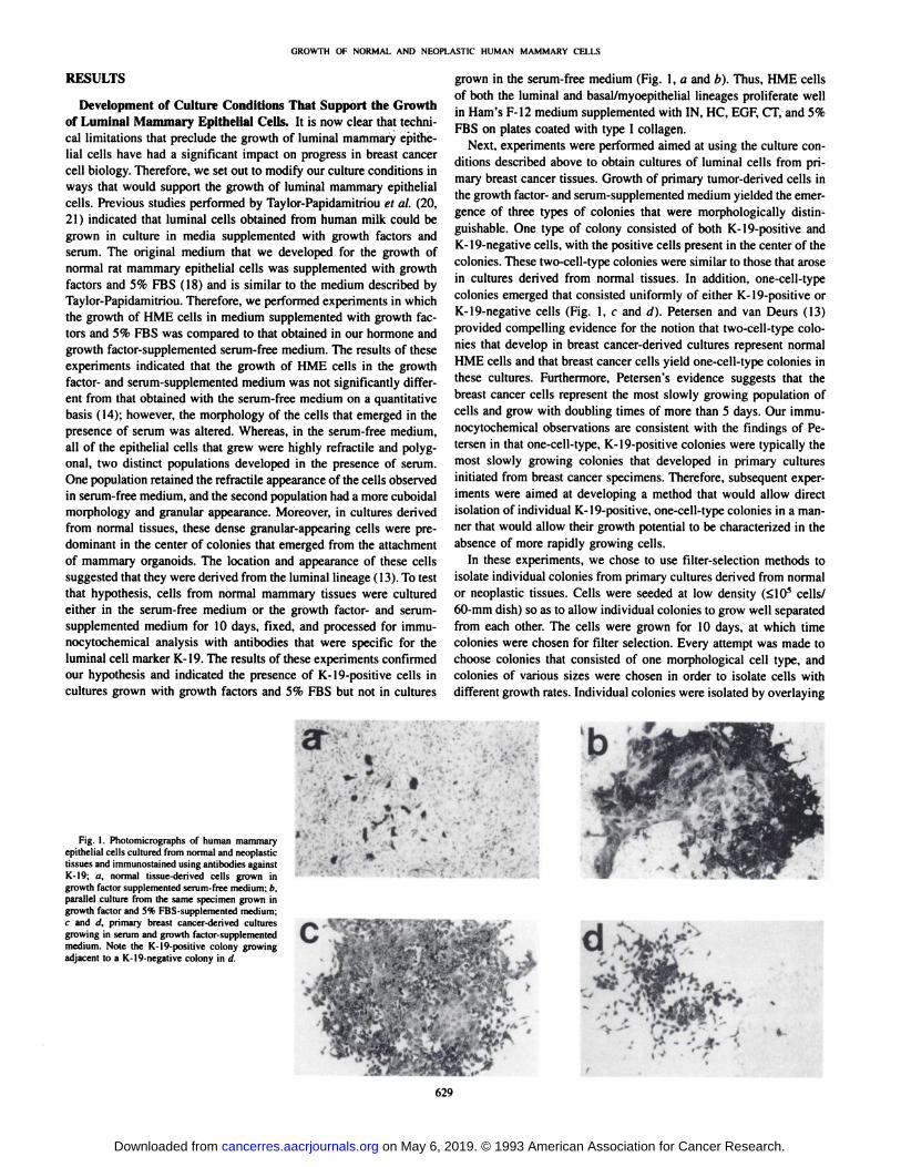
Development of Culture Conditions That Support the Growthof Luminal Mammary Epithelial Cells. It is now clear that technical limitations that preclude the growth of luminal mammary epithelial cells have had a significant impact on progress in breast cancercell biology. Therefore, we set out to modify our culture conditions inways that would support the growth of luminal mammary epithelialcells. Previous studies performed by Taylor-Papidamitriou et al. (20,
21) indicated that luminal cells obtained from human milk could begrown in culture in media supplemented with growth factors andserum. The original medium that we developed for the growth ofnormal rat mammary epithelial cells was supplemented with growthfactors and 5% FBS (18) and is similar to the medium described byTaylor-Papidamitriou. Therefore, we performed experiments in which
the growth of HME cells in medium supplemented with growth factors and 5% FBS was compared to that obtained in our hormone andgrowth factor-supplemented serum-free medium. The results of these
experiments indicated that the growth of HME cells in the growthfactor- and serum-supplemented medium was not significantly different from that obtained with the serum-free medium on a quantitative
basis (14); however, the morphology of the cells that emerged in thepresence of serum was altered. Whereas, in the serum-free medium,
all of the epithelial cells that grew were highly refractile and polygonal, two distinct populations developed in the presence of serum.One population retained the refractile appearance of the cells observedin serum-free medium, and the second population had a more cuboidal
morphology and granular appearance. Moreover, in cultures derivedfrom normal tissues, these dense granular-appearing cells were pre
dominant in the center of colonies that emerged from the attachmentof mammary organoids. The location and appearance of these cellssuggested that they were derived from the luminal lineage ( 13). To testthat hypothesis, cells from normal mammary tissues were culturedeither in the serum-free medium or the growth factor- and serum-supplemented medium for 10 days, fixed, and processed for immu-
nocytochemical analysis with antibodies that were specific for theluminal cell marker K-19. The results of these experiments confirmedour hypothesis and indicated the presence of K-19-positive cells in
cultures grown with growth factors and 5% FBS but not in cultures
grown in the serum-free medium (Fig. 1, a and b). Thus, HME cells
of both the luminal and basal/myoepithelial lineages proliferate wellin Ham's F-12 medium supplemented with IN, HC, EGF, CT, and 5%
FBS on plates coated with type I collagen.Next, experiments were performed aimed at using the culture con
ditions described above to obtain cultures of luminal cells from primary breast cancer tissues. Growth of primary tumor-derived cells inthe growth factor- and serum-supplemented medium yielded the emer
gence of three types of colonies that were morphologically distinguishable. One type of colony consisted of both K-19-positive andK-19-negative cells, with the positive cells present in the center of thecolonies. These two-cell-type colonies were similar to those that arosein cultures derived from normal tissues. In addition, one-cell-typecolonies emerged that consisted uniformly of either K-19-positive orK-19-negative cells (Fig. 1, c and d). Petersen and van Deurs (13)provided compelling evidence for the notion that two-cell-type colonies that develop in breast cancer-derived cultures represent normalHME cells and that breast cancer cells yield one-cell-type colonies inthese cultures. Furthermore, Petersen's evidence suggests that the
breast cancer cells represent the most slowly growing population ofcells and grow with doubling times of more than 5 days. Our immu-
nocytochemical observations are consistent with the findings of Petersen in that one-cell-type, K-19-positive colonies were typically the
most slowly growing colonies that developed in primary culturesinitiated from breast cancer specimens. Therefore, subsequent experiments were aimed at developing a method that would allow directisolation of individual K-19-positive, one-cell-type colonies in a man
ner that would allow their growth potential to be characterized in theabsence of more rapidly growing cells.
In these experiments, we chose to use filter-selection methods to
isolate individual colonies from primary cultures derived from normalor neoplastic tissues. Cells were seeded at low density (<10S cells/
60-mm dish) so as to allow individual colonies to grow well separated
from each other. The cells were grown for 10 days, at which timecolonies were chosen for filter selection. Every attempt was made tochoose colonies that consisted of one morphological cell type, andcolonies of various sizes were chosen in order to isolate cells withdifferent growth rates. Individual colonies were isolated by overlaying
Fig. 1. Photomicrographs of human mammaryepithelial cells cultured from normal and neoplastictissues and immunostained using antibodies againstK-19; a, normal tissue-derived cells grown ingrowth factor supplemented serum-free medium; b.parallel culture from the same specimen grown ingrowth factor and 5% FBS-supplemented medium;c and (I. primary breast cancer-derived culturesgrowing in serum and growth factor-supplementedmedium. Note the K-19-positive colony growingadjacent to a K-19-negative colony in d.
-. »t
•¿�
629
on May 6, 2019. © 1993 American Association for Cancer Research. cancerres.aacrjournals.org Downloaded from

GROWTH OF NORMAL AND NEOPLASTIC HUMAN MAMMARY CELLS
the colonies with filter discs that were soaked with trypsin:EDTA andthen transferring the discs to 35-mm wells containing fresh growth
medium. Cells isolated in this way were then grown to confluence andsplit at a ratio of 1:3. At the end of the second passage, cells from onewell were subcultured, and the cells in the remaining two wells weretested for keratin expression. In all cases, the cultures derived fromindividual colonies reacted positively with broad-spectrum anti-kera
tin antibodies confirming the epithelial nature of the cells. However,certain colony-derived cultures were uniformly positive for K-19,
indicating the luminal origin of the cells, whereas others were uniformly K-19-negative, indicating the basal or myoepithelial nature of
those cells. The filter isolation methods were used to analyze thegrowth rate and proliferative life span of approximately 50 colony-
derived cultures originating from normal tissues and 5 different primary breast cancers. The proliferative life span of colonies derivedfrom tumor specimens was consistently greater than that of culturesderived from normal cell colonies. Whereas colonies isolated fromnormal tissues typically senesced in the second or third passage,colonies derived from tumor tissues proliferated for three to fivepassages before senescing. One slow-growing colony isolated from a
tumor specimen proliferated for 10 passages or approximately 6months in culture before senescing. In no case did we isolate animmortal cell line from a primary tumor using these methods toselectively grow K-19-positive cells.
The ability to isolate individual K-19-positive colonies from breastcancer-derived primary cultures allows genetic analysis to be carried
out on these cultures to determine unequivocally if they are normal orneoplastic. To perform the genetic analysis, a marker is required thatwill distinguish normal from neoplastic cells from an individual patient and which can be detected with a small number of cells. Wechose to examine breast cancer specimens for the presence of apolymorphism in the BstU-l restriction site of the p53 gene using
nested PCR (19). The p53 gene is an important genetic locus in humanbreast cancer (22, 23), and the presence or absence of the restrictionsite can be examined by PCR analysis. The first step in our approachwas to determine, for patients whose cells are frozen in our cell bank,which are constitutionally heterozygous at this locus in normal cellsand which have lost one alÃelein their cancer cells. To do this experiment, new histológica! sections were cut from formalin-fixed tissue
blocks from a series of patients. Next, normal and neoplastic cellswere isolated from different areas of the sections, and these sampleswere prepared for PCR analysis of the p53 gene. The samples wereamplified using a nested PCR approach, incubated with the restrictionenzyme BxtU-l, and analyzed on Nusieve/agarose gels. From our
initial series, we identified four patients that were heterozygous at thislocus and one that had a LOH at that locus in the cancer cells (Fig. 2).Thus, this LOH can be used as a genetic signature that distinguishesnormal from neoplastic cells from that patient. Accordingly, cells fromthe patient found to have lost an alÃelein the tumor cells (SUM-43;
Fig. 2, Lanes 4 and 5) were reactivated from frozen stocks andcultured at low density, and individual colonies were isolated by filterselection. Cultures that arose from the isolated colonies were analyzedby PCR as described above. Fifteen separate colonies were analyzedin this way, and all of the colonies had the genetic signature of normalcells; i.e., both alÃelespresent (Fig. 2, Lanes 6-8). It must be noted
that the presence of any normal cells in a colony that was composedpredominantly of cancer cells would be read as normal in this assaybecause of the sensitivity of the PCR method. Our current data indicate that we were not able to isolate pure colonies of neoplastic cellsfrom SUM-43 by filter selection of colonies proliferating in medium
that supports the growth of luminal mammary cells.
12345678Fig. 2. LOH at the BifU-I restriction site in patient sample designated SUM-43. Lane
I, size markers. iMnes 2 and 3 were obtained from DNA prepared from a patient that washeterozygous for the Bsru-l site in both normal (Lane 2) and neoplastic (Lane 3) cells.
Lanes 4 and 5, heterozygosity for the site in normal cells (Lane 4) and loss of an alÃelein the cancer cells (Lane 5) of specimen SUM-43. Lanes 6-8, LOH analysis of threerepresentative colonies isolated from cultures obtained from SUM-43. Upper band, 219-base pair PCR fragment that does not contain the Bst\i-\ site; two lower bands, 141- and78-base pair PCR fragments that result from cleavage wilh Bsta-l.
A second approach that would allow the detection of neoplasticcells in primary cultures derived from tumor tissues involves theidentification of specimens in which the neoplastic cells overexpressa specific protein as a result of a genetic alteration in the tumor cells.We screened a series of histológica! sections of primary breast cancerspecimens of patients whose cells were frozen in our cell bank, foroverexpression of the erbB-2 protein. This protein is dramaticallyoverexpressed in cells that have an amplified c-erbB-2 gene. Although
a number of the specimens analyzed expressed moderate levels of thec-erbB-2 protein, in one specimen the protein was dramatically over-expressed by the neoplastic cells (SUM-33; Fig. 3a). Tumor-derived
cells from this patient were then reactivated from frozen stocks andcultured for 7 days in the medium that supports the growth of luminalmammary epithelial cells and then tested immunocytochemically forerbB-2 expression and K-19 expression. For this experiment, SKBr-3
cells were used as positive controls for immunocytochemical detection of erbB-2 protein. K-19-positive colonies did emerge in primary
cultures derived from this specimen (Fig. 3c). However, none of thecolonies stained positively for erbB-2 protein (Fig. 3b). By contrast,SKBR-3 cells yielded strong positive cell surface and cytoplasmic
staining under identical conditions (Fig. 3d). The results of this experiment are consistent with the results described above and indicatethat primary HBC cells do not proliferate well in a growth mediumthat supports the growth of luminal mammary epithelial cells.
Isolation and Growth of HBC Cells from Metastatic Sites.Since experiments with cells obtained from primary human breastcancer specimens indicated that normal mammary epithelial cells arethe predominant cell type to emerge in these cultures and that thesecells proliferate rapidly, yielding confluent monolayers of normalcells, experiments were initiated aimed at isolating neoplastic HBCcells from metastatic sites. In the first experiment performed withpleural effusion-derived cells (SUM-44PE), we made use of the cul
ture conditions that we had shown previously support the growth ofHME cells of the luminal lineage. SUM-44PE cells were isolated from1 liter of pleural effusion fluid, seeded onto type I collagen-coatedtissue culture plates, and grown in Ham's F-12 medium supplemented
with IN, HC, EGF, CT, and 5% FBS. In addition, attempts were madeto culture these cells in similar media in which individual factors hadbeen deleted. The results shown in Fig. 4 indicate that SUM-44 PEcells grew poorly in the fully growth factor- and serum-supplemented
medium that supports the growth of normal luminal HME cells. However, deletion of CT resulted in improved proliferation. In thisexperiment, the best growth was obtained in media supplemented with
630
on May 6, 2019. © 1993 American Association for Cancer Research. cancerres.aacrjournals.org Downloaded from

GROWTH OF NORMAL AND NEOPLASTIC HUMAN MAMMARY CELLS
.
I
/ *
>d W**ï*• *•¿�;••¿�'; * /•.,, . -li*:1 m4, -r '
Fig. 3. Immuncytochemical analysis of erhB-2 expression in specimen designated SUM-33. a. deteclion of frfeB-2-overexpressing neoplaslic cells but not normal cells in Ihehistological section from this patient, b and d, immunocytochemical analysis of erbB-2 expression of cultured SUM-33 cells and SkBr-3 cells, r, immunoslaining of cultured SUM-33cells with K-19 antibodies.
5% FBS and IN plus HC, and under these conditions the cells grewwith a population doubling time of approximately 12 days. The presence or absence of EGF had no effect on the growth of these breastcancer cells. In the next experiment, SUM-44 PE cells were thawed
and grown in media supplemented with 5% FBS, IN, HC, and otherhormones and growth factors to determine if the culture conditionscould be improved further. Progesterone, estradici, acidic fibroblastgrowth factor, and basic fibroblast growth factor had no significanteffect on the growth of these cells when added to the medium supplemented with IN, HC, and 5% FBS (data not shown). However, thisexperiment did indicate that SUM-44 PE cells grew better in serum-free medium supplemented with IN plus HC than in the serum-
containing medium supplemented with the same factors (Fig. 5).When grown in serum-free medium, omission of IN in primary culture
had a significant effect on cell growth with little or no proliferationtaking place in the absence of this factor (Fig. 6). Next, cells that grewto confluence in primary culture were subcultured and tested for theirability to grow on tissue culture plastic versus tissue culture platescoated with type I collagen. In this experiment, the cells exhibitedimproved attachment and viability and appeared to grow better ontissue culture plastic than on the collagen-coated plates. These experiments suggested that the growth of SUM-44PE cells could be im
proved by the omission of serum and by growing cells on tissueculture plastic. To test those observations, cells were reactivated fromfrozen stocks, and a primary culture growth curve was obtained for the
cells growing under the improved culture conditions. The data in Fig.7 indicate that SUM-44 PE cells grown in serum-free medium with IN
and HC and on tissue culture plastic proliferate with a populationdoubling time of approximately 5 days. This is significantly fasterthan the 12-day doubling time observed in the original experimentswith cells grown in the presence of serum and on collagen-coated
plates. Thus, metastatic breast cancer cells obtained from the patientdesignated SUM-44 PE exhibit a number of cellular phenotypes that
distinguish them from normal luminal mammary epithelial cells.SUM-44 PE cells are positive for the luminal cell marker K-19, yet,unlike K-19 positive normal HME cells, these breast cancer cells do
not require serum factors for growth in culture. Whereas normal HMEcells respond to EGF and CT by rapid proliferation, SUM-44PE cells
do not respond to exogenous EGF and are growth inhibited by CT.These cells do exhibit a requirement, however, for exogenous IN andHC for growth in culture. SUM-44 PE cells grow better on tissueculture plastic than on a collagen substratum. Finally, SUM-44PE
cells grow very slowly in culture and have not exhibited signs ofsenescence. This characteristic is dramatically different from that expressed by normal cells, which grow very rapidly in culture (population doubling times of 24 to 36 h) and then uniformly senesce after 30to 60 days. SUM-44PE cells have now been in culture for over 15
months and are in their 17th passage (split ratios 1:3). Thus, weestimate that these cells have undergone approximately 30 populationdoublings. Preliminary karyotypic analysis indicates that SUM-44PE
631
on May 6, 2019. © 1993 American Association for Cancer Research. cancerres.aacrjournals.org Downloaded from

GROWTH OF NORMAL AND NEOPLASTIC HUMAN MAMMARY CELLS
cells have a model chromosome number of 60 and exhibit severalmarker chromosomes that are being characterized further (Figs. 8aand 9A).
The results obtained with SUM-44 PE cells prompted experiments
to determine if cells that express similar altered phenotypes could bedetected in cells from other metastatic breast cancer specimens. In oneexperiment, cells from a metastatic lymph node were reactivated fromfrozen stocks (SUM-16LN) and tested for their ability to grow eitherin the growth factor- and serum-supplemented medium required by
normal cells or in the medium supplemented with IN, HC, and 5%FBS that was used originally to isolate SUM-44PE cells. After 6
weeks in primary culture, plates cultured in the medium supplementedwith 5% FBS, IN, HC, EOF. and CT were completely overgrown bystromal cells, and there was no sign of HBC cell growth. By contrast,the plates cultured in the 5% IN, HC medium contained a confluentlawn of density-arrested stromal cells in which slowly growing epithelial colonies emerged. After 6 weeks in primary culture, the neo-
plastic epithelial cells were harvested by treatment of the cultures with
120000 -
100000 -
20000 -
DAYS IN CULTURE
Fig. 4. Primary culture growth obtained with SUM-44PE cells. Cells were seeded ontolype-l collagen-coated 60-mm culture dishes at 5 X \<f cells/60-mm culture dish andgrown in Ham's F-12 medium with 5% FBS plus IN (/); IN and HC (IH}: IN, HC. and
EOF (IHE). or IN. HC. EGF. and CT (IHEC). The cells were grown for 4 weeks, andtriplicate dishes were counted at each time point. Bars, range in the data.
200000
GC
£100000 -
SF-IH 5% IH
Fig. 5. Primary culture growth of SUM-44PE cells in media supplemented with 5^FBS. IN. and HC or in serum-free medium with IN and HC (SF-IH). Cells were seededat 10" cells/35-mm well and cultured for 2 weeks, at which time triplicate wells were
counted. Bars, range in the data.
DCVCL
20000 -
SF-IH SF-H
Fig. 6. Influence of IN on growth of SUM-44 PE cells in primary culture. Cells werecultured for 2 weeks in medium supplemented with HC in the presence or absence of IN.The experiment was performed as described in Fig. 5.
650000
IV)5
10 15
DAYS IN CULTURE
Fig. 7. Primary culture growth curve for SUM-44PE cells cultured on tissue cultureplastic in serum-free medium .supplemented with IN and HC. Cells were seeded at 2.5 XIO5 cells/35-mm well and grown for 3 weeks. At each time point, triplicate wells were
counted. Bars, range in the data.
0.1 M EDTA in calcium- and magnesium-free Hanks' balanced salt
solution. These cells reattached in the first passage and gave rise toslowly growing epithelial colonies that grew to confluence after 4weeks in passage one. These cells proliferate slowly in culture likeSUM-44PE cells and have now been in culture for over 6 months.Preliminary karyotypic analysis indicates that SUM-16LN cells have
a model chromosome number of 62 and exhibit several marker chromosomes (Figs. 8¿and 9ß).The results obtained with SUM-16LNcells coupled with those obtained with SUM-44PE cells indicate that
a medium supplemented with 5% FBS, I, and HC, which does notsupport the growth of normal mammary epithelial cells, can be usedto isolate HBC cells from some metastatic breast cancer specimens.
We have now tested the 5% FBS, I, HC medium and the serum-free
I, HC medium described above with several primary and metastaticHBC specimens to determine if these media can be used for theroutine isolation of breast cancer cells from patient specimens. Ourresults to date indicate that only a subset of tumor specimens can becultured in these media. Among metastatic specimens, we have initiated cultures from 6 additional pleural effusion specimens and 5additional metastatic lymph node specimens. Of these specimens, onlyone additional lymph node-derived specimen and two additional pleural effusion-derived specimens gave rise to subculturable HBC cellcolonies. Growth of primary tumor-derived cells in the media used toisolate SUM-44PE and SUM-16LN often resulted in the slow emer
gence of epithelial colonies that were morphologically distinct fromnormal mammary epithelial cell colonies. The tumor designatedSUM-55 consisted of normal mammary epithelial cells that grewrapidly in the fully growth factor- and serum-supplemented medium.
In addition, this specimen consisted of cells that gave rise to manytightly packed epithelial cell colonies in the medium supplementedwith IN, HC, and 5% FBS (Fig. 8c). These colonies continued toproliferate in primary culture for 5 months, yet none of the colonies
632
on May 6, 2019. © 1993 American Association for Cancer Research. cancerres.aacrjournals.org Downloaded from

GROWTH OF NORMAL AND NEOPLASTIC HUMAN MAMMARY CELLS
Fig. 8. Photomicrographs of cultured HBC cells, a, SUM-44PE cells growing inserum-free medium with insulin and hydrocortisone on tissue culture plastic; h. SUM-I6LN cells growing in similar culture conditions; c. typical colony that arose in culturesfrom SUM-55 after 2 months in selective medium.
yielded viable cultures upon passage, despite numerous attempts usingseveral different methods to remove the cells from the primary dish.We have now attempted to isolate HBC cells from more than 10primary tumors using the media described above. Although we havedetected long-lived colonies in these media in at least three specimens,
we have yet to isolate subculturable HBC cells from any primarytumor using these growth conditions.
In summary, our results indicate that selective growth media can beused to isolate human breast cancer cells from primary and metastatic
sites; however, only a subset of these tumors consists of cells that canproliferate continuously in these media.
DISCUSSION
The routine isolation and culture of neoplastic mammary epithelialcells from primary breast cancer specimens has still not been accomplished. One hypothesis regarding the inability to culture these cellscenters around the observation that the culture media developed forthe growth of normal mammary epithelial cells does not support thegrowth of mammary epithelial cells of the luminal lineage. Sincebreast cancer arises from luminal mammary epithelial cells, development of a culture medium that supports the growth of these cellsshould allow the growth of neoplastic cells from breast cancer specimens. The results of our experiments do not support this hypothesis.In our studies, we used a culture medium that supports the primaryculture and serial passage of luminal mammary epithelial cells. Inaddition, we used a filter isolation method that allowed for the isolation of individual mammary epithelial cell colonies. The use of thesemethods did not result in the isolation of HBC cells from primarytumor specimens. Rather, primary tumor-derived cells yielded rapidly
growing epithelial cell cultures that expressed all the features ofnormal cells. The cells grew rapidly and underwent a limited numberof population doublings before senescing. The limited growth potential of K-19-positive mammary epithelial cells obtained from breast
cancer tissues was uniformly observed in mass cultures and in culturesstarted from colonies isolated by filter selection. Finally, the use ofgenetic and protein markers that defined the neoplastic versus thenormal cell compartment from their respective tissues demonstratedthat the cells cultured from these specimens were normal and notneoplastic. In the filter isolation experiments, we consistently observed the extended proliferation of keratin-19-positive cells obtained
from breast cancer specimens relative to cells obtained from normaltissues. The mechanistic significance of this observation is not clear;however, it is noteworthy that we previously made similar observations regarding the growth factor requirements of normal cells derivedfrom primary tumors (14). Thus, it is possible that the tumor microen-
vironment affects many aspects of the proliferati ve capacity of normalmammary epithelial cells within the tumor.
The results obtained with metastatic HBC specimens are consistentwith the results obtained with primary tumors. Thus far, we haveattempted to isolate HBC cells from 8 metastatic breast cancer specimens using the culture conditions that support the growth of luminalmammary epithelial cells. Thus far, this medium has not resulted inthe isolation of breast cancer cells from these specimens. However, wehave isolated two HBC cell lines from the metastatic specimensusing a selective medium from which growth factors required bynormal cells were omitted. Indeed, we found that omission of thesefactors was critical for the selective isolation of these breast cancercells. Thus, the results of our studies suggest that HBC cells havegrowth factor requirements distinct from those of normal humanmammary epithelial cells and express dramatically different growthphenotypes in culture. The inabilty to isolate HBC cells from allmetastatic specimens or from primary tumors using our selectiveculture conditions suggests further that these cells have requirementsfor exogenous growth factors that are still to be defined.
The most common phenotype that we have observed in HBC cellsobtained from primary and metastatic sites is their slow proliferationrate. Our observations are consistent with the findings of other groups.Many cell lines have been developed from human breast cancers, andvirtually all of these cell lines grew very slowly in culture in earlypassages and acquired rapid growth rates after months or years in
633
on May 6, 2019. © 1993 American Association for Cancer Research. cancerres.aacrjournals.org Downloaded from

GROWTH OF NORMAL AND NEOPLAST1C HUMAN MAMMARY CELLS
r IM miiii j«n a M/I'' BU
6 7 8 9 10» 11 12
M MM13 14 15
Mit "ft*15 1J
ai17 18
19 20
Fig. 9. Karyotypes of SUM-44PE and SUM-16LN cells. A, near-triploid karyolype of SUM-44PE displaying the following representativeclonal structural abnormalities: add (I)(p22); i(2)-(qlO)x2; del (7)(q32)x2; I(l;lO)(q2l;q24); dic-(Il;l6)(pl3;q22); +8-14 UMARS. B. representative hypotriploid karyotype from SUM-16 LN
displaying multiple clonal structural alterations, including del(7)(pl 1.2), add ( I3)(pl I ), add(20)(pl3),and +10-16 UMARS.
21
C A
22 X/Y
i Y i
10 11 12
ÃŽf Hi u16 17 18
21 22
«X/Y
B-Umars-
634
on May 6, 2019. © 1993 American Association for Cancer Research. cancerres.aacrjournals.org Downloaded from
GROWTH OF NORMAL AND NEOPI.ASTIC HUMAN MAMMARY CELLS
culture (24, 25). Thus, the rapid growth rates that characterize manyhuman breast cancer cell lines currently in use are a characteristicacquired by the cells over many years in culture and do not reflect theoriginal growth potential of the cells. In experiments with primarytumor-derived cells, Petersen and van Deurs (13) used an enzymaticmethod for detecting breast cancer cells growing in primary culture inan enriched growth medium while growing in the midst of normalmammary epithelial cells. The results of their growth rate studies alsoindicated that primary human breast cancer cells grow very slowly inculture (doubling times > 120 h), even when grown in a well-defined,highly growth factor-enriched medium. It is important to note that the
observed growth rates of primary breast cancer cells are entirelyconsistent with the rate at which these tumors grow in vivo (26-28).
Thus, given the consistency of the experimental observations made bymany workers, the fact that breast cancer cells grow slowly despite thepresence of many exogenous growth factors, and the agreement withclinical observations of breast cancer growth rates, the slow proliferation of breast cancer cells in culture is likely to reflect the true growthpotential of the cells and is unlikely to be an in vitro artifact. Indeed,it is the slow rate at which HBC cells grow in culture that precludesthe use of enriched media for the growth of neoplastic cells obtainedfrom primary tumors. Media that support the growth of normal mammary epithelial cells yields confluent monolayers of cells within 1-2
weeks of the initiation of cultures. Thus, even HBC cells that doproliferate in fully growth factor-supplemented media are likely to be
rapidly overgrown by normal cells under those culture conditions. Itappears that improved success in isolating HBC cells from primaryand metastatic sites will require selective culture conditions that provide appropriate growth factor stimulation for the neoplastic cellswhile at the same time not supporting the rapid proliferation of normalcells.
Some human breast cancer cells can be isolated in highly enrichedculture media. In recent years, Petersen et al. (29), Band et al. (30),and Meltzer et al. (31) have developed HBC cell lines from primarytumors specimens using enriched growth media. However, in each ofthese cases, the lines were established from 1% to 2% of the specimens tested. Thus. HBC cells that can be isolated under these conditions are relatively rare. It is interesting to note that the cell linesdeveloped by Band et al. (30) and Meltzer et al. (31) both have anamplified ERBB-2 gene, and both expressed requirements for EOF inculture. Thus, HBC cells that overexpress erbB-2 protein and the EOF
receptor may represent the subset of primary breast cancers mostamenable to isolation using enriched culture media.
In conclusion, the results of our studies indicate that a subset ofHBC cells can be isolated and cultured under selective conditions withminimal growth factor supplementation. Our results also suggest thatbreast cancer cells that do not proliferate under the selective cultureconditions have requirements for exogenous growth factors that haveyet to be determined and which are distinct from the growth factorsrequired by normal human mammary epithelial cells.
ACKNOWLEDGMENTS
The authors wish to express their appreciation to Drs. David August, VernonSondak, Alfred Chang, and the members of the University of Michigan BreastCare Center for providing human breast cancer specimens to the laboratory. Wealso acknowledge the technical expertise of Dr. Jeffry Trent and Tom Dennisand the University of Michigan Cytogenetics Core facility for their assistancein the karyotypic analysis of human breast cancer cell strains.
REFERENCES
1. Ethier, S. P.. Kudla, A., and Cundiff, K. C. The influence of hormone and growthfactor interactions on the proliferative potential of normal rat mammary epithelialcells in vilrn. !. Cell. Physiol., 132: 161-167, 1987.
2. Ethier, S. P.. and Cundiff, K. C. Importance of extended growth potential and growthfactor independence on in vivo neoplastic potential of primary rat mammary carcinoma cells. Cancer Res.. 47: 5316-5322, 1987.
3. Elhier, S. P.. Chiodino, C., and Jones, R. F. Role of growth factor synthesis in theacquisition of insulin/insulin-like growth factor I independence in rat mammarycarcinoma cells. Cancer Res., 50: 5351-5357. 1990.
4. Ethier, S. P., and Moorthy. R. Multiple growth factor independence in rat mammarycarcinoma cells. Breast Cancer Res. Treat.. 18: 73-81, 1991.
5. Ethier. S. P.. Moorthy. R., and Dills, C. A. Secretion of an epidemial growth factor-like growth factor by epidermal growth factor-independent rat mammary carcinomacells. Cell Growlh Differ., 2: 593-602, 1991.
6. Chiodino, C.. Jones. R. F., and Ethier. S. P. The role of Ha-ra.s oncogenes in growthfactor independence in rat mammary carcinoma cells. Mol. Carcinog., 4: 286-2%,
1991.7. Stampfer, M. R., Hallowes, R. C., and Hacken, A. J. Growth of normal human
mammary epithelial cells in culture. In Vitro, 16: 415-425. 1980.8. Smith, H. S., Lan, S., Ceriani, R.. Hacken, A. J., and Stampfer, M. R. Clonal
proliferation of cultured nonmalignant and malignant human breast epithelia. CancerRes., 41: 4637-4643. 1981.
9. Stampfer, M. R. Cholera toxin stimulation of human mammary epithelial cells inculture. In Vitro. 18: 531-537. 1982.
10. Hammond. S. L., Ham. R. G.. and Stampfer. M. R. Serum-free growth of human
mammary epithelial cells: rapid clonal growth in defined medium and extended serialpassage with pituilary extract. Proc. Nati. Acad. Sci. USA. 81: 5435-5439,
1984.11. Stampfer. M. R., and Bartley. J. C. Induction of transformation and continuous cell
lines from normal human mammary epithelial cells after exposure lo benzo|u]pyrene.Proc. Nati. Acad. Sci. USA, 82: 2394-2398, 1985.
12. Band. V, and Sager, R. Distinctive trails of normal and tumor derived human mammary epithelial cells expressed in a medium that supports long-term growth of bothcell types. Proc. Nati. Acad. Sci. USA, 86: 1249-1253, 1989.
13. Petersen, O. W., and van Deurs. B. Preservation of defined phenolypic traits inshort-term cultured human breast carcinoma derived epithelial cells. Cancer Res., 47:856-866, 1987.
14. Ethier. S. P., Summerfelt, R. M., Cundiff. K. C., and Asch, B. B. The influence ofgrowth factors on the proliferative potential of normal and primary breast cancer-derived human breast epithelial cells. Breast Cancer Res. Treat., 17: 221-230,
1990.15. Bartek. J., Papadimilriou. J. T. Miller. N.. and Millis. R. Patterns of expression of
keratin 19 as detecled with monoclonal antibodies in human breasl tissues and tumors.Int. J. Cancer. 36: 299-306, 1985.
16. Papadimitriou, J.T.. Stampfer. M.. Barter, J., Lewis, A., Boshell. M., Lane, E. B.. andLeith, I. M. Keratin expression in human mammary cells cultured from normal andmalignant tissue: relation lo in vivo phenotypes and influence of medium. J. Cell Sci.,94: 403-413. 1989.
17. Wolman, S. R.. Smith, H. S., Stampfer, M., and Hacken, A. J. Growlh of diploid cellsfrom breast cancers. Cancer Genet. Cytogenet., 16: 49-64. 1985.
18. Elhier, S. P. Primary culture and serial passage of normal and carcinogen-treated ratmammary epithelial cells in vitro. 1. Nati. Cancer Inst.. 74: 1307-1318. 1985.
19. Ära, S., Lee. P. S.. Hansen. M. F., and Soya. H. Codon 72 polymorphism of the TP53 gene. Nucleic Acids Res., 18: 4961-4962. 1990.
20. Taylor-Papadimitriou, J.. Shearer. M.. and Tilly, R. Some properties of cells culturedfrom early-lactation human milk. J. Nati. Cancer Inst., 58: 1563-1571, 1977.
21. Taylor-Papadimitriou. J.. Purkis, P.. and Fentiman. I. Cholera toxin and analogues ofcyclic AMP stimulate the growth of cultured human mammary epithelial cells. J. Cell.Physiol., /02/ 317-321, 1980.
22. Davidoff, A. M.. Humphrey, P. A.. Iglehart. J. D.. and Marks. J. R. Genetic basis forp53 overexpression in human breast cancer. Proc. Nati. Acad. Sci. USA, 88: 5006-
5010. 1991.23. Osborne, R. J., Merlo. G. R., Mitsudomi, T., Venesio, T., Liscia, D. S., Cappa. A. P.
M.. Chiba, I., Takahashi, T., Nau, M. M.. Callahan, R., and Minna, J. D. Mutations inthe p53 gene in primary human breast cancers. Cancer Res., 51: 6194-6198,
1991.24. Cailleau. R., Young. R., Olive, M., and Reeves. W. J. Breast tumor cell lines from
pleural effusions. J. Nati. Cancer Inst.. 53: 661-674. 1974.
25. Soule, H. D., Vazguez, J., Long, A., Albert, S.. and Brennan, M. A human cell linefrom a pleura! effusion derived from a breast carcinoma. J. Nati. Cancer. Inst., 51:1409-1416, 1973.
26. Collins. V. Loeffler. R. K., and Tivey, H. Observations on growth rates of humantumors. Am. J. Roentgenol.. 76: 988-HXX). 1956.
27. Gershon-Cohen. J., Berger, S. M.. and Klickstein, H. S. Roentgenography of breastcancer moderating concept of biological predeterminism. Cancer (Phila.), 16: 961-
964. 1963.28. Fournier. D. V. Weber. E., and Hoeffken. W. Growth rate of 147 mammary carcino
mas. Cancer (Phila.). 45: 2198-2207, 1980.29. Petersen, O. W., van Deurs. B.. Nielsen, K. V, Madsen, M. W., Laursen. I.. Balslev,
I., and Briand, P. Differential tumorigenicity of two autologous human breast carcinoma cell lines. HMT-3909SI and HMT-3909S8. established in serum-free medium.Cancer Res., JO.- 1257-1270, 1990.
30. Band. V.. Zajchowski, D., Swisshelm. D., Trask, D., Kulesa, V, Cohen, C., Connolly.J., and Sager, R. Tumor progression in four mammary epithelial cell lines derivedfrom the same patient. Cancer Res., 50: 7351-7357, 1990.
31. Meltzer, P., Leibovitz, A., Dalton, W., Villar. H.. Kute. T, Davis. J., Nagle, R., andTrent. J. Establishment of two new cell lines derived from human breast carcinomaswith HER-2/nc«amplification. Br. J. Cancer. 63: 727-735. 1991.
635
on May 6, 2019. © 1993 American Association for Cancer Research. cancerres.aacrjournals.org Downloaded from

1993;53:627-635. Cancer Res Stephen P. Ethier, Michael L. Mahacek, William J. Gullick, et al. Using Selective Media
SitesCells and Breast Cancer Cells from Primary and Metastatic Differential Isolation of Normal Luminal Mammary Epithelial
Updated version
http://cancerres.aacrjournals.org/content/53/3/627
Access the most recent version of this article at:
E-mail alerts related to this article or journal.Sign up to receive free email-alerts
Subscriptions
Reprints and
To order reprints of this article or to subscribe to the journal, contact the AACR Publications
Permissions
Rightslink site. Click on "Request Permissions" which will take you to the Copyright Clearance Center's (CCC)
.http://cancerres.aacrjournals.org/content/53/3/627To request permission to re-use all or part of this article, use this link
on May 6, 2019. © 1993 American Association for Cancer Research. cancerres.aacrjournals.org Downloaded from