Differential host cell gene expression regulated by the porcine reproductive and respiratory...
10
Differential host cell gene expression regulated by the porcine reproductive and respiratory syndrome virus GP4 and GP5 glycoproteins Changhee Lee a , Aimee Bachand a , Michael P. Murtaugh b , Dongwan Yoo a, * a Department of Pathobiology, Ontario Veterinary College, University of Guelph, Guelph, Ont., Canada N1G 2W1 b Department of Veterinary and Biomedical Sciences, University of Minnesota, 1971 Commonwealth Avenue, St. Paul, MN 55108, USA Abstract The porcine reproductive and respiratory syndrome virus (PRRSV) GP4 and GP5 proteins are two membrane-associated viral glycoproteins that have been shown to induce neutralizing antibodies. In the present study, the host cell gene expression profiles altered by the GP4 and GP5 proteins were investigated by the use of DNA microarrays. Sublines of Marc-145 and HeLa cells were established by stable transfection with open reading frame (ORF)4 and ORF5 of PRRSV, respectively, and differential gene expressions were studied using microarray chips embedded with 1718 human-expressed sequence tags. The genes for protein degradation, protein synthesis and transport, and various other biochemical pathways were identified. No genes involved in the apoptosis pathway appeared to be regulated in GP5-expressing cells. The microarray data may provide insights into the specific cellular responses to the GP4 and GP5 proteins during PRRSV infection. # 2004 Elsevier B.V. All rights reserved. Keywords: PRRS; GP4 protein; GP5 protein; Microarray; Gene regulation 1. Introduction For viruses to replicate, they must enter a host cell and utilize host cell biosynthetic machinery and energy supplies. Infected cells activate innate and adaptive immune responses, and host antiviral defense is switched on to eliminate invading viruses. Viruses may persist in infected cells when antiviral defenses are insufficient. The series of interactive processes cause the differential expression of cellular genes, and it has been of interest to understand how altered gene expression plays a role during virus infection. RT- PCR, RNase protection assays, and Northern and Western blot analyses are commonly used techniques to identify altered gene expression. RT-PCR in particular, has been used to study porcine reproductive and respiratory syndrome virus (PRRSV)-mediated altered gene expression for IFN-a, IFN-g, IL-10, IL- 12, and TNF-a (Johnsen et al., 2002; Feng et al., 2003; Suradhat and Thanawongnuwech, 2003; Thanawong- www.elsevier.com/locate/vetimm Veterinary Immunology and Immunopathology 102 (2004) 189–198 * Corresponding author. Tel.: +1 519 824 4120x54729; fax: +1 519 767 0809. E-mail address: [email protected] (D. Yoo). 0165-2427/$ – see front matter # 2004 Elsevier B.V. All rights reserved. doi:10.1016/j.vetimm.2004.09.020
-
Upload
changhee-lee -
Category
Documents
-
view
213 -
download
1
Transcript of Differential host cell gene expression regulated by the porcine reproductive and respiratory...
www.elsevier.com/locate/vetimm
Veterinary Immunology and Immunopathology 102 (2004) 189–198
Differential host cell gene expression regulated by the
porcine reproductive and respiratory syndrome virus
GP4 and GP5 glycoproteins
Changhee Leea, Aimee Bachanda, Michael P. Murtaughb, Dongwan Yooa,*
aDepartment of Pathobiology, Ontario Veterinary College, University of Guelph, Guelph, Ont., Canada N1G 2W1bDepartment of Veterinary and Biomedical Sciences, University of Minnesota,
1971 Commonwealth Avenue, St. Paul, MN 55108, USA
Abstract
The porcine reproductive and respiratory syndrome virus (PRRSV) GP4 and GP5 proteins are two membrane-associated
viral glycoproteins that have been shown to induce neutralizing antibodies. In the present study, the host cell gene expression
profiles altered by the GP4 and GP5 proteins were investigated by the use of DNA microarrays. Sublines of Marc-145 and HeLa
cells were established by stable transfection with open reading frame (ORF)4 and ORF5 of PRRSV, respectively, and differential
gene expressions were studied using microarray chips embedded with 1718 human-expressed sequence tags. The genes for
protein degradation, protein synthesis and transport, and various other biochemical pathways were identified. No genes involved
in the apoptosis pathway appeared to be regulated in GP5-expressing cells. The microarray data may provide insights into the
specific cellular responses to the GP4 and GP5 proteins during PRRSV infection.
# 2004 Elsevier B.V. All rights reserved.
Keywords: PRRS; GP4 protein; GP5 protein; Microarray; Gene regulation
1. Introduction
For viruses to replicate, they must enter a host cell
and utilize host cell biosynthetic machinery and
energy supplies. Infected cells activate innate and
adaptive immune responses, and host antiviral defense
is switched on to eliminate invading viruses. Viruses
may persist in infected cells when antiviral defenses
* Corresponding author. Tel.: +1 519 824 4120x54729;
fax: +1 519 767 0809.
E-mail address: [email protected] (D. Yoo).
0165-2427/$ – see front matter # 2004 Elsevier B.V. All rights reserved
doi:10.1016/j.vetimm.2004.09.020
are insufficient. The series of interactive processes
cause the differential expression of cellular genes, and
it has been of interest to understand how altered gene
expression plays a role during virus infection. RT-
PCR, RNase protection assays, and Northern and
Western blot analyses are commonly used techniques
to identify altered gene expression. RT-PCR in
particular, has been used to study porcine reproductive
and respiratory syndrome virus (PRRSV)-mediated
altered gene expression for IFN-a, IFN-g, IL-10, IL-
12, and TNF-a (Johnsen et al., 2002; Feng et al., 2003;
Suradhat and Thanawongnuwech, 2003; Thanawong-
.
C. Lee et al. / Veterinary Immunology and Immunopathology 102 (2004) 189–198190
nuwech and Thacker, 2003). Such techniques, how-
ever, are often time-consuming and labor-intensive
and lead to high degrees of experimental variation. For
such reasons, little is known concerning the molecular
changes in cells upon PRRSV infection.
The recent development of DNA microarray
technology allows for the simultaneous assessment
of mRNA transcription patterns for thousands of
genes, and is commonly applied to determine patterns
of differential gene expression (Schena et al., 1995).
Using this technique, it is now possible to define
changes in gene expression that evaluate host cell–
virus interaction and to obtain specific insights into the
molecular nature of viral pathogenesis (Browne et al.,
2001; Johnston et al., 2001). Microarrays are
particularly useful in studying whether cellular
mRNAs, differentially regulated by each viral protein,
play a crucial role for virus multiplication in the cell.
The PRRSV-2 (North American) genome contains
nine open reading frames (ORFs). ORF1a and ORF1b
code for two partly overlapping non-structural
polyproteins that are predicted to be post-translation-
ally processed to 13 cleavage products. These non-
structural proteins are believed to participate in viral
genome replication and subgenomic mRNA transcrip-
tion. ORFs 2–7 code for six structural proteins: GP2–
GP5, membrane (M), and nucleocapsid (N) proteins
(Meulenberg et al., 1995; Snijder and Meulenberg,
1998). A small internal ORF is found within ORF2,
which encodes the E protein (Wu et al., 2001).
The GP5 protein, consisting of 200 amino acids, is a
major glycosylated structural component of the virion.
It resembles a type I integral membrane protein with a
putative endoplasmic reticulum (ER) translocational
signal of 31 amino acids at its N-terminus. GP5 however
lacks the typical C-terminal hydrophobic anchor
sequence. Instead, a triple membrane spanning region
is found in the middle of the protein between residues 65
and 130, leaving a large stretch of a 70 amino acid
cytoplasmic tail at the C-terminus (Mardassi et al.,
1995; Meulenberg et al., 1995). GP5 exists as a
heterodimer with the M protein in the virion (Mardassi
et al., 1996), and heterodimerization has been shown to
be essential for virus infectivity in LDV (Faaberg et al.,
1995) and EAV (Snijder et al., 2003). The GP5 protein is
reported to cause apoptosis (Suarez et al., 1996) and is
able to induce neutralizing monoclonal antibodies in
mice (Weiland et al., 1999; Ostrowski et al., 2002).
The GP4 protein is a minor structural protein
consisting of 178 amino acids (Murtaugh et al., 1995),
and as with GP5, is also able to induce neutralizing
antibodies (Meulenberg et al., 1997; Weiland et al.,
1999). The electrophoretic migration of the mature
protein incorporated into virions is 31–35 kDa,
suggesting that the GP4 protein is heavily glycosylated
during transport through the ER–Golgi complex (van
Nieuwstadt et al., 1996). Four potential N-glycosyla-
tion sites are found on the protein. The amino acid
sequence shows that GP4 contains a putative N-
terminal signal sequence at positions 1–22, and an
additional hydrophobic sequence at positions 162–178
at the C-terminus. The hydrophobicity profile of GP4
suggests that it resembles a class I integral membrane
protein. However, GP4 has a unique feature uncom-
monly seen in this class of proteins—the lack of a
hydrophilic cytoplasmic tail on the carboxy terminus
of the hydrophobic transmembrane region. This unique
topology of GP4 is found to mimic a glycosylpho-
sphatidylinositol (GPI) anchored protein (Ferguson
and Williams, 1988). Indeed, GP4 has been shown to
be a GPI-anchored protein in our laboratory (Bachand,
2003). The function of GPI anchors is poorly under-
stood, but limited evidence suggests that they are
involved in ‘lipid rafts’ (Varma and Mayor, 1998) or in
cellular signal transduction (Jacobs et al., 2000).
The present study was designed to examine
regulation of specific host cell gene expression by
two PRRSV structural proteins. We established two
independent cell sublines to stably express the GP4
and GP5 proteins of the North American type PRRSV,
and investigated their effect on host cell gene
expression using DNA microarray technology.
2. Materials and methods
2.1. Cells
Marc-145 cells (a subclone of MA 104 cells (Kim et
al., 1993)) were grown in DMEM containing 8% fetal
bovine serum (Invitrogen), 50 units/ml of penicillin,
and 50 mg/ml of streptomycin. HeLa-Tet-off cells were
purchased from Clontech. These cells constitutively
express the chimeric tetracycline transactivator
(Gossen and Bujard, 1992). HeLa-Tet-off cells and
Marc-GP4 cells were maintained in Dulbecco’s
C. Lee et al. / Veterinary Immunology and Immunopathology 102 (2004) 189–198 191
modified Eagle’s medium (DMEM) supplemented
with 10% serum, 2 mM L-glutamine, 50 units/ml of
penicillin, 50 mg/ml of streptomycin, and 100 mg/ml of
G418 (Geneticin; Invitrogen). HeLa-GP5 cells were
maintained in DMEM containing 10% serum, 2 mM
L-glutamine, 50 units/ml of penicillin, 50 mg/ml of
streptomycin, 100 mg/ml of G418, 1 mg/ml of dox-
ycycline (Clontech), and 100 mg/ml of hygromycin B
(Invitrogen). All cells were maintained at 37 8C with
5% CO2 in a humidified incubator.
2.2. Cloning and DNA manipulation
DNA was manipulated according to standard
procedures (Sambrook and Russell, 2001). The
ORF4 gene of the North American type PRRSV
strain ATCC VR2332 was PCR-amplified from the
parental plasmid pGEM3zf-ORF4 (Wootton et al.,
2000) and subcloned into the EcoRI and HindIII sites
of the pCI-Neo mammalian expression vector (Pro-
mega) to generate pCI-Neo-ORF4. The GP5 expres-
sion plasmid pTRE-hyg-ORF5 was constructed by
subcloning the VR2332 ORF5 gene from the parental
plasmid pGEM3zf-ORF5 (Wootton et al., 2000) into
pTRE-hyg (Clontech) using the BamHI site such that
the ORF5 gene was placed under control of the
tetracycline-responsive element along with the hygro-
mycin-resistance gene.
2.3. Generation of stably expressing cells
Marc-145 cells were grown to approximately 75%
confluence in a 35 mm diameter dish and then
transfected for 24 h with 1.5 mg pCI-Neo-ORF4
DNA using Lipofectin (Invitrogen) according to the
directions of the manufacturer. After 24 h, the
transfection solution was replaced with DMEM and
incubated for 12 h to allow the cells to divide at least
once. Cells were then trypsinized and seeded into fresh
35 mm diameter dishes at approximately 6 � 104 cells
per dish. The pCI-Neo-ORF4 plasmid contains a gene
conferring neomycin resistance and allows for the
selection of cells that have integrated ORF4 into the
cellular DNA. Freshly seeded cells were selected for
neomycin resistance using 1 mg/ml of G418 (Invitro-
gen). Selection continued over the course of approxi-
mately 3 weeks, with G418 being replaced at least
every 4 days. When the majority of cells died, resistant
colonies of cells were picked using cell cloning
cylinders and amplified for further characterization.
To generate cells expressing GP5, the Tet-off
inducible gene expression system was chosen to
prevent cell death that might occur due to the GP5
protein expression since GP5 was reported to induce
apoptosis. The Tet-off inducible cell system was
purchased from Clontech. HeLa-Tet-off cells were
transfected with pTRE-hyg-ORF5 for 24 h using
Lipofectin according to the manufacturer’s instruction
(Invitrogen). The transfection solution was removed
and the cells were grown for additional 24 h in DMEM.
For the transfected HeLa-Tet-off cells, the medium
contained 1 mg/ml of doxycycline (Clontech). At 24 h
of incubation, 300 mg/ml of hygromycin B was added,
and cells were further incubated for 2 weeks until the
majority of cells died. Hygromycin-resistant cell
colonies were picked using cell-cloning cylinders
and amplified in 24-well tissue culture plates.
2.4. Immunofluorescence
Cells were grown on microscope slide coverslips
placed in 35 mm-diameter culture dishes in the
maintaining medium. After 12 h, doxycycline was
removed for 24 or 48 h to induce GP5 expression in
GP5 expressing cells. Cells were washed twice in
phosphate-buffered saline (PBS) and fixed immedi-
ately with cold methanol for 10 min. For immuno-
fluorescence, cells on microscope coverslips were
blocked using 1% bovine serum albumin (BSA) in PBS
for 30 min at room temperature. The cells were then
incubated with a 1:50 dilution of porcine anti-PRRSV
hyperimmune sera for 2 h. The cells were washed five
times in PBS and incubated for 1 h at room temperature
with a 1:100 dilution of fluorescein isothiocyanate
(FITC)-labeled goat anti-swine secondary antibody
(KPL). Cells were washed five times in PBS and the
coverslips were mounted on microscope glass slides in
the mounting buffer (60% glycerol and 0.1% sodium
azide in PBS). Cell staining was visualized by a
fluorescent microscope (model AX70, Olympus).
2.5. Protein expression and immunoprecipitation
Marc-GP4 and HeLa-GP5 cells were seeded in
100 mm-diameter cell culture dishes. HeLa-GP5 cells
were grown in the presence of 1 mg/ml doxycycline. To
C. Lee et al. / Veterinary Immunology and Immunopathology 102 (2004) 189–198192
induce GP5 protein expression, doxycycline was
removed and incubation was continued. At 48 h
post-seeding or 48 h of induction, cells were starved
for 30 min in methionine-deficient medium (Invitro-
gen) and labeled for 5 h with 100 mCi/ml of EasyTag
EXPRESS protein labeling mix ([35S]methionine and
[35S]cysteine, specific activity, 407 MBq/ml) (Perkin-
Elmer). After labeling, cells were harvested, washed
twice with cold PBS, and lysed with lysis buffer
(50 mM Tris–HCl [pH 7.5], 150 mM NaCl, 1% NP-40)
containing 1 mM phenylmethyl-sulfonyl fluoride
(PMSF). After incubation on ice for 20 min, cell
lysates were centrifuged at 14,000 rpm for 30 min in a
microcentrifuge (model 5415, Eppendorf), and super-
natants were recovered. For immunoprecipitation, cell
lysates equivalent to 1:15 of a 100 mm diameter dish
were adjusted with RIPA buffer (1% Triton X-100, 1%
sodium deoxycholate, 150 mM NaCl, 50 mM Tris–
HCl [pH 7.4], 10 mM EDTA, 0.1% SDS) to a final
volume of 100 ml and incubated for 2 h at RT with 5 ml
of swine anti-PRRSV hyperimmune serum. The
immune complexes were adsorbed to 7 mg of
protein-A Sepharose CL-4B beads (Amersham Bios-
ciences) for 16 h at 4 8C. The beads were collected by
centrifugation at 6000 rpm for 5 min, washed twice
with RIPA buffer and once with wash buffer (50 mM
Tris–HCl [pH 7.4], 150 mM NaCl). The beads were
resuspended in 20 ml of SDS–PAGE sample buffer
(10 mM Tris–HCl [pH 6.8], 25% glycerol, 10% SDS,
0.12% (w/v) bromophenol blue) with 10% b-mercap-
toethanol, boiled for 5 min, and analyzed by sodium
dodecyl sulfate (SDS)–12% polyacrylamide gel elec-
trophoresis (PAGE). Gels were dried on filter paper and
radiographic images were obtained using Phosphor-
Imager (Molecular Dynamics PhosphorImager SI,
Amersham Biosciences).
2.6. Microarray analysis
The DNA microarray used in this study was
comprised of 1718 human expression sequence tag
(EST) clones printed at the Microarray Centre (Toronto,
Ont., Canada). The genes were arrayed in duplicate on
one slide. Detailed information on the layout of the
microarray is found at the Microarray Centre (http://
www.microarrays.ca/support/glists.html). At 48 h post-
induction, total cellular RNAs were extracted from
each line of established cells, Marc-GP4 and HeLa-
GP5, and from their corresponding parental cells,
Marc-145 and HeLa-Tet-off, respectively. Comple-
mentary DNAs were synthesized by reverse transcrip-
tion using 500 units of SuperScript II (Invitrogen) in a
total reaction volume of 50 ml. Briefly, 10 mg of total
RNA was primed with the AncT primer (Sigma
Genosys; 50T(20)-V-N 30) and reverse transcription
was carried out in the presence of 0.5 mM each of
dATP, dCTP, and dGTP (Invitrogen), 0.15 mM dTTP,
amino-allyl 0.15 mM dUTP, 10 mM DTT in 10� first-
strand synthesis buffer (Invitrogen; 250 mM Tris–HCl
[pH 8.3], 375 mM KCl, 15 mM MgCl2). The mixture
was heated at 65 8C for 5 min followed by 42 8C for
5 min. SuperScript II was added, and the reverse
transcription reaction was further incubated at 42 8Cfor 2 h. The reaction was stopped by heating to 95 8Cfor 5 min, and the RNA template was degraded by the
addition 10 ml of 1N NaOH followed by incubation at
65 8C for 5 min. The mixture was neutralized by the
addition 10 ml of 1 M HCl and 5 ml of 1 M Tris–HCl
(pH 7.5). The cDNA was purified using Microcon
columns (Millipore) and labeled with Alexa dyes
(Molecular Probes) at room temperature for 1 h in
darkness. For each microarray, control cDNA from
parental cell lines was labeled with Alexa 546 (Cy3
equivalent), whereas cDNA from Marc-GP4 or HeLa-
GP5 cells was labeled with Alexa 647 (Cy5
equivalent). The fluorescent labeled cDNAs were
purified again with the Qiaquick PCR purification kit
(Qiagen) and precipitated by adding one volume of
isopropanol and incubating on ice for 40 min. After
rinsing with 70% ethanol, the labeled cDNA was
resuspended in 2.5 ml of DNase-free, RNase-free
water (Invitrogen). For hybridization, 40 ml of calf
thymus DNA (10 mg/ml) was added to 80 ml of DIG
Easy hybridization buffer (Roche) and the solution
was heated at 65 8C for 2 min. Two 2.5 ml samples of
labeled cDNA were combined with 30 ml of the
hybridization solution as prepared above. This
solution was then incubated at 65 8C for 3 min and
pipetted onto the chip. The array chip was then
covered with a 24 mm � 30 mm coverslip and
incubated at 37 8C for 16 h. The slides were washed
three times in 1 � SSC containing 0.1% SDS for
15 min at 50 8C, rinsed twice in 0.1 � SSC for 5 min
each at room temperature, and dried. Array chips were
scanned on a GenPix 4000A scanner (Axon Instru-
ments Inc., Union City, CA). The normalization of raw
C. Lee et al. / Veterinary Immunology and Immunopathology 102 (2004) 189–198 193
data and the analysis of the data sets were performed
using GeneTraffic microarray data analysis software
(Iobion Informatics, La Jolla, CA). The log2 R/G
(where R and G represent Cy5 and Cy3, respectively)
normalized ratio was selected as the value, which was
further calculated to a fold change in regulation.
3. Results
3.1. Establishment of cells expressing the PRRSV
GP4 or GP5 proteins
To examine the effects of the PRRSV GP4 or GP5
protein on cell function, cells were first established to
express the GP4 or GP5 proteins. Marc-145 cells were
chosen to establish a cell line stably expressing the
GP4 protein since they are cells susceptible to
infection by PRRSV. For GP5 expression, we used
the tetracycline-dependent inducible gene expression
system (Tet-on/Tet-off) to avoid a possible cell death
that may result from GP5 expression since the PRRSV
GP5 protein was reported to induce cytotoxicity in
African green monkey kidney cells (Suarez et al.,
1996). HeLa-Tet-off cells, that were previously
transformed with the regulatory plasmid pTet-off
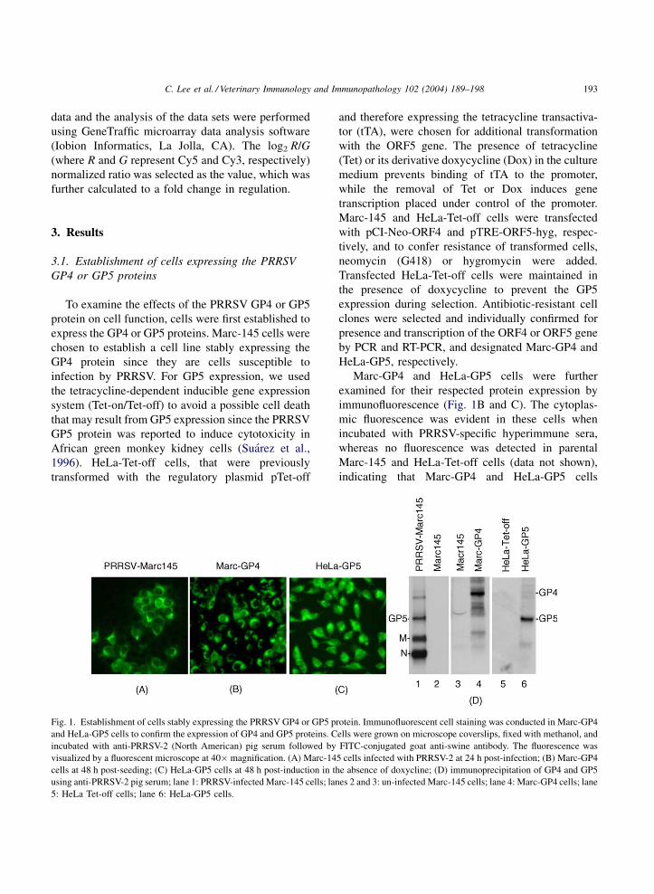
Fig. 1. Establishment of cells stably expressing the PRRSV GP4 or GP5 p
and HeLa-GP5 cells to confirm the expression of GP4 and GP5 proteins. C
incubated with anti-PRRSV-2 (North American) pig serum followed by
visualized by a fluorescent microscope at 40� magnification. (A) Marc-14
cells at 48 h post-seeding; (C) HeLa-GP5 cells at 48 h post-induction in th
using anti-PRRSV-2 pig serum; lane 1: PRRSV-infected Marc-145 cells; lan
5: HeLa Tet-off cells; lane 6: HeLa-GP5 cells.
and therefore expressing the tetracycline transactiva-
tor (tTA), were chosen for additional transformation
with the ORF5 gene. The presence of tetracycline
(Tet) or its derivative doxycycline (Dox) in the culture
medium prevents binding of tTA to the promoter,
while the removal of Tet or Dox induces gene
transcription placed under control of the promoter.
Marc-145 and HeLa-Tet-off cells were transfected
with pCI-Neo-ORF4 and pTRE-ORF5-hyg, respec-
tively, and to confer resistance of transformed cells,
neomycin (G418) or hygromycin were added.
Transfected HeLa-Tet-off cells were maintained in
the presence of doxycycline to prevent the GP5
expression during selection. Antibiotic-resistant cell
clones were selected and individually confirmed for
presence and transcription of the ORF4 or ORF5 gene
by PCR and RT-PCR, and designated Marc-GP4 and
HeLa-GP5, respectively.
Marc-GP4 and HeLa-GP5 cells were further
examined for their respected protein expression by
immunofluorescence (Fig. 1B and C). The cytoplas-
mic fluorescence was evident in these cells when
incubated with PRRSV-specific hyperimmune sera,
whereas no fluorescence was detected in parental
Marc-145 and HeLa-Tet-off cells (data not shown),
indicating that Marc-GP4 and HeLa-GP5 cells
rotein. Immunofluorescent cell staining was conducted in Marc-GP4
ells were grown on microscope coverslips, fixed with methanol, and
FITC-conjugated goat anti-swine antibody. The fluorescence was
5 cells infected with PRRSV-2 at 24 h post-infection; (B) Marc-GP4
e absence of doxycline; (D) immunoprecipitation of GP4 and GP5
es 2 and 3: un-infected Marc-145 cells; lane 4: Marc-GP4 cells; lane
C. Lee et al. / Veterinary Immunology and Immunopathology 102 (2004) 189–198194
expressed the GP4 and GP5 proteins, respectively.
Every cell expressed GP4 or GP5 indicating a
homogenous population of cells. The GP4 and GP5
protein expressions were further confirmed by radio-
immunoprecipitation (Fig. 1D). A specific band of
31 kDa protein was identified in Marc-GP4 cells (lane
4). This band was absent in the parental Marc-145
cells (lane 3) and was considered the GP4 protein. We
were not, however, able to detect the same protein
from PRRSV-infected cells, and this is probably due to
the low abundance of GP4 in PRRSV-infected cells as
it is a minor protein. The GP5 protein was readily
produced in HeLa-GP5 cells (lane 6), and its migration
was similar to that of the authentic GP5 protein seen in
PRRSV-infected cells (lane 1).
3.2. Gene expression profiles in Marc-GP4 and
HeLa-GP5 cells
To investigate the effects of the PRRSV GP4 and
GP5 proteins on cellular gene expressions, a micro-
array DNA chip technology was employed. Total
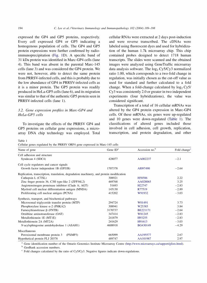
Table 1
Cellular genes regulated by the PRRSV ORF4 gene expressed in Marc-1
Name of gene G
Cell adhesion and structure
Syndecan 4 (SDC4)
Cell cycle regulators and cancer signals
Growth factor independent 1B (GFI1B) 1
Replication, transcription, translation, degradation machinery, and protein
Cathepsin L (CTSL)
Zinc finger protein 36, C3H type-like 2 (ZFP36L2)
Angiotensinogen proteinase inhibitor (Clade A; AGT)
Myeloid cell nuclear differentiation antigen (MNDA)
Proliferating cell nuclear antigen (PCNA)
Synthesis, transport, and biochemical pathways
Microsomal triglyceride transfer protein (MTP)
Phosphorylase kinase a-2 (PHKA2)
Farnesyltransferase b (FNTB) 3
Ornithine aminotransferase (OAT)
Metallothionein 1E (MT1E)
Metallothionein 2A (MT2A)
N-acylsphingosine amidohydrolase 1 (ASAH1) 4
Miscellaneous
Peroxisomal membrane protein 3 (PXMP3)
Hypothetical protein FLJ 20378
a Gene identification number of the Ontario Genomics Institute Microb GenBank accession numbers.c Fold changes calculated by the ratio of Cy5/Cy3. Negative figures i
cellular RNAs were extracted at 2 days post-induction
and were reverse transcribed. The cDNAs were
labeled using fluorescent dyes and used for hybridiza-
tion of the human 1.7k microarray chip. This chip
contained probes designed to detect 1718 human
transcripts. The slides were scanned and the obtained
images were analyzed using GeneTraffic microarray
data analysis software. The log2 Cy5/Cy3 normalized
ratio 1.00, which corresponds to a two-fold change in
regulation, was initially chosen as the cut-off value as
used for standard and further calculated to a fold
change. When a fold-change calculated by log2 Cy5/
Cy3 was consistently 2.0 or greater in two independent
experiments (four hybridizations), the value was
considered significant.
Transcription of a total of 16 cellular mRNAs was
altered by the GP4 protein expression in Marc-GP4
cells. Of these mRNAs, six genes were up-regulated
and 10 genes were down-regulated (Table 1). The
classifications of altered genes included those
involved in cell adhesion, cell growth, replication,
transcription, and protein degradation, and other
45 cells
ene IDa Accession no.b Fold changec
428077 AA002237 �2.1
707378 AI097490 �2.64
modification
308921 H58586 2.22
469768 AA028065 3.25
51693 H22747 �2.83
145130 R77519 �2.89
415202 W91932 �3.03
294724 W01491 3.73
308941 W25385 3.84
170737 BE221171 �2.64
347414 W81245 �2.83
241879 H93255 �2.83
241629 H91613 �3.03
608918 BG430149 �4.29
665099 AA195577 2.67
489747 AA101987 2.97
array Centre (http://www.microarrays.ca/support/glists.html).
ndicate down-regulations.
C. Lee et al. / Veterinary Immunology and Immunopathology 102 (2004) 189–198 195
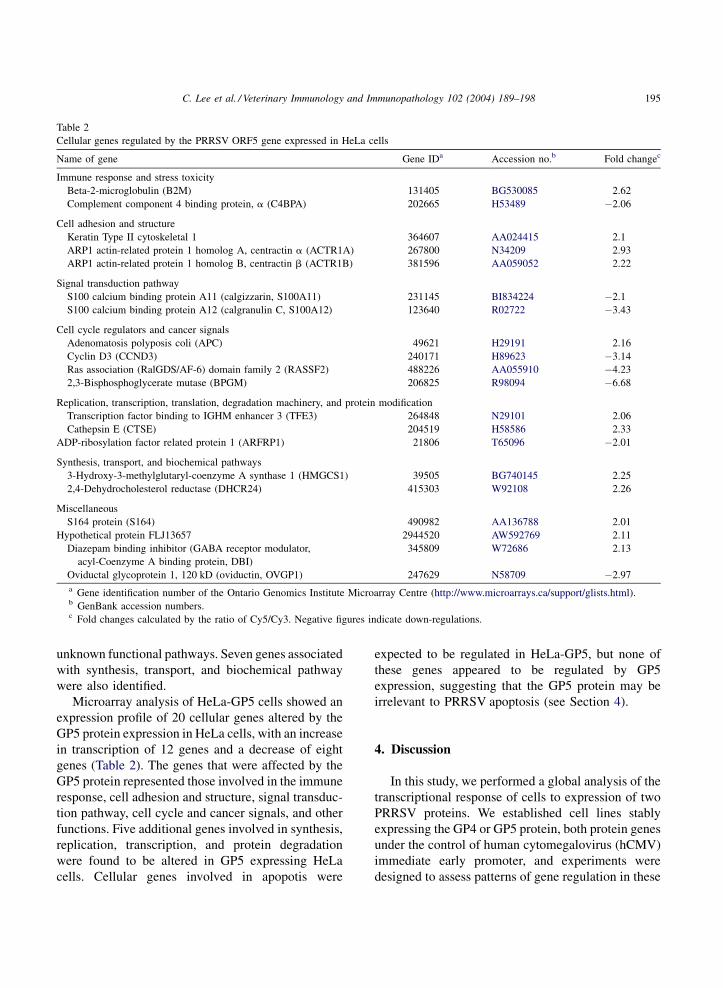
Table 2
Cellular genes regulated by the PRRSV ORF5 gene expressed in HeLa cells
Name of gene Gene IDa Accession no.b Fold changec
Immune response and stress toxicity
Beta-2-microglobulin (B2M) 131405 BG530085 2.62
Complement component 4 binding protein, a (C4BPA) 202665 H53489 �2.06
Cell adhesion and structure
Keratin Type II cytoskeletal 1 364607 AA024415 2.1
ARP1 actin-related protein 1 homolog A, centractin a (ACTR1A) 267800 N34209 2.93
ARP1 actin-related protein 1 homolog B, centractin b (ACTR1B) 381596 AA059052 2.22
Signal transduction pathway
S100 calcium binding protein A11 (calgizzarin, S100A11) 231145 BI834224 �2.1
S100 calcium binding protein A12 (calgranulin C, S100A12) 123640 R02722 �3.43
Cell cycle regulators and cancer signals
Adenomatosis polyposis coli (APC) 49621 H29191 2.16
Cyclin D3 (CCND3) 240171 H89623 �3.14
Ras association (RalGDS/AF-6) domain family 2 (RASSF2) 488226 AA055910 �4.23
2,3-Bisphosphoglycerate mutase (BPGM) 206825 R98094 �6.68
Replication, transcription, translation, degradation machinery, and protein modification
Transcription factor binding to IGHM enhancer 3 (TFE3) 264848 N29101 2.06
Cathepsin E (CTSE) 204519 H58586 2.33
ADP-ribosylation factor related protein 1 (ARFRP1) 21806 T65096 �2.01
Synthesis, transport, and biochemical pathways
3-Hydroxy-3-methylglutaryl-coenzyme A synthase 1 (HMGCS1) 39505 BG740145 2.25
2,4-Dehydrocholesterol reductase (DHCR24) 415303 W92108 2.26
Miscellaneous
S164 protein (S164) 490982 AA136788 2.01
Hypothetical protein FLJ13657 2944520 AW592769 2.11
Diazepam binding inhibitor (GABA receptor modulator,
acyl-Coenzyme A binding protein, DBI)
345809 W72686 2.13
Oviductal glycoprotein 1, 120 kD (oviductin, OVGP1) 247629 N58709 �2.97
a Gene identification number of the Ontario Genomics Institute Microarray Centre (http://www.microarrays.ca/support/glists.html).b GenBank accession numbers.c Fold changes calculated by the ratio of Cy5/Cy3. Negative figures indicate down-regulations.
unknown functional pathways. Seven genes associated
with synthesis, transport, and biochemical pathway
were also identified.
Microarray analysis of HeLa-GP5 cells showed an
expression profile of 20 cellular genes altered by the
GP5 protein expression in HeLa cells, with an increase
in transcription of 12 genes and a decrease of eight
genes (Table 2). The genes that were affected by the
GP5 protein represented those involved in the immune
response, cell adhesion and structure, signal transduc-
tion pathway, cell cycle and cancer signals, and other
functions. Five additional genes involved in synthesis,
replication, transcription, and protein degradation
were found to be altered in GP5 expressing HeLa
cells. Cellular genes involved in apopotis were
expected to be regulated in HeLa-GP5, but none of
these genes appeared to be regulated by GP5
expression, suggesting that the GP5 protein may be
irrelevant to PRRSV apoptosis (see Section 4).
4. Discussion
In this study, we performed a global analysis of the
transcriptional response of cells to expression of two
PRRSV proteins. We established cell lines stably
expressing the GP4 or GP5 protein, both protein genes
under the control of human cytomegalovirus (hCMV)
immediate early promoter, and experiments were
designed to assess patterns of gene regulation in these
C. Lee et al. / Veterinary Immunology and Immunopathology 102 (2004) 189–198196
cells. In GP5-expressing HeLa cells, actin-related
protein 1 (ARP1) homologs A and B were identified
to be up-regulated. These genes encode a subunit
of dynactin that binds to both microtubules and
cytoplasmic dynein, and is involved in ER-to-Golgi
transport (Lees-Miller et al., 1992). This may
implicate an important function of GP5 in the
transport of the viral components. The bisphospho-
glycerate mutase (BPGM) gene was down-regulated
by GP5 by more than six-folds. BPGM is an
erythrocyte specific trifunctional enzyme regulating
the level of 2,3-BPG in red blood cells. 2,3-BPG is the
main allosteric effector of hemoglobin, shifting the
equilibrium between the oxy and deoxy conformation
of hemoglobins by stabilizing the unliganded form.
Sickle cell anemia in humans is characterized by
polymerization of deoxygenated hemoglobin mutants
giving rise to deformed erythrocytes and vasoocclu-
sive complications. 2,3-BPG has been shown to
facilitate this polymerization in sickle cell anemia. In
humans, deficiency of BPGM has been shown to be
associated with anemia (Jacobasch and Rapoport,
1996). RASSF2 was also down-regulated by GP5.
RASSF2 is a new member of the RASSF1 family and
shares the properties of Ras effector/tumor suppres-
sors (Vos et al., 2003). Similarly, cyclin D3 gene
expression was found to be suppressed. D-type cyclins
are the key regulators along with cyclin E for cell cycle
progression from G1 to S phase. Complexes formed
between cyclin D or cyclin E and their kinase partners
are involved in phosphorylation of retinoblastoma
protein, which ultimately leads to activation of E2F
transcription factor and progression to S phase of the
cell cycle. A recent study demonstrates a clear
reduction of cyclin D3 and cell cycle arrest in
G0/G1 phase in cells infected with mouse hepatitis
coronavirus, a member of nidoviruses (Chen and
Makino, 2004).
In cell expressing GP4, the majority of differentially
expressed genes were involved in synthesis, transport,
and biochemical pathways. This observation implicates
that the GP4 protein may utilize or change host cell
machinery to transport viral or cellular components to
the cell surface. Zinc finger protein 36 (zfp36) and
microsomal triglyceride transfer protein were readily
up-regulated, while metallothineins, proliferating cell
nuclearprotein,andN-acylsphingosineamidohydrolase
were down-regulated. Zinc finger protein 36-like 1 is a
member of the tristetraprolin family of tandem CCCH
finger proteins. Tristetraprolin can bind to AU-rich
elements within the 30-untranslated regions of the
mRNAs encoding tumor necrosis factor (TNF) and
granulocyte-macrophage colony-stimulating factor
(GM-CSF), leading to accelerated mRNA degradation
(Stumpo et al., 2004). Tristetraprolin-knockout mice
exhibit an inflammatory phenotype that is largely due to
increased TNF secretion (Taylor et al., 1996). Micro-
somal triglyceride transfer protein is a protein complex
required for the assembly of lipoprotein particles
(Gordon et al., 1995). It is noteworthy that GP4 has
recently been shown to be a lipid-anchored protein
(Bachand, 2003). Metallothionein is a metal binding
proteinandhas beenshowntobe regulatedbya common
virus infection (Ilback et al., 2004). Coxsackievirus B-
type 3 virus infection altered the normal physiological
trace element balance in the liver, kidney, spleen, and
increased metallothionein in theseorgans.This maybe a
normal response in common infections that could
adversely influence the pathogenesis when the host is
concomitantly exposed to potentially toxic trace
elements, even at levels in the physiological range.
The function of N-acylsphingosine amidohydrolase is
unclear.
We have observed consistent increases in the
expression of cathepsin genes in both Marc-GP4 and
HeLa-GP5 cells. Cathepsin is involved in protein
degradation. Despite a specific role of cathepsin
during virus replication remains to be determined, the
up-regulated expression of the gene encoding protease
may represent a cellular defense against expression of
foreign proteins.
Interestingly, no pro-apoptotic genes were identi-
fied in HeLa-GP5 cells. This observation is contra-
dictory to the previous report (Suarez et al., 1996) but
is consistent with our recent finding that the GP5
expressing cells did not show any detectable level of
cytotoxicity or cell death (Lee et al., 2004). Zhang et
al. (1999) have shown that PRRSV infection induced
the expression of IFN-inducible gene Mx1 and an
ubiquitin-specific protease in porcine alveolar macro-
phages. These genes were not identifiable in the
present study. This difference may be due to the use of
different cell types since Marc-145 or HeLa cells were
used in the present study to express the single GP4 or
GP5 protein rather than using the whole virus to infect
porcine macrophages in the previous report.

C. Lee et al. / Veterinary Immunology and Immunopathology 102 (2004) 189–198 197
The DNA microarray has allowed us to identify the
differential effects of PRRSV proteins on cellular
genes. Confirmatory studies are further required as to
the significance of the genes that have been identified
in the present study. The method of choice to measure
and confirm the differential mRNA expression
mediated by the GP4 and GP5 proteins is real-time
quantitative RT-PCR. It is also possible that altered
mRNA profiles may not necessarily reflect altered
production of corresponding proteins (Gygi et al.,
1999). In this regard, additional techniques such as
Western blot analysis or protein arrays may be needed
to support our findings. Nevertheless, our data
obtained from the microarray study will provide
future insights into the understanding of host cell virus
interactions and eventually of the pathogenic mechan-
isms of PRRSV and the host responses to PRRSV
infection.
Acknowledgments
The authors are grateful to Ryan Dowling for his
technical assistance for microarray analysis. This
study was supported by NSERC, Ontario Pork, the
Ontario Ministry of Agriculture and Food (Animal
Program).
References
Bachand, A., 2003. Characterization of PRRSV GP4. MSc. Thesis.
University of Guelph, Guelph, Ont.
Browne, E.P., Wing, B., Coleman, D., Shenk, T., 2001. Altered
cellular mRNA levels in human cytomegalovirus-infected fibro-
blasts: viral block to the accumulation of antiviral mRNAs. J.
Virol. 75, 12319–12330.
Chen, C.J., Makino, S., 2004. Murine coronavirus replication
induces cell cycle arrest in G0/G1 phase. J. Virol. 78, 5658–
5669.
Faaberg, K.S., Even, C., Palmer, G.A., Plagemann, P.G., 1995.
Disulfide bonds between two envelope proteins of lactate dehy-
drogenase-elevating virus are essential for viral infectivity. J.
Virol. 69, 613–617.
Feng, W.H., Tompkins, M.B., Xu, J.S., Zhang, H.X., McCaw, M.B.,
2003. Analysis of constitutive cytokine expression by pigs
infected in-utero with porcine reproductive and respiratory
syndrome virus. Vet. Immunol. Immunopathol. 94, 35–45.
Ferguson, M.A., Williams, A.F., 1988. Cell surface anchoring of
proteins via glycosyl-phosphatidylinositol structures. Annu.
Rev. Biochem. 57, 285–320.
Gordon, D.A., Wetterau, J.R., Gregg, R.E., 1995. Microsomal
triglyceride transfer protein: a protein complex required for
the assembly of lipoprotein particles. Trends Cell Biol. 8,
317–321.
Gossen, M., Bujard, H., 1992. Tight control of gene expression in
mammalian cells by tetracycline-responsive promoters. Proc.
Natl. Acad. Sci. U.S.A. 89, 5547–5551.
Gygi, S., Rochon, Y., Franza, B.R., Aebersold, R., 1999. Correlation
between protein and mRNA abundance in yeast. Mol. Cell Biol.
19, 1720–1730.
Ilback, N.G., Glynn, A.W., Wilberg, L., Netzel, E., Lindh, U., 2004.
Metallothionein is induced and trace element balance changed in
target organs of a common viral infection. Toxicology 199,
241–250.
Jacobasch, G., Rapoport, S.M., 1996. Hemolytic anemias due to
erythrocyte enzyme deficiencies. Mol. Aspects Med. 17, 143–
170.
Jacobs, M.G., Robinson, P.J., Bletchly, C., Mackenzie, J.M., Young,
P.R., 2000. Dengue virus non-structural protein 1 is expressed in
a glycosyl-phosphatidylinositol-linked form that is capable of
signal transduction. FASEB J. 14, 1603–1610.
Johnsen, C.K., Botner, A., Kamstrup, S., Lind, P., Nielsen, J., 2002.
Cytokine mRNA profiles in bronchoalveolar cells of piglets
experimentally infected in utero with porcine reproductive
and respiratory syndrome virus: association of sustained expres-
sion of IFN-g and IL-10 after viral clearance. Viral. Immunol.
15, 549–556.
Johnston, C., Jiang, W., Chu, T., Levine, B., 2001. Identification of
genes involved in the host response to neurovirulent alphavirus
infection. J. Virol. 75, 10431–10445.
Kim, H.S., Kwang, J., Yoon, I.J., Joo, H.S., Frey, M.L., 1993.
Enhanced replication of porcine reproductive and respiratory
syndrome (PRRS) virus in a homogeneous subpopulation of
MA-104 cell line. Arch. Virol. 133, 477–483.
Lee, C., Rogan, D., Erickson, L., Zhang, J., Yoo, D., 2004. Char-
acterization of the porcine reproductive and respiratory syn-
drome virus GP5 in stably expressing cells. Virus Res. 104, 33–
38.
Lees-Miller, J.P., Helfman, D.M., Schroer, T.A., 1992. A vertebrate
actin-related protein is a component of a multisubunit complex
involved in microtubule-based vesicle motility. Nature 359,
244–246.
Mardassi, H., Mounir, S., Dea, S., 1995. Molecular analysis of the
ORFs 3–7 of porcine reproductive and respiratory syndrome
virus, Quebec reference strain. Arch. Virol. 140, 1405–1418.
Mardassi, H., Massie, B., Dea, S., 1996. Intracellular synthesis,
processing, and transport of proteins encoded by ORFs 5–7 of
porcine reproductive and respiratory syndrome virus. Virology
221, 98–112.
Meulenberg, J.J.M., den Besten, A., de Kluyer, F.P., Moormann,
R.J.M., Schaaper, V.M.M., Wensvoort, G., 1995. Characteriza-
tion of proteins encoded by ORFs 2–7 of Lelystad virus.
Virology 206, 155–163.
Meulenberg, J.J., van Nieuwstadt, A.P., van Essen-Zandbergen, A.,
Langeveld, J.P., 1997. Posttranslational processing and identi-
fication of a neutralization domain of the GP4 protein encoded
by ORF4 of Lelystad virus. J. Virol. 71, 6061–6067.
C. Lee et al. / Veterinary Immunology and Immunopathology 102 (2004) 189–198198
Murtaugh, M.P., Elam, M.R., Kakach, L.T., 1995. Comparison of the
structural protein coding sequences of the VR-2332 and Lelys-
tad virus strains of the PRRS virus. Arch. Virol. 140, 1451–1460.
Ostrowski, M., Galeota, J.A., Jar, A.M., Platt, K.B., Osorio, F.A.,
Lopez, O.J., 2002. Identification of neutralizing and nonneu-
tralizing epitopes in the porcine reproductive and respiratory
syndrome virus GP5 ectodomain. J. Virol. 76, 4241–4250.
Sambrook, J., Russell, D.W., 2001. Molecular Cloning: A Labora-
tory Manual, 3rd ed. Cold Spring Harbor Laboratory, Cold
Spring Harbor, NY.
Schena, M., Shalon, R., Davis, W., Brown, P.O., 1995. Quantitative
monitoring of gene expression patterns with complementary
DNA microarray. Science 270, 467–470.
Snijder, E.J., Meulenberg, J.J., 1998. The molecular biology of
arteriviruses. J. Gen. Virol. 79, 961–979.
Snijder, E.J., Dobbe, J.C., Spaan, W.J.M., 2003. Heterodimerization
of the two major envelope proteins is essential for arterivirus
infectivity. J. Virol. 77, 97–104.
Stumpo, D.J., Byrd, N.A., Phillips, R.A., Ghosh, S., Maronpot, R.R.,
Castranio, T., Meyers, E.N., Mishina, Y., Blackshear, P.J., 2004.
Chorioallantoic fusion defects and embryonic lethality resulting
from disruption of Zxp36L1, a gene encoding CCCH tandom
zinc finger protein of the tristetraprol family. Mol. Cell Biol. 24,
6445–6455.
Suarez, P., Dıaz-Guerra, M., Prieto, C., Esteban, M., Castro, J.M.,
Nieto, A., Ortın, J., 1996. Open reading frame 5 of porcine
reproductive and respiratory syndrome virus as a cause of virus-
induced apoptosis. J. Virol. 70, 2876–2882.
Suradhat, S., Thanawongnuwech, R., 2003. Upregulation of inter-
leukin-10 gene expression in the leukocytes of pigs infected with
porcine reproductive and respiratory syndrome virus. J. Gen.
Virol. 84, 2755–2760.
Taylor, G.A., Carballo, E., Lee, D.M., Lai, W.S., Thompson, M.J.,
Patel, D.D., Schenkman, D.I., Gilkeson, G.S., Broxmeyer, H.E.,
Haynes, B.F., Blackshear, P.J., 1996. A pathogenic role for TNF
alpha in the syndrome of cachexia, arthritis, and autoimmunity
resulting from tristetraprolin (TTP) deficiency. Immunity 4,
445–454.
Thanawongnuwech, R., Thacker, E.L., 2003. Interleukin-10, inter-
leukin-12, and interferon-g levels in the respiratory tract follow-
ing mycoplasma hyopneumoniae and PRRSV infection in pigs.
Viral Immunol. 16, 357–367.
van Nieuwstadt, A.P., Meulenberg, J.J.M., van Essen-Zandbergen,
A., Peterson-den Besten, A., Bende, R.J., Moormann, R.J.M.,
Wensvoort, G., 1996. Proteins encoded by ORFs 3 and 4 of the
genome of Lelystad virus (Arteriviridae) are structural proteins
of the virion. J. Virol. 70, 4767–4772.
Varma, R., Mayor, S., 1998. GPI-anchored proteins are organized in
submicron domains at the cell surface. Nature (London) 394,
798–801.
Vos, M.D., Ellis, C.A., Elam, C., Ulku, A.S., Taylor, B.J., Clark,
G.J., 2003. RASSF2 is a novel K-Ras-specific effector and
potential tumor suppressor. J. Biol. Chem. 278, 28045–
28054.
Weiland, E., Wieczorek-Krohmer, M., Kohl, D., Conzelmann, K.K.,
Weiland, F., 1999. Monoclonal antibodies to the GP5 of porcine
reproductive and respiratory syndrome virus are more effective
in virus neutralization than monoclonal antibodies to the GP4.
Vet. Microbiol. 66, 171–186.
Wootton, S., Yoo, D., Rogan, D., 2000. Full-length sequence of a
Canadian porcine reproductive and respiratory syndrome virus
(PRRSV) isolate. Arch. Virol. 145, 2297–2323.
Wu, W.H., Fang, Y., Farwell, R., Steffen-Bien, M., Rowland, R.R.,
Christopher-Hennings, J., Nelson, E.A., 2001. A 10 kDa struc-
tural protein of porcine reproductive and respiratory syndrome
virus encoded by ORF2b. Virology 287, 183–191.
Zhang, X., Shin, J.H., Molitor, T.W., Schook, L.B., Rutherford,
M.S., 1999. Molecular responses of macrophages to porcine
reproductive and respiratory syndrome virus infection. Virology
262, 152–162.