Differential Expression of IL-36 Family Members and IL-38...
13
Research Article Differential Expression of IL-36 Family Members and IL-38 by Immune and Nonimmune Cells in Patients with Active Inflammatory Bowel Disease Gabriela Fonseca-Camarillo , 1 Janette Furuzawa-Carballeda , 2 Emilio Iturriaga-Goyon, 1,3 and Jesús K. Yamamoto-Furusho 1 1 Inflammatory Bowel Disease Clinic, Department of Gastroenterology, Instituto Nacional de Ciencias M´ edicas y Nutrici´ on, Salvador Zubir´ an, Mexico City, Mexico 2 Department of Immunology and Rheumatology, Instituto Nacional de Ciencias M´ edicas y Nutrici´ on, Salvador Zubir´ an, Mexico City, Mexico 3 MD/PhD (PECEM) Program, Facultad de Medicina, Universidad Nacional Autonoma de M´ exico, Mexico Correspondence should be addressed to Jes´ us K. Yamamoto-Furusho; [email protected] Received 5 March 2018; Accepted 19 November 2018; Published 10 December 2018 Academic Editor: Koichiro Wada Copyright © 2018 Gabriela Fonseca-Camarillo et al. is is an open access article distributed under the Creative Commons Attribution License, which permits unrestricted use, distribution, and reproduction in any medium, provided the original work is properly cited. IL-1 family includes IL-38 (IL-1F10) and the subfamily of IL-36 and is the central mediators of inflammatory diseases, including pustular psoriasis, atopic dermatitis, rheumatoid arthritis, and gut inflammation. e purpose of the study was to evaluate on tissue of the patients with inflammatory bowel disease (IBD), the IL-36, IL-36, IL-36, IL-36Ra, and IL-38 gene and cell expression and its correlation with clinical activity. Patients and Methods. A cross-sectional and comparative study was performed. Seventy patients with IBD and 30 noninflamed non-IBD controls were enrolled. Gene expression was measured by RT-PCR. Protein expression was detected by double-staining immunohistochemistry. Results. e mRNA expression of IL-36 family members but not IL-38 was increased in colonic mucosa from patients with active ulcerative colitis versus Crohn’s disease group and noninflammatory control group (P<0.05). However, only gene expression of IL-38 was increased in tissue from patients with inactive ulcerative colitis versus active disease and control group (P<0.005). Conversely, gene expression of IL-36Ra was significantly higher in colonic tissue from patients with active versus inactive ulcerative colitis and noninflamed control group (P<0.05). A differential protein overexpression of IL-36, IL-36, IL-36, IL-36Ra, and IL-38 by intestinal epithelial cells, macrophages, CD8+ T cells, and/or versus dendritic cells (pDCs) was found in patients with active inflammatory bowel disease compared with noninflamed controls. Conclusion. IL-38 and IL-36 family members’ expression was increased by immune and nonimmune cells in patients with active inflammatory bowel disease. ese cytokines and IL-36Ra might represent novel therapeutic targets in patients with gut inflammation. 1. Introduction Inflammatory Bowel Disease (IBD) is characterized by an imbalance between innate and adaptive immunity leading to the stimulation of T helper responses with preponderance of proinflammatory cytokines [1]. An increase production of proinflammatory cytokines such as IL-1 and TNF- has been involved in the development of chronic inflammation of the gut, founded on the recent knowledge about the role of cytokine-driven pathways in intestinal immunity and it is based on animal models of acute and chronic intestinal injury and inflammation [2] e IL-1 family (IL-1F) includes IL-38 (IL-1F10) and the subfamily of IL-36 (IL-36Ra or IL-1F5; IL-36 or IL- 1F6; IL-36 or IL-1F8; and IL-36 or IL-1F9). It has been considered one of the most important key regulators in the pathophysiology of inflammatory autoimmune diseases including Crohn’s disease (CD), rheumatoid arthritis, and psoriasis [3–5]. Hindawi BioMed Research International Volume 2018, Article ID 5140691, 12 pages https://doi.org/10.1155/2018/5140691
Transcript of Differential Expression of IL-36 Family Members and IL-38...

Research ArticleDifferential Expression of IL-36 Family Membersand IL-38 by Immune and Nonimmune Cells in Patients withActive Inflammatory Bowel Disease
Gabriela Fonseca-Camarillo 1 Janette Furuzawa-Carballeda 2
Emilio Iturriaga-Goyon13 and Jesuacutes K Yamamoto-Furusho 1
1 Inflammatory Bowel Disease Clinic Department of Gastroenterology Instituto Nacional de Ciencias Medicas y NutricionSalvador Zubiran Mexico City Mexico2Department of Immunology and Rheumatology Instituto Nacional de Ciencias Medicas y Nutricion Salvador ZubiranMexico City Mexico3MDPhD (PECEM) Program Facultad de Medicina Universidad Nacional Autonoma de Mexico Mexico
Correspondence should be addressed to Jesus K Yamamoto-Furusho kazuofurushohotmailcom
Received 5 March 2018 Accepted 19 November 2018 Published 10 December 2018
Academic Editor Koichiro Wada
Copyright copy 2018 Gabriela Fonseca-Camarillo et al This is an open access article distributed under the Creative CommonsAttribution License which permits unrestricted use distribution and reproduction in any medium provided the original work isproperly cited
IL-1 family includes IL-38 (IL-1F10) and the subfamily of IL-36 and is the central mediators of inflammatory diseases includingpustular psoriasis atopic dermatitis rheumatoid arthritis and gut inflammationThe purpose of the study was to evaluate on tissueof the patients with inflammatory bowel disease (IBD) the IL-36120572 IL-36120573 IL-36120574 IL-36Ra and IL-38 gene and cell expression andits correlationwith clinical activityPatients andMethods A cross-sectional and comparative study was performed Seventy patientswith IBD and 30 noninflamed non-IBD controls were enrolled Gene expression wasmeasured by RT-PCR Protein expression wasdetected by double-staining immunohistochemistry Results The mRNA expression of IL-36 family members but not IL-38 wasincreased in colonic mucosa from patients with active ulcerative colitis versus Crohnrsquos disease group and noninflammatory controlgroup (Plt005) However only gene expression of IL-38 was increased in tissue from patients with inactive ulcerative colitis versusactive disease and control group (Plt0005) Conversely gene expression of IL-36Ra was significantly higher in colonic tissue frompatients with active versus inactive ulcerative colitis and noninflamed control group (Plt005) A differential protein overexpressionof IL-36120572 IL-36120573 IL-36120574 IL-36Ra and IL-38 by intestinal epithelial cells macrophages CD8+ T cells andor versus dendriticcells (pDCs) was found in patients with active inflammatory bowel disease compared with noninflamed controls Conclusion IL-38and IL-36 family membersrsquo expression was increased by immune and nonimmune cells in patients with active inflammatory boweldisease These cytokines and IL-36Ra might represent novel therapeutic targets in patients with gut inflammation
1 Introduction
Inflammatory Bowel Disease (IBD) is characterized by animbalance between innate and adaptive immunity leading tothe stimulation of T helper responses with preponderanceof proinflammatory cytokines [1] An increase productionof proinflammatory cytokines such as IL-1120573 and TNF-120572 hasbeen involved in the development of chronic inflammationof the gut founded on the recent knowledge about the roleof cytokine-driven pathways in intestinal immunity and it is
based on animal models of acute and chronic intestinal injuryand inflammation [2]
The IL-1 family (IL-1F) includes IL-38 (IL-1F10) andthe subfamily of IL-36 (IL-36Ra or IL-1F5 IL-36120572 or IL-1F6 IL-36120573 or IL-1F8 and IL-36120574 or IL-1F9) It has beenconsidered one of the most important key regulators inthe pathophysiology of inflammatory autoimmune diseasesincluding Crohnrsquos disease (CD) rheumatoid arthritis andpsoriasis [3ndash5]
HindawiBioMed Research InternationalVolume 2018 Article ID 5140691 12 pageshttpsdoiorg10115520185140691
2 BioMed Research International
The IL-1 family overproduction but not from antagonistreceptor (IL-36Ra) or IL-38 results in inflammation in arobust immune response that acts as first line of defenseagainst invasive pathogenic microorganisms and damageand when there is an aberrant immune response underappropriate genetic and environmental backgrounds in anautoimmune disease [6]
IL38IL-1F10 is a protein that in humans is encoded bythe il1f10 gene [7] IL-38 is expressed in a range of tissuesincluding heart placenta fetal liver skin spleen thymusand tonsil IL-38 is also expressed mostly in the skin andin proliferating B cells [8] This cytokine participates in anetwork of IL-1 family members to regulate adapted andinnate immune responses by the inhibition of the productionof T cell cytokines (IL-17 and IL-22) IL-38 also inhibitsthe production of IL-8 induced by IL-36120574 thus regulatinginflammatory responses [9]
In addition other secreted protein is IL-36 receptorantagonist (IL-36Ra) also knownas IL-1F5 a natural inhibitorfor IL-36 activity The IL-36Ra is expressed by immune cellssuch as monocytes B cells dendritic cellsLangerhans cellskeratinocytes and gastric parietal cells [10]
Veerdonk et al showed that IL-38 binds to the IL-36receptor (IL-36R) and has similar biological effects to IL-36Ra on immune cells [10] According to its activity low con-centrations of IL-38may have an anti-inflammatory functionblocking IL-36R and IL-1R pathways which suppressed IL-22 and IL-17 synthesis and secretion Strikingly IL-38 shareshigh sequence homology with IL-1Ra (41) and IL-36Ra(43) [11]
The distinct expression of IL-36120572 IL-36120573 and IL-36120574their antagonist (IL-36Ra) and IL-38 in autoimmune diseasehas been shown They showed an increased expression of IL-36120572 and IL-38 only in patients with CD but did not showthe expression of IL-36 family by producing effector immunecells regarding clinical activity in patients with ulcerativecolitis (UC) [5]
Nonetheless little is known about the presence of IL-38IL-36Ra and IL-36120572 IL-36120573 and IL-36120574 producing intestinaleffector immune cells (T cells plasmacytoid dendritic cellsand monocytes) and nonimmune cells in Mexican Mestizopatients with IBD
The purpose of the study was to evaluate on intestinaltissue IL-36120572- IL-36120573- IL-36120574- IL-36Ra- and IL-38 pro-ducing cells as well as gene expression in immune (cytotoxicT cells macrophages and plasmacytoid dendritic cells) andnonimmune cells from patients with IBD compared withnoninflamed controls
2 Materials and Methods
21 Study Subjects For the cross-sectional and comparativestudy a total of 70 patients with IBD were recruited Patientswere categorized into the following groups 30 active UC(aUC) 20 inactive UC (iUC) 10 active CD (aCD) and10 inactive CD (iCD) patients All patients were includedbetween January 2014 and May 2015 from the InflammatoryBowel Disease Clinic at the Instituto Nacional de CienciasMedicas y Nutricion Salvador Zubiran (a tertiary referral
center in Mexico City Mexico) The UC and CD diagnosiswas done by the correlation of clinical endoscopic andhistopathological findings Colonic samples were obtainedfrom IBD patients and noninflamed controls after a signedinformed consent Patientsrsquo clinical records were reviewedand personal interviews were done and the followinginformation were collected for all IBD patients age sextreatment (mesalazine azathioprine prednisonemercaptop-urine etc) and the presence of extraintestinal manifestations(present or absent) The disease severity was evaluated incolon biopsy by Mayo score disease activity index for UC[12] and Harvey-Bradshaw Index for CD patients [13] Allcolonoscopies were performed for cancer surveillance Exclu-sion criteria included patients with indeterminate colitispostradiation infectious colitis and other types of colitis
The control group consisted of 30 noninflamed colonicbiopsies (without endoscopic and histological evidence of anytype of colitis neoplasia inflammatory disease or other doc-umented diseases) All participants underwent colonoscopydue to screening of polyps or the study of weight loss forevaluation of anemia Controls were matched by age and sexwith IBD patients
22 Tissue Samples
221 Sample Processing and Gene Expression Analysis Tostudy gene and in situ expression we followed the methodsof Fonseca-Camarillo G et al 2015 [14] The colonic biopsieswere taken by colonoscopy and immediately submerged inRNA later solution (Ambion Austin TX USA) for storageThen total RNA was isolated using High Pure RNA Tissue(Roche Diagnostics Mannheim Germany) following themanufacturerrsquos guidelines
Electrophoresis of one aliquot was made for each oneof the RNA products in an agarose gel at 1 it was thenvisualized by staining with ethidium bromide and then it wasdocumented using an UV transilluminator
Two hundred nanograms of total RNA was reverse-transcribed into cDNA with random hexamer primers(Roche Diagnostics Mannheim Germany)
cDNA synthesis from total RNA through reverse tran-scription was made taking 20 120583L from total RNA of eachof the products with the following protocol preincubation25∘C x 10 minutes incubation 55∘C x 30 minutes followedby denaturalization 85∘C x 5 minutes using a thermocycler(Perkin-Elmer)
Quantitative real-time PCR was used to measure theRNA transcription level of target genes Expression ofglyceraldehyde-3-phosphate dehydrogenase (GAPDH) geneas housekeeping was analyzed for normalization purposes
PCR amplificationwas performedwith a concentration of20 ng of cDNA 200 nM forward reverse primer and TaqmanMasterMix (RocheDiagnosticsMannheim GermanyRocheDiagnostics Mannheim Germany) in a final volume of 10120583l PCR reactions were run in a Light Cycler 480 (RocheDiagnostics Mannheim Germany)
The mRNA relative quantification of target genes wasconducted using the LightCycler software 41 according tothe 2-delta-delta Ct method Table 1 shows the details of
BioMed Research International 3
Table 1 Primers designs
Gene Genebank Oligonucleotides Probe UPL
IL-36120572 NM 0005722 CATAAATTAGAGGTCCAAAATCGAAGGGGCTGGGTCAGCTAT 45
IL-36szlig NM 0015583 GTCTTGGCTCAGACGCTCATTGCTTCAAACCACACAGACG 23
IL-36Υ NM 0006283 GGTCGTGTGCTTGGAGGAGGTACCATTCCCAATGCTGA
20
IL-38 NM 0187243 AAGAAGGACCTCCGGCTCTTGACTCAGAATCTGGC5GTATTTC
69
IL-36RN NM 0144322 TCCATCAACATGAAGAATGTCCAGCCATTTCTTTTGCCCATA
22
GADPH NM 0020463 AGCCACATCGCTCAGACACGCCCAATACGACCAAATCC
60
the primerrsquos designs and number of UPL (Universal ProbeLibrary Roche Diagnostics Mannheim Germany) used forthe RT-PCR assay
222 Patient Samples for Protein Expression Analysis Atotal of 10 surgical samples from patients with active severeUC refractory to conventional therapy were included forprotein detection Also 10 patients with definitive diagnosisof active severe CD were enrolled in the study Ten con-trols were obtained from noninflamed non-IBD intestinaltissue All biopsies were obtained from whole colon in UCand right colon in CD patients The Riley Index scorewas used for grading the severity of colonic inflammation[15]
23 Immunohistochemistry Todetermine the IL-38 IL-36Raand secondary IL-36 family expressing cells 4 120583m thickformalin-fixed and paraffin-embedded tissue from patientsand noninflamednon-IBD controls were placed on posi-tively charged slides Sections were deparaffinized in xyleneand rehydrated in water Morphometric evaluation of theimmune-stained sections was performed in a blinded man-ner
231 Double-Staining Procedure After deparaffinization anddemasking of antigens with the immunohistochemistry(IHCh) enzyme antigen retrieval reagent (Enzo Life Sci-ences Inc Farmingdale NY USA) endogenous peroxidasewas quenched with peroxidase block (Enzo Life SciencesInc) Then nonspecific background staining was avoidedwith a serum-free solution which eliminates the need tomatch species with the link antibody (IHCh backgroundblocker Enzo Life Sciences Inc) To determine subpopu-lations of IL-38 IL-36Ra and CD14+IL-36120572 CD123+IL-36120572 CD14+IL-36120573 CD123+IL-36120573 CD8120572+IL-36120574 andCD123+IL-36120574 expressing cells a simultaneous detectionwith a nonbiotin one-step detection was performed (Multi-View (mouse-HRPrabbit-AP) Enzo Life Sciences Inc)Theprocedure is a sequential double staining where the rab-bit polyclonal anti-CD8120572 IgG or anti-CD123 IgG antibody(Santa Cruz Biotechnology CA USA)mouse monoclonal
anti- IL-36120574 IgG (Abcam) and the mouse monoclonal anti-CD14 IgG2a antibody or anti-CD123 IgG1 antibody (SantaCruz Biotechnology Santa Cruz CA USA)rabbit polyclonalanti-IL-38 IgG anti-IL-36Raanti-IL-36120572 and anti-IL-36120573(Abcam) at 10 120583gmL were incubated during 30 min at roomtemperature Slides were washed with IHCh wash buffer(Enzo Life Sciences Inc) and then incubated with PolyViewIHCh-HRP reagent for mouse antibody and PolyView IHCh-AP reagent for rabbit antibody for 20 min Finally antigenswere visualized using horseradish peroxidase (HRP)3 31015840-diaminobenzidine (DAB) for 10 min and the second antigenwith alkaline phosphatase (AP)Permanent Red for 5 minTissues were counterstained with the nonalcoholic Mayerrsquoshematoxylin (DAKO) and mounted in aqueous mountingmedium (DAKO) Negative control staining was performedwith the universal negative control (a reagent mixture ofpurified goat rabbit and mouse immunoglobulins) whichwas tittered to work with polymer-based secondary systems(IHCh universal negative control reagent Enzo Life SciencesInc)The reactive blank was incubated with phosphate buffersaline-and IHCh background blocker instead of the primaryantibody Both controls excluded nonspecific staining andendogenous enzymatic activities IL-38 IL-36Ra and IL-36 producing cells were reported as the single and doublepositive staining cells in at least twofields (times320)Histologicalanalysis was performed by the program Image Pro Plusversion 511
24 Ethical Considerations This work was performed ac-cording to the principles expressed in the Declaration ofHelsinki 1989The study was carried out with approval by theethical committee in our institution and a written informedconsent was obtained from all patients recruited prior to theirinclusion in the study
25 Statistical Analysis Statistical analysis was performedusing the SPSS 19 program by the Kruskal-Wallis One-WayAnalysis of Variance on Ranks Data expressed as the medianrange and mean plusmn SE A P value le 005 was considered assignificant
4 BioMed Research International
Table 2 Demographic and clinical characteristics of Crohnrsquos disease and ulcerative colitis patients
Variable Non-inflamedControls (n=30)
Inactive UC patients(n=20)
Active UC patients(n=30)
Inactive CD patients(n=10)
Active CD patients(n=10)
Age yearsMean 489 408 3905 493 25Sex 1416 614 1020 46 37femalemaleTreatmentMesalazine ND 1920 1930 110 110Azathioprine ND 420 130 510 310Prednisone ND 320 330 210 110Mercaptopurine ND 020 330 010 110DiseaseExtension ND 720 130 110 010Distal colitis ND 1220 830 710 610PancolitisExtra-intestinalmanifestationsabsent ND 1520 1730 710 610present ND 420 1230 110 010CD Crohnrsquos Disease patient group UC ulcerative colitis patient group ND not determined
3 Results
31 Demographic and Clinical Characteristics A total of100 individuals were recruited for the study Patients werecategorized in 4 groups (1) active UC (n=30) (2) remissionUC (n=20) (3) active CD (n=10) (4) remission CD (n=10)A fifth group consisted of 30 noninflamednon-IBD controlgroups All demographic and clinical characteristics frompatients with IBD and noninflamed controls are depicted inTable 2
32 In Situ Expression of IL-36RA IL-38 and IL-36 Family inInflammatory Bowel Disease
321 IL-36Ra and IL-38 Histochemical analysis showedthat the intestinal tissue from IBD patients had higherexpression of IL-36Ra and IL-38 throughout the mucosasubmucosa muscular and serosa layers when comparedwith noninflamed control group IL-36Ra producing cellswere by mucosal epithelial cells and cell from submucosaand perivascular mononuclear cells CD14+IL-36Ra+ doublepositive cells from intestinal tissue from noninflamed controltissue were practically undetectable versus aUC and CD(Figure 1(a))
The IL-38 expression in tissue from patients with UCand CD was mostly by epithelial and parenchyma cellsNevertheless there were some perivascular inflammatoryCD123minus cells that expressed this cytokine In addition therewas a small subpopulation of CD123+IL-38 producing cellsdistributed along serosa muscular submucosa and mucosafrom IBDpatientsThe IL-38 expressing cellswere plentiful inserosa muscular and submucosa from active UC comparedto active CD and noninflamed control tissue (Figure 1(b))
The protein expression of IL-38 was scarce in mucosa fromIBD patients
322 IL-36120572 The most important IL-36120572 production wasdetected in nonimmune cells including gut epithelial andparenchyma cells
Immunohistochemical double staining revealed that bothCD14+ macrophages and CD123+ plasmacytoid dendriticcells expressed IL-36120572
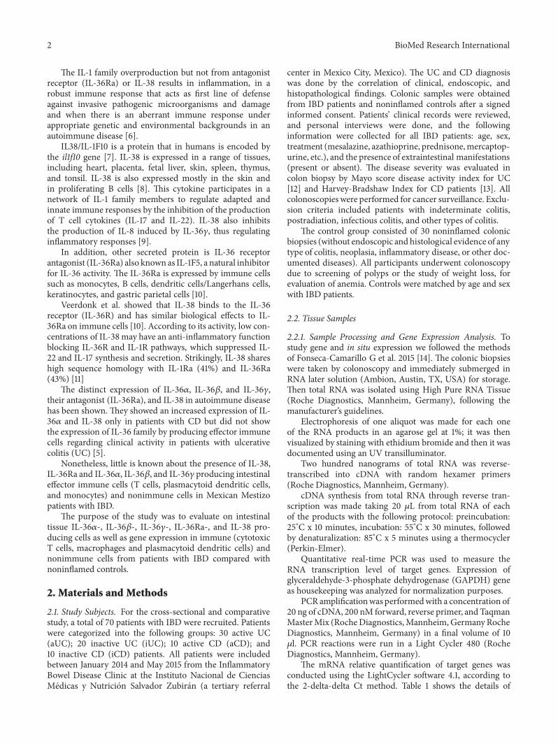
CD14IL-36120572 cells and CD123IL-36120572 pDCs were moreabundant on muscular and serosa from active UC patientscompared with active CD patients and noninflamed controltissue (Figures 2(a) and 2(b)) The IL-36120572 expressing cells inactive UC tissue were mainly CD14+ cells (Figure 2(a)) asmaller pDC positive subpopulation CD123+ (Figure 2(b))some plasma cells and lymphocytes A similar pattern of IL-36120572 expression was observed in samples from CD patientsThere was no expression of IL-36120572 on intestinal biopsiesfrom controls Staining for IL-36120572 protein in inflamed gutcorrelated with the pattern of mRNA expression
323 IL-36120573 CD14+IL-36120573+ immunoreactive macropha-ges were noticeably higher in active CD patients comparedwith active UC and noninflamed control tissue (Figures 3(a)and 3(b)) Double positive cells were localized mainly inthe muscular and serosa Furthermore IL-36120573 producingcells potentially lymphocytes were increased in submucosamuscular serosa and perivascular inflammatory infiltratesfrom active CD patients compared with noninflamed controltissue
Biopsies from patients with active CD had a significantnumber of CD123minusIL-36 expressing cells A conspicuous
BioMed Research International 5
Mucosa
Submucosa
Muscular
Serosa
CD14
IL-3
6Ra
Ulcerative Colitis Crohnrsquos Disease Control
(a)
Serosa
Mucosa
Submucosa
Muscular
CD12
3IL
-38
Ulcerative Colitis Crohnrsquos Disease Control
(b)
Figure 1CD14+IL-36Ra- andCD123+IL-38 expressing cells in intestinal tissue from IBDpatients CD14+IL-36Ra- and (b) CD123+IL-38 expressing cells Representative immunoperoxidase in tissue from active ulcerative colitis patients (n=10 left panel) active Crohnrsquos diseasepatients (n=10middle panel) and noninflamed colonic tissue (n=10 right panel) Photomicrographs representmucosa submucosamuscularand serosa Dotted arrows depict the cytokine expression solid arrows show expression of cell-surface marker (leukocytes) double arrowsindicate CD14+ or CD123+ (in brown)IL-36Ra or IL-38 (in pink) double positive cells (burgundy) and circles highlight the positive cellsfrom perivascular inflammatory infiltrates Original magnification was X320
number of IL-36120573 cells were observed in serosa muscularand submucosa and smaller proportion was observed inmucosa Furthermore in patients with active UC a consid-erably lower number of CD123+IL-36120573+ was detected inmucosa submucosa muscular and serosa although themostimmunoreactive cells were CD123minusIL-36120573+ immune andnonimmune cells Tissue cells from noninflamednon-IBDcontrols had a high number of IL-36120573 producing cells inserosa followed by muscular and submucosa (Figures 3(a)and 3(b))
324 IL-36120574 CD8120572IL-36120574 double positive cells from pa-tients with active UC were found in a similar proportion tothe CD8120572minusIL-36120574 producing cells (potentially monocyteslymphocytes and plasma cells) Tissues from patients withactive CD had lower number of IL-36 producing CD8120572+ Tlymphocytes when compared with UC These cells infiltrateprimarily the mucosa submucosa and muscular In biopsiesfrom noninflamed non-IBD patients no positive cells weredetermined (Figure 4(a))
6 BioMed Research International
Ulcerative Colitis Crohnrsquos Disease
Mucosa
Submucosa
Muscular
Serosa
CD14
IL-3
6
Control
(a)
Serosa
Mucosa
Submucosa
Muscular
Ulcerative Colitis Crohnrsquos Disease Control
CD12
3IL
-36
(b)
Figure 2 IL-36120572 expression in immune and nonimmune intestinal cells from IBD patients (a) CD14+IL-36120572 expressing cells and (b)CD123+IL-36120572 pDCs Representative immunoperoxidase in tissue fromactiveUCpatients (n=10 left panel) active CDpatients (n=10middlepanel) and noninflamed colonic tissue (n=10 right panel) Photomicrographs represent mucosa submucosa muscular and serosa Dottedarrows depict the cytokine expression solid arrows show expression of cell-surface marker (leukocytes) double arrows indicate CD14+ orCD123+ (in brown)IL-36120572 (in pink) double positive cells (burgundy) and circles highlight the immunoreactive perivascular inflammatoryinfiltrates Original magnification was X320
In contrast CD123+IL-36120574+ plasmacytoid dendritic cellswere numerous onmucosa submucosa muscular and serosafrom active CD patients compared with active UC patientsand noninflamed control tissue
Inflammatory infiltrates from CD biopsies had a signifi-cant number of IL-36120574 expressing cellsTheywere distributedthroughout all layers of the submucosa (and deeper layers)whereas inUC theywere accentuated in the upper submucosanear the mucosal border (predominant mucosal inflamma-tion in UC) (Figure 4(b))
33 Gene Expression of IL-38 IL-36RA and IL-36 Family inColonic Mucosa from UC and CD Patients The IL-38 IL-36Ra and IL-36 family gene expression were detected andquantified by RT-qPCR in colonic biopsies of UC and CDpatients as well as noninflamed control tissue Bars showmean plusmn standard error of the mean of transcript levels frompatients with GAPDH as housekeeping gene determined by2-Ct lowastP value lt005 was considered as significant
The gene expression of IL-38 was increased in colonictissue from noninflamed patients with UC when compared
BioMed Research International 7
Ulcerative Colitis Crohnrsquos Disease
Mucosa
Submucosa
Muscular
Serosa
CD14
IL-3
6
Control
(a)
Serosa
Mucosa
Submucosa
Muscular
Ulcerative Colitis Crohnrsquos Disease Control
CD12
3IL
-36
(b)
Figure 3 IL-36120573 production in immune and nonimmune intestinal cells from IBD patients (a) CD14+IL-36120573 expressing cells and (b)CD123+IL-36120573 pDCs Representative immunoperoxidase in inflamed tissue from active ulcerative colitis patients (n=10 left panel) activeCrohnrsquos disease patients (n=10 middle panel) and noninflamed colonic tissue (n=10 right panel) Photomicrographs represent mucosasubmucosa muscular and serosa Dotted arrows depict the cytokine expression solid arrows show expression of cell-surface marker(leukocytes) double arrows indicate CD14+ or CD123+ (in brown)IL-36120573 (in pink) double positive cells (burgundy) and circles highlightthe immunoreactive perivascular inflammatory infiltrates Original magnification was X320
with inflamed UC and the control group (P= 0009 andP= 0008 respectively) No statistically significant differencewas found among patients with active UC compared withnoninflamed CD (Figure 5(a))
The gene expression of IL-36Ra was significantly higherin inflamed colonic tissue from patients with active UCwhencompared with inactive UC and noninflamed control group(P= 0006 and P= 0007) (Figure 5(b))
The gene expression of IL-36120572was significantly increasedin colonic inflamed tissue of patients with active UC when
compared with inactive UC and noninflamed control grouprespectively (P= 0006 and P= 0007 Figure 6(a)) We alsofound a significant difference among inactive UC versusinactive CD group as shown in Figure 6(a) (P= 0050)
IL-36120573 gene expression was higher in inflamed colonicmucosa from patients with active UC when compared withinactiveUCand controls (P= 0032 andP=0036 Figure 6(b))
IL-36120574 gene expression was also significantly up-regulated in colonic mucosa from patients with active UCin comparison with inactive UC and control group (P= 002
8 BioMed Research International
Ulcerative Colitis Crohnrsquos Disease
Mucosa
Submucosa
Muscular
Serosa
CD8
IL-3
6
Control
(a)
Serosa
Mucosa
Submucosa
Muscular
CD12
3IL
-36
Ulcerative Colitis Crohnrsquos Disease Control
(b)
Figure 4 IL-36120574 production in intestinal tissue from IBD patients (a) CD8+IL-36120574 expressing cells (b) CD123+IL-36120574 pDCRepresentative immunoperoxidase in tissue from active ulcerative colitis patients (n=10 left panel) active Crohnrsquos disease patients (n=10middle panel) and noninflamed colonic tissue (n=10 right panel) Photomicrographs represent mucosa submucosa muscular and serosaDotted arrows depict the cytokine expression solid arrows show expression of cell-surface marker (leukocytes) double arrows indicateCD8120572+ or CD123+ (in pink)IL-36120574 (in brown) double positive cells (burgundy) and circles highlight the immunoreactive perivascularinflammatory infiltrates Original magnification was X320
and P= 003 Figure 6(c)) We also determined a significantdifference among inactive UC versus CD group (P= 0050Figure 6(c))
4 Discussion
To the best of our knowledge this study demonstrated theintestinal expression of IL-38 and IL-36 family membersexpression by immune and nonimmune cells in patients withinflammatory bowel disease
The most recently identified IL-36 family members arewidely expressed in inflammatory epithelial and othernonimmune cells These cytokines combine with the cell-surface heterodimeric receptor IL-36R (IL1RAcPIL-1Rrp2)and activate downstream nuclear transcripts such as nuclearfactor-120581B (NF-120581B) and activator protein-1 (AP-1) andMAPKspathways like IL-1Therefore most molecules involved in IL-36-induced signaling such as MMPs antimicrobial peptidescytokines chemokines and adhesion molecules all of themmediators of inflammatory diseases [9] IL-36 cytokine has
BioMed Research International 9
000E+00
500E-04
100E-03
150E-03
200E-03
250E-03
300E-03
350E-03
400E-03
ACTI
VE
REM
ISSI
ON
CRO
HN
DIS
EASE
CO
NTR
OL
Rela
tive g
ene e
xpre
ssio
n of
IL-3
8G
APD
H
P=0008P=0009
(a)
0
01
02
03
04
05
ACTI
VE
UC
REM
ISSI
ON
UC
CRO
HN
DIS
EASE
CO
NTR
OL
Rela
tive g
ene e
xpre
ssio
n of
IL-3
6Ra
GA
PDH
P=0006
P=0007
P=0023
(b)
Figure 5 IL-38 and IL-36Ra gene expression in IBD patients (a) IL-38 and (b) IL-36Ra relative gene expression quantified by RT-PCRBars show mean plusmn standard error of the mean of transcript levels in colonic mucosa from IBD patients with GAPDH as housekeeping genedetermined by 2-Ct
significant in vivo effects on DCs and T cells in humanimmune responses via its role in the differentiation ofinflammatory Th1 cells [16]
To identify the different cell populations of immunesystem we followed the methods of Fonseca-Camarillo G etal 2015 [14] and we detected different subpopulations suchas CD8120572+ T cells CD123+ pDCs and CD14+ monocytesand nonimmune cells including epithelial and parenchymacells that were remarkably increased in active UC patientscompared with active CD and noninflamed controls
Previously IL-36120572 has been reported as an IL-1 fam-ily member expressed on monocytes macrophages andordendritic cells T cells [11] but we also showed the proteinexpression of IL-36120574 by CD8 T cells and plasmacytoiddendritic cells on intestinal tissue from patients with IBD
Russell et al [17] recently demonstrated that expressionlevels of IL-36120572 are specifically elevated in the colonicmucosaof UC pediatric patients and this finding was also reportedin the inflamed colonic mucosa of mice wherein Il36rminusminusexhibited reduced disease severity in acute dextran sulfatesodium- (DSS-) induced model of colitis of IBD
Kanda et al demonstrated that IL-36120572 and IL-36120574 con-tribute to gut inflammation through the induction of proin-flammatory mediators such as IL-6 and CXC chemokines(CXCL1 CXCL2 and CXCL8) by human colonic subepithe-lial myofibroblasts [18]
Moreover Boutet et al reported that IL-36120572 IL-36120574 andIL-38 were induced at low levels and correlated with IL-1120573 M-CSF and some chemokines but not with IL-17A inthe colon of mice with DSS-induced colitis and in patientswith CD when compared with psoriasis [5] Besides IL-36and IL-38 share a common receptor IL-36R and exhibit
dual proinflammatory effects on autoimmune diseases par-ticularly in psoriasis and rheumatoid arthritis These aresecreted by Langerhans cells keratinocytesstratified squa-mous epithelium chief cells and parietal cells [8]
Medina Contreras showed that IL-36R-deficient(Il1rl2(minusminus)) mice exhibited defective recovery followingDSS-induced damage and an important reduction in IL-22 expression a cytokine involved in tissue repair andregeneration particularly by colonic neutrophils and suggestthe important role of IL-36IL-36R axis in the resolution ofintestinal mucosal wounds [19]
We also decided to explore the gene and protein expres-sion of IL-38 and IL-36Ra receptor in intestinal tissue frompatients with IBD We found that IL-38 and IL-36Ra mRNAexpression were increased in the tissue from active andremission IBD patients compared with noninflamed tissues
Analysis of the whole samples showed that IL-38 mRNAlevels were higher in colonic mucosa from patients in remis-sion UC when compared with active UC Interestingly IL-38 gene expression was decreased in patients with active UCand conversely percentage of IL-38 immunoreactive cells inactive UC patients and active CD were increased comparedwith noninflamed tissues
Increased IL-38 protein expression in mucosa frompatients with active IBD suggests that the upregulation is adefense mechanism in the colonic epithelia in response todecreased bacterial invasion and inflammatory activity
An increase of IL-36Ra mRNA expression was deter-mined in active UC patients versus inactive CD patients
Those results are relevant because previously it has beenreported that the binding of IL-1Ra and IL-36Ra to theirreceptor reduces inflammation by blocking the binding of
10 BioMed Research International
ACTI
VE
UC
REM
ISSI
ON
UC
CRO
HN
DIS
EASE
CO
NTR
OL
Rela
tive g
ene e
xpre
ssio
n of
IL-3
6AG
APD
H
900E-03
800E-03
700E-03
600E-03
500E-03
400E-03
300E-03
200E-03
100E-03
000E+00
P=0006
P=0050
P=0007
(a)AC
TIV
E U
C
REM
ISSI
ON
UC
CRO
HN
DIS
EASE
CO
NTR
OL
Rela
tive g
ene e
xpre
ssio
n of
IL-3
6BG
APD
H
400E-01
350E-01
300E-01
250E-01
200E-01
150E-01
100E-01
500E-02
000E+00
P=0032
P=0036
(b)
ACTI
VE
UC
REM
ISSI
ON
UC
CRO
HN
DIS
EASE
CO
NTR
OL
300E-02
250E-02
200E-02
150E-02
100E-02
500E-03
000E+00
Rela
tive g
ene e
xpre
ssio
n of
IL-3
6G
APD
H
P=0027
P=0050
P=0039
(c)
Figure 6 IL-36 family relative gene expression quantified by RT-PCR (a) IL-36120572 gene expression (b) IL-36szlig gene expression (c) IL-36120574 gene expression RT-qPCR was performed to assess mRNA levels in colonic mucosa biopsies from IBD patients bars show meanswith standard error of the mean of IL-36120572 IL-36szlig and IL-36120574 transcript levels with GAPDH as housekeeping gene determined by 2 Ctdifferences among groups were assessed by Kruskall Wallis test and p values are presented in the figure
BioMed Research International 11
receptor ligands Yuan et al suggest the regulatory role of IL-38 by the production of fungal-induced IL-17 IL-22 and IL-36120574-derived IL-8 was decreased by IL-38 which may play animportant anti-inflammatory role in inflammatory diseases[9]
This study described the expression of IL-36 family IL-36Ra and IL-38 in colonic cells and immune cells in patientswith IBD
Additional functional studies about IL-36 family and IL-38 in the gut mucosal immune response can confirm its roleand support the proinflammatory role of this cytokines inpatients with IBD
There are some limitations of our study As expected itwould be desirable to include a group of intestinal tissuefrompatients with remission IBD for immunohistochemistryanalysis but it is not possible since this set of patients do notneed to be colectomized for treatment of the disease
It is important to note that this study evaluated thepresence of IL-36 family IL-36Ra and IL-38 in monocytesCD8 T cell and plasmacytoid dendritic cell subpopulationsin IBD patients
Summing up current knowledge supports the conceptthat the pathophysiology of IBD is characterized by a robustelevation of IL-36 family members and IL-36Ra and IL-38probably promoting agonist andor healing activity whoseprimary source is mononuclear cells and epithelial cells
Our findings showed that clinical active IBD patientshave an increased gene expression and production ofIL-36Ra by macrophages The IL-38 and IL-36Ra-relatedsignaling pathway is poorly understood and certainlyrequires further studies to elucidate its role in patients withIBD
In conclusion our results suggest the role for IL-38 andIL-36R120572 signaling in the colonic inflammation by effectorimmune cells and indicate that the IL-36Ra and IL-38pathway may represent an innovative and rational target fortherapeutic treatment in patients with IBD
Data Availability
The data used to support the findings of this study areavailable from the corresponding author upon request
Disclosure
This manuscript was presented as an abstract on ldquo12thCongress of ECCOndashEuropean Crohnrsquos and Colitis Organisa-tionrdquo
Conflicts of Interest
The authors declare that they have no conflicts of interest
Acknowledgments
This work was supported by a research grant of CONACYT(Mexico) no 178925
References
[1] D K Podolsky ldquoInflammatory bowel diseaserdquoTheNewEnglandJournal of Medicine vol 347 no 6 pp 417ndash429 2002
[2] G Bamias D Corridoni T T Pizarro and F Cominelli ldquoNewinsights into the dichotomous role of innate cytokines in guthomeostasis and inflammationrdquoCytokine vol 59 no 3 pp 451ndash459 2012
[3] J E Towne B R Renshaw J Douangpanya et al ldquoInterleukin-36 (IL-36) ligands require processing for full agonist (IL-36120572 IL-36120573 and IL-36120574) or antagonist (IL-36Ra) activityrdquoTheJournal of Biological Chemistry vol 286 no 49 pp 42594ndash42602 2011
[4] S Frey A Derer M Messbacher et al ldquoThe novel cytokineinterleukin-36120572 is expressed in psoriatic and rheumatoid arthri-tis synoviumrdquo Annals of the Rheumatic Diseases vol 72 no 9pp 1569ndash1574 2013
[5] M-A Boutet G Bart M Penhoat et al ldquoDistinct expressionof interleukin (IL)-36120572 120573 and 120574 their antagonist IL-36Ra andIL-38 in psoriasis rheumatoid arthritis and Crohnrsquos diseaserdquoClinical amp Experimental Immunology vol 184 no 2 pp 159ndash173 2016
[6] K A Milora H Fu O Dubaz and L E Jensen ldquoUnprocessedinterleukin-36120572 regulates psoriasis-like skin inflammation incooperation with interleukin-1rdquo Journal of Investigative Derma-tology vol 135 no 12 pp 2992ndash3000 2015
[7] J T Bensen P A Dawson J C Mychaleckyj and D WBowden ldquoIdentification of a novel human cytokine gene in theinterleukin gene cluster on chromosome 2q12-14rdquo Journal ofInterferonampCytokine Research vol 21 no 11 pp 899ndash904 2001
[8] H Lin A S Ho D Haley-Vicente et al ldquoCloning and char-acterization of IL-1HY2 a novel interleukin-1 family memberrdquoThe Journal of Biological Chemistry vol 276 no 23 pp 20597ndash20602 2001
[9] X Yuan X Peng Y Li and M Li ldquoRole of Il-38 and its relatedcytokines in inflammationrdquo Mediators of Inflammation vol2015 Article ID 807976 7 pages 2015
[10] F L van de Veerdonk A K Stoeckman G Wu et al ldquoIL-38binds to the IL-36 receptor andhas biological effects on immunecells similar to IL-36 receptor antagonistrdquo Proceedings of theNational Acadamy of Sciences of the United States of Americavol 109 no 8 pp 3001ndash3005 2012
[11] L R Lopetuso S Chowdhry and T T Pizarro ldquoOpposingfunctions of classic and novel IL-1 family members in gut healthand diseaserdquo Frontiers in Immunology vol 4 article 181 2013
[12] L R Sutherland F Martin S Greer et al ldquo5-Aminosalicylicacid enema in the treatment of distal ulcerative colitis proc-tosigmoiditis and proctitisrdquoGastroenterology vol 92 no 6 pp1894ndash1898 1987
[13] R F Harvey and J M Bradshaw ldquoA simple index of Crohnrsquos-disease activityrdquoThe Lancet vol 1 no 8167 p 514 1980
[14] G Fonseca-Camarillo J Furuzawa-Carballeda and J KYamamoto-Furusho ldquoInterleukin 35 (IL-35) and IL-37 Intesti-nal and peripheral expression by T and B regulatory cells inpatients with Inflammatory Bowel Diseaserdquo Cytokine vol 75no 2 pp 389ndash402 2015
[15] S A Riley V Mani M J Goodman M E Herd S Dutt and LA Turnberg ldquoComparison of delayed release 5 aminosalicyclicacid (mesazaline) and sulphalazine in the treatment of mild tomoderate ulcerative colitis relapserdquoGut vol 29 no 5 pp 669ndash674 1988
12 BioMed Research International
[16] E Dunn J E Sims M J H Nicklin and L A J OrsquoNeillldquoAnnotating genes with potential roles in the immune systemsix newmembers of the IL-1 familyrdquo Trends in Immunology vol22 no 10 pp 533ndash536 2001
[17] S E Russell R M Horan M Stefanska et al ldquoIL-36120572expression is elevated in ulcerative colitis and promotes colonicinflammationrdquo Mucosal Immunol vol 9 no 5 pp 1193ndash12042016
[18] T Kanda A Nishida K Takahashi et al ldquoInterleukin(IL)-36120572and IL-36120574 induce proinflammatory mediators from humancolonic subepithelial myofibroblastsrdquo Frontiers inMedicine vol2 2015
[19] O Medina-Contreras A Harusato H Nishio et al ldquoCuttingedge IL-36 receptor promotes resolution of intestinal damagerdquoThe Journal of Immunology vol 196 no 1 pp 34ndash38 2016
Stem Cells International
Hindawiwwwhindawicom Volume 2018
Hindawiwwwhindawicom Volume 2018
MEDIATORSINFLAMMATION
of
EndocrinologyInternational Journal of
Hindawiwwwhindawicom Volume 2018
Hindawiwwwhindawicom Volume 2018
Disease Markers
Hindawiwwwhindawicom Volume 2018
BioMed Research International
OncologyJournal of
Hindawiwwwhindawicom Volume 2013
Hindawiwwwhindawicom Volume 2018
Oxidative Medicine and Cellular Longevity
Hindawiwwwhindawicom Volume 2018
PPAR Research
Hindawi Publishing Corporation httpwwwhindawicom Volume 2013Hindawiwwwhindawicom
The Scientific World Journal
Volume 2018
Immunology ResearchHindawiwwwhindawicom Volume 2018
Journal of
ObesityJournal of
Hindawiwwwhindawicom Volume 2018
Hindawiwwwhindawicom Volume 2018
Computational and Mathematical Methods in Medicine
Hindawiwwwhindawicom Volume 2018
Behavioural Neurology
OphthalmologyJournal of
Hindawiwwwhindawicom Volume 2018
Diabetes ResearchJournal of
Hindawiwwwhindawicom Volume 2018
Hindawiwwwhindawicom Volume 2018
Research and TreatmentAIDS
Hindawiwwwhindawicom Volume 2018
Gastroenterology Research and Practice
Hindawiwwwhindawicom Volume 2018
Parkinsonrsquos Disease
Evidence-Based Complementary andAlternative Medicine
Volume 2018Hindawiwwwhindawicom
Submit your manuscripts atwwwhindawicom
2 BioMed Research International
The IL-1 family overproduction but not from antagonistreceptor (IL-36Ra) or IL-38 results in inflammation in arobust immune response that acts as first line of defenseagainst invasive pathogenic microorganisms and damageand when there is an aberrant immune response underappropriate genetic and environmental backgrounds in anautoimmune disease [6]
IL38IL-1F10 is a protein that in humans is encoded bythe il1f10 gene [7] IL-38 is expressed in a range of tissuesincluding heart placenta fetal liver skin spleen thymusand tonsil IL-38 is also expressed mostly in the skin andin proliferating B cells [8] This cytokine participates in anetwork of IL-1 family members to regulate adapted andinnate immune responses by the inhibition of the productionof T cell cytokines (IL-17 and IL-22) IL-38 also inhibitsthe production of IL-8 induced by IL-36120574 thus regulatinginflammatory responses [9]
In addition other secreted protein is IL-36 receptorantagonist (IL-36Ra) also knownas IL-1F5 a natural inhibitorfor IL-36 activity The IL-36Ra is expressed by immune cellssuch as monocytes B cells dendritic cellsLangerhans cellskeratinocytes and gastric parietal cells [10]
Veerdonk et al showed that IL-38 binds to the IL-36receptor (IL-36R) and has similar biological effects to IL-36Ra on immune cells [10] According to its activity low con-centrations of IL-38may have an anti-inflammatory functionblocking IL-36R and IL-1R pathways which suppressed IL-22 and IL-17 synthesis and secretion Strikingly IL-38 shareshigh sequence homology with IL-1Ra (41) and IL-36Ra(43) [11]
The distinct expression of IL-36120572 IL-36120573 and IL-36120574their antagonist (IL-36Ra) and IL-38 in autoimmune diseasehas been shown They showed an increased expression of IL-36120572 and IL-38 only in patients with CD but did not showthe expression of IL-36 family by producing effector immunecells regarding clinical activity in patients with ulcerativecolitis (UC) [5]
Nonetheless little is known about the presence of IL-38IL-36Ra and IL-36120572 IL-36120573 and IL-36120574 producing intestinaleffector immune cells (T cells plasmacytoid dendritic cellsand monocytes) and nonimmune cells in Mexican Mestizopatients with IBD
The purpose of the study was to evaluate on intestinaltissue IL-36120572- IL-36120573- IL-36120574- IL-36Ra- and IL-38 pro-ducing cells as well as gene expression in immune (cytotoxicT cells macrophages and plasmacytoid dendritic cells) andnonimmune cells from patients with IBD compared withnoninflamed controls
2 Materials and Methods
21 Study Subjects For the cross-sectional and comparativestudy a total of 70 patients with IBD were recruited Patientswere categorized into the following groups 30 active UC(aUC) 20 inactive UC (iUC) 10 active CD (aCD) and10 inactive CD (iCD) patients All patients were includedbetween January 2014 and May 2015 from the InflammatoryBowel Disease Clinic at the Instituto Nacional de CienciasMedicas y Nutricion Salvador Zubiran (a tertiary referral
center in Mexico City Mexico) The UC and CD diagnosiswas done by the correlation of clinical endoscopic andhistopathological findings Colonic samples were obtainedfrom IBD patients and noninflamed controls after a signedinformed consent Patientsrsquo clinical records were reviewedand personal interviews were done and the followinginformation were collected for all IBD patients age sextreatment (mesalazine azathioprine prednisonemercaptop-urine etc) and the presence of extraintestinal manifestations(present or absent) The disease severity was evaluated incolon biopsy by Mayo score disease activity index for UC[12] and Harvey-Bradshaw Index for CD patients [13] Allcolonoscopies were performed for cancer surveillance Exclu-sion criteria included patients with indeterminate colitispostradiation infectious colitis and other types of colitis
The control group consisted of 30 noninflamed colonicbiopsies (without endoscopic and histological evidence of anytype of colitis neoplasia inflammatory disease or other doc-umented diseases) All participants underwent colonoscopydue to screening of polyps or the study of weight loss forevaluation of anemia Controls were matched by age and sexwith IBD patients
22 Tissue Samples
221 Sample Processing and Gene Expression Analysis Tostudy gene and in situ expression we followed the methodsof Fonseca-Camarillo G et al 2015 [14] The colonic biopsieswere taken by colonoscopy and immediately submerged inRNA later solution (Ambion Austin TX USA) for storageThen total RNA was isolated using High Pure RNA Tissue(Roche Diagnostics Mannheim Germany) following themanufacturerrsquos guidelines
Electrophoresis of one aliquot was made for each oneof the RNA products in an agarose gel at 1 it was thenvisualized by staining with ethidium bromide and then it wasdocumented using an UV transilluminator
Two hundred nanograms of total RNA was reverse-transcribed into cDNA with random hexamer primers(Roche Diagnostics Mannheim Germany)
cDNA synthesis from total RNA through reverse tran-scription was made taking 20 120583L from total RNA of eachof the products with the following protocol preincubation25∘C x 10 minutes incubation 55∘C x 30 minutes followedby denaturalization 85∘C x 5 minutes using a thermocycler(Perkin-Elmer)
Quantitative real-time PCR was used to measure theRNA transcription level of target genes Expression ofglyceraldehyde-3-phosphate dehydrogenase (GAPDH) geneas housekeeping was analyzed for normalization purposes
PCR amplificationwas performedwith a concentration of20 ng of cDNA 200 nM forward reverse primer and TaqmanMasterMix (RocheDiagnosticsMannheim GermanyRocheDiagnostics Mannheim Germany) in a final volume of 10120583l PCR reactions were run in a Light Cycler 480 (RocheDiagnostics Mannheim Germany)
The mRNA relative quantification of target genes wasconducted using the LightCycler software 41 according tothe 2-delta-delta Ct method Table 1 shows the details of
BioMed Research International 3
Table 1 Primers designs
Gene Genebank Oligonucleotides Probe UPL
IL-36120572 NM 0005722 CATAAATTAGAGGTCCAAAATCGAAGGGGCTGGGTCAGCTAT 45
IL-36szlig NM 0015583 GTCTTGGCTCAGACGCTCATTGCTTCAAACCACACAGACG 23
IL-36Υ NM 0006283 GGTCGTGTGCTTGGAGGAGGTACCATTCCCAATGCTGA
20
IL-38 NM 0187243 AAGAAGGACCTCCGGCTCTTGACTCAGAATCTGGC5GTATTTC
69
IL-36RN NM 0144322 TCCATCAACATGAAGAATGTCCAGCCATTTCTTTTGCCCATA
22
GADPH NM 0020463 AGCCACATCGCTCAGACACGCCCAATACGACCAAATCC
60
the primerrsquos designs and number of UPL (Universal ProbeLibrary Roche Diagnostics Mannheim Germany) used forthe RT-PCR assay
222 Patient Samples for Protein Expression Analysis Atotal of 10 surgical samples from patients with active severeUC refractory to conventional therapy were included forprotein detection Also 10 patients with definitive diagnosisof active severe CD were enrolled in the study Ten con-trols were obtained from noninflamed non-IBD intestinaltissue All biopsies were obtained from whole colon in UCand right colon in CD patients The Riley Index scorewas used for grading the severity of colonic inflammation[15]
23 Immunohistochemistry Todetermine the IL-38 IL-36Raand secondary IL-36 family expressing cells 4 120583m thickformalin-fixed and paraffin-embedded tissue from patientsand noninflamednon-IBD controls were placed on posi-tively charged slides Sections were deparaffinized in xyleneand rehydrated in water Morphometric evaluation of theimmune-stained sections was performed in a blinded man-ner
231 Double-Staining Procedure After deparaffinization anddemasking of antigens with the immunohistochemistry(IHCh) enzyme antigen retrieval reagent (Enzo Life Sci-ences Inc Farmingdale NY USA) endogenous peroxidasewas quenched with peroxidase block (Enzo Life SciencesInc) Then nonspecific background staining was avoidedwith a serum-free solution which eliminates the need tomatch species with the link antibody (IHCh backgroundblocker Enzo Life Sciences Inc) To determine subpopu-lations of IL-38 IL-36Ra and CD14+IL-36120572 CD123+IL-36120572 CD14+IL-36120573 CD123+IL-36120573 CD8120572+IL-36120574 andCD123+IL-36120574 expressing cells a simultaneous detectionwith a nonbiotin one-step detection was performed (Multi-View (mouse-HRPrabbit-AP) Enzo Life Sciences Inc)Theprocedure is a sequential double staining where the rab-bit polyclonal anti-CD8120572 IgG or anti-CD123 IgG antibody(Santa Cruz Biotechnology CA USA)mouse monoclonal
anti- IL-36120574 IgG (Abcam) and the mouse monoclonal anti-CD14 IgG2a antibody or anti-CD123 IgG1 antibody (SantaCruz Biotechnology Santa Cruz CA USA)rabbit polyclonalanti-IL-38 IgG anti-IL-36Raanti-IL-36120572 and anti-IL-36120573(Abcam) at 10 120583gmL were incubated during 30 min at roomtemperature Slides were washed with IHCh wash buffer(Enzo Life Sciences Inc) and then incubated with PolyViewIHCh-HRP reagent for mouse antibody and PolyView IHCh-AP reagent for rabbit antibody for 20 min Finally antigenswere visualized using horseradish peroxidase (HRP)3 31015840-diaminobenzidine (DAB) for 10 min and the second antigenwith alkaline phosphatase (AP)Permanent Red for 5 minTissues were counterstained with the nonalcoholic Mayerrsquoshematoxylin (DAKO) and mounted in aqueous mountingmedium (DAKO) Negative control staining was performedwith the universal negative control (a reagent mixture ofpurified goat rabbit and mouse immunoglobulins) whichwas tittered to work with polymer-based secondary systems(IHCh universal negative control reagent Enzo Life SciencesInc)The reactive blank was incubated with phosphate buffersaline-and IHCh background blocker instead of the primaryantibody Both controls excluded nonspecific staining andendogenous enzymatic activities IL-38 IL-36Ra and IL-36 producing cells were reported as the single and doublepositive staining cells in at least twofields (times320)Histologicalanalysis was performed by the program Image Pro Plusversion 511
24 Ethical Considerations This work was performed ac-cording to the principles expressed in the Declaration ofHelsinki 1989The study was carried out with approval by theethical committee in our institution and a written informedconsent was obtained from all patients recruited prior to theirinclusion in the study
25 Statistical Analysis Statistical analysis was performedusing the SPSS 19 program by the Kruskal-Wallis One-WayAnalysis of Variance on Ranks Data expressed as the medianrange and mean plusmn SE A P value le 005 was considered assignificant
4 BioMed Research International
Table 2 Demographic and clinical characteristics of Crohnrsquos disease and ulcerative colitis patients
Variable Non-inflamedControls (n=30)
Inactive UC patients(n=20)
Active UC patients(n=30)
Inactive CD patients(n=10)
Active CD patients(n=10)
Age yearsMean 489 408 3905 493 25Sex 1416 614 1020 46 37femalemaleTreatmentMesalazine ND 1920 1930 110 110Azathioprine ND 420 130 510 310Prednisone ND 320 330 210 110Mercaptopurine ND 020 330 010 110DiseaseExtension ND 720 130 110 010Distal colitis ND 1220 830 710 610PancolitisExtra-intestinalmanifestationsabsent ND 1520 1730 710 610present ND 420 1230 110 010CD Crohnrsquos Disease patient group UC ulcerative colitis patient group ND not determined
3 Results
31 Demographic and Clinical Characteristics A total of100 individuals were recruited for the study Patients werecategorized in 4 groups (1) active UC (n=30) (2) remissionUC (n=20) (3) active CD (n=10) (4) remission CD (n=10)A fifth group consisted of 30 noninflamednon-IBD controlgroups All demographic and clinical characteristics frompatients with IBD and noninflamed controls are depicted inTable 2
32 In Situ Expression of IL-36RA IL-38 and IL-36 Family inInflammatory Bowel Disease
321 IL-36Ra and IL-38 Histochemical analysis showedthat the intestinal tissue from IBD patients had higherexpression of IL-36Ra and IL-38 throughout the mucosasubmucosa muscular and serosa layers when comparedwith noninflamed control group IL-36Ra producing cellswere by mucosal epithelial cells and cell from submucosaand perivascular mononuclear cells CD14+IL-36Ra+ doublepositive cells from intestinal tissue from noninflamed controltissue were practically undetectable versus aUC and CD(Figure 1(a))
The IL-38 expression in tissue from patients with UCand CD was mostly by epithelial and parenchyma cellsNevertheless there were some perivascular inflammatoryCD123minus cells that expressed this cytokine In addition therewas a small subpopulation of CD123+IL-38 producing cellsdistributed along serosa muscular submucosa and mucosafrom IBDpatientsThe IL-38 expressing cellswere plentiful inserosa muscular and submucosa from active UC comparedto active CD and noninflamed control tissue (Figure 1(b))
The protein expression of IL-38 was scarce in mucosa fromIBD patients
322 IL-36120572 The most important IL-36120572 production wasdetected in nonimmune cells including gut epithelial andparenchyma cells
Immunohistochemical double staining revealed that bothCD14+ macrophages and CD123+ plasmacytoid dendriticcells expressed IL-36120572
CD14IL-36120572 cells and CD123IL-36120572 pDCs were moreabundant on muscular and serosa from active UC patientscompared with active CD patients and noninflamed controltissue (Figures 2(a) and 2(b)) The IL-36120572 expressing cells inactive UC tissue were mainly CD14+ cells (Figure 2(a)) asmaller pDC positive subpopulation CD123+ (Figure 2(b))some plasma cells and lymphocytes A similar pattern of IL-36120572 expression was observed in samples from CD patientsThere was no expression of IL-36120572 on intestinal biopsiesfrom controls Staining for IL-36120572 protein in inflamed gutcorrelated with the pattern of mRNA expression
323 IL-36120573 CD14+IL-36120573+ immunoreactive macropha-ges were noticeably higher in active CD patients comparedwith active UC and noninflamed control tissue (Figures 3(a)and 3(b)) Double positive cells were localized mainly inthe muscular and serosa Furthermore IL-36120573 producingcells potentially lymphocytes were increased in submucosamuscular serosa and perivascular inflammatory infiltratesfrom active CD patients compared with noninflamed controltissue
Biopsies from patients with active CD had a significantnumber of CD123minusIL-36 expressing cells A conspicuous
BioMed Research International 5
Mucosa
Submucosa
Muscular
Serosa
CD14
IL-3
6Ra
Ulcerative Colitis Crohnrsquos Disease Control
(a)
Serosa
Mucosa
Submucosa
Muscular
CD12
3IL
-38
Ulcerative Colitis Crohnrsquos Disease Control
(b)
Figure 1CD14+IL-36Ra- andCD123+IL-38 expressing cells in intestinal tissue from IBDpatients CD14+IL-36Ra- and (b) CD123+IL-38 expressing cells Representative immunoperoxidase in tissue from active ulcerative colitis patients (n=10 left panel) active Crohnrsquos diseasepatients (n=10middle panel) and noninflamed colonic tissue (n=10 right panel) Photomicrographs representmucosa submucosamuscularand serosa Dotted arrows depict the cytokine expression solid arrows show expression of cell-surface marker (leukocytes) double arrowsindicate CD14+ or CD123+ (in brown)IL-36Ra or IL-38 (in pink) double positive cells (burgundy) and circles highlight the positive cellsfrom perivascular inflammatory infiltrates Original magnification was X320
number of IL-36120573 cells were observed in serosa muscularand submucosa and smaller proportion was observed inmucosa Furthermore in patients with active UC a consid-erably lower number of CD123+IL-36120573+ was detected inmucosa submucosa muscular and serosa although themostimmunoreactive cells were CD123minusIL-36120573+ immune andnonimmune cells Tissue cells from noninflamednon-IBDcontrols had a high number of IL-36120573 producing cells inserosa followed by muscular and submucosa (Figures 3(a)and 3(b))
324 IL-36120574 CD8120572IL-36120574 double positive cells from pa-tients with active UC were found in a similar proportion tothe CD8120572minusIL-36120574 producing cells (potentially monocyteslymphocytes and plasma cells) Tissues from patients withactive CD had lower number of IL-36 producing CD8120572+ Tlymphocytes when compared with UC These cells infiltrateprimarily the mucosa submucosa and muscular In biopsiesfrom noninflamed non-IBD patients no positive cells weredetermined (Figure 4(a))
6 BioMed Research International
Ulcerative Colitis Crohnrsquos Disease
Mucosa
Submucosa
Muscular
Serosa
CD14
IL-3
6
Control
(a)
Serosa
Mucosa
Submucosa
Muscular
Ulcerative Colitis Crohnrsquos Disease Control
CD12
3IL
-36
(b)
Figure 2 IL-36120572 expression in immune and nonimmune intestinal cells from IBD patients (a) CD14+IL-36120572 expressing cells and (b)CD123+IL-36120572 pDCs Representative immunoperoxidase in tissue fromactiveUCpatients (n=10 left panel) active CDpatients (n=10middlepanel) and noninflamed colonic tissue (n=10 right panel) Photomicrographs represent mucosa submucosa muscular and serosa Dottedarrows depict the cytokine expression solid arrows show expression of cell-surface marker (leukocytes) double arrows indicate CD14+ orCD123+ (in brown)IL-36120572 (in pink) double positive cells (burgundy) and circles highlight the immunoreactive perivascular inflammatoryinfiltrates Original magnification was X320
In contrast CD123+IL-36120574+ plasmacytoid dendritic cellswere numerous onmucosa submucosa muscular and serosafrom active CD patients compared with active UC patientsand noninflamed control tissue
Inflammatory infiltrates from CD biopsies had a signifi-cant number of IL-36120574 expressing cellsTheywere distributedthroughout all layers of the submucosa (and deeper layers)whereas inUC theywere accentuated in the upper submucosanear the mucosal border (predominant mucosal inflamma-tion in UC) (Figure 4(b))
33 Gene Expression of IL-38 IL-36RA and IL-36 Family inColonic Mucosa from UC and CD Patients The IL-38 IL-36Ra and IL-36 family gene expression were detected andquantified by RT-qPCR in colonic biopsies of UC and CDpatients as well as noninflamed control tissue Bars showmean plusmn standard error of the mean of transcript levels frompatients with GAPDH as housekeeping gene determined by2-Ct lowastP value lt005 was considered as significant
The gene expression of IL-38 was increased in colonictissue from noninflamed patients with UC when compared
BioMed Research International 7
Ulcerative Colitis Crohnrsquos Disease
Mucosa
Submucosa
Muscular
Serosa
CD14
IL-3
6
Control
(a)
Serosa
Mucosa
Submucosa
Muscular
Ulcerative Colitis Crohnrsquos Disease Control
CD12
3IL
-36
(b)
Figure 3 IL-36120573 production in immune and nonimmune intestinal cells from IBD patients (a) CD14+IL-36120573 expressing cells and (b)CD123+IL-36120573 pDCs Representative immunoperoxidase in inflamed tissue from active ulcerative colitis patients (n=10 left panel) activeCrohnrsquos disease patients (n=10 middle panel) and noninflamed colonic tissue (n=10 right panel) Photomicrographs represent mucosasubmucosa muscular and serosa Dotted arrows depict the cytokine expression solid arrows show expression of cell-surface marker(leukocytes) double arrows indicate CD14+ or CD123+ (in brown)IL-36120573 (in pink) double positive cells (burgundy) and circles highlightthe immunoreactive perivascular inflammatory infiltrates Original magnification was X320
with inflamed UC and the control group (P= 0009 andP= 0008 respectively) No statistically significant differencewas found among patients with active UC compared withnoninflamed CD (Figure 5(a))
The gene expression of IL-36Ra was significantly higherin inflamed colonic tissue from patients with active UCwhencompared with inactive UC and noninflamed control group(P= 0006 and P= 0007) (Figure 5(b))
The gene expression of IL-36120572was significantly increasedin colonic inflamed tissue of patients with active UC when
compared with inactive UC and noninflamed control grouprespectively (P= 0006 and P= 0007 Figure 6(a)) We alsofound a significant difference among inactive UC versusinactive CD group as shown in Figure 6(a) (P= 0050)
IL-36120573 gene expression was higher in inflamed colonicmucosa from patients with active UC when compared withinactiveUCand controls (P= 0032 andP=0036 Figure 6(b))
IL-36120574 gene expression was also significantly up-regulated in colonic mucosa from patients with active UCin comparison with inactive UC and control group (P= 002
8 BioMed Research International
Ulcerative Colitis Crohnrsquos Disease
Mucosa
Submucosa
Muscular
Serosa
CD8
IL-3
6
Control
(a)
Serosa
Mucosa
Submucosa
Muscular
CD12
3IL
-36
Ulcerative Colitis Crohnrsquos Disease Control
(b)
Figure 4 IL-36120574 production in intestinal tissue from IBD patients (a) CD8+IL-36120574 expressing cells (b) CD123+IL-36120574 pDCRepresentative immunoperoxidase in tissue from active ulcerative colitis patients (n=10 left panel) active Crohnrsquos disease patients (n=10middle panel) and noninflamed colonic tissue (n=10 right panel) Photomicrographs represent mucosa submucosa muscular and serosaDotted arrows depict the cytokine expression solid arrows show expression of cell-surface marker (leukocytes) double arrows indicateCD8120572+ or CD123+ (in pink)IL-36120574 (in brown) double positive cells (burgundy) and circles highlight the immunoreactive perivascularinflammatory infiltrates Original magnification was X320
and P= 003 Figure 6(c)) We also determined a significantdifference among inactive UC versus CD group (P= 0050Figure 6(c))
4 Discussion
To the best of our knowledge this study demonstrated theintestinal expression of IL-38 and IL-36 family membersexpression by immune and nonimmune cells in patients withinflammatory bowel disease
The most recently identified IL-36 family members arewidely expressed in inflammatory epithelial and othernonimmune cells These cytokines combine with the cell-surface heterodimeric receptor IL-36R (IL1RAcPIL-1Rrp2)and activate downstream nuclear transcripts such as nuclearfactor-120581B (NF-120581B) and activator protein-1 (AP-1) andMAPKspathways like IL-1Therefore most molecules involved in IL-36-induced signaling such as MMPs antimicrobial peptidescytokines chemokines and adhesion molecules all of themmediators of inflammatory diseases [9] IL-36 cytokine has
BioMed Research International 9
000E+00
500E-04
100E-03
150E-03
200E-03
250E-03
300E-03
350E-03
400E-03
ACTI
VE
REM
ISSI
ON
CRO
HN
DIS
EASE
CO
NTR
OL
Rela
tive g
ene e
xpre
ssio
n of
IL-3
8G
APD
H
P=0008P=0009
(a)
0
01
02
03
04
05
ACTI
VE
UC
REM
ISSI
ON
UC
CRO
HN
DIS
EASE
CO
NTR
OL
Rela
tive g
ene e
xpre
ssio
n of
IL-3
6Ra
GA
PDH
P=0006
P=0007
P=0023
(b)
Figure 5 IL-38 and IL-36Ra gene expression in IBD patients (a) IL-38 and (b) IL-36Ra relative gene expression quantified by RT-PCRBars show mean plusmn standard error of the mean of transcript levels in colonic mucosa from IBD patients with GAPDH as housekeeping genedetermined by 2-Ct
significant in vivo effects on DCs and T cells in humanimmune responses via its role in the differentiation ofinflammatory Th1 cells [16]
To identify the different cell populations of immunesystem we followed the methods of Fonseca-Camarillo G etal 2015 [14] and we detected different subpopulations suchas CD8120572+ T cells CD123+ pDCs and CD14+ monocytesand nonimmune cells including epithelial and parenchymacells that were remarkably increased in active UC patientscompared with active CD and noninflamed controls
Previously IL-36120572 has been reported as an IL-1 fam-ily member expressed on monocytes macrophages andordendritic cells T cells [11] but we also showed the proteinexpression of IL-36120574 by CD8 T cells and plasmacytoiddendritic cells on intestinal tissue from patients with IBD
Russell et al [17] recently demonstrated that expressionlevels of IL-36120572 are specifically elevated in the colonicmucosaof UC pediatric patients and this finding was also reportedin the inflamed colonic mucosa of mice wherein Il36rminusminusexhibited reduced disease severity in acute dextran sulfatesodium- (DSS-) induced model of colitis of IBD
Kanda et al demonstrated that IL-36120572 and IL-36120574 con-tribute to gut inflammation through the induction of proin-flammatory mediators such as IL-6 and CXC chemokines(CXCL1 CXCL2 and CXCL8) by human colonic subepithe-lial myofibroblasts [18]
Moreover Boutet et al reported that IL-36120572 IL-36120574 andIL-38 were induced at low levels and correlated with IL-1120573 M-CSF and some chemokines but not with IL-17A inthe colon of mice with DSS-induced colitis and in patientswith CD when compared with psoriasis [5] Besides IL-36and IL-38 share a common receptor IL-36R and exhibit
dual proinflammatory effects on autoimmune diseases par-ticularly in psoriasis and rheumatoid arthritis These aresecreted by Langerhans cells keratinocytesstratified squa-mous epithelium chief cells and parietal cells [8]
Medina Contreras showed that IL-36R-deficient(Il1rl2(minusminus)) mice exhibited defective recovery followingDSS-induced damage and an important reduction in IL-22 expression a cytokine involved in tissue repair andregeneration particularly by colonic neutrophils and suggestthe important role of IL-36IL-36R axis in the resolution ofintestinal mucosal wounds [19]
We also decided to explore the gene and protein expres-sion of IL-38 and IL-36Ra receptor in intestinal tissue frompatients with IBD We found that IL-38 and IL-36Ra mRNAexpression were increased in the tissue from active andremission IBD patients compared with noninflamed tissues
Analysis of the whole samples showed that IL-38 mRNAlevels were higher in colonic mucosa from patients in remis-sion UC when compared with active UC Interestingly IL-38 gene expression was decreased in patients with active UCand conversely percentage of IL-38 immunoreactive cells inactive UC patients and active CD were increased comparedwith noninflamed tissues
Increased IL-38 protein expression in mucosa frompatients with active IBD suggests that the upregulation is adefense mechanism in the colonic epithelia in response todecreased bacterial invasion and inflammatory activity
An increase of IL-36Ra mRNA expression was deter-mined in active UC patients versus inactive CD patients
Those results are relevant because previously it has beenreported that the binding of IL-1Ra and IL-36Ra to theirreceptor reduces inflammation by blocking the binding of
10 BioMed Research International
ACTI
VE
UC
REM
ISSI
ON
UC
CRO
HN
DIS
EASE
CO
NTR
OL
Rela
tive g
ene e
xpre
ssio
n of
IL-3
6AG
APD
H
900E-03
800E-03
700E-03
600E-03
500E-03
400E-03
300E-03
200E-03
100E-03
000E+00
P=0006
P=0050
P=0007
(a)AC
TIV
E U
C
REM
ISSI
ON
UC
CRO
HN
DIS
EASE
CO
NTR
OL
Rela
tive g
ene e
xpre
ssio
n of
IL-3
6BG
APD
H
400E-01
350E-01
300E-01
250E-01
200E-01
150E-01
100E-01
500E-02
000E+00
P=0032
P=0036
(b)
ACTI
VE
UC
REM
ISSI
ON
UC
CRO
HN
DIS
EASE
CO
NTR
OL
300E-02
250E-02
200E-02
150E-02
100E-02
500E-03
000E+00
Rela
tive g
ene e
xpre
ssio
n of
IL-3
6G
APD
H
P=0027
P=0050
P=0039
(c)
Figure 6 IL-36 family relative gene expression quantified by RT-PCR (a) IL-36120572 gene expression (b) IL-36szlig gene expression (c) IL-36120574 gene expression RT-qPCR was performed to assess mRNA levels in colonic mucosa biopsies from IBD patients bars show meanswith standard error of the mean of IL-36120572 IL-36szlig and IL-36120574 transcript levels with GAPDH as housekeeping gene determined by 2 Ctdifferences among groups were assessed by Kruskall Wallis test and p values are presented in the figure
BioMed Research International 11
receptor ligands Yuan et al suggest the regulatory role of IL-38 by the production of fungal-induced IL-17 IL-22 and IL-36120574-derived IL-8 was decreased by IL-38 which may play animportant anti-inflammatory role in inflammatory diseases[9]
This study described the expression of IL-36 family IL-36Ra and IL-38 in colonic cells and immune cells in patientswith IBD
Additional functional studies about IL-36 family and IL-38 in the gut mucosal immune response can confirm its roleand support the proinflammatory role of this cytokines inpatients with IBD
There are some limitations of our study As expected itwould be desirable to include a group of intestinal tissuefrompatients with remission IBD for immunohistochemistryanalysis but it is not possible since this set of patients do notneed to be colectomized for treatment of the disease
It is important to note that this study evaluated thepresence of IL-36 family IL-36Ra and IL-38 in monocytesCD8 T cell and plasmacytoid dendritic cell subpopulationsin IBD patients
Summing up current knowledge supports the conceptthat the pathophysiology of IBD is characterized by a robustelevation of IL-36 family members and IL-36Ra and IL-38probably promoting agonist andor healing activity whoseprimary source is mononuclear cells and epithelial cells
Our findings showed that clinical active IBD patientshave an increased gene expression and production ofIL-36Ra by macrophages The IL-38 and IL-36Ra-relatedsignaling pathway is poorly understood and certainlyrequires further studies to elucidate its role in patients withIBD
In conclusion our results suggest the role for IL-38 andIL-36R120572 signaling in the colonic inflammation by effectorimmune cells and indicate that the IL-36Ra and IL-38pathway may represent an innovative and rational target fortherapeutic treatment in patients with IBD
Data Availability
The data used to support the findings of this study areavailable from the corresponding author upon request
Disclosure
This manuscript was presented as an abstract on ldquo12thCongress of ECCOndashEuropean Crohnrsquos and Colitis Organisa-tionrdquo
Conflicts of Interest
The authors declare that they have no conflicts of interest
Acknowledgments
This work was supported by a research grant of CONACYT(Mexico) no 178925
References
[1] D K Podolsky ldquoInflammatory bowel diseaserdquoTheNewEnglandJournal of Medicine vol 347 no 6 pp 417ndash429 2002
[2] G Bamias D Corridoni T T Pizarro and F Cominelli ldquoNewinsights into the dichotomous role of innate cytokines in guthomeostasis and inflammationrdquoCytokine vol 59 no 3 pp 451ndash459 2012
[3] J E Towne B R Renshaw J Douangpanya et al ldquoInterleukin-36 (IL-36) ligands require processing for full agonist (IL-36120572 IL-36120573 and IL-36120574) or antagonist (IL-36Ra) activityrdquoTheJournal of Biological Chemistry vol 286 no 49 pp 42594ndash42602 2011
[4] S Frey A Derer M Messbacher et al ldquoThe novel cytokineinterleukin-36120572 is expressed in psoriatic and rheumatoid arthri-tis synoviumrdquo Annals of the Rheumatic Diseases vol 72 no 9pp 1569ndash1574 2013
[5] M-A Boutet G Bart M Penhoat et al ldquoDistinct expressionof interleukin (IL)-36120572 120573 and 120574 their antagonist IL-36Ra andIL-38 in psoriasis rheumatoid arthritis and Crohnrsquos diseaserdquoClinical amp Experimental Immunology vol 184 no 2 pp 159ndash173 2016
[6] K A Milora H Fu O Dubaz and L E Jensen ldquoUnprocessedinterleukin-36120572 regulates psoriasis-like skin inflammation incooperation with interleukin-1rdquo Journal of Investigative Derma-tology vol 135 no 12 pp 2992ndash3000 2015
[7] J T Bensen P A Dawson J C Mychaleckyj and D WBowden ldquoIdentification of a novel human cytokine gene in theinterleukin gene cluster on chromosome 2q12-14rdquo Journal ofInterferonampCytokine Research vol 21 no 11 pp 899ndash904 2001
[8] H Lin A S Ho D Haley-Vicente et al ldquoCloning and char-acterization of IL-1HY2 a novel interleukin-1 family memberrdquoThe Journal of Biological Chemistry vol 276 no 23 pp 20597ndash20602 2001
[9] X Yuan X Peng Y Li and M Li ldquoRole of Il-38 and its relatedcytokines in inflammationrdquo Mediators of Inflammation vol2015 Article ID 807976 7 pages 2015
[10] F L van de Veerdonk A K Stoeckman G Wu et al ldquoIL-38binds to the IL-36 receptor andhas biological effects on immunecells similar to IL-36 receptor antagonistrdquo Proceedings of theNational Acadamy of Sciences of the United States of Americavol 109 no 8 pp 3001ndash3005 2012
[11] L R Lopetuso S Chowdhry and T T Pizarro ldquoOpposingfunctions of classic and novel IL-1 family members in gut healthand diseaserdquo Frontiers in Immunology vol 4 article 181 2013
[12] L R Sutherland F Martin S Greer et al ldquo5-Aminosalicylicacid enema in the treatment of distal ulcerative colitis proc-tosigmoiditis and proctitisrdquoGastroenterology vol 92 no 6 pp1894ndash1898 1987
[13] R F Harvey and J M Bradshaw ldquoA simple index of Crohnrsquos-disease activityrdquoThe Lancet vol 1 no 8167 p 514 1980
[14] G Fonseca-Camarillo J Furuzawa-Carballeda and J KYamamoto-Furusho ldquoInterleukin 35 (IL-35) and IL-37 Intesti-nal and peripheral expression by T and B regulatory cells inpatients with Inflammatory Bowel Diseaserdquo Cytokine vol 75no 2 pp 389ndash402 2015
[15] S A Riley V Mani M J Goodman M E Herd S Dutt and LA Turnberg ldquoComparison of delayed release 5 aminosalicyclicacid (mesazaline) and sulphalazine in the treatment of mild tomoderate ulcerative colitis relapserdquoGut vol 29 no 5 pp 669ndash674 1988
12 BioMed Research International
[16] E Dunn J E Sims M J H Nicklin and L A J OrsquoNeillldquoAnnotating genes with potential roles in the immune systemsix newmembers of the IL-1 familyrdquo Trends in Immunology vol22 no 10 pp 533ndash536 2001
[17] S E Russell R M Horan M Stefanska et al ldquoIL-36120572expression is elevated in ulcerative colitis and promotes colonicinflammationrdquo Mucosal Immunol vol 9 no 5 pp 1193ndash12042016
[18] T Kanda A Nishida K Takahashi et al ldquoInterleukin(IL)-36120572and IL-36120574 induce proinflammatory mediators from humancolonic subepithelial myofibroblastsrdquo Frontiers inMedicine vol2 2015
[19] O Medina-Contreras A Harusato H Nishio et al ldquoCuttingedge IL-36 receptor promotes resolution of intestinal damagerdquoThe Journal of Immunology vol 196 no 1 pp 34ndash38 2016
Stem Cells International
Hindawiwwwhindawicom Volume 2018
Hindawiwwwhindawicom Volume 2018
MEDIATORSINFLAMMATION
of
EndocrinologyInternational Journal of
Hindawiwwwhindawicom Volume 2018
Hindawiwwwhindawicom Volume 2018
Disease Markers
Hindawiwwwhindawicom Volume 2018
BioMed Research International
OncologyJournal of
Hindawiwwwhindawicom Volume 2013
Hindawiwwwhindawicom Volume 2018
Oxidative Medicine and Cellular Longevity
Hindawiwwwhindawicom Volume 2018
PPAR Research
Hindawi Publishing Corporation httpwwwhindawicom Volume 2013Hindawiwwwhindawicom
The Scientific World Journal
Volume 2018
Immunology ResearchHindawiwwwhindawicom Volume 2018
Journal of
ObesityJournal of
Hindawiwwwhindawicom Volume 2018
Hindawiwwwhindawicom Volume 2018
Computational and Mathematical Methods in Medicine
Hindawiwwwhindawicom Volume 2018
Behavioural Neurology
OphthalmologyJournal of
Hindawiwwwhindawicom Volume 2018
Diabetes ResearchJournal of
Hindawiwwwhindawicom Volume 2018
Hindawiwwwhindawicom Volume 2018
Research and TreatmentAIDS
Hindawiwwwhindawicom Volume 2018
Gastroenterology Research and Practice
Hindawiwwwhindawicom Volume 2018
Parkinsonrsquos Disease
Evidence-Based Complementary andAlternative Medicine
Volume 2018Hindawiwwwhindawicom
Submit your manuscripts atwwwhindawicom
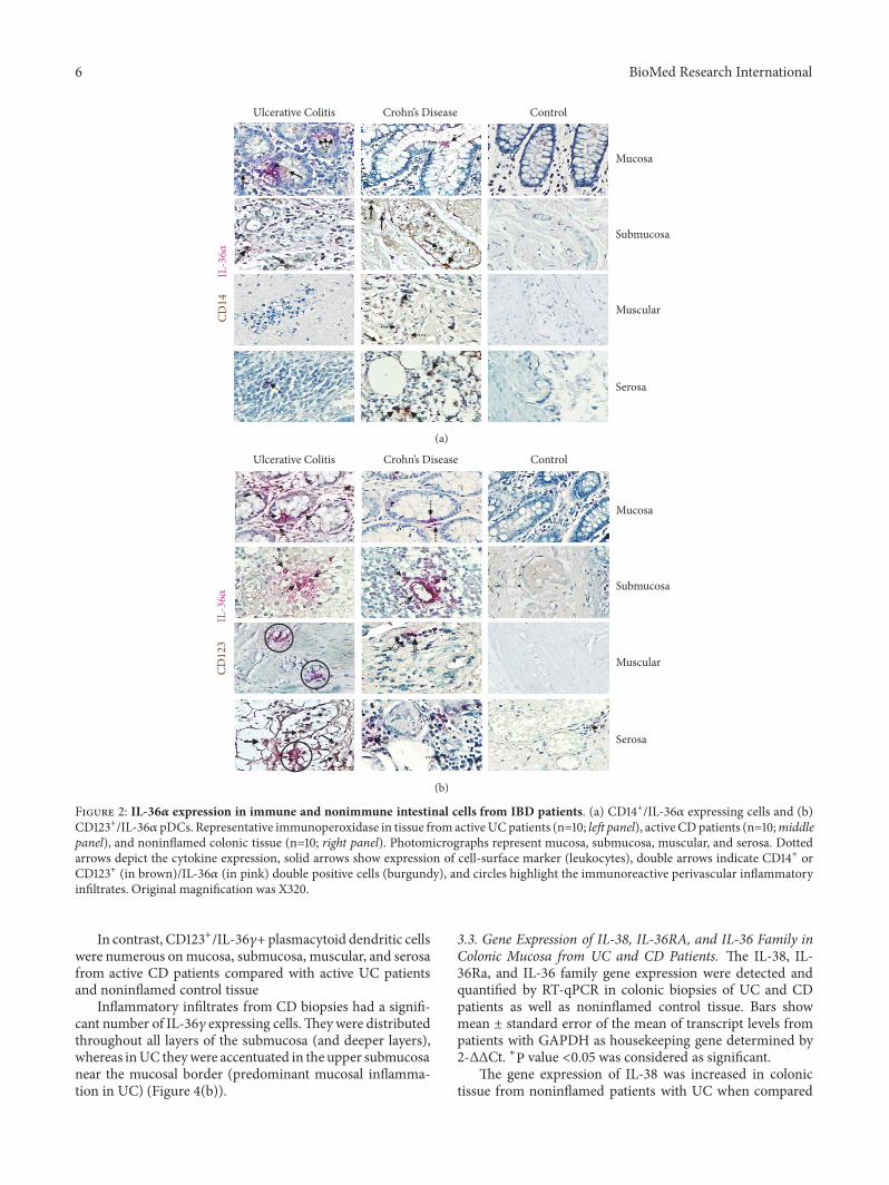
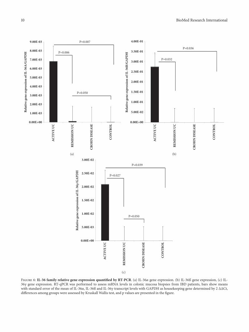