Differential diagnosis of syndromic craniosynostosis: a ...
9
Vol.:(0123456789) 1 3 Archives of Gynecology and Obstetrics https://doi.org/10.1007/s00404-021-06263-9 MATERNAL-FETAL MEDICINE Differential diagnosis of syndromic craniosynostosis: a case series Tamara Casteleyn 1 · Denise Horn 2 · Wolfgang Henrich 3 · Stefan Verlohren 3 Received: 15 February 2021 / Accepted: 15 September 2021 © The Author(s) 2021 Abstract Purpose Syndromic craniosynostosis is a rare genetic disease caused by premature fusion of one or multiple cranial sutures combined with malformations of other organs. The aim of this publication is to investigate sonographic signs of different syndromic craniosynostoses and associated malformations to facilitate a precise and early diagnosis. Methods We identified in the period of 2000–2019 thirteen cases with a prenatal suspected diagnosis of syndromic cranio- synostosis at our department. We analyzed the ultrasound findings, MRI scans, genetic results as well as the mode of delivery, and postnatal procedures. Results Eight children were diagnosed with Apert Syndrome, two with Saethre Chotzen syndrome, one with Crouzon syn- drome, and one with Greig cephalopolysyndactyly syndrome. One child had a mutation p.(Pro253Leu) in the FGFR2 gene. We identified characteristic changes of the head shape as well as typical associated malformations. Conclusion Second trimester diagnosis of syndromic craniosynostosis is feasible based on the identified sonographic signs. In case of a suspected diagnosis a genetic, neonatal as well as surgical counseling is recommended. We also recommend to offer a fetal MRI. The delivery should be planned in a perinatal center. Keywords Syndromic craniosynostosis · Apert syndrome · Saethre Chotzen syndrome · Prenatal ultrasound Introduction Craniosynostosis is the result of a premature fusion of one or multiple cranial sutures. Depending on the affected sutures, the head can develop asymmetrically which is detectable in in utero with prenatal ultrasound. Postnatally, surgery may be necessary in case of an increase in intracranial pressure. Isolated craniosynostosis is mostly sporadic, with an inci- dence of 1:2000–2500 [1]. In contrast, syndromic cranio- synostosis usually involves multiple sutures combined with malformations of other organs [2]. Syndromes most fre- quently associated with craniosynostosis are Apert-, Crou- zon-, Pfeiffer-, and Saethre Chotzen syndrome. Of these, the Apert syndrome is the most common with a prevalence of 1:100,000 [3]. A mutation of Fibroblast Growth Factor Receptor 2 (FGFR2) gene causes the autosomal dominantly inherited Apert syndrome [4]. However, most individuals with Apert syndrome develop the disorder as the result of a de novo mutation in the FGFR2 gene. The Apert syndrome causes variable deformation of the skull due to bicoronal craniosynostosis, midface hypoplasia and complex syndactyly of hands and feet [5, 6]. It can also be associated with the central nervous system's abnormali- ties, including malformations of the corpus callosum, the limbic system and abnormal gyration [7]. Neurological development disorders are possible that mostly lead to mild and rarely moderate to severe impairment [6]. After birth, high intracranial pressure can indicate a need for surgery. The autosomal dominant Crouzon syndrome is also caused by mutations in FGFR2 gene [8]. Similar to Apert syndrome, the clinical features of Crouzon syndrome are a tall, flattened forehead caused by bicoronal craniosynostosis, and midface hypoplasia. The degree of the malformations is milder, and limbs are usually not affected [9]. Bicoronal craniosynostosis and syndactyly characterize Pfeiffer syn- drome that is caused by autosomal dominant mutations of FGFR1- or FGFR2 gene [10, 11]. Additional sutures can be * Stefan Verlohren [email protected] 1 Department of Gynecology and Obstetrics, Sana Klinikum Lichtenberg, Berlin, Germany 2 Institute of Medical Genetics and Human Genetics, Charité – Universitätsmedizin, Berlin, Germany 3 Department of Obstetrics, Charité – Universitätsmedizin, Berlin, Germany
Transcript of Differential diagnosis of syndromic craniosynostosis: a ...

Vol.:(0123456789)1 3
Archives of Gynecology and Obstetrics https://doi.org/10.1007/s00404-021-06263-9
MATERNAL-FETAL MEDICINE
Differential diagnosis of syndromic craniosynostosis: a case series
Tamara Casteleyn1 · Denise Horn2 · Wolfgang Henrich3 · Stefan Verlohren3
Received: 15 February 2021 / Accepted: 15 September 2021 © The Author(s) 2021
AbstractPurpose Syndromic craniosynostosis is a rare genetic disease caused by premature fusion of one or multiple cranial sutures combined with malformations of other organs. The aim of this publication is to investigate sonographic signs of different syndromic craniosynostoses and associated malformations to facilitate a precise and early diagnosis.Methods We identified in the period of 2000–2019 thirteen cases with a prenatal suspected diagnosis of syndromic cranio-synostosis at our department. We analyzed the ultrasound findings, MRI scans, genetic results as well as the mode of delivery, and postnatal procedures.Results Eight children were diagnosed with Apert Syndrome, two with Saethre Chotzen syndrome, one with Crouzon syn-drome, and one with Greig cephalopolysyndactyly syndrome. One child had a mutation p.(Pro253Leu) in the FGFR2 gene. We identified characteristic changes of the head shape as well as typical associated malformations.Conclusion Second trimester diagnosis of syndromic craniosynostosis is feasible based on the identified sonographic signs. In case of a suspected diagnosis a genetic, neonatal as well as surgical counseling is recommended. We also recommend to offer a fetal MRI. The delivery should be planned in a perinatal center.
Keywords Syndromic craniosynostosis · Apert syndrome · Saethre Chotzen syndrome · Prenatal ultrasound
Introduction
Craniosynostosis is the result of a premature fusion of one or multiple cranial sutures. Depending on the affected sutures, the head can develop asymmetrically which is detectable in in utero with prenatal ultrasound. Postnatally, surgery may be necessary in case of an increase in intracranial pressure.
Isolated craniosynostosis is mostly sporadic, with an inci-dence of 1:2000–2500 [1]. In contrast, syndromic cranio-synostosis usually involves multiple sutures combined with malformations of other organs [2]. Syndromes most fre-quently associated with craniosynostosis are Apert-, Crou-zon-, Pfeiffer-, and Saethre Chotzen syndrome. Of these, the Apert syndrome is the most common with a prevalence
of 1:100,000 [3]. A mutation of Fibroblast Growth Factor Receptor 2 (FGFR2) gene causes the autosomal dominantly inherited Apert syndrome [4]. However, most individuals with Apert syndrome develop the disorder as the result of a de novo mutation in the FGFR2 gene.
The Apert syndrome causes variable deformation of the skull due to bicoronal craniosynostosis, midface hypoplasia and complex syndactyly of hands and feet [5, 6]. It can also be associated with the central nervous system's abnormali-ties, including malformations of the corpus callosum, the limbic system and abnormal gyration [7]. Neurological development disorders are possible that mostly lead to mild and rarely moderate to severe impairment [6]. After birth, high intracranial pressure can indicate a need for surgery.
The autosomal dominant Crouzon syndrome is also caused by mutations in FGFR2 gene [8]. Similar to Apert syndrome, the clinical features of Crouzon syndrome are a tall, flattened forehead caused by bicoronal craniosynostosis, and midface hypoplasia. The degree of the malformations is milder, and limbs are usually not affected [9]. Bicoronal craniosynostosis and syndactyly characterize Pfeiffer syn-drome that is caused by autosomal dominant mutations of FGFR1- or FGFR2 gene [10, 11]. Additional sutures can be
* Stefan Verlohren [email protected]
1 Department of Gynecology and Obstetrics, Sana Klinikum Lichtenberg, Berlin, Germany
2 Institute of Medical Genetics and Human Genetics, Charité – Universitätsmedizin, Berlin, Germany
3 Department of Obstetrics, Charité – Universitätsmedizin, Berlin, Germany

Archives of Gynecology and Obstetrics
1 3
affected, and skeletal, central nervous system and gastroin-testinal abnormalities can occur [12]. Saethre Chotzen syn-drome is characterized by mild craniosynostosis of different cranial sutures and syndactyly and is caused by the auto-somal dominant mutation of TWIST gene and the FGFR2 gene [13, 14].
The focus of our case series is to identify the contribu-tion of prenatal ultrasound for an early and precise prenatal diagnosis of syndromic craniosynostosis [5]. In case of a suspected diagnosis, genetic counselling and testing includ-ing the newest methods of whole genome/exome sequencing and/or targeted panel diagnosis is recommended. A precise differentiation between syndromic and nonsyndromic causes is paramount to allow for specific counselling [15]. A fetal MRI should also be performed to confirm the diagnosis and identify possible central nervous malformations [5].
We aim to describe the prenatal sonographic signs and their contribution in the diagnostic work up in cases with suspected syndromic craniosynostoses. We aim to raise awareness for this disease complex to facilitate a precise and early diagnosis which is essential for perinatal management and the interdisciplinary counseling of the parents.
Materials and methods
We identified thirteen cases of syndromic craniosynosto-sis in the Viewpoint (GE, Solingen, Germany) and the SAP (Walldorf, Germany) patient databases of the Department of Obstetrics and the Department of Pediatric Surgery at Char-ité—Universitätsmedizin Berlin in the years 2000–2019. We searched for keywords indicating abnormal biometric parameters of the head, brain anomalies or both, and event-ful findings of the limbs. We furthermore used exact key-word search for the keywords Apert syndrome, craniosyn-ostosis, Crouzon-, Saethre Chotzen- or Pfeiffer syndrome.
This search resulted in 389 cases of abnormal findings. A subsequent manual review identified syndromic craniosyn-ostosis in thirteen fetuses.
In addition, we compared the results with the surgery records of the Department of Pediatric Neurosurgery. No additional cases were identified. After retrieving the patients, we analyzed ultrasound findings, MRI scans, genetic results and the mode of delivery and postnatal procedures.
Results
Between 2000 and 2019, we identified thirteen pregnancies with high suspicion of syndromic craniosynostosis in our department. A detailed description of the sonographic find-ings is found in Table 1. In ten cases, Apert syndrome was suspected due to specific sonographic features. Molecular
genetic testing revealed a p.(Pro253Arg) mutation in the FGFR2 gene and confirmed the diagnosis in five cases. However, in one case, the postnatal genetic test detected a mutation in GLI3-gene, which causes Greig cephalopoly-syndactyly syndrome, which is associated with craniosyn-ostosis [16]. In another case, the genetic test revealed a p.(Pro253Leu) mutation in the FGFR2 gene. The subsequent tests of the parents identified the same mutation in the father who had not been diagnosed previously. Three patients did not give consent for genetic testing.
Two children were diagnosed with Saethre Chotzen syn-drome, one child with Crouzon syndrome. The gestational age when the diagnosis was suspected was between 20 + 1 and 33 + 4 weeks of gestation. Nine patients received the diagnosis in the second trimester, four patients in the third trimester. In all cases after 2017, we recommended a fetal MRI. This was conducted in three cases and confirmed the sonographic results.
In the fetuses with Apert syndrome, typical sonographic features were frontal bossing (5/8 cases) as well as a clo-verleaf skull (4/8) (Table 1 and Figs. 1a, b, 2a–e). In two cases, the examiner described a turricephaly, a tall head shape caused by coronal craniosynostosis (Tables 1, 2). In all cases, a prenatal ultrasound revealed a diagnosis of syn-dactyly (Fig. 1c).
In two separate Saethre Chotzen syndrome cases, the diagnosis of a bicoronal craniosynostosis occurred in one case before birth, along with identification of a saddle nose and a flat profile (Table 1). The diagnosis of syndactyly of
Table 1 Sonographic findings in syndromic craniosynostosis
Syndrome Sonographic findings
Apert syndrome (n = 8) Syndactyly (8/8)Frontal bossing (5/8)Cloverleaf skull (4/8)Turricephaly (2/8)Dolichocephaly (1/8)Polyhydramnios (1/8)A. lusoria dextra (1/8)Mild ventriculomegaly (1/8)Dysgenesis of corpus callosum
(1/8)Cleft palate (1/8)Retracted bridge of the nose (1/8)
Saethre Chotzen syndrome (n = 2) Turricephaly (1/2)Saddle nose (1/2)Flat facial profile (1/2)
Crouzon syndrome (n = 1) Flattened occiputDepressed frontoparietal bonesProtruded bulbiSmall thorax with short ribs
Greig cephalopolysyndactyly syndrome (n = 1)
Agenesis of corpus callosumHypertelorismRight-sided aortic archPolydactyly

Archives of Gynecology and Obstetrics
1 3
hands and feet occurred after birth. In the second fetus with Saethre Chotzen syndrome, the only anomaly detected was a prominent forehead.
The patient with fetal Crouzon syndrome presented with an abnormal shape of the head with a flat occiput, depressed frontoparietal bones (Fig. 3) and protruding eyes, and a small thorax with short ribs (Table 1).
The fetus with Greig cephalopolysyndactyly syndrome exhibited hypertelorism, agenesis of the corpus callosum, a right-sided aortic arch, and polydactyly (Table 1, Figs. 4a, 5). Postnatally, these findings were confirmed (Figs. 4b, c, 5b); furthermore, additional identification of malfor-mations in the child with Greig cephalopolysyndactyly
Fig. 1 Fetus with Apert syndrome in 30 + 3 weeks of gestation. The ultrasound examination shows a prominent forehead with frontal bossing (a, b). Bilateral syndactyly can be imaged (c)
Fig. 2 Child with suspected Apert syndrome. The prenatal ultrasound exam in 31 + 3 weeks of gestation shows a prominent shape of the skull with bicoronal and sagittal craniosynostosis as well as frontal bossing (a, c). The prenatal MRI confirms the findings (e). After the birth, a scaphocephaly with a long and narrow skull and high fore-
head is seen (d). The genetic examination showed a mutation in FGFR2-gene (Pro253Leu), the father had the same mutation. At this amino acid position is the pathogen mutation p.Pro253Arg located, which leads to Apert syndrome

Archives of Gynecology and Obstetrics
1 3
syndrome included a subaortic ventricular septal defect and a deformation of the feet.
After the prenatal diagnosis of a fetal syndromic cranio-synostosis and after extensive interdisciplinary counselling, seven couples decided to terminate the pregnancy. In all of these cases, a fetal Apert syndrome was diagnosed. Of these couples, four decided for an autopsy of the fetus. The patho-anatomical examinations and the radiological fetograms confirmed the sonographic findings. Of the other children, five were delivered by cesarean section and one child was delivered through vaginal birth. In one case the cesarean section was indicated due to fetal breech position. Three patients decided to have a preventive cesarean section and one patient had a repeat cesarean section.
For analyzing the long-term postnatal outcome, the data of six children who were treated at our department for pedi-atric neurosurgery was available. One child (born 2011) with Crouzon syndrome received a decompressive craniectomy in his first year of life due to high intracranial pressure. Follow-ing that multiple surgeries (last in 2020) for fronto-orbital remodeling and multiple corrections of craniofacial defects were performed. This child also suffers a hearing loss due to aural atresia and subsequent speech development disorder.
Of the two children diagnosed with Saethre Chotzen syn-drome one (born 2014) underwent fronto-orbital remodeling ten months after birth. The other one (born 2019) was treated with a strip craniectomy four months after birth. As of now, no surgery was indicated for the child with Apert syndrome (born 2019). The child diagnosed with the p.(Pro253Leu) mutation (born 2019, Fig. 1) had a biparietal strip craniec-tomy two months after birth due to raised intracranial pres-sure. The child with Greig cephalopolysyndactyly syndrome had a foot surgery to correct the hexadactyly and the pes supinatus (Fig. 5).
Discussion
Syndromic craniosynostosis is a rare disease complex that shows characteristic features detectable in prenatal ultra-sound. An early precise diagnosis is important for the inter-disciplinary counseling of the parents and the perinatal management. Our study confirms that a prenatal detection of syndromic craniosynostosis in the second trimester is pos-sible. In our case series, the diagnosis was suspected at the time of the second trimester screening in 9/13 patients and in 4/13 patients between 27 + 0 and 33 + 4 weeks of gesta-tion. However, the diagnosis can be challenging as the extent of the skull deformity can vary and the standardized meas-urements of the head (biparietal diameter and head circum-ference) are not necessarily outside the normal range [17]. To confirm the diagnosis, it can be helpful to use a three-dimensional ultrasonic skeletal imaging mode to image the Ta
ble
2 O
verv
iew
of m
alfo
rmat
ions
in sy
ndro
mic
cra
nios
ynos
tosi
s
Synd
rom
eA
ffect
ed su
ture
sH
ead
Ass
ocia
ted
mal
form
atio
ns
Ape
rt sy
ndro
me
Usu
ally
mul
tiple
sutu
res,
espe
cial
ly b
ilate
ral c
oron
al
sutu
res a
nd v
aria
ble
othe
r sut
ures
[5, 6
]A
crob
rach
ycep
haly
, flat
occ
iput
, hyp
erte
loris
m, fl
at
fore
head
, mid
face
hyp
opla
sia,
asy
mm
etric
cra
nial
sh
ape
[24]
, tur
ricep
haly
[5]
Synd
acty
ly, c
entra
l ner
vous
mal
form
atio
ns (d
ysge
n-es
is o
f cor
pus c
allo
sum
, abn
orm
al g
yrat
ion)
[5, 2
4],
neur
olog
ical
dev
elop
men
t dis
orde
rs p
ossi
ble
Cro
uzon
synd
rom
eVa
riabl
e; o
ften
coro
nal a
nd sa
gitta
l sut
ures
[24]
Acr
obra
chyc
epha
ly, (
sym
met
ric) m
idfa
ce h
ypo-
plas
ia, h
yper
telo
rism
[24]
,Ex
trem
ities
not
invo
lved
, no
neur
olog
ical
dev
elop
men
t di
sord
ers
Saet
hre
Cho
tzen
synd
rom
eU
ni-/b
ilate
ral c
oron
al su
ture
s, ot
her s
utur
es p
ossi
ble
[25]
Plag
ioce
phal
y, b
rach
ycep
haly
, acr
ocep
haly
, dee
p ha
irlin
e, sm
all e
ars,
high
fore
head
, asy
mm
etric
fa
ce (o
vera
ll m
ilder
than
Ape
rt sy
ndro
me)
[25]
Synd
acty
ly p
ossi
ble,
bra
chyd
acty
ly, r
arel
y ne
urol
ogi-
cal d
evel
opm
ent d
isor
ders
, ske
leta
l and
car
diac
m
alfo
rmat
ions
[25]
Gre
ig c
epha
lopo
ly- s
yn-
dact
yly
synd
rom
eFr
onta
l und
sagi
ttal s
utur
es [1
6]M
acro
ceph
aly,
pro
min
ent f
oreh
ead,
hyp
erte
loris
m
[26]
, trig
onoc
epha
ly [1
6]Po
lyda
ctyl
y, b
road
thum
bs/b
ig to
es, c
utan
eous
synd
ac-
tyly
, neu
rolo
gica
l dev
elop
men
t dis
orde
rs p
ossi
ble
[26]
, dys
gene
sis o
f cor
pus c
allo
sum
[16]
Pfei
ffer s
yndr
ome
Bila
tera
l cor
onal
- und
lam
bdoi
dal s
utur
es, r
arel
y sa
gitta
l sut
ure
[27]
Flat
occ
iput
, hig
h fo
rehe
ad, h
yper
telo
rism
, clo
verle
af
skul
l, va
riabl
e m
idfa
ce h
ypop
lasi
a [1
2, 2
7]B
road
thum
bs/b
ig to
es w
ith d
evia
tion,
synd
acty
ly p
os-
sibl
e, n
euro
logi
cal d
evel
opm
ent d
isor
ders
pos
sibl
e [1
2, 2
7]

Archives of Gynecology and Obstetrics
1 3
skull with the sutures (Figs. 6, 7, 8). Though diagnosis is mostly feasible without using the skeleton mode, as we have used B-mode and conventional 3D ultrasound in our cases to establish the diagnosis.
Value of sonographic signs: skull shapes
In accordance to the literature, an abnormal shape of the skull was the leading sonographic sign in our case series. The basic biometric parameters may be out of range. Depending on the affected sutures the biparietal diameter (BPD) and the cephalic index (CI) can be raised or lower [17].
Fig. 3 Fetus with bilateral coro-nal synostosis caused by Crou-zon syndrome. The ultrasound shows a flattened occiput and mild bilateral frontal depres-sions of the skull
Fig. 4 Sonographic and postnatal images of a child with Greig cepha-lopolysyndactyly syndrome. The cranial biometric parameters were in the normal range, a dysgenesis of the corpus callosum was suspected.
After the birth, a high forehead with down-slanting palpebral fissures and a low nose root is seen
Fig. 5 Child with Greig cephalopolysyndactyly syndrome. The sono-graphic diagnosis of postaxial polysyndactyly (a) was confirmed after the birth (b)
Archives of Gynecology and Obstetrics
1 3
In our cohort, all cases with Apert syndrome exhibited an abnormal shape of the skull (Figs. 1, 2). We detected frontal bossing in 5/8 cases, a cloverleaf skull in 4/8 cases, a turricephaly in 2/8 cases and a dolichocephaly in 1/8 cases (see Table 1). In one of the fetuses with Apert syndrome, agenesis of corpus callosum was diagnosed. Malformations of midline structures like dysgenesis of the corpus callosum are reported in up to 11% as well as alterations of the temporal lobe [7, 18].
An abnormal shape of the skull was the leading ultra-sonographic finding also in the other cases of syndromic craniosynostosis. In one case of Saethre Chotzen syn-drome, the fetus presented with turricephaly with flat profile and saddle nose. In the other case the head was small without other sonomorphological alterations. In the fetus with Greig cephalopolysyndactyly syndrome a hyper-telorism and agenesis of corpus callosum were noted and
the fetus with Crouzon syndrome had protruding bulbi of the eyes.
In accordance to other published case series the diagnosis of syndromic craniosynostosis was suspected in most cases in the second trimester as the skull deformation develops at this time [14]. Owing to premature fusion of sutures, the underlying structures are harder to visualize. This effect has been identified as an early indirect sign for craniosynosto-sis called `brain shadowing sign’. It can be noted prior to changes of the skull shape and is also reported in fetuses with mild changes [19]. In this retrospective study, cases of syndromic craniosynostosis were detected in 25 weeks of gestation. However, the brain shadowing sign is not spe-cific for craniosynostosis and can also be seen in fetal head molding or open spina bifida. The authors also report that in accordance to our results an abnormal head shape was seen in 23 of 24 cases. Other signs were facial abnormalities, syndactyly and ventriculomegaly [19]. Furthermore, the cra-nial bones with the sutures and facial alterations can be seen more detailed with three-dimensional ultrasound, especially with the feature “skeleton mode”, as compared to B-mode (Figs. 6, 7, 8) [20]. In the skeleton mode, the premature fusion of the suture can be imaged exactly (Fig. 8).
Value of sonographic signs: other malformations
Other organ malformations may occur in syndromic cranio-synostosis. A thorough sonographic examination of hands and feet is mandatory, as malformations of extremities are common and may be used to distinguish between isolated and syndromic craniosynostosis. The type of limb abnor-malities indicates which type of syndromic craniosynosto-sis is likely. In accordance to the literature, in all our cases of Apert syndrome, syndactyly of upper and partly of the lower extremities were diagnosed by ultrasound (Table 1 and Fig. 1c) [17, 19, 20]. In one case of fetal Saethre Chotzen syndrome, syndactyly was diagnosed only postnatally. In another case series, anal atresia and a patent ductus arterio-sus were detected postnatally in a child with Saethre Chotzen syndrome [19]. In accordance to Hurst et al. we diagnosed a polydactyly in a fetus with Greig cephalopolysyndactyly syndrome [16]. Besides the thorough sonographic assess-ment of the skull, the central nervous system and limbs, it is important to examine the other organ systems as well to detect possible accompanied malformations which might have an impact on the child’s prognosis. Our case of Greig cephalopolysyndactyly syndrome was associated with a right aortic arch. A subaortic ventricular septal defect was addi-tionally diagnosed postnatally. In the literature, congenital heart defects are described in Greig cephalopolysyndactyly syndrome and include ventricular septal defects, atrial septal defects and patent ductus arteriosus as well as double outlet right ventricle [16]. Congenital heart defects in combination
Fig. 6 Fetus with normal face and skull shape imaged by three-dimensional skeletal imaging mode in 27 + 5 weeks of gestation
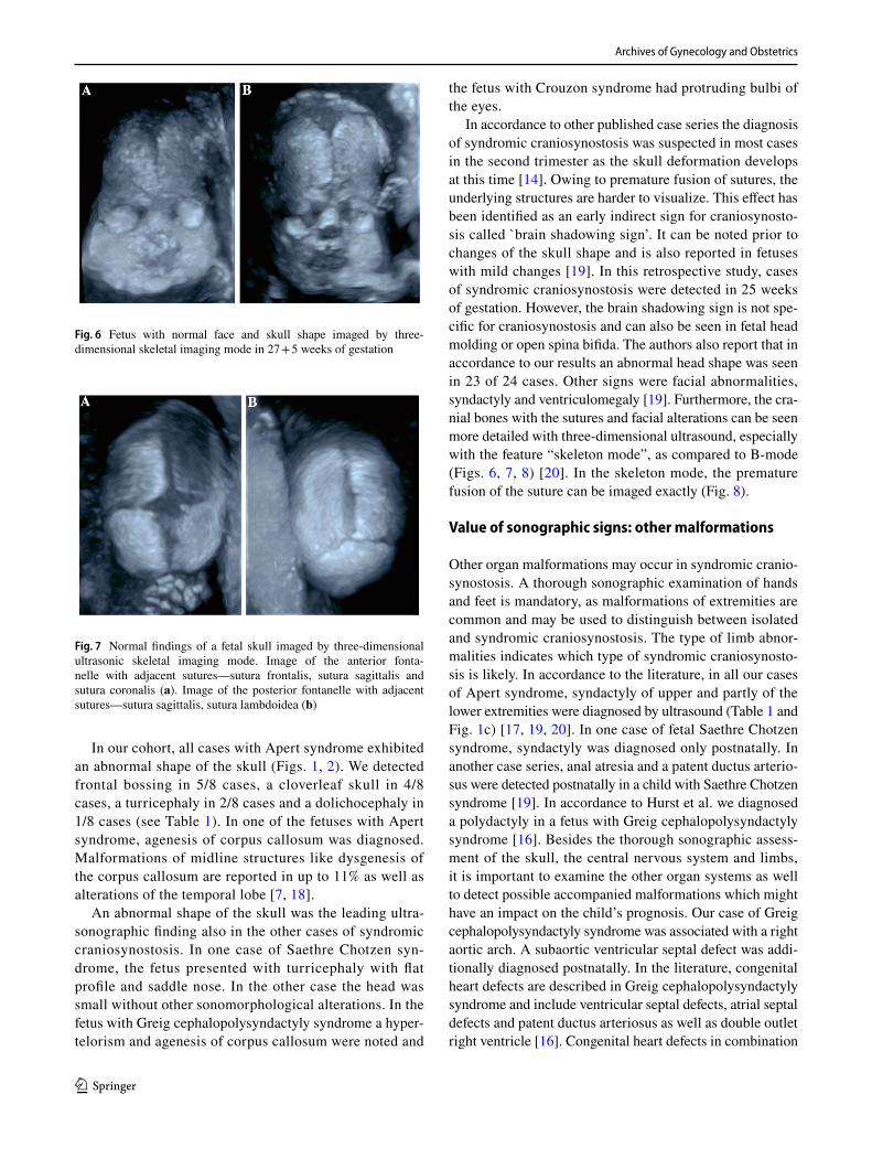
Fig. 7 Normal findings of a fetal skull imaged by three-dimensional ultrasonic skeletal imaging mode. Image of the anterior fonta-nelle with adjacent sutures—sutura frontalis, sutura sagittalis and sutura coronalis (a). Image of the posterior fontanelle with adjacent sutures—sutura sagittalis, sutura lambdoidea (b)

Archives of Gynecology and Obstetrics
1 3
with craniosynostosis and polydactyly can also be seen in Pfeiffer syndrome and the rare Carpenter syndrome [21]. The rare Antley Bixler syndrome is characterized by cranio-synostosis, humero-radial synostosis, a curved femur and contractures of the joints. Cardiac and urogenital defects are possible [22].
Value of fetal MRI
In our case series, a fetal MRI was performed in three cases. This examination confirmed the sonographic findings and identified no further abnormalities of the central nervous system (Fig. 2e). However, a study of Rubio et al., com-pared the results of ultrasound exams and fetal MRI after the diagnosis of syndromic craniosynostosis. The MRI detected two cases of dysgenesis of corpus callosum and one teth-ered cord syndrome that were not detected with ultrasound [5]. Malformations of the central nervous system can cause neurodevelopmental disorders and are important when counseling the parents. In conclusion, a fetal MRI should be offered to all patients with suspected syndromic cranio-synostosis. However, a precise prenatal prognosis regarding developmental disorders is not possible.
Value of molecular genetic tests
Genetic testing must be offered to the patients in order to distinguish between isolated and syndromic craniosynostosis and to confirm the entity of the craniosynostosis. A genetic examination of the parents can be performed since not only sporadic mutations, but also autosomal dominant inheritance with variable symptoms is possible. In our study, one parent
was previously diagnosed with Saethre Chotzen syndrome. In another case of suspected Apert syndrome a genetic test-ing was conducted. The result showed a p.(Pro253Leu) mutation in the FGFR2 gene not only in the DNA of the fetus, but also in the father’s DNA who had not been diag-nosed with craniosynostosis previously.
Mode of delivery
In our case series, five patients had a cesarean section and one patient a vaginal birth. Similar to our results Harada et al., described a high rate of cesarean sections (73%) in patients with fetal craniosynostosis [17]. If the fetus is in cephalic position and the head circumference is not raised excessively there is no absolute indication for a cesarean section. The patients should be informed about the higher risk of arrested labor and emergency cesarean section in case of significant skull deformities. The delivery should be planned in a perinatal center to assure an ideal postnatal care of the newborn especially regarding the airway management. Prenatally a Pediatric Neurosurgeon should be consulted and inform the parents about possible operative procedures.
After birth a cranial ultrasound and a cranial MRI should be performed. Owing to the higher prevalence of cardiac defects an echocardiography is recommended as well as an examination by a pediatric surgeon.
Limitations
Due to the low incidences of syndromic craniosynostosis we could only analyze thirteen cases with suspected cranio-synostosis. Another limitation is the retrospective character
Fig. 8 Fetus with partial sagittal craniosynostosis in 23 + 4 weeks of gestation. By three-dimensional ultrasonic skeletal imaging mode, a partial premature fusion of the sagittal suture can be shown (a). The
B-mode image shows a prominent shape of the skull, a sagittal crani-osynostosis is suspected (b)

Archives of Gynecology and Obstetrics
1 3
of the study. A comparison between ultrasound and MRI is limited as we did not perform an MRI in all cases.
Conclusion
In conclusion, syndromic craniosynostosis is a rare disease. A prenatal diagnosis in the second trimester is feasible based on the sonographic signs described here. An early and pre-cise diagnosis should be achieved to allow for targeted coun-selling. In case of a suspected diagnosis a genetic, neonatal and surgical workup is recommended and a fetal MRI should be conducted. Owing to midface hypoplasia and alterations of the upper airway the newborns can be respiratory com-promised. Consequently the delivery should be planned in a perinatal center [23].
Author contribution TC: manuscript writing, data collection, data analysis, project development. DH: manuscript editing, data analy-sis. WH: manuscript editing, data analysis. SV: project development, manuscript editing.
Funding Open Access funding enabled and organized by Projekt DEAL.
Availability of data and material The data available on request from the corresponding author.
Code availability Not applicable.
Declarations
Conflict of interest The authors declare that they have no conflict of interest.
Open Access This article is licensed under a Creative Commons Attri-bution 4.0 International License, which permits use, sharing, adapta-tion, distribution and reproduction in any medium or format, as long as you give appropriate credit to the original author(s) and the source, provide a link to the Creative Commons licence, and indicate if changes were made. The images or other third party material in this article are included in the article's Creative Commons licence, unless indicated otherwise in a credit line to the material. If material is not included in the article's Creative Commons licence and your intended use is not permitted by statutory regulation or exceeds the permitted use, you will need to obtain permission directly from the copyright holder. To view a copy of this licence, visit http:// creat iveco mmons. org/ licen ses/ by/4. 0/.
References
1. Slater BJ, Lenton KA, Kwan MD et al (2008) Cranial sutures: a brief review. Plast Reconstr Surg 121:170e–178e. https:// doi. org/ 10. 1097/ 01. prs. 00003 04441. 99483. 97
2. Schramm T, Mommsen H (2018) Fetal Skeletal Disorders. Ultra-schall Med 39:610–634. https:// doi. org/ 10. 1055/a- 0660- 9417
3. Tolarova MM, Harris JA, Ordway DE et al (1997) Birth preva-lence, mutation rate, sex ratio, parents’ age, and ethnicity in Apert syndrome. Am J Med Genet 72:394–398. https:// doi. org/ 10. 1002/ (sici) 1096- 8628(19971 112) 72:4% 3c394:: aid- ajmg4% 3e3.0. co;2-r
4. Wilkie AO, Slaney SF, Oldridge M et al (1995) Apert syndrome results from localized mutations of FGFR2 and is allelic with Crouzon syndrome. Nat Genet 9:165–172. https:// doi. org/ 10. 1038/ ng0295- 165
5. Rubio EI, Blask A, Bulas DI (2016) Ultrasound and MR imag-ing findings in prenatal diagnosis of craniosynostosis syn-dromes. Pediatr Radiol 46:709–718. https:// doi. org/ 10. 1007/ s00247- 016- 3550-x
6. Cohen MM Jr, Kreiborg S (1993) An updated pediatric per-spective on the Apert syndrome. Am J Dis Child 147:989–993. https:// doi. org/ 10. 1001/ archp edi. 1993. 02160 33007 9025
7. Breik O, Mahindu A, Moore MH et al (2016) Central nerv-ous system and cervical spine abnormalities in Apert syn-drome. Childs Nerv Syst 32:833–838. https:// doi. org/ 10. 1007/ s00381- 016- 3036-z
8. Reardon W, Winter RM, Rutland P et al (1994) Mutations in the fibroblast growth factor receptor 2 gene cause Crouzon syn-drome. Nat Genet 8:98–103. https:// doi. org/ 10. 1038/ ng0994- 98
9. Kreiborg S, Cohen MM Jr (1998) Is craniofacial morphology in Apert and Crouzon syndromes the same? Acta Odontol Scand 56:339–341. https:// doi. org/ 10. 1080/ 00016 35984 28275
10. Cornejo-Roldan LR, Roessler E, Muenke M (1999) Analysis of the mutational spectrum of the FGFR2 gene in Pfeiffer syn-drome. Hum Genet 104:425–431. https:// doi. org/ 10. 1007/ s0043 90050 979
11. Muenke M, Schell U, Hehr A et al (1994) A common muta-tion in the fibroblast growth factor receptor 1 gene in Pfeiffer syndrome. Nat Genet 8:269–274. https:// doi. org/ 10. 1038/ ng1194- 269
12. Cohen MM Jr (1993) Pfeiffer syndrome update, clinical sub-types, and guidelines for differential diagnosis. Am J Med Genet 45:300–307. https:// doi. org/ 10. 1002/ ajmg. 13204 50305
13. Johnson D, Horsley SW, Moloney DM et al (1998) A compre-hensive screen for TWIST mutations in patients with craniosyn-ostosis identifies a new microdeletion syndrome of chromosome band 7p21.1. Am J Hum Genet 63:1282–1293. https:// doi. org/ 10. 1086/ 302122
14. Delahaye S, Bernard JP, Renier D et al (2003) Prenatal ultra-sound diagnosis of fetal craniosynostosis. Ultrasound Obstet Gynecol 21:347–353. https:// doi. org/ 10. 1002/ uog. 91
15. Yoon AJ, Pham BN, Dipple KM (2016) Genetic screening in patients with craniofacial malformations. J Pediatr Genet 5:220–224. https:// doi. org/ 10. 1055/s- 0036- 15924 23
16. Hurst JA, Jenkins D, Vasudevan PC et al (2011) Metopic and sagittal synostosis in Greig cephalopolysyndactyly syndrome: five cases with intragenic mutations or complete deletions of GLI3. Eur J Hum Genet 19:757–762. https:// doi. org/ 10. 1038/ ejhg. 2011. 13
17. Harada A, Miyashita S, Nagai R et al (2019) Prenatal sono-graphic findings and prognosis of craniosynostosis diagnosed during the fetal and neonatal periods. Congenit Anom (Kyoto) 59:132–141. https:// doi. org/ 10. 1111/ cga. 12308
18. Stark Z, McGillivray G, Sampson A et al (2015) Apert syn-drome: temporal lobe abnormalities on fetal brain imaging. Prenat Diagn 35:179–182. https:// doi. org/ 10. 1002/ pd. 4515
19. Krajden Haratz K, Leibovitz Z, Svirsky R et al (2016) The “brain shadowing sign”: a novel marker of fetal craniosynos-tosis. Fetal Diagn Ther 40:277–284. https:// doi. org/ 10. 1159/ 00044 4298
20. Chaoui R, Levaillant JM, Benoit B et al (2005) Three-dimen-sional sonographic description of abnormal metopic suture in

Archives of Gynecology and Obstetrics
1 3
second- and third-trimester fetuses. Ultrasound Obstet Gynecol 26:761–764. https:// doi. org/ 10. 1002/ uog. 2650
21. Haye D, Collet C, Sembely-Taveau C et al (2014) Prenatal find-ings in carpenter syndrome and a novel mutation in RAB23. Am J Med Genet A 164A:2926–2930. https:// doi. org/ 10. 1002/ ajmg.a. 36726
22. Lee HJ, Cho DY, Tsai FJ et al (2001) Antley-Bixler syndrome, description of two new cases and review of the literature. Pedi-atr Neurosurg 34:33–39. https:// doi. org/ 10. 1159/ 00005 5989
23. Fujimoto T, Imai K, Matsumoto H et al (2011) Tracheobronchial anomalies in syndromic craniosynostosis with 3-dimensional CT image and bronchoscopy. J Craniofac Surg 22:1579–1583. https:// doi. org/ 10. 1097/ SCS. 0b013 e3182 2e5d15
24. Cohen MM Jr, Kreiborg S (1996) A clinical study of the crani-ofacial features in Apert syndrome. Int J Oral Maxillofac Surg 25:45–53. https:// doi. org/ 10. 1016/ s0901- 5027(96) 80011-7
25. Gallagher ER, Ratisoontorn C, Cunningham ML (2003) Sae-thre-Chotzen syndrome. In: Adam MP, Ardinger HH, Pagon RA, Wallace SE, Bean LJH, Mirzaa G, Amemiya A (eds) GeneReviews® [Internet]. Seattle (WA): University of Wash-ington, Seattle; 1993–2021
26. Biesecker LG (2008) The Greig cephalopolysyndactyly syndrome. Orphanet J Rare Dis 3:10. https:// doi. org/ 10. 1186/ 1750- 1172-3- 10
27. Vogels A, Fryns JP (2006) Pfeiffer syndrome. Orphanet J Rare Dis 1:19. https:// doi. org/ 10. 1186/ 1750- 1172-1- 19
Publisher's Note Springer Nature remains neutral with regard to jurisdictional claims in published maps and institutional affiliations.