Different Functions of the Insect Soluble and … · Different Functions of the Insect Soluble and...
13
Different Functions of the Insect Soluble and Membrane- Bound Trehalase Genes in Chitin Biosynthesis Revealed by RNA Interference Jie Chen 1 , Bin Tang 1,2 , Hongxin Chen 1 , Qiong Yao 1 , Xiaofeng Huang 1 , Jing Chen 1 , Daowei Zhang 1 , Wenqing Zhang 1 * 1 State Key Laboratory of Biocontrol, School of Life Sciences, Sun Yat-sen University, Guangzhou, China, 2 Hangzhou Key Laboratory of Animal Adaptation and Evolution, Hangzhou Normal University, Hangzhou, Zhejiang, China Abstract Background: Trehalase, an enzyme that hydrolyzes trehalose to yield two glucose molecules, plays a pivotal role in various physiological processes. In recent years, trehalase proteins have been purified from several insect species and are divided into soluble (Tre-1) and membrane-bound (Tre-2) trehalases. However, no functions of the two trehalases in chitin biosynthesis in insects have yet been reported. Principal Findings: The membrane-bound trehalase of Spodoptera exigua (SeTre-2) was characterized in our laboratory previously. In this study, we cloned the soluble trehalase gene (SeTre-1) and investigated the tissue distribution and developmental expression pattern of the two trehalase genes. SeTre-1 was expressed highly in cuticle and Malpighian tubules, while SeTre-2 was expressed in tracheae and fat body. In the midgut, the two trehalase genes were expressed in different locations. Additionally, the expression profiles of both trehalase mRNAs and their enzyme activities suggest that they may play different roles in chitin biosynthesis. The RNA interference (RNAi) of either SeTre-1 or SeTre-2 was gene-specific and effective, with efficiency rates up to 83% at 72 h post injection. After RNAi of SeTre-1 and SeTre-2, significant higher mortality rates were observed during the larva-pupa stage and pupa-adult stage, and the lethal phenotypes were classified and analyzed. Additionally, the change trends of concentration of trehalose and glucose appeared reciprocally in RNAi-mutants. Moreover, knockdown of SeTre-1 gene largely inhibited the expression of chitin synthase gene A (CHSA) and reduced the chitin content in the cuticle to two-thirds relative to the control insects. The chitin synthase gene B (CHSB) expression, however, was inhibited more by the injection of dsRNA for SeTre-2, and the chitin content in the midgut decreased by about 25%. Conclusions: SeTre-1 plays a major role in CHSA expression and chitin synthesis in the cuticle, and SeTre-2 has an important role in CHSB expression and chitin synthesis in the midgut. Citation: Chen J, Tang B, Chen H, Yao Q, Huang X, et al. (2010) Different Functions of the Insect Soluble and Membrane-Bound Trehalase Genes in Chitin Biosynthesis Revealed by RNA Interference. PLoS ONE 5(4): e10133. doi:10.1371/journal.pone.0010133 Editor: Fernando Rodrigues-Lima, University Paris Diderot-Paris 7, France Received January 30, 2010; Accepted March 22, 2010; Published April 12, 2010 Copyright: ß 2010 Chen et al. This is an open-access article distributed under the terms of the Creative Commons Attribution License, which permits unrestricted use, distribution, and reproduction in any medium, provided the original author and source are credited. Funding: This work was supported by National Natural Science Foundation of China (30871634), Research Fund for the Doctoral Program of Higher Education of China (20070558027) and Science Foundation of the State Key Laboratory of Biocontrol (SKLBC09A01). The funders had no role in study design, data collection and analysis, decision to publish, or preparation of the manuscript. Competing Interests: The authors have declared that no competing interests exist. * E-mail: [email protected] Introduction Trehalase (a-glucoside-1-glucohydrolase, EC 3.2.1.28) is an enzyme that hydrolyzes trehalose to yield two glucose molecules, and it is present in almost all tissues in different forms. Trehalase plays a pivotal role in various physiological processes, including flight metabolism[1], chitin synthesis during molting [2], and cold tolerance[3]. In insects, all these function of trehalase are achieved through the hydrolysis of trehalose (a-D-glucopyranosyl-a-D- glucopyranoside), the principal hemolymph sugar in insects that acts as an indispensable substrate for energy production and macromolecular biosynthesis[4]. The chitin biosynthesis pathway starts with trehalose[5], and the first enzyme involved in the pathway is trehalase, and the last one is chitin synthase. Other enzymes involved include hexokinase, Glucose-6-P isomerase (G-6-P-I), UDP-N-acetylglucosamine pyr- ophosphorylase (UAP) and so on. The activity of enzymes involved in chitin metabolism were affected by temperature, especially at elevated temperature[6]. To date, trehalase proteins have been purified from various insect species and are divided into the soluble (Tre-1) and the membrane-bound (Tre-2) trehalases [7,8,9,10,11,12]. The first insect trehalase gene, a soluble trehalase gene, was reported in 1992[13], and a membrane-bound trehalase gene was not reported until 2005[14]. The number of genes encoding a family of proteins in insects varies widely among species. For example, two trehalase genes, two chitin synthase genes, and 13 to 16 chitinase-related genes have been identified thus far in insects[15,16,17]. Different genes in a family often have different functions. The chitin synthase gene A of Tribolium castaneum (TcCHSA) is the sole contributor to cuticular chitin synthesis, and chitin synthase gene B (TcCHSB) is the major/sole contributor to the chitin synthesis in peritrophic PLoS ONE | www.plosone.org 1 April 2010 | Volume 5 | Issue 4 | e10133
Transcript of Different Functions of the Insect Soluble and … · Different Functions of the Insect Soluble and...

Different Functions of the Insect Soluble and Membrane-Bound Trehalase Genes in Chitin Biosynthesis Revealedby RNA InterferenceJie Chen1, Bin Tang1,2, Hongxin Chen1, Qiong Yao1, Xiaofeng Huang1, Jing Chen1, Daowei Zhang1,
Wenqing Zhang1*
1 State Key Laboratory of Biocontrol, School of Life Sciences, Sun Yat-sen University, Guangzhou, China, 2 Hangzhou Key Laboratory of Animal Adaptation and Evolution,
Hangzhou Normal University, Hangzhou, Zhejiang, China
Abstract
Background: Trehalase, an enzyme that hydrolyzes trehalose to yield two glucose molecules, plays a pivotal role in variousphysiological processes. In recent years, trehalase proteins have been purified from several insect species and are dividedinto soluble (Tre-1) and membrane-bound (Tre-2) trehalases. However, no functions of the two trehalases in chitinbiosynthesis in insects have yet been reported.
Principal Findings: The membrane-bound trehalase of Spodoptera exigua (SeTre-2) was characterized in our laboratorypreviously. In this study, we cloned the soluble trehalase gene (SeTre-1) and investigated the tissue distribution anddevelopmental expression pattern of the two trehalase genes. SeTre-1 was expressed highly in cuticle and Malpighian tubules,while SeTre-2 was expressed in tracheae and fat body. In the midgut, the two trehalase genes were expressed in differentlocations. Additionally, the expression profiles of both trehalase mRNAs and their enzyme activities suggest that they may playdifferent roles in chitin biosynthesis. The RNA interference (RNAi) of either SeTre-1 or SeTre-2 was gene-specific and effective, withefficiency rates up to 83% at 72 h post injection. After RNAi of SeTre-1 and SeTre-2, significant higher mortality rates wereobserved during the larva-pupa stage and pupa-adult stage, and the lethal phenotypes were classified and analyzed.Additionally, the change trends of concentration of trehalose and glucose appeared reciprocally in RNAi-mutants. Moreover,knockdown of SeTre-1 gene largely inhibited the expression of chitin synthase gene A (CHSA) and reduced the chitin content inthe cuticle to two-thirds relative to the control insects. The chitin synthase gene B (CHSB) expression, however, was inhibitedmore by the injection of dsRNA for SeTre-2, and the chitin content in the midgut decreased by about 25%.
Conclusions: SeTre-1 plays a major role in CHSA expression and chitin synthesis in the cuticle, and SeTre-2 has an importantrole in CHSB expression and chitin synthesis in the midgut.
Citation: Chen J, Tang B, Chen H, Yao Q, Huang X, et al. (2010) Different Functions of the Insect Soluble and Membrane-Bound Trehalase Genes in ChitinBiosynthesis Revealed by RNA Interference. PLoS ONE 5(4): e10133. doi:10.1371/journal.pone.0010133
Editor: Fernando Rodrigues-Lima, University Paris Diderot-Paris 7, France
Received January 30, 2010; Accepted March 22, 2010; Published April 12, 2010
Copyright: � 2010 Chen et al. This is an open-access article distributed under the terms of the Creative Commons Attribution License, which permitsunrestricted use, distribution, and reproduction in any medium, provided the original author and source are credited.
Funding: This work was supported by National Natural Science Foundation of China (30871634), Research Fund for the Doctoral Program of Higher Education ofChina (20070558027) and Science Foundation of the State Key Laboratory of Biocontrol (SKLBC09A01). The funders had no role in study design, data collectionand analysis, decision to publish, or preparation of the manuscript.
Competing Interests: The authors have declared that no competing interests exist.
* E-mail: [email protected]
Introduction
Trehalase (a-glucoside-1-glucohydrolase, EC 3.2.1.28) is an
enzyme that hydrolyzes trehalose to yield two glucose molecules,
and it is present in almost all tissues in different forms. Trehalase
plays a pivotal role in various physiological processes, including
flight metabolism[1], chitin synthesis during molting [2], and cold
tolerance[3]. In insects, all these function of trehalase are achieved
through the hydrolysis of trehalose (a-D-glucopyranosyl-a-D-
glucopyranoside), the principal hemolymph sugar in insects that
acts as an indispensable substrate for energy production and
macromolecular biosynthesis[4].
The chitin biosynthesis pathway starts with trehalose[5], and the
first enzyme involved in the pathway is trehalase, and the last one
is chitin synthase. Other enzymes involved include hexokinase,
Glucose-6-P isomerase (G-6-P-I), UDP-N-acetylglucosamine pyr-
ophosphorylase (UAP) and so on. The activity of enzymes involved
in chitin metabolism were affected by temperature, especially at
elevated temperature[6]. To date, trehalase proteins have been
purified from various insect species and are divided into the
soluble (Tre-1) and the membrane-bound (Tre-2) trehalases
[7,8,9,10,11,12]. The first insect trehalase gene, a soluble trehalase
gene, was reported in 1992[13], and a membrane-bound trehalase
gene was not reported until 2005[14].
The number of genes encoding a family of proteins in insects
varies widely among species. For example, two trehalase genes,
two chitin synthase genes, and 13 to 16 chitinase-related genes
have been identified thus far in insects[15,16,17]. Different genes
in a family often have different functions. The chitin synthase gene
A of Tribolium castaneum (TcCHSA) is the sole contributor to
cuticular chitin synthesis, and chitin synthase gene B (TcCHSB) is
the major/sole contributor to the chitin synthesis in peritrophic
PLoS ONE | www.plosone.org 1 April 2010 | Volume 5 | Issue 4 | e10133

matrix chitin synthesis[18]. The chitinase genes in Tribolium
castaneum differ in their expression patterns during development
and in different tissues, and genes in different groups have different
functions[19]. In some situations, isoforms of a gene are
functionally redundant[20]. However, the functions of the two
trehalases in insect chitin biosynthesis have not yet been reported.
The beet armyworm Spodoptera exigua is a serious lepidopteran pest.
Its membrane-bound trehalase gene was previously characterized in
our laboratory[16]. Here we cloned the soluble trehalase gene.
Different expression pattern of the two trehalase genes suggest that
they may have different functions. The transcript levels of the two
genes were substantially down-regulated by injection of gene-specific
dsRNAs of the two trehalases. The results above revealed that the two
genes have different functions in insect chitin biosynthesis.
Results
Cloning and sequence analysis of SeTre-1 cDNAThe full-length cDNA sequence of the soluble trehalase in S.
exigua (SeTre-1) is 2144 nucleotides with an open reading frame of
1755 nucleotides, which encodes a protein of 585 amino acids with
a predicted mass of approximately 66.48 KDa and an isoelectric
point of 4.75. The deduced amino acid sequence of SeTre-1 was
aligned with the trehalases of other insect species. Compared with
the membrane-bound trehalase gene (SeTreh-2)[16], they have only
37% homeology.
A phylogenic tree was constructed based on the full-length
sequences of known trehalases from insects and other organisms
(Figure 1). The results showed that the overall amino acid
sequence of SeTre-1 shares high identity with Spodoptera frugiperda
Tre-1 (80%) (DQ447188), Ostrinia furnacalis Tre-1 (55%)
(EF426724), Bombyx mori Tre-1 (55%) (D86212), followed by
Tribolium castaneum Tre-1 (44%) (XM_968826), Nilaparvata lugens
Tre-1 (41%) (EJ790319), S. exigua Tre-2 (37%) (EU106080), S.
frugiperda Tre-2 (36%) (EU872435), B. mori Tre-2 (36%)
(NM_001043445), N. lugens Tre-2 (36%) (GQ397451), and T.
castaneum Tre-2 (35%) (XM_967517).
Immuno-blotting in tissuesImmuno-blot analysis was performed with anti-SeTre-1 or anti-
SeTre-2 serums. Only one major protein at 68 or 74 kDa was
detected in the tissues on day 2 of fifth instars (Figure 2). The size
was consistent with the predicted mass of SeTre-1 or SeTre-2
deduced from amino acid sequence. These data suggested that the
polyclonal antibodies specifically recognized SeTre-1 or SeTre-2.
Immunocytochemical analysis of SeTre-1 and SeTre-2The localization of SeTre-1 and SeTre-2 proteins in the midgut,
cuticle, tracheae, Malpighian tubules and fat body were determined
by immunohistochemistry. Different expression levels of SeTre-1 and
SeTre-2 were detected in all tissues (Figure 3). SeTre-1 was highly
expressed in the cuticle, middle section of the midgut and Malpighian
tubules but weakly in the fat body, while SeTre-2 was expressed in the
verge section of the midgut, and highly expressed in the tracheae and
fat body but very weakly in the cuticle. Additionally, the expressions
of SeTre-1 and SeTre-2 mRNA in different tissues were also
determined by semi-quantitative RT-PCR, coincident results were
obtained (data not shown).
Developmental expression pattern of SeTreh-1 andSeTre-2
The expression patterns of the two trehalases during S. exigua
development from day one of the first instar larva through pupa to
adult were determined by quantitative real-time PCR. As the
mRNA levels of Seb-actin gene were nearly constant during the
whole development stages (Figure 4), it was used to standardize the
relative expression levels of SeTre-1 and SeTre-2 genes.[21] SeTre-1
and SeTre-2 were continuously expressed in all developmental
stages. After day one of the fifth instar, the SeTre-1 mRNA
expression increased gradually and reached a maximal level at day
one of the pupae, while the mRNA level of SeTre-2 remained at a
constant level and increased to a high level at day four of the
Figure 1. Phylogenetic analysis of the trehalase amino acidsequences. The phylogenetic analysis of the trehalases amino acidswas conducted using the MEGA3.1 program to generate a phylogenetictree (1, the soluble trehalase gene; 2, the membrane-bound trehalasegene) and the robustness of each cluster was verified in 1000 replicates.The scale on the x-axis represents the estimated branch lengths andnumbers indicate the bootstrap values. Trehalases were from Aedesaegypti (Aa), Anopheles gambiae(Ag), Apis mellifera (Am), Bombyx mori(Bm), Drosophila melanogaster (Dm), Drosophila simulans (Ds), Nasoniavitripennis (Nv), Ostrinia furnacalis (Of), Pimpla hypochondriaca (Ph),Spodoptera exigua (Se), Spodoptera frugiperda (Sf), Tribolium castaneum(Tc), Nilaparvata lugensand (Nl), Tenebrio molitor (Tm), Mus musculus(Mm) and Homo sapiens (Hs). The GenBank numbers (cDNA) are asfol lows: A a - 2 (XM_001660243) , A g - 2 (XM_320471) , A m - 2(NM_001112671), Bm-2 (NM_001043445), Dm-2 (DQ864060), Ds-2(DQ864075), Nv-2 (XM_001602129), Of-2 (EF426723), Tc-2(XM_967517), Nl-2 (GQ397451), Sf-2 (EU872435), Se-2 (EU106080), Bm-1 (D86212), Of-1 (EF426724), Ph-1 (AJ459958), Sf-1 (DQ447188), Se-1(EU427311), Tc-1 (XM_968826), Tm-1 (D11338), Am-1 (XM_393963), Nl-1(EJ790319), Mm (NM_021481), Hs (NM_007180).doi:10.1371/journal.pone.0010133.g001
Trehalases Functions by RNAi
PLoS ONE | www.plosone.org 2 April 2010 | Volume 5 | Issue 4 | e10133

Figure 2. Immuno-blot analysis of SeTre-1 and SeTre-2 in various tissues. 10 ug protein extracts from the crude tissue extracts were loadedon 12 % SDS-PAGE. Lanes were immuno stained with anti-SeTre-1 serum or anti-SeTre-2 serum. Arrows indicated the positions of molecular weightmarkers. Cu: cuticle; Mg: midgut; Ma: Malpighian tubules; Tr: tracheae; Ft: fat body.doi:10.1371/journal.pone.0010133.g002
Figure 3. Immunocytochemical analysis of SeTre-1 and SeTre-2 in various tissues. Various tissues were dissected on day two of the fifithinstar larvae and incubated with anti-SeTre-1 or 2 sera followed by goat anti-rabbit IgG conjugated with DAB. Cuticle (A), midgut (B), Malpighiantubules (C), tracheae (D) and fat body (E) were stained (red arrow heads indicate the positive staining). Tissues immunostained with anti-SeTre-1 (Ai-Ei) serum and anti-SeTre-2 (Aii-Eii) serum were stained respectively. Cryosections and tissues immunostained with preimmune serum were used asnegative controls (Aiii-Eiii).doi:10.1371/journal.pone.0010133.g003
Trehalases Functions by RNAi
PLoS ONE | www.plosone.org 3 April 2010 | Volume 5 | Issue 4 | e10133

pupae (Figure 4). The different temporal expression patterns
suggested distinct physiological roles of the two trehalases.
Enzyme activity of the two trehalasesIn addition, enzyme activities of SeTre-1 and SeTre-2 were
determined at the three important metamorphosis stages, from the
fourth larva to fifth larva, from larva to pupa and from pupa to
adult. In the haemolymph, the SeTre-1/SeTre-2 ratio remained
almost constant, reflecting a dynamic balance of the insect blood
sugar, but the total activity of SeTre-1 and SeTre-2 in individual
insects changed dramatically at various stages. For example,
SeTre-2 activity was about two-fold higher than that of SeTre-1 at
the late fourth instar stage, while SeTre-1 activity was about three-
fold and about 1.5-fold higher than SeTre-2 in the prepupal stage
and late pupal stage, respectively (Table 1). These results suggest
that SeTre-1 plays a crucial role in the larva-pupa and pupa-adult
stages, and SeTre-2 contributes more in the pupa-adult stage.
Efficiency and specificity of RNAi for SeTre-1 and SeTre-2To verify the specificity of RNAi for SeTre-1 and SeTre-2 genes,
the dsSeTre-1 fragment (460 bp) was aligned with the dsSeTre-2
fragment (475 bp), and 19-bp consecutive identical sequences
between the two fragments were not found (Figure 5A). Because
SeTre-1 was expressed at a higher level in the prepupal stage,
whereas SeTre-2 had high expression level at day one of the fifth
instar larvae (Figure 4), the dsRNAs for SeTre-1 or SeTre-2 were
injected at early day one of the fifth instar stage, prior to the
anticipated time of high expression of the two trehalase genes, in
order to achieve the maximum effect of down-regulation.
Quantitative real-time PCR was carried out using total RNA
extracted from dsRNA-injected insects as templates. According to
three independent experiments, the efficiency rates of RNAi for
both SeTre-1 and SeTre-2 increased gradually and reached 83% at
72 hours post-injection (An individual with more than a 10% of
decrease of target gene expression was regarded as displaying an
effective RNAi response) (Figure 5B). The transcript levels of the
target genes were compared to those of the controls(non-injection),
but decrease in the SeTre-1 expression level was less than that of
SeTre-2 transcript for equal amounts of dsRNAs, with a 60%
decrease in SeTre-1 gene expression and an 80% decrease in SeTre-
2 gene expression after 72 hours (Figure 5C). Moreover, the
protein level changes of the target genes showed that both SeTre-1
and SeTre-2 decreased immune signals in response to the dsRNAs
injection from 48 hours post-injection, especially at 72 hours
(Figure 5D). Enzyme activities of both trehalaes also presented
significant decrease from 48 hours, which substantiates the success
of the RNAi (Figure 5E).
As shown in figure 5C, after RNAi of SeTre-1 gene, the SeTre-2
expression did not decrease even though SeTre-1 expression
dropped obviously at 24 hours post-injection, and after RNAi of
SeTre-2, the SeTre-1 expression increased while SeTre-2 expression
reduced markedly at 12 hours post-injection, which demonstrated
gene specific RNAi of the either gene in the early period of post-
injection. On the other hand, although significant decrease of
SeTre-2 expression from 48 to 72 hours in response to dsSeTre-1
injection and of SeTre-1 in response to dsSeTre-2 injection at
48 hours was detected, it was due to feedback mechanism of the
two trehalases (Figure 5C). These results indicated the dsRNA-
mediated silencing of trehalase was gene-specific within at least
24 hours post-injection.
Figure 4. Developmental profiles of SeTre-1 and SeTre-2 mRNA expression during the larva-pupa-adult metamorphosis. The levels ofboth SeTre-1 and SeTre-2 mRNAs relative to the Seb-actin mRNA level were measured with real-time PCR. Each point represents the mean 6 SEM fromthree independent experiments. The age of the insects is indicated in days: 1-1, the first day of the first instar larva; P1, the first day of the pupal stage.doi:10.1371/journal.pone.0010133.g004
Table 1. Enzyme activity of SeTre-1 and SeTre-2 during the fourth larva to the fifith larva, larva-pupa and pupa-adult stages in S.exigua.
typeEnd of 4th instar larva(nmol glucose/ug protein/min)
Prepupa (nmol glucose/ugprotein/min)
7th day of pupa (nmol glucose/ug protein/min)
hemolymph SeTre-1 7.5361.42 5.6660.60 4.4560.62
SeTre-2 1.9660.64 1.3460.62 1.8360.71
individual SeTre-1 16.3262.29 103.960.83 22.9560.61
SeTre-2 29.1461.10 36.9062.05 14.3560.59
Each data point is the mean 6 SEM of three independent experiments with ten individuals each (n = 30).doi:10.1371/journal.pone.0010133.t001
Trehalases Functions by RNAi
PLoS ONE | www.plosone.org 4 April 2010 | Volume 5 | Issue 4 | e10133

Phenotype analysis after RNAiAfter the successfully gene silencing of SeTre-1 and SeTre-2, we
investigated whether the inhibition of mRNA expression of the
two trehalase genes leads to abnormal or lethal phenotypes.
During the larva-pupa stage, 48% and 23% of individual insects
injected with dsRNA for SeTre-1 and SeTre-2, respectively, died
before the pupal stage, ang these were significantly higher than the
mortality rates of the naive insects or insects injected with dsRNA
for GFP(Figure 6). During the pupa-adult stage, 13% and 30% of
individual insects injected with dsRNA for SeTre-1 and SeTre-2,
respectively, exhibited the malformation phenotype, and these
were significantly higher than the two control groups (Figure 6).
More specifically, during the prepupa-pupa metamorphosis, 27%
and 13% of individuals (about 32 and 16 individuals in three
experiments, respectively) injected with dsRNAs for SeTre-1 and
SeTre-2 exhibited an obviously abnormal phenotype, respectively
(Figure 6). Three different lethal phenotypes were observed after the
knockdown of SeTre-1 (Figure 7Bi-iii), with 64% of the malformed
individuals exhibiting the ‘‘severe-abnormal’’ phenotype overall,
while four different lethal phenotypes were observed after knockdown
of SeTre-2 (Figure 7Ci-iv), with 50% of the malformed individuals
exhibiting the ‘‘abdomen-abnormal’’ phenotype. In addition, after
24–48 hours, many individuals injected with dsRNA for SeTre-2
shrank in body size and had a reduced food intake (data not shown).
During the pupa-adult metamorphosis, 13% of the individuals (about
16 total in three experiments) injected with dsRNA for SeTre-1
displayed an abnormal phenotype, while 30% of the individuals
(about 36 total in three experiments) injected with dsRNA for SeTre-2
displayed an abnormal phenotype (Figure 6). Two eclosion
malformation phenotypes were observed with dsRNA for SeTre-1
(Figure 7Biv-v), and 67% individuals were still alive with misshapen
wings (Figure 7Biv). Three eclosion malformation phenotypes were
Figure 5. Changes in the mRNA, protein and enzyme activity levels of SeTre-1 and SeTre-2 after specific RNAi. (A) An alignment of thenucleotide sequences of SeTre-1 and SeTre-2 in the region of the dsRNAs. (B) The efficiency of RNAi for SeTre-1 and SeTre-2. Each point represents themean 6 SEM from three independent experiments. (C) The transcription levels of target genes and the other trehalase gene were detected. Both themRNA levels of SeTre-1 and SeTre-2 relative to the Seb-actin mRNA level were measured with real-time PCR. The mRNA expression level in the non-injection group is designated as one. Each point represents the mean 6 SEM from three independent experiments with all effective individuals fortarget gene detection and with three effective individuals for the other trehalase gene detection. An asterisk indicates significant differences of themRNA levels between the dsGFP and dsSeTre-1 or dsSeTre-2 groups measured at the same time (p,0.05, T test). (D) The protein levels of target geneswere detected by Western blot. A total of 30 ug of proteins from each individual treated with dsGFP or dsRNA was applied to each lane and separatedby 12% SDS-polyacrylamide gel electrophoresis. Lanes 1, 3 and 5: 24, 48 and 72 hours after dsGFP injection, respectively; Lanes 2, 4 and 6: 24, 48 and72 hours after dsSeTre-1 or 2 injection, respectively. (E) SeTre-1 and SeTre-2 activities were measured after dsRNAs injection. Each point represents themean 6 SEM from three independent experiments with three effective individuals in each replicate. Asterisks indicate significant differences of theactivities between the two groups measured at the same time (p,0.05, T test).doi:10.1371/journal.pone.0010133.g005
Trehalases Functions by RNAi
PLoS ONE | www.plosone.org 5 April 2010 | Volume 5 | Issue 4 | e10133

Figure 6. Survival rates after injection of dsRNA of SeTre-1 and SeTre-2. The survival rate of insects after dsRNA of SeTre-1 and 2 injectionsduring the fifith instar to adult stage. Each point represents 12 hours after the injection before the pupa stage. The age of insects is indicated in days:5–1, the first day of the fifth instar larva; 5–2 and 5–29 represent the two 12 hours in one day; P1, the first day of the pupal stage. Each pointrepresents the mean 6 SEM from three independent experiments with forty individuals in each group. Different letters indicate significant differencesof the survival rates (p,0.05, LSR, SPSS).doi:10.1371/journal.pone.0010133.g006
Figure 7. Lethal phenotypes caused by the RNAi for SeTre-1 or SeTre-2. During the larva-pupa stage, the insects injected with dsRNA of SeTre-1 produced three different lethal phenotypes, with a 63.6% proportion of the ‘‘severe-abnormal’’ phenotype (Bi-iii), while the insects injected withdsRNA of SeTre-2 produced four different lethal phenotypes, with a 50% proportion of the ‘‘abdomen-abnormal’’ phenotype during the larva-pupastage (Ci-iv). During the pupa-adult stage, the insects injected with dsRNA of SeTre-1 produced two different lethal phenotypes, with a 66.7%proportion of the ‘‘misshapen-wings’’ phenotype (Biv-v), while the insects injected with dsRNA of SeTre-2 produced three different lethal phenotypes,with a 68.6% proportion of the ‘‘half-eclosion’’ phenotype (Cv-vii). The pupae and adults in the dsGFP-injected control are shown in A.doi:10.1371/journal.pone.0010133.g007
Trehalases Functions by RNAi
PLoS ONE | www.plosone.org 6 April 2010 | Volume 5 | Issue 4 | e10133

observed with dsRNA for SeTre-2 (Figure 7Cv-vii), and 69%
individuals died without any splitting of the old cuticle (Figure 7Cvii).
A correlation analysis showed that a positive correlation
between the efficiency rates of RNAi for SeTre-1 or SeTre-2 and
the mortality rates 12 h to 72 h post-injection (Pearson correlation
coefficient is equal to 0.957* for SeTre-1 and 0.930 for SeTre-2,
* means the correlation is significant, SPSS). This result confirmed
that malformation phenotypes were caused by accumulative
decrease in transcription levels of the two trehalases.
Changes of trehalose and glucose concentration afterRNAi
From 24 to 72 hours after the injection of dsSeTre-1, the
trehalose level increased while the glucose level dropped gradually,
but the glucose level increased while the trehalose level decreased
after the injection of dsSeTre-2 (Figure 8). As already shown, after
RNAi for SeTre-1, the expression level of SeTre-2 increased initially
followed by a persistent reduction until the prepupal stage, leading
to a severe shortage of glucose in the pupal progress, which may
cause pupation malformation (Figure 5C, Figure 8). On the other
hand, after the injection of dsRNA for SeTre-2, the SeTre-1
expression level increased rapidly at the beginning (Figure 5C),
while persistent RNAi for SeTre-2 inhibited the trehalose
hydrolyzation from external food-intake, and the transcriptional
level of SeTre-1 dropped significantly, leading to an abundance of
trehalose and a shortage of glucose at 24 hours (Figure 5C,
Figure 8). After 24 hours, in order to complete metamorphosis and
maintain normal activity, trehalose was excessively hydrolyzed
into glucose, resulting in enough glucose for the larva-pupa stage.
However, this resulted in a severe shortage of trehalose in the
pupal stage and may have caused eclosion malformation
(Figure 5C, Figure 8).
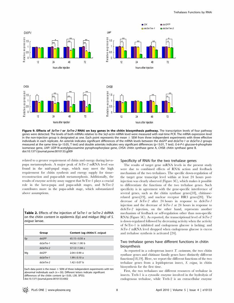
Effects of SeTre-1 and SeTre-2 RNAi on the expressions ofkey genes in the chitin biosynthesis pathway
Quantitative real-time PCR was used to detect the mRNA levels
of four genes in the chitin biosynthesis pathway after the RNAi
knockdown of SeTre-1 and SeTre-2. The results showed that G-6-P-
I (glucose-6-phosphate isomerase) gene was down-regulated
extremely significantly by the the RNAi of SeTre-1 from 24 hours
and by the RNAi of SeTre-2 from 48 hours, while UAP (UDP-N-
acetylglucosamine pyrophosphorylase) gene expression was affect-
ed obviously only by the RNAi of SeTre-2 from 12 to 48 hours
(Figure 9). Moreover, knockdown of SeTre-1 affected the
expression of CHSA (chitin synthase gene A) more significantly
than CHSB (chitin synthase gene B), while the RNAi of SeTre-2
affected CHSB expression more significantly (Figure 9).
Chitin content in cuticle and midgut after RNAiThe significant decrease in the CHSA and CHSB transcript levels
and the abnormal phenotypes of insects after RNAi of SeTre-1/SeTre-
2 suggest that the chitin content in the insect cuticle and midgut may
be decreased. To confirm this hypothesis, the chitin contents in the
cuticle and midgut of dsRNA-injected insects were determined at 60
hours post-injection. Compared to the control group, the chitin
content in the cuticle of insects injected with dsRNA for SeTre-1 was
reduced greatly, and the mean chitin content per insect was only
about two thirds of that in the control insects. In contrast, the chitin
content in the insects injected with dsRNA for SeTre-2 was only
slightly reduced. However, compared to the chitin content in the
midgut of the control insects, the chitin content in the midgut of the
insects injected with dsRNA for SeTre-2 was reduced about 25%,
while the chitin content in the insects injected with the dsRNA of
SeTre-1 did not decrease significantly (Table 2).
Effects of trehalose injection on expressions of key genesin the chitin biosynthesis pathway
In addition to the down-regulation of trehalase expression, an
examination of an indirect up-regulation of trehalases by the
injection of trehalose was conducted. The results showed that the
trehalose injection increased the mRNA levels of SeTre-1 and
SeTre-2 and significantly increased the expression of UAP, CHSA
and CHSB at some time points (Figure 10). This further confirmed
that the expression patterns of the genes in the chitin biosynthesis
pathway are affected by changes in the expression of trehalase
genes.
Discussion
Characterization of the two trehalase genesIn two lepidopteran insects, Bombyx mori and Spodoptera frugiperda,
Tre-1 and Tre-2 were expressed in the midgut[14,22], which is
consistent with our findings (Figure 3). However, we found that the
two trehalases in S. exigua were distributed in other tissues as well,
including the cuticle, tracheae, Malpighian tubules and fat body. A
possible explanation is that two trehalases are devoted to utilizing
intracellular and extracellular trehalose to supply energy and
material for the whole body, and the soluble trehalase is located in
haemolymph that flows throughout the body. The expression
patterns of SeTre-1 and SeTre-2 were different in the tissues
(Figure 3). The high expression of SeTre-1 in the cuticle suggests
its close relationship with slough during metamorphosis[23], while
the high expression of SeTre-2 in the tracheae, the main structure
of wings, may suggest its contribution to flight in the adult stage.
The expression of SeTre-1 gene in the prepupal stage was found
to be much higher than in other stages (Figure 4), which may be
Figure 8. Concentration changes of trehalose and glucose. Theconcentrations of trehalose and glucose were detected by HPLC. Eachpoint represents the mean 6 SEM from three independent experimentswith five abnormal individuals mixed in each replicate.doi:10.1371/journal.pone.0010133.g008
Trehalases Functions by RNAi
PLoS ONE | www.plosone.org 7 April 2010 | Volume 5 | Issue 4 | e10133
related to a greater requirement of chitin and energy during larva-
pupa metamorphosis. A major peak of SeTre-2 mRNA level was
found in the mid-pupal stage, which may meet the high
requirement for chitin synthesis and energy supply for tissue-
reconstruction and pupa-adult metamorphosis. Additionally, the
results of enzyme activity assay suggest that SeTre-1 plays a crucial
role in the larva-pupa and pupa-adult stages, and SeTre-2
contributes more in the pupa-adult stage, which substantiated
above assumptions.
Specificity of RNAi for the two trehalase genesThe results of target gene mRNA levels in the present study
were due to combined effects of RNAi action and feedback
mechanism of the two trehalases. The specific down-regulation of
the target gene transcript level within at least 24 hours post-
injection was clearly observed (Figure 5C), which makes it possible
to differentiate the functions of the two trehalase genes. Such
specificity is in agreement with the gene-specific interference of
several genes, such as the chitin synthase genes[18], chitinase-
related genes[19], and nuclear receptor HR3 genes[20]. The
decrease of SeTre-2 after 24 hours in response to dsSeTre-1
injection and the decrease of SeTre-1 at 24 hours in response to
dsSeTre-2 injection, on the other hand, represents another
mechanism of feedback or self-regulation other than non-specific
RNAi (Figure 5C). As reported, the transcriptional level of SeTre-2
is down-regulated followed by decreasing activity when the activity
of SeTre-1 is inhibited and endogenous glucose is lacking; and
SeTre-1 mRNA level dropped when endogenous glucose is excess
and trehalose synthesis is activated [24].
Two trehalase genes have different functions in chitinbiosynthesis
As reported in a coleopteran insect T. castaneum, the two chitin
synthase genes and chitinase family genes have distinctly different
functions[18,19]. Here, we report the different functions of the two
trehalase genes from a lepidopteran insect, S. exigua, in chitin
biosynthesis for the first time.
First, the two trehalases use different resources of trehalose in
insects. Treh-1 is a cytosolic enzyme involved in the hydrolysis of
endogenous trehalose, while Treh-2 is an extracellular enzyme
Figure 9. Effects of SeTre-1 or SeTre-2 RNAi on key genes in the chitin biosynthesis pathway. The transcription levels of four pathwaygenes were detected. The levels of both mRNAs relative to the Seb-actin mRNA level were measured with real-time PCR. The mRNA expression levelin the non-injection group is designated as one. Each point represents the mean 6 SEM from three independent experiments with three effectiveindividuals in each replicate. An asterisk indicates significant differences of the mRNA levels between the dsGFP and dsSeTre-1 or dsSeTre-2 groupsmeasured at the same time (p,0.05, T test) and double asterisks indicates very significant differences (p,0.01, T test). G-6-P-I: glucose-6-phosphateisomerase gene, UAP: UDP-N-acetylglucosamine pyrophosphorylase gene, CHSA: chitin synthase gene A, CHSB: chitin synthase gene B.doi:10.1371/journal.pone.0010133.g009
Table 2. Effects of the injection of SeTre-1 or SeTre-2 dsRNAon the chitin content in epidermis (Ep) and midgut (Mg) of S.exigua larvae.
Group Content (ug chitin/S. exigua)
Ep dsGFP 65.1560.50 a
dsSeTre-1 44.5661.90 b
dsSeTre-2 57.1261.04 c
Mg dsGFP 2.0460.90 a
dsSeTre-1 1.9960.10 a
dsSeTre-2 1.4260.07 b
Each data point is the mean 6 SEM of three independent experiments with tenabnormal individuals each (n = 30). Different letters indicate significantdifferences of the chitin content (p,0.05, LSR, SPSS).doi:10.1371/journal.pone.0010133.t002
Trehalases Functions by RNAi
PLoS ONE | www.plosone.org 8 April 2010 | Volume 5 | Issue 4 | e10133

proposed to have a role in the assimilation of exogenous trehalose
as a carbon source[25,26]. After RNAi for SeTre-1, the expression
level of SeTre-2 dropped significantly in the prepupal stage, leading
to a severe shortage of glucose that caused a pupation
malformation (Figure 5C, Figure 8). This result demonstrates that
the activity of SeTre-2 dropped when trehalose synthesis was
stopped[24]. After RNAi of SeTre-2, however, the reducation of
SeTre-1 expression led to an abundance of trehalose and a shortage
of glucose after 24 hours that caused a reduced body
size(Figure 5C, Figure 8). In the puapl stage, there was sufficient
glucose for the larva-pupa stage, but a severe shortage of trehalose
that led to eclosion malformation (Figure 5C, Figure 8). In other
words, SeTre-1 and SeTre-2 have different effects on the
concentrations of glucose and trehalose, which implies that they
may have different functions in insect chitin biosynthesis through
the chitin biosynthesis pathway and the glycometabolism pathway.
Second, the interference of the SeTre-1 and SeTre-2 caused
decrease in the survival rates at different times. The remarkable
reduction of transcriptional levels of SeTre-1 and SeTre-2 at 72
hours post-injection caused by RNAi of SeTre-1 resulted in high
mortality during the larva-pupa stage, while the severe shortage of
trehalose before pupal stage owing to RNAi of SeTre-2 caused high
mortality during the pupa-adult stage (Figure 6) and a high
percentage of failure in eclosion (Figure 7). The phenotypes caused
by RNAi are due to combined effects of RNAi action and feedback
mechanism of the two trehalases as discussed above.
Eight enzymes are involved in the insect chitin biosynthesis
pathway, starting with trehalase[5]. After the gene-specific
interference of SeTre-1 and SeTre-2, the UAP gene expression was
affected only by the injection of dsRNA to SeTre-2, and the
expression of the CHSA and CHSB genes were affected by RNAi
for both dsSeTre-1 and dsSeTre-2 (Figure 9). The possible reason is
that CHSA mainly exists in cuticle, the primary location for
slough[27], and Tre-1 is regulated by 20E to participate in slough
during the larva-pupa transformation[2]. On the other hand,
Treh-2 was involved in incorporating trehalose from the blood
into muscular cells and then providing the energy required for the
visceral muscles to strongly support the peristaltic movement of the
midgut during active feeding[23], SeTre-2 appeared to be involved
in food intake and the muscle movement of midgut, thus it may
have a greater effect on CHSB. Besides downregulation of
trehalase expression, the overexpress of trehalase genes by
injection of trehalose increased the mRNA levels of key genes in
the chitin biosynthesis pathway (Figure 10). These further
confirmed that expressions of the genes in the chitin biosynthesis
pathway are affected by changes in expressions of trehalase genes.
Figure 10. Effects of trehalose injection on expression of the chitin biosynthesis pathway genes, SeTre-1, SeTre-2, UAP, CHSA andCHSB. The transcription levels of the five genes were detected after injection with a 500 ug dose. Both of the mRNA levels relative to the Seb-actinmRNA level were measured with real-time PCR. The mRNA expression level in the dsGFP-injection group is designated as one. Each point representsthe mean 6 SEM from three independent experiments with three individuals in each replicate. UAP: UDP-N-acetylglucosamine pyrophosphorylasegene, CHSA: chitin synthase gene A, CHSB: chitin synthase gene B.doi:10.1371/journal.pone.0010133.g010
Trehalases Functions by RNAi
PLoS ONE | www.plosone.org 9 April 2010 | Volume 5 | Issue 4 | e10133

The two trehalase genes have different effects on CHSA and
CHSB, which was supported by the chitin content measurements.
The chitin content in the epidermis was mainly affected by dsSeTre-
1, and that in midgut was only affected by dsSeTre-2 (Table 2).
Because CHSA is responsible for chitin synthesis in the cuticle and
trachea and CHSB mainly for chitin synthesis in the peritrophic
matrix in the gut[15,18], it can be concluded that SeTre-1 affected
the expression of CHSA and chitin synthesis in the cuticle more
significantly. SeTre-2, however, has a major effect on the
expression of CHSB and chitin synthesis in the midgut.
Possible mechanisms for two trehalase genes to affectchitin biosynthesis
The insect chitin biosynthesis pathway starts with trehalase. As
reported, the enzyme activity, which can be affected by substrate
feedback mechanisms, is controlled at both the levels of
transcription and post-translation[27,28,29,30]. In the present
study, RNAi of the two trehalase genes decreased their
transcription levels (Figure 5C), and their enzyme activities
dropped accordingly (Figure 5E). In this way, chitin, the last
product in the chitin biosynthesis pathway, is affected through
changes in the substrates, products and activities of a series of
enzymes in the pathway. On the other hand, the interference of
the two trehalase genes resulted in a shortage of glucose (Figure 8)
and decreased the transcription level of a signal molecule,
ecdysone receptor (EcR) gene (data not shown). Moreover, the
transcription levels of the pathway genes such as SeTre-1, G-6-P-I,
UAP, CHSA and CHSB were affected at different times after RNAi
of SeEcR gene (Yao, et al, unpublished data in the laboratory). In
short, possible mechanisms for the two trehalase genes to affect
chitin biosynthesis were summarized (Figure 11). Additional
research will be conducted in the near future.
Materials and Methods
Insect culturesS. exigua larvae were reared at 2561uC with an L14:D10
photoperiod using an artificial diet [27,30]. The developmental
stages were synchronized at each molt by collecting new larvae or
pupae. The midgut, fat body, cuticle and other tissues from the
fifth instar larvae were dissected in insect saline containing 0.75%
NaCl and stored at 280uC until further use.
RNA isolation, cDNA synthesis and rapid amplification ofthe full-length cDNA
Total RNA was isolated at day two of the fifth instar larvae
using TRIzol reagent (Invitrogen, USA) and the first strand cDNA
synthesis was carried out according to the reverse transcriptase XL
(AMV) (TaKaRa, Japan) protocol with oligo dT18. The first-strand
cDNA (1 mL) was used as a template for PCR, and the
components of the PCR mix were PCR buffer containing 0.1
mM dNTPs, 5 mM each primer, and 1.0 U of HiFi-Taq DNA
polymerase (Transgene, China) in a total volume of 25 mL. Two
pairs of degenerate primers (Table 3), SeTre-1-F1, SeTre-1-F2 and
SeTre-1-R1, SeTre-1-R2 were designed from the conserved SeTre-1
cDNA sequences of other insects. The first PCR reaction was
performed with primers SeTre-1-F1 and SeTre-1-R1 using the
following conditions: three cycles of 30 s at 95uC, 30 s at 45uC and
60 s at 72uC followed by 30 cycles of 30 s at 95uC, 30 s at 48uCand 60 s at 72uC. A second PCR was carried out using the nested
Figure 11. Possible mechanisms for the two trehalase genes to affect chitin biosynthesis. Solid arrows in the picture represent theinternal interaction of the pathway genes, and broken arrows represent indirect effects through the glycometabolism pathway. The solid blue arrowsrepresent greater effects, and the solid yellow arrows represent lower effects. G-6-P-I: glucose-6-phosphate isomerase gene, UAP: UDP-N-acetylglucosamine pyrophosphorylase gene, CHSA: chitin synthase gene A, CHSB: chitin synthase gene B.doi:10.1371/journal.pone.0010133.g011
Trehalases Functions by RNAi
PLoS ONE | www.plosone.org 10 April 2010 | Volume 5 | Issue 4 | e10133

primers SeTre-1-F2 and SeTre-1-R2 using the same conditions as
for the first PCR. The amplified product was separated on an
agarose gel and purified using the Gel Extraction Kit (OMEGA,
USA). Purified DNA was ligated into the pMD18-T vector
(TaKaRa, Japan) and sequenced completely in both directions.
A BD SMART RACE cDNA amplification kit (BD Bioscience
Clontech, CA, USA) was used to obtain the full-length Tre-1
cDNA. Specific primers, 5-SeTre-1-1 and 5-SeTre-1-2, for 59-
RACE and 3-SeTre-1-1 and 3-SeTre-1-2 for 39-RACE (Table 3)
were synthesized based on the cDNA sequence obtained from the
identified fragment. PCR was performed with the Tre-1-1 primer
and Universal Primer Mix (UPM, Clontech) by denaturing at
95uC for 30 s, 35 cycles of 95uC for 30 s, 55uC for 30 s and 72uCfor 2 min, followed by a final extension at 72uC for 10 min. Nested
PCR was carried out with the first- round PCR product as a
template and the Nested Universal Primer A (NUP, Clontech) and
Tre-1-2 primers. The RACE products were purified and
sequenced as described above[16].
Analysis of the cDNAs and protein sequences of SeTre-1The sequences of the two trehalase cDNAs were compared with
other trehalase sequences deposited in the GenBank using the
‘‘BLAST-N’’ or ‘‘BLAST-X’’ tools at the National Center for
Biotechnology Information (NCBI) website. The amino acid
sequence were deduced from the corresponding cDNA sequences
using the translation tool at the ExPASy Proteomics website
(http://expasy.org/tools/dna.html). Other protein sequence anal-
ysis tools used in this study, including MW, pI, and topology
prediction tools, were obtained from the ExPASy Proteomics
website (http://expasy.org/). Multiple sequence alignments of
deduced amino acid sequences were made using the ClustalW
multiple-alignment software (http://www.ebi.ac.uk/clustalw/
index.html). The phylogenetic tree was constructed using the
MEGA 3.1 software based on the amino acid sequences of the
known trehalases. A bootstrap analysis was carried out and the
robustness of each cluster was verified in 1000 replicates.
Expression of recombinants and antibodies productionfor SeTre-1 and SeTre-2
Two cDNA fragments containing the SeTre-1 partial sequence
(547–927 bp) and SeTre-2 partial sequence (1438–1863 bp) were
inserted into the pET32a vector (Takara) for the expression of
recombinant SeTre-1 and SeTre-2 proteins (rSeTre-1 and rSeTre-
2) according to the manufacturer’s instructions. After purification
with an Ni SepharoseTM 6 Fast Flow (GE Healthcare), rSeTre-1
and rSeTre-2 were used to immunize rabbits as described
previously[31]. The sera of the immunized rabbits were collected
as the anti-SeTre-1 and anti-SeTre-2 sera.
Western-blotting analysis for SeTre-1 and SeTre-2Individuals or tissues were homogenized in two volumes of cold
PBS (20 mM sodium phosphate and 130 mM NaCl, pH 7.2) plus
protease inhibitor cocktail. The homogenate was centrifuged at
12,0006g for 20 min at 4uC. The supernatant was then incubated at
70uC for 10 min to remove impurity, the homogenate was
centrifuged at 12,0006g for 10 min at 4uC again. The supernatant
was immediately mixed with an equal volume of buffer for SDS-
polyacrylamide gel electrophoresis and boiled for 10 min. After
centrifugation at 12,0006g for 5 min, each supernatant was collected
and stored at 240uC [32]. The amount of total protein in 1 ul sample
buffer supernatant was determined by BCA kit. Electrophoresis on
12% SDS- polyacrylamide gels and Western-blotting analysis were
carried out according to the methods of Mitsumasu et al. (2008). A
purified primary antibody (anti-SeTre-1 or anti-SeTre-2 antibody,
1:1000 dilution) and a secondary antibody (1:5000 dilution), an anti-
rabbit lgG antibody conjugated with HRP (BOSTER), were used.
Immunocytochemical analysis of SeTre-1 and SeTre-2 invarious tissues
Small pieces of midgut and cuticle were prepared from the fifth-
instar larvae using a Frigocut cryotome (Mod. 2700, Reichert and
Jung) at 226uC. The immunolabeling of the cuticle and midgut
cryosections was performed according to Klein et al (1991).[33]
The distributions of SeTre-1 and SeTre-2 immunoreactivity in the
fatbody, tracheae and Malpighian tubule were investigated using
the whole mount immunocytochemistry described previously[34].
For the localizations of SeTre-1 and SeTre-2, the sections and
tissues were treated with a 1:1000 dilution of anti-Tre serum. The
control samples were treated with the preimmune serum. The
visualization of the primary antibody was performed with HRP-
conjugated anti-rabbit IgG (BOSTER). The sections and tissues
Table 3. PCR primers used in this study.
Primers Primer sequence
Degenerate primers
SeTre-1-F1 59-AGYGGYTGGGAYTTCTC-39
SeTre-1-F2 59-TGGATYATBGAAGGTCT-39
SeTre-1-R1 59-GCCADGCGTTRGGGAAGTCC-39
SeTre-1-R2 59-CGCRTCRTAYTTCTCRAACAT-39
For cDNA cloning
5-SeTre-1-1 59-CACTATATTCGGATCGACAG-39
5-SeTre-1-2 59-GGAGCCAGGTTAGATGGGT-39
3-SeTre-1-1 59-CACACCAGATACATCATACC-39
3-SeTre-1-2 59-GGAATCACCGGGATGCTG-39
For real-time PCR
QSeTre-1-F 59-ATTCGCCAGAAACATCACCAAC-39
QSeTre-1-R 59-TTCCACTTATCAGCAGACCTCC-39
QSeTre-2-F 59-GGACTCTTGGGTTGATGGTGT-39
QSeTre-2-R 59-AGGCTTCTCAGTTCCGTGTAGG-39
QActin-F 59-TGCGTGACATCAAGGAGAAGC-39
QActin-R 59-CCATACCCAAGAAGGAAGGCT-39
QG-6-P-I-F
eG-6-P-I-R
QUAP-F 59-AGCAGACGGCAGACTAACTTTC-39
QUAP-R 59-GGACTCCTTCGTGGTCAACATAA-39
QCHSA-F 59-TAAGGCAAAGATTCGTCACAGG-39
QCHSA-R 59-CAGGGTCAGCAGATAGGTGTTC-39
QCHSB-F 59-CGCTGAGTCTTGTTGGTCCTGT-39
QCHSB-R 59-TCCACGCTACCTCTTTCCCTA-39
For dsRNA synthesis
dsSeTre-1-F 59-ACCAGGAACCGTCAGTAG-39
dsSeTre-1-R 59-GCAACCATAGCTGTCAACA-39
dsSeTre-2-F 59-GCCAGGACAGGTTCACATC-39
dsSeTre-2-R 59- GCTTCACCATCGGAATTAGG-39
GFP-F 59-AAGGGCGAGGAGCTGTTCACCG-39
GFP-R 59-CAGCAGGACCATGTGATCGCGC-39
F: forward, R: reverse.doi:10.1371/journal.pone.0010133.t003
Trehalases Functions by RNAi
PLoS ONE | www.plosone.org 11 April 2010 | Volume 5 | Issue 4 | e10133

were rinsed three times with PBS, covered with glycerol and
viewed under a microscope.
Developmental expression analysis of SeTre-1 and SeTre-2Total RNA was isolated from S. exigua on every day of its life
cycle, including the larval, pupal and adult stages. 10 ug total
RNA extracted from each stage was used as template to
demonstrate the stability of Seb-actin. The PCR reaction was
performed with primers QActin-F and QActin-R using the following
conditions: 95uC for 30 s, 28 cycles of 95uC for 30 s, 60uC for 30 s
and 72uC for 20 s, followed by a final extension at 72uC for
10 min. The expression of SeTre-1 and SeTre-2 were estimated by
real-time quantitative PCR (qRT-PCR) using a LightCycler480
system (Roche, Germany) and SYBR Premix Ex Taq (Takara,
Japan). Two pairs of primers, QSeTre-1-F and QSeTre-1-R and
QSeTre-2-F and QSeTre-2-R (Table 3), were designed to determine
the expression of SeTre-1 and SeTre-2. The cycling for each
reaction was done in a final volume of 10 mL containing 0.3 mL of
the cDNA sample (or standard), 0.2 mL (l0 mmol/ml) of each
primer, and 5 mL of SYBR premix Ex Taq. After 10 s of initial
denaturation at 95uC, the cycling protocol consisted of 45 cycles of
denaturation at 95uC for 5 s, annealing at 58uC for 15 s, and
elongation at 72uC for 20 s. The b-actin (EU179846) cDNA
fragment was amplified with the QActin-F and QActin-R primers
(Table 3) as an internal control. Standard curves were obtained
using a ten-fold serial dilution of pooled total RNA. All the data
were presented as the relative mRNA expression (mean 6 SEM).
Injection bioassays and samplingEarly day one of the fifth instar larvae were used for the
injection experiment because larvae at earlier stages proved to be
too small for satisfactory injections (data not shown). All of the
reagents and enzymes used for the dsRNA synthesis were from the
T7 RiboMAXTM Express RNAi System Kit (Promega) and all
primers are shown in table 3. Five micrograms of dsRNA were
injected into the side of the abdomen of the larvae using a 10 ml
micro-syringe (Hamilton), and the injection point was sealed
immediately with wax as described previously[27]. Two controls
were performed, an equivalent volume of dsGFP (all experiments)
and no treatment (the survival rate analysis and pathway gene
detection only). In the phenotypic observation experiment, each
group had 40 individual larvae with three replicates, and the
observation was performed every 12 hours after injection during
the larva stage and every 24 hours during the pupal stage. In the
target gene detection experiment, each group had 80 individuals
with three replicates, and 10 larvae were selected randomly at
12 h, 24 h, 48 h and 72 h after the injection for mRNA level
detection independently. An individual with more than a 10% of
decrease of the target gene expression was regarded as an effective
RNAi, which was used to calculate the efficiency of RNAi. In the
pathway gene detection experiment, three individuals confirmed
as effective above were used for the detection independently. In
the chitin content assay, each group had 50 individuals with three
replicates, and 30 abnormal larvae were chosen for experiment. In
the trehalose and glucose concentration assay, each group had 30
individuals with three replicates, and 5 abnormal larvae mixed at
24 h, 48 h and 72 h after injection were chosen for experiment.
Trehalase activity assayTen crude larvae, prepupae and pupae in the late part of the
fourth instar stage, the prepupal stage and day seven of the pupal
stage were used for the soluble and membrane-bound trehalase
activity analysis of individuals for three independent experiments.
Ten crude individuals and the haemolymph of ten individuals
from each of the three stages above were used for the activity
analysis of the two trehalases. In the target gene experiment, three
effective individuals were selected randomly at 48 h and 72 h post-
injection for activity measurement independently in three
replicates. The assay of trehalase activity was carried out
according to the method described by Tatun et al.[2,3]
Trehalose and glucose content assay by HPLCThe concentrations of trehalose and glucose in S. exigua larvae
were determined using high performance liquid chromatography
(HPLC) with a 2414 refractive index detector[35]. An acetonitrile-
water mixture (75:25) was used as the mobile phase on a
Carbohydrate Column (4.6 mm6250 mm, Waters). The working
conditions were as follows: the flow rate was 1 ml/min, and the
detector temperature was 37uC. Before the quantitative and
qualitative determination of the concentrations of the sugars in the
samples, standard solutions of trehalose and glucose (Sigma) were
prepared and run on the same column to obtain standard curves
for each sugar. The concentrations of trehalose and glucose in the
samples were then calculated using the standard curves above.
Chitin analysisTen abnormal larvae cuticles and thirty abnormal larvae
midguts were choosen for the chitin content detection for three
independent experiments. A piece of cuticle or three midgut mixed
segments were measured in one sample. The assay of the chitin
content was carried out according to the method described by
Arakane et al (2005).[18]
Trehalose injection assayEarly day one of the fifth instar larvae were used for the injection of
500 ug of trehalose per individual (the fifith instar larva has about
250 ug trehalose in the haemolymph). Each group had 30 individuals
with three replicates, and three larvae were selected randomly per
time point for mRNA level detection by quantitative real-time PCR
independently. The methods used were the same as above.
Acknowledgments
We are grateful to Dr. Xiaoqiang Yu (University of Missouri-Kansas City,
USA) and Dr. Sheng Li (Institute of Plant Physiology and Ecology,
Shanghai, China) for their suggestions and reading of the manuscript.
Author Contributions
Conceived and designed the experiments: JC BT DZ. Performed the
experiments: JC XH. Analyzed the data: JC XH. Contributed reagents/
materials/analysis tools: JC BT HC QY JC DZ. Wrote the paper: JC WZ.
References
1. Clegg JS, Evans DR (1961) Blood trehalose and flight metabolism in the blowfly.
Science 134: 54–55.
2. Tatun N, Singtripop T, Sakurai S (2008) Dual control of midgut trehalase
activity by 20-hydroxyecdysone and an inhibitory factor in the bamboo borer
Omhisa fuscidentalis Hampson. Journal of Insect Physiology 54: 351–357.
3. Tatun N, Singtripop T, Tungjitwitayakul J, Sakurai S (2008) Regulation of
soluble and membrane-bound trehalase activity and expression of the enzyme in
the larval midgut of the bamboo borer Omphisa fuscidentalis. Insect Biochemistry
and Molecular Biology 38: 788–795.
4. Friedman S (1978) Trehalose regulation, one aspect of metabolic homeostasis.
Annual Review of Entomology 23: 389–407.
5. Kramer KJ, Dziadik-Turner C, Koga D (1985) Chitin metabolism in insects.
Comprehensive Insct Physiology,Biochemistry,and Pharmacology 3: 75–
115.
Trehalases Functions by RNAi
PLoS ONE | www.plosone.org 12 April 2010 | Volume 5 | Issue 4 | e10133

6. Borgia PT, Miao Y, Dodge CL (1996) The orlA gene from Aspergillus nidulans
encodes a trehalose-6-phosphate phosphatase necessary for normal growth and
chitin synthesis at elevated temperatures. Mol Microbiol 20: 1287–1296.
7. Becker A, Schloder P, Steele JE, Wegener G (1996) The regulation of trehalose
metabolism in insects. Experientia 52: 433–439.
8. Friedman S (1985) Carbohydrate metabolism. In Comparative Insect Physiol-
ogy, Biochemistry, and Pharmacology 10: 43–76.
9. Sumida M, Yamashita O (1983) Purification and some properties of soluble
trehalase from midgut of pharate adult of the silkworm, Bombyx mori. Insect
Biochem Mol Biol 13: 257–265.
10. Terra WR, Ferreira C (1994) Insect digestive enzymes: properties, compart-
mentalization and function. Comp Biochemistry Physiology 109B: 1–62.
11. Thompson SN (2003) Trehalose – the insect ‘‘blood’’ sugar. Adv Insect Physiol
31: 203–285.
12. Yaginuma T, Mizuno T, Mizuno C, Ikeda M, Wada T, et al. (1996) Trehalase
in the spermatophore from the bean-shaped accessory gland of the male
mealworm beetle,Tenebrio molitor: purification, kinetic properties and localization
of the enzyme. J Comp Physiol B 166: 1–10.
13. Takiguchi M, Niimi T, Su ZH, Yaginuma T (1992) Trehalase from male
accessory gland of an insect, Tenebrio molitor cDNA sequencing and develop-
mental profile of the gene expression. Biochem J 288: 19–22.
14. Mitsumasu K, Azuma M, Niimi T, Yamashita O, Yaginuma T (2005)
Membrane-penetrating trehalase from silkworm Bombyx mori. Molecular cloning
and localization in larval midgut. Insect Molecular Biology 14: 501–508.
15. Merzendorfer H, Zimoch L (2003) Chitin metabolism in insects: structure,
function and regulation of chitin synthases and chitinases. J Exp Biol 206:
4393–4412.
16. Tang B, Chen X, Liu Y, Tian H, Liu J, et al. (2008) Characterization and
expression patterns of a membrane-bound trehalase from Spodoptera exigua. BMC
Mol Biol 9: 51.
17. Zhu QS, Arakane Y, Beeman RW, Kramer KJ, Muthukrishnan S (2008)
Functional specialization among insect chitinase family genes revealed by RNA
interference. Proceedings of the National Academy of Sciences of the United
States of America 105: 6650–6655.
18. Arakane Y, Muthukrishnan S, Kramer KJ, Specht CA, Tomoyasu Y, et al.
(2005) The Tribolium chitin synthase genes TcCHS1 and TcCHS2 are
specialized for synthesis of epidermal cuticle and midgut peritrophic matrix.
Insect Mol Biol 14: 453–463.
19. Zhu Q, Arakane Y, Banerjee D, Beeman RW, Kramer KJ, et al. (2008) Domain
organization and phylogenetic analysis of the chitinase-like family of proteins in
three species of insects. Insect Biochem Mol Biol 38: 452–466.
20. Cruz J, Martin D, Belles X (2007) Redundant ecdysis regulatory functions of
three nuclear receptor HR3 isoforms in the direct-developing insect Blattella
germanica. Mech Dev 124: 180–189.
21. Gu J, Shao Y, Zhang C, Liu Z, Zhang Y (2009) Characterization of putative
soluble and membrane-bound trehalases in a hemipteran insect, Nilaparvata
lugens. J Insect Physiol 55: 997–1002.
22. Silva MC, Ribeiro AF, Terra WR, Ferreira C (2009) Sequencing of Spodoptera
frugiperda midgut trehalases and demonstration of secretion of soluble trehalaseby midgut columnar cells. Insect Mol Biol 18: 769–784.
23. Azuma M, Yamashita O (1985) Cellular localization and proposed function ofmidguttrehalase in the silkworm larva, Bombyx mori. Tissue Cell 17: 539–551.
24. Sanchez-Fresneda R, Gonzalez-Parraga P, Esteban O, Laforet L, Valentin E,
et al. (2009) On the biochemical classification of yeast trehalases: Candida albicans
contains two enzymes with mixed features of neutral and acid trehalase activities.
Biochemical and Biophysical Research Communications 383: 98–102.25. Almeida FMd, Bonini BM (2009) Heterologous expression in Escherichia coli of
Neurospora crassa neutral trehalase as an active enzyme. Protein Expression andPurification 65: 185–189.
26. de Almeida FM, Bonini BM, Beton D, Jorge JA, Terenzi HF, et al. (2009)
Heterologous expression in Escherichia coli of Neurospora crassa neutral trehalase asan active enzyme. Protein Expr Purif 65: 185–189.
27. Chen XF, Yang X, Kumar NS, Tang B, Sun XJ, et al. (2007) The class A chitinsynthase gene of Spodoptera exigua: Molecular cloning and expression patterns.
Insect Biochemistry and Molecular Biology 37: 409–417.
28. Kato N, Mueller CR, Fuchs JF, Wessely V, Lan Q, et al. (2006) Regulatorymechanisms of chitin biosynthesis and roles of chitin in peritrophic matrix
formation in the midgut of adult Aedes aegypti. Insect Biochemistry and MolecularBiology 36: 1–9.
29. Zimoch L, Hogenkamp DG, Kramer KJ, Muthukrishnan S, Merzendorfer H(2005) Regulation of chitin synthesis in the larval midgut of Manduca sexta. Insect
Biochemistry and Molecular Biology 35: 515–527.
30. Kumar NS, Tang B, Chen XF, Tian HG, Zhang WQ (2008) Molecular cloning,expression pattern and comparative analysis of chitin synthase gene B in
Spodoptera exigua. Comparative Biochemistry and Physiology B-Biochemistry &Molecular Biology 149: 447–453.
31. Cui SY, Xu WH (2006) Molecular characterization and functional distribution
of N-ethylmaleimide-sensitive factor in Helicoverpa armigera. Peptides 27:1226–1234.
32. Mitsumasu K, Azuma M, Niimi T, Yamashita O, Yaginuma T (2008) Changesin the expression of soluble and integral-membrane trehalases in the midgut
during metamorphosis in Bombyx mori. Zoolog Sci 25: 693–698.33. Klein U, Loffelmann G, Wieczorek H (1991) The midgut as a model system for
insect K+-transporting epithelia—immunocytochemical localization of a vacu-
olar-type H+ pump. JExpBiol 161: 61–75.34. Su ZH, Sato Y, Yamashita O (1993) Purification, cDNA cloning and northern
blot analysis of trehalase of pupal midgut of the silkworm, Bombyx mori. BiochimBiophys Acta 1173: 217–224.
35. Hallsworth J, Magan N (1997) A rapid HPLC protocol for detection of polyols
and trehalose. Journal of Microbiological Methods 29: 7–13.
Trehalases Functions by RNAi
PLoS ONE | www.plosone.org 13 April 2010 | Volume 5 | Issue 4 | e10133