Diethyl ether exposure of EEG technicians using collodian.
72
Transcript of Diethyl ether exposure of EEG technicians using collodian.
DIETHYL ETHER EXPOSURE OF EEG
TECHNICIANS USING COLLODIAN
by
Jeri L. Lockman
A thesis submitted to the faculty of The University of Utah
in partial fulfillment of the requirements for the degree of
Master of Science
College of Nursing
The University of Utah
August 1984

THE UNIVERSITY OF UTAH GRADUATE SCHOOL
SUPERVISORY COMMITTEE APPROVAL
of a thesis submitted by
Jeri L. Lockman
This thesis has been read by each member of the following supervisory committee and by majority vote has been found to be satisfactory.
j, I

THE UNIVERSITY OF UTAH GRADUATE SCHOOL
FINAL READING APPROVAL
To the Graduate Council of The University of Utah:
Jeri L. Lockman I have read the thesis of in its
final form and have found that (1) its format, citations. and bibliographic style are
consistent and acceptable; (2) its illustrative materials including figures. tables. and charts are in place; and (3) the final manuscript is satisfactory to the Supervisory
Committee and is ready for submission to the Graduate School.
Margaret Armstrong \-1ember. Supenisury Committee
Approved for the Major Department
Llnda K. Amos
Chairman Dean
Approved for the Graduate Council

copyright~ 1984 Jeri L. Lockman
All Rights Reserved
ABSTRACT
The purpose of this study was to determine if EEG
technicians using collodian are exposed to diethyl
ether and to determine if there was an accumulation
of ether in the body after repeated exposures. Breathing
zone air and expired-air samples were collected on four
EEG technicians for five consecutive days. Expired-
air samples were also collected the following Monday
morning.
Measurable amounts of ether were found in all
samples collected from the subjects. No relationship
was found between the levels of ether in the breathing
zone and levels found in the individual subject's
expired-air. Also, levels of ether in the expired
air did not increase over the week nor was there any
consistent increase in levels at the end of the day
immediately after exposure as compared to the morning
level. The scatter diagram of the number of EEGs per
formed and the levels of ether in the breathing zone
indicate a positive trend between these two factors.
It can be concluded from this study that EEG tech
nicians are exposed to diethyl ether via collodian use.
It can also be concluded that a body burden exists in

EEG technicians with repeated exposure to low concen-
trations of ether. Expired-air sample results were
considered invalid because of analytical problems.
For this reason, no conclusions can be drawn regarding
a relationship between exposure levels and expired
air levels of ether, nor can any statement be made
regarding the accumulation of ether in the body with
repeated exposures.
v

CONTENTS
ABSTRACT . . . .
LIST OF FIGURES.
Chapter
I. INTRODUCTION.
Problem . . Purpose
II. LITERATURE REVIEW.
III.
Biotrans format ... .... . Effects of Diethyl Ether.. .. . Epidemiologic Studies . . . . . . . Animal Studies. . . . . . . . . . Adverse Effects from Industrial Use of Diethyl Ether . .. ....... . Research Questions. ... .. .
METHODOLOGY .
Population .... Instrumentation . . . . . Sampling Method . Procedure Sequence. Analysis of Samples
IV. DATA ANALYSIS ...
Study Population .. Analysis of Samples Data Analysis . . . .
V. DISCUSSION ..
Recommendations for Future Research . Conclusion.
iv
. viii
1
1 2
3
4 8
12 13
17 17
19
19 19 20 21 22
23
23 23 24
31
36 38
Appendices
A.
B.
C.
D.
E.
F.
CONSENT FORMS ..
INFORMED CONSENT PROCEDURE FORM. . . . . . .
PRESAMPLING QUESTIONNAIRE.. . .....
SAMPLING RECORD. . .
ENVIRONMENTAL SAMPLE CONVERSION DATA
INFORMATION ON COLLODIAN . . . . . . .
40
42
45
47
49
51
SELECTED BIBLIOGRAPHY . . . . . . . . . . . . . . . . 58
vii

LIST OF FIGURES
1. Biotransformation of diethyl ether .. 6
2. Ether elimination results 9
3. Uptake and elimination of ether in chick embryos. 10
4. Number of EEGs per day and breathing zone levels. . 28
5. Breathing zone levels and expired air levels of ether. . . . . .. .... .... 29
CHAPTER I
INTRODUCTION
Collodian is a substance containing 4% dinitro
cellulose, 24% ethyl alcohol, and 70% diethyl ether
(Gosselin, Hodge, Smith & Gleason, 1976). It is a thick,
colorless liquid with an ether-like odor (U.S. Coast
Guard [Chris], 1978). It is used to adhere electro
encephalogram electrodes to the scalp_ The solvent
mixture of diethyl ether and ethyl alcohol is reportedly
the chief source of toxicity in collodian (Gosselin
et al., 1976). Collodian making is recognized as an
occupation in which exposure to diethyl ether may occur
(Occupational Diseases, A Guide to Their Recognition,
Department of Health, Education and Welfare, 1977).
However, EEG technicians using collodian are not included
in the list of occupations with a potent I exposure
risk to diethyl ether.
Problem
Diethyl ether is a chemical that has been reported
to have biological effects on animals and humans which
have beeri exposed to it. EEG technicians represent
an occupation associated with a potential exposure to

chronic low levels of diethyl ether.
Purpose
The purpose of this study was to determine if EEG
technicians are indeed exposed to low doses of hyl
ether. The study was not intended to look at the long
term effects of such an exposure but rather to ascertain
if the exposure exists. In addition, this study will
determine whether a body burden exists from chronic
exposure to ether as demonstrated by detectible levels
of ether in the expired air; and if so, whether there
is a correlation between exposure levels and expired
air levels of ether.
2

CHAPTER II
LITERATURE REVIEW
The occupational standard of diethyl ether as
promulgated by the Occupational Safety and Health Admini
stration is 400 ppm (1200 mg/m 3 ) for an 8-hour time-
weighted average concentration basis. This standard
was adopted from the recommendations of the American
Conference of Governmental Industrial Hygienists (ACGIH)
and was based on the prevention of nasal irritation
and possible narcosis (NIOSH criteria, 1977). The most
current ACGIH threshold limit value (TLV) for ethyl
ether remains at 400 ppm or 1200 mg/m3 , with an asso-
ciated 500 ppm or 1500 mg/m3 Short-Term Exposure Limit.
In 1974, the Hospital Engineering Cooperative Groups
of Denmark recommended that the highest permissible
average concentration of diethyl ether in the breathing
3 zone be 3 ppm or 9 mg/m (NIOSH document, 1977). They
state that since the lowest concentration of anesthetic
gases which offers any risk upon long-term exposure
is unknown it is necessary to attempt to remove all
excesses (NIOSH document, 1977). NIOSH recommended
that occupational exposure to waste gases, including
diethyl ether, from anesthetic procedures be controlled

in order to minimize potential adverse effects on the
health and safety of workers and their unborn children.
Studies to determine these adverse effects will be dis
cussed in depth later in this review.
Biotransformation
Diethyl ether [(C 2HS )2=0] is a colorless, highly
volatile liquid with a boiling point of 3So C. It
possesses a pungent odor and gives off an irritating
vapor (Goodman & Gilman, 1970). Diethyl ether has
enjoyed long clinical and industrial use; however, there
are a number of significant gaps in our knowledge of
this substance and little information about its elimi
nation or metabolism is available.
Until the late 1960s, ethyl ether was considered
4
to be eliminated unchanged from the body. Haggard (1924)
studied the recovery of diethyl ether from the expired
air of dogs. He found that an average of 87% of the
administered amount was recovered unchanged in the
expired air. Onchi and Asao (1961) studied the recovery
rate of diethyl ether in human subjects using lower
percentages of ether. Ether was administered by
inhalation, and then expired air was collected for three
consecutive 40-minute periods. In the first subject,
the amount eliminated in 120 minutes was 83.9% of the
amount absorbed in the 20 minutes of inhalation. In
the second and third subjects, the amounts recovered

in 120 minutes were lower, being 49.9% and 37%, re
spectively. The two explanations offered by Onchi and
Asao for the difference in recovery rate as compared to
Haggard's findings included a difference in the sizes
of dogs and human subjects and the difference in the
concentration of the ether vapor used.
5
The metabolic pathways for the degradation of
diethyl ether have not been worked out in detail. The
cleavage of ether by the biological system was estab
lished by Axelrod (1956) but beyond this we know little
about the biotransformation of diethyl ether. Van Poznak
(1974) postulated that an aldehyde intermediate is
formed following ether cleavage. The aldehyde so formed
may then undergo reduction to ethanol or oxidation to
carbon dioxide or both in the Krebs cycle (see Figure
1 ) .
In 1978, Aune, Ranek, Bessesen and Midrland,
measured blood-acetaldehyde concentrations in patients
to determine if concentrations were affected by ether
anesthesia. Findings of the study showed that a compound
behaving like acetaldehyde in the chromatographic system
appeared during ether anesthesia and no blood-acetalde
hyde increase was seen in patients not receiving ether.
This study would appear to validate the postulation
that an aldehyde intermediate is formed following ether
cleavage.

H H H H H--C-C-O-C-C-H
H H H H >
6
H H H H H-C-C=O+H-C-C--OH
H H H
~ H H
H-C-C-OH --j. CO2
H H
Figure 1. Biotransformation of diethyl ether.

Van Dyke, Chenoweth and Van Poznak (1964) studied
the fate of diethyl-1-14 C-ether in rats. The ether
was administered by intraperitoneal injection. Four
7
percent of the total radioactivity injected was collected
over a 24-hour period as 14 co2 , and during the same
period an additional 2% was recovered as nonvolatile
radioactivity in the urine. A portion of the admini
stered ether was also stored in the fat and released
slowly. As this slow release occurred, the amount of
ether in the animal was maintained at a constant,
although low level.
Cohen and Hood (1969) following autoradiographic
studies in mice observed that initially ether was uni
formly distributed throughout the tissue slice. After
2 hours, most of the radioactivity had disappeared but
some was still present in liver, kidney, intestine and
the nasal mucous membranes. When these tissue slices
were heated to drive off volatile 14c compound, radio
activity was still seen to be present in the liver and
intestines. The extracts from the liver were then
separated by thin-layer chromatography. Exposure to
photographic film established the presence of a nonvola-
tile radioactive metabolite. Following exposure of these
metabolites to B-glucuronidase, they performed repeat
radiochromatography and the major metabolic factor was
identified as a glucoronide of ether. The appearance

of 14C02 could be explained by the breakdown of diethyl
ether through the addition of a hydrosyl group to form
ethyl alcohol and acetaldehyde. Additional pathways
are required to account for the presence of other non
volatile metabolites (Geddes, 1972). No information
was available regarding the biological half-life of
diethyl ether.
In the study conducted by Onchi and Asao (1961),
two phases of ether elimination were described. The
first was a rapid phase followed by a phase of slow
decrease. They found that traces of ether were detec
table by gas-chromatography in the expired air until
20 hours after inhalation of ether. A diagram of ether
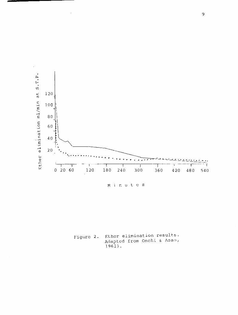
elimination from that study is shown in Figure 2.
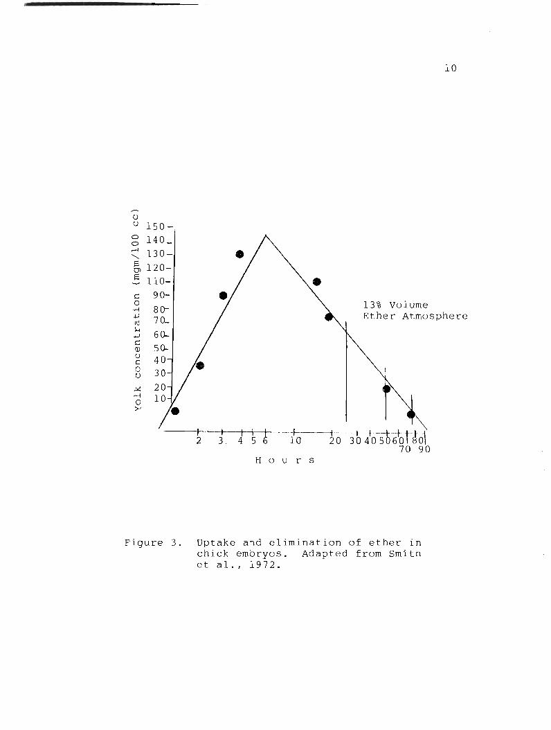
Figure 3 demonstrates yolk concentration during uptake
and excretion of 13% ether in 3-day living embryos
(Smith, Caub & Lehrer, 1972).
Effects of Diethyl Ether
Much of the data on the effects of diethyl ether
exposure either have been obtained by studying patients
who experienced some type of complication following
clinical anesthesia or by evaluating the effects of
diethyl ether as an anesthetic. These studies do give
us information about the acute, high dose effects of
ether on humans. Stevens, Eger, Joas, Cromwell, White
and Dolan (1973) studied the effects of various anes-
8
120
100 I \
80 \ \
60_ \
40 \
9
~. ",~ '. '---20 1 ' • . ..
I ••• ." ...... • ... II' •• ' •
o 20 60 r--- 'f
120 180 240 300 360 420 480 540
Min ute s
Figure 2. Ether elimination results. Adapted from Onchi & Asao, 1961).
u u 150-g 140_
~ 130-& 120-~ 110-
c:: o
.,-i
10
13% Volume .j...l
m l>-I
.j...l
90-
80-70_ Ether Atmosphere
c:: Q)
u c:: o u
...:x:: r-I o :>-i
60-
2 3 10
H 0 u r s
Figure 3. Uptake and elimination of ether in chick embryos. Adapted from Smith et al., 1972.

thetics including a diethyl ether on cardiorespiratory,
renal and hepatic function using young, healthy human
volunteers. In the ether group, blood urea nitrogen
(BUN) decreased significantly but returned to control
values within 7 days; potassium values increased on the
seventh day; and bicarbonate content tended to increase
on the first day after anesthesia and continued to
increase further at 7 days.
Oyama and Takazawa (1971) explored the effects
11
of ether during anesthesia on carbohydrate and fat meta
bolism by measuring plasma growth hormone (HGH), insulin,
blood glucose, free fatty acid (FFA), and cortisone,
using 20 male patients between the ages of 22-56 years.
The plasma HGH concentration during ether anesthesia
showed a significant elevation 1 hour after the start
of the operation. There was a relative decrease in
plasma insulin level; increases in blood glucose and
the plasma cortisol level significantly increased.
There was no demonstrable variation of blood FFA.
Rosenmann, Dishon, Durst and Boss (1972) reported
that ether anesthesia causes vasoconstriction in the
wake of which minor morphological changes ensue. Though
ether, per se, is not considered to be hepatoxic, its
administration is accompanied by mild hepatic functional
derangements with or without minimal cytological evi
dence of injury. While agreeing that ether is not a

12
true hepatoxin, other investigators have described alter-
ations in liver histology (i.e., reduced glycogen con-
tent, slight fatty changes, and central necrosis)
(Adrian, 1970; Robertson, 1959).
Epidemiologic Studies
Several epidemiologic studies have been conducted
in an attempt to identify the health effects associated
with chronic exposure to waste anesthetic gases. How-
ever, none of the studies provided information on the
anesthetic agent or on the concentrations of the gases
present.
NIOSH (NIOSH criteria, 1971, p. 29) reports:
In 1966, Vaisman surveyed by questionnaire 303 Russian anesthesiologists (193 men and 110 women). Ninety-eight percent reported using diethyl ether; 59% nitrous oxide; 28% halothane; and 21% other agents. A high incidence of headache, fatigue, irritability, nausea, and itching was reported. The authors also noted that 18 of 31 pregnancies among anesthesiologists who were between the ages of 24 and 38 ended in spontaneous abortions. In addition, there were two premature births and one child was born with a congenital malformation. The anesthesiologists with abnormal pregnancies had exposure of 25 hours per week or more while those with normal pregnancies did not exceed 15 hours per week.
Other epidemiologic studies will not be reviewed here
because they give us no information regarding the agents
used. However, the study discussed above suggests that
chronic exposure to low concentrations of anesthetic

13
gases, which include diethyl ether, may have deleterious
effects on the health of workers.
Animal Studies
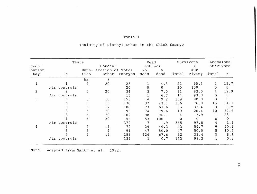
In order to determine teratogenic effects of
diethyl ether, Smith et al. (1972) conducted studies
on chicken embryos exposed to varying levels of ether
during incubation. The mortality and anomaly rates
of chick embryos after exposure to ether atmospheres
are shown in Table 1. The types of anomalies produced
by exposure to ether on the third and fourth day general
ly were similar both in type and incidence (see Table
2). However, the incidence of eye anomalies was 30.4%
after all ether treatments. Only 8% of the anomalies
encountered in 1,110 control eggs during this same
period were of the eye. Defects included anophthalmia,
and microphthalmia. This study demonstrated the
teratogenic capability of diethyl ether in the verte
brate, although the exact mechanism for that effect
is not known (Smith et al., 1972).
Stevens et al. (1975) reported the effects of 35-day
exposures to subanesthetic concentrations of diethyl
ether in mice, rats and guinea pigs which were in a
phase of rapid body growth. Diethyl ether reportedly
had a negligible effect on rat or mouse weight gain
and a detrimental effect in guinea pigs at the one per
cent dose. Although no hepatic injury was seen with
Tab
le
1
To
xic
ity
o
f D
ieth
yl
Eth
er
in
the
Ch
ick
E
mb
ryo
Tests
D
ead
S
urv
ivo
rs
Incu
-C
on
cen
-em
bry
os
~
0
bat io
n
Du
ra-
trati
on
o
f T
ota
l N
o.
~
0 su
r-D
ay
N
tio
n
Eth
er
Em
bry
os
dead
d
ead
T
ota
l v
ivin
g
1 1
6 2
0
23
1
4.5
2
2
95
.5
Air
co
ntr
ols
2
0
0 0
20
1
00
2
2 5
20
34
3
7.0
3
1
93
.0
Air
co
ntr
ols
1
5
1 6
.7
14
93
.3
3 5
6 1
0
15
3
14
9
.2
13
9
90
.8
5 6
13
1
38
3
2
23
.1
10
6
76
.9
3 6
17
1
08
73
6
7.6
3
5
32
.4
3 5
20
9
3
74
79
.6
19
2
0.4
3
6 2
0
10
2
98
9
6.1
4
3.9
1
6 3
0
53
5
3
10
0
0 0
Air
co
ntr
ols
3
72
7
1.9
3
65
9
7.8
4
3 5
11
72
2
9
40
.3
43
5
9.7
3
6 9
94
47
5
0.0
4
7
50
.0
6 6
13
1
88
1
26
6
7.6
6
2
32
.4
Air
co
ntr
ols
1
34
1
0.7
1
33
9
9.3
No
te.
Ad
ap
ted
fr
om
S
mit
h et
al.
, 1
97
2.
An
om
alo
us
Su
rviv
ors
To
tal
~
0
3 1
3.7
0
0 4
12
.9
0 0
0 0
15
1
4.1
3
8.5
1
0
52
.6
1 2
5
0 0
4 1
.1
9 2
0.9
5
10
.6
5 8
.1
1 0
.8
~
.t;..

Tab
le
2
Ty
pes
o
f A
no
mali
es
in
the
Ch
ick
A
fter
Ex
po
sure
to
E
ther
3 d
ay
s 4
day
s T
ota
l C
on
tro
la
%
%
2- 0
Ty
pe
N
an
om
aly
N
an
om
aly
N
%
N
an
om
aly
Bra
in
12
2
2.7
2
12
.5
14
20
.3
3 2
3.0
Ey
es
13
22
.8
8 5
0
21
3
0.4
1
8.0
Bea
k
12
22
.7
4 2
5.0
1
6
23
.2
3 2
3.0
Ex
trem
itie
s
6 1
1.3
0
0 6
8.7
1
8.0
Bo
dy
10
1
8.8
2
12
.5
12
1
7.4
5
38
.0
To
tal
an
om
ali
es
53
1
6
69
13
No
te.
aCo
mb
ined
ex
peri
en
ce
wit
h
1,1
10
co
ntr
ol
em
bry
os
ex
am
ined
in
th
is
an
d
oth
er
ex
peri
men
ts
du
rin
g th
e
sam
e ti
me p
eri
od
. A
dap
ted
fr
om
S
mit
h et
al.
, 1
97
2.
I-'
V1
16
the higher dose of ether, this dose was lethal to mice
and guinea pigs but not rats for unknown reasons. The
mortality was unrelated to changes in blood or the
appearance of histologic changes in any tissue examined,
including bone marrow. These animals did manifest gross
hepatic enlargement. Perhaps ether or its metabolism
to ethyl alcohol stimulated growth of the liver, but
how that is related to ether lethality is unclear.
In another study, rabbits, rats and guinea pigs
were exposed to 2000 ppm of diethyl ether for 7 hours
per day, 5 days per week for a total of 7 weeks. During
the exposures, the animals were observed for signs of
possible toxicity, including alterations in activity,
symptoms of eye and nasal irritation, skin condition
and respiratory distress. The results of the study
demonstrated no consistent increases in organ ratios,
no hematological deviations, and no changes in SGOT
and SGPT levels (Chenoweth, Leong, Sparschu & Torkelson,
1972).
In a study regarding exposure to lower concen
trations of diethyl ether (7 hours, 5 days a week for
6-7 weeks) using rats, rabbits and guinea pigs at 1/10
minimum alveolar concentration (minimum alveolar con
centration is defined as the minimum concentration in
the alveolus at which 50% of subjects moved in response
to skin incision [Wood & Wood, 1982]), ether was without

reported deleterious effect and compared well with
the air-exposed controls (Chenoweth, 1971).
Adverse Effects from Industrial Use of Diethyl Ether
As well as an inhalation anesthetic, diethyl ether
is used as a solvent for waxes, fats, oils, perfumes,
alkaloids, dyes, gums, resins, nitrocellulose, hydro-
17
carbons, raw rubber and smokeless powder; a refrigerant,
in diesel fuels, in dry cleaning, as an extractant,
and as a chemical reagent for various organic reactions.
Yet, despite this wide industrial use, there is prac-
tically nothing in the literature about ether as an
industrial poison. The literature that is available
states that industrial exposure to chronic low concen-
trations may cause a variety of symptoms including loss
of appetite, nausea, vomiting, faintness, exhaustion,
headache, dizziness, drowsiness, excitation, psychic
disturbances and constipation. Albuminuria has been
reported (Occupational diseases, 1977) as well as poly-
cythemia, increased white count, and occasionally slight
anemia (Hamilton & Minot, 1920). Local effects of ether
vapors are irritation to the eye, nose and throa~ and
contact to the liquid may produce a dermatitis (Occu-
pational diseases, 1977).
Research Questions
The following research questions were considered
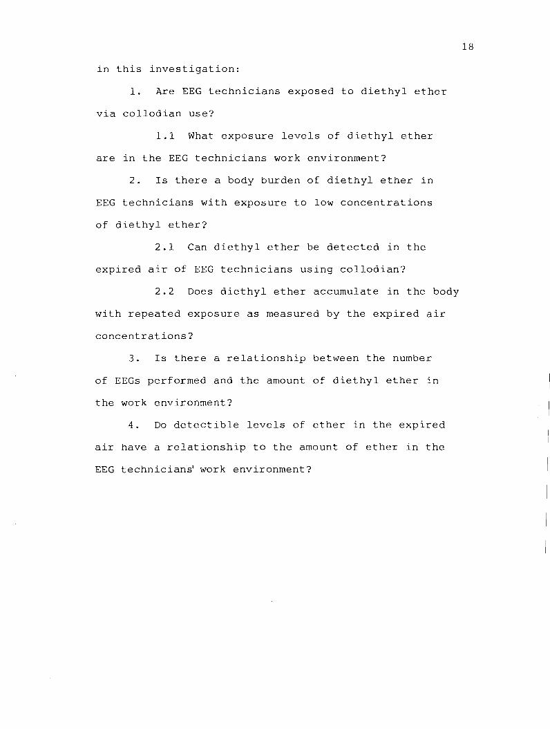
in this investigation:
1. Are EEG technicians exposed to diethyl ether
via collodian use?
1.1 What exposure levels of diethyl ether
are in the EEG technicians work environment?
2. Is there a body burden of diethyl ether in
EEG technicians with exposure to low concentrations
of diethyl ether?
2.1 Can diethyl ether be detected in the
expired air of EEG technicians using collodian?
2.2 Does diethyl ether accumulate in the body
with repeated exposure as measured by the expired air
concentrations?
3. Is there a relationship between the number
of EEGs performed and the amount of diethyl ether in
the work environment?
4. Do detectible levels of ether in the expired
air have a relationship to the amount of ether in the
EEG technicians' work environment?
18

CHAPTER III
METHODOLOGY
The design of the study was descriptive. Air
samples were collected from the breathing zone of each
subject at the place of work on five consecutive days.
During the same period, expired air samples were col
lected for each subject pre- and postwork. An expired
air sample was also collected prior to work the following
week.
Population
The study population was composed of 4 EEG techni
cians in the Salt Lake City area. All subjects were
contacted via telephone and questioned as to their use
of collodian. All subjects met the following criteria:
1. Currently working 40 hours per week as an EEG
technician performing 2-4 electroencephalograms per
day.
2. Using collodian routinely in the electro
encephalogram procedure.
Instrumentation
T~e following instruments were employed in this
investigation:

1. A questionnaire eliciting demographic infor
mation about all subjects and their use of collodian
(Appendix C).
20
2. A personal sampling pump. Each pump was cali
brated before and after each sampling period and visually
checked at regular intervals during the sampling period
to maintain a flow rate of 2 liters/minute.
3. Charcoal collection tube: a glass tube with
both ends flame sealed containing two sections of 400/200
mesh activated charcoal separated by a 2 mm portion
of urethane foam.
4. An expired air sampling device: AS-litre
saran bag with a 2-way sealable valve.
Sampling Method
1. The questionnaire responses were elicited
by the researcher.
2. The personal sampling pump and charcoal col
lection tube were attached to each subject at the onset
of work. At the end of the work day, the pump and
collection tube were removed, the charcoal tube was
sealed and labeled. This method was repeated each day
for 5 consecutive days. A control charcoal tube
was opened and sealed for analysis on each of the sam
pling days.
3. Expired air samples were collected by filling
the 5-litre bag with approximately 3-4 vital capacity

21
maneuvers. When the bag was full, the subject rebreathed
into the bag 3-5 times allowing for an equilibrium
between alveolar air and the expired air sample. This
method was repeated twice daily, before and after work
for 5 consecutive days and on the morning of the
following Monday.
Procedure Sequence
One week prior to sampling:
1. Informed consent form was signed by subjects.
2. Demographic information was obtained from sub
jects.
3. The date and times for each sampling were con
firmed with the subjects.
Sample Collection
1. The morning of the first sampling day, an
expired air sample was collected from subjects before
exposure as per the sampling method at the subject's
workplace.
2. After collection of the expired air sample,
a personal pump and charcoal collection tube were
attached to each subject.
3. At the end of the subject's work day, the after
noon expired air sample was collected and the charcoal
collection tube removed and sealed.
4. Charcoal tube and expired air samples were
taken to the Utah Biomedical Test Laboratory at the
end of each day.
5. This procedure sequence was repeated for 5
consecutive days.
6. An additional expired air sample was collected
the following Monday morning.
Analysis of Samples
Samples were analyzed by Gas Chromatography at
the Utah Biomedical Test Laboratory (NIOSH, Method
No. 127).
22

CHAPTER IV
DATA ANALYSIS
Study Population
Four subjects were selected who met the criter-
ia for inclusion in the study. The subjects ranged
in age from 22-47 years. Two subjects were smokers
and two nonsmokers. The number of years of collodian
use ranged from 10 months to 13 years. No subject
had any history of lung disease or any acute respir-
atory problems at the time of this study. One sub-
ject reported being on the following medications:
Synthyroid and Megace.
Analysis of Sample
Both air and breath samples were analyzed by
the Utah Biomedical Test Laboratory. Results of
the charcoal tube samples were then converted to
3 mg/m . The personal sampling pumps were calli-
brated before and after each sampling period and the
average was used in the calculations. Refer to
Appendix E for the data used in the calculations.
Data Analysis
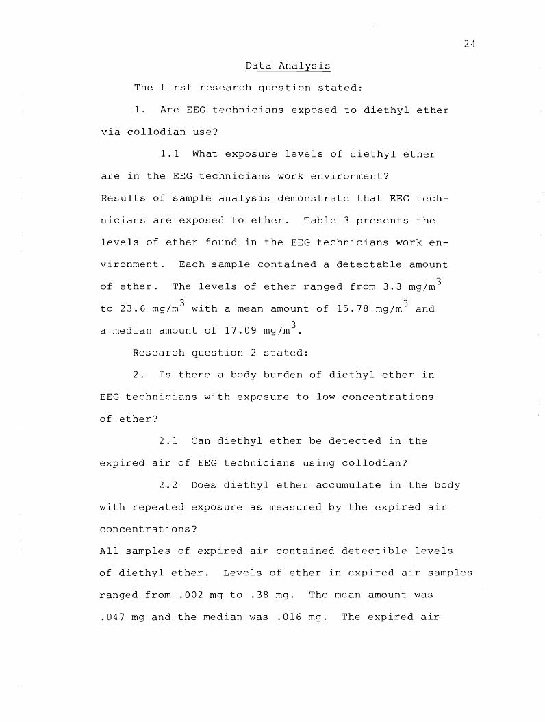
The first research question stated:
1. Are EEG technicians exposed to diethyl ether
via collodian use?
1.1 What exposure levels of diethyl ether
are in the EEG technicians work environment?
Results of sample analysis demonstrate that EEG tech
nicians are exposed to ether. Table 3 presents the
levels of ether found in the EEG technic work en-
vironment. Each sample contained a detectable amount
of ether. The levels of ether ranged from 3.3 mg/m3
to 23.6 mg/m 3 with a mean amount of 15.78 mg/m 3 and
a median amount of 17.09 mg/m 3 .
Research question 2 stated:
2. Is there a body burden of diethyl ether in
EEG technic
of ether?
with exposure to low concentrations
2.1 Can diethyl ether be detected in the
expired air of EEG technicians using collodian?
2.2 Does diethyl ether accumulate in the body
with repeated exposure as measured by the expired air
concentrations?
All samples of expired air contained detectible levels
24
of diethyl ether. Levels of ether in expired air samples
ranged from .002 mg to .38 mg. The mean amount was
.047 mg and the median was .016 mg. The expired air

25
Table 3
Sampling Results
Levels of Diethyl Ether in Breathing Zone Air Mg/m 3
Monday Tuesday Wednesday Thursday Friday Subject Day 1 Day 2 Day 3 Day 4 Day 5
A 8.65 19.84 20.14 9. 1 16.58
B 14.67 18.6 20.22 9.01
c 20.13 17.09 23.26 23.27 23.6
D 11.2 3.3 8.5 12.29 20.42

26
sample results are shown in Table 4. The levels of
ether in expired air did not increase during the week
and in fact were lower on the last day of the week than
on the first. Sample results demonstrated no consistent
increase in ether levels immediately after exposure.
Levels of ether were lower in the afternoon immediately
after exposure than in the morning 12 out of the 18
combined sampling days.
Research question 3 stated:
3. Is there a relationship between the number
of EEGs performed and the amount of diethyl ether in
the work environment?
The breathing zone ether levels and the number of EEGs
done per day were plotted on a scatter diagram. The
graph indicates a trend toward a positive relationship
between these two factors (Figure 4) (correlation
coefficient r .536; 95% confidence interval .05-.8).
Research question 4 stated:
4. Do detectible levels of ether in the expired
air have a relationship to the amount of ether in the
EEG technicians work environment?
The environmental sample results and expired sample
results were plotted on a scatter diagram. There is
no indication of a trend or relationship between the
expired air and breathing zone sample results (Figure 5).
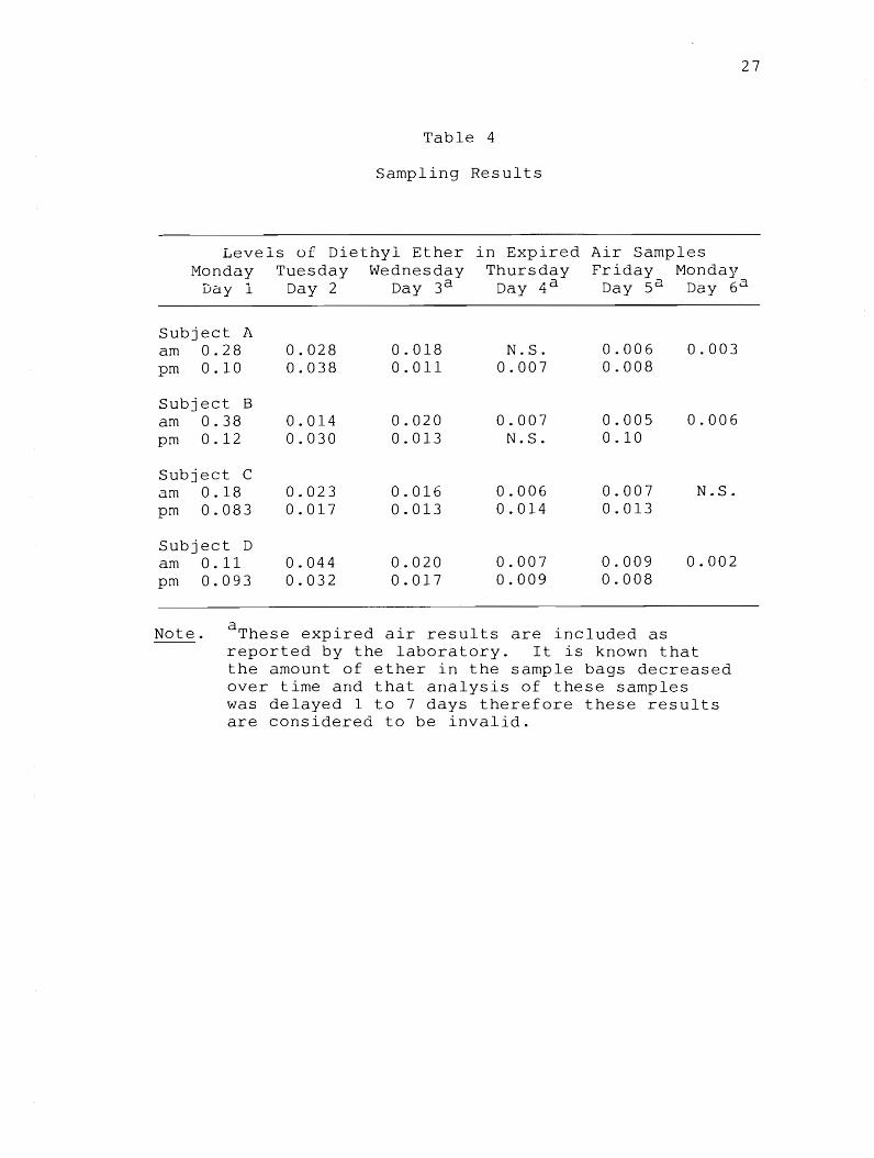
27
Table 4
Sampling Results
Levels of Diethyl Ether in ired Air Samples Monday Tues Wednesday Thursday Friday Monday
Day 1 Day 2 Day 3a Day 4 a Day Sa Day 6a
Subject A am 0.28 0.028 0.018 N.S. 0.006 0.003 pm 0.10 0.038 0.011 0.007 0.008
Subject B am 0.38 0.014 0.020 0.007 0.005 0.006 pm 0.12 0.030 0.013 N.S. 0.10
Subject C am 0.18 0.023 0.016 0.006 0.007 N.S. pm 0.083 0.017 0.013 0.014 0.013
Subject D am 0.11 0.044 0.020 0.007 0.009 0.002 pm 0.093 0.032 0.017 0.009 0.008
Note. se expired a results are included as reported by the laboratory. It is known that the amount of ether in the sample bags decreased over time and that analysis of these samples was delayed 1 to 7 days therefore these results are considered to be invalid.

6
5
NO. 4 EEGs Per Day
3
2
1
o
•
41 •
• ,
. , • •
5 10 15 20
Breathing Zone Level of Ether mg/m 3
II II
Figure 4. Number of EEGs per day and breathing zone levels.
28
25

Expired Air mg
.40
.30
.20
.10
.08
.06
.04
.02
o
Figure 5.
o 5 10 15
Breathing Zone Air mg/m 3
. ..
20
.. • II •
25
Breathing zone levels and expired air levels of ether.
29
No further statistical analysis was deemed necessary
because of the randomness of the scattergram.
30

CHAPTER V
DISCUSSION
One conclusive finding of this study is that EEG
technicians using collodian are exposed to diethyl ether.
This is demonstrated by the detectible levels of ether
found in the breathing zone air of the subjects. Based
on the assumption that exposure during the nonsampling
period was the same as during the sampling time, the
levels of ether found in this study can be compared
to the recommended standard for an 8-hour time weighted
3 average of 1200 mg/m. The levels of ether found in
this study were far below this recommended standard.
However, study levels were above those recommended by
the Hospital Engineering Cooperative Group of Denmark
3 for long-term exposure of 9 mg/m (NIOSH document, 1977).
A second finding of this study was that there is
a direct relationship between the amount of ether found
in the breathing zone air and the number of electro-
encephalograms that were performed by the technicians
during the sampling period. A possible explanation
is that the more that EEGs are performed, the more collo
dian use is required, hence increasing the amount of
ether present in the environment. Other factors that

would influence the amount of collodian used are the
number of electrodes that are applied for a particular
procedure and the amount of time needed to apply the
electrodes such as with a cooperative patient versus
an uncooperative one. Another factor that may affect
32
the amount of ether in the environment is the ventilation
rate and room size of the EEG lab and the storage of
used collodian.
The third finding of this study is that there is
a body burden of diethyl ether in EEG technicians with
exposure to low concentrations of ether. Detectible
levels of ether were found in all samples of expired
air including those collected after 2 days with no ether
exposure. However, the data in this study do not show
an accumulation of ether in the body with repeated
exposures as measured by the expired air concentrations.
There was no consistent increase in expired air levels
of ether over the 5 day period of consecutive sampling.
Levels in the expired air were lower on the last day
of sampling than on the first day. Also, on 12 of the
18 combined sampling days ether levels were lower in
the afternoon immediately after exposure than in the
morning before exposure. The levels of ether on the
second Monday morning after 2 days with no exposure
were only slightly lower than on Friday afternoon.
Possible explanations for this are:

33
1. The expired air sample results do not accurately
reflect expired air levels in the subject.
1.1 One reason for this is the loss of ether
from the collection bag. It was found during the analy-
sis of samples that the concentration of ether in the
sampling bags decreased over a period of time. The
samples collected on Day 1 (Monday) were analyzed within
a few hours of collection. The next day, analysis of
these same samples was repeated and the ether concen
tration was found to be approximately 30% lower than
the results from the previous day. Samples collected
on the following days were analyzed 1 to 7 days later-
the longest delays occurring with the samples that were
collected on the third, fourth and fifth days and with
the samples collected the following Monday morning.
This loss of ether from the sampling bag over time would
result in lower amounts of ether in the samples collected
on the third, fourth and fifth days, as well as the
following Monday. Several possibilities may account
for the loss of ether over time.
1.1.1 One possibility is the presence
of a leak from the sampling bag values or the bag walls
themselves. Each of the sampling bags were checked
for air tightness prior to the onset of sampling but
the presence of a very slow leak cannot be excluded.
1.1.2 A second explanation may be that

the ether interacted with the bag1s surface or with
moisture in the sample bags which condensed on the bag
surface causing a physical nonreactive absorption of
the gas to the condensate. The analyst reported that
during sample measuring there was extensive deposition
of moisture on the inside wall of the syringes after
withdrawing air from the sampling bags.
The mylar sampling bag used in this study may not
be an appropriate sampling device for diethyl ether
and/or the collection of expired air. A study done
on this bag used vinyl chloride monomer gas to test
for permeability and storage stability; it showed no
loss of VCM over a period of one week. Also, it was
found that after cleansing the bag with compressed air,
emptying and repeating this cycle, no detectable levels
of VCM were found in the bags. This study collected
34
VCM from the breathing zone not from expired air (Levine,
Hebel, Bolton & Kugel, 1975). No studies were found
which tested the bag's reliability with expired air
samples. Other studies regarding the collection of
expired air samples used a glass sampling tube (Brug
none, Perbellini, Gaffuri & Apostoli, 1980; Pasquini,
1978: Stewart, Hake & Peterson, 1974). This instru-
ment was not available to the researcher for this study.
The research team did not determine prior to this study
the mylar bag's reliability and stability in terms

of expired air samples of diethyl ether.
1.2 Invalid expired air sample results may
also be attributed to problems encountered in the
analytical process.
analysis were:
Problems encountered during sample
1.2.1 The use of nonairtight syringes
to draw air from sampling bag and inject into the gas
chromatograph injection port. The nonairtight syringes
leaked extensively, especially when injected into the
GC injection port. The extent this altered the results
is not known.
1.2.2 It was not possible to obtain
reproducible injections. Several injections (~ 6-7)
of the same sample were performed. The outliers were
eliminated and the average value of median values was
computed for reporting the results. The analyst used
the following process to obtain samples for analysis
from the sampling bags: The bags were shaken well,
then 2 ml of air volume was drawn from the top side
of the bag while lying horizontally in a 5 ml syringe
then injected into the gas chromatograph injection
port.
2. The second possible explanation is that the
ether content in expired air does not reflect the
amount of ether exposure. As stated in the literature
review, very little is known of the metabolism and
35

36
elimination of ether especially in subjects exposed
to repeated low levels of ether. One study found that
a portion of administered ether was stored in fat and
released slowly. As the slow release occurs, the amount
of ether in the animal was maintained at a constant,
although low, level (Van Dyke et al., 1964). Although
this phenomenon is not clearly understood, its possible
that the individual subject maintains a fairly constant
body level of ether despite fluctuations in the environ
mental levels. This may also be influenced by an
individual's metabolic and body characteristics, as
well as lifestyle.
These results did not demonstrate a relationship
between levels of ether in the expired air of subjects
and those found in the breathing zone. Possible explan
ations for this are the same as those discussed earlier.
The first is that the expired air sample results do
not accurately reflect expired air levels in the subject
and the second, the ether content in expired air does
not reflect the amount of ether exposure.
Recommendations for Future Research
Since it has been determined that EEG technicians
using collodian are exposed repeatedly to low concen
trations of ether and a body burden of ether does exist
from this exposure, it is recommended that EEG techni
cians using collodian be added to the list of occupations

37
with potential ether exposure. The long-term effects
of low concentrations of ether are not known~ therefore,
levels in the environment should be kept as low as
possible. This should be taken in consideration in
the construction of EEG labs, in the engineering of
air flow and ventilation in these areas, and in work
practice techniques.
Because of the invalid expired air sample results,
no recommendations can be made regarding the accumulation
of diethyl ether in the body with repeated exposure
or the relationship between expired air concentrations
and breathing zone air levels of diethyl ether.
This study was limited by the paucity of knowledge
regarding the sampling and analysis of ether in expired
air as well as the biotransformation and elimination of
ether from the body. Before replication of this type
of study is attempted, additional data on the following
areas would be useful:
1. Additional studies on the reliability and
validity of various sampling instruments to determine
expired air levels of ether. One instrument that could
be considered for this sampling is the charcoal sampling
tube. Its "Limit of Detection" (LOD) as reported on the
breathing zone air samples was .01 mg. If the first day
of expired air sample results in this study are can
sidered the most accurate, this LOD would be far below

38
the levels of ether in expired air. It is recommended
that in future studies of this type, analysis of samples
be performed within a few hours of sampling if the relia
bility and stability of the sampling instrument cannot
or has not been determined.
2. Research on the metabolism and elimination
of ether through the respiratory system to arrive at
a reliable breath decay curve. Studies comparing the
elimination of diethyl ether through the respiratory
system after repeated exposures to low concentrations
of ether and elimination of ether after isolated exposure
to higher concentrations to determine if biotransfor
mation and elimination is dose affected.
3. Research on other biological monitoring methods,
such as blood and tissue sampling.
Future areas of research might include the longi
tudinal study of long-term exposure to ether, at both
high and low concentrations. Subjects with high exposure
levels may give earlier indications of adverse effects
and the reliability of sampling methods. Subjects with
low exposure levels may provide information especially
relevant to the usual hospital or clinic work setting,
and may serve to evaluate, develop, or revise the present
occupational standards.
Conclusion
A general statement can be made that EEG technicians

who use collodian are exposed to diethyl ether. Levels
found in this study were below the recommended TLV of
400 ppm. However, no studies were available regarding
the effects of long-term exposure to low concentrations
of ether. EEG technicians who use collodian may be
a good population to study regarding possible effects
of long-term exposure. In terms of using expired air
sampling to study exposure to ether, more data need
39
to be acquired. A sampling instrument needs to be
tested for it's validity and reliability. Also, more
information is needed regarding the metabolism and elimi
nation of ether, the breath decay curve of ether, as
well as other methods of biological testing for ether
exposure.

APPENDIX A
CONSENT FORMS
SUBJECT INFORMATION
AND
INFORMED CONSENT FORM
Information about "Diethyl Ether Exposure to EEG Technicians Using Collodian" study:
41
The purpose of this study is to determine if EEG technicians using collodian are exposed to low concentrations of diethyl ether and to determine if diethyl ether is accumulated in the body with repeated exposures.
Levels of diethyl ether in the breathing zone air of EEG technicians will be determined as well as levels of ether in expired air. A personal sampling pump and charcoal collection tube will be attached to the EEG technician for 8 hours on 5 consecutive days. Expired air samples will be collected in a 5-litre bag twice a day, pre and postwork. Environmental monitoring and sample collection will be done for 5 consecutive days. A final expired air sample will be collected prework on the following Monday. All sampling instruments will be delivered to and collected at the subject's workplace.
Details of this study have been discussed with the subject and an opportunity given to ask questions regarding the study and participation in this study. If there are any questions before or during the study, participants are free to contact Jeri Lockman at 485-3995.
The risk involved in participating in this study is the one-half to one hour per day of the subject's time. Sample collection will be done at the workplace and coordinated prior to the onset of the study.
Benefits of this study will be an increased knowledge regarding ether levels in the environment and expired air from the use of collodian.
All samples will be coded so as to insure confidentiality.
The subjects are free to withdraw from this study at any time without prejudice to themselves.
Date
APPENDIX B
INFORMED CONSENT PROCEDURE FORM

43
Informed Consent Procedure
1 . Each subject will be given information regarding
the nature of this study and study objectives.
2 • A detailed description of the sampling methods
will be explained to the subjects. A personal sampling
pump, charcoal collection tube and expired air sampling
bag will be shown and a demonstration of its use given
at this time.
3. The sample procedure will be described. The
exact times for sample collection will be coordinated
with each subject so as to eliminate any interference
with the subject's work schedule. Samples will be
collected at the workplace to eliminate travel expense
and time for the subjects. The amount of time involved
will be approximately one-half to one hour per day for
each subject.
4. Potential benefits of this study will be dis
cussed with each subject. Benefits include increased
knowledge regarding collodian use and the level of
diethyl ether in the air.
5. Each subject will be informed of his/her right
to withdraw from the study at any time without prejudice
to him/herself.
6. Each subject will be given an opportunity to
ask questions regarding the study and any procedure
in the study.

44
7. All samples will be coded so as to insure confi
dential handling. Subjects may be given results of
their individual samples upon request.
8. An informed consent form will be signed by
each subject agreeing to participate in this study.
APPENDIX C
PRESAMPLING QUESTIONNAIRE

46
Name: Subject Code:
Sex: Male Female Age
History of lung disease (asthma, bronchitis)
Smoker: Nonsmoker:
Medications:
Hours worked per week with collodian:
Number of years of collodian use:
How is collodian stored:
Where is collodian stored:
How is collodian disposed of:

APPENDIX D
SAMPLING RECORD

48
Sampling Record
Subject Code:
Day 1
Expired air collection time: Sample A1 ____ Sample B1
Personal Sampling Pump: On Off
No. of EEGs Time of last EEG -----Day 2
Expired air collection time: Sample A2 Sample B2
Personal Sampling Pump: On Off
No. of EEGs Time of last EEG -----Day 3
Expired air collection time: Sample A3 Sample B3 --Personal Sampling Pump: On Off
No. of EEGs Time of last EEG
Day 4
Expired air collection time: Sample A4 Sample B4
Personal Sampling Pump: On Off
No. of EEGs Time of last EEG
Day 5
Expired air collection time: Sample AS Sample 85
Personal Sampling Pump: On Off
No. of EEGs Time of last EEG -----Day 6
Expired air collection time: Sample A6
APPENDIX E
ENVIRONMENTAL SAMPLE CONVERSION
DATA

50
Subject A
Sampling Time 6hr 38min 6hr 41min 5hr 44 min 6hr 31min 6hr 25min Sample Result ~.3 mg 14 mg 9.7 mg 6.9 mg 12 mg Flow Rate 1.54 1 ~m 1.76 1 pm 1.4 1 pm 1.94 1 pm 1.84 1 pm
Subject B
Sampling Time ~hr 40min 6hr 24min 6hr 25min -- 5hr 55min Sample lResult 8.8 mg 13 mg 13 mg -- 6.4 mg Flow IRate 1.5 1 2!fl 1.82 1 pm 1.67 1 pm -- 2 1 p_m
Subject C
Sampling! Time ~hr 39min 6hr 49min 7hr 20min 7hr 05min 7hr 25min Sample I
I
Result 113 mg 10 mg 18 mg 18 mg 21 mg Flow !
Rate 11.62 1 pm i 1.43 1 pm 1.76 1 pm 1.82 1 pm 2 1 pm
Subject D
Sampling I I
I Time ~hr 21min 7hr 30min 7hr 45min j 7hr 56min 7hr 45min Sample i 1
b mg !
Result 2.7 mg 6.4 mg i 11 mg i 15 mg Flow I Rate .62 1 pm 1.82 1 pm 1.62 1 pm I 1.88 1 pm 1.58 1 pm
APPENDIX F
INFORMATION ON COLLODIAN

52
u.s. Coast Guard Chemical Hazards Response Information System (Chris) 9-11-78
CLD COLLODION
Common Synonyms
Cellulose nitrate solution Nitrocellulose solution Pyroxylin solution
Thick liquid Colorless Ether-like
Box toe gum Nitrocellulose gum
odor Floats on water. Flammable, irritating vapor is produced. Boiling point is around 94°F.
Shut off ignition sources. Call fire department. stop discharge if possible. Isolate and remove discharged material. Notify local health and pollution control agencies.
Fire
FLAMMABLE. POISONOUS GASES MAY BE PRODUCED IN FIRE. Containers may explode in fire. Flashback along vapor trail may occur. Vapor may explode if ignited in an enclosed area. Extinguish with dry chemicals, alcohol foam, or carbon dioxide. Cool exposed containers with water.
Exposure
Call for medical aid. VAPOR Irritating to eyes, nose and throat. If inhaled will cause dizziness, difficult breathing, or loss of consciousness. Move victim to fresh air. If breathing has stopped, give artificial respiration. If breathing is difficult, give oxygen. LIQUID Not harmful.
Water Pollution
Effect of low concentrations on aquatic life is unknown. Fouling to shoreline. May be dangerous if it enters water intakes. Notify local health and wildlife officials. Notify operators of nearby water intakes.
53
1. Response to Discharge (See Response Methods Handbook, CG446-4).
Issue warning, high flammability Restrict access Mechanical containment Should be removed Chamical and physical treatment
2. Label
3. Chemical Designations
3.1 Synonyms: Cellulose nitrate solution; nitrocellulose solution; pyroxylin solution; box toe gum; nitrocellulose gum.
3.2 Coast Guard compatability classification: Not applicable.
3.3 Chemical Formula: Not pertinent.
3.4 IMCO/United Nations Numerical Designation: 3.2/2059.2061 3.3/2060.2062
4. Observable Characteristics
4.1 Physical State (as shipped); Viscous liquid.
4.2 Color: Colorless.
4.3 Odor: Depends on solvent used; often that of ether.
5. Health Hazards
5.1 Personal Protective Equipment: Self-contained breathing apparatus: rubber gloves; goggJes or face shield.
5.2 Symptoms Following Exposure: High concentration of ether fumes may cause narcosis, loss of consciousness and respiratory paralysis if inhaled. Contact with eyes causes irritation.
5.3 Treatment for Exposure: INHALATION: remove victim to fresh air; initiate artificial respiration if breathing has stopped; call physician.
54
5.4 Toxicity by inhalation (Threshold Limit Value): 400 ppm>
5.5 Short-Term Inhalation Limits: able.
Data not avail-
5.6 Toxicity by Ingestion: Grade 0: LD30 >15 g/kg.
5.7 Late Toxicity: Data not available.
5.8 Vapor (Gas) Irritant Characteristics: Vapors cause a slight smarting of the eyes or respiratory system if present in high concentrations. The effect is temporary.
5.9 Liquid or Solid Irritant Characteristics: No appreciable hazard. Practically harmless to the skin.
5.10 Odor Threshold: Data not available.
6. Fire Hazards
6.1 Flash Point: -49°F C.C. (ether)
6.2 Flammable Limits in Air: solut ion) .
6.3 Fire Extinguishing Agents: foam, carbon dioxide.
1.9%-36% (ether
Dry chemical, alcohol
6.4 Fire Extinguishing Agents Not to be Used: Water may be ineffective.
6.5 Special Hazards of Combusion Products: The formation of extremely toxic gases, notably oxides of nitrogen, hydrogen cyanide, and carbon monoxide is possible.
6.6 Behavior in Fire: Highly flammable solvent vapors are formed. May travel a long distance to a source of ignition and flash back.
6.7 Ignition Temperature: 356 ° F (ether).
6.8 Electrical Hazard: Class I, Group C.
6.9 Burning Rate: Data not available.

55
7. Chemical Reactivity
7.1 Reactivity with Water: No reaction.
7.2 Reactivity with Common Materials: No reaction.
7.3 Stability During Transport: Stable.
7.4 Neutralizing Agents for Acids and Caustics: Not pertinent.
7.5 Polymerization: Not pertinent.
7.6 Inhibitor of Polymerization: Not pertinent.
8. Water Pollution
8.1 Aquatic Toxicity: Data not available.
8.2 Waterfowl Toxicity: Data not available.
8.3 Biological Oxygen Demand (BOD): able.
Data not avail-
8.4 Food Chain Concentration Potential: None.
9. Selected Manufacturers
1. Mallinckrodt Chemical Works Second and Mallinckrodt Streets St. Louis, MO 63160
2. Polysciences , Inc. Paul Valley Industrial Park Warrington, PA 18976
10. Shipping Information
10.1 Grades or Purity: USP. All grades contain less than 60% nitrocellulose by weight.
10.2 Storage Temperature: Ambient.
10.3 Inert Atmosphere: No requirement.
10.4 Venting: Pressure-vacuum/
11. Hazard Assessment Code (See Hazard Assessment Handbook, CG 446-3) A-T-U-V-W.

56
12. Hazard Classifications
12.1 Code of Federal Regulations: Flammable liquid.
12.2 NAS Hazard Rating for Bulk Water Transportation:
Category Rating
Fire. . . . . . . . . . . . . . . . . . . . . . . . . . . . . . . . . . . . . 4 Health
Vapor Irritant .......................... 1 Liquid or Solid Irri tant. . . . . . . . . . . . . . . . 0 Poisons. . . . . . . . . . . . . . . . . . . . . . . . . . . . . . . . . 2
Water Pollution Human Toxicity .......................... 0 Aquatic Toxicity ........................ 1 Aesthetic Effect ........................ 1
Reactivity Other Chemicals......................... 1 Water. . . . . . . . . . . . . . . . . . . . . . . . . . . . . . . . . . . 0 Self-Reaction. . . . . . . . . . . . . . . . . . . . . . . . . . . 0
12.3 NFPA Hazard Classifications:
Category Classification*
Heal th Haz ard (Bl ue ) ............ . Flammability (Red) .............. . Reactivity (yellow) ............. .
o 3 3
*First column refers to nonfire situation.
13. Physical and Chemical Properties
13.1 Physical State at 15°C and 1 atm: Liquid.
13.2 Molecular Weight: Not pertinent.
13.3 Boiling Point at 1 atm: 93°F = 34°C (ether solvent)
13.4 Freezing Point: Not pertinent.
13.5 Critical Temperature: Not pertinent.
13.6 Critical Pressure: Not pertinent.
13.7 Specific Gravity: 0.772 at 25°C (liquid).
13.8 Liquid Surface Tension: Not pertinent.
2 3 3

13.9 Liquid-Water Interfacial Tension: Not pertinent.
13.10 Vapor (Gas) Specific Gravity: Not pertinent.
13.11 Ratio of Specific Heats of Vapor (Gas): Not pertinent.
13.12 Latent Heat of Vaporization: Not pertinent.
13.13 Heat of Combustion: Data not available.
13.14 Heat of Decomposition: Not pertinent.
13.15 Heat of Solution: Not pertinent.
13.16 Heat of Polymerization: Not pertinent.
Notes
57

SELECTED BIBLIOGRAPHY

59
Adrian, J. (1970). The pharmacology of anesthetic drugs (5th ed.). Springfield: Charles C. Thomas, pp. 46, 95.
Aune, H., Ranck, H., Bessesen, A. & Midrland, J. (1978, July). Metabolism of diethyl ether to acetaldehyde in man. The Lancet, l, 97.
Axelrod, J. (1956). The enzymic cleavage of aromatic ethers. Biochemical Journal, i1, 634-639.
Brown, B.R. & Vandam, D. (1971). A review of current advances in metabolism of inhalation anesthetics. Annals of the New York Academy of Science, 179, 235-243.
Brugnone, F., Perbellini, L., Gaffuri, E. & Apostali, P. (1980). Biomonitoring of industrial solvent exposures in workers' alveolar air. International Archives of Occupational and Environmental Health, !I, 245-261.
Chenoweth, M.B., Leong, B.K.J., Sparschu, G.L. & Torkelson, T.R. (1972). Toxicities of methoxyflurane, halothane, and diethyl ether in laboratory animals on repeated inhalation at subanesthetic concentrations. In B.R. Fink, (Ed.), Cellular biology and toxicity of anesthetics. Baltimore: The Williams and Wilkins Co.
Chenoweth, M.B. (1971, February). of inhalational anesthetics. College of Surgeons, ~, 79.
Chronic toxicity Annals of the Royal
Cohen, E.N. & Hood, N. (1969, July). Application of low temperature autoradiography to studies of the uptake and metabolism of volatile anesthetics in the mouse; II diethyl ether. Anesthesiology, }!(1), 61-68.
Cohen, E.N. & Van Dyke, R.A. (1977). Metabolism of volatile anesthetics. In Implications for toxicity, Reading, Massachusetts: Addison Wesley Publishing Company, Inc., p. 116.
Corbett, T.H. (1973, July-August). Retention of anesthetic agents following occupational exposure. Anesthes Ana , ~(4), 614-617.
Cowles, A.L., Borgstedt, H.H. & Gillies, A.J. (1971, August). Solubilities of ethylene, cyclopropane, halothane and diethyl ether in human and dog blood

at low concentrations. Anesthesiology, 12(2), 203-211.
Elder, B.F., Beal, H., DeWald, W. & Cobb, S. (1971). Exacerbation of subclinical myasthenia by occupational exposure to an anesthetic. Anesthesia and Analgesia, 2Q, 383 387.
60
Geddes, I.C. (1972). Metabolism of volatile anesthetics. British Journal of Anesthesia, !i, 953-960.
Goodman, L.S. & Gilman, A. (1970). The pharmacological basis of therapeutics, (4th ed.). New York: The Macmillan Company, pp. 79-82.
Gosselin, R.E., Hodge, H.C., Smith, R.P. & Gleason, M.N. (1976). Toxicology of commercial products (4th ed.). Baltimore: The Williams and Wilkins Company.
Haggard, H.H. (1924). The absorption, distribution, and elimination of ethyl ether. Journal of Biological Chemistry, 59, 737-751.
Hamilton, A. & Minot, G.R. (1920, June). Ether poisoning in the manufacture of smokeless powder. Journal of Industrial Hygiene, 2(2), 41-49.
Land, P.C., Owen, E.L. & Kinde, H.W. (1981). Morphologic changes in mouse spermatozoa after exposure to inhalational anesthetics during early spermato-genesis. Anesthe , 2!, 53-56.
Levine, S.P., Hebel, K.G., Bolton, J.R. & Kugel, K.E. (1975, October). Industrial analytical chemists and OSHA regulations for vinyl chloride. Analytical Chemistry, il, 1078A.
Mazur, J.F., Podolak, G.E., Rinehard, D.S. & Glenn, R.E. (1980). Evaluation of a passive dosimeter for collection of 2-bromo-2-chloro-1, 1, 1-trifluroethane and 2-chloro-1,l, 2-trifluoroethy difluromethyl ether in hospital operating rooms. Industrial Hygiene Association Journal, !l(5), 317-321.
NIOSH Criteria for
Education and Welfare.
Occupational diseases: A guide to their recognition. (1977, June). Bethesda: U.S. Department of Health,
Education and Welfare.
Onchi, Y. & Asao, Y. (1961). Absorption, distribution and elimination of diethyl ether in man. Brit Journal of Anaesthesiology, 11, 544.
61
Oyama, T. & Takazawa, T. (1971, January). Effects of diethyl ether anesthesia and surgery on carbohydrate and fat metabolism in man. Canadian Anesthesiology Society Journal, ~(1), 51-59.
Pasquini, D.A. (1978, January). Evaluation of glass sampling tubes for industrial breath analysis. American Industrial Hygiene Association Journal, ~, 55-62.
Practical anesthetic pharmacology. (1978). In R.R. Attia and A.W. Grogono (eds.). New York, New York: Appleton-Century Crofts.
Robertson, J.D. (1959). General anesthesia. In F.T. Evans & T.C. Gray (Eds.), Anesthesia and the difficult patient. London: Butterworth, p. 334.
Rosenmann, E., Dishon, T., Durst, A. & Boss, J.H. (1972). Kidney and liver damage following anesthesia with ether and pentobarbitone. British Journal of thesiology, 44, 465-468.
Schwetz, B.A. & Becker, B.A. (1971). Comparison of the lethality of inhaled diethyl ether in neonatal and adult rats. Toxicology and Applied Pharmacology, ~, 703-706.
Smith, B.E. (1979, September-October). Gas chromatography in the operating room. Anesthesia and Analgesia ... Current Research, !2(5), 740-745.
Smith, B.E., Caub, M.L. & Lehrer, S.B. of Anesthetics (B.R. Fink, ed.). Williams and Wilkins Company.
(1972). Toxicity Baltimore: The
Stevens, W.C., Eger, E.I., Joas, T.A., Cromwell, T.H., White, A. & Dolan, W.M. (1973, May). Comparative toxicity of isoglurane, halothane, fluroxene and diethyl ether in human volunteers. Canadian Anesthetists Society Journal, lQ.(3), 35 -
Stevens, W.C., Eger, E.I., White, A., Halsey, M.J., Munger, W., Gibbons, R.D., Dolen, W. & Shargel, R. (1975). Comparative toxicities of halothane, iso flurane, and diethyl ether at subanesthetic concen-

trations in laboratory animals. Anesthesiology, g(4), 408-419.
Stewart, R., Hake, C.L. & Peterson, J.E. (1974, July).
62
Use of breath analysis to monitor trichloroethylene exposures. Archives of ironmental Health, l2, 6-13.
Stewart, R.A. & Awu, A. (1976). Use of breath analysis to monitor methylene chloride exposure. Scandinavian Journal of Work, Environment and Health, 2, 57-70.
U.S. Coast Guard (1978). Chemical hazards response information system (Chris): Collodion. Washington: Author.
Van Dyke, R.A. & Chenoweth, M.B. (1965). Metabolism of volatile anesthestics. Anesthesiology,~,
348-356.
Van Dyke, R.A., Chenoweth, M.B. & Van Poznak, (1964). Metabolism of volatile anesthetics--I, conversion in vivo of several anesthetics to 14/C02 and chlo-ride. , 13, 1239-1247.
Van Dyke, R.A. (1973, January). Biotransformation of volatile anesthetics with special emphasis on the role of metabolism in the toxicity of anesthetics. Canadian Anesthetists' Society Journal, 20(1), 21 33.
Van Poznak, A. (1974, Summer). Biotransformation of diethyl ether and chloroform. International Anes-______ ~~ _______ , ll(2), 35-
Van Oettingen, W.F. (1958). Poisoni --------~~--------------------and Treatme
Waritz, R.S. (1979). Biological indications of chemical dosage and burden. In L.J. Cralley and L.V. Cralley (eds.), Patty's industrial hygiene and toxicology, (Vol. III), pp. 295-302. New York: John Wiley & Sons.
Winek, C.L., Collom, W.D. & Wecht, C.H. with plastic bag and ethyl ether.
(1970). Suicide Lancet, 1, 365.
Wood, M. & Wood, A. (1982). Drugs and anesthesia in pharmacology for anesthesiologists. Baltimore: Williams and Wilkins.

Zielhues, R.L. (1978). Biological monitoring. Scandinavian Journal of ork, Encironment and Health,
!, 1-18.
63