Dialectical behavior therapy alters emotion regulation and amygdala ... - UW Blogs … ·...
9
Dialectical behavior therapy alters emotion regulation and amygdala activity in patients with borderline personality disorder Marianne Goodman a, b, d, * , David Carpenter c , Cheuk Y. Tang c , Kim E. Goldstein a , Jennifer Avedon b , Nicolas Fernandez b , Kathryn A. Mascitelli b , Nicholas J. Blair b , Antonia S. New a, b , Joseph Triebwasser a, d , Larry J. Siever a, b, d , Erin A. Hazlett a, b a Department of Psychiatry, Icahn School of Medicine at Mount Sinai, New York, NY, USA b VISN 3 Mental Illness Research, Education, and Clinical Center (MIRECC), James J. Peters VA Medical Center, Bronx, NY, USA c Department of Radiology, Icahn School of Medicine at Mount Sinai, USA d Oupatient Psychiatry, James J. Peters VA Medical Center, Bronx, NY, USA article info Article history: Received 29 January 2014 Received in revised form 20 June 2014 Accepted 24 June 2014 Keywords: Borderline personality disorder Emotion regulation Amygdala Habituation fMRI abstract Objective: Siever and Davis' (1991) psychobiological framework of borderline personality disorder (BPD) identifies affective instability (AI) as a core dimension characterized by prolonged and intense emotional reactivity. Recently, deficient amygdala habituation, defined as a change in response to repeated relative to novel unpleasant pictures within a session, has emerged as a biological correlate of AI in BPD. Dia- lectical behavior therapy (DBT), an evidence-based treatment, targets AI by teaching emotion-regulation skills. This study tested the hypothesis that BPD patients would exhibit decreased amygdala activation and improved habituation, as well as improved emotion regulation with standard 12-month DBT. Methods: Event-related fMRI was obtained pre- and post-12-months of standard-DBT in unmedicated BPD patients. Healthy controls (HCs) were studied as a benchmark for normal amygdala activity and change over time (n ¼ 11 per diagnostic-group). During each scan, participants viewed an intermixed series of unpleasant, neutral and pleasant pictures presented twice (novel, repeat). Change in emotion regulation was measured with the Difficulty in Emotion Regulation (DERS) scale. Results: fMRI results showed the predicted Group Time interaction: compared with HCs, BPD patients exhibited decreased amygdala activation with treatment. This post-treatment amygdala reduction in BPD was observed for all three pictures types, but particularly marked in the left hemisphere and during repeated-emotional pictures. Emotion regulation measured with the DERS significantly improved with DBT in BPD patients. Improved amygdala habituation to repeated-unpleasant pictures in patients was associated with improved overall emotional regulation measured by the DERS (total score and emotion regulation strategy use subscale). Conclusion: These findings have promising treatment implications and support the notion that DBT targets amygdala hyperactivitydpart of the disturbed neural circuitry underlying emotional dysregu- lation in BPD. Future work includes examining how DBT-induced amygdala changes interact with frontal-lobe regions implicated in emotion regulation. Published by Elsevier Ltd. 1. Introduction Borderline Personality Disorder (BPD) affects 2% of the popula- tion and is characterized by impulsivity and poor affect regulation (Links et al., 1998), and severe morbidity and mortality, including reported suicide rates 50 times the general population (Skodol et al., 2002). BPD patients experience more frequent psychiatric hospitalizations, greater use of outpatient psychotherapy and emergency room use than individuals with any other psychiatric disorder (Bender et al., 2001; Lieb et al., 2004b). Affective Instability (AI) is responsible for the considerable morbidity across psychiatric disorders including aggression, suici- dality, and disrupted relationships. AI is defined as “rapid and reactive oscillations of intense affect, with a difficulty in regulating * Corresponding author. MIRECC, Room 6A-44, James J. Peters VA Medical Center, Bronx, NY 10468, USA. Tel.: þ1 718 584 9000x5188; fax: þ1 718 364 3576. E-mail addresses: [email protected], [email protected] (M. Goodman). Contents lists available at ScienceDirect Journal of Psychiatric Research journal homepage: www.elsevier.com/locate/psychires http://dx.doi.org/10.1016/j.jpsychires.2014.06.020 0022-3956/Published by Elsevier Ltd. Journal of Psychiatric Research xxx (2014) 1e9 Please cite this article in press as: Goodman M, et al., Dialectical behavior therapy alters emotion regulation and amygdala activity in patients with borderline personality disorder, Journal of Psychiatric Research (2014), http://dx.doi.org/10.1016/j.jpsychires.2014.06.020
Transcript of Dialectical behavior therapy alters emotion regulation and amygdala ... - UW Blogs … ·...
lable at ScienceDirect
Journal of Psychiatric Research xxx (2014) 1e9
Contents lists avai
Journal of Psychiatric Research
journal homepage: www.elsevier .com/locate/psychires
Dialectical behavior therapy alters emotion regulation and amygdalaactivity in patients with borderline personality disorder
Marianne Goodman a, b, d, *, David Carpenter c, Cheuk Y. Tang c, Kim E. Goldstein a,Jennifer Avedon b, Nicolas Fernandez b, Kathryn A. Mascitelli b, Nicholas J. Blair b,Antonia S. New a, b, Joseph Triebwasser a, d, Larry J. Siever a, b, d, Erin A. Hazlett a, b
a Department of Psychiatry, Icahn School of Medicine at Mount Sinai, New York, NY, USAb VISN 3 Mental Illness Research, Education, and Clinical Center (MIRECC), James J. Peters VA Medical Center, Bronx, NY, USAc Department of Radiology, Icahn School of Medicine at Mount Sinai, USAd Oupatient Psychiatry, James J. Peters VA Medical Center, Bronx, NY, USA
a r t i c l e i n f o
Article history:Received 29 January 2014Received in revised form20 June 2014Accepted 24 June 2014
Keywords:Borderline personality disorderEmotion regulationAmygdalaHabituationfMRI
* Corresponding author. MIRECC, Room 6A-44, JameBronx, NY 10468, USA. Tel.: þ1 718 584 9000x5188; f
E-mail addresses: [email protected], m(M. Goodman).
http://dx.doi.org/10.1016/j.jpsychires.2014.06.0200022-3956/Published by Elsevier Ltd.
Please cite this article in press as: Goodmanwith borderline personality disorder, Journa
a b s t r a c t
Objective: Siever and Davis' (1991) psychobiological framework of borderline personality disorder (BPD)identifies affective instability (AI) as a core dimension characterized by prolonged and intense emotionalreactivity. Recently, deficient amygdala habituation, defined as a change in response to repeated relativeto novel unpleasant pictures within a session, has emerged as a biological correlate of AI in BPD. Dia-lectical behavior therapy (DBT), an evidence-based treatment, targets AI by teaching emotion-regulationskills. This study tested the hypothesis that BPD patients would exhibit decreased amygdala activationand improved habituation, as well as improved emotion regulation with standard 12-month DBT.Methods: Event-related fMRI was obtained pre- and post-12-months of standard-DBT in unmedicatedBPD patients. Healthy controls (HCs) were studied as a benchmark for normal amygdala activity andchange over time (n ¼ 11 per diagnostic-group). During each scan, participants viewed an intermixedseries of unpleasant, neutral and pleasant pictures presented twice (novel, repeat). Change in emotionregulation was measured with the Difficulty in Emotion Regulation (DERS) scale.Results: fMRI results showed the predicted Group � Time interaction: compared with HCs, BPD patientsexhibited decreased amygdala activation with treatment. This post-treatment amygdala reduction in BPDwas observed for all three pictures types, but particularly marked in the left hemisphere and duringrepeated-emotional pictures. Emotion regulation measured with the DERS significantly improved withDBT in BPD patients. Improved amygdala habituation to repeated-unpleasant pictures in patients wasassociated with improved overall emotional regulation measured by the DERS (total score and emotionregulation strategy use subscale).Conclusion: These findings have promising treatment implications and support the notion that DBTtargets amygdala hyperactivitydpart of the disturbed neural circuitry underlying emotional dysregu-lation in BPD. Future work includes examining how DBT-induced amygdala changes interact withfrontal-lobe regions implicated in emotion regulation.
Published by Elsevier Ltd.
1. Introduction
Borderline Personality Disorder (BPD) affects 2% of the popula-tion and is characterized by impulsivity and poor affect regulation
s J. Peters VA Medical Center,ax: þ1 718 364 [email protected]
M, et al., Dialectical behaviorl of Psychiatric Research (201
(Links et al., 1998), and severe morbidity and mortality, includingreported suicide rates 50 times the general population (Skodolet al., 2002). BPD patients experience more frequent psychiatrichospitalizations, greater use of outpatient psychotherapy andemergency room use than individuals with any other psychiatricdisorder (Bender et al., 2001; Lieb et al., 2004b).
Affective Instability (AI) is responsible for the considerablemorbidity across psychiatric disorders including aggression, suici-dality, and disrupted relationships. AI is defined as “rapid andreactive oscillations of intense affect, with a difficulty in regulating
therapy alters emotion regulation and amygdala activity in patients4), http://dx.doi.org/10.1016/j.jpsychires.2014.06.020
M. Goodman et al. / Journal of Psychiatric Research xxx (2014) 1e92
these oscillations or their behavioral consequences” (Marwahaet al., 2013). It is further characterized by heightened intensity ofaffect, usually in negative valence, rapid affective shifts lastingminutes to hours, hypersensitivity to environmental triggers, usu-ally interpersonal in nature and, dysregulated affect modulation(Koenigsberg, 2010; Renaud and Zacchia, 2012; Carpenter and Trull,2013; Links et al., 2008). AI contrasts with the affective/mooddysregulation seen in major depression and bipolar disorder wherethe mood disorder is sustained for days to weeks and relativelyautonomous of environmental triggers.
While AI is expressed in other disorders, AI is at the core of BPDand subsumes many of the diagnostic criteria including affectivelability, intense anger, chronic emptiness and behaviors like suicideand self-mutilation that may reflect misguided efforts to modulatestrong and aversive emotional states (Linehan, 1993). Siever andDavis' (1991) psychobiological approach to the understanding ofpersonality disorders highlights the dimension of AI in BPD.
Linehan's Biosocial Theory (1993) conceptualization ofemotional dysregulation in BPD, overlaps with the construct of AIand delineates two components: a) heightened emotionalresponsivity characterized by high sensitivity to emotional stimuliand heightened emotional intensity, and b) difficulties in effortfulmodulation of negative affect. The emotional hyper-responsivity ispostulated to be biologically mediated, arising from genetic vul-nerabilities, intrauterine and/or early childhood events that interactwith an “invalidating” environment (Linehan,1993), as is supportedby multiple studies indicating high rates of childhood trauma andneglect in this population (Fossati et al., 1999; Goodman et al.,2004). Empirical research on emotional hyper-responsivity in BPDincludes subjective reports of heightened affective experiences tovarious emotional stimuli such as films, audiotapes, and picturesfrom the International Affective Picture System (IAPS) (Arntz et al.,2000; Herpertz et al., 1999; Yen et al., 2002). More recently, the useof objective psychophysiological parameters including affectivestartle modulation, skin conductance, and heart rate measures ofemotional arousal have been employed, also revealing heightenedresponsivity in BPD, e.g., (Hazlett et al., 2007; Herpertz andKoetting, 2005). The second part of Linehan's Biosocial Theoryfocusing on difficulties in effortful modulation of negative affect issupported by neuroimaging data demonstrating inefficient regu-latory control of the amygdala by prefrontal cortex (PFC) regions(Lis et al., 2007; Minzenberg et al., 2007; Wingenfeld et al., 2009;Silbersweig et al., 2007) and dysfunctional coupling of fronto-limbic structures (New et al., 2007).
Building on the substantial literature in both animals andhumans that implicates amygdala in emotional processes,including the perception and production of emotion (Davidsonet al., 1999), there is growing evidence supporting the role ofamygdala in the emotion processing disturbances observed in BPD.Functional magnetic resonance imaging (fMRI) studies in BPD showincreased amygdala activity to specific types of stimuli, e.g.,“unre-solved” life events (Schmahl et al., 2006), emotional faces (Doneganet al., 2003), positive and negative emotional pictures (Herpertzet al., 2001) and emotionally-triggering scripts (Beblo et al., 2006).
More recently, our group has demonstrated exaggeratedamygdala response to repeated emotional pictures in two separateBPD studies. The first, in the largest sample size of unmedicatedBPD patients (n¼ 33) studiedwith fMRI to date (Hazlett et al., 2012)were compared with healthy controls (HC) and schizotypal per-sonality disorder (SPD) patients while viewing socially-relevantIAPS pictures. The main finding was that compared with theother two groups, BPD patients failed to show amygdala habitua-tion to repeated emotional (unpleasant and pleasant) but notneutral pictures. The second study, (Koenigsberg et al., 2014), usinga similar IAPS habituation paradigm but an avoidant personality
Please cite this article in press as: Goodman M, et al., Dialectical behaviowith borderline personality disorder, Journal of Psychiatric Research (201
disorder psychiatric-control group, examined functional connec-tivity differences between groups. The BPD group showed greateramygdala activity to unpleasant pictures collapsed across novel andrepeated conditions compared with both the HC and avoidantgroups and less functional connectivity between the midposteriorinsula and the left and right amygdala relative to the HC group(Koenigsberg et al., 2014). Taken together, these findings suggestthat affective instability in BPD may be mediated by an overactiveamygdala that manifests as increased emotionality, sensitivity andslow return to baseline.
In addition to functional differences in amygdala activity, some(Driessen et al., 2000; Tebartz van Elst et al., 2003) but not all(Goldstein et al., 2009; Rusch et al., 2003) structural MRI studiesreport significantly smaller amygdala volumes in BPD patientscompared with HCs, with discrepant findings possibly due toposttraumatic stress disorder (PTSD) comorbidity (de-Almeidaet al., 2012).
Dialectical Behavior Therapy (DBT) emphasizes the role ofemotion regulation (Bohus et al., 2004; Linehan et al., 1991) andtargets the acquisition of skills and techniques to encouragecognitive control over maladaptive behavioral patterns (Neacsiuet al., 2010). It has achieved widespread proliferation due to itsrobust empirical basis and exportability and is included as acomponent of the APA Practice Guideline for the treatment of BPD.With over 17 randomized clinical trials, DBT is the psychotherapyapproach for BPD with the largest empirical base, however, mini-mal data exists regarding neurobiological mechanisms of changewith DBT, or the existence of specific predictors for positive treat-ment response (Kleindienst et al., 2011) that might guide cliniciantreatment decisions.
While neuroimaging and psychophysiological studies of a psy-chotherapeutic treatment have been conducted inmajor depressivedisorder (MDD) (Brody et al., 2001; Goldapple et al., 2004;Mayberg, 2003), few such studies exist in BPD. A small neuro-imaging pilot on DBT (Schnell and Herpertz, 2007) highlights therole of amygdala normalization. This study scanned six BPD and sixHC participants at five time points over a 12-week inpatient DBTprogram, with an IAPS paradigm. DBT treatment response was notoperationalized but rather was defined as whether two of threetreatment goals were met. DBT treatment decreased activity inanterior cingulate cortex (ACC), posterior cingulate, and insula tounpleasant stimuli. DBT responders (four of six) also demonstrateddiminished activation in left amygdala and bilateral hippocampus(Schnell and Herpertz, 2007).
The present study examines DBT treatment effect on emotionregulation in unmedicated outpatients with BPD as measured bychanges in the Difficulties in Emotion Regulation Scale (DERS)(Gratz and Roemer, 2004) and uses an emotional processing task toinvestigate amygdala changes after a standard 12-month course ofDBT. A yoked HC group was included as a benchmark for normalamygdala activity and change over 12 months. Given our priorfinding of exaggerated amygdala response and impaired habitua-tion to unpleasant pictures in BPD, this investigation focused on theeffects of DBT on the amygdaladour a priori region of interest. In-dividual differences in treatment response were also examinedwith correlations between the change in emotion regulation asmeasured by the DERS and amygdala activity from pre- to post-DBTtreatment. Lastly, we examined change in emotion regulation withthe DERS, independent of the amygdala as well, comparing base-line, 6, and 12 months. We hypothesized that the BPD patientswould show a decrease in amygdala reactivity following treatment,and that the magnitude of this change would be associated withimproved emotion regulation as measured by the DERS. In contrast,we hypothesized that the HC group would show consistent amyg-dala activation over time and their emotion regulation scores on
r therapy alters emotion regulation and amygdala activity in patients4), http://dx.doi.org/10.1016/j.jpsychires.2014.06.020
M. Goodman et al. / Journal of Psychiatric Research xxx (2014) 1e9 3
the DERS would also remain stable over time (i.e. baseline, 6months, 12 months).
2. Methods
2.1. Participants
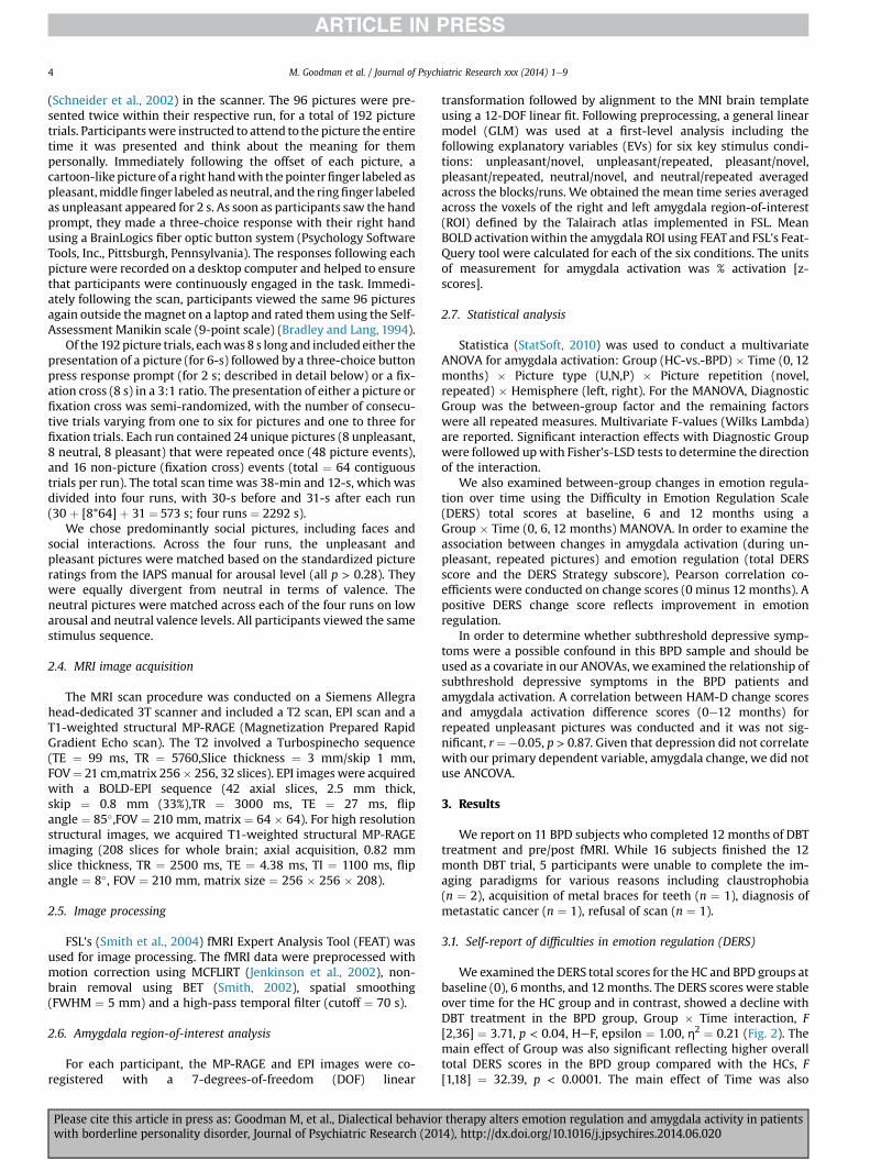
Twenty-twoage- and gender-matched unmedicated BPD andHCparticipants (11 in each group)were included in this study (Table 1).All eligible participants received a full diagnostic structured inter-view which included the Structured Clinical Interview for DSM-IVAxis I disorders (SCID-I) (First et al., 1996a) and the StructuredInterview for DSM-IV Personality Disorders (SIDP) (First et al.,1996b) conducted by a clinical psychologist specifically trained inthe assessment of Axis II disorders. Weekly consensus and diag-nostic meetings were led by a second clinical psychologist orresearch psychiatrist. In our laboratory, the intra-class correlationfor BPD diagnosis is 0.80. All patients met DSM-IV criteria for BPD.HCs had noAxis I or II disorder, or family history of anAxis I disorder.
Exclusion criteria for all participants included severe medical orneurological illness, head injury, or substance dependence or abuseduring the prior six months. All participants had a negative urine
Table 1Demographic/Clinical Descriptors for Borderline Personality Disorder (BPD) andHealthy Control (HC) groups.
Characteristic HC (n ¼ 11) BPD (n ¼ 11)
N % N %
SexFemale 9 81.8 9 81.8Male 2 18.2 2 18.2
Psychiatric comorbidityMood disorder (present) 0 0 0 0Mood disorder (past) 0 0 8 72.7PTSD (present) 0 0 1 9.1Other anxiety disorder (present) 0 0 6 54.5Substance use disorder (present) 0 0 0 0Substance use disorder (past) 0 0 2 18.2Psychosis 0 0 0 0
Baseline 12-MonthFollow-Up
Baseline 12-MonthFollow-Up
Mean SD Mean SD Mean SD Mean SD
Age 30.4 10.4 32.8 11.5Educationa 7.2 0.4 6.7 1.4DERSTotal 56.6 14.9 54.8 14.4 120.5** 24.2 101.5**D 25.7Nonacceptance 8.7 2.9 9.0 3.4 20.5** 6.5 17.2** 7.1Goals 9.2 4.4 8.7 3.9 20.9** 4.2 18.0**D 5.0Impulsivity 7.2 2.0 7.4 2.3 19.9** 5.1 15.1**D 5.2Awareness 13.2 4.7 12.0 4.8 17.7* 6.0 15.3 6.2Strategies 11.2 4.2 11.2 3.9 26.2** 6.3 22.5**D 6.2Clarity 7.1 1.1 6.5 1.4 15.4** 4.7 13.5**D 3.9
ZAN 0.6 1.3 0.7 0.7 18.1** 5.2 9.4**D 6.2HAM-D e e e e 13.8 5.6 12.0** 6.0PANASPositive Affect 38.3 5.1 39.7 5.0 23.1** 6.5 26.5** 8.7Negative Affect 14.2 4.1 14.8 3.6 30.0** 10.9 27.1**D 10.4
**p < 0.004, BPD > HC, t-test.*p < 0.08, BPD > HC, t-test.DERS ¼ Difficulties in Emotion Regulation Scale, ZAN ¼ Zanarini Rating Scale forBorderline Personality Disorder, HAM-D ¼ Hamilton Depression Rating Scale, andPANAS¼Positive and Negative Affective Scale.D denotes significant change (0e12), paired t-test, p < 0.05.Note: One HC did not have DERS, ZAN, or PANAS data, two HCs did not have edu-cation data.
a Education ¼ highest degree earned: 1 ¼ no high school diploma; 2 ¼ GED;3 ¼ high school diploma; 4 ¼ technical training; 5 ¼ some college, no degree;6 ¼ Associate's degree; 7 ¼ Bachelor's degree; 8 ¼Master's degree; 9 ¼MD/PhD/JD/PharmD.
Please cite this article in press as: Goodman M, et al., Dialectical behaviorwith borderline personality disorder, Journal of Psychiatric Research (201
toxicology screen for drugs of abuse during the study's screeningvisit and on each fMRI scan day, women also had a negative preg-nancy test on each scan day. Exclusion criteria for patients includedmeeting DSM-IV criteria for any schizophrenia-related psychoticdisorder, bipolar (Type I) disorder, or current MDD (no episode inthe prior 6 months). Patients were excluded if they had a history ofa suicide attempt, or inpatient psychiatric hospitalization withinthe past six months as required by the local IRB for safety concernsgiven that concurrent psychoactive medication was not allowed inthis study. Written informed consent approved by the InstitutionalReview Board was provided by all participants.
In addition to the DERS, symptoms were also assessed duringthe DBT trial using the Zanarini Rating Scale for Borderline Per-sonality Disorder (ZAN-BPD) (Frankenburg and Zanarini, 2002),Hamilton Depression Rating Scale (Hamilton, 1960), and Positiveand Negative Affective Scale (PANAS) (Watson et al., 1988).
2.2. DBT treatment
The BPD patients received standard 12-month DBT treatment,including weekly skills training group (90 min), weekly individualtreatment (50e60 min) and telephone coaching as needed. DBTtherapists participated in a weekly 60-min consultation meeting.DBT therapists were experienced Ph.D. or M.D. clinicians whoreceived intensive 10-day training. Individual DBT therapists wererated for adherence on taped sessions and judged by members ofthe Linehan research group. Adherence monitoring for researchparticipants who agreed to videotaping included review of theinitial two tapes for any therapist-patient dyad and random tapesranging from 6 to 8 week intervals for the remainder of the year-long treatment. Adherence ratings ranged from 3.7 to 4.2. Anyrating <4 was reviewed in weekly consultation meetings.
2.3. Event-related fMRI task measuring emotion processing
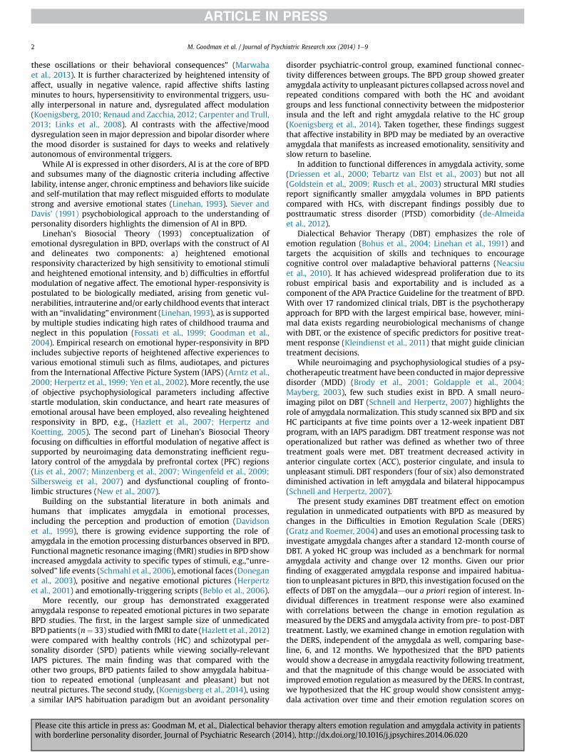
The fMRI task employed in this study was identical to the onepreviously published in a larger study of BPD patients (Hazlett et al.,2012). During the fMRI scan, participants viewed unpleasant,neutral, and pleasant photographic pictures from the IAPS (Lang andBradley, 2007; Fig. 1). A total of 96 intermixed unpleasant, neutral,and pleasant photographic images were presented using E-Primesoftware (Psychology Software Tools, Pittsburgh, Pennsylvania)
Fig. 1. A schematic of the event-related picture processing fMRI task paradigm isshown. Participants viewed an intermixed series of unpleasant, neutral, and pleasantpictures for 6-s each. Following each picture, they were prompted with a screen tomake a 3-choice button press to rate how the picture made them feel (unpleasant,neutral, or pleasant). Some trials had no picture presented during the 6-s period (seeMethods section for additional details).
therapy alters emotion regulation and amygdala activity in patients4), http://dx.doi.org/10.1016/j.jpsychires.2014.06.020
M. Goodman et al. / Journal of Psychiatric Research xxx (2014) 1e94
(Schneider et al., 2002) in the scanner. The 96 pictures were pre-sented twice within their respective run, for a total of 192 picturetrials. Participantswere instructed to attend to the picture the entiretime it was presented and think about the meaning for thempersonally. Immediately following the offset of each picture, acartoon-likepicture of a right handwith thepointerfinger labeled aspleasant,middlefinger labeled asneutral, and the ringfinger labeledas unpleasant appeared for 2 s. As soon as participants saw the handprompt, they made a three-choice response with their right handusing a BrainLogics fiber optic button system (Psychology SoftwareTools, Inc., Pittsburgh, Pennsylvania). The responses following eachpicture were recorded on a desktop computer and helped to ensurethat participants were continuously engaged in the task. Immedi-ately following the scan, participants viewed the same 96 picturesagain outside themagnet on a laptop and rated them using the Self-Assessment Manikin scale (9-point scale) (Bradley and Lang, 1994).
Of the 192picture trials, eachwas 8 s long and included either thepresentation of a picture (for 6-s) followed by a three-choice buttonpress response prompt (for 2 s; described in detail below) or a fix-ation cross (8 s) in a 3:1 ratio. The presentation of either a picture orfixation cross was semi-randomized, with the number of consecu-tive trials varying from one to six for pictures and one to three forfixation trials. Each run contained 24 unique pictures (8 unpleasant,8 neutral, 8 pleasant) that were repeated once (48 picture events),and 16 non-picture (fixation cross) events (total ¼ 64 contiguoustrials per run). The total scan time was 38-min and 12-s, which wasdivided into four runs, with 30-s before and 31-s after each run(30 þ [8*64] þ 31 ¼ 573 s; four runs ¼ 2292 s).
We chose predominantly social pictures, including faces andsocial interactions. Across the four runs, the unpleasant andpleasant pictures were matched based on the standardized pictureratings from the IAPS manual for arousal level (all p > 0.28). Theywere equally divergent from neutral in terms of valence. Theneutral pictures were matched across each of the four runs on lowarousal and neutral valence levels. All participants viewed the samestimulus sequence.
2.4. MRI image acquisition
The MRI scan procedure was conducted on a Siemens Allegrahead-dedicated 3T scanner and included a T2 scan, EPI scan and aT1-weighted structural MP-RAGE (Magnetization Prepared RapidGradient Echo scan). The T2 involved a Turbospinecho sequence(TE ¼ 99 ms, TR ¼ 5760,Slice thickness ¼ 3 mm/skip 1 mm,FOV¼ 21 cm,matrix 256� 256, 32 slices). EPI imageswere acquiredwith a BOLD-EPI sequence (42 axial slices, 2.5 mm thick,skip ¼ 0.8 mm (33%),TR ¼ 3000 ms, TE ¼ 27 ms, flipangle ¼ 85�,FOV ¼ 210 mm, matrix ¼ 64 � 64). For high resolutionstructural images, we acquired T1-weighted structural MP-RAGEimaging (208 slices for whole brain; axial acquisition, 0.82 mmslice thickness, TR ¼ 2500 ms, TE ¼ 4.38 ms, TI ¼ 1100 ms, flipangle ¼ 8�, FOV ¼ 210 mm, matrix size ¼ 256 � 256 � 208).
2.5. Image processing
FSL's (Smith et al., 2004) fMRI Expert Analysis Tool (FEAT) wasused for image processing. The fMRI data were preprocessed withmotion correction using MCFLIRT (Jenkinson et al., 2002), non-brain removal using BET (Smith, 2002), spatial smoothing(FWHM ¼ 5 mm) and a high-pass temporal filter (cutoff ¼ 70 s).
2.6. Amygdala region-of-interest analysis
For each participant, the MP-RAGE and EPI images were co-registered with a 7-degrees-of-freedom (DOF) linear
Please cite this article in press as: Goodman M, et al., Dialectical behaviowith borderline personality disorder, Journal of Psychiatric Research (201
transformation followed by alignment to the MNI brain templateusing a 12-DOF linear fit. Following preprocessing, a general linearmodel (GLM) was used at a first-level analysis including thefollowing explanatory variables (EVs) for six key stimulus condi-tions: unpleasant/novel, unpleasant/repeated, pleasant/novel,pleasant/repeated, neutral/novel, and neutral/repeated averagedacross the blocks/runs. We obtained the mean time series averagedacross the voxels of the right and left amygdala region-of-interest(ROI) defined by the Talairach atlas implemented in FSL. MeanBOLD activationwithin the amygdala ROI using FEATand FSL's Feat-Query tool were calculated for each of the six conditions. The unitsof measurement for amygdala activation was % activation [z-scores].
2.7. Statistical analysis
Statistica (StatSoft, 2010) was used to conduct a multivariateANOVA for amygdala activation: Group (HC-vs.-BPD) � Time (0, 12months) � Picture type (U,N,P) � Picture repetition (novel,repeated) � Hemisphere (left, right). For the MANOVA, DiagnosticGroup was the between-group factor and the remaining factorswere all repeated measures. Multivariate F-values (Wilks Lambda)are reported. Significant interaction effects with Diagnostic Groupwere followed upwith Fisher's-LSD tests to determine the directionof the interaction.
We also examined between-group changes in emotion regula-tion over time using the Difficulty in Emotion Regulation Scale(DERS) total scores at baseline, 6 and 12 months using aGroup � Time (0, 6, 12 months) MANOVA. In order to examine theassociation between changes in amygdala activation (during un-pleasant, repeated pictures) and emotion regulation (total DERSscore and the DERS Strategy subscore), Pearson correlation co-efficients were conducted on change scores (0 minus 12 months). Apositive DERS change score reflects improvement in emotionregulation.
In order to determine whether subthreshold depressive symp-toms were a possible confound in this BPD sample and should beused as a covariate in our ANOVAs, we examined the relationship ofsubthreshold depressive symptoms in the BPD patients andamygdala activation. A correlation between HAM-D change scoresand amygdala activation difference scores (0e12 months) forrepeated unpleasant pictures was conducted and it was not sig-nificant, r¼�0.05, p > 0.87. Given that depression did not correlatewith our primary dependent variable, amygdala change, we did notuse ANCOVA.
3. Results
We report on 11 BPD subjects who completed 12 months of DBTtreatment and pre/post fMRI. While 16 subjects finished the 12month DBT trial, 5 participants were unable to complete the im-aging paradigms for various reasons including claustrophobia(n ¼ 2), acquisition of metal braces for teeth (n ¼ 1), diagnosis ofmetastatic cancer (n ¼ 1), refusal of scan (n ¼ 1).
3.1. Self-report of difficulties in emotion regulation (DERS)
We examined the DERS total scores for the HC and BPD groups atbaseline (0), 6 months, and 12months. The DERS scores were stableover time for the HC group and in contrast, showed a decline withDBT treatment in the BPD group, Group � Time interaction, F[2,36] ¼ 3.71, p < 0.04, HeF, epsilon ¼ 1.00, ƞ2 ¼ 0.21 (Fig. 2). Themain effect of Group was also significant reflecting higher overalltotal DERS scores in the BPD group compared with the HCs, F[1,18] ¼ 32.39, p < 0.0001. The main effect of Time was also
r therapy alters emotion regulation and amygdala activity in patients4), http://dx.doi.org/10.1016/j.jpsychires.2014.06.020

Fig. 2. DERS total scores for the HC and BPD groups at baseline (0), 6 months, and 12months. DERS scores showed a decline with DBT treatment in the BPD group, but werestable over time for the HC group, Group � Time interaction, F[2,36] ¼ 3.71, p < 0.04,HeF, epsilon ¼ 1.00.
M. Goodman et al. / Journal of Psychiatric Research xxx (2014) 1e9 5
significant, primarily reflecting the overall decline across time inDERS scores for patients, F[2,36] ¼ 7.09, p < 0.003.
3.2. fMRI
The HC group showed overall amygdala activation (averagedacross picture type, hemisphere, and novel/repeated pictures) thatwas similar at baseline and 12 months, whereas the BPD groupexhibited an overall decrease in amygdala activation post-treatment, Group � Time interaction, F[1,20] ¼ 4.89, p < 0.04;ƞ2 ¼ 0.24, HC > BPD post-hoc, p < 0.08, trend level, Fig. 3). In orderto confirm that BPD patients showed a change (i.e. decrease) inamygdala activity with DBT while the HC group showed no changeover 12 months, we followed-up this significant interaction effectwith post-hoc t-tests examining the amygdala change scores (0e12months) for each group (compared to 0) and between-group dif-ferences. The resulted indicated that the HC group did not show a
Fig. 3. Compared with the healthy control (HC) group (which was yoked and did notreceive treatment), the BPD group showed a pattern of higher amygdala activity atbaseline (pre-treatment) that decreased following a standard 12-month DBT inter-vention. The HC group was scanned to provide a benchmark for normal amygdalaactivity at baseline and a 12-month interval. *p ¼ 0.08, Fisher's LSD post-hoc, trend-level.
Please cite this article in press as: Goodman M, et al., Dialectical behaviorwith borderline personality disorder, Journal of Psychiatric Research (201
significant change in amygdala activity over time, whereas the BPDgroup did (0e12 month difference scores: HC: 1.99 ± 16.67 (vs. 0,p ¼ 0.70) vs. BPD:-11.54 ± 11.73 (vs. 0, t(10) ¼ 3.26, p < 0.01);(Cohen's d ¼ 0.95), and this between-group difference was signif-icant, t(20) ¼ 2.20, p < 0.04.
There was also a significant for Group � Time � Picturetype � Picture repetition � Hemisphere interaction, F[2,19] ¼ 3.71p < 0.05, ƞ2 ¼ 0.16, Fig. 4. Compared with healthy controls, in-dividuals with BPD showed a pattern of greater decrease from pre-to post-treatment in amygdala activity for all three pictures types,but particularly in the left hemisphere and during the repeatedemotional picture conditions (unpleasant and pleasant). None ofthe other interactions with Group reached statistical significance.
3.3. Self-report ratings of picture valence
Neither the Group � Time � Picture Type interaction, nor anyinteraction with Diagnostic group was significant for the SAM self-report ratings of picture valence. Both groups showed the standard,linear, stepwise self-report rating pattern of pleasant pictures beingthe most unpleasant, pleasant being the least unpleasant, andneutral being intermediate. The valence ratings for the HC groupdid not change over time (i.e. 0-vs.-12 months). However, it isnoteworthy that compared with the HC group, the BPD groupshowed a trend for rating the unpleasant pictures as less unpleas-ant following DBT (p < 0.06). We followed this finding up with apaired t-test for the BPD group comparing ratings for unpleasantpictures pre- and post-DBT which indicated that the patients ratedthe unpleasant pictures as less unpleasant post-DBT (pre-DBT:7.99 ± 0.52 post-DBT: 7.42 ± 0.62, t(9) ¼ 2.90, p < 0.02). Comparedwith HCs, there was a trend for the BPD group to rate the pleasantpictures as less pleasant at both the baseline and post-DBT timepoints (HC-vs.-BPD, p < 0.07).
3.4. Amygdala and clinical change with DBT
Among the BPD patients, reduction in amygdala activity torepeated unpleasant pictures following DBT was associated withimproved emotion regulation as measured by the change in totalDERS score and the DERS Strategy subscale score (r¼ 0.70, p < 0.02,r ¼ 0.69, p < 0.02, respectively; Fig. 5).
4. Discussion
This is the first study to examine pre-post changes in amygdalaactivity with standard 1-year DBT treatment for BPD. The mainfindings are: a) BPD patients showed a reduction in overall amyg-dala activation following 12 months of DBT treatment (ƞ2 ¼ 0.24);b) this reduced amygdala activation in BPD patients post treatmentwas present in all three pictures types, but particularly notable inthe left hemisphere and during the repeated emotional pictureconditions (ƞ2 ¼ 0.16); c) among the BPD group, improvement inemotion regulation and strategy as measured by the DERS wasassociated with decreased amygdala activity to repeated unpleas-ant pictures (r ¼ 0.70, r ¼ 0.69, respectively).
Strengths of our study include the use of: HC participants tocapture longitudinal scanning effects of our paradigm, unmedi-cated, rigorously diagnosed patients with BPD without currentMDD or bipolar disorder, a validated fMRI task of emotional pro-cessing, and DBT adherence ratings during delivery of the 1-yeartreatment.
Our findings are consistent with the only previous fMRI study ofDBT (Schnell and Herpertz, 2007), which also found normalizationof amygdala hyper-responsivity with successful DBT treatment in a3-month pilot study. Our results build on these findings with a HC
therapy alters emotion regulation and amygdala activity in patients4), http://dx.doi.org/10.1016/j.jpsychires.2014.06.020
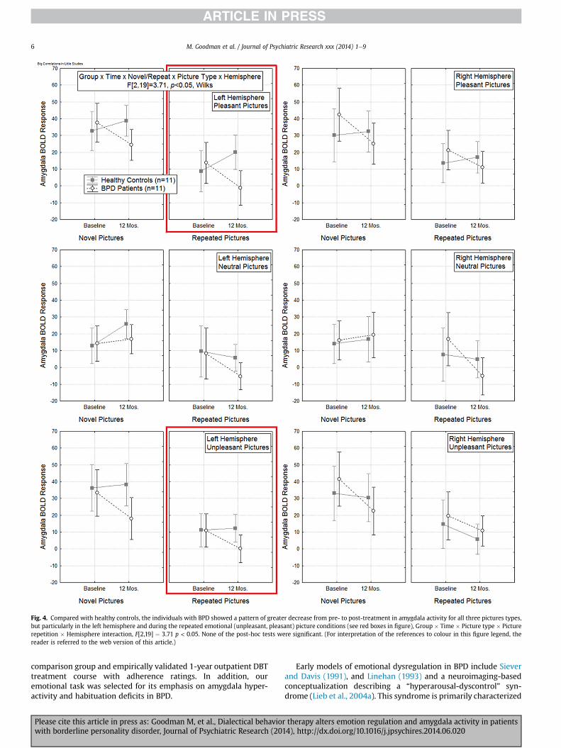
Fig. 4. Compared with healthy controls, the individuals with BPD showed a pattern of greater decrease from pre- to post-treatment in amygdala activity for all three pictures types,but particularly in the left hemisphere and during the repeated emotional (unpleasant, pleasant) picture conditions (see red boxes in figure), Group � Time � Picture type � Picturerepetition � Hemisphere interaction, F[2,19] ¼ 3.71 p < 0.05. None of the post-hoc tests were significant. (For interpretation of the references to colour in this figure legend, thereader is referred to the web version of this article.)
M. Goodman et al. / Journal of Psychiatric Research xxx (2014) 1e96
comparison group and empirically validated 1-year outpatient DBTtreatment course with adherence ratings. In addition, ouremotional task was selected for its emphasis on amygdala hyper-activity and habituation deficits in BPD.
Please cite this article in press as: Goodman M, et al., Dialectical behaviowith borderline personality disorder, Journal of Psychiatric Research (201
Early models of emotional dysregulation in BPD include Sieverand Davis (1991), and Linehan (1993) and a neuroimaging-basedconceptualization describing a “hyperarousal-dyscontrol” syn-drome (Lieb et al., 2004a). This syndrome is primarily characterized
r therapy alters emotion regulation and amygdala activity in patients4), http://dx.doi.org/10.1016/j.jpsychires.2014.06.020

Fig. 5. Scatterplots and Pearson correlation coefficients for the BPD group show therelationship between change (pre-treatment minus post-treatment, i.e. 0e12 months)in amygdala activation to repeated unpleasant pictures and emotional regulation(measured by change in the DERS total score (Top) and the DERS strategy subscale(Bottom). Among the patient group, greater reduction in amygdala activity torepeated-unpleasant pictures (i.e. better habituation) following DBT was associatedwith greater clinical improvement in emotional regulation and use of emotion regu-lation strategies.
M. Goodman et al. / Journal of Psychiatric Research xxx (2014) 1e9 7
by dysfunction in anterior regulatory regions including ACC andPFC, coupled with limbic structure hyper-reactivity, notably inamygdala and insula and supported by the findings of Silbersweiget al. (2007) and echoed in other populations of emotionally-dysregulated individuals including adolescents (Hare et al., 2008).A recentmeta-analysis of neural correlates of negative emotionalityin BPD (Ruocco et al., 2013) concluded that there is increasedactivation in the posterior cingulate and insula but diminishedactivation in a network extending from the amygdala to prefrontalregulatory regions including dorsolateral and subgenual PFC. Ourresults conflict with these findings but could be attributed to dif-ferences in study design, use of ROI methodology, patient selectionincluding Axis I co-morbidity, hospitalization and/or medicationstatus. For example, the studies reporting amygdala hyperactivity(e.g., Koenigsberg et al., 2009; Minzenberg et al., 2007; Schulzeet al., 2011) involved unmedicated BPD patients, while the studiesdemonstrating diminished amygdala responsivity included partic-ipants currently taking psychotropic medications (Smoski et al.,2011). Similarly, Axis I co-morbidities such as PTSD may influenceamygdala reactivity, particularly in relation to pain perception
Please cite this article in press as: Goodman M, et al., Dialectical behaviorwith borderline personality disorder, Journal of Psychiatric Research (201
(Kraus et al., 2009). Cullen et al. (2011) reported increased amyg-dala connectivity during fear states in 12 females with BPD, sug-gesting increased use of both overt and automatic fear processing;however, the neutral state revealed lower connectivity betweenboth bilateral amygdala and mid-cingulate regions. This inconsis-tency may stem, too, from differences in the type of stimuliemployed and their personal relevance to the individual. Givenresearch (e.g., Hazlett et al., 2007; Limberg et al., 2011) showing theimportance of using BPD-salient stimuli (e.g., with abandonment,rejection themes) to elicit HC-BPD differences in affective startlemodulationda defensive response linked to amygdala activation,we chose IAPS pictures that had an interpersonal-social focuswhich may be of particular importance for delineating HC-BPDdifferences.
The finding of 12-month DBT treatment normalizing amygdalahyperactivity overlaps with findings of other psychotherapies foraffective disorders. Treatment response to long-term psychody-namic psychotherapy has been found to correlate with decreases inanterior hippocampus/amygdala activity along with subgenual andmedial PFC in MDD (Buchheim et al., 2012). Cognitive behavioraltherapy treatment response is predicted by reduced medial pre-frontal activity and increased amygdala activation (Siegle et al.,2006). In MDD, pharmacologic interventions also target normali-zation of amygdala function (Sheline et al., 2001). While all ourparticipants were free of current MDD, the role of amygdala hy-peractivity in several affective disorders raises questions as to itsspecificity to BPD and as to the uniqueness of DBT vs. other forms ofpsychotherapy.
4.1. Study limitations
Limitations of the study include the pilot nature of our study,low power and a small sample size. While this limitation rendersour findings preliminary, significant between-group differencesemerged, suggesting that further research and replication in thisarea is warranted. Given our small sample, we focused on theamygdala with an a priori hypothesis based upon our prior workshowing habituation abnormalities in a larger sample (n ¼ 33) ofunmedicated BPD patients compared with HCs, using an identicalfMRI task and amygdala-centric focus (Hazlett et al., 2012).
The small size precludes differentiation of the sample based ontreatment responders and nonresponders. Future work with alarger sample size will benefit from examining neurobiologicalparameters of treatment response, as has been argued by others(Schnell and Herpertz, 2007). In addition, our small sample sizelikely enhanced the magnitude of the correlation between thechange in amygdala activity with treatment and DERS strategy andtotal scores (see Fig. 4). This limitation was discussed by Yarkoni(2009) and Vul et al. (2009) and highlights the potential for anexaggerated magnitude of correlations found in fMRI studies ofemotion, personality and social cognition.
Additionally, our emotional task was a “passive viewing task”which means that we did not examine “active” emotion regulation,per se which might be considered a study limitation by some.However, given our prior work indicating that BPD patients evincea mismatch between their psychophysiological and self-reportmeasures of emotion/valence (Hazlett et al., 2007, 2012), it couldbe argued that asking BPD participants whether they successfullyregulated their emotions (i.e. using self-report) during a task in-volves a participant bias or demand characteristic which is aconfound. Nevertheless, additional research is needed given recentresearch showing skill acquisition is critical to DBT treatment effi-cacy. This line of work will help us better understand the neuro-biological changes that accompany amygdala quieting withsuccessful psychotherapy.
therapy alters emotion regulation and amygdala activity in patients4), http://dx.doi.org/10.1016/j.jpsychires.2014.06.020
M. Goodman et al. / Journal of Psychiatric Research xxx (2014) 1e98
It may also be argued that the community sample of subjects inthis study were not fully representative of the larger pool of pa-tients with BPD, limiting the generalizability of the findings. Theywere required to remain off all psychoactive medications duringthe duration of the 12-month DBT trial and pre/post fMRI imaging.Perhaps, this selected for a more cooperative, less symptomaticcohort. Lastly, without a comparison treatment condition, wecannot rule out the possibility that the amygdala changes observedin BPD were not due to other life events common to patients withBPD, but not controls.
4.2. Future directions
Our investigation of the amygdala before and after 12-months ofDBT treatment in BPD highlights the role of DBT treatment inquieting amygdala activity and the importance of enhancingemotional regulation strategies. Since our subjects were not treatedwith any psychiatric medication, these amygdala effects result fromthe psychotherapy intervention and suggest that patients arelearning an adaptive process that counters emotionally-relevantactivation of the amygdala. Future studies will benefit fromparsing out which emotion regulation skills and strategies arenecessary for a particular patient and how they combine for ther-apeutic and neurobiological effect.
Additional work with a larger BPD sample allowing for whole-brain analyses involving functional connectivity of the amygdalawith other regions including prefrontal and anterior cingulatecortex is necessary to clarify how DBT-induced amygdala changesinteract with other brain regions implicated in emotion regulation(Ochsner et al., 2002; Etkin et al., 2011). Future research similar toour study, examining the effects of evidence-based psychotherapyon underlying psychopathological mechanisms is critical toadvance our understanding of the neuropathology of BPD, emotionregulation processes, and development of new treatment targets.
Role of the funding source
This research was supported by a Veterans' AdministrationAdvanced Career Development Award (3277-06-109) to MG, NIMHGrant R01-MH073911 to EAH, Grant UL1TR000067 from the Na-tional Center for Advancing Translational Sciences, a component ofthe National Institutes of Health, and additional resources from theMount Sinai GCRC, VISN 3 Mental Illness Research, Education andClinical Center (MIRECC), James J. Peters Research Foundation.
Contributors
Marianne Goodman e Dr. Goodman was the study PI andresponsible for all aspects of the study.
David Carpenter e Dr. Carpenter was responsible for imageanalysis under Dr. Hazlett's supervision.
Cheuk Y. Tang e Dr. Tang was responsible for all aspects of fMRIacquisition.
Kim E. Goldstein e Dr. Goldstein assisted with the coordinationand collection of healthy control and patient data.
Jennifer Avedon e Ms. Avedon was responsible for studyimplementation, recruitment of the healthy controls, and patientdata collection.
Nicolas Fernandez e Mr. Fernandez was tasked with datacollection and assisted in data analysis.
Kathryn A. Mascitelli e Ms. Mascitelli helped with data coor-dination and organization of the database for this study.
Nicholas J. Blair e Mr. Blair helped with the manuscript prep-aration including references and formatting of the figures and table.
Please cite this article in press as: Goodman M, et al., Dialectical behaviowith borderline personality disorder, Journal of Psychiatric Research (201
Antonia S. New e Dr. New was a study design mentor for theCareer Development award that partially funded this study.
Joseph TriebwassereDr. Triebwasser assistedwith patient datacollection.
Larry J. Siever e Dr. Siever assisted with the study design, andwas the primary mentor for the Career Development award thatpartially funded this study.
Erin A. Hazlett e Dr. Hazlett was responsible for overseeing allaspects of the fMRI component of the study, conducting the sta-tistical analyses, and write-up of the Results section. She was amentor on Dr. Goodman's Career Development award that partiallyfunded this study.
Conflict of interest
None of the authors have any biomedical financial interests orpotential conflicts of interest to declare.
Acknowledgment
We acknowledge the DBT treatment provided by Drs. LaurenHelm, Gail Maurer and Lucia Vail, cooperation of the mental healthclinical services of the James J. Peters VAMC.
References
Arntz A, Appels C, Sieswerda S. Hypervigilance in borderline disorder: a test withthe emotional Stroop paradigm. J Personal Disord 2000;14:366e73.
Beblo T, Driessen M, Mertens M, Wingenfeld K, Piefke M, Rullkoetter N, et al.Functional MRI correlates of the recall of unresolved life events in borderlinepersonality disorder. Psychol Med 2006;36:845e56.
Bender DS, Dolan RT, Skodol AE, Sanislow CA, Dyck IR, McGlashan TH, et al.Treatment utilization by patients with personality disorders. Am J Psychiatry2001;158:295e302.
Bohus M, Haaf B, Simms T, Limberger MF, Schmahl C, Unckel C, et al. Effectiveness ofinpatient dialectical behavioral therapy for borderline personality disorder: acontrolled trial. Behav Res Ther 2004;42:487e99.
Bradley MM, Lang PJ. Measuring emotion: the self-assessment manikin and thesemantic differential. J Behav Ther Exp Psychiatry 1994;25:49e59.
Brody AL, Barsom MW, Bota RG, Saxena S. Prefrontal-subcortical and limbic circuitmediation of major depressive disorder. Semin Clin Neuropsychiatry 2001;6:102e12.
Buchheim A, Viviani R, Kessler H, Kachele H, Cierpka M, Roth G, et al. Changes inprefrontal-limbic function in major depression after 15 months of long-termpsychotherapy. PLoS ONE 2012;7:e33745.
Carpenter RW, Trull TJ. Components of emotion dysregulation in borderline per-sonality disorder: a review. Curr Psychiatry Rep 2013;15:335.
Cullen KR, Vizueta N, Thomas KM, Han GJ, Lim KO, Camchong J, et al. Amygdalafunctional connectivity in young women with borderline personality disorder.Brain Connect 2011;1:61e71.
Davidson RJ, Abercrombie H, Nitschke JB, Putnam K. Regional brain function,emotion and disorders of emotion. Curr Opin Neurobiol 1999;9:228e34.
de-Almeida CP, Wenzel A, De-Carvalho CS, Powell VB, Araujo-Neto C, Quarantini LC,et al. Amygdalar volume in borderline personality disorder with and withoutcomorbid post-traumatic stress disorder: a meta-analysis. CNS Spectr 2012;17:70e5.
Donegan NH, Sanislow CA, Blumberg HP, Fulbright RK, Lacadie C, Skudlarski P, et al.Amygdala hyperreactivity in borderline personality disorder: implications foremotional dysregulation. Biol Psychiatry 2003;54:1284e93.
Driessen M, Herrmann J, Stahl K, Zwaan M, Meier S, Hill A, et al. Magnetic resonanceimaging volumes of the hippocampus and the amygdala in women withborderline personality disorder and early traumatization. Arch Gen Psychiatry2000;57:1115e22.
Etkin A, Egner T, Kalisch R. Emotional processing in anterior cingulate and medialprefrontal cortex. Trends Cogn Sci 2011;15:85e93.
First M, Gibbon M, Spitzer R. Structured clinical interview for DSM-IV Axis I Dis-orders (SCID-I). Washington, D.C.: American Psychiatric Publishing; 1996a.
First M, Gibbon M, Spitzer R. Structured clinical interview for DSM-IV PersonalityDisorders (SCIP-II). Washington, D.C.: American Psychiatric Publishing; 1996b.
Fossati A, Madeddu F, Maffei C. Borderline personality disorder and childhoodsexual abuse: a meta-analytic study. J Personal Disord 1999;13:268e80.
Frankenburg FR, Zanarini MC. Divalproex sodium treatment of women withborderline personality disorder and bipolar II disorder: a double-blind placebo-controlled pilot study. J Clin Psychiatry 2002;63:442e6.
Goldapple K, Segal Z, Garson C, Lau M, Bieling P, Kennedy S, et al. Modu-lation of cortical-limbic pathways in major depression: treatment-specific
r therapy alters emotion regulation and amygdala activity in patients4), http://dx.doi.org/10.1016/j.jpsychires.2014.06.020

M. Goodman et al. / Journal of Psychiatric Research xxx (2014) 1e9 9
effects of cognitive behavior therapy. Arch Gen Psychiatry 2004;61:34e41.
Goldstein KE, Hazlett EA, New AS, Haznedar MM, Newmark RE, Zelmanova Y, et al.Smaller superior temporal gyrus volume specificity in schizotypal personalitydisorder. Schizophr Res 2009;112:14e23.
Goodman M, New A, Siever L. Trauma, genes, and the neurobiology of personalitydisorders. Ann N Y Acad Sci 2004;1032:104e16.
Gratz KL, Roemer L. Multidimensional assessment of emotion regulation and dys-regulation: development, factor structure, and initial validation of the diffi-culties in emotion regulation scale. J Psychopathol Behav Assess 2004;26:41e54.
Hamilton M. A rating scale for depression. J Neurol Neurosurg Psychiatr 1960;23:56e62.
Hare T, Tottenham N, Galvan A, Voss H, Glover G, Casey BJ. Biological substrates ofemotional reactivity and regulation in adolescence during an emotional go-nogo task. Biol Psychiatry 2008;63:927e34.
Hazlett EA, Speiser LJ, Goodman M, Roy M, Carrizal M, Wynn JK, et al. Exaggeratedaffect-modulated startle during unpleasant stimuli in borderline personalitydisorder. Biol Psychiatry 2007;62:250e5.
Hazlett EA, Zhang J, New AS, Zelmanova Y, Goldstein KE, Haznedar MM, et al.Potentiated amygdala response to repeated emotional pictures in borderlinepersonality disorder. Biol Psychiatry 2012;72:448e56.
Herpertz SC, Kunert HJ, Schwenger UB, Sass H. Affective responsiveness inborderline personality disorder: a psychophysiological approach. Am J Psychi-atry 1999;156:1550e6.
Herpertz SC, Dietrich TM, Wenning B, Krings T, Erberich SG, Willmes K, et al. Evi-dence of abnormal amygdala functioning in borderline personality disorder: afunctional MRI study. Biol Psychiatry 2001;50:292e8.
Herpertz SC, Koetting K. Startle response in inpatients with borderline personalitydisorder vs. healthy controls. J Neural Transm 2005;112:1097e106.
Jenkinson M, Bannister P, Brady M, Smith S. Improved optimization for the robustand accurate linear registration and motion correction of brain images. Neu-roimage 2002;17:825e41.
Kleindienst N, Limberger MF, Ebner-Priemer UW, Keibel-Mauchnik J, Dyer A,Berger M, et al. Dissociation predicts poor response to dialectical behavioraltherapy in femal patients with borderline personality disoder. J Personal Disord2011;25:432e47.
Koenigsberg HW, Siever LJ, Lee H, Pizzarello S, New AS, Goodman M, et al. Neuralcorrelates of emotion processing in borderline personality disorder. PsychiatryRes 2009;172:192e9.
Koenigsberg HW. Affective instability: toward an integration of neuroscience andpsychological perspectives. J Personal Disord 2010;24:60e82.
Koenigsberg HW, Denny BT, Fan J, Liu X, Guerreri S, Mayson SJ, et al. The neuralcorrelates of anomalous habituation to negative emotional pictures in border-line and avoidant personality disorder patients. Am J Psychiatry 2014;171:82e90.
Kraus A, Esposito F, Seifritz E, Di Salle F, Ruf M, Valerius G, et al. Amygdala deac-tivation as a neural correlate of pain processing in patients with borderlinepersonality disorder and co-occurrent posttraumatic stress disorder. Biol Psy-chiatry 2009;65:819e22.
Lang P, Bradley MM. The International Affective Picture System (IAPS) in the studyof emotion and attention. Handbook of emotion elicitation and assessment;2007. p. 29.
Lieb K, Rexhausen JE, Kahl KG, Schweiger U, Philipsen A, Hellhammer DH, et al.Increased diurnal salivary cortisol in women with borderline personality dis-order. J Psychiatr Res 2004a;38:559e65.
Lieb K, Zanarini MC, Schmahl C, Linehan MM, Bohus M. Borderline personalitydisorder. Lancet 2004b;364:453e61.
Limberg A, Barnow S, Freyberger HJ, Hamm AO. Emotional vulnerability inborderline personality disorder is cue specific and modulated by traumatiza-tion. Biol Psychiatry 2011;69:574e82.
Linehan MM, Armstrong HE, Suarez A, Allmon D, Heard HL. Cognitive-behavioraltreatment of chronically parasuicidal borderline patients. Arch Gen Psychiatry1991;48:1060e4.
Linehan MM. Cognitive behavioral treatment of borderline personality disorder.New York: The Guilford Press; 1993.
Links PS, Heslegrave R, van Reekum R. Prospective follow-up study of borderlinepersonality disorder: prognosis, prediction of outcome, and Axis II comorbidity.Can J Psychiatry 1998;43:265e70.
Links PS, Eynan R, Heisel MJ, Nisenbaum R. Elements of affective instability asso-ciated with suicidal behaviour in patients with borderline personality disorder.Can J Psychiatry 2008;53:112e6.
Lis E, Greenfield B, Henry M, Guile JM, Dougherty G. Neuroimaging and ge-netics of borderline personality disorder: a review. J Psychiatry Neurosci2007;32:162e73.
Marwaha S, He Z, Broome M, Singh SP, Scott J, Eyden J, et al. How is affectiveinstability defined and measured? A systematic review. Psychol Med 2013:1e16.
Please cite this article in press as: Goodman M, et al., Dialectical behaviorwith borderline personality disorder, Journal of Psychiatric Research (201
Mayberg HS. Modulating dysfunctional limbic-cortical circuits in depression: to-wards development of brain-based algorithms for diagnosis and optimisedtreatment. Br Med Bull 2003;65:193e207.
Minzenberg MJ, Fan J, New AS, Tang CY, Siever LJ. Fronto-limbic dysfunction inresponse to facial emotion in borderline personality disorder: an event-relatedfMRI study. Psychiatry Res 2007;155:231e43.
New AS, Hazlett EA, Buchsbaum MS, Goodman M, Mitelman SA, Newmark R, et al.Amygdala-prefrontal disconnection in borderline personality disorder. Neuro-psychopharmacology 2007;32:1629e40.
Neacsiu AD, Rizvi SL, Linehan MM. Dialectical behavior therapy skills use as amediator and outcome of treatment for borderline personality disorder. BehavRes Ther 2010; Sep;48(9):832e9. Epub 2010 May 23.
Ochsner KN, Bunge SA, Gross JJ, Gabrieli JD. Rethinking Feelings: an fMRI study ofcognitive regulation of emotion. J Cogn Neurosci 2002;14:1215e29.
Renaud SM, Zacchia C. Toward a definition of affective instability. Harv Rev Psy-chiatry 2012;20:298e308.
Ruocco AC, Amirthavasagam S, Choi-Kain LW, McMain SF. Neural correlates ofnegative emotionality in borderline personality disorder: an activation-likelihood-estimation meta-analysis. Biol Psychiatry 2013;73:153e60.
Rusch N, van Elst LT, Ludaescher P, Wilke M, Huppertz HJ, Thiel T, et al. A voxel-based morphometric MRI study in female patients with borderline personalitydisorder. Neuroimage 2003;20:385e92.
Schmahl C, Bohus M, Esposito F, Treede RD, Di Salle F, Greffrath W, et al. Neuralcorrelates of antinociception in borderline personality disorder. Arch GenPsychiatry 2006;63:659e67.
Schneider W, Eschmann A, Zuccolotto A. E-Prime user's guide. Pittsburgh, PA:Psychology Software Tools, Inc.; 2002.
Schnell K, Herpertz SC. Effects of dialectic-behavioral-therapy on the neural cor-relates of affective hyperarousal in borderline personality disorder. J PsychiatrRes 2007;41:837e47.
Schulze L, Domes G, Kruger A, Berger C, Fleischer M, Prehn K, et al. Neuronal cor-relates of cognitive reappraisal in borderline patients with affective instability.Biol Psychiatry 2011;69:564e73.
Sheline YI, Barch DM, Donnelly JM, Ollinger JM, Snyder AZ, Mintun MA.Increased amygdala response to masked emotional faces in depressed sub-jects resolves with antidepressant treatment: an fMRI study. Biol Psychiatry2001;50:651e8.
Siegle GJ, Carter CS, Thase ME. Use of FMRI to predict recovery from unipolardepression with cognitive behavior therapy. Am J Psychiatry 2006;163:735e8.
Siever LJ, Davis KL. A psychobiological perspective on the personality disorders. AmJ Psychiatry 1991;148:1647e58.
Silbersweig D, Clarkin J, Goldstein M, Kernberg O, Tuescher O, Levy K, et al. Failureof frontolimbic inhibitory function in the context of negative emotion inborderline personality disorder. Am J Psychiatry 2007;164:1832e41.
Skodol AE, Siever LJ, Livesley WJ, Gunderson JG, Pfohl B, Widiger TA. The borderlinediagnosis II: biology, genetics, and clinical course. Biol Psychiatry 2002;51:951e63.
Smith SM. Fast robust automated brain extraction. Hum Brain Mapp 2002;17:143e55.
Smith SM, Jenkinson M, Woolrich MW, Beckmann CF, Behrens TE, Johansen-Berg H,et al. Advances in functional and structural MR image analysis and imple-mentation as FSL. Neuroimage 2004;23:S208e19.
Smoski MJ, Salsman N, Wang L, Smith V, Lynch TR, Dager SR, et al. Functional im-aging of emotion reactivity in opiate-dependent borderline personality disor-der. Personal Disord 2011;2:230e41.
StatSoft Inc. Statistica, Version 9.1; 2010. Tulsa, OK, www.statsoft.com.Tebartz van Elst L, Hesslinger B, Thiel T, Geiger E, Haegele K, Lemieux L, et al.
Frontolimbic brain abnormalities in patients with borderline personality dis-order: a volumetric magnetic resonance imaging study. Biol Psychiatry2003;54:163e71.
Vul E, Harris C, Winkielman P, Pashler H. Puzzingly high correlations in fMRI studiesof emotion, personality, and social cognition. Perspect Psychol Sci 2009;4:274e90.
Watson D, Clark LA, Tellegen A. Development and validation of brief measures ofpositive and negative affect: the PANAS scales. J Pers Soc Psychol 1988;54:1063e70.
Wingenfeld K, Rullkoetter N, Mensebach C, Beblo T, Mertens M, Kreisel S, et al.Neural correlates of the individual emotional Stroop in borderline personalitydisorder. Psychoneuroendocrinology 2009;34:571e86.
Yarkoni T. Big Correlations in little studies: Inflated fMRI Correlations Reflect LowStatistical Power e Commentary on Vul et al. (2009). Perspect Psychol Sci2009;4:294e8.
Yen S, Zlotnick C, Costello E. Affect regulation in womenwith borderline personalitydisorder traits. J Nerv Ment Dis 2002;190:693e6.
therapy alters emotion regulation and amygdala activity in patients4), http://dx.doi.org/10.1016/j.jpsychires.2014.06.020