Diagnostic Problems Severe Neonatal Jaundice G6PD Deficiency · value of G6PD activity in normal...
6
Arch. Dis. Childh., 1968, 43, 36. Diagnostic Problems in Severe Neonatal Jaundice and G6PD Deficiency in Greece* L. ZANNOS-MARIOLEA, TH. THOMAIDIS, G. GEORGIZASt., E. GAVRIELIDOU, and S. BENETOS From the Blood Division of the Choremion Research Laboratory, Department of Pediatrics, University of Athens Since the first observation in Greece by Doxiadis, Fessas, and Valaes in 1960, it is now well established that a considerable proportion of full-term Greek newborns with severe jaundice (and/or anaemia) have G6PD deficiency. Similar cases have been described in other parts of the world (Freier, Mayer, Levene, and Abrahamov, 1965). G6PD deficiency and increased haemolysis are obviously cause and effect when newborns have been exposed to drugs and chemicals known to cause haemolysis in G6PD deficiency in general. However, about 50% of deficient newborns who develop severe jaundice with or without obvious haemolysis have not been exposed to one of these drugs. This particular group of babies has aroused considerable interest, research, and speculation; nevertheless, the pathogenesis of the jaundice remains obscure. In the present study we intend to expose the difficulties involved in the diagnosis of G6PD deficiency in the newborn, and especially in newborns who develop severe jaundice. The diagnostic aspects of this problem have both a practical and a theoretical interest. They are not irrelevant to the pathogenesis of jaundice. Any speculation on the mechanism of severe jaundice in babies with G6PD deficiency, and particularly in those who have not been exposed to the action of a known haemolytic compound, should take into consideration the causes underlying the diagnostic difficulties. Material and Methods Studies were made of 302 full-term newborns (185 male; 117 female) younger than 15 days who were referred to the Paediatric Clinic of Athens University during a three-year period (1964-1967) because of severe jaundice and/or anaemia. Received April 12, 1967. * This work was supported in part by a grant (743) of the Royal Hellenic Research Foundation. t Fellow of Royal Hellenic Research Foundation. The level of indirect bilirubin was above 20 mg./100 ml. in 241 cases (79 7%). In 55 newborns it ranged from 15 to 19 9 mg.1100 ml. In neonates with Rh isoimmunization, in whom the diagnosis was made early, and in 2 others with G6PD deficiency and naph- thalene-induced acute haemolysis, indirect bilirubin levels were below 15 mg./100 ml., but in these cases there was also severe anaemia (Hb less than 8 g./100 ml.). In the 302 above-mentioned newborns the following diagnostic procedures were carried out. Serum bilirubin was determined by the method of Malloy and Evelyn (1937); haemoglobin, reticulocyte count, Heinz bodies, Rh factor, and blood group deter- mination by the standard techniques; dry blood smears, in almost all cases. Direct Coombs test in all cases; and liver function tests and haemoglobin electrophoresis on starch-gel (pH 8 6), in selected cases. Determination of G6PD of red cell activity was assessed by three different methods: quantitative, methaemoglobin-elution test, and glutathione stability test. (1) Quantitative measurement of G6PD activity. The reagents supplied in the G6PD kit (Biochemica, Boehringer) were used and G6PD activity was measured by following the rate of reduction of triphosphopyridine nucleotide (TPN) at 340 mit. The enzyme unit was defined as the activity of G6PD which produced an optical density change of 1 00 per minute per 3 ml. reaction mixture at 25° C. In our laboratory the mean value of G6PD activity in normal full-term neonates is 434 units, with a standard deviation of ±56. Values below 322 units, i.e. two standard deviations below the mean, are considered pathological. (2) 'Methaemoglobin-elution' test (as applied by Sansone, Rasore-Quartino, and Veneziano, 1963). This test is a combination of Brewer's methaemoglobin reduction test and Kleihauer and Betke's cyanmethaemo- globin elution technique. It permits the diagnosis of G6PD deficiency in the individual red cell, since deficient erythrocytes do not reduce methaemoglobin efficiently, and upon elution of methaemoglobin they appear empty. The percentage of methaemoglobin present at the end of the incubation period (Brewer's test) was not measured spectrophotometrically. The results of Brewer's test were noted by visual examination of a 36 copyright. on 16 May 2019 by guest. Protected by http://adc.bmj.com/ Arch Dis Child: first published as 10.1136/adc.43.227.36 on 1 February 1968. Downloaded from
Transcript of Diagnostic Problems Severe Neonatal Jaundice G6PD Deficiency · value of G6PD activity in normal...
Arch. Dis. Childh., 1968, 43, 36.
Diagnostic Problems in Severe Neonatal Jaundice andG6PD Deficiency in Greece*
L. ZANNOS-MARIOLEA, TH. THOMAIDIS, G. GEORGIZASt.,E. GAVRIELIDOU, and S. BENETOS
From the Blood Division of the Choremion Research Laboratory, Department of Pediatrics, University of Athens
Since the first observation in Greece by Doxiadis,Fessas, and Valaes in 1960, it is now well establishedthat a considerable proportion of full-term Greeknewborns with severe jaundice (and/or anaemia)have G6PD deficiency. Similar cases have beendescribed in other parts of the world (Freier, Mayer,Levene, and Abrahamov, 1965).G6PD deficiency and increased haemolysis are
obviously cause and effect when newborns havebeen exposed to drugs and chemicals known tocause haemolysis in G6PD deficiency in general.However, about 50% of deficient newborns whodevelop severe jaundice with or without obvioushaemolysis have not been exposed to one of thesedrugs. This particular group of babies has arousedconsiderable interest, research, and speculation;nevertheless, the pathogenesis of the jaundiceremains obscure.
In the present study we intend to expose thedifficulties involved in the diagnosis of G6PDdeficiency in the newborn, and especially innewborns who develop severe jaundice. Thediagnostic aspects of this problem have both apractical and a theoretical interest. They are notirrelevant to the pathogenesis of jaundice. Anyspeculation on the mechanism of severe jaundice inbabies with G6PD deficiency, and particularly inthose who have not been exposed to the action of aknown haemolytic compound, should take intoconsideration the causes underlying the diagnosticdifficulties.
Material and MethodsStudies were made of 302 full-term newborns (185
male; 117 female) younger than 15 days who werereferred to the Paediatric Clinic of Athens Universityduring a three-year period (1964-1967) because of severejaundice and/or anaemia.
Received April 12, 1967.* This work was supported in part by a grant (743) of the
Royal Hellenic Research Foundation.
t Fellow of Royal Hellenic Research Foundation.
The level of indirect bilirubin was above 20 mg./100ml. in 241 cases (79 7%). In 55 newborns it rangedfrom 15 to 19 9 mg.1100 ml. In neonates with Rhisoimmunization, in whom the diagnosis was madeearly, and in 2 others with G6PD deficiency and naph-thalene-induced acute haemolysis, indirect bilirubinlevels were below 15 mg./100 ml., but in these casesthere was also severe anaemia (Hb less than 8 g./100 ml.).
In the 302 above-mentioned newborns the followingdiagnostic procedures were carried out.Serum bilirubin was determined by the method of
Malloy and Evelyn (1937); haemoglobin, reticulocytecount, Heinz bodies, Rh factor, and blood group deter-mination by the standard techniques; dry blood smears,in almost all cases. Direct Coombs test in all cases; andliver function tests and haemoglobin electrophoresis onstarch-gel (pH 8 6), in selected cases. Determinationof G6PD of red cell activity was assessed by threedifferent methods: quantitative, methaemoglobin-elutiontest, and glutathione stability test.
(1) Quantitative measurement ofG6PD activity.The reagents supplied in the G6PD kit (Biochemica,Boehringer) were used and G6PD activity was measuredby following the rate of reduction of triphosphopyridinenucleotide (TPN) at 340 mit. The enzyme unit wasdefined as the activity of G6PD which produced anoptical density change of 1 00 per minute per 3 ml.reaction mixture at 25° C. In our laboratory the meanvalue of G6PD activity in normal full-term neonatesis 434 units, with a standard deviation of ±56. Valuesbelow 322 units, i.e. two standard deviations below themean, are considered pathological.
(2) 'Methaemoglobin-elution' test (as applied bySansone, Rasore-Quartino, and Veneziano, 1963). Thistest is a combination of Brewer's methaemoglobinreduction test and Kleihauer and Betke's cyanmethaemo-globin elution technique. It permits the diagnosis ofG6PD deficiency in the individual red cell, since deficienterythrocytes do not reduce methaemoglobin efficiently,and upon elution of methaemoglobin they appearempty. The percentage of methaemoglobin present atthe end of the incubation period (Brewer's test) was notmeasured spectrophotometrically. The results ofBrewer's test were noted by visual examination of a
36
copyright. on 16 M
ay 2019 by guest. Protected by
http://adc.bmj.com
/A
rch Dis C
hild: first published as 10.1136/adc.43.227.36 on 1 February 1968. D
ownloaded from
Diagnostic Problems in Severe Neonatal Jaundice and G6PD Deficiency in Greece
AW~Win*S *%r t
Ca~~~~~~~~~~~~~~~~~A
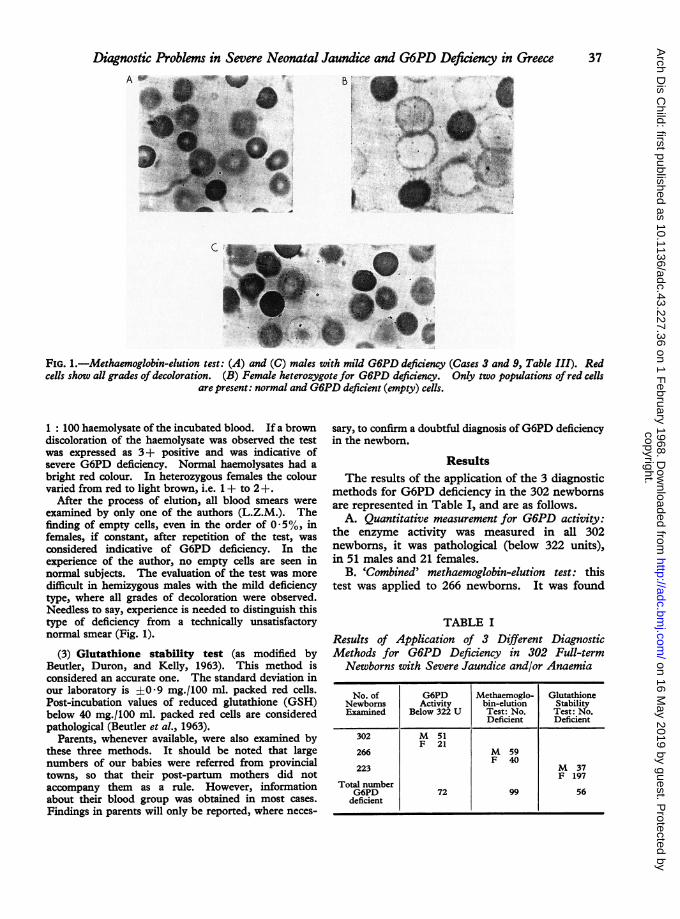
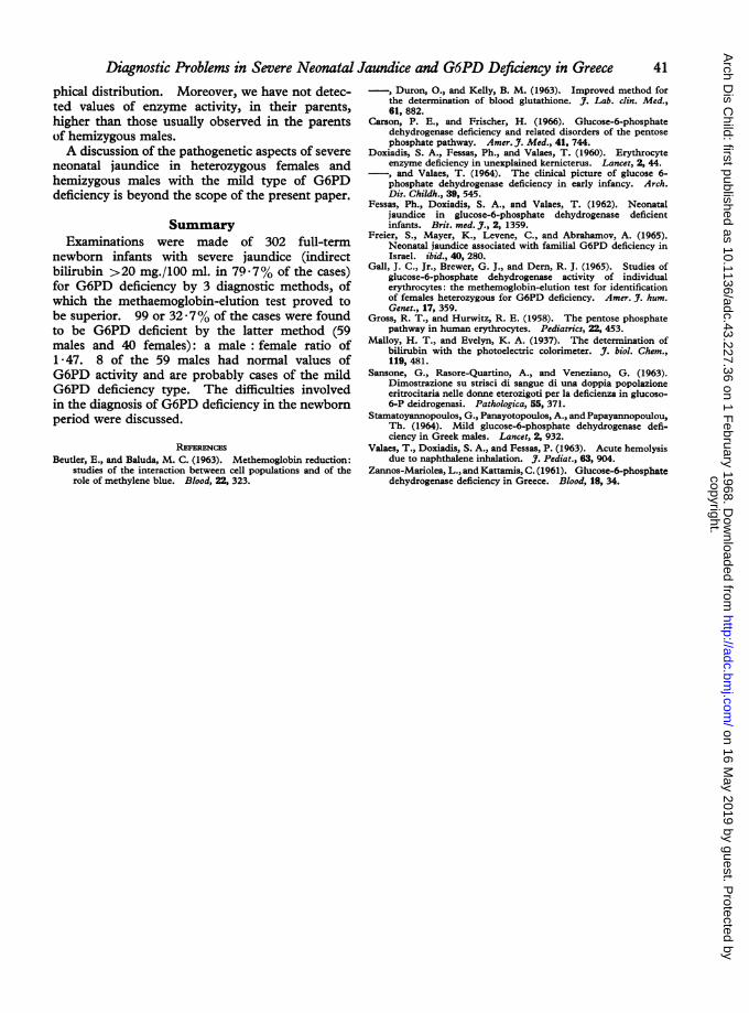
FIG. 1.-Methaemoglobin-elution test: (A) and (C) males with mild G6PD deficiency (Cases 3 and 9, Table III). Redcells show all grades of decoloration. (B) Female heterozygote for G6PD deficiency. Only two populations ofred cells
are present: normal and G6PD deficient (empty) cells.
1: 100 haemolysate of the incubated blood. If a browndiscoloration of the haemolysate was observed the testwas expressed as 3+ positive and was indicative ofsevere G6PD deficiency. Normal haemolysates had a
bright red colour. In heterozygous females the colourvaried from red to light brown, i.e. 1 + to 2 +.
After the process of elution, all blood smears wereexamined by only one of the authors (L.Z.M.). Thefinding of empty cells, even in the order of 0 5%, infemales, if constant, after repetition of the test, wasconsidered indicative of G6PD deficiency. In theexperience of the author, no empty cells are seen innormal subjects. The evaluation of the test was moredifficult in hemizygous males with the mild deficiencytype, where all grades of decoloration were observed.Needless to say, experience is needed to distinguish thistype of deficiency from a technically unsatisfactorynormal smear (Fig. 1).
(3) Glutathione stability test (as modified byBeutler, Duron, and Kelly, 1963). This method isconsidered an accurate one. The standard deviation inour laboratory is ±0 9 mg./100 ml. packed red cells.Post-incubation values of reduced glutathione (GSH)below 40 mg./100 ml. packed red cells are consideredpathological (Beutler et al., 1963).
Parents, whenever available, were also examined bythese three methods. It should be noted that largenumbers of our babies were referred from provincialtowns, so that their post-partum mothers did notaccompany them as a rule. However, informationabout their blood group was obtained in most cases.
Findings in parents will only be reported, where neces-
sary, to confirm a doubtful diagnosis of G6PD deficiencyin the newborn.
ResultsThe results of the application of the 3 diagnostic
methods for G6PD deficiency in the 302 newbornsare represented in Table I, and are as follows.
A. Quantitative measurement for G6PD activity:the enzyme activity was measured in all 302newborns, it was pathological (below 322 units),in 51 males and 21 females.
B. 'Combined' methaemoglobin-elution test: thistest was applied to 266 newborns. It was found
TABLE IResults of Application of 3 Different DiagnosticMethods for G6PD Deficiency in 302 Full-term
Newborns with Severe Jaundice and/or Anaemia
No. of G6PD Methaemoglo- GlutathioneNewborns Activity bin-elution StabilityExamined Below 322 U Test: No. Test: No.
Deficient Deficient
302 M 51F 21
266 M 59F 40
223 M 37F 197
Total numberG6PD 72 99 56
deficient
37
copyright. on 16 M
ay 2019 by guest. Protected by
http://adc.bmj.com
/A
rch Dis C
hild: first published as 10.1136/adc.43.227.36 on 1 February 1968. D
ownloaded from
Zannos-Mariolea, Thomaidis, Georgizas, Gavrielidou, and BenetosTABLE II
Non-G6PD Deficient Newborns with Severe Jaundiceand/or Anaemia, Grouped According to Aetiology
Groups Newborn Males Females M:F ratio
I: ABO isoim-munization .. 54 31 23 1*34
II: ABOset-up.. 29 17 12 1-41III: Rh isoimn-
munization .. 21 13 8 1*62IV: Naphthalene
exposure .. 14 12 2 6V: Unknown .. 70 42 28 1*5
VI: Miscellaneous* 15 11 4 2-75
Total . .. 203 126 77 1*63
* Miscellaneous = newborns with cyanosis, septicaemia, mon-golism, etc.
pathological in 59 males and 40 females, i.e. in33% of the total cases. Of the 36 cases whichcould not be examined by this method, 5 werecases of Rh isoimmunization, 8 of ABO isoim-munization (spherocytosis present), 9 of doubtfulABO isoimmunization (ABO set up, withoutspherocytosis), and 14 of 'unknown aetiology'.
C. Glutathione (GSII) stability test: this test wasapplied to 223 newborns and found to be patholo-gical in 37 males and 19 females.These results indicate that the greatest number
of G6PD deficient newborns were detected by themethaemoglobin-elution test, i.e. 99 newborns ascompared to 72 by the quantitative method and 56by the GSH stability test. Of the 99 deficientbabies, 59 were male and 40 were female. Hencethe male: female ratio was 1 47.Of the 99 G6PD deficient babies, 34 have been
exposed to one of the known toxic drugs, mostlynaphthalene. 9 were cases ofABO incompatibilityas well, one of Rh isoimmunization, and 55 had notbeen exposed to any of the drugs known to causehaemolysis in 'sensitive' subjects. However, themothers of these babies were usually given pethidine,hyoscine butylbromide, and vasopressin duringlabour, and in the great majority tetracycline wasgiven during the first few days post partum, whiletheir babies were breast feeding.The remaining 203 newborns who did not have
G6PD deficiency were divided into 6 groups (I-VI)according to certain aetiological criteria, as shown inTable II. In 70 cases, or 23% of the total ofnewborns, the aetiology of severe jaundice remainedunknown. This percentage is in reality higher ifit is considered that a number of babies of group 2were probably not cases of ABO isoimmunization.The male: female ratio of these 203 babies is
1-63 (males 126; females 77; Table II), i.e. not
significantly different from that observed in theG6PD deficient group. Interestingly enough,males predominated in all groups. Group IVdeserves comment; it included babies who developedsevere jaundice after exposure to naphthalene,despite their normal G6PD activity. Such caseshave been described already in Greece (Valaes,Doxiadis, and Fessas, 1963). The impressivepredominance of males is reminiscent of childhoodfavism, where the male: female ratio is about 10(Zannos-Mariolea and Kattamis, 1961). It isunfortunate that the number of cases in this groupis too small to draw any conclusions.
Analysis of Results in 99 Newborn G6PDDeficient Newborns by Methaemoglobin
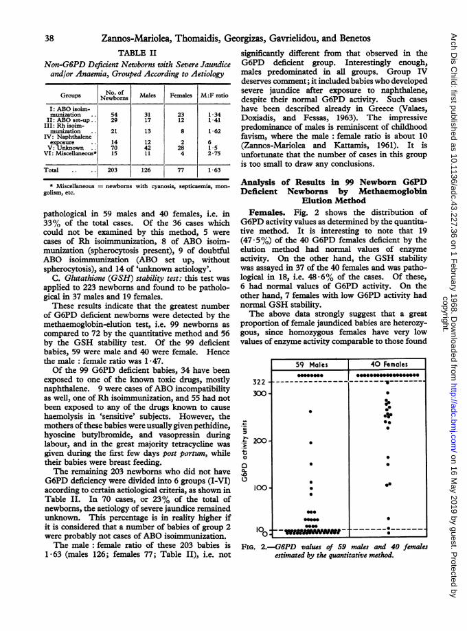
Elution MethodFemales. Fig. 2 shows the distribution of
G6PD activity values as determined by the quantita-tive method. It is interesting to note that 19(47- 5%) of the 40 G6PD females deficient by theelution method had normal values of enzymeactivity. On the other hand, the GSH stabilitywas assayed in 37 of the 40 females and was patho-logical in 18, i.e. 48 6% of the cases. Of these,6 had normal values of G6PD activity. On theother hand, 7 females with low G6PD activity hadnormal GSH stability.The above data strongly suggest that a great
proportion of female jaundiced babies are heterozy-gous, since homozygous females have very lowvalues of enzyme activity comparable to those found
322 -
300-
C
.t 200-
a.
a
100
la0
FIG. 2.-G6PD values of 59 males and 40 femalesestimated by the quantitative method.
38
59 Mal,es ] 40 Females
0.S S0
0
*
0~~~~~~~
000
000 00
0000-wDe*
copyright. on 16 M
ay 2019 by guest. Protected by
http://adc.bmj.com
/A
rch Dis C
hild: first published as 10.1136/adc.43.227.36 on 1 February 1968. D
ownloaded from
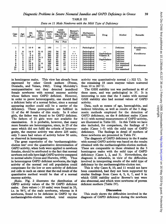
Diagnostic Problems in Severe Neonatal Jaundice and G6PD Deficiency in GreeceTABLE III
Data on 11 Male Newborns with the Mild Type of Deficiency
Indirect G6PDdGSH Brewer'sHb Bili- Acti Stability Methae- Methaemo-Age 53 2 38(g+/ rubing 1 m. oglobin globin-elution Drug Mother Remarks
No. (dy.) 100 nil.) (m vity packed red Reuton Ts100 m.) (units) cells)
Reuto Test
Pre Post
1 9 1089 46 150 - - + + + Pathological _ Deficient Kerikterus;died
2 6 14-3 318 193 54 32 - Pathological N Deficient3 11 8 40 267 86 65 + + Pathological Naphthalene Deficient Kemikterus4 5 5 3 27 385 32 28 +++ Pathological Vitamin K Deficient5 5 18-4 19-3 393 85 49 +++ Pathological -Deficient6 7 13-3 24 616 81 81 +++ Pathological Naphthalane Deficient7 6 20 34 345 80 11 +++ Pathological - Deficient8 5 14-8 31-6 388 90 67 + + + Pathological - Not examined9 5 14-5 28 431 66 27 + + + Pathological - Not examined Kernikterus10 6 18-4 22-4 576 - - + + + Pathological - Normal11 4 16*5 20 454 56 38 Pathological - Not examined
in hemizygous males. This view has already beenexpressed by other Greek authors (Fessas,Doxiadis, and Valaes, 1962). Using Motulsky'ssemiquantitative test they detected jaundicedfemale newborns with normal enzyme activitywhose fathers were G6PD deficient. However,in order to prove heterozygosity, one should finda deficient baby of a normal father, since a normalappearing mother could still be a carrier of thedeficiency. These prerequisites are fulfilled in11 of the 40 females of this study. In 8 othergirls, the father was found to be G6PD deficient.The fathers of 21 girls were not available forexamination. It is probable, however, that manymore females are heterozygotes, since, in 25 of thecases which did not fulfil the criteria of heterozy-gosity, the enzyme activity was above 225 units;only 3 cases had values of activity below 50 units,as observed in homozygotes.The great superiority of the 'methaemoglobin-
elution test' over the quantitative determination ofG6PD activity, when both were applied to newbornfemales, should be attributed to the fact that normalnewborn red cells have high G6PD activity comparedto normal adults (Gross and Hurwitz, 1958). Thusin heterozygous G6PD deficient newborns, the highactivity of the normal red cell population couldcompensate for the low or zero activity of deficientred cells to such an extent that the end result of thequantitative method would be that of a normalenzyme activity.
Males. Fig. 2 illustrates the distribution ofG5PD activity values of the 59 G6PD deficientmales. Zero values (< 10 units) were found in 33,i.e. in 56% of the male newborns, whereas in 8newborns, found to be deficient in G6PD by themethaemoglobin-elution method, total enzyme
activity was quantitatively normal (>322 U). Inthe remaining 18 cases enzyme values scatteredfrom 10 to 267 U.The GSH stability test was performed in 40 of
these cases, and was pathological in 37. It isinteresting to note that the 3 males with normalGSH stability also had normal values of G6PDactivity.
Data, such as means of age, haemoglobin, andindirect bilirubin, as well as results of the 3 diag-nostic methods employed for the detection ofG6PD deficiency, on the 8 deficient males (Cases4-11) with normal measurements of G6PD activity,are illustrated in Table III. In this Table we havealso included, for comparison, the findings of 3males (Cases 1-3) with the mild type of G6PDdeficiency. The findings in detail of mothers ofthese newborns are presented in Table IV.The diagnosis of G6PD deficiency in the 8 males
with normal G6PD activity was based on the resultsobtained with the methaemoglobin-elution method.These are comparable to those obtained in the 3hemizygous males with the mild type of G6PDdeficiency (Cases 1, 2, and 3). Nevertheless, thediagnosis is debatable, in view of the difficultiesinvolved in interpreting results of the mild type ofdeficiency by the methaemoglobin-elution test.This is why Cases 8, 10, and 11 would not have
been considered, had they not been supported bysimilar findings from Cases 4, 5, 6, 7, and 9 inwhich GSH stability and/or Brewer's test werealso abnormal, and which were newborns of provendeficient mothers (Table IV).
DiscussionThis study shows the difficulties involved in the
diagnosis of G6PD deficiency during the newborn
39
copyright. on 16 M
ay 2019 by guest. Protected by
http://adc.bmj.com
/A
rch Dis C
hild: first published as 10.1136/adc.43.227.36 on 1 February 1968. D
ownloaded from
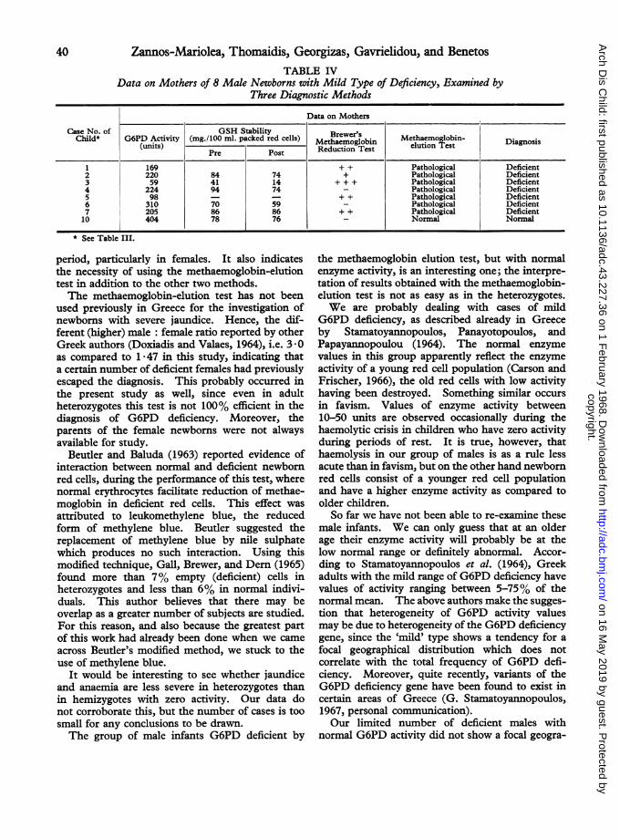
Zannos-Mariolea, Thomaidis, Georgizas, Gavrielidou, and BenetosTABLE IV
Data on Mothers of 8 Male Newborns with Mild Type of Deficiency, Examined byThree Diagnostic Methods
Case No. ofChild* G6PD Activity
(units)
I~IGSH Stability
(mg./100 ml. packed red cells)
Pre Post
Data on Mothers
Brewer'sMethaemoglobinReduction Test
Methaemoglobin-elution Test Diagnosis
169 + + Pathological Deficient220 84 74 + Pathological Deficient59 41 14 + + + Pathological Deficient
224 94 74 - Pathological Deficient98 - - + + Pathological Deficient310 70 59 - Pathological Deficient205 86 86 + + Pathological Deficient404 78 76 - Normal Normal
* See Table III.
period, particularly in females. It also indicatesthe necessity of using the methaemoglobin-elutiontest in addition to the other two methods.The methaemoglobin-elution test has not been
used previously in Greece for the investigation ofnewborns with severe jaundice. Hence, the dif-ferent (higher) male: female ratio reported by otherGreek authors (Doxiadis and Valaes, 1964), i.e. 3 0as compared to 1 47 in this study, indicating thata certain number of deficient females had previouslyescaped the diagnosis. This probably occurred inthe present study as well, since even in adultheterozygotes this test is not 100% efficient in thediagnosis of G6PD deficiency. Moreover, theparents of the female newborns were not alwaysavailable for study.
Beutler and Baluda (1963) reported evidence ofinteraction between normal and deficient newbornred cells, during the performance of this test, wherenormal erythrocytes facilitate reduction of methae-moglobin in deficient red cells. This effect wasattributed to leukomethylene blue, the reducedform of methylene blue. Beutler suggested thereplacement of methylene blue by nile sulphatewhich produces no such interaction. Using thismodified technique, Gall, Brewer, and Dern (1965)found more than 7% empty (deficient) cells inheterozygotes and less than 6% in normal indivi-duals. This author believes that there may beoverlap as a greater number of subjects are studied.For this reason, and also because the greatest partof this work had already been done when we came
across Beutler's modified method, we stuck to theuse of methylene blue.
It would be interesting to see whether jaundiceand anaemia are less severe in heterozygotes thanin hemizygotes with zero activity. Our data donot corroborate this, but the number of cases is toosmall for any conclusions to be drawn.The group of male infants G6PD deficient by
the methaemoglobin elution test, but with normalenzyme activity, is an interesting one; the interpre-tation of results obtained with the methaemoglobin-elution test is not as easy as in the heterozygotes.We are probably dealing with cases of mild
G6PD deficiency, as described already in Greeceby Stamatoyannopoulos, Panayotopoulos, andPapayannopoulou (1964). The normal enzyme
values in this group apparently reflect the enzymeactivity of a young red cell population (Carson andFrischer, 1966), the old red cells with low activityhaving been destroyed. Something similar occurs
in favism. Values of enzyme activity between10-50 units are observed occasionally during thehaemolytic crisis in children who have zero activityduring periods of rest. It is true, however, thathaemolysis in our group of males is as a rule lessacute than in favism, but on the other hand newbornred cells consist of a younger red cell populationand have a higher enzyme activity as compared toolder children.
So far we have not been able to re-examine thesemale infants. We can only guess that at an olderage their enzyme activity will probably be at thelow normal range or definitely abnormal. Accor-ding to Stamatoyannopoulos et al. (1964), Greekadults with the mild range of G6PD deficiency havevalues of activity ranging between 5-75% of thenormal mean. The above authors make the sugges-tion that heterogeneity of G6PD activity valuesmay be due to heterogeneity of the G6PD deficiencygene, since the 'mild' type shows a tendency for a
focal geographical distribution which does notcorrelate with the total frequency of G6PD defi-ciency. Moreover, quite recently, variants of theG6PD deficiency gene have been found to exist incertain areas of Greece (G. Stamatoyannopoulos,1967, personal communication).Our limited number of deficient males with
normal G6PD activity did not show a focal geogra-
40
copyright. on 16 M
ay 2019 by guest. Protected by
http://adc.bmj.com
/A
rch Dis C
hild: first published as 10.1136/adc.43.227.36 on 1 February 1968. D
ownloaded from
Diagnostic Problems in Severe Neonatal Jaundice and G6PD Deficiency in Greece 41phical distribution. Moreover, we have not detec-ted values of enzyme activity, in their parents,higher than those usually observed in the parentsof hemizygous males.A discussion of the pathogenetic aspects of severe
neonatal jaundice in heterozygous females andhemizygous males with the mild type of G6PDdeficiency is beyond the scope of the present paper.
SummaryExaminations were made of 302 full-term
newborn infants with severe jaundice (indirectbilirubin >20 mg./100 ml. in 79- 7% of the cases)for G6PD deficiency by 3 diagnostic methods, ofwhich the methaemoglobin-elution test proved tobe superior. 99 or 32 *7% of the cases were foundto be G6PD deficient by the latter method (59males and 40 females): a male : female ratio of1 47. 8 of the 59 males had normal values ofG6PD activity and are probably cases of the mildG6PD deficiency type. The difficulties involvedin the diagnosis of G6PD deficiency in the newbornperiod were discussed.
REEMRENCFSBeutler, E., and Baluda, M. C. (1963). Methemoglobin reduction:
studies of the interaction between cell populations and of therole of methylene blue. Blood, 22, 323.
, Duron, O., and Kelly, B. M. (1963). Improved method forthe determination of blood glutathione. J. Lab. clin. Med.,61, 882.
Carson, P. E., and Prischer, H. (1966). Glucose-6-phosphatedehydrogenase deficiency and related disorders of the pentosephosphate pathway. Amer. J. Med., 41, 744.
Doxiadis, S. A., Fessas, Ph., and Valaes, T. (1960). Erythrocyteenzyme deficiency in unexplained kernicterus. Lancet, 2, 44., and Valaes, T. (1964). The clinical picture of glucose 6-phosphate dehydrogenase deficiency in early infancy. Arch.Dis. Childh., 39, 545.
Fessas, Ph., Doxiadis, S. A., and Valaes, T. (1962). Neonataljaundice in glucose-6-phosphate dehydrogenase deficientinfants. Brit. med. J., 2, 1359.
Freier, S., Mayer, K., Levene, C., and Abrahamov, A. (1965).Neonatal jaundice associated with familial G6PD deficiency inIsrael. ibid., 40, 280.
Gall, J. C., Jr., Brewer, G. J., and Dern, R. J. (1965). Studies ofglucose-6-phosphate dehydrogenase activity of individualerythrocytes: the methemoglobin-elution test for identificationof females heterozygous for G6PD deficiency. Amer. J. hum.Genet., 17, 359.
Gross, R. T., and Hurwitz, R. E. (1958). The pentose phosphatepathway in human erythrocytes. Pediatrics, 22, 453.
Malloy, H. T., and Evelyn, K. A. (1937). The determination ofbilirubin with the photoelectric colorimeter. J. biol. Chem.,119, 481.
Sansone, G., Rasore-Quartino, A., and Veneziano, G. (1963).Dimostrazione su strisci di sangue di una doppia popolazioneeritrocitaria nelle donne eterozigoti per la deficienza in glucoso-6-P deidrogenasi. Pathologica, 55, 371.
Stamatoyannopoulos, G., Panayotopoulos, A., and Papayannopoulou,Th. (1964). Mild glucose-6-phosphate dehydrogenase defi-ciency in Greek males. Lancet, 2, 932.
Valaes, T., Doxiadis, S. A., and Fessas, P. (1963). Acute hemolysisdue to naphthalene inhalation. J. Pediat., 63, 904.
Zannos-Mariolea, L., and Kattamis, C. (1961). Glucose-6-phosphatedehydrogenase deficiency in Greece. Blood, 18, 34.
copyright. on 16 M
ay 2019 by guest. Protected by
http://adc.bmj.com
/A
rch Dis C
hild: first published as 10.1136/adc.43.227.36 on 1 February 1968. D
ownloaded from