DIAGNOSTIC POTENTIALITIES OF THREE- DIMENSIONAL NLS · PDF fileDIAGNOSTIC POTENTIALITIES OF...
16
1 DIAGNOSTIC POTENTIALITIES OF THREE- DIMENSIONAL NLS-GRAPHY V.I.Nesterov, V.I.Nesterova This publication contains modern principles of three-dimensional images rendering in accordance with NLS-graphy data. Also it gives overall evaluation of three-dimensional NLS graphy diagnostic value for revealing of various diseases in comparison with other methods of hardware diagnostics, such as roentgenography, computerized tomography and magnetic resonance imaging. Special attention is paid to advantages and disadvantages of various techniques of three-dimensional images rendering. Three-dimensional pictures of human’s internal organs became a part of general practice in early 90’s after computer tomographic scanners were equipped with powerful computing systems capable of controlled processing of two-dimensional crosscuts. Nowadays three- dimensional representation of elements of diagnostically interesting zone is an everyday reality in leading clinics of the world. Method of three-dimensional representation of diagnostic data is generally related to powerful hardware resources, i.e. acquiring of parallel (or placed at previously specified angles) magnetic-resonance, roentgen or NLS-graphic images with their following integration into single visual array where an operator can separately visualize bones, muscles, soft tissues, vessels, nerves etc. highlighting them with color while other tissues are shown in gray semitransparent tone. Rendering of three-dimensional NLS-graphic images of abdominal cavity organs is considered nowadays to be an experimental task mainly. Rarely used due to its low information value method of creating of separate two-dimensional images of abdominal cavity organs by means of NLS-visualization is much more interesting for rendering three-dimensional diagnostic images. In addition to solving of usual objectives on the basis of two-dimensional NLS-graphy of abdominal cavity organs data many parallel diagnostic tasks are solved by means of non-linear algorithms and massive calculations application. Maybe that is how should be explained so accurate results described in one of the first reports about three-dimensional reconstruction on the basis of transabdominal NLS-data (T.G. Kuznetsova, R.A. Sorokina). We place our great hopes on three-dimensional NLS-graphy, first of all related to its application in endoscopy. In 2010 V.I. Melushko theoretically justified an opinion that application of three-dimensional NLS-research in laparoendoscopic surgery will allow to “look beyond the horizon” and see anatomic structures which cannot be visualized when using laparoscope. He believed that NLS-graphy is diagnostically efficient, safe and affordable procedure.
Transcript of DIAGNOSTIC POTENTIALITIES OF THREE- DIMENSIONAL NLS · PDF fileDIAGNOSTIC POTENTIALITIES OF...

1
DIAGNOSTIC POTENTIALITIES OF THREE-
DIMENSIONAL NLS-GRAPHY
V.I.Nesterov, V.I.Nesterova
This publication contains modern principles of three-dimensional images rendering in
accordance with NLS-graphy data. Also it gives overall evaluation of three-dimensional NLS
graphy diagnostic value for revealing of various diseases in comparison with other methods of
hardware diagnostics, such as roentgenography, computerized tomography and magnetic
resonance imaging. Special attention is paid to advantages and disadvantages of various
techniques of three-dimensional images rendering.
Three-dimensional pictures of human’s internal organs became a part of general practice
in early 90’s after computer tomographic scanners were equipped with powerful computing
systems capable of controlled processing of two-dimensional crosscuts. Nowadays three-
dimensional representation of elements of diagnostically interesting zone is an everyday reality
in leading clinics of the world.
Method of three-dimensional representation of diagnostic data is generally related to
powerful hardware resources, i.e. acquiring of parallel (or placed at previously specified angles)
magnetic-resonance, roentgen or NLS-graphic images with their following integration into single
visual array where an operator can separately visualize bones, muscles, soft tissues, vessels,
nerves etc. highlighting them with color while other tissues are shown in gray semitransparent
tone.
Rendering of three-dimensional NLS-graphic images of abdominal cavity organs is
considered nowadays to be an experimental task mainly. Rarely used due to its low information
value method of creating of separate two-dimensional images of abdominal cavity organs by
means of NLS-visualization is much more interesting for rendering three-dimensional diagnostic
images.
In addition to solving of usual objectives on the basis of two-dimensional NLS-graphy of
abdominal cavity organs data many parallel diagnostic tasks are solved by means of non-linear
algorithms and massive calculations application.
Maybe that is how should be explained so accurate results described in one of the first
reports about three-dimensional reconstruction on the basis of transabdominal NLS-data (T.G.
Kuznetsova, R.A. Sorokina).
We place our great hopes on three-dimensional NLS-graphy, first of all related to its
application in endoscopy. In 2010 V.I. Melushko theoretically justified an opinion that
application of three-dimensional NLS-research in laparoendoscopic surgery will allow to “look
beyond the horizon” and see anatomic structures which cannot be visualized when using
laparoscope. He believed that NLS-graphy is diagnostically efficient, safe and affordable
procedure.

2
A.Y. Shvack and L.P. Goltsova (Pic.1) found out that at size changing of hepatic focal
formations two- and three-dimensional NLS-graphy are equally precise when shape of
neoplasms is relatively simple - close to roundish or oval. However, the authors state that if
shape of neoplasms is complex, results of two-dimensional NLS-graphy matched in 65% of
cases only, when at three-dimensional visualization – in 92% of cases.
Pic.1. 3D-image of hepatic neoplasms foci
In 2010 S.A. Volkova and A.V. Zaitsev reported about results of three-dimensional
transabdominal NLS-graphy application in diagnostics of hepatobiliary system, in particular for
specifying diagnostics of previously revealed neoplasms. The objective of their research was
accuracy evaluation of three-dimensional NLS-graphy method with application of spectral-
entropic analysis (SEA). Acquired results were compared with information gathered by
computed tomographic portal venography carried out on spiral tomograph. Altogether 62 cases
were analyzed including those with hepatic neoplasms sized from 1.5 cm to 12.3 cm. The
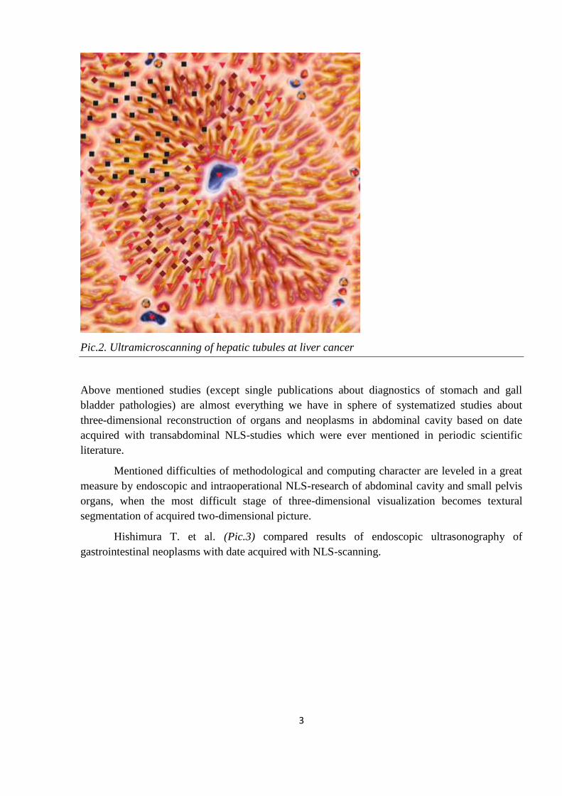
researches showed that three-dimensional model built on the basis of NLS-data (Pic.2) contains
more precise and valuable diagnostic information in comparison with usual two-dimensional
NLS-graphy or data acquired with CT. The authors believe that three-dimensional NLS-graphy
is financially affordable procedure for diagnostic of tumor affections.
3
Pic.2. Ultramicroscanning of hepatic tubules at liver cancer
Above mentioned studies (except single publications about diagnostics of stomach and gall
bladder pathologies) are almost everything we have in sphere of systematized studies about
three-dimensional reconstruction of organs and neoplasms in abdominal cavity based on date
acquired with transabdominal NLS-studies which were ever mentioned in periodic scientific
literature.
Mentioned difficulties of methodological and computing character are leveled in a great
measure by endoscopic and intraoperational NLS-research of abdominal cavity and small pelvis
organs, when the most difficult stage of three-dimensional visualization becomes textural
segmentation of acquired two-dimensional picture.
Hishimura T. et аl. (Pic.3) compared results of endoscopic ultrasonography of
gastrointestinal neoplasms with date acquired with NLS-scanning.

4
Pic.3. 3D NLS-image of large intestine cancer
In two cases of esophagus cancer, in two cases of rectum cancer and in 7 of 10 cases
(Pic.4) of stomach cancer results of three-dimensional reconstruction made on the basis of NLS-
graphy were accurate in relation to depth of tumorous invasion.
Pic.4. 3D-image of stomach cancer

5
Japanese scientists Katamaki S. et аl. (Pic.5) reported in 2011 about preliminary results
of three-dimensional NLS-graphy application in 26 patients. Three-dimensional reconstruction
made on the basis of three-dimensional pictures allowed to define presence of metastatic
affection of surrounding organs and in 4 of 6 cases of bile duct cancer – to define volume of
tumorous tissue.
Pic.5. 3D NLS-picture of common bile duct cancer
with affection of regional lymph nodes
Fox P. et аl. (Pic.6) used NLS-study for three-dimensional reconstruction of esophagus picture in
5 patients suffering form esophagus cancer. The research was carried out with sensor’s
frequency of 4.9 GHz. Three-dimensional pictures were reconstructed on the basis of series of
cross-cut pictures. Precise three-dimensional visualization of tumor’s structure and surrounding
tissues were acquired in all cases. In this study proper determining of tumorous process stage
was achieved in three of four cases. The authors note special diagnostic value of pictures
showing longitudinal sections of tumor which allow to define precisely its length and connection
with mediastinal structures.

6
Pic.6. Longitudinal 3D NLS-image of esophagus cancer
Among of the most standardized methods of research are three-dimensional NLS-graphy
of rectum and NLS-scanning of prostate.
German oncoproctologists Operbein K. et аl. (Pic.7) have studied more than 100 patients
suffering from rectum tumors with three-dimensional NLS-graphy.
Pic.7. 3D NLS-image of rectum cancer

7
Diagnostic accuracy of three-dimensional ultrasound research in this group was 89%, at the same
time accuracy of two-dimensional NLS-graphy in the same group of patients was 76%. Accuracy
of these methods in defining of pararectal lymph nodes affection was 85% at three-dimensional
visualization and 71% in accordance with two-dimensional data. The authors believe that three-
dimensional NLS-graphy allows to overcome well known limitations of two-dimensional NLS-
study which are usually faced at obstructing tumor and in revealing of recurrent cancer of
rectum.
Operbein K. et аl. also used three-dimensional NLS-graphy to detect recurrent
malignization of rectum in patients after surgical treatment of rectum cancer. Possibilities of
two-dimensional NLS-study in these cases are strictly limited due to massive growth of fibrous
tissue. Data for three-dimensional reconstruction was acquired by Operbein K. and Schmidt A.
who used non-linear scanner co-manufactured by IPP (Omsk, Russia) and the firm's Clinic Tech
Inc. (USA) with operating frequency of 9.6 GHz. Three-dimensional NLS-graphy have helped to
find pararectal neoplasms in 28 of 163 patients. Spectral-entropic analysis confirmed recurrent
cancer in 7 patients and metastatic affection of lymph nodes in 2 patients correspondingly.
Therapists of pediatric surgery department Stubdreier H. et аl. (Pic.8) of Tübingen
University (Germany) used three-dimensional NLS-graphy of pelvic floor as an auxiliary
diagnostic instrument for determining of surgical tactics for treatment of children suffering from
stool incontinence. The study included visualization of sphincter muscles and rectum walls;
results of the study were confirmed by data acquired with spiral computed tomography. A
distinctive feature of this study was using of interactive tissue segmentation of organs and tissues
by means of 4D TISSUE mode application during their three-dimensional visualization.
Pic.8. 3D-ultramicroscanning of rectum cancer

8
Chun L.G. et аl. (Pic.9) carried out three-dimensional NLS-graphy of malignant prostate
gland in 46 patients. In accordance with gathered diagnostic data cryoablation of prostate gland
was fulfilled.
Pic.9. 3D NLS-graphy of malignant prostate gland
Possible diagnostic value of three-dimensional NLS-graphy is most clearly seen during
examination of small pelvis organs in women, where conventional diagnostic methods allow to
see advantages and disadvantages of each of them.
Akuda T. et аl. have not got any additional information using three-dimensional NLS-
graphy in comparison with usual two-dimensional NLS-study which revealed uterine cavity
septa in one woman, fibroid foci in three women and endometrial polyp in one woman of five
examined patients.
Spanish gynecologists Brisoles H. et аl. have studied possibilities of three-dimensional
NLS-graphy, transvaginal sonography, transvaginal Doppler sonography, hysteroscopy and usual
hysterosonography in diagnostics of endometrial tumors and defining of endometrium thickness
during hormonal treatment in 16 patients. Three-dimensional NLS-study was found the most
accurate diagnostic method of all methods studied.
Chun L.G. et аl., a group of gynecologists of Temple University (Philadelphia) in 2011
reported about results of comparative study of diagnostic efficiency of three-dimensional and
two-dimensional NLS-graphy. Altogether 8 women (Pic.10) suffering from surgical diseases of
ovaries were examined. Later on all patients were subjected to explorative laparotomy or
diagnostic laparoscopy. NLS-graphy data was compared with intraoperational findings and
results of macroscopic and histological studies. In all eight cases three-dimensional NLS-graphy
managed to define correct diagnosis before surgical intervention. It is especially important that in
absence of difference in diagnostic accuracy of three-dimensional and two-dimensional study of

9
cysts and benign neoplasms, two-dimensional NLS-graphy turned out to bell less accurate in
detecting of malignant tissues, giving both false-positive and false-negative results.
Pic.10. 3D-ultramicroscanning of ovarian cancer
Spoun N. et аl. (Pic.11) have carried out comparative evaluation of two-dimensional and
three-dimensional NLS-graphy accuracy in specifying diagnostics of uterine neck cancer. The
study was carried out in 61 women one day before surgical intervention; accuracy of three-
dimensional examination was in range of +6.68 to -6.1 ml, two-dimensional – from +12.46 to -
10.98 ml.
Pic.11. 3D-ultramicroscanning of uterus cervix cancer

10
French gynecologists Sarida M. et аl. used three-dimensional NLS-graphy for diagnostics
of atropic pregnancy in 12 women with amenorrhea period above 6 weeks. Laparoscopy detected
atropic pregnancy in 9 cases, only in 2 of them it was found on NLS-pictures. Hence the authors
believe that this diagnostic method is ineffective, especially for early non-invasive detection of
gestational sac development in fallopian tube.
Other methodologically valuable results for evaluation of advantages of three-
dimensional representation of NLS-graphic data were results acquired at NLS-study of
mammary glands.
Bluhmberg S.O. et аl. (Pic.12) have used three-dimensional NLS-graphy of mammary
gland in study of 48 women, 18 of which had malignant neoplasms of mammary gland. The
therapists reported about 4 false-positive mammary gland cancer diagnoses against two cases of
false-positive diagnoses when two-dimensional NLS-graphy was used. At the same time the
authors note that three-dimensional NLS-graphy has advantages in evaluation of edge zones and
shape of pathological nidus and also at multifocal affection of mammary gland.
Pic.12. 3D-image of mammary gland cancer
English breast physicians Havies L. et аl. (Pic.13) of Bristol University used three-dimensional
NLS-graphy of mammary gland and in some cases managed to reveal and detect internal
structure of intraductal carcinoma by means of spectral-entropic analysis, and neoplasms
simulating microinvasive carcinoma. Such outstanding diagnostic results authors achieved using
non-linear sensor of 9.6 GHz frequency that allowed to get resolution of 30 microns.

11
Pic.13. 3D-ultramicroscanning of mammary gland cancer
A peculiar etalon test of diagnostic efficiency of three-dimensional reconstruction at complex
configuration of source two-dimensional data is considered NLS-scanning of bloodstream,
results of which are compared with similar data acquired by traditional and well known
radiographic contrast study.
In 2011 Bljger K. et аl. (Pic.14) reported about results of a study than have been carried out in
University hospital of Antwerp and were devoted for “reconstruction of bloodstream’s
geometry” in accordance with data of radiographic contrast angiography and intracoronary NLS-
study. After evaluation of reconstruction precision of studies performed on special phantoms, the
authors agreed to consider acquired results “very realistic” and offered a number of criteria for
comparative evaluation of three-dimensional bloodstream reconstruction precision. Clinical part
of the study contained comparison of reconstructed three-dimensional images of coronary
arteries with data acquired with usual angiograms. The difference was less than 5%.

12
Pic.14. 3D-image of coronary artery
Prust G.J. et аl. note the following advantages of reconstructed images of arteries,
acquired with intravascular NLS-study: 1) a vessel may be seen from different sides and angles,
2) its direction may be seen, 3) changes of vessel’s diameter are clearly visible, 4) a possibility to
get a cross-section of a vessel in any plane of scanning, 5) condition and location of intravascular
stent may be found with high precision.
Kogan S. et аl. (Pic.15) have determined high diagnostic efficiency of three-dimensional
intravascular NLS-graphy in visualization of arterial wall structure and its pathological changing.
The authors believe that intravascular NLS-graphy with following three-dimensional
reconstruction significantly excels coronary angiography, because it have better resolution and
allow to reveal many hidden neoplasms; that is why it is irreplaceable at study of coronary
arteries condition after heart transplantation.

13
Pic.15. 3D-ultramicroscanning of vascular wall
The most impressive was using of three-dimensional NLS-study data for intraoperative
navigation and computed modeling of surgical manipulations.
Serbia S. – a head of obstetrics and gynecology department of Washington University
reasonably stated that three-dimensional NLS-study is a potential diagnostic instrument which in
near future will become the main source of data for preoperative surgical training which uses
individual diagnostic data.
Three-dimensional NLS-graphy may be widely used in cardiosurgery as a method of
intraoperational navigation. Quite character in this relation is a study of cardiothoracic surgeons
from North Carolina Abrams O.H. et аl. (Pic.16) who presented in 2011 a detailed report about
wide clinical application of intraoperational three-dimensional NLS-graphy during replacement
of cardiac valves. The authors say that this diagnostic method helped them to get important
intraoperational data when they used spectral-entropic analysis about morphological peculiarities
of valves, which weren’t revealed by echocardiography and Doppler sonography.

14
Pic.16. 3D NLS-picture of cardiac valves
The authors say that this diagnostic method helped them to get important intraoperational
data when they used spectral-entropic analysis about morphological peculiarities of valves,
which weren’t revealed by echocardiography and Doppler sonography.
Going beyond the most optimistic ways of possible application of three-dimensional
NLS-graphy, Parris D. et аl. (Pic.17) use data about prostate gland structure acquired by this
method for computerized control of robot (!) movement during fulfillment of transrectal
resection.
Pic.17. 3D-ultramicroscanning of prostate gland

15
An unique and apparently having no equals in efficiency navigation algorithm was
offered by Hatayama T. et аl. – scientists of Tokio University in 2012. They reported about
possibility of precise matching of three-dimensional pictures acquired by CT and MRI in
preoperational period with data of three-dimensional NLS-research acquired during
neurosurgical intervention. NLS scanner of 4.9 GHz frequency was an integral part of a system
and had built-in positioning device. The authors report about successful clinical application of a
system in three cases and note good complementarity of NLS-graphic pictures and MRI and CT
data. On the one hand, not visualized on NLS-picture parts of surgical intervention zone were
filled by pictures got during preoperational period; on the other hand precise intraoperational
positioning of various anatomic objects on NLS-sonogram allowed to identify them without any
doubts. According to the authors, even preliminary results of this study allow to think about
beginning of a new stage in intraoperational navigation based on NLS-graphic three-dimensional
pictures of human body, which was not possible at separate application of above mentioned
methods or inaccurate matching of pictures acquired with this method.
On the whole in comparing of diagnostic efficiency of indirect visualization various
methods, NLS-graphy, significantly pressed in surgical room by such hardware titans as CT and
MRI, in 2 recent years started to get new significance because of improving of both visualization
method itself and results of three-dimensional reconstruction on the basis of NLS-graphy data.
Improving of diagnostic equipment
Thanks to a number of qualitative changes of NLS-diagnostic devices, NLS-graphy
potentials were widened to extent unthinkable even few years ago, when CT and MRI had
obviously growing advantage in surgical gastroenterology.
Application of high-frequency sensors during NLS-studies in gastroenterology allowed to
move early diagnostics of pathology to a qualitatively new and higher level. By means of
spectral-entropic analysis and ultramicroscanning it is possible to diagnose superficial carcinoma
of esophagus or stomach, when cancerous cells infiltrate mucous and sub-mucous levels only
and initial manifestations of metastases in perioesophageal and periogastric lymph nodes. It is
considered that application of high-frequency sensors at three-dimensional visualization ensures
determining of depth and length of tumorous infiltration into stomach walls in 93.6% of cases.
Besides using of high-frequency sensors allows to excel all known methods of polyps and bile
papilla cancer diagnostic, at the same time specificity of the method at preoperational diagnostic
of choledocholithiasis reaches 94%. And finally, there is information that application of high-
frequency sensors allows to visualize concrements not visible even at intraoperational
cholengiography.
Conclusion
Even now when methods of three-dimensional NLS-diagnostics are in stage of formation
and standardization, it is possible to conjecture direction of their further development in relation
to mutual competence and complementarity.

16
In 2011 Dupta D. and Supuy X.E. while noting that for the last 5 years significant
difference in visualization methods at acute abdominal pathology was related to CT mainly,
stated that at the present time, thanks to technological improvement of NLS-scanners, NLS-
graphy must become the first diagnostic instrument for patients suffering from acute stomach
pain and women suffering from acute pain in right lower quadrant of stomach and small pelvis.
The authors believe that computed tomography is necessary only when NLS-graphy turned out
to be diagnostically inefficient. According to the authors place of MRI in diagnostic sequence at
acute surgical diseases of abdominal cavity organs is uncertain due to absence of its wide
application.
Sun K., Bergman P. and Flager W. of Cambridge University hav e developed an
algorithm of “wire” model of organs building on the basis of their frequency characteristics,
acquired by various methods of medical registration. The most demonstrative result of this
algorithm application is its application of liver model reconstruction on the basis of three-
dimensional NLS-study, the authors say.
Thus role of three-dimensional NLS-research in combined diagnostics is constantly
growing and becoming quite unique. At the same time in modern high-quality representation of
diagnostic data, in particular in surgical gastroenterology, two-dimensional models in many
cases are insufficient for acquiring of comprehensive diagnostic information.
Other important aspect is improving of applied methods of three-dimensional diagnostic
data presentation, which will help to implement numerous possibilities of efficient visualization
of researched objects and use their graphic images for computer training simulators and real
intraoperational navigation.
Solving of these problems is the most efficient way to improve quality of preoperational
diagnostics and boosting of surgical accuracy.
Achieved hardware-software level of ultramicroscanning and spectral-entropic analysis,
gathered positive experience of topological and tissue segmentation of organ’s virtual pictures,
rapid development of three-dimensional animation software available for average personal
computer allow to put a reasonable question about need in development and clinical approval of
practically acceptable and unified methods of three-dimensional parametric animation on the
basis of NLS-data.