Diagnostic approach to hereditary renal hypouricemia Ivan Sebesta Institute of Inherited Metabolic...
25
Diagnostic approach to hereditary renal hypouricemia Ivan Sebesta Institute of Inherited Metabolic Disorders, Institute of Medical Biochemistry and Laboratory Diagnostics, First Faculty of Medicine, Charles University in Prague
-
Upload
cody-wilkinson -
Category
Documents
-
view
217 -
download
0
Transcript of Diagnostic approach to hereditary renal hypouricemia Ivan Sebesta Institute of Inherited Metabolic...

Diagnostic approach to hereditary renal hypouricemia
Ivan Sebesta
Institute of Inherited Metabolic Disorders, Institute of Medical Biochemistry and Laboratory Diagnostics, First Faculty of Medicine,
Charles University in Prague

Introduction – hypouricemia
- hereditary renal hypouricemia
- hereditary xanthinuria
Characteristics of Czech patients
Problems of diagnosis
- incidence
- dg. flow charts


Hypoexcretion of urate

secretion50%
glomerular filtration 100%
reabsorption 99%
post-secretory reabsorption 40%
urine excretion
urate
4-component model of urate handling
1%
51%
10%

• Enomoto, A., et al., Molecular identification of a renal urate anion exchanger that regulates blood urate levels. Nature, 2002. 417(6887): p. 447-52.
• OMIM 607096, GeneID 116085• 11q13, 2 transcript variants (3206 and 2940 bp)553 amino acids• expressed in fetal and adult kidney
Urate transporter
URAT 1- gene SLC22A12
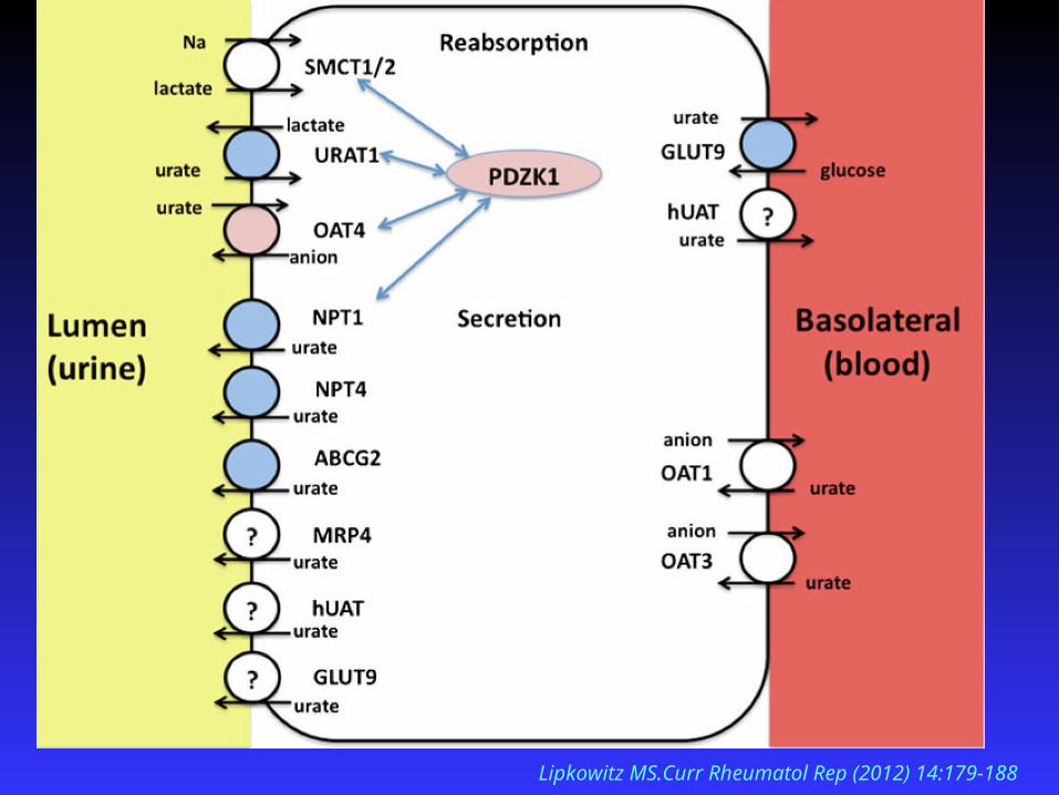
Lipkowitz MS.Curr Rheumatol Rep (2012) 14:179-188

Hypouricemia < 119 µmol/l (2 mg/dL)
it is important to distinguish :
primary genetic defect - hereditary xanthinuria
transport defect - primary renal hypouricemia (RHUC1, RHUC2)
secondary increased renal secretion (Fanconi sy.,Wilson´s disease) medication (allopurinol,salicylates )
severe liver disease
thyrotoxicosis, diabetes mellitus, acute respiratory sy.

Hereditary xanthinuria
xanthine oxidoreductase ( XO) deficiency type I
XO def. + aldehyde oxidase deficiency type II
molybdenum cofactor def. “ “ + sulfite oxidase def.
dg.markers: hypouricemia
high urinary concentration of xanthine
symptoms: cca 50% patients - hematuria,renal colic acute renal failure,
crystalluria,urolithiasis
th: low purine diet,high fluid intake
(alkalization of urine is of no value)

• new transport defect of uric acid
• biochemical markers– hypouricemia (SKM<120 μmol/l)
– increased excretion fraction of uric acid (EFKM >10% )
• clinical features
– urolithiasis– acute renal failure (exercise-induced)
RHUC 1 - URAT1 (SLC22A12 gene) RHUC 2 - GLUT 9 (SLC2A9 gene)
Hereditary renal hypouricemia
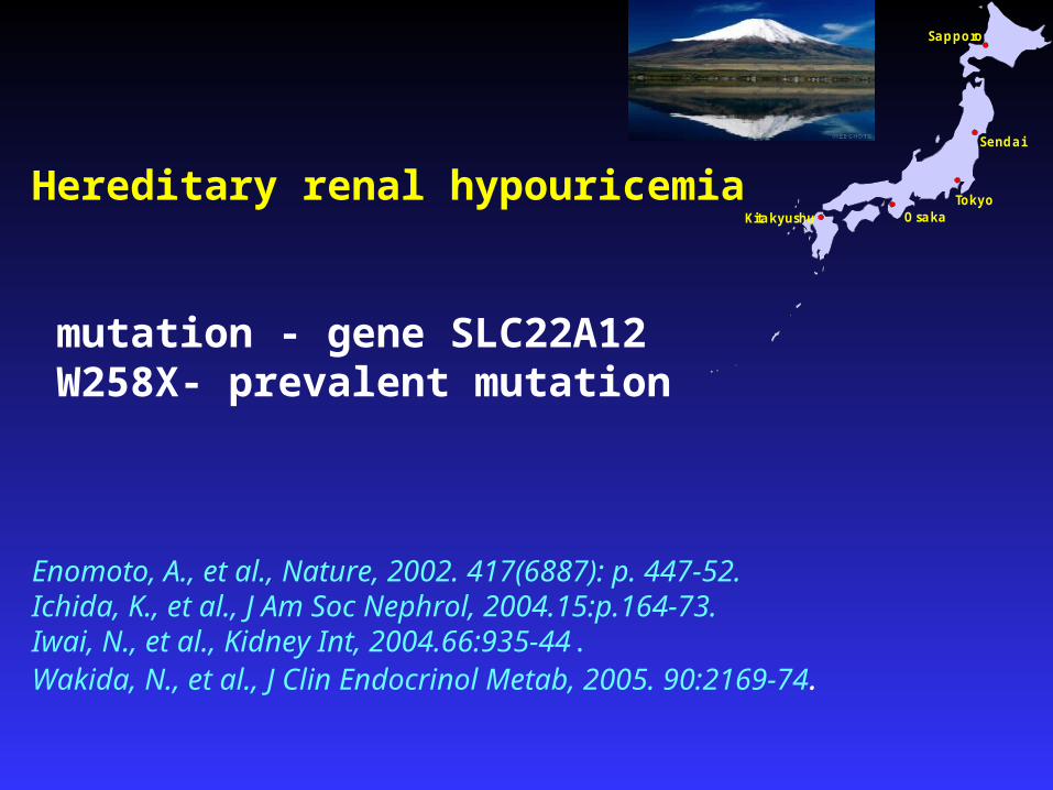
Hereditary renal hypouricemia mutation - gene SLC22A12 W258X- prevalent mutation
Enomoto, A., et al., Nature, 2002. 417(6887): p. 447-52. Ichida, K., et al., J Am Soc Nephrol, 2004.15:p.164-73.Iwai, N., et al., Kidney Int, 2004.66:935-44.Wakida, N., et al., J Clin Endocrinol Metab, 2005. 90:2169-74.
Sendai
Sapporo
TokyoOsakaKitakyushu
Institute of Inherited Metabolic Disorder, First Faculty of of Medicine, Charles University, Prague
( patients with HPRT def., FJHN, APRT def, ASL def., ADA def.)
Are disorders with hypouricemia also
in the Czech population ?

Investigation of unexplained hypouricemia
exclusion of secondary causes of hypouricemia !
1.assessment of uric acid - serum , urine
2.urinary purine metabolites
(+ allopur.loading test)
3.molecular genetic analysis
SLC22A12, SLC2A9
(in cooperation with Japan – SLC17A3, ABCC4, ABCG2 )
AU
0.00
0.02
0.04
0.06
0.08
0.10
0.12
0.14
Minutes
2.00 3.00 4.00 5.00 6.00 7.00 8.00 9.00 10.00 11.00 12.00 13.00 14.00
XO. def.HX Xanthine
UA

Allopurinol loading test
patients with with XO def. type I - able
II - not able to metabolize
allopurinol to oxipurinol
1. 300 mg of allopurinol (adults) …… after overnight fasting
2. Oxipurinol determined in plasma … after 1 hour
Ichida K et al (1997) J Clin Invest 99, 2391-97

Clinical and biochemical findings in patients with XDH deficiency
case age of dg . first sign uric acid Kaufman xanthine (years) in serum index in urine
(µmol/l) (UA/Cr) (mmol/mol Cr)
1. 3 hematuria not detectable 0.002 598.0 renal stone
2. 8 hematuria 53.0 0.04 370.0
3. 9 none 16.0 0.08 327.0
4. 30 none not detectable 180.0
controls 120- 360 0.7 30.0
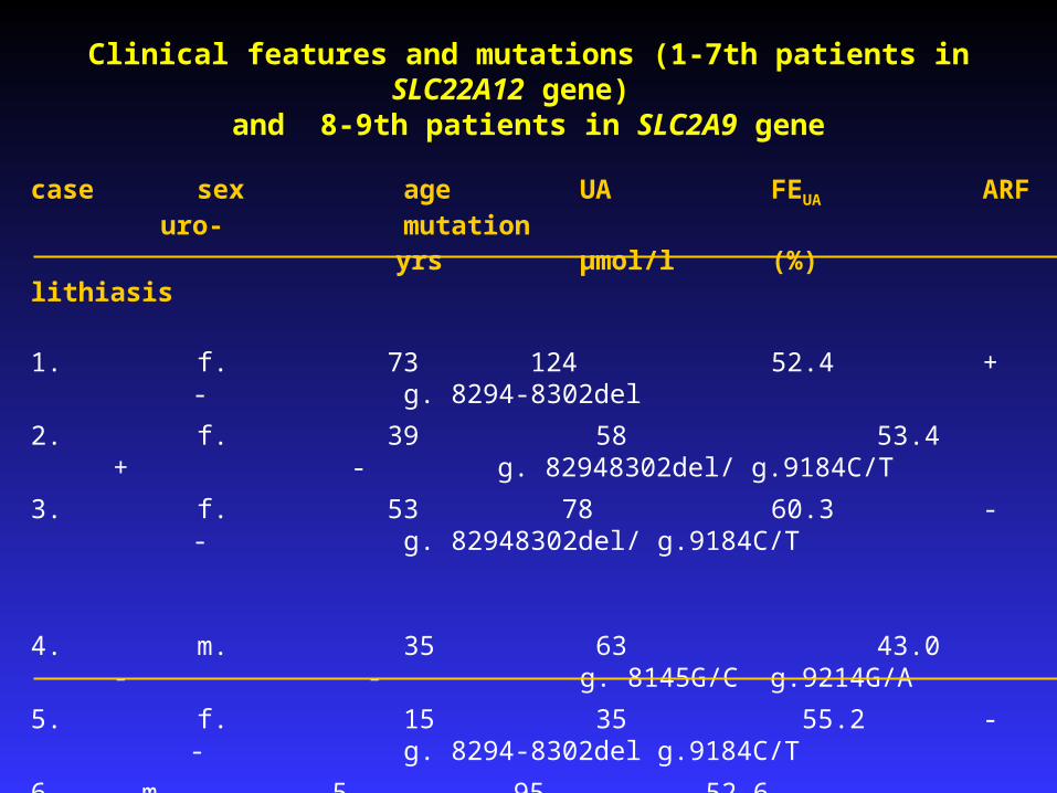
Clinical features and mutations (1-7th patients in SLC22A12 gene) and 8-9th patients in SLC2A9 gene
case sex age UA FEUA ARF uro- mutation
yrs μmol/l (%) lithiasis
1. f. 73 124 52.4 + - g. 8294-8302del
2. f. 39 58 53.4 + - g. 82948302del/ g.9184C/T
3. f. 53 78 60.3 - - g. 82948302del/ g.9184C/T
4. m. 35 63 43.0 - - g. 8145G/C g.9214G/A
5. f. 15 35 55.2 - - g. 8294-8302del g.9184C/T
6. m. 5 95 52.6 - + 1242-1250delGCTGGCAGG
7. m. 5 50. - - 1245-1253delGGCAGGGCT
8 f. 18 11 240.0 - - g. 43412_43413insC
9. m. 23 10 220.0 - - g. 43412_43413insC

Clinical features (two UK patients with acute renal failure-ARF) and mutations in SLC2A9 gene
case sex age UA FEUA Cr ARF mutation
yrs μmol/l (%) μmol/l
1. m 12 40 93.0 297 + p.G216R; p.N333S
2. m 14 58 53.4 202 + p.G216R
- further evidence … …..SLC2A9 is a causative gene in RHUC2
- supports the prediction….both URAT1 and GLUT9 are essential for UA reabsorption
.
Sebesta I. Adv Chronic Kidney Dis 2012,19(6):398-403 Stiburkova B ,Ichida K,Sebesta I. Mol Genet Metab.2011,102(4):1411-5
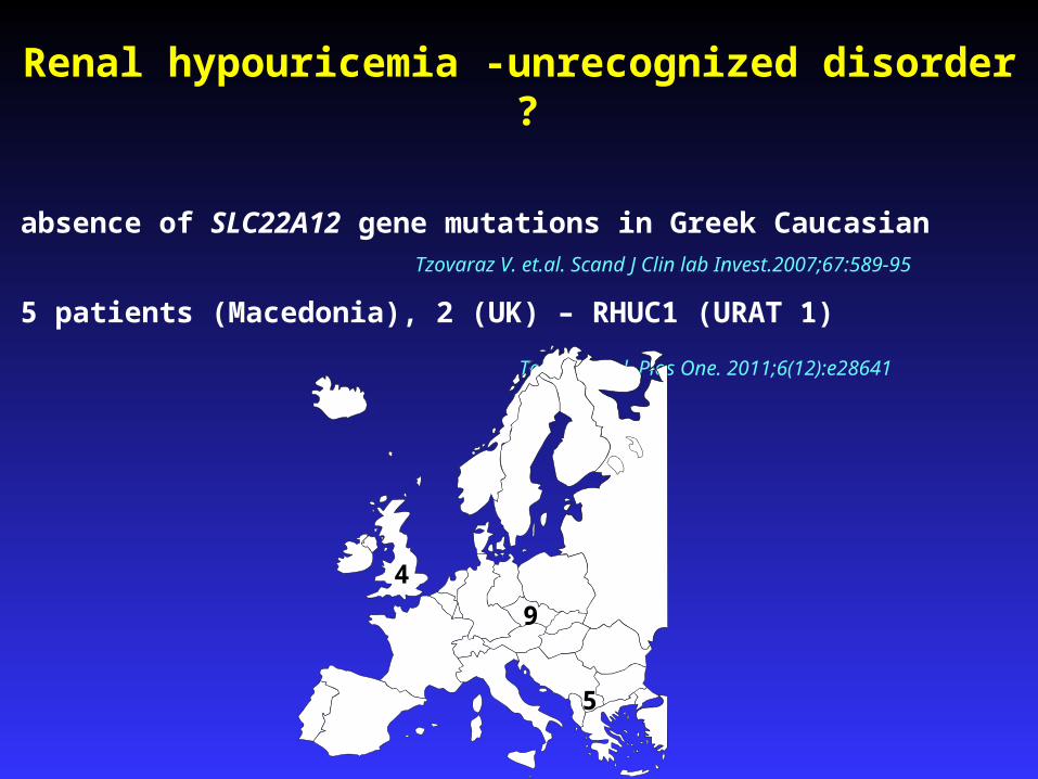
Renal hypouricemia -unrecognized disorder ?
absence of SLC22A12 gene mutations in Greek Caucasian Tzovaraz V. et.al. Scand J Clin lab Invest.2007;67:589-95
5 patients (Macedonia), 2 (UK) – RHUC1 (URAT 1)
Tesic V. et.al. Plos One. 2011;6(12):e28641
9
5
4
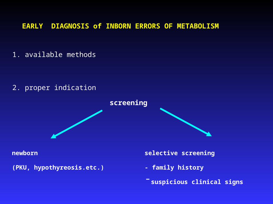
EARLY DIAGNOSIS of INBORN ERRORS OF METABOLISM
1. available methods
2. proper indication
screening
newborn
(PKU, hypothyreosis.etc.)
selective screening
- family history
-suspicious clinical signs
diagnostic guidelines

Dg. flow chart for unexplained hypouricemia (SUA: <120 μmol/l )
Evaluation of case history ( urolithiasis, seizures, immunodeficiency) Exclusion of secondary causes ( drugs /allopurinol/, Fanconi sy. etc.)
↓1. Estimation of EXCRETION FRACTION OF UA ↓
if high→ - mol.genet.analysis of URAT1, GLUT9
2. Urinary concentration of XANTHINE, S-SULFOCYSTEIN, THIOSULFATE
3. Urinary concentration of (DEOXY) GUANOSINE, (DEOXY) INOSINE ↓ if positive - assay of purine nucleoside phoshorylase (PNP) in ery.
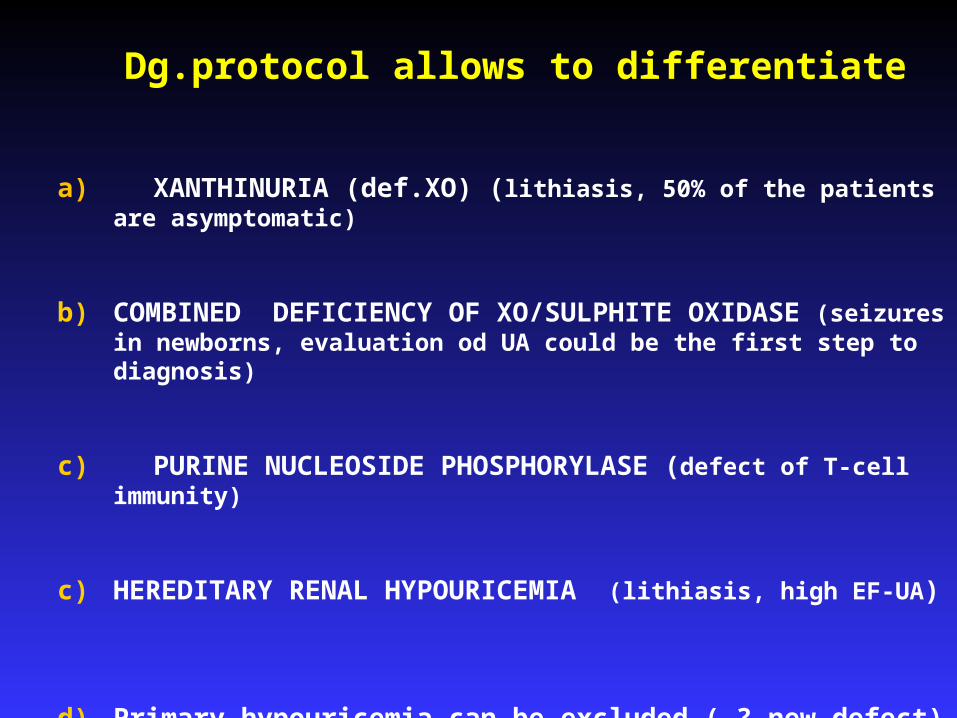
Dg.protocol allows to differentiate
a) XANTHINURIA (def.XO) (lithiasis, 50% of the patients are asymptomatic)
b) COMBINED DEFICIENCY OF XO/SULPHITE OXIDASE (seizures in newborns, evaluation od UA could be the first step to diagnosis)
c) PURINE NUCLEOSIDE PHOSPHORYLASE (defect of T-cell
immunity)
c) HEREDITARY RENAL HYPOURICEMIA (lithiasis, high EF-UA)
d) Primary hypouricemia can be excluded ( ? new defect)
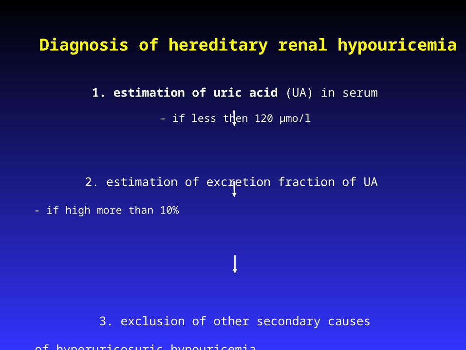
Diagnosis of hereditary renal hypouricemia
1. estimation of uric acid (UA) in serum
- if less then 120 µmo/l
2. estimation of excretion fraction of UA
- if high more than 10%
3. exclusion of other secondary causes
of hyperuricosuric hypouricemia
if excluded
4. molecular analysis of SLC22A and SLC2A9 genes

Conclusions
• hypouricemia → risk factor for kidney injury
→ indication for detailed purine metabolic investigation
• hypouricemia can be good diagnostic tool – enables to find asymptomatic patients
• available guidelines will help for early diagnosis of purine disorders with hypouricemia
Conclusions
• first patients with hereditary renal hypouricemia and xanthinuria were diagnosed in Czech population
• findings of a defect in the SLC2A9 gene provides further evidence that SLC2A9 is a causative gene in renal hypouricemia and support the prediction that normal function of both URAT1 and GLUT 9 are essential for normal uric reabsorption
• renal hypouricemia is still unrecognized disorder and probably not wide spread in Asia only