Diagnosis and treatment of endometrial cancer · endometrial fluid and an endometrial thickness...
45
Prof Vanita Suri Head Dept of Obstetrics and Gynecology PGIMER, Chandigarh Diagnosis and Surgical Treatment of Endometrial cancer
Transcript of Diagnosis and treatment of endometrial cancer · endometrial fluid and an endometrial thickness...
Prof Vanita Suri
Head Dept of Obstetrics and Gynecology
PGIMER, Chandigarh
Diagnosis and Surgical Treatment of
Endometrial cancer
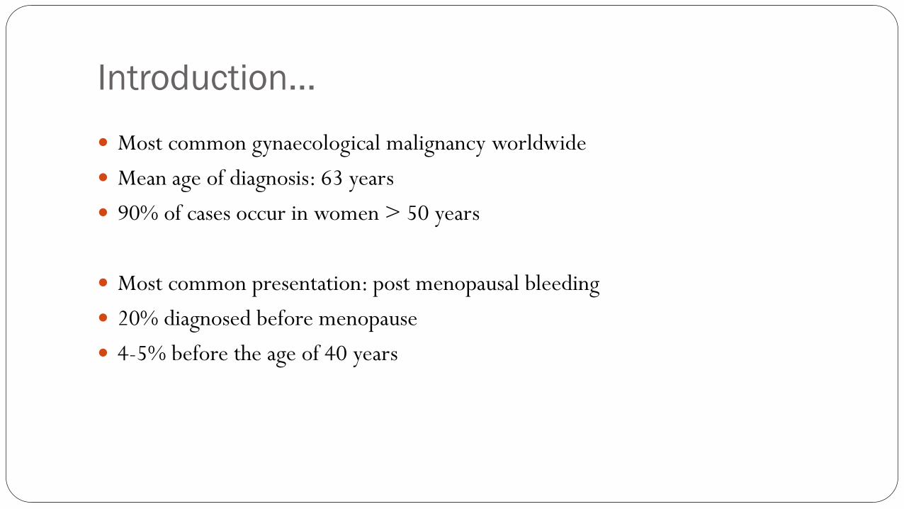
Introduction…
Most common gynaecological malignancy worldwide
Mean age of diagnosis: 63 years
90% of cases occur in women > 50 years
Most common presentation: post menopausal bleeding
20% diagnosed before menopause
4-5% before the age of 40 years

REFERENCES FOR TODAY
• NCCN CLINICAL PRACTICE GUIDELINES IN ONCOLOGY
• APRIL 2017

Important points to ponder----
Who are at risk…whom to screen and how??
Approach to postmenopausal lady with bleeding P/V
Approach to postmenopausal lady with incidentally diagnosed thick ET/ fluid
Premenopausal lady: when to suspect EC
Once decided for biopsy: how to sample
Once diagnosed: how to work-up
Evidence based treatment options
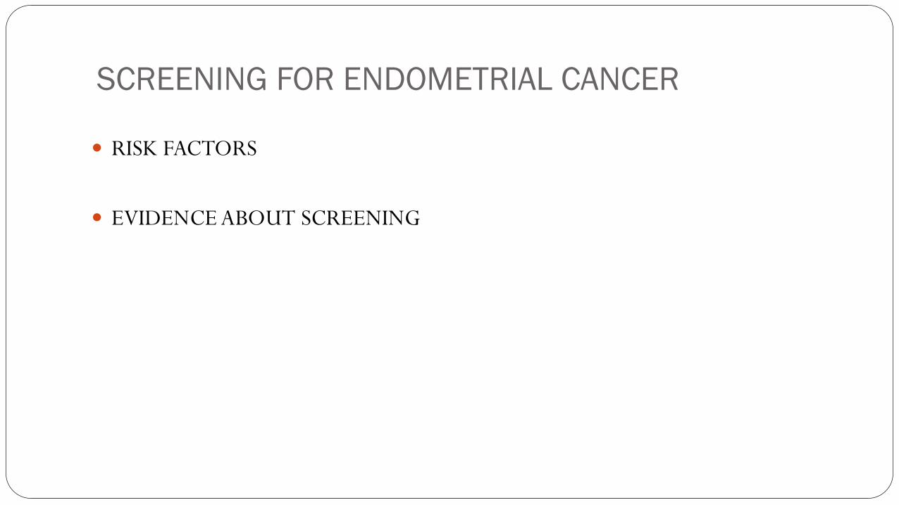
SCREENING FOR ENDOMETRIAL CANCER
RISK FACTORS
EVIDENCE ABOUT SCREENING

Risk Factors
Prolonged estrogen exposure
Tamoxifen
Obesity
Diabetes
Age (15% less than 50 years)
Hypertension
Reproductive factors (Early menarche, nulliparity, Late menopause)
OCPs
Medroxy progesterone
Progesterone IUCD
Smoking
Genetic predisposition
Lynch/ HNPCC
Autosomal dominant
Germline mutations in mismatch
repair genes
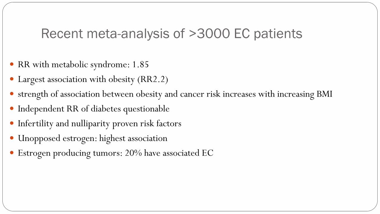
Recent meta-analysis of >3000 EC patients
RR with metabolic syndrome: 1.85
Largest association with obesity (RR2.2)
strength of association between obesity and cancer risk increases with increasing BMI
Independent RR of diabetes questionable
Infertility and nulliparity proven risk factors
Unopposed estrogen: highest association
Estrogen producing tumors: 20% have associated EC

Screening in general population
There is no evidence for endometrial cancer screening in the general population
Level of evidence: II
Strength of recommendation: A

Screening in high risk groups
Routine surveillance in asymptomatic women with obesity, PCOS, diabetes mellitus,
infertility, nulliparity or late menopause is not recommended
Level of evidence: III
Strength of recommendation: B
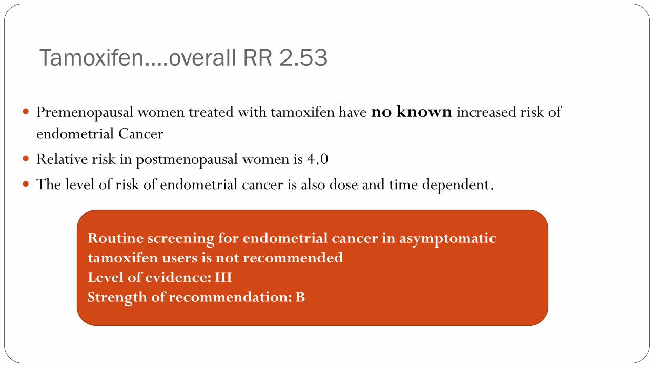
Tamoxifen….overall RR 2.53
Premenopausal women treated with tamoxifen have no known increased risk of
endometrial Cancer
Relative risk in postmenopausal women is 4.0
The level of risk of endometrial cancer is also dose and time dependent.
Routine screening for endometrial cancer in asymptomatic
tamoxifen users is not recommended
Level of evidence: III
Strength of recommendation: B
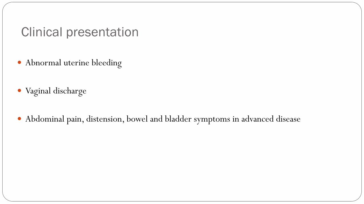
Clinical presentation
Abnormal uterine bleeding
Vaginal discharge
Abdominal pain, distension, bowel and bladder symptoms in advanced disease

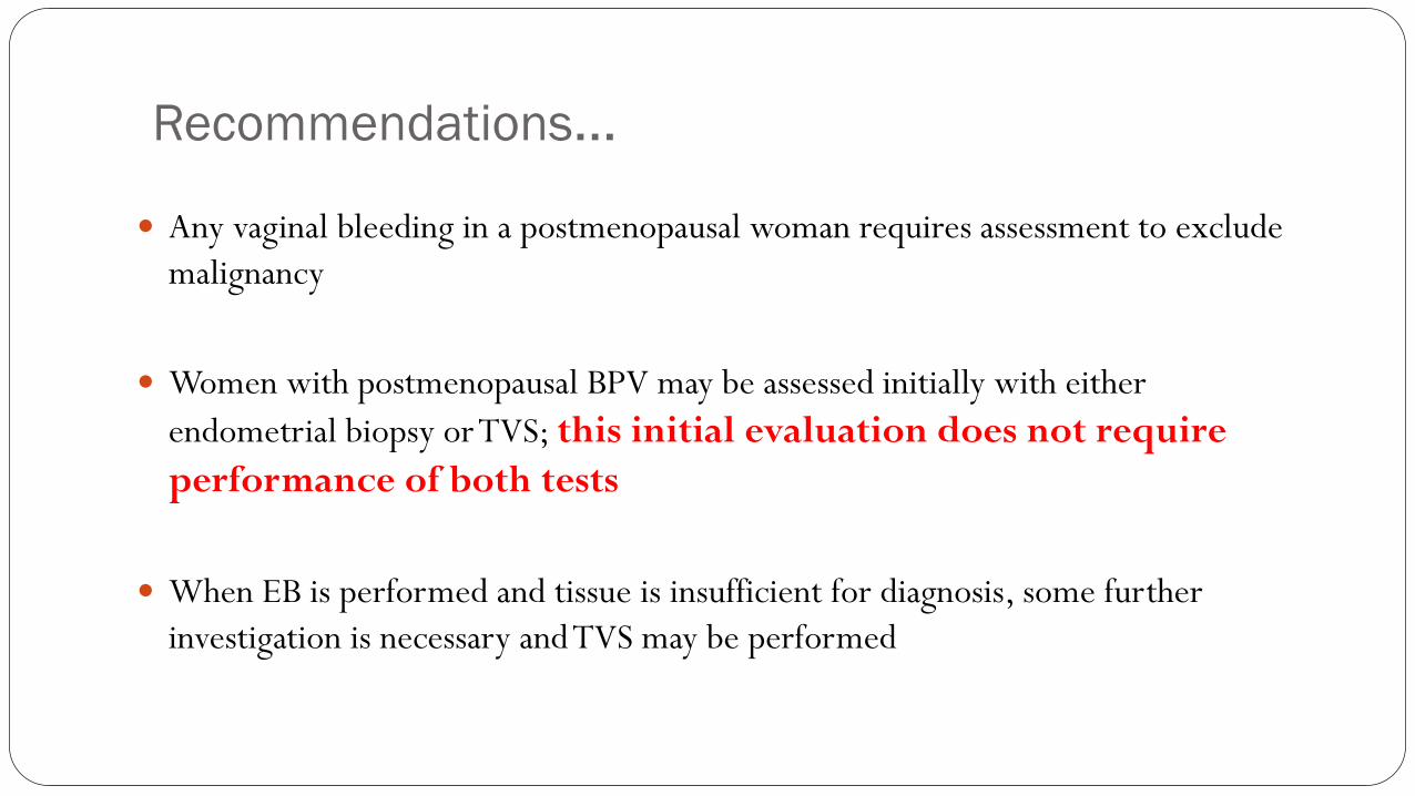
Recommendations…
Any vaginal bleeding in a postmenopausal woman requires assessment to exclude
malignancy
Women with postmenopausal BPV may be assessed initially with either
endometrial biopsy or TVS; this initial evaluation does not require
performance of both tests
When EB is performed and tissue is insufficient for diagnosis, some further
investigation is necessary and TVS may be performed

Recommendations
When TVS is performed and ET is less than or equal to 4mm, endometrial
sampling is not required.
ET >4mm should trigger alternative evaluation, as should an inability to
adequately visualize thickness.
When bleeding persists despite negative initial evaluation, additional assessment is
indicated. Because of extremely high negative predictive value, non-invasive
nature, low cost and easy availability: TVS is a reasonable first approach
to PMB

TVS
ACOG ≤ 4mm
SRU ≤ 5mm
supported by a meta-analysis of 35 prospective studies that included data from
almost 6000 women with postmenopausal bleeding.
sensitivity and specificity of TVUS for detection of endometrial cancer
at a 4 mm thickness threshold were 96 and 53 percent, and at a 5 mm
threshold the sensitivity and specificity were 96 and 61 percent.
At 3 mm: sensitivity is 98%
3-5mm

Post menopausal lady with incidentally diagnosed thick
ET
15-20% endometrial cancers occur in women without vaginal bleeding
Significance of ET>4mm in asymptomatic postmenopausal lady has not been
established, does not routinely trigger evaluation
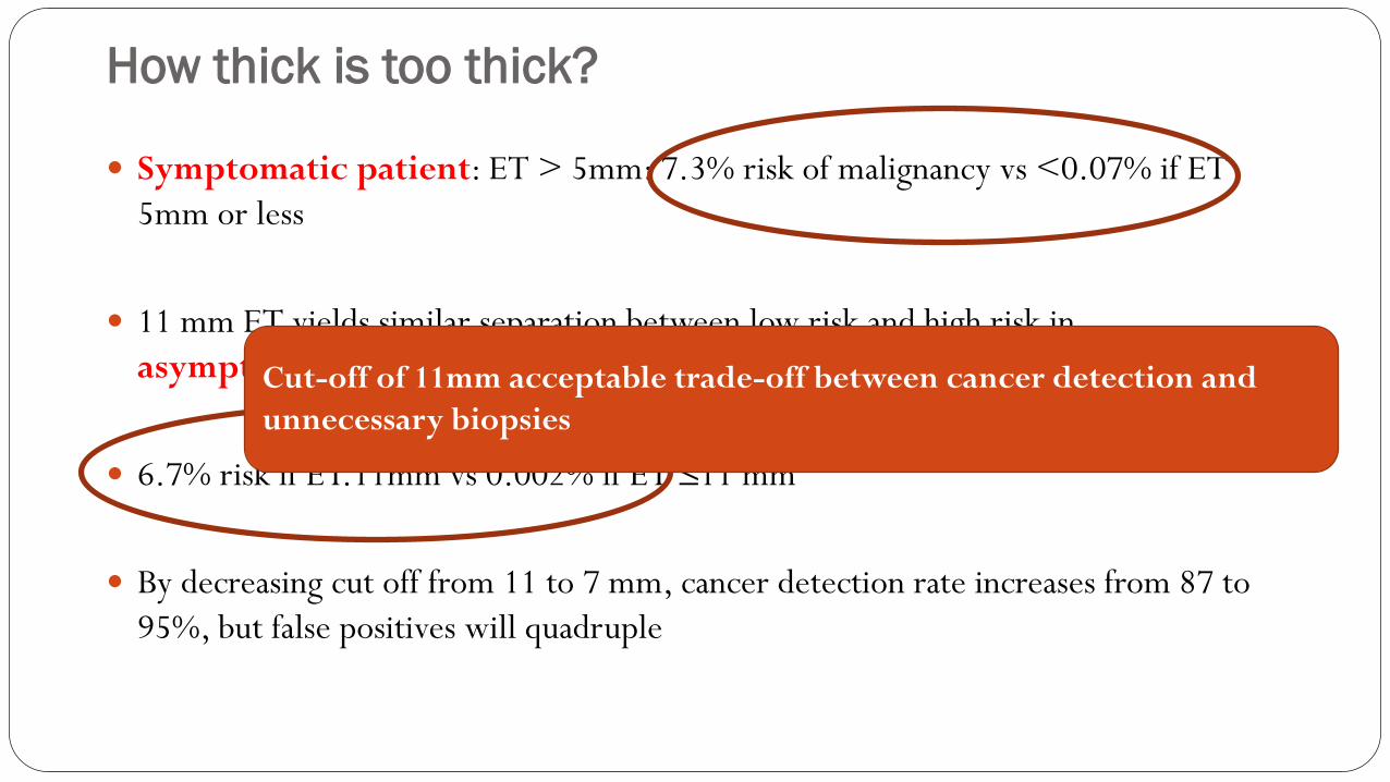
How thick is too thick?
Symptomatic patient: ET > 5mm: 7.3% risk of malignancy vs <0.07% if ET
5mm or less
11 mm ET yields similar separation between low risk and high risk in
asymptomatic post-menopausal women
6.7% risk if ET.11mm vs 0.002% if ET ≤11 mm
By decreasing cut off from 11 to 7 mm, cancer detection rate increases from 87 to
95%, but false positives will quadruple
Cut-off of 11mm acceptable trade-off between cancer detection and
unnecessary biopsies
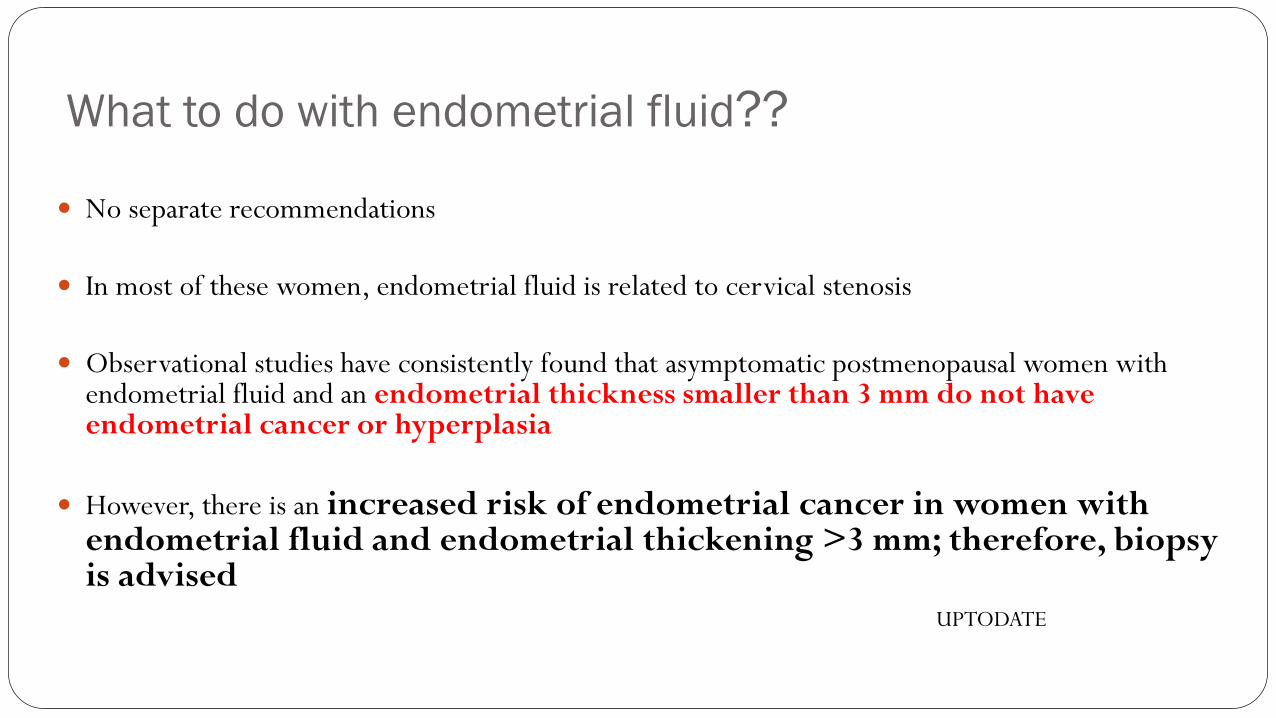
What to do with endometrial fluid??
No separate recommendations
In most of these women, endometrial fluid is related to cervical stenosis
Observational studies have consistently found that asymptomatic postmenopausal women with endometrial fluid and an endometrial thickness smaller than 3 mm do not have endometrial cancer or hyperplasia
However, there is an increased risk of endometrial cancer in women with endometrial fluid and endometrial thickening >3 mm; therefore, biopsy is advised
UPTODATE

Pre-menopausal lady
Endometrial evaluation for abnormal bleeding at > 40 years or history suggestive of
excess estrogen exposure irrespective of endometrial thickness
For asymptomatic women:
Optimal cut-off not defined
Cut-off of 16mm recommended by some

Once decided we need to sample…
how to sample??
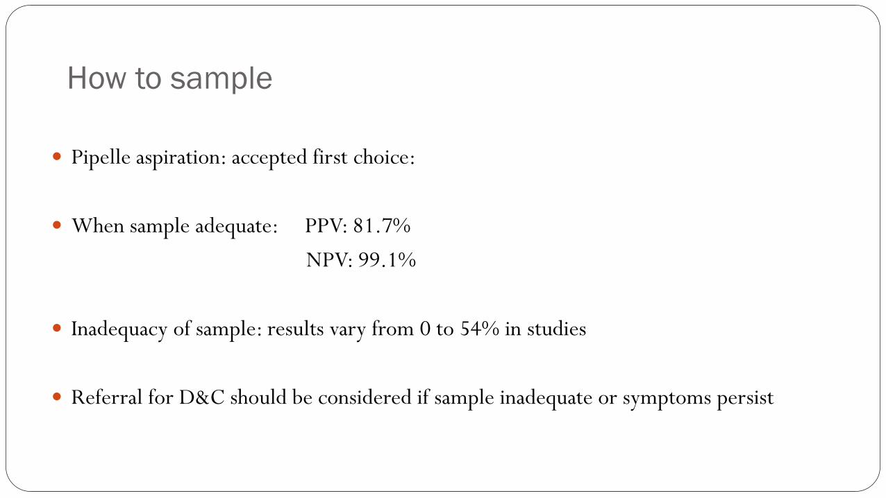
How to sample
Pipelle aspiration: accepted first choice:
When sample adequate: PPV: 81.7%
NPV: 99.1%
Inadequacy of sample: results vary from 0 to 54% in studies
Referral for D&C should be considered if sample inadequate or symptoms persist
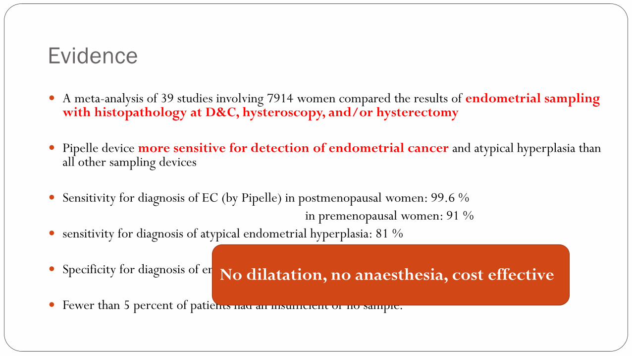
Evidence
A meta-analysis of 39 studies involving 7914 women compared the results of endometrial sampling with histopathology at D&C, hysteroscopy, and/or hysterectomy
Pipelle device more sensitive for detection of endometrial cancer and atypical hyperplasia than all other sampling devices
Sensitivity for diagnosis of EC (by Pipelle) in postmenopausal women: 99.6 %
in premenopausal women: 91 %
sensitivity for diagnosis of atypical endometrial hyperplasia: 81 %
Specificity for diagnosis of endometrial carcinoma: 98 to 100 percent.
Fewer than 5 percent of patients had an insufficient or no sample.
No dilatation, no anaesthesia, cost effective
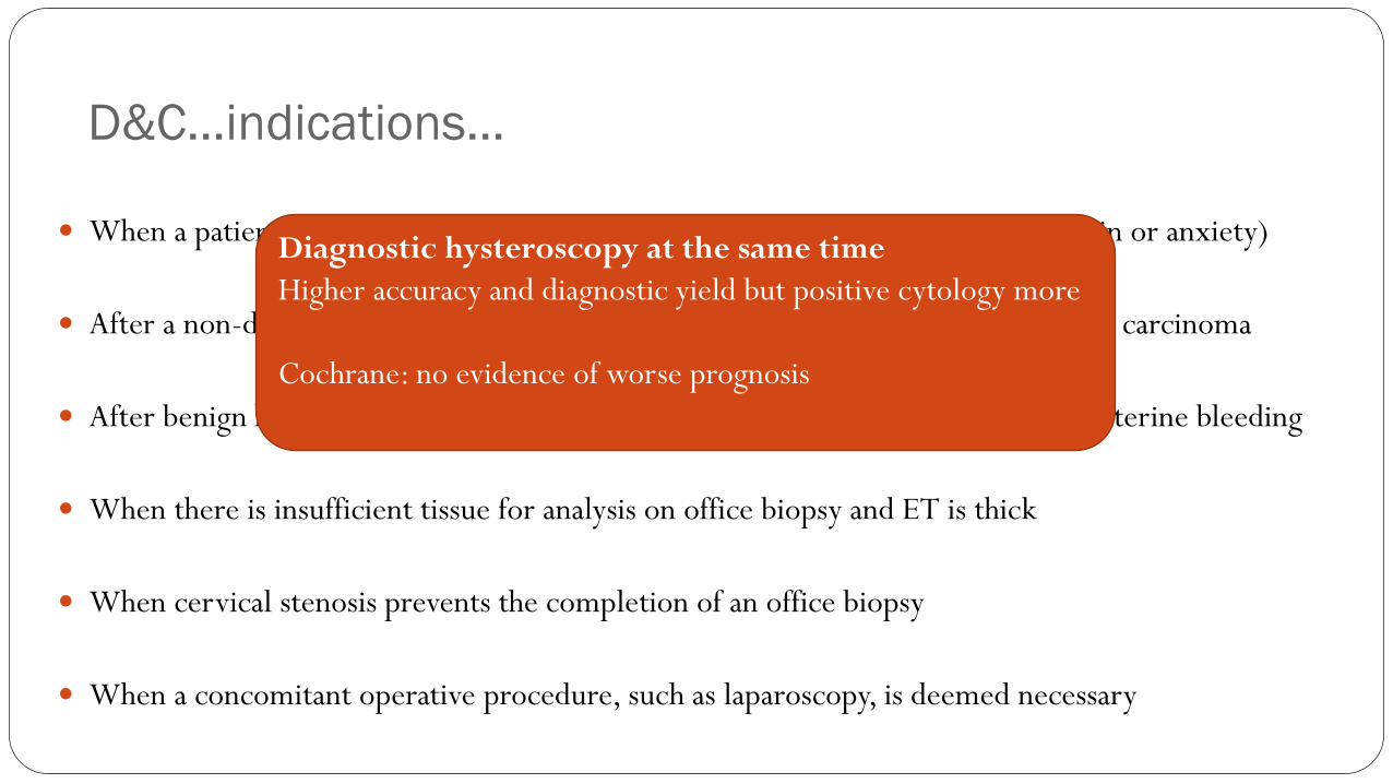
D&C…indications…
When a patient is not able to tolerate an office endometrial biopsy (eg, due to pain or anxiety)
After a non-diagnostic office biopsy in women who are at high risk of endometrial carcinoma
After benign histology on office biopsy in women who have persistent abnormal uterine bleeding
When there is insufficient tissue for analysis on office biopsy and ET is thick
When cervical stenosis prevents the completion of an office biopsy
When a concomitant operative procedure, such as laparoscopy, is deemed necessary
Diagnostic hysteroscopy at the same time
Higher accuracy and diagnostic yield but positive cytology more
Cochrane: no evidence of worse prognosis

Once diagnosed;
how to work-up
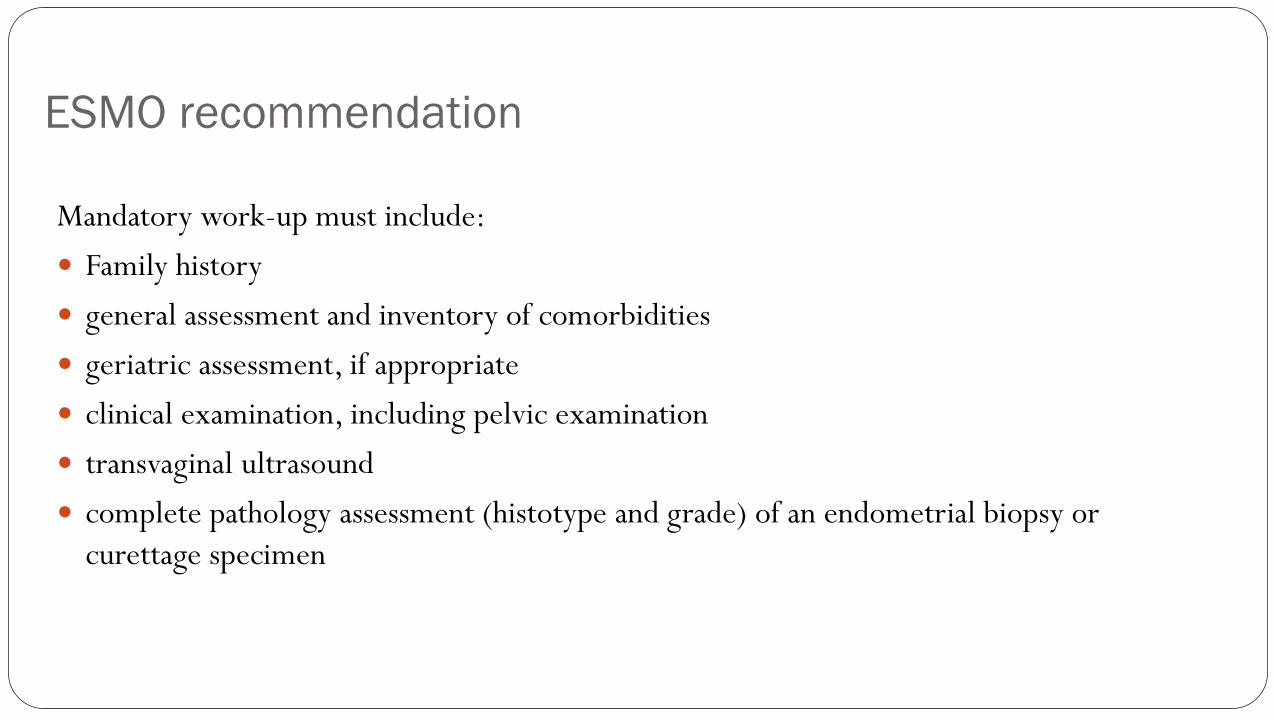
ESMO recommendation
Mandatory work-up must include:
Family history
general assessment and inventory of comorbidities
geriatric assessment, if appropriate
clinical examination, including pelvic examination
transvaginal ultrasound
complete pathology assessment (histotype and grade) of an endometrial biopsy or
curettage specimen
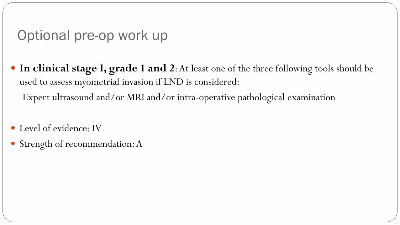
Optional pre-op work up
In clinical stage I, grade 1 and 2: At least one of the three following tools should be
used to assess myometrial invasion if LND is considered:
Expert ultrasound and/or MRI and/or intra-operative pathological examination
Level of evidence: IV
Strength of recommendation: A
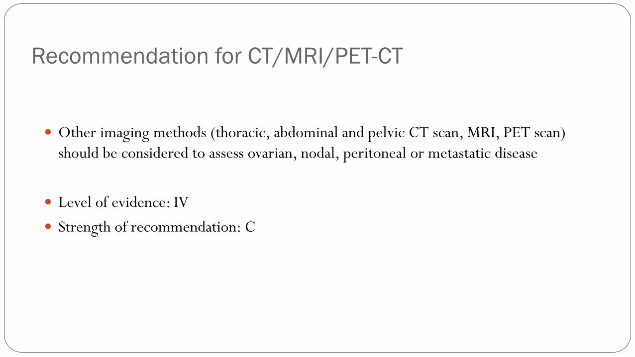
Recommendation for CT/MRI/PET-CT
Other imaging methods (thoracic, abdominal and pelvic CT scan, MRI, PET scan)
should be considered to assess ovarian, nodal, peritoneal or metastatic disease
Level of evidence: IV
Strength of recommendation: C
Place of MRI
Endometrial carcinomas are divided into two histologic subtypes. Endometrioid adenocarcinoma (type 1), Type 2 endometrial carcinomas include serous papillary and clear cell adenocarcinomas
Serous papillary, clear cell, and grade 3 endometrioid adenocarcinomas demonstrate more aggressive tumor biologic characteristics and have a 50% pretest probability of locally advanced or distant disease at manifestation
information provided by MR imaging is invaluable in managing endometrial carcinoma In response to growing evidence, the National Cancer Institute in France incorporated preoperative MR imaging into its guidelines for managing endometrial carcinoma. MR imaging is also recommended by the ESUR for staging high-risk endometrial carcinoma, including all histologic subtype 2 and high-grade subtype 1 tumors
MR imaging plays an important role in the treatment stratification of patients with endometrial carcinoma. Accurate preoperative delineation of local disease extent and involved lymph nodes is essential
Tumor markers….ESMO
There is evidence that CA-125 and HE-4 are significantly correlated with histological grade,
stage, lymph node metastases, myometrial invasion and cervical involvement
However, the appropriate cut off has not been established and evidence that
serum marker assessment is clinically useful is lacking
There is no evidence for the clinical usefulness of serum tumour markers, including CA-125
Level of evidence: IV
Strength of recommendation: B Selective in unstaged patients and
serous tumors

Whom to offer genetic screening??
Facts
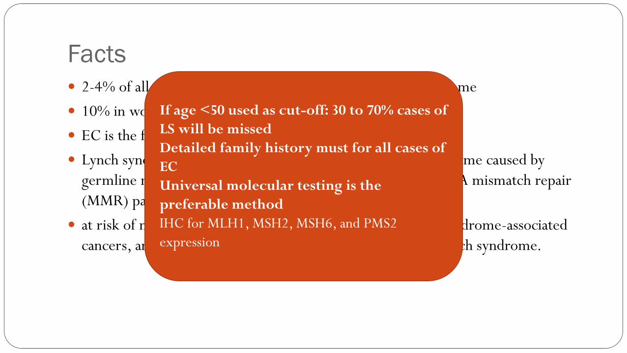
2-4% of all those with endometrial cancer have Lynch Syndrome
10% in women diagnosed under the age of 50
EC is the first malignancy in 50% of LS women
Lynch syndrome, an autosomal dominant cancer-prone syndrome caused by
germline mutations in genes encoding components of the DNA mismatch repair
(MMR) pathway.
at risk of metachronous colorectal cancer and other Lynch syndrome-associated
cancers, and their first-degree relatives are at 50% risk of Lynch syndrome.
If age <50 used as cut-off: 30 to 70% cases of
LS will be missed
Detailed family history must for all cases of
EC
Universal molecular testing is the
preferable method
IHC for MLH1, MSH2, MSH6, and PMS2
expression

MANAGEMENT OF ENDOMETRIAL CANCER
TAH with BSO: mainstay of treatment till 30 years back
In 1988: mounting evidence that extrauterine disease was associated with poor outcomes
and that patients with advanced disease required more than just surgical intervention
EC: converted to a surgically staged disease
Comprehensive surgical staging
Advantages of comprehensive surgical staging: diagnosis, prognosis, and proper triage of
patients for adjuvant therapy
FIGO’s surgical staging system for endometrial cancer is based on surgical pathology, and
comprehensive staging allows for accurate definition of disease extent
Hence, the recommended initial management of endometrial cancer should include
comprehensive surgical staging (total hysterectomy, BSO, pelvic and para-aortic
lymphadenectomy, and the collection of peritoneal cytology [pelvic washings])
Cytology to be taken (though does not affect prognosis)
Omental biopsy: for clear cell, serous and carcinosarcoma
Thorough visual inspection of all peritoneal surfaces of abdomen
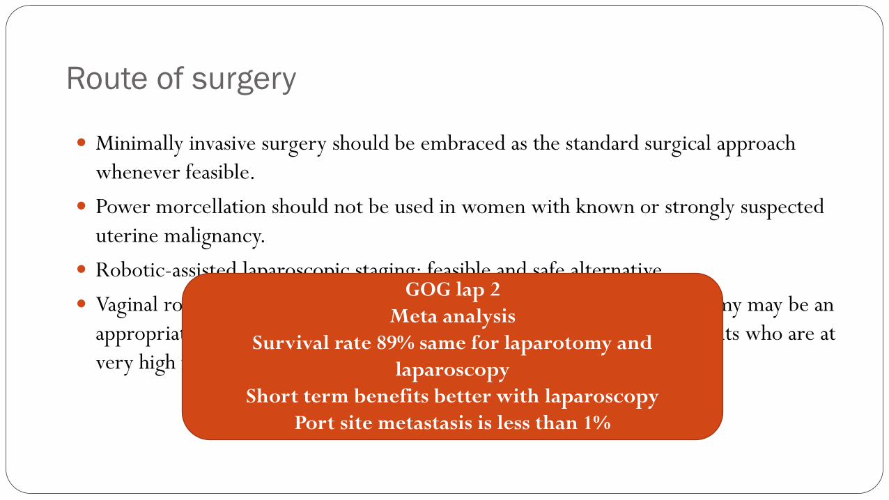
Route of surgery
Minimally invasive surgery should be embraced as the standard surgical approach
whenever feasible.
Power morcellation should not be used in women with known or strongly suspected
uterine malignancy.
Robotic-assisted laparoscopic staging: feasible and safe alternative
Vaginal route precludes thorough abdominal survey and lymphadenectomy may be an
appropriate treatment for early-stage endometrial cancer in select patients who are at
very high risk of surgical morbidity.
GOG lap 2
Meta analysis
Survival rate 89% same for laparotomy and
laparoscopy
Short term benefits better with laparoscopy
Port site metastasis is less than 1%
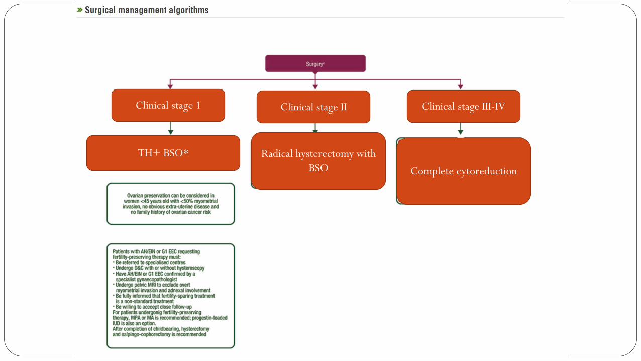
Clinical stage 1 Clinical stage II Clinical stage III-IV
TH+ BSO* Radical hysterectomy with
BSO Complete cytoreduction

Clinical Stage 1
Low risk:
clinical stage
1A, G1/2
Endometrioid
intermediate risk:
clinical stage 1A, G3
or clinical stage IB,
G1/2
Endometrioid
High risk: clinical stage
1B,G3
Endometrioid
All stages with non-
endometrioid
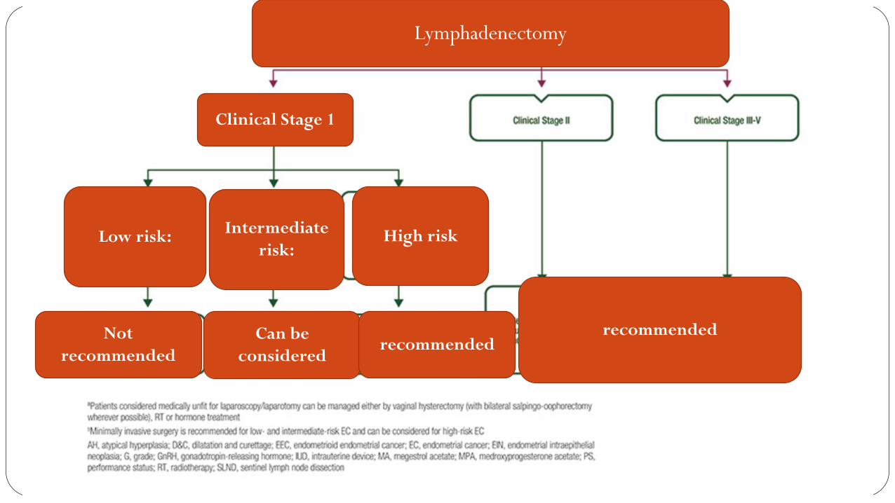
Clinical Stage 1
Low risk:
Not
recommended
Intermediate
risk:
Can be
consideredrecommended
recommended
High risk
Lymphadenectomy
Lymphadenectomy
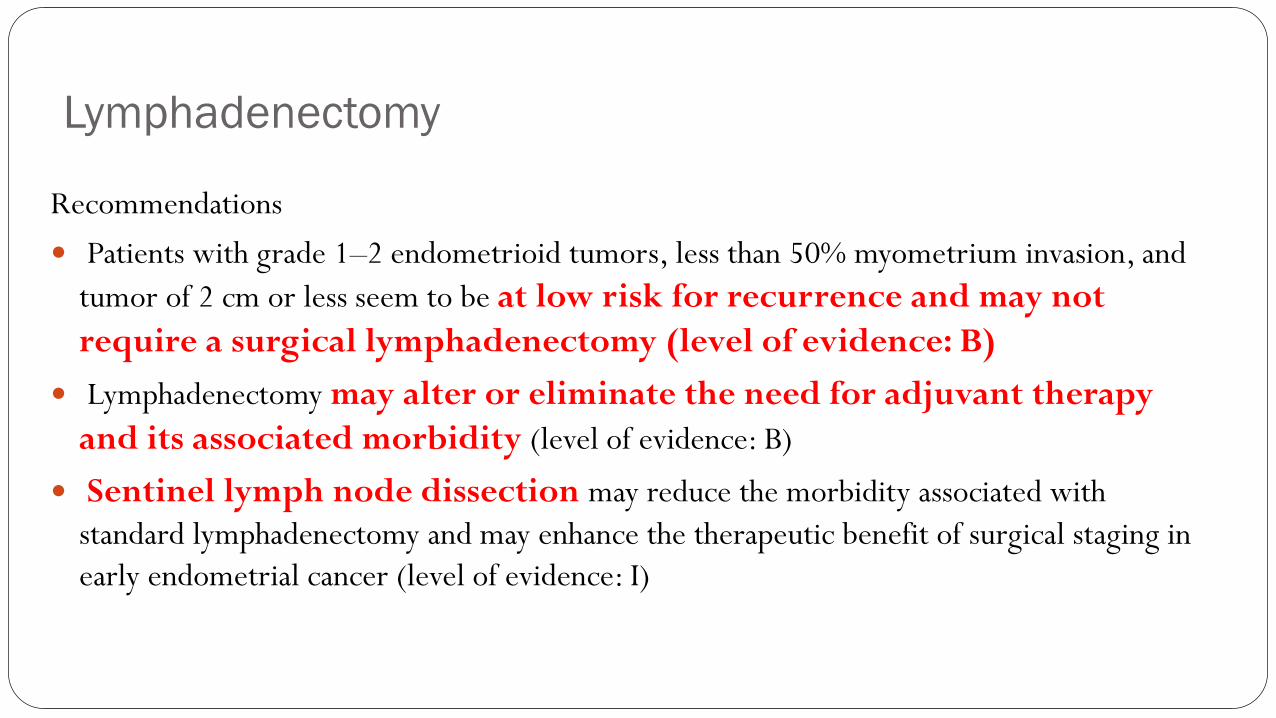
Recommendations
Patients with grade 1–2 endometrioid tumors, less than 50% myometrium invasion, and
tumor of 2 cm or less seem to be at low risk for recurrence and may not
require a surgical lymphadenectomy (level of evidence: B)
Lymphadenectomy may alter or eliminate the need for adjuvant therapy
and its associated morbidity (level of evidence: B)
Sentinel lymph node dissection may reduce the morbidity associated with
standard lymphadenectomy and may enhance the therapeutic benefit of surgical staging in
early endometrial cancer (level of evidence: I)
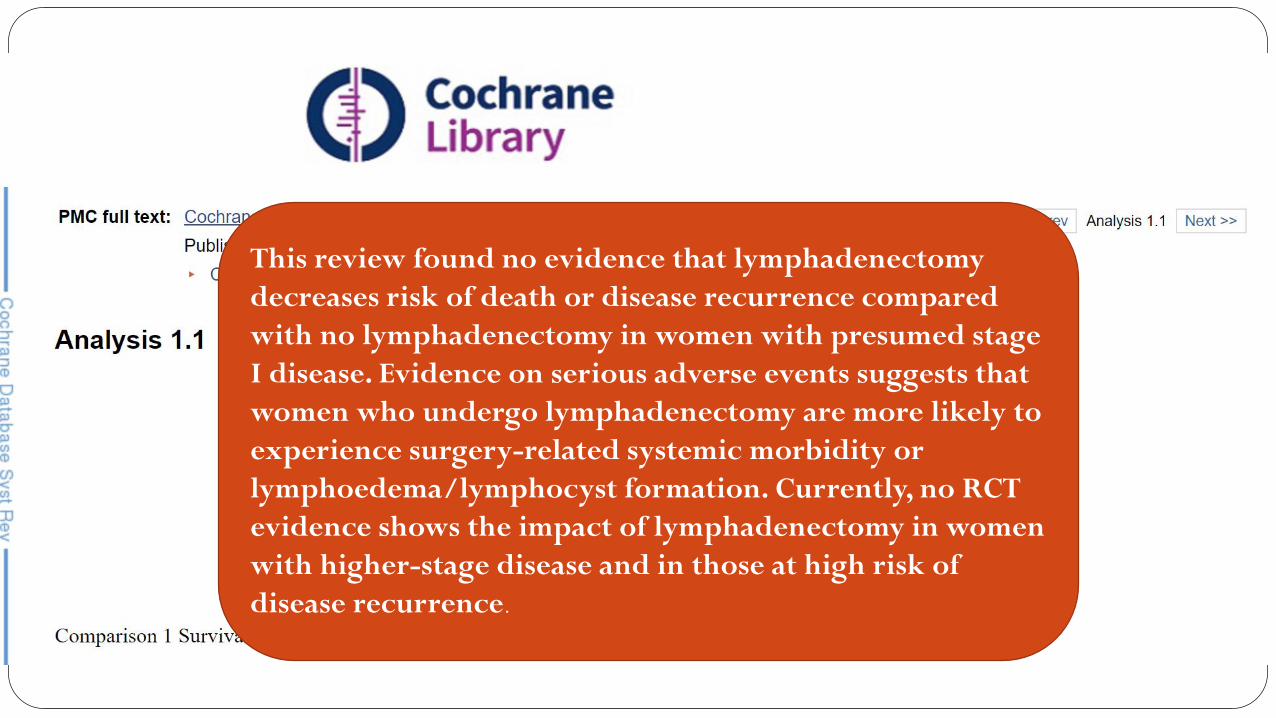
This review found no evidence that lymphadenectomy
decreases risk of death or disease recurrence compared
with no lymphadenectomy in women with presumed stage
I disease. Evidence on serious adverse events suggests that
women who undergo lymphadenectomy are more likely to
experience surgery-related systemic morbidity or
lymphoedema/lymphocyst formation. Currently, no RCT
evidence shows the impact of lymphadenectomy in women
with higher-stage disease and in those at high risk of
disease recurrence.

Advanced-stage or recurrent endometrial cancer
10–15% of all new cases
account for more than 50% of all uterine cancer-related deaths
Is there a benefit from cytoreduction?
Optimal surgical cytoreduction (variably defined as less than or equal to 1 cm or 2
cm) has been found to improve progression-free and overall survival rates in patients
with advanced-stage or recurrent endometrial cancer.
Post-operatively…
Appropriate pathological assessment
Decision for adjuvant therapy
Discussion in tumor board (Team of pathologists, Radiotherapist )
Carry home---- The commonest cancer of genital tract in women world wide (second in India)
Women in the US have a 2.8 percent lifetime risk of being diagnosed with uterine cancer
Known high risk factors
Clinical staging for deciding extent of surgery
Adjuvant treatment based on surgico-pathological staging
Role of lymphadenectomy in early stage?
Role of chemotherapy?
Targeted therapy, Immunotherapy--
No role of screening till now---
Prevention is possible? Genetic testing---, metformin and OCP in PCOS?