Diagnosis and Prognosis of Seizures and Epilepsy in Childhood Hans - Diagnosis.pdfDiagnosis and...
152
Diagnosis and Prognosis of Seizures and Epilepsy in Childhood (Dutch study of epilepsy in childhood) Diagnose en prognose van epileptische aanvallen en epilepsie op de kinderleeftijd (Dutch study of epilepsy in childhood) Hans Stroink
-
Upload
nguyencong -
Category
Documents
-
view
229 -
download
0
Transcript of Diagnosis and Prognosis of Seizures and Epilepsy in Childhood Hans - Diagnosis.pdfDiagnosis and...

Diagnosis and Prognosis of Seizures and Epilepsy in Childhood
(Dutch study of epilepsy in childhood)
Diagnose en prognose van epileptische aanvallen en epilepsie op
de kinderleeftijd(Dutch study of epilepsy in childhood)
Hans Stroink
290158_Stroink_BW.indd 1 11-04-2008 10:53:43
Cover: “In the shade of awareness...”painted by Hans Innemée for this thesis, 2008, design Peter de Jong.
Toelichting schilderij “In the shade of awareness...” door Hans Innemée.
Aanwezigheid, afwezigheid zijn in mijn beleving relatieve begrippen. Tijdens epileptische aanvallen is
mijn zoon Martijn even niet meer bewust in het hier en nu, althans voor hem zelf niet.
Voor mij als vader des te meer, ik beleef zijn “even weg zijn” met een intens bewustzijn.
De wereld gaat “even op zijn kop”. Het beeld van de vogel als heldere tekening tussen de donkere
banen waarin het negatief van de tekening van de vogel is verwerkt verbeeldt “To be AND not to
be...”(vrij naar Shakespeare ) maar dan in één beeld, in één tijd, tegelijkertijd.
© 2008 Hans Stroink, Tilburg, The Netherlands
ISBN/EAN: 978-90-9022994-2
Diagnosis and Prognosis of Seizures and Epilepsy in Childhood
(Dutch study of epilepsy in childhood)
290158_Stroink_BW.indd 2 11-04-2008 10:53:43
Diagnosis and Prognosis of Seizures and Epilepsy in Childhood
(Dutch study of epilepsy in childhood)
Diagnose en prognose van epileptische aanvallen en epilepsie op de kinderleeftijd
(Dutch study of epilepsy in childhood)
Proefschrift
ter verkrijging van de graad van doctor aan de
Erasmus Universiteit Rotterdam
op gezag van de
rector magnificus
Prof.dr. S.W.J. Lamberts
en volgens besluit van het College voor Promoties.
De openbare verdediging zal plaatsvinden op
donderdag 5 juni 2008 om 13.30 uur
door
Hans Stroink
geboren te Zwijndrecht
290158_Stroink_BW.indd 3 11-04-2008 10:53:43
Promotiecommissie Promotor:
Prof.dr. W.F.M Arts
Overige leden:
Prof.dr. A.P. Aldenkamp
Prof.dr. M.M.B. Breteler
Prof.dr. P.A. Sillevis Smit
Copromotor:
Dr. C.A. van Donselaar
The publication of this thesis was supported by - Het Nationale Epilepsiefonds
- Epilepsia Rotterdam
- UCB Nederland Pharma BV
- Cyberonics Europe SA/NV
- sanofi-aventis Nederland BV
- Janssen-Cilag Nederland BV
- Eli Lilly Nederland
- GlaxoSmithKline BV
290158_Stroink_BW.indd 4 11-04-2008 10:53:43
5
Contents
Chapter 1General introduction
PART I DIAGNOSIS
Chapter 2How confident are we of the diagnosis of epilepsy?
Chapter 3Interrater agreement of the diagnosis and classification of a first seizure in childhood
Chapter 4The accuracy of the diagnosis of paroxysmal events in children
Chapter 5Interobserver reliability of visual interpretation of electroencephalograms in children with newly diagnosed seizures
PART II PROGNOSIS
Chapter 6The first unprovoked, untreated seizure in childhood: a hospital based study of the accuracy of the diagnosis, rate of recurrence, and long term outcome after recurrence
Chapter 7Status epilepticus in children with epilepsy
Chapter 8General discussion
Summary
Samenvatting
List of abbreviations
Acknowledgements
Dankwoord
Curiculum vitae
List of publications H. Stroink
List of publications DSEC
7
19
21
33
47
59
69
71
87
105
119
123
141
142
143
144
145
149
290158_Stroink_BW.indd 5 11-04-2008 10:53:43

290158_Stroink_BW.indd 6 11-04-2008 10:53:43

7
Chapter 1
General introduction
290158_Stroink_BW.indd 7 11-04-2008 10:53:43

290158_Stroink_BW.indd 8 11-04-2008 10:53:43
9
Introduction
Many people suffer from one or more epileptic seizures during life, but not all
these people have epilepsy. Moreover, epilepsy is not one disease or syndrome,
but a collection of different disorders, which have in common the repeated occur-
rence of unprovoked epileptic seizures during some time in life.1-3 There are many
genetically determined and acquired causes of epilepsy. The symptomatology of
the seizures can be very different.2 3 Also the course in time of epilepsies is very
diverging. The cause, symptoms, signs and course are influenced to a great deal
by age. Therefore, the epilepsies in childhood and in adulthood differ in many
aspects.1 3 For this reason, children and adults should not be mixed in studies on
epilepsy.
A recent population based study in Denmark found an incidence of 83 new epi-
lepsy patients per 100,000 person-years at risk. The prevalence of active epilepsy
was 0.6% with the highest prevalence in childhood and the lowest between 20 and
40 years.4 In 70% of patients epilepsy starts before the age of 20 years with the
highest incidence in young children.5 In the Danish study the incidence in the first
year of life was 200 per 100,000 children. The cumulative incidence of a period of
active epilepsy was 1% at the age of 10 years and 2% at the age of 25 years.4 So
epilepsies are more common in childhood.
Many studies on the cause and prognosis of epilepsy are done in specialised epi-
lepsy centres. This may cause a bias to more severely affected patients. Moreover
little is known of the natural history of epilepsy because almost all patients are
treated with anti-epileptic drugs (AEDs). Many children are still treated even after
a single seizure.6
“Het Zuid-Hollands Kinderepilepsie Onderzoek” (ZHKO) started in1988. The ZHKO
was initiated by the paediatric neurologists Willem Frans Arts, Boudewijn Peters,
Oebo Brouwer and Hans Stroink (at the time they were employed at the West-
einde Hospital and Juliana Children’s Hospital in The Hague, and at the university
hospitals of Leiden and Rotterdam), by the neurologist Cees van Donselaar and by
the epidemiologist Ada Geerts (Rotterdam). Due to the move of participating pae-
diatric neurologists to other hospitals, later on the departments of paediatric neu-
rology of the University Hospitals of Groningen and Utrecht, and the departments
of paediatric neurology of the St. Elisabeth Hospital and TweeSteden Hospital in
Tilburg joined, whereas the University Hospital of Leiden left the study group. In
1998 “Het Zuid-Hollands Kinderepilepsie Onderzoek” was renamed “Dutch Study
of Epilepsy in Childhood” (DSEC).
To prevent the bias mentioned before, we created a cohort of unselected children
290158_Stroink_BW.indd 9 11-04-2008 10:53:43
10
by enrolling consecutively all children aged one month through 15 years with
new onset epilepsy referred to the participating hospitals from August 1, 1988, to
August 1, 1992. The intake of children with a single unprovoked seizure started
somewhat earlier on January 1, 1988. Children treated before with AEDs and chil-
dren referred from other hospitals were excluded, again to prevent bias to more
severely affected patients.7-10 The object was to study many aspects of childhood
epilepsy in this cohort with unselected children, as reliability and accuracy of the
diagnosis, prognosis, comorbidity, quality of life, and cognitive and behavioural
disturbances. The attending paediatric neurologist completed extensive question-
naires on the description of the events including postictal signs, possible provok-
ing factors, previous medical history and family history. We performed a standard
electroencephalogram (EEG) in each child . If this did not show epileptiform dis-
charges, a re cording after partial sleep depri va tion was made, or in the case of
very young children, during a daytime nap. In the first years of the study in most
children a CT scan of the brain was performed, later on in most children a MRI
scan. All recruited children were discussed before inclusion by a panel consisting
of three out of the four participating paediatric neurologists in an attempt to make
the possibility of a misdiagnosis of epilepsy as low as possible and to prevent ex-
clusion of children due to an unjustified diagnosis of a non-epileptic paroxysmal
event. They also classified the type of seizure and the epilepsy syndrome.1 2 7-10 To
prevent bias, none of the paediatric neurologists was allowed to judge his own
patients, but only those who were seen by one of the other panel members. The
panel included 760 children for follow-up: 170 with a single seizure, 412 children
with epilepsy according to the judgment of the panel; 54 children had one and
124 children multiple paroxysmal events without a clear diagnosis; 121 children
were excluded because the events were diagnosed as non-epileptic.8 The cohort
comprised about 75% of the expected incidence of childhood epilepsy in the re-
ferral areas of the participating hospitals.10 So a rather unique cohort of children,
who were not treated before with AEDs, with single unprovoked epileptic seizures
and new onset epilepsy was obtained. Children with single unprovoked seizures
were followed initially for at least two years and children diagnosed with epilepsy
for five years. Children with one or more unclear events (including children with
presumed pseudo-seizures) were followed for one year to assess whether new
episodes might yield firm evidence for a definite diagnosis. An unclear event was
defined as a paroxysmal event not considered as an epileptic seizure, but without
another obvious explanation. Later on the follow-up of children with epilepsy was
extended to a median of 14.8 years. Children with a single unprovoked seizure and
children with unclear events were not treated with AEDs. In children diagnosed
290158_Stroink_BW.indd 10 11-04-2008 10:53:43
11
with epilepsy, the decision whether or not to treat the child with AEDs was made
by the child’s paediatric neurologist. The paediatric neurologist was free in select-
ing and dosing AED(s). Many of the investigated aspects of childhood epilepsy
in this cohort have been published by various authors, such as cognition and be-
haviour11-14, quality of life15-17, diagnostic yield of a second EEG after partial sleep
deprivation18, the relation between the duration of treatment with AEDs and the
recurrence rate after withdrawal19 20, prediction of intractability and good outcome
early in the course of epilepsy9 10 21, mortality22, familial occurrence of epilepsy23
and immunology of childhood epilepsy.24 A complete list of papers is found in this
book in the publication list of the DSEC.
In this thesis, several topics concerning the diagnosis and the prognosis of single
seizures and epilepsy in childhood will be discussed: reliability and accuracy of
the diagnoses seizure and epilepsy; the interobserver reliability of the EEG inter-
pretation; short-term and long-term prognosis of children with a single seizure and
of children with status epilepticus.
Diagnosis
The diagnoses seizure and epilepsy may be difficult in many cases. The diagnosis
depends on the description of the event by an eyewitness and the interpretation
of the description by the physician.25 Most studies on prognosis and treatment of
seizure(s) do not mention the problem of making the right diagnosis at the time
of inclusion. Nor does it become clear in how many patients during follow-up the
diagnosis proved to be wrong.26-28 An exception is one study in adults with a single
seizure (chapter 2).28
For these reasons we studied the reliability and accuracy of the diagnosis seizure(s)
and the reliability of the EEG interpretation in (a part of) our cohort. To study
the reliability of the diagnosis of a single seizure several experienced paediatric
neurologists had to diagnose one paroxysmal event of 100 children. The written
description of the event was presented to them. If they concluded that the child
suffered a seizure they had to classify the type of the seizure according to the ILAE
classification (chapter 3).2 29
As mentioned above, children with the diagnosis epilepsy were followed for five
years, whereas children with a single seizure initially for two years. To study the
accuracy after each new event during follow-up the patient was re-evaluated. The
diagnosis was also reassessed for all children diagnosed with a single seizure or
epilepsy after two years follow-up. Children with an unclear event were followed
290158_Stroink_BW.indd 11 11-04-2008 10:53:43
12
for one year to reassess whether new episodes might yield firm evidence for a
definite diagnosis (chapter 4).7 8
The electroencephalogram (EEG) is an important tool in the diagnostic process in
children suspected to have epilepsy. In most cases, the results of the EEG solely are
not appropriate to confirm or refute the diagnosis of epilepsy in childhood. Howe-
ver, in a few syndromes like childhood absences and West syndrome the correla-
tion is 100%. EEG findings are mainly used for the classification of epileptic seizu-
res and syn dromes, in combination with the information from the history, physical
examination and possibly other additional investigations. The EEG may also aid
in the choice of the appropriate AEDs. The presence of epileptiform discharges
is also used to pre dict the risk of recur ren ce after a single seizure, although the
recurrence rates are rather diverging in studies published before.30-44 The reliability
and accuracy determine the value of a diagnostic or prognostic tool. Data on the
reliability of the visual interpretation of EEG-findings are scarce, however.45-50 We
examined the interobserver reliability of the visual interpretation of the EEG in
children with new-onset epilepsy (chapter 5).51-53
Prognosis
The DSEC started with the inclusion of children with a single unprovoked seizure
several months earlier than with the inclusion of the other children. Other studies
on single seizures were published before30-44 and of course also after our study.6
54-63 In earlier studies differences in the way children were included and followed
may explain the large variation of the recurrence rate: 23-71% after three years of
follow-up. The criteria for the diagnosis seizure were never mentioned. So infor-
mation on accuracy of the diagnosis is missing. In several studies children and
adults were included without analysing the data separately. The interval between
the seizure and the inclusion in studies is often not mentioned. This interval is of
great importance because if recurrences do occur they mostly do so soon after
the first seizure. Also in all studies a minority or even a majority of children were
treated with AEDs after a single seizure. In our own study methods were adapted
to adjust for these shortcomings (chapter 6).7
Status epilepticus is defined in most studies as seizures lasting 30 minutes or
longer, although this duration is discussed in recent years.64 65 When the DSEC
started, information on the long term prognosis of unprovoked status epilepticus
was almost completely lacking. We investigated the incidence, causes and short-
and long-term outcome of status epilepticus in our cohort of children.66 We used
290158_Stroink_BW.indd 12 11-04-2008 10:53:43
13
the definition of seizures lasting at least 30 minutes. After the DSEC started a few
other studies have been published on this subject, which also used this definition
(chapter 7).67-70
References
1. Commission ILAE. Proposal for revised classification of epilepsies and epileptic syndromes.
From the commission on Classification and Terminology of the International League
Against Epilepsy. Epilepsia 1989;30:389-399.
2. Commission ILAE. Proposal for revised clinical and electroencephalographic classification
of epileptic seizures. From the commission on Classification and Terminology of the
International League Against Epilepsy. Epilepsia 1981;22:489-501.
3. Engel J. Report of the ILAE Classification Core Group. Epilepsia 2006;47:1558-1568.
4. Christensen J, Vestergaard M, Pedersen MG, Pedersen CB, Olsen J, Sidenius P. Incidence
and prevalence of epilepsy in Denmark. Epilepsy Res 2007;76:60-65.
5. Annegers F. Epilepsy. In: Nelson L, Tanner C, Van Den Eeden S, McGuire V, editors.
Neuroepidemiology. From Principles to Practice. New York: Oxford University Press
2004:303–318.
6. Jallon P, Loiseau P, Loiseau J. Newly Diagnosed Unprovoked Epileptic Seizures:
Presentation at Diagnosis in CAROLE Study. Epilepsia 2001;42:464-475.
7. Stroink H, Brouwer OF, Arts WF, Geerts AT, Peters AC, van Donselaar CA. The first
unprovoked, untreated seizure in childhood: a hospital based study of the accuracy of
the diagnosis, rate of recurrence, and long term outcome after recurrence. Dutch Study of
Epilepsy in Childhood. J Neurol Neurosurg Psychiatry 1998;64:595-600.
8. Stroink H, van Donselaar CA, Geerts AT, Peters AC, Brouwer OF, Arts WF. The accuracy of
the diagnosis of paroxysmal events in children. Neurology 2003;60:979-982.
9. Arts WF, Geerts AT, Brouwer OF, Peters AC, Stroink H, van Donselaar CA. The early
prognosis of epilepsy in childhood: the prediction of a poor outcome. The Dutch Study of
Epilepsy in Childhood. Epilepsia 1999;40:726-734.
10. Arts WF, Brouwer OF, Peters AC, Stroink H, Peeters EA, Schmitz PI, van Donselaar CA,
Geerts AT. Course and prognosis of childhood epilepsy: 5-year follow-up of the Dutch
Study of Epilepsy in Childhood. Brain 2004;127:1774-1784.
11. Schouten A, Oostrom KJ, Pestman WR, Peters AC, Jennekens-Schinkel A. Learning and
memory of school children with epilepsy: a prospective controlled longitudinal study. Dev
Med Child Neurol 2002;44:803-811.
12. Oostrom KJ, van Teeseling H, Smeets-Schouten A, Peters AC, Jennekens-Schinkel A. Three
to four years after diagnosis: cognition and behaviour in children with ‘epilepsy only’. A
prospective, controlled study. Brain 2005;128:1546-1555.
13. Oostrom KJ, Schouten A, Kruitwagen CL, Peters AC, Jennekens-Schinkel A. Behavioral
problems in children with newly diagnosed idiopathic or cryptogenic epilepsy attending
normal schools are in majority not persistent. Epilepsia 2003;44:97-106.
290158_Stroink_BW.indd 13 11-04-2008 10:53:43
14
14. Oostrom KJ, Smeets-Schouten A, Kruitwagen CL, Peters AC, Jennekens-Schinkel A. Not
only a matter of epilepsy: early problems of cognition and behavior in children with
“epilepsy only”-a prospective, longitudinal, controlled study starting at diagnosis. Pediatrics
2003;112:1338-1344.
15. Carpay HA, Arts WF, Vermeulen J, Stroink H, Brouwer OF, Peters AC, van Donselaar CA,
Aldenkamp AP. Parent-completed scales for measuring seizure severity and severity of
side-effects of antiepileptic drugs in childhood epilepsy: development and psychometric
analysis. Epilepsy Res 1996;24:173-181.
16. Carpay JA, Vermeulen J, Stroink H, Brouwer OF, Peters AC, Aldenkamp AP, van Donselaar
CA, Arts WF. Seizure severity in children with epilepsy: a parent-completed scale
compared with clinical data. Epilepsia 1997;38:346-352.
17. Carpay HA, Vermeulen J, Stroink H, Brouwer OF, Peters AC, van Donselaar CA,
Aldenkamp AP, Arts WF. Disability due to restrictions in childhood epilepsy. Dev Med
Child Neurol 1997;39:521-526.
18. Carpay JA, de Weerd AW, Schimsheimer RJ, Stroink H, Brouwer OF, Peters AC, van
Donselaar CA, Geerts AT, Arts WF. The diagnostic yield of a second EEG after partial sleep
deprivation: a prospective study in children with newly diagnosed seizures. Epilepsia
1997;38:595-599.
19. Peters AC, Brouwer OF, Geerts AT, Arts WF, Stroink H, van Donselaar CA. Randomized
prospective study of early discontinuation of antiepileptic drugs in children with epilepsy.
Neurology 1998;50:724-730.
20. Geerts AT, Niermeijer JM, Peters AC, Arts WF, Brouwer OF, Stroink H, Peeters EA,
van Donselaar CA. Four-year outcome after early withdrawal of antiepileptic drugs in
childhood epilepsy. Neurology 2005;64:2136-2138.
21. Carpay HA, Arts WF, Geerts AT, Stroink H, Brouwer OF, Boudewyn Peters AC, van
Donselaar CA. Epilepsy in childhood: an audit of clinical practice. Arch Neurol
1998;55:668-673.
22. Callenbach PM, Westendorp RG, Geerts AT, Arts WF, Peeters EA, van Donselaar CA, Peters
AC, Stroink H, Brouwer OF. Mortality risk in children with epilepsy: the Dutch Study of
Epilepsy in Childhood. Pediatrics 2001;107:1259-1263.
23. Callenbach PM, Geerts AT, Arts WF, van Donselaar CA, Peters AC, Stroink H, Brouwer OF.
Familial occurrence of epilepsy in children with newly diagnosed multiple seizures: Dutch
Study of Epilepsy in Childhood. Epilepsia 1998;39:331-336.
24. Callenbach PM, Jol-Van Der Zijde CM, Geerts AT, Arts WF, Van Donselaar CA, Peters AC,
Stroink H, Brouwer OF, Van Tol MJ. Immunoglobulins in children with epilepsy: the
Dutch Study of Epilepsy in Childhood. Clin Exp Immunol 2003;132:144-151.
25. Fowle AJ, Binnie CD. Uses and abuses of the EEG in epilepsy. Epilepsia 2000;41 Suppl
3:S10-18.
26. Sackett DL, Haynes RB, Guyatt GH, Tugwell P. Clinical epidemiology: a basic science for
clinical medicine. 2nd edition. Boston, Toronto, London: Little, Brown and Company, 1991.
27. van Donselaar CA, Stroink H, Arts WF. How confident are we of the diagnosis of epilepsy?
Epilepsia 2006;47 Suppl 1:9-13.
28. van Donselaar CA, Geerts AT, Meulstee J, Habbema JD, Staal A. Reliability of the diagnosis
of a first seizure. Neurology 1989;39:267-271.
290158_Stroink_BW.indd 14 11-04-2008 10:53:43
15
29. Stroink H, van Donselaar CA, Geerts AT, Peters AC, Brouwer OF, van Nieuwenhuizen O,
de Coo RF, Geesink H, Arts WF, Dutch Study of Epilepsy in C. Interrater agreement of the
diagnosis and classification of a first seizure in childhood. The Dutch Study of Epilepsy in
Childhood. J Neurol Neurosurg Psychiatry 2004;75:241-245.
30. Blom S, Heijbel J, Bergfors PG. Incidence of epilepsy in children: a follow-up study three
years after the first seizure. Epilepsia 1978;19:343-350.
31. Thomas MH. The single seizures: its study and management. J Am Med Assoc
1959;169:457-459.
32. Pearce JL, Mackintosh HT. Prospective study of convulsions in childhood. N Z Med J
1979;89:1-3.
33. Camfield PR, Camfield CS, Dooley JM, Tibbles JAR, Fung T, Garner B. Epilepsy after a first
unprovoked seizure in childhood. Neurology 1985;35:1657-1660.
34. Elwes RD, Chesterman P, Reynolds EH. Prognosis after a first untreated tonic-clonic
seizure. Lancet 1985;2:752-753.
35. Annegers JF, Shirts SB, Hauser WA, Kurland LT. Risk of recurrence after an initial
unprovoked seizure. Epilepsia 1986;27:43-50.
36. Boulloche J, Leloup P, Mallet E, Parain D, Tron P. Risk of recurrence after a single,
unprovoked, generalized tonic-clonic seizure. Dev Med Child Neurol 1989;31:626-632.
37. Camfield P, Camfield C, Dooley J, Smith E, Garner B. A randomized study of
carbamazepine versus no medication after a first unprovoked seizure in childhood.
Neurology 1989;39:851-852.
38. Hauser WA, Rich SS, Annegers JF, Anderson VE. Seizure recurrence after a 1st unprovoked
seizure: An extended follow-up. Neurology 1990;40:1163-1170.
39. Shinnar S, Berg AT, Moshe SL, Petix M, Maytal J, Kang H, Goldensohn ES, Hauser WA. Risk
of Seizure Recurrence Following a First Unprovoked Seizure in Childhood: A Prospective
Study. Pediatrics 1990;85:1076-1085.
40. Hart YM, Sander JW, Johnson AL, Shorvon SD. National General Practice Study of Epilepsy:
recurrence after a first seizure. Lancet 1990;336:1271-1274.
41. FIR.S.T. GROUP. Randomized clinical trial on the efficacy of antiepileptic drugs in reducing
the risk of relapse after a first unprovoked tonic-clonic seizure. First Seizure Trial Group.
Neurology 1993;43:478-483.
42. Berg AT, Shinnar S. The risk of seizure recurrence following a first unprovoked seizure: A
quantitative review. Neurology 1991;41:965-972.
43. Shinnar S, Berg AT, Moshe SL, O’Dell C, Alemany M, Newstein D, Kang H, Goldensohn ES,
Hauser WA. The Risk of Seizure Recurrence After a First Unprovoked Afebrile Seizure in
Childhood: An Extended Follow-up. Pediatrics 1996;98:216-225.
44. Martinovic Z, Jovic N. Seizure recurrence after a first generalized tonic-clonic seizure, in
children, adolescents and young adults. Seizure 1997;6:461-465.
45. Rose SW, Penry JK, White BG, Sato S. Reliability and validity of visual EEG assessment in
third grade children. Clin Electroencephalogr 1973;4:197–205.
46. Struve FA, Becka DR, Green MA, Howard A. Reliability of clinical interpretation of
electroencephalogram. Clinic Electroencephalogr 1975;6:54-60.
47. Houfek EE, Ellingson RJ. On the reliability of clinical EEG interpretation. J Nerv Ment Dis
1959;128:425-437.
290158_Stroink_BW.indd 15 11-04-2008 10:53:43
16
48. Woody RH. Inter-judge reliability in clinical electroencephalography. J Clin Psychol
1968;24:251-256.
49. Blum RH. A note on the reliability of electroencephalographic judgments. Neurology
1954;4:143-146.
50. Walczak TS, Radtke RA, Lewis DV. Accuracy and interobserver reliability of scalp ictal EEG.
Neurology 1992;42:2279-2285.
51. Stroink H, Schimsheimer RJ, de Weerd AW, Geerts AT, Arts WF, Peeters EA, Brouwer OF,
Boudewijn Peters A, van Donselaar CA. Interobserver reliability of visual interpretation of
electroencephalograms in children with newly diagnosed seizures. Dev Med Child Neurol
2006;48:374-377.
52. Gilbert DL. Interobserver reliability of visual interpretation of electroencephalograms in
children with newly diagnosed seizures. Dev Med Child Neurol 2006;48:1009-1010.
53. Stroink H, Schimsheimer RJ, de Weerd AW, Geerts AT, Arts WF, Peeters EA, Brouwer
OF, Peters AC, van Donselaar CA. ‘Stroink et al. reply’ Dev Med Child Neurol
2006;48:1010–1011.
54. Shinnar S, Berg AT, O’Dell C, Newstein D, Moshe SL, Hauser WA. Predictors of multiple
seizures in a cohort of children prospectively followed from the time of their first
unprovoked seizure. Ann Neurol 2000;48:140-147.
55. Ramos Lizana J, Cassinello Garcia E, Carrasco Marina LL, Vazquez Lopez M, Martin
Gonzalez M, Munoz Hoyos A. Seizure recurrence after a first unprovoked seizure in
childhood: a prospective study. Epilepsia 2000;41:1005-1013.
56. Camfield P, Camfield C. Epilepsy Can Be Diagnosed When the First Two Seizures Occur on
the Same Day. Epilepsia 2000;41:1230-1233.
57. Shinnar S, O’Dell C, Berg AT. Mortality following a first unprovoked seizure in children: a
prospective study. Neurology 2005;64:880-882.
58. Marson A, Jacoby A, Johnson A, Kim L, Gamble C, Chadwick D, Medical Research Council
MSG. Immediate versus deferred antiepileptic drug treatment for early epilepsy and single
seizures: a randomised controlled trial. Lancet 2005;365:2007-2013.
59. Kim LG, Johnson TL, Marson AG, Chadwick DW on behalf of the MRC MESS Study group.
Prediction of risk of seizure recurrence after a single seizure and early epilepsy: further
results from the MESS trial. Lancet Neurol 2006;5:317-322.
60. Leone MA, Solari A, Beghi E. FIRST Group. Treatment of the first tonic-clonic seizure does
not affect long-term remission of epilepsy. Neurology 2006;67:2227-2229.
61. Hamiwka LD, Singh N, Niosi J, Wirrell EC. Diagnostic inaccuracy in children referred with
“first seizure”: role for a first seizure clinic. Epilepsia 2007;48:1062-1066.
62. Hirtz D, Ashwal S, Berg A, Bettis D, Camfield C, Camfield P, Crumrine P, Elterman R,
Schneider S, Shinnar S. Practice parameter: Evaluating a first nonfebrile seizure in children:
Report of the Quality Standards Subcommittee of the American Academy of Neurology, the
Child Neurology Society, and the American Epilepsy Society. Neurology 2000;55:616-623.
63. Hirtz D, Berg A, Bettis D, Camfield C, Camfield P, Crumrine P, Gaillard WD, Schneider
S, Shinnar S. Practice parameter: Treatment of the child with a first unprovoked seizure:
Report of the Quality Standards Subcommittee of the American Academy of Neurology and
the Practice Committee of the Child Neurology Society. Neurology 2003;60:166-175.
290158_Stroink_BW.indd 16 11-04-2008 10:53:44
17
64. Shinnar S, Pellock JM. Update on the Epidemiology and Prognosis of Pediatric Epilepsy. J
Child Neurol 2002;17:S4-17.
65. Lowenstein DH, Bleck T, Macdonald RL. It’s Time to Revise the Definition of Status
Epilepticus. Epilepsia 1999;40:120-122.
66. Stroink H, Geerts AT, van Donselaar CA, Peters ACB, Brouwer OF, Peeters EA, Arts WF.
Status Epilepticus in Children with Epilepsy: Dutch Study of Epilepsy in Childhood.
Epilepsia 2007;48:1708-1715.
67. Maytal J, Shinnar S, Moshe SL, Alvarez LA. Low morbidity and mortality of status
epilepticus in children. Pediatrics 1989;83:323-331.
68. Shinnar S, Pellock JM, Moshe SL, Maytal J, O’Dell C, Driscoll SM, Alemany M, Newstein D,
DeLorenzo RJ. In whom does status epilepticus occur: age-related differences in children.
Epilepsia 1997;38:907-914.
69. Sillanpaa M, Shinnar S. Status epilepticus in a population-based cohort with childhood-
onset epilepsy in Finland. Ann Neurol 2002;52:303-310.
70. Chin RF, Neville BG, Peckham C, Bedford H, Wade A, Scott RC, Nlstepss Collaborative
Group. Incidence, cause, and short-term outcome of convulsive status epilepticus in
childhood: prospective population-based study. Lancet 2006;368:222-229.
290158_Stroink_BW.indd 17 11-04-2008 10:53:44

290158_Stroink_BW.indd 18 11-04-2008 10:53:44
19
Part I Diagnosis
290158_Stroink_BW.indd 19 11-04-2008 10:53:44

290158_Stroink_BW.indd 20 11-04-2008 10:53:44
21
Chapter 2
How confident are we of the diagnosis
of epilepsy?
van Donselaar CA, Stroink H, Arts WFEpilepsia 2006;47 Suppl 1:9-13
290158_Stroink_BW.indd 21 11-04-2008 10:53:44

290158_Stroink_BW.indd 22 11-04-2008 10:53:44
23
Abstract
The diagnosis of a first seizure or epilepsy may be subject to interobserver vari-
ation and inaccuracy with possibly far-reaching consequences for the patients in-
volved. We reviewed the current literature.
Studies on the interobserver variation of the diagnosis of a first seizure show that
such a diagnosis is subject to considerable interobserver disagreement. Interpreta-
tion of the electroencephalogram (EEG) findings is also subject to interobserver
disagreement and is influenced by the threshold of the reader to classify EEG
findings as epileptiform. The accuracy of the diagnosis of epilepsy varies from a
misdiagnosis rate of 5% in a prospective childhood epilepsy study in which the
diagnosis was made by a panel of three experienced paediatric neurologists to at
least 23% in a British population-based study, and may be even higher in every-
day practice. The level of experience of the treating physician plays an important
role. The EEG may be helpful but one should be reluctant to make a diagnosis of
epilepsy mainly on the EEG findings without a reasonable clinical suspicion based
on the history.
Being aware of the possible interobserver variation and inaccuracy, adopting a
systematic approach to the diagnostic process, and timely referral to specialized
care may be helpful to prevent the misdiagnosis of epilepsy.
Introduction
Diagnosing epilepsy is usually straightforward, but in everyday clinical practice
often doubts arise. Not everyone who faints with myoclonic jerks has epilepsy
and the differential diagnosis of “fits, faints, and funny turns” is extensive. The
diagnosis of a first epileptic seizure or epilepsy has proven to be subject to error.1-3
The mainstay of the diagnosis is a good eyewitness account of the episode(s), but
this information may be not available, be incomplete, misleading, or rather vague,
and may not correspond to the definition of the various epileptic seizures. The re-
sults of the additional investigations like the electroencephalogram (EEG) may be
noninformative, provide information that is difficult to interpret or may seem to be
contradictory to the clinical data. The level of experience of the treating physician
is also important.3 4
For any clinical diagnosis two questions should be considered. The first is whether
others agree with the diagnosis: the interobserver variability, interobserver varia-
tion, or reliability. The second question is whether the diagnosis is correct: the ac-
curacy or validity. To prove the accuracy of a diagnosis, a gold standard is needed.
290158_Stroink_BW.indd 23 11-04-2008 10:53:44
24
However, for most patients with epilepsy seen in everyday care, no gold standard
is available. Clearly it is not possible to admit all patients for EEG-video monitoring
to assess that the episodes are really epileptic seizures. An excellent response to
antiepileptic drug (AED) treatment does not preclude misdiagnosis.
The diagnosis of a first seizure or epilepsy is often subjective. Patients may be mis-
diagnosed as suffering from epilepsy or the diagnosis of epilepsy may be missed.
The consequences of a misdiagnosis may be far-reaching: psychosocial and soci-
oeconomic problems such as driving restrictions and loss of employment may oc-
cur, the episodes may continue, an effective treatment may be withheld or patients
may be treated with ineffective drugs often in high dosages or with polytherapy.
If a diagnosis of epilepsy is incorrectly made in a patient with cardiac arrhythmia,
the consequences may be serious, even life-threatening.5
This paper reviews the diagnostic process, the interobserver variation and the ac-
curacy of the diagnosis of a first seizure or epilepsy. The paper reflects our opinion
based upon the main papers on this subject and our own work in this field.
The diagnostic process
To make a diagnosis of epilepsy the patient should have had two unprovoked
epileptic seizures. The definition of epilepsy is not completely clear in case of
sporadic unprovoked seizures. For example, if a person has a single unprovoked
seizure and then a recurrence five years later, the diagnosis of epilepsy may not
be justified. If a patient with a congenital hemiplegia or previous stroke has one
epileptic seizure, epilepsy will be diagnosed by many neurologists.6
The first question is whether the episode was an epileptic seizure or a nonepileptic
event. If it was an epileptic seizure, what type of seizure?7 Additional investigations
may determine the aetiology (third step). The fourth step is using all information to
classify the epilepsy syndrome.8 Then, one can fully advise the patient and decide
about starting treatment and the selection of AEDs.
This paper focuses on the first step: was it an epileptic seizure? The main problem
is that we do not have validated criteria to diagnose an epileptic seizure. We have
a classification scheme to categorize the seizure.7 But which descriptive elements
of the episode warrant the diagnosis of a seizure? What if the episode only men-
tions loss of consciousness with some myoclonic jerks and loss of urine? Syncope,
for example, may be accompanied by myoclonic jerks in many patients, by other
motor symptoms like head turning and oral movements or attempts to sit up, and
by incontinence for urine.9 Cardiac arrhythmias such as occur in the prolonged
QT-interval syndrome, may mimic epileptic seizures.5 Validated descriptive criteria
290158_Stroink_BW.indd 24 11-04-2008 10:53:44
25
to distinguish between these entities are lacking. Additional investigations and
especially EEG findings may be helpful, but may also be misleading. If the clinical
description points to a certain seizure type or epilepsy syndrome and the EEG
findings correspond, the EEG is a useful tool for the classification of the seizure
type and the epilepsy syndrome. However, the EEG may be normal in a substantial
proportion of patients with epilepsy and the EEG may show epileptiform abnor-
malities in about 1% of aircrews undergoing routine EEGs as part of the screening
procedure.10 The prevalence of epileptiform discharges in normal children is 3.5%
and even higher in children with other neurological or behavioural disorders (e.g.,
ADHD).11 12 Those with epileptiform abnormalities have only a slightly increased
risk to develop epilepsy.10 Misinterpreting normal EEG patterns as epileptiform
abnormalities can lead to the misdiagnosis of epilepsy.13 The EEG may show ab-
normalities that seem to contradict the clinical diagnosis. For example, the clini-
cal description may point to a partial onset whereas the EEG shows generalized
epileptiform abnormalities. If the diagnosis is doubtful on clinical grounds, but
the EEG shows epileptiform abnormalities, one may be tempted to make a (mis)
diagnosis of epilepsy. Decision rules are lacking.
Interobserver reliabilityIf the interobserver variation of a diagnostic process or classification scheme is
poor, such a system cannot be accurate. Hence, one should improve the interob-
server variation first. Unfortunately, this phase is often skipped, probably explai-
ning the poor performance of many of these diagnostic systems or classification
schemes when applied by others or used in different circumstances.
Studies on the interobserver variation of the diagnosis of an epileptic seizure or
epilepsy often focus rather artificially on one aspect such as different neurologists
assessing written descriptions of the episodes, reading of EEGs, evaluating neu-
roimaging studies, or interpreting videos of possible epileptic seizures. The results
may be influenced by the way the information is presented, the instructions to the
observers (make only a diagnosis if you are absolutely sure), the views and the
levels of experience of the observers. Poor reliability results in these studies may
not automatically point to similar poor performance in real life since the real life
situation allows consideration of all available information. An eyewitness account
is the mainstay of diagnosing epilepsy. The interobserver agreement of history
taking has not been investigated.14
First seizureThe interobserver variation of the diagnosis of a first seizure has been studied
290158_Stroink_BW.indd 25 11-04-2008 10:53:44
26
in adults and in children.15 16 Written descriptions of the episodes were presented
to (paediatric) neurologists and the interobserver agreement was assessed. The
observers did not have the EEG results. Kappa statistics were used to correct the
observed agreement due to chance. Interobserver agreement in adults was mode-
rate (kappa 0.58) when the neurologists were asked to give their clinical opinion. It
was substantial (kappa 0.73) if descriptive criteria were used. For example, uncon-
sciousness without an apparent cause with incontinence of urine is not considered
to be an epileptic seizure compared with unconsciousness without an apparent
cause plus a tongue bite.15 In children, the interobserver agreement was poorer:
according to clinical judgment it was moderate (kappa 0.41) and improved only
slightly when comparable descriptive criteria were used (kappa 0.45).11 So, the di-
agnosis of first seizure is subject to considerable interobserver disagreement.
EEGThe EEG can help classify episodes as epileptic seizures but only if the eyewit-
ness account is very suspicious of epileptic seizures. EEG interpretation is subject
to interobserver disagreement. Disagreement on whether or not the EEG shows
epileptiform abnormalities is substantial and is influenced by the threshold of the
reader for classifying the EEG as epileptiform. Making a syndrome diagnosis will
probably also vary among different readers.17-19
AccuracyTo actually prove that a diagnosis is correct a gold standard is needed. In 26% of
the patients who were referred because of intractable epilepsy to a tertiary centre,
another diagnosis was made often based upon long-term EEG-video monitoring.20
Good clinical practice is to refer patients in case of intractability to assess whether
the diagnosis of epilepsy or the classification of the epileptic syndrome is correct.
Hence, these referrals cannot be considered to be a random sample of patients
being cared for in nontertiary centers. Their referral to the tertiary center was cor-
rect and one may not assume beforehand that this 26% reflects the actual “mis-
diagnosis rate” in everyday clinical practice. Alarming however is a misdiagnosis
rate of epilepsy of 23% found in a population-based study in the United Kingdom,
whereas in another 12% of patients the diagnosis proved to be disputable.21 The
authors assume that their results are representative for the entire United Kingdom.
Major concern was also raised in the United Kingdom after reviewing the medical
records of 214 children with epilepsy from the practice of one English paediatri-
cian.21 Over one-third of children diagnosed as having epilepsy were thought not
to have epilepsy, and below one-third was probably overtreated. Similar rates of
290158_Stroink_BW.indd 26 11-04-2008 10:53:44
27
misdiagnosis were found among generalist paediatricians with an interest in neu-
rology.4 21Not all patients can be studied with long-term EEG-video monitoring.
Several other strategies have been adopted in studies on the accuracy of the diag-
nosis of a seizure. One approach is “wait and see” to assess whether new informa-
tion will cast doubt on the initial diagnosis. If seizures do not recur with or without
AEDs (i.e., no new information), then it may be assumed that the initial diagnosis
of epilepsy was correct. Yet, such an assumption may be false. Another approach is
to have the medical records re-evaluated by one or more experienced neurologists,
but this approach is still not an objective substitute for a gold standard.
First seizureThe accuracy of the diagnosis of a first seizure in adulthood was good in a study
of 165 adults with a first seizure.22 The diagnosis was made exclusively on the ac-
count of the episodes by a panel of three neurologists. All patients were followed
according to the wait and see policy. In 6% doubts about the initial diagnosis arose
mainly because a cardiac arrhythmia was found during follow-up.
The Dutch Study of Epilepsy in Childhood was a prospective cohort study of child-
ren referred with a possible first seizure or epilepsy.2 23 The diagnosis was made
by three paediatric neurologists exclusively on the description of the episodes for
the children with a single event. In most of the children with multiple events the
diagnosis was based only on the description of the events, but in 11% the results of
both the history and EEG were combined. In all children a standard EEG was re-
corded, followed by an EEG after partial sleep deprivation if the first EEG showed
no epileptiform discharges. A diagnosis of a first seizure was made in 170 children
of whom 94 had epileptiform EEG abnormalities.2 In none of these children doubts
arose about the initial diagnosis during follow-up, but only 53 of them suffered a
recurrence. In 54 children, the panel classified the episode as a single uncertain
event including 14 children (21%) with epileptiform EEG discharges. In four child-
ren (7%) a diagnosis of epilepsy was made during a follow-up of one year. Three
of these belonged to the group of 14 with epileptiform EEG abnormalities. Thus,
the diagnosis of a first seizure was probably quite accurate in these two studies
in which the diagnosis was made by a panel of experienced adult and paediatric
neurologists.
EpilepsyA diagnosis of epilepsy was made by the panel in 412 children of whom 285 had
epileptiform abnormalities in their EEG.2 In 19 (5%) children there were doubts
about the initial diagnosis. The diagnoses finally made were uncertain episodes
290158_Stroink_BW.indd 27 11-04-2008 10:53:44
28
(6), pseudo-seizures (2), syncope (2), daydreaming (3) and acute symptomatic sei-
zure, breath holding spell, hair dressers syncope, TIA (in a child with moya-moya
syndrome), anxiety disorder, and alternating hemiplegia in one child each. The
misdiagnosis rate was only 2% for the children in whom the EEG had shown epi-
leptiform abnormalities and 11% in the group without epileptiform abnormalities.
In 124 children, the panel decided that the nature of the events was uncertain des-
pite epileptiform EEG abnormalities in 27 children. In seven children (6%) finally a
diagnosis of epilepsy was made; one in the group with epileptiform abnormalities
(4%) and six (6%) in the group without epileptiform abnormalities.
In two British series the initial diagnosis of epilepsy was re-evaluated. Misdiagno-
sis of epilepsy was at least 23% in a population based study and 16% in a hospital
based study.1 3 The misdiagnosis rate for neurologists (5.6%) was much lower than
for nonspecialists (19.3%) in the hospital based study.3 In a series of 684 children
referred for paroxysmal disorders, the events were classified initially as an isola-
ted seizure (51), epilepsy (83), possible epilepsy (90), or nonepileptic events (243
children).24 All cases were reviewed at 6–30 months after the initial diagnosis by
the same physician. Of the 90 children with possible epilepsy 31 were reclassified
as having epilepsy (34%), none of the children diagnosed as having epilepsy was
reclassified. The remarkable accuracy, in contrast to other studies, may be ex-
plained by the relatively high number of events classified initially as uncertain or
by the design of the study with the same physician making the initial and the final
classification. The authors advocated the use of a diagnostic category of “uncertain
events” or “unclassified paroxysmal events” instead of “possible epilepsy,” and to
follow these children without a straightforward diagnosis to prevent an unneces-
sary misdiagnosis of epilepsy.
EEGExcluding the diagnosis of epilepsy because of normal EEG findings or making
a diagnosis mainly on the EEG findings may be an abuse of the EEG.25 26 In the
Dutch study, children with a diagnosis other than epilepsy were excluded. If a di-
agnosis of a single seizure was made in all of the remaining 224 children referred
with a single event exclusively on the basis of epileptiform abnormalities on the
EEG, 11 of the 108 children (9%) would have been misdiagnosed.2 If epilepsy was
diagnosed in all children with multiple events only on the basis of EEG epilepti-
form abnormalities, the error rate would have been 10% (31 of 312 children).2 The
EEG appears helpful to classify seizures or epilepsy syndromes but one should not
base the diagnosis of a single seizure or epilepsy mainly on EEG findings if good
historical data are lacking.
290158_Stroink_BW.indd 28 11-04-2008 10:53:44
29
Discussion
How confident are we of the diagnosis epilepsy? Clearly, the diagnosis of a single
seizure or epilepsy is subjective and will be subject to interobserver disagreement
and inaccuracy. The misdiagnosis rate of 5% for the diagnosis of epilepsy found
in our childhood study, must be considered an absolute minimum.2 The diagnosis
was made by a panel of three experienced paediatric neurologists who discussed
all patients, events could be classified as uncertain and the accuracy of the diagno-
sis was evaluated according to the wait and see policy. If there were no recurren-
ces, there was no reason to change the initial diagnosis. The misdiagnosis rate of at
least 23% in a British population-based study may reflect general practitioners and
paediatricians without special training in epilepsy playing a central role in diagno-
sis and treatment.1 4 21 More specialized physicians do better. Neurologists (mistake
rate 5.6%) did better than nonspecialists (mistake rate 18.9%) in another hospital-
based British study.3 So ideally, all patients suspected of having epilepsy should
have an assessment by a neurologist, but this is not possible in many countries due
to available resources. The diagnosis will remain uncertain for some patients even
if specialists are involved. Adopting the policy of making a diagnosis of epilepsy
only when the data are beyond all doubts is unrealistic and may delay effective
treatment in many patients. The risk of a diagnostic error can be minimized by ta-
king into account all available information, especially a good eyewitness account.
A home video of an event may be of great value. The previous medical history
may contribute. One might argue that an EEG is indicated only if the eyewitness
account has led to a reasonable suspicion of seizure(s). When events are uncertain
on clinical grounds, an EEG can result in misdiagnosis due to overinterpretation,
the finding of nonspecific abnormalities or the presence of (nonrelevant) epilepti-
form discharges that can occur in patients without epilepsy.
All these elements may be subject to interobserver variation and inaccuracy. If all
the information fits into a clear pattern, a diagnosis of epilepsy can be made. If not,
efforts should be made to make the information as complete as possible. When
there is doubt about the diagnosis of epilepsy, patients should be classified as ha-
ving an uncertain diagnosis .2 4 24 If uncertain events occur frequently, referral to a
tertiary centre for a second opinion or long term EEG-video monitoring may pre-
vent a misdiagnosis of epilepsy. When the diagnosis is uncertain, prescribing AEDs
with the hope that the response will clarify the diagnosis may harm the patient
because of the psychosocial and socioeconomic consequences of the diagnosis,
and the possible side effects of AEDs. Moreover, other important diagnoses such
as a cardiac arrhythmia may be overlooked. If a patient with diagnosed epilepsy
290158_Stroink_BW.indd 29 11-04-2008 10:53:44
30
continues to have seizures after treatment with two AEDs it may be useful to re-
evaluate the diagnosis and classification of the seizures or epilepsy syndrome and
consider tertiary centre referral.
Conclusions
The diagnosis of a first epileptic seizure or epilepsy is subjective and subject to in-
terobserver variation and inaccuracy. This cannot be prevented completely in our
everyday care, but being aware of this problem, adopting a systematic, careful ap-
proach to the diagnosis, reassessment if AEDs fail, and timely referral to a tertiary
centre may be helpful to prevent a misdiagnosis of epilepsy.
References
1. Scheepers B, Clough P, Pickles C. The misdiagnosis of epilepsy: findings of a population
study. Seizure 1998;7:403–406.
2. Stroink H, Van Donselaar CA, Geerts AT, Peters AC, Brouwer OF, Arts WF. The accuracy of
the diagnosis of paroxysmal events in children. Neurology 2003;60:979–982.
3. Leach JP, Lauder R, Nicolson A, Smith DF. Epilepsy in the UK: misdiagnosis, mistreatment,
and undertreatment? The Wrexham area epilepsy project. Seizure 2005;14:514–1520.
4. Chadwick D, Smith D. The misdiagnosis of epilepsy. BMJ 2002;324:495–496.
5. Zaidi A, Clough P, Cooper P, Scheepers B, Fitzpatrick AP. Misdiagnosis of epilepsy: many
seizure-like attacks have a cardiovascular cause. J Am Coll Cardiol 2000;36:181–184.
6. Shinnar S, Berg AT, O’Dell C, Newstein D, Moshe SL, Hauser WA. Predictors of multiple
seizures in a cohort of children prospectively followed from the time of their first
unprovoked seizure. Ann Neurol 2000;48:140–147.
7. Commission ILAE. Proposal for revised clinical and electroencephalographic classification
of epileptic seizures. From the Commission on Classification and Terminology of the
International League Against Epilepsy. Epilepsia 1981;22:489–501.
8. Commission ILAE. Proposal for revised classification of epilepsies and epileptic syndromes.
Commission on Classification and Terminology of the International League Against
Epilepsy. Epilepsia 1989;30:389–399.
9. Lempert T, Bauer M, Schmidt D. Syncope: a videometric analysis of 56 episodes of
transient cerebral hypoxia. Ann Neurol 1994;36:233–237.
10. Gregory RP, Oates T, Merry RT. Electroencephalogram epileptiform abnormalities in
candidates for aircrew training. Electroencephalogr Clin Neurophysiol 1993;86:75–77.
11. Cavazzuti GB, Cappella L, Nalin A. Longitudinal study of epileptiform EEG patterns in
normal children. Epilepsia 1980;21:43–55.
12. Richer LP, Shevell MI, Rosenblatt BR. Epileptiform abnormalities in children with attention-
deficit-hyperactivity disorder. Paediatr Neurol 2002;26:125–129.
13. Benbadis SR, Tatum WO. Overintepretation of EEGs and misdiagnosis of epilepsy. J Clin
Neurophysiol 2003;20:42–44.
290158_Stroink_BW.indd 30 11-04-2008 10:53:44
31
14. Camfield P, Camfield C. Childhood epilepsy: what is the evidence for what we think and
what we do? J Child Neurol 2003;18:272–287.
15. van Donselaar CA, Geerts AT, Meulstee J, Habbema J D, Staal A. Reliability of the diagnosis
of a first seizure. Neurology 1989;39:267–271.
16. Stroink H, Van Donselaar CA, Geerts AT, Peters AC, Brouwer OF, van Nieuwenhuizen
O, de Coo RF, Geesink H, Arts WF, Dutch Study of Epilepsy in Childhood . Interrater
agreement of the diagnosis and classification of a first seizure in childhood. The Dutch
Study of Epilepsy in Childhood. J Neurol Neurosurg Psychiatr 2004;75:241–245.
17. van Donselaar CA, Schimsheimer RJ, Geerts AT, Declerck AC. Value of the
electroencephalogram in adult patients with untreated idiopathic first seizures. Arch Neurol
1992;49:231–237.
18. Gilbert DL, Sethuraman G, Kotagal U, Buncher CR. Meta-analysis of EEG test performance
shows wide variation among studies. Neurology 2003;60:564–570.
19. Stroink H, Schimsheimer RJ, de Weerd AW, Geerts AT, Arts WF, Peeters EA, Brouwer OF,
Boudewijn Peters A, van Donselaar CA. The reliability of the visual interpretation of the
electroencephalogram in children with newly diagnosed seizures. The Dutch Study of
Epilepsy in Childhood. Dev Med Child Neurol 2006;48:374–377.
20. Smith D, Defalla BA, Chadwick DW. The misdiagnosis of epilepsy and the management of
refractory epilepsy in a specialist clinic. QJM 1999;92:15–23.
21. White C. Rate of misdiagnosis of childhood epilepsy “may not be unusual.” BMJ
2003;326:355.
22. van Donselaar CA, Geerts AT, Schimsheimer RJ. Idiopathic first seizure in adult life: who
should be treated? BMJ 1991;302:620–623.
23. Arts WF, Brouwer OF, Peters AC, Stroink H, Peeters EA, Schmitz PI, van Donselaar CA,
Geerts AT. Course and prognosis of childhood epilepsy: 5-year follow-up of the Dutch
study of epilepsy in childhood. Brain 2004;127:1774–1784.
24. Beach R, Reading R. The importance of acknowledging clinical uncertainty in the
diagnosis of epilepsy and non-epileptic events. Arch Dis Child 2005;90:1219–1222.
25. Fowle AJ, Binnie CD. Uses and abuses of the EEG in epilepsy. Epilepsia 2000;41(suppl.
3):S10–18.
26. Smith D, Bartolo R, Pickles RM, Tedman BM. Requests for electroencephalography in a
district general hospital: retrospective and prospective audit. BMJ 2001;322:954–957.
290158_Stroink_BW.indd 31 11-04-2008 10:53:44

290158_Stroink_BW.indd 32 11-04-2008 10:53:44
33
Chapter 3
Interrater agreement of the diagnosis and
classification of a first seizure in childhood
Stroink H, van Donselaar CA, Geerts AT, Peters AC, Brouwer OF, van Nieuwenhuizen O, de Coo RF, Geesink H, Arts WFJ Neurol Neurosurg Psychiatry 2004;75:241-245
290158_Stroink_BW.indd 33 11-04-2008 10:53:44

290158_Stroink_BW.indd 34 11-04-2008 10:53:44
35
Abstract
Objective: To assess the interrater agreement of the diagnosis and the classification
of a first paroxysmal event in childhood.
Methods: The descriptions of 100 first paroxysmal events were submitted to two
panels each consisting of three experienced paediatric neurologists. Each observer
independently made a diagnosis based on clinical judgment and thereafter a di-
agnosis based on predefined descriptive criteria. Then, the observers discussed all
patients within their panel. The agreement between the six individual observers
was assessed before discussion within each panel and after that, between the two
panels.
Results: Using their clinical judgement, the individual observers reached only fair
to moderate agreement on the diagnosis of a first seizure (mean (SE) kappa 0.41
(0.03)). With use of defined descriptive criteria the mean (SE) kappa was 0.45
(0.03). The kappa for agreement between both panels after intra-panel discussion
increased to 0.60 (0.06). The mean (SE) kappa for the seizure classification by in-
dividual observers was 0.46 (0.02) for clinical judgment and 0.57 (0.03) with use
of criteria. After discussion within each panel the kappa between the panels was
0.69 (0.06). In 24 out of 51 children considered to have had a seizure, agreement
was reached between the panels on a syndrome diagnosis. However, the epileptic
syndromes were in most cases only broadly defined.
Conclusions: The interrater agreement on the diagnosis of a first seizure in child-
hood is just moderate. This phenomenon hampers the interpretation of studies
on first seizures in which the diagnosis is only made by one observer. The use of
a panel increased the interrater agreement considerably. This approach is recom-
mended at least for research purposes. Classification into clinically relevant syn-
dromes is possible only in a very small minority of children with a single seizure.
Introduction
The diagnosis and classification of a first seizure in childhood may be difficult.
The differential diagnosis of a single paroxysmal event is extensive, particularly in
young children. The consequences of the diagnosis of a first seizure are far reach-
ing: it causes an emotional shock in the family and leads to restriction of activities.
The subsequent classification may have consequences for the prognosis. Accord-
ing to the recent practice parameter, treatment with anti-epileptic drugs does not
prevent the development of epilepsy, and treatment should be considered only in
special circumstances.1 Nevertheless, many children are at present still treated with
290158_Stroink_BW.indd 35 11-04-2008 10:53:44
36
anti-epileptic drugs after a first unprovoked seizure.2 An objective test to confirm
or refute the diagnosis of first seizure is missing. Epileptiform discharges on EEG
recordings are not rare in children without epilepsy,3–5 whereas as many as 41%
of patients with epilepsy and 56% of children with a first seizure have no epilep-
tiform discharges on their standard EEG.6,7 The very low diagnostic value of EEG
in children with single events of disputable origin was shown in an earlier study.7 8
Therefore, the diagnosis has to be based on the description of the episode given by
an eyewitness, or sometimes by the child itself if he or she is old enough. For these
reasons it is difficult to assess the accuracy8 9 of the diagnosis and classification of a
first paroxysmal event, and little is known about the reliability (consistency, inter-
rater and intrarater agreement) of the diagnosis. Earlier studies on children with
single seizures did not mention these diagnostic problems.10–19 A study in adult
patients showed that the use of diagnostic criteria formulated in simple descriptive
terms and discussion between neurologists improved the diagnostic agreement.20
In a prospective hospital based multicentre study (Dutch Study of Epilepsy in Child-
hood, DSEC), we enrolled all children with suspected single seizures7 or epilepsy.21
22 We used previously defined descriptive criteria to diagnose seizures. In this part
of the study under experimental conditions we evaluated the interrater agreement
on the diagnosis and classification of a first paroxysmal event in childhood, and
compared the results with the original diagnosis. We assessed whether the use of
predefined criteria and discussion of the available data in a panel improved the
interrater agreement.
Patients and methods
Two hundred and thirty three children, aged one month to 16 years, were included
in the DSEC after a single unprovoked paroxysmal episode. This episode was
considered as either a seizure or an unclear event by the paediatric neurologist
of one of the four participating hospitals.7 Children with a clear diagnosis other
than epileptic seizure were not referred systematically. The mean age was 6.2
years, median 6.0 years (25th percentile 2.0; 75th percentile 9.0); 110 were boys.
The paediatric neurologist made a description of the event, and completed an
extensive questionnaire on the episode, previous medical history, and findings on
physical examination. All children were discussed in the original panel of the four
paediatric neurologists participating in the DSEC (HS, AP, OB, WA) to assess the
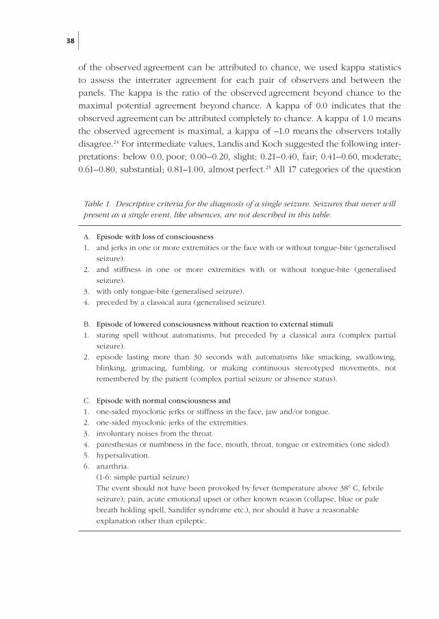
diagnosis according to predefined diagnostic criteria (table 1). This list contained
descriptions of all possible seizure types, but in table 2 of this paper we only men-
tion seizures which may present as a single event. The events were classified as
290158_Stroink_BW.indd 36 11-04-2008 10:53:44
37
epileptic seizure (170), other diagnosis (9), or unclear event (54). The study on the
prognosis and prognostic determinants of these children was published in 1998.7
One year after the intake for children with a single event into the DSEC had been
closed, two of the authors (HS, CD) selected 100 events from the diagnostic catego-
ries mentioned above. The intake panel of the DSEC considered 51 children to have
had an epileptic seizure, nine an event with a clear other diagnosis (like breath
holding spell or syncope), and 40 an unclear event. The number of children with
an unclear event was set proportionally higher than in the original cohort of the
DSEC to encourage discussion on their diagnosis and to diminish agreement due
to chance. The mean age of the children was 5.6 years, median 6.0 years (25th per-
centile 2.0; 75th percentile 9.0); 52 were boys. Two new panels were formed. Panel
A consisted of three of the four paediatric neurologists from the original panel,
each with at least 10 years of experience in paediatric epilepsy and in working in
such an interactive way (AP, OB, WA). Together with HS, they started the DSEC in
1988. Panel B consisted of three experienced senior paediatric neurologists at that
time working in other hospitals. One (HG) was attached to an epilepsy clinic, one
(ON) to a university centre for epilepsy surgery, and one (RC) to a university hos-
pital for children. The members of both panels received anonymous descriptions
of the 100 events, as given in the letter to the family physician. This included pos-
sible provoking factors and postictal signs, the previous medical history, the results
of the physical examination, and an assessment of the mental development. They
were distributed in random order and did not include the results of additional in-
vestigations (EEG, imaging, etc). The paediatric neurologists were not aware of the
stratification policy. Firstly, each member decided independently on the question
“Was it a seizure?” and, if applicable, on seizure classification according to his per-
sonal judgment. Subsequently, they independently repeated this process using the
predefined descriptive criteria (table 2). Then the observers discussed all patients
within their own panel until they reached consensus on the diagnosis and, if ap-
plicable, classification of the event according to the predefined descriptive criteria.
Next the panels received information on the results of the EEG, imaging study, and
possible other relevant information. With this new information, both panels were
independently asked again to classify the seizure in an epileptic syndrome, accord-
ing to the classification of the International League Against Epilepsy (ILAE), despite
the single occurrence of the event.23 The panels were forced to reach consensus
in all cases.
We evaluated the interrater agreement between the individual paediatric neurolo-
gists, between both panels after discussion between their members, and between
both panels and the original panel deciding on inclusion in the DSEC. As part
290158_Stroink_BW.indd 37 11-04-2008 10:53:45
38
of the observed agreement can be attributed to chance, we used kappa statistics
to assess the interrater agreement for each pair of observers and between the
panels. The kappa is the ratio of the observed agreement beyond chance to the
maximal potential agreement beyond chance. A kappa of 0.0 indicates that the
observed agreement can be attributed completely to chance. A kappa of 1.0 means
the observed agreement is maximal, a kappa of –1.0 means the observers totally
disagree.24 For intermediate values, Landis and Koch suggested the following inter-
pretations: below 0.0, poor; 0.00–0.20, slight; 0.21–0.40, fair; 0.41–0.60, moderate;
0.61–0.80, substantial; 0.81–1.00, almost perfect.25 All 17 categories of the question
Table 1. Descriptive criteria for the diagnosis of a single seizure. Seizures that never will present as a single event, like absences, are not described in this table.
A. Episode with loss of consciousness
1. and jerks in one or more extremities or the face with or without tongue-bite (generalised
seizure).
2. and stiffness in one or more extremities with or without tongue-bite (generalised
seizure).
3. with only tongue-bite (generalised seizure).
4. preceded by a classical aura (generalised seizure).
B. Episode of lowered consciousness without reaction to external stimuli
1. staring spell without automatisms, but preceded by a classical aura (complex partial
seizure).
2. episode lasting more than 30 seconds with au to matisms like smacking, swallowing,
blinking, grimacing, fumbling, or making continuous stereotyped movements, not
remembered by the patient (com plex partial seizure or absence status).
C. Episode with normal consciousness and
1. one-sided myoclonic jerks or stiffness in the face, jaw and/or tongue.
2. one-sided myoclonic jerks of the extremities.
3. involuntary noises from the throat.
4. paresthesias or numbness in the face, mouth, throat, tongue or extremities (one sided).
5. hypersalivation.
6. anarthria.
(1-6: simple partial seizure)
The event should not have been provoked by fever (temperature above 380 C, febrile
seizure); pain, acute emotional upset or other known reason (collapse, blue or pale
breath holding spell, Sandifer syndrome etc.), nor should it have a reasonable
explanation other than epileptic.
290158_Stroink_BW.indd 38 11-04-2008 10:53:45

39
Table 2. Questionnaires.
Diagnosis based on your own judgment
1. If you would be the attending physician, would you con sider this epi sode to be an
epileptic seizure, irrespective of the criteria presented in table 1?
Yes/no/ unclear
2. If you answered no, your diagnosis of the event is:
3. If you answered yes, how would you classify the seizure?
a. simple partial
b. complex partial
c. generalised with partial onset
d. generalised without partial onset
e. equivocal generalised or partial
Diagnosis with use of previously defined criteria
4. Do you think the history of the event satisfies the descriptive criteria listed in table 1?
Yes/no/unclear
5. If you answered yes, which category is applicable? (answers one or more)?
A1 / A2 / A3 / A4
B1 / B2
C1 / C2 / C3 / C4 / C5 / C6
6. Do you consider the seizure had a partial onset / partial characteristics?
Yes/no/ unclear
Diagnosis as a panel
After your diagnosis and classification, your panel will discuss the case to reach a final
common decision about the questions 4, 5, 6. After this, you will receive the results of
the physical examination, EEG, imaging study and other additional investigations.
7. If your panel diagnosed the event as an epileptic seizure, the aetiology according to this
panel is:
a. idiopathic
b. remote symptomatic
c. acute symptomatic
d. associated with mental retardation
e. equivocal
8. Are you able to classify the type of epilepsy according to the ILAE classification,
knowing the results of the additional investigations?
290158_Stroink_BW.indd 39 11-04-2008 10:53:45
40
concerning seizure classification using predefined criteria were collapsed into three
new categories (A1 to A4, B1 to B2, and C1 to C6; table 1). Simplification of the
complex ILAE syndrome classification was reached by grouping together the cat-
egories 4, 5, and 6; 7 and 8; 9 and 10 (table 5).
Results
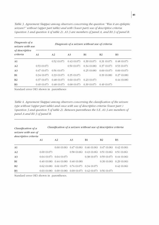
The kappa for pairs of individual observers for the question “Was it an epileptic
seizure?” according to personal judgment varied between 0.19 and 0.60, median
kappa 0.43, mean kappa 0.41 (SE 0.03). The use of the diagnostic criteria resulted
in kappa values for pairs of individual observers between 0.23–0.68, median kappa
0.48, mean kappa 0.45 (0.03) (table 3).
Both panels succeeded in all cases to reach consensus on the diagnosis after dis-
cussion. The kappa between the panels was 0.60 (0.06).
The kappas for the agreement between the diagnoses made by the panels partici-
pating in this experiment and the original panel deciding on entry into the DSEC
were 0.72 (SE 0.07) for the experienced panel and 0.66 (0.08) for the inexperienced
panel. Conspicuously, the experimental panels agreed on the epileptic nature of
the event in 61 children, whereas the paediatric neurologists deciding on entry into
the DSEC considered the event to be epileptic in only 51 cases.
For seizure classification, the kappas for pairs of individual observers without use
of descriptive criteria varied between 0.29 and 0.60 (median 0.46, mean 0.46, SE
0.02). The use of the predefined criteria resulted in kappas of 0.34–0.74 (median
0.62, mean 0.57, SE 0.03; table 4). The kappa between the panels after discussion
within each panel was 0.69 (0.06).
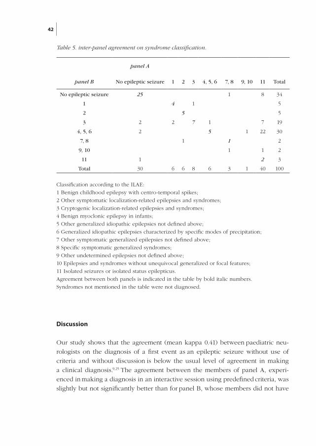
Finally, after the results of the electroencephalograms and imaging study had been
made available, each panel was asked to classify the epilepsy syndrome for the
children diagnosed with an epileptic seizure. In 24 of the 61 children in whom both
teams agreed there had been an epileptic seizure, the panels reached consensus on
the syndrome diagnosis (table 5).
290158_Stroink_BW.indd 40 11-04-2008 10:53:45
41
Diagnosis of a
seizure with use
of descriptive
criteria
Diagnosis of a seizure without use of criteria
A1 A2 A3 B1 B2 B3
A1 0.52 (0.07) 0.43 (0.07) 0.39 (0.07) 0.31 (0.07) 0.48 (0.07)
A2 0.53 (0.07) 0.50 (0.07) 0.34 (0.08) 0.37 (0.07) 0.53 (0.07)
A3 0.47 (0.07) 0.56 (0.07) 0.25 (0.08) 0.60 (0.07) 0.60 (0.07)
B1 0.34 (0.07) 0.33 (0.07) 0.35 (0.07) 0.19 (0.08) 0.27 (0.08)
B2 0.37 (0.07) 0.48 (0.07) 0.60 (0.07) 0.23 (0.07) 0.44 (0.08)
B3 0.49 (0.07) 0.48 (0.07) 0.68 (0.07) 0.39 (0.07) 0.49 (0.07)
Standard error (SE) shown in parentheses
Table 3. Agreement (kappa) among observers concerning the question “Was it an epileptic seizure?” without (upper part table) and with (lower part) use of descriptive criteria (question 1 and question 4 of table 2). A1-3 are members of panel A, and B1-3 of panel B.
Classification of a
seizure with use of
descriptive criteria
Classification of a seizure without use of descriptive criteria
A1 A2 A3 B1 B2 B3
A1 0.60 (0.06) 0.47 (0.06) 0.46 (0.06) 0.47 (0.06) 0.42 (0.06)
A2 0.69 (0.07) 0.58 (0.06) 0.43 (0.06) 0.51 (0.06) 0.51 (0.06)
A3 0.64 (0.07) 0.64 (0.07) 0.38 (0.07) 0.59 (0.07) 0.44 (0.06)
B1 0.40 (0.08) 0.44 (0.08) 0.40 (0.08) 0.39 (0.06) 0.29 (0.06)
B2 0.62 (0.08) 0.61 (0.07) 0.74 (0.07) 0.34 (0.07) 0.42 (0.06)
B3 0.63 (0.08) 0.69 (0.08) 0.69 (0.07) 0.42 (0.07) 0.56 (0.07)
Standard error (SE) shown in parentheses.
Table 4. Agreement (kappa) among observers concerning the classification of the seizure type without (upper part table) and once with use of descriptive criteria (lower part ) (question 3 and question 5 of table 2). Between parentheses the S.E. A1-3 are members of panel A and B1-3 of panel B.
290158_Stroink_BW.indd 41 11-04-2008 10:53:45
42
Discussion
Our study shows that the agreement (mean kappa 0.41) between paediatric neu-
rologists on the diagnosis of a first event as an epileptic seizure without use of
criteria and without discussion is below the usual level of agreement in making
a clinical diagnosis.9,25 The agreement between the members of panel A, experi-
enced in making a diagnosis in an interactive session using predefined criteria, was
slightly but not significantly better than for panel B, whose members did not have
panel B
panel A
No epileptic seizure 1 2 3 4, 5, 6 7, 8 9, 10 11 Total
No epileptic seizure 25 1 8 34
1 4 1 5
2 5 5
3 2 2 7 1 7 19
4, 5, 6 2 5 1 22 30
7, 8 1 1 2
9, 10 1 1 2
11 1 2 3
Total 30 6 6 8 6 3 1 40 100
Classification according to the ILAE:
1 Benign childhood epilepsy with centro-temporal spikes;
2 Other symptomatic localization-related epilepsies and syn dromes;
3 Cryptogenic localization-related epilepsies and syn dromes;
4 Benign myoclonic epilepsy in infants;
5 Other generalized idiopa thic epilepsies not defined above;
6 Generalized idiopa thic epilepsies characterized by specific modes of precipitation;
7 Other symptomatic genera lized epilepsies not defined above;
8 Specific symptomatic genera lized syndromes;
9 Other undetermined epilep sies not defined above;
10 Epilepsies and syndromes without unequivocal generalized or focal features;
11 Isolated seizures or isolated status epilepticus.
Agreement between both panels is indicated in the table by bold italic numbers.
Syndromes not mentioned in the table were not diagnosed.
Table 5. inter-panel agreement on syndrome classification.
290158_Stroink_BW.indd 42 11-04-2008 10:53:45
43
this experience (tables 3 and 4). Agreement could be improved only slightly by the
use of descriptive criteria, but discussion led to a better agreement (kappa 0.60).
We also found substantial, but not perfect, agreement between both experimental
panels and the panel originally deciding on the diagnosis at the moment of inclu-
sion in the DSEC. The experienced panel did slightly better than the panel whose
members were not used to working in such a collaborative way. This was probably
not because of the effect of memory, despite the fact that the experienced panel
contained three of the four members of the original panel. All case descriptions
had been anonymised and the time elapsed between inclusion in the DSEC and
the experiment described here varied between one and six years. On the contrary,
it is surprising that the agreement between the opinions of this panel at entry into
the DSEC and some years later was not better than 0.72. Both the interrater and the
“intra-panel” disagreement illustrated here suggest that the diagnosis of an isolated
epileptic seizure may be extremely difficult and should always be looked at with
some suspicion, especially in the context of a clinical research study.
For this study we had deliberately selected 100 cases with a 1:1 ratio between
epileptic seizures on the one hand and unclear or non-epileptic events on the
other. Although kappa statistics take into account the agreement due to chance,
the results are influenced by the distribution of the possible diagnoses. In case
of an askew distribution, kappa statistics will be lower than in case of a 1:1 ratio
between the various diagnoses.26 Therefore, the fair to moderate agreement rates
found before discussion cannot be explained by an askew distribution between
epileptic seizures versus unclear or non-epileptic events. One may even argue that
our findings are biased towards a higher agreement.
Only in 24 cases was consensus reached on the syndrome diagnosis (table 5). A
conspicuous discrepancy existed between the ways the panels used the ILAE clas-
sification. Panel B often tried to reach consensus on such classifications as “crypto-
genic localisation-related epilepsy” or “idiopathic generalised epilepsy not otherwi-
se defined”. Panel A classified most of these seizures as “isolated seizure or isolated
status”. These classifications of both panels are safe when evidence concerning the
nature of the seizure is lacking or inconclusive, but they do not contribute to our
knowledge on the causal diagnosis of the child, the prognosis, or the way in which
he or she should be treated. Only in the four children with benign childhood
epilepsy with rolandic spikes did agreement exist on a syndrome with prognostic
significance. King et al stated that syndrome diagnosis is possible in most patients
presenting with only one seizure.27 However, 45% of the patients in their study had
suffered more than one seizure.28 These patients were carefully excluded in our
study by using standardised questionnaires.6 Moreover, most patients in the study
290158_Stroink_BW.indd 43 11-04-2008 10:53:45
44
of King et al were adults over 30 years old, so many had probably suffered a remote
symptomatic partial seizure.28–30 More recently, the CAROLE group also made a
syndrome classification in patients with newly diagnosed epilepsy or only a single
seizure.2 In this study a panel made the diagnosis. However, the patients with a sin-
gle seizure were much older than in our study (mean age 19 years) and most clas-
sifications were broadly defined as well. In our study, classification in a clinically
relevant syndrome was possible only in a very small minority of children.
We know of only one study in which the reliability of the diagnosis of a first seizu-
re was studied by assessing interrater agreement. This study was done in adults.20
Other studies in epilepsy, in which kappa statistics were used, concerned genera-
lised or partial seizure onset in adults,31 and seizure classification32 and syndrome
classification in children,33 all patients with multiple seizures. The study on seizure
classification in children32 showed poor interrater correlations, and suggested that
specific criteria for the categorisation of symptoms could reduce the interrater vari-
ability. Combined with our results, this suggests that the best agreement could be
obtained if the seizures were not only classified according to pre-defined criteria
(like in our study), but also the symptoms categorised according to a standardised
questionnaire to the patient and any witnesses of the seizure.
The study on interrater agreement on classification of childhood epilepsy syndro-
mes33 showed excellent agreement using the ILAE classification of epilepsy syndro-
mes, although a substantial proportion of children were classified into relatively
non-specific syndromes.23 However, in this study only children with newly diag-
nosed epilepsy were classified, not children with a single seizure.
Even experienced paediatric neurologists frequently disagree about the diagnosis
and classification of a first seizure in children. In this study the diagnosis was
based on a careful written description made by experts with the aim of an exten-
sive questionnaire. The agreement among neurologists may be even lower when
they have to listen to the actual histories from the parents themselves.
Our results may at least partly explain the widely discrepant recurrence risks repor-
ted in first seizure studies.10–19 In this study, the use of a panel was the best means
to increase the interrater agreement. We recommend such an approach for research
purposes, although even then in many cases the diagnosis will remain uncertain.
However, better ways to diagnose first seizures are not currently available.
290158_Stroink_BW.indd 44 11-04-2008 10:53:45
45
References
1 Hirtz D, Berg A, Bettis D, Camfield C, Camfield P, Crumrine P, Gaillard WD, Schneider
S, Shinnar S. Practice parameter: Treatment of the child with a first unprovoked seizure:
Report of the Quality Standards Subcommittee of the American Academy of Neurology and
the Practice Committee of the Child Neurology Society. Neurology 2003;60:166–175.
2. Jallon P, Loiseau P, Loiseaue J. Newly diagnosed unprovoked epileptic seizures:
presentation at diagnosis in CAROLE Study. Epilepsia 2001;42:464–475.
3. Cavazzuti GB, Cappella L, Nalin A. Longitudinal study of epileptiform EEG patterns in
normal children. Epilepsia 1980;21:43–55.
4. Binnie CD. What’s the use of the EEG in epilepsy? Br J Hosp Med 1988;39:99–102.
5. Gibbs J, Appleton RE. False diagnosis of epilepsy in children. Seizure 1992;1:15–18.
6. Carpay JA, de Weerd AW, Schimsheimer RJ, Stroink H, Brouwer OF, Peters AC, van
Donselaar CA, Geerts AT, Arts WF. The diagnostic yield of a second EEG after partial sleep
deprivation: a prospective study in children with newly diagnosed seizures. Epilepsia
1997;38:595–599.
7. Stroink H, Brouwer OF, Arts WF, Geerts AT, Peters AC, van Donselaar CA. The first
unprovoked, untreated seizure in childhood: a hospital-based study of the accuracy of
the diagnosis, rate of recurrence, and long-term outcome after recurrence. Dutch study of
epilepsy in childhood. J Neurol Neurosurg Psychiatry 1998;64:595–600.
8. Stroink H, van Donselaar CA, Geerts AT, Peters AC, Brouwer OF, Arts WF. The accuracy of
the diagnosis of paroxysmal events in children. Neurology 2003;60:979–982.
9. Sacket DL, Haynes RB, Tugwell P. Clinical epidemiology, a basic science for clinical
medicine, 2nd edn. Boston, Toronto, London: Little, Brown and Company, 1991.
10. Camfield PR, Camfield CS, Dooley JM, Tibbles JAR, Fung T, Garner B. Epilepsy after a first
unprovoked seizure in childhood. Neurology 1985;35:1657–1560.
11. Boullouche J, Leloup P, Mallet E, Parain D, Tron P. Risk of recurrence after a single,
unprovoked, generalised tonic-clonic seizure. Dev Med Child Neurol 1989;31:626–632.
12. Camfield P, Camfield C, Dooley J, Smith E, Garner B. A randomised study of
carbamazepine versus no medication after a first unprovoked seizure in childhood.
Neurology 1989;39:851–852.
13. Hauser WA, Rich SS, Annegers JF, Anderson VE. Seizure recurrence after a 1st unprovoked
seizure: an extended follow-up. Neurology 1990;40:1163–1170.
14. Shinnar S, Berg AT, Moshe SL, O’Dell C, Alemany M, Newstein D, Kang H, Goldensohn ES,
Hauser WA. Risk of seizure recurrence following a first unprovoked seizure in childhood:
an extended follow-up. Paediatrics 1996;98:216–225.
15. First Seizure Trial Group. Randomised clinical trial on the efficacy of antiepileptic drugs
in reducing the risk of relapse after a first unprovoked tonic-clonic seizure. Neurology
1993;43:478–483.
16. Berg AT, Shinnar S. The risk of seizure recurrence following a first unprovoked seizure: a
quantitative review. Neurology 1991;41:965–972.
17. Hopkins A, Garman A, Clarke C. The first seizure in adult life: value of clinical features,
electroencephalography and computerized tomographic scanning in prediction of seizure
recurrence. Lancet 1988;1:721–726.
290158_Stroink_BW.indd 45 11-04-2008 10:53:45
46
18. Elwes RDC, Chesterman P, Reynolds EH. Prognosis after a first untreated tonic-clonic
seizure. Lancet 1985;2:752–753.
19. Hauser WA, Anderson VE, Loewenson RB, et al. Seizure recurrence after a first
unprovoked seizure. N Engl J Med 1982;26:522–528.
20. Van Donselaar CA, Geerts AT, Meulstee J, Habbema JD, Staal A. Reliability of the diagnosis
of a first seizure. Neurology 1989;39:267–271.
21. Arts WFM, Geerts AT, Brouwer OF, Peters AC, Stroink H, van Donselaar CA. The early
prognosis of epilepsy in childhood: the prediction of a poor outcome. The Dutch Study of
Epilepsy in Childhood. Epilepsia 1999;40:726–734.
22. Carpay JA, Arts WFM, Geerts AT, Stroink H, Brouwer OF, Boudewyn Peters AC,
van Donselaar CA. Epilepsy in childhood: an audit of clinical practice. Arch Neurol
1998;55:668–673.
23. Commission on Classification and Terminology of the International League Against
Epilepsy. Proposal for revised classification of epilepsies and epileptic syndromes. Epilepsia
1989;30:389–399.
24. Cohen JA. Coefficient of agreement for nominal scales. Educational and Psychological
Measurement 1960;20:37–46.
25. Landis JR, Koch GG. The measurement of observer agreement for categorical data.
Biometrics 1977;33:159–174.
26. Kraemer HC. Ramifications of a population model for Kappa as a coefficient of reliability.
Psychometrika 1979;44:461–472.
27. King MA, Newton MR, Jackson GD, Fitt GJ, Mitchell LA, Silvapulle MJ, Berkovic SF.
Epileptology of the first-seizure presentation: a clinical, electroencephalographic, and
magnetic resonance imaging study of 300 consecutive patients. Lancet 1998;352:1007–1011.
28. Lhatoo SD, Shorvon SD. Relevance of the first seizure. Lancet 1998;352:1003–1004.
29. Chadwick D, Smith D. Comment on epileptology of the first-seizure presentation. Lancet
1998;35:1855.
30. Addy DP. Comment on epileptology of the first-seizure presentation. Lancet 1998;35:1857.
31. Ottman R, Lee JH, Hauser WA, Hong S, Hesdorffer D, Schupf N, Pedley TA, Scheuer
ML. Reliability of seizure classification using a semi-structured interview. Neurology
1993;43:2526–2530.
32. Bodensteiner JB, Brownsworth RD, Knapik JR, Kanter MC, Cowan LD, Leviton A.
Interobserver variability in the ILAE classification of seizures in childhood. Epilepsia
1988;29:123–128.
33. Berg AT, Levy SR, Testa FM, Shinnar S. Classification of childhood epilepsy syndromes in
newly diagnosed epilepsy: interrater agreement and reasons for disagreement. Epilepsia
1999;40:439–444.
290158_Stroink_BW.indd 46 11-04-2008 10:53:45
47
Chapter 4
The accuracy of the diagnosis of
paroxysmal events in children
Stroink H, van Donselaar CA, Geerts AT, Peters AC, Brouwer OF, Arts WFNeurology 2003;60:979-982
290158_Stroink_BW.indd 47 11-04-2008 10:53:45

290158_Stroink_BW.indd 48 11-04-2008 10:53:45
49
Abstract
Objective: To assess the accuracy of the diagnosis of epileptic seizures in child-
ren.
Methods: The Dutch Study of Epilepsy in Childhood is a prospective hospital-
based study of 881 children referred because of possible seizures. The diagnosis
was based on predefined descriptive criteria, as applied by a panel of three paedia-
tric neurologists. Children with a definite other diagnosis were excluded. All child-
ren with unclear events were followed up for one year and children with seizures
were followed up for two years to assess the accuracy of the diagnosis.
Results: In 170 of 224 children seen after a single event, the incident was classified
initially as epileptic, in 54 as unclear. In none of the 170 children did the diagnosis
prove to be wrong. In four of the 54 children, recurrent episodes enabled a defi-
nite diagnosis of epilepsy. In 412 of the 536 children seen with multiple events, an
initial diagnosis of epilepsy was made. After follow-up, this initial diagnosis was
probably incorrect in 19. In contrast, seven of 124 children with multiple unclear
episodes at intake later received the diagnosis epilepsy.
Conclusions: A false positive diagnosis of epilepsy was made in 4.6%, whereas
a definite diagnosis of epilepsy or seizure was delayed in 5.6% of children with
multiple unclear events and in 7.4% of children with one unclear event.
Introduction
The diagnosis of a single seizure or epilepsy is based on a description of the epi-
sodes and may be subject to error. To prove that a single event was an epileptic
seizure is often impossible. Generally accepted and validated diagnostic criteria
are lacking. The widely used International Classifications of Seizures1 and Epileptic
Syndromes2 provide no guidelines for the diagnosis of epileptic seizures. They only
permit classification once the diagnosis has been made.3 4 Surprisingly, most clinical
or epidemiologic studies do not mention patients in whom the diagnosis proved to
be incorrect.5 The Dutch Study of Epilepsy in Childhood (DSEC) is a prospective
hospital-based study of children with newly diagnosed possible single or multiple
seizures.6-8 A panel of three paediatric neurologists classified the events as epileptic
seizures, unclear episodes, or events of definitely other origin. Children with one
or more unclear events were followed up for one year and children with a diagno-
sis of a first seizure or epilepsy for five years. In this part of the study, we describe
the accuracy of the initial diagnosis after one or more paroxysmal events.
290158_Stroink_BW.indd 49 11-04-2008 10:53:45
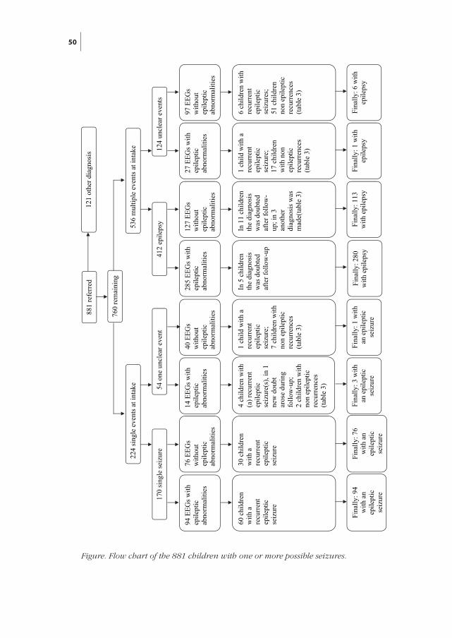
50
881
refe
rred
12
1 ot
her d
iagn
osis
760
rem
aini
ng
224
sing
le e
vent
s at i
ntak
e 53
6 m
ultip
le e
vent
s at i
ntak
e
170
sing
le se
izur
e 54
one
unc
lear
eve
nt
412
epile
psy
124
uncl
ear e
vent
s
14 E
EGs w
ith
epile
ptic
ab
norm
aliti
es
76 E
EGs
with
out
epile
ptic
ab
norm
aliti
es
94 E
EGs w
ith
epile
ptic
ab
norm
aliti
es
60 c
hild
ren
with
a
recu
rren
t ep
ilept
ic
seiz
ure
30 c
hild
ren
with
a
recu
rren
t ep
ilept
ic
seiz
ure
40 E
EGs
with
out
epile
ptic
ab
norm
aliti
es
4 ch
ildre
n w
ith
(a) r
ecur
rent
ep
ilept
ic
seiz
ure(
s), i
n 1
new
dou
bt
aros
e du
ring
follo
w-u
p;
2 ch
ildre
n w
ith
non
epile
ptic
re
curr
ence
s (ta
ble
3)
1 ch
ild w
ith a
re
curr
ent
epile
ptic
se
izur
e;
7 ch
ildre
n w
ith
non
epile
ptic
re
curr
ence
s (ta
ble
3)
285
EEG
s with
ep
ilept
ic
abno
rmal
ities
In 5
chi
ldre
n th
e di
agno
sis
was
dou
bted
af
ter f
ollo
w-u
p
127
EEG
s w
ithou
t ep
ilept
ic
abno
rmal
ities
In 1
1 ch
ildre
n th
e di
agno
sis
was
dou
bted
af
ter f
ollo
w-
up; i
n 3
anot
her
diag
nosi
s was
m
ade(
tabl
e 3)
97 E
EGs
with
out
epile
ptic
ab
norm
aliti
es
6 ch
ildre
n w
ith
recu
rren
t ep
ilept
ic
seiz
ures
; 51
chi
ldre
n no
n ep
ilept
ic
recu
rren
ces
(tabl
e 3)
27 E
EGs w
ith
epile
ptic
ab
norm
aliti
es
1 ch
ild w
ith a
re
curr
ent
epile
ptic
se
izur
e;
17 c
hild
ren
with
non
ep
ilept
ic
recu
rren
ces
(tabl
e 3)
Fina
lly: 1
13
with
epi
leps
y Fi
nally
: 1 w
ith
epile
psy
Fina
lly: 6
with
ep
ileps
y Fi
nally
: 280
w
ith e
pile
psy
Fina
lly: 1
with
an
epi
lept
ic
seiz
ure
Fina
lly: 9
4 w
ith a
n ep
ilept
ic
seiz
ure
Fina
lly: 7
6 w
ith a
n ep
ilept
ic
seiz
ure
Fina
lly: 3
with
an
epi
lept
ic
seiz
ure
Figure. Flow chart of the 881 children with one or more possible seizures.
290158_Stroink_BW.indd 50 11-04-2008 10:53:45
51
Patients and methods
The prospective multicenter hospital-based DSEC started in 1988. After informed
consent, we enrolled consecutively all children aged one month to 16 years who
were referred because of a possible single seizure or epilepsy. Most children were
referred directly by their general practitioner or by the paediatricians of the par-
ticipating hospitals (who routinely refer all children with possible seizures to the
departments of paediatric neurology). Some were first seen in the emergency de-
partment. We excluded children with only neonatal, febrile, or other acute symp-
tomatic seizures as well as children who were referred from other hospitals for a
second opinion.
The attending paediatric neurologist completed an extensive questionnaire on the
description of the events including postictal signs, possible provoking factors, me-
dical history, and family history. In addition, the descriptions of the episodes ac-
cording to the letters to the general practitioners were made available to the panel
judging the diagnoses (see below). We performed a standard EEG in each child. If
this did not show epileptiform discharges, a recording after partial sleep depriva-
tion or, in small children, during a daytime nap was made. Details on the recording
and classification of the EEG have been described elsewhere.8 A brain CT scan
was performed in all children unless anaesthesia was required or the child had
idiopathic generalized epilepsy with absences. Seventy-two percent of the children
with one seizure and 66% of the children with multiple seizures had a CT scan.
In former publications on the DSEC, more extensive information concerning the
results of imaging can be found.6 7
A panel of three of four paediatric neurologists with at least 10 years of experience
in paediatric epilepsy (H.S., A.C.B.P., O.F.B., and W.F.M.A.) judged whether the
description of the ictal events fulfilled the predefined descriptive diagnostic criteria
for seizures as adapted from an earlier Dutch study in adults9 (table E1 on the Neu-
rology Web site). To prevent bias, none of the paediatric neurologists was allowed
to judge his patients, but only those who were treated by one of the other panel
members. These paediatric neurologists in 1988 founded the DSEC and have been
involved in it since then. They discussed all 881 children referred during mont-
hly sessions. Unanimous decisions were reached in all cases. The 121 children in
whom another diagnosis was made were excluded. The study group consisted of
760 children: mean age 5.4 years, median age four years (25 to 75% range: two
to nine years); 367 were boys. A flow chart of all children in the study and their
subgroups is presented in the figure. For a single event, the diagnosis was based
exclusively on the description of the event without knowledge of the results of the
290158_Stroink_BW.indd 51 11-04-2008 10:53:46
52
EEGs or other ancillary studies. We considered a single unclear event in a child
with an abnormal EEG not as an epileptic seizure, as epileptiform discharges are
present in some children without any clinical seizures. In case of multiple events,
however, the results of the EEGs were considered if the panel agreed based on
their clinical experience that the events were suspect for seizures, although the
criteria were not completely satisfied.
In the DSEC we followed up all children with a diagnosis of epilepsy for five years
and all children with a single seizure for two years to assess the prognosis.6 7 At
two years, the accuracy of the original diagnosis was assessed. In an attempt to
include all children with epilepsy in the DSEC, children with unclear events (in-
cluding children with presumed pseudo-seizures) were followed for one year to
assess whether new episodes might yield firm evidence for a definite diagnosis.
An unclear event was defined as a paroxysmal event not considered as an epilep-
tic seizure, but without another obvious explanation. After each new event, the
patient was re-evaluated and the diagnosis reconsidered. Point estimates regarding
false positive or false negative diagnoses are given with their 95% CIs. The CIs were
calculated according to the efficient-score method (corrected for continuity).10
Results
Paroxysmal events Of the 881 children in the study group, 121 received a definite other diagnosis and
were excluded from follow-up (see the figure). A total of 224 children were refer-
red because of a single event. In 170 of these 224, the panel diagnosed a single
seizure (table 1); 90 of them had at least one more event. During the follow-up pe-
riod, doubts on the diagnosis seizure arose in none of the children with recurrent
events, whereas the diagnosis was maintained in all children without a recurrence
(false positive rate, 0%; 95% CI, 0 to 2.8).
The panel was uncertain on the diagnosis of a first event in 54 children, of whom
14 had at least one more event. We eventually made a diagnosis of epilepsy after
1-year follow-up in five of these. After two years of follow-up, new doubts arose
in one of these children. Her first event was associated with teeth brushing. We
considered this event as uncertain diagnosis. Within 12 months, she had a second
paroxysmal event. Her EEG showed epileptiform abnormalities; a diagnosis of epi-
lepsy was made and an antiepileptic drug was prescribed. After about 20 months,
a new event took place while her hair was being combed. Based on the description
of this event and the circumstances during two of the three events (teeth brushing,
hair combing) this was thought to be a hairdresser’s syncope,11 after which the
290158_Stroink_BW.indd 52 11-04-2008 10:53:46
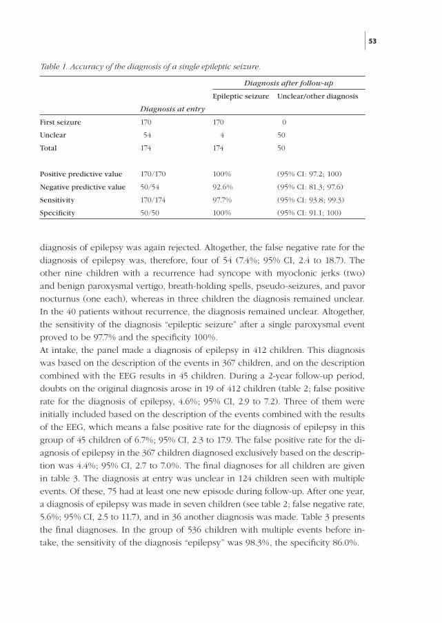
53
diagnosis of epilepsy was again rejected. Altogether, the false negative rate for the
diagnosis of epilepsy was, therefore, four of 54 (7.4%; 95% CI, 2.4 to 18.7). The
other nine children with a recurrence had syncope with myoclonic jerks (two)
and benign paroxysmal vertigo, breath-holding spells, pseudo-seizures, and pavor
nocturnus (one each), whereas in three children the diagnosis remained unclear.
In the 40 patients without recurrence, the diagnosis remained unclear. Altogether,
the sensitivity of the diagnosis “epileptic seizure” after a single paroxysmal event
proved to be 97.7% and the specificity 100%.
At intake, the panel made a diagnosis of epilepsy in 412 children. This diagnosis
was based on the description of the events in 367 children, and on the description
combined with the EEG results in 45 children. During a 2-year follow-up period,
doubts on the original diagnosis arose in 19 of 412 children (table 2; false positive
rate for the diagnosis of epilepsy, 4.6%; 95% CI, 2.9 to 7.2). Three of them were
initially included based on the description of the events combined with the results
of the EEG, which means a false positive rate for the diagnosis of epilepsy in this
group of 45 children of 6.7%; 95% CI, 2.3 to 17.9. The false positive rate for the di-
agnosis of epilepsy in the 367 children diagnosed exclusively based on the descrip-
tion was 4.4%; 95% CI, 2.7 to 7.0%. The final diagnoses for all children are given
in table 3. The diagnosis at entry was unclear in 124 children seen with multiple
events. Of these, 75 had at least one new episode during follow-up. After one year,
a diagnosis of epilepsy was made in seven children (see table 2; false negative rate,
5.6%; 95% CI, 2.5 to 11.7), and in 36 another diagnosis was made. Table 3 presents
the final diagnoses. In the group of 536 children with multiple events before in-
take, the sensitivity of the diagnosis “epilepsy” was 98.3%, the specificity 86.0%.
Diagnosis after follow-up
Epileptic seizure Unclear/other diagnosis
Diagnosis at entry
First seizure 170 170 0
Unclear 54 4 50
Total 174 174 50
Positive predictive value 170/170 100% (95% CI: 97.2; 100)
Negative predictive value 50/54 92.6% (95% CI: 81.3; 97.6)
Sensitivity 170/174 97.7% (95% CI: 93.8; 99.3)
Specificity 50/50 100% (95% CI: 91.1; 100)
Table 1. Accuracy of the diagnosis of a single epileptic seizure.
290158_Stroink_BW.indd 53 11-04-2008 10:53:46
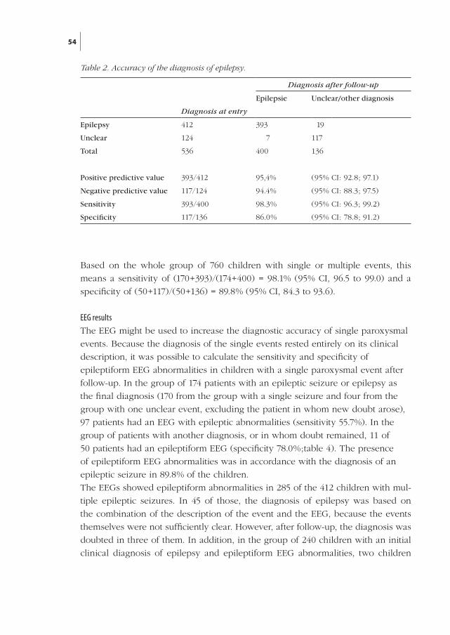
54
Based on the whole group of 760 children with single or multiple events, this
means a sensitivity of (170+393)/(174+400) = 98.1% (95% CI, 96.5 to 99.0) and a
specificity of (50+117)/(50+136) = 89.8% (95% CI, 84.3 to 93.6).
EEG resultsThe EEG might be used to increase the diagnostic accuracy of single paroxysmal
events. Because the diagnosis of the single events rested entirely on its clinical
description, it was possible to calculate the sensitivity and specificity of
epileptiform EEG abnormalities in children with a single paroxysmal event after
follow-up. In the group of 174 patients with an epileptic seizure or epilepsy as
the final diagnosis (170 from the group with a single seizure and four from the
group with one unclear event, excluding the patient in whom new doubt arose),
97 patients had an EEG with epileptic abnormalities (sensitivity 55.7%). In the
group of patients with another diagnosis, or in whom doubt remained, 11 of
50 patients had an epileptiform EEG (specificity 78.0%;table 4). The presence
of epileptiform EEG abnormalities was in accordance with the diagnosis of an
epileptic seizure in 89.8% of the children.
The EEGs showed epileptiform abnormalities in 285 of the 412 children with mul-
tiple epileptic seizures. In 45 of those, the diagnosis of epilepsy was based on
the combination of the description of the event and the EEG, because the events
themselves were not sufficiently clear. However, after follow-up, the diagnosis was
doubted in three of them. In addition, in the group of 240 children with an initial
clinical diagnosis of epilepsy and epileptiform EEG abnormalities, two children
Diagnosis after follow-up
Epilepsie Unclear/other diagnosis
Diagnosis at entry
Epilepsy 412 393 19
Unclear 124 7 117
Total 536 400 136
Positive predictive value 393/412 95,4% (95% CI: 92.8; 97.1)
Negative predictive value 117/124 94.4% (95% CI: 88.3; 97.5)
Sensitivity 393/400 98.3% (95% CI: 96.3; 99.2)
Specificity 117/136 86.0% (95% CI: 78.8; 91.2)
Table 2. Accuracy of the diagnosis of epilepsy.
290158_Stroink_BW.indd 54 11-04-2008 10:53:46
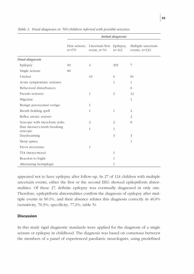
55
Table 3. Final diagnoses in 760 children referred with possible seizures.
Initial diagnosis
First seizure, n=170
Uncertain first event, n=54
Epilepsy,n=412
Multiple uncertain events, n=124
Final diagnosis
Epilepsy 90 4 393 7
Single seizure 80
Unclear 43 6 81
Acute symptomatic seizures 1 1
Behavioral disturbances 6
Pseudo-seizures 1 2 12
Migraine 1
Benign paroxysmal vertigo 1
Breath holding spell 1 1 2
Reflex anoxic seizure 2
Syncope with myoclonic jerks 2 2 8Hair dresser’s/teeth brushingsyncope
1 1
Daydreaming 3 3
Sleep apnea 1
Pavor nocturnus 1
TIA (moya-moya) 1
Reaction to fright 1
Alternating hemiplegia 1
appeared not to have epilepsy after follow-up. In 27 of 124 children with multiple
uncertain events, either the first or the second EEG showed epileptiform abnor-
malities. Of these 27, definite epilepsy was eventually diagnosed in only one.
Therefore, epileptiform abnormalities confirm the diagnosis of epilepsy after mul-
tiple events in 90.1%, and their absence refutes this diagnosis correctly in 46.9%
(sensitivity, 70.3%; specificity, 77.2%; table 5).
Discussion
In this study rigid diagnostic standards were applied for the diagnosis of a single
seizure or epilepsy in childhood. The diagnosis was based on consensus between
the members of a panel of experienced paediatric neurologists, using predefined
290158_Stroink_BW.indd 55 11-04-2008 10:53:46
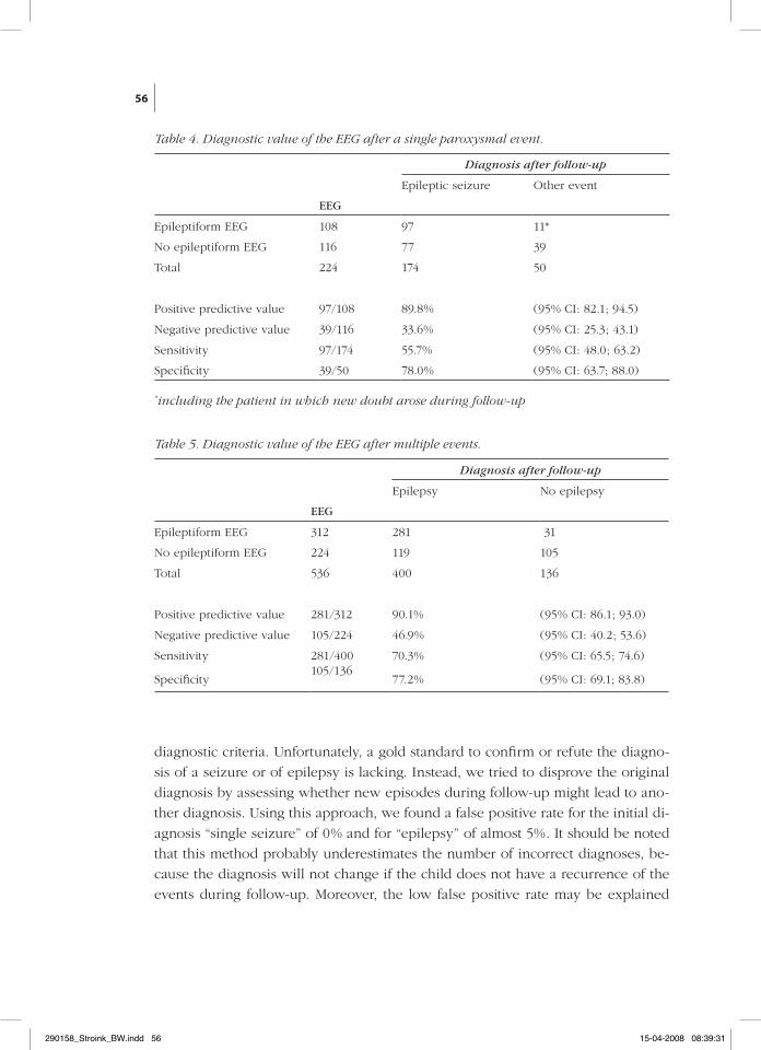
56
diagnostic criteria. Unfortunately, a gold standard to confirm or refute the diagno-
sis of a seizure or of epilepsy is lacking. Instead, we tried to disprove the original
diagnosis by assessing whether new episodes during follow-up might lead to ano-
ther diagnosis. Using this approach, we found a false positive rate for the initial di-
agnosis “single seizure” of 0% and for “epilepsy” of almost 5%. It should be noted
that this method probably underestimates the number of incorrect diagnoses, be-
cause the diagnosis will not change if the child does not have a recurrence of the
events during follow-up. Moreover, the low false positive rate may be explained
Table 5. Diagnostic value of the EEG after multiple events.
Diagnosis after follow-up
Epilepsy No epilepsy
EEG
Epileptiform EEG 312 281 31
No epileptiform EEG 224 119 105
Total 536 400 136
Positive predictive value 281/312 90.1% (95% CI: 86.1; 93.0)
Negative predictive value 105/224 46.9% (95% CI: 40.2; 53.6)
Sensitivity 281/400 70.3% (95% CI: 65.5; 74.6)
Specificity105/136
77.2% (95% CI: 69.1; 83.8)
Diagnosis after follow-up
Epileptic seizure Other event
EEG
Epileptiform EEG 108 97 11*
No epileptiform EEG 116 77 39
Total 224 174 50
Positive predictive value 97/108 89.8% (95% CI: 82.1; 94.5)
Negative predictive value 39/116 33.6% (95% CI: 25.3; 43.1)
Sensitivity 97/174 55.7% (95% CI: 48.0; 63.2)
Specificity 39/50 78.0% (95% CI: 63.7; 88.0)
Table 4. Diagnostic value of the EEG after a single paroxysmal event.
*including the patient in which new doubt arose during follow-up
290158_Stroink_BW.indd 56 15-04-2008 08:39:31
57
by the fact that the panel tended to prefer the diagnosis unclear and not seizure or
epilepsy in case of any doubt or disagreement between the panel members.
In addition, we studied the fate of the children with an unclear diagnosis at enroll-
ment. The false negative rate was 7.4% for children recruited after a single unclear
event and 5.6% for children enrolled after more unclear events. In other words,
irrespective of the fact that a child was enrolled after one or more unclear events,
one of every 15 children was not recognized to have epileptic seizures. However,
none of the children experienced any harm due to this delay.
Most of the children in whom the diagnosis seizures was changed during follow-
up, as well as the children with unclear events in whom a diagnosis became obvi-
ous during follow-up, turned out to have a harmless paroxysmal disorder (see table
3). The majority was not harmed by unnecessary delay of medical treatment. The
only exception was the child with TIAs based on moyamoya disease diagnosed
initially as epilepsy.
In view of the rigid standards we used in the diagnostic process (diagnosis by a
unanimous decision of a panel of three experienced paediatric neurologists with
use of predefined criteria), the figures found in our study may be a minimum esti-
mate for the errors in everyday clinical and research situations. This should be kept
in mind when judging studies on the prognosis of single seizures and epilepsy in
children. We recommend that in this type of research, the initial diagnosis should
be reconsidered after sufficient follow-up. From a clinical point of view, however,
a false negative diagnosis of epilepsy is probably less harmful for the patient than
a false positive. A conservative approach in children with paroxysmal events of
uncertain nature seems, as before, warranted.
References
1. Commission on Classification and Terminology of the International League Against
Epilepsy. Proposal for revised clinical and electroencephalographic classification of
epileptic seizures. Epilepsia 1981;22:489–501.
2. Commission on Classification and Terminology of the International League Against
Epilepsy. Proposal for revised classification of epilepsies and epileptic syndromes. Epilepsia
1989;30:389–399.
3. Berg AT, Levy SR, Testa FM, Shinnar S . Classification of childhood epilepsy syndromes in
newly diagnosed epilepsy: interrater agreement and reasons for disagreement. Epilepsia
1999; 40:439–444.
4. Jallon P, Loiseau P, Loiseau J. Newly diagnosed unprovoked epileptic seizures: presentation
at diagnosis in CAROLE Study. Epilepsia 2001;42:464–475.
5. Sander JW, Hart YM, Johnson AL, Shorvon SD. National General Practice Study of Epilepsy:
newly diagnosed epileptic seizures in a general population. Lancet 1990; 336: 1267–1271.
290158_Stroink_BW.indd 57 11-04-2008 10:53:46
58
6. Stroink H, Brouwer OF, Arts WF, Geerts AT, Peters AC, van Donselaar CA. The first
unprovoked, untreated seizure in childhood: a hospital-based study of the accuracy of
the diagnosis, rate of recurrence, and long-term outcome after recurrence. Dutch study of
epilepsy in childhood. J Neurol Neurosurg Psychiatry 1998;64:595–600.
7. Arts WFM, Geerts AT, Brouwer OF, Peters AC, Stroink H, van Donselaar CA. The early
prognosis of epilepsy in childhood: the prediction of a poor outcome. The Dutch Study of
Epilepsy in Childhood. Epilepsia 1999;40:726–734.
8. Peters AC, Brouwer OF, Geerts AT, Arts WF, Stroink H, van Donselaar CA. Randomized
prospective study of early discontinuation of antiepileptic drugs in children with epilepsy.
Neurology 1998;50:724-730.
9. van Donselaar CA, Geerts AT, Meulstee J, Habbema JD, Staal A. Reliability of the diagnosis
of a first seizure. Neurology 1989;39:267-271.
10. Newcombe, Robert G. Two-sided confidence intervals for the single proportion:
comparison of seven methods. Stats Med 1998;17:857–872.
11. Stephenson JB. In: Fits and faints. Philadelphia: JB Lippincott, 1990.
290158_Stroink_BW.indd 58 11-04-2008 10:53:46
59
Chapter 5
Interobserver reliability of visual interpre-
tation of electroencephalograms in children
with newly diagnosed seizures
Stroink H, Schimsheimer RJ, de Weerd AW, Geerts AT, Arts WF, Peeters EA, Brouwer OF, Boudewijn Peters A, van Donselaar CADev Med Child Neurol 2006;48:374-37
290158_Stroink_BW.indd 59 11-04-2008 10:53:46

290158_Stroink_BW.indd 60 11-04-2008 10:53:46
61
Abstract
The reliability of visual interpretation of electroencephalograms (EEG) is of great
importance in assessing the value of this diagnostic tool. We prospectively obtained
50 standard EEGs and 61 EEGs after partial sleep deprivation from 93 children (56
males, 37 females) with a mean age of six years 10 months (SE 5 mo; range 4 mo-15
y 7 mo) with one or more newly diagnosed, unprovoked seizures. Two clinical
neurophysiologists independently classified the background pattern and the pre-
sence of epileptiform discharges or focal non-epileptiform abnormalities of each
EEG. The agreement was substantial for the interpretation of the EEG as normal or
abnormal (kappa 0.66), almost perfect for the presence of epileptiform discharges
(kappa 0.83), substantial for the occurrence of an abnormal background pattern
(kappa 0.73), and moderate for the presence of focal non-epileptiform discharges
(kappa 0.54). In conclusion, the reliability of the visual interpretation of EEGs in
children is almost perfect as regards the presence of epileptiform abnormalities,
and moderate to substantial for the presence of other abnormalities.
Introduction
The electroencephalogram (EEG) is an important tool in the diagnosis of children
with epilepsy. EEG findings are used for the classification of epileptic syndromes
and may determine the choice of anti-epileptic drugs. The presence of epileptiform
discharges is a strong predictor for the risk of recurrence after a first seizure.1-3
The reliability (interobserver consistency or interobserver variation) and validity
(accuracy) of a diagnostic or prognostic tool determine its value.4 However, studies
on the reliability of the visual interpretation of EEG findings are scarce.5-10 Most in-
vestigations have focused on items not directly relevant to the treatment of patients
with epilepsy. Observed agreement rates were often not corrected for agreement
due to chance, making the results difficult to interpret. The visual interpretation of
the EEG in adults with first seizures was subject to considerable interobserver vari-
ation.11 Interobserver agreement on the correct site of seizure origin was excellent
in patients with complex partial seizures.10
We investigated the reliability of the visual interpretation of the EEG in children
with newly diagnosed unprovoked seizures. We tried to delineate those aspects
that might serve to enhance or reduce interobserver variability.
290158_Stroink_BW.indd 61 11-04-2008 10:53:46
62
Methods
This study is part of the prospective multicentre Dutch Study of Epilepsy in Child-
hood (DSEC). We enrolled all children aged one month to 16 years with one or
more newly diagnosed unprovoked seizures who were referred to two university
hospitals (Rotterdam, Leiden), a university children’s hospital (Rotterdam), a gener-
al hospital (The Hague), and a children’s hospital (The Hague) in The Netherlands.
The DSEC was approved by the Ethics Committees of all involved hospitals, and
informed consent was obtained in all cases before enrolment.3 12
A committee of three child neurologists judged whether the description of the
ictal event(s) fulfilled predefined descriptive diagnostic criteria, and classified the
seizures and epilepsies. We ordered a standard EEG and an EEG after partial sleep
deprivation in each child on 16 to 21 channel machines with both referential and
bipolar recordings using the International 10 to 20 electrode placement system. If
the standard EEG showed epileptiform discharges, the recording after sleep depri-
vation could be cancelled. The standard EEG included intermittent photic stimula-
tion and, if the child was able to cooperate, hyperventilation. The recording of the
partial sleep deprivation EEG took place early in the afternoon after five hours of
sleep the night before for children aged 11 to 15 years, and after seven hours of
sleep for children aged three to 10 years. In younger children the second EEG was
made at the time of their daytime nap.
EEGs were classified in accordance with a standardized questionnaire. The observ-
ers had no access to clinical data except the age of the child. The questionnaire
contained items regarding the background pattern, the sleep stages, the occur-
rence of focal non-epileptiform abnormalities, and the presence of epileptiform
discharges. The following questions had to be answered: is the EEG normal? If not,
is the background pattern normal? Are epileptiform discharges present? Are focal
non-epileptiform abnormalities present?
If epileptiform discharges were present, the clinical neurophysiologist had to de-
cide whether these discharges consisted of spikes or spike-waves, whether the
frequency of the discharges was more or less than 3Hz, whether the discharges
occurred more or less often than once per 20 seconds, and whether the discharges
were generalized or (multi)focal. Paroxysmal fast activity or electrodecremental
activity did not occur in this material but would have been scored as epileptiform.
Intermittent rhythmic delta activity was not scored as epileptiform but as focal
non-epileptiform abnormality, and frontal intermittent rhythmic delta activity as
part of a ground pattern abnormality.
The EEGs were classified initially by the clinical neurophysiologist from the hospi-
290158_Stroink_BW.indd 62 11-04-2008 10:53:46
63
tal in which the recording was done. We mailed the EEGs for a second judgement
at random to one of five clinical neurophysiologists from the other participating
hospitals. The second observer scored the EEG without access to the initial clas-
sification. They used a fixed protocol to classify the EEGs. All five observers were
experienced full-time working clinical neurophysiologists.
The EEGs were obtained from 93 children (56 males, 37 females) with one or more
seizures out of the cohort of the DSEC (mean age 6y 10mo; range 4mo-15y 7mo;
(SE 5mo); 7.5% were aged less than Iy, 33.4% l-6y, 49.4% 6-12y. and 9.7% 12-l6y).
We started with a sample of 72 EEGs; 16 were randomly chosen from the EEGs
scored as normal and 56 from the EEGs scored as not normal by the first observer.
The participating clinical neurophysiologists were not aware of the distribution of
the number of normal or abnormal EEGs, nor in which order they were submitted
for their judgement.
The distinction between focal non-epileptiform abnormalities and abnormal back-
ground patterns often led to disagreement. After discussion with the participating
neurophysiologists we defined non-epileptiform abnormalities as focal if they were
restricted to a maximum of three adjacent electrodes. We then took a second ran-
dom sample of 39 EEGs (37 abnormal and two normal recordings according to the
first observer).
We used kappa statistics to adjust the observed interobserver agreement
(pobserved
) for the proportion of agreement due to chance
(Pchance
): kappa=(pobserved
-pchance
)/(1-pchance
).
The value of kappa ranges from +1, denoting perfect agreement, to -1 for total
disagreement. A value of 0 indicates that the agreement is not better than would
be expected by chance alone (Cohen 1960, Schouten 1982).13 14 Kappa values of
0 to 0.2 are considered to be slight, 0.2 to 0.4 fair, 0.4 to 0.6 moderate, 0.6 to 0.8
substantial, and 0.8 to 1.0 almost perfect.4 Because the EEGs were scored by two
out of a group of five observers we used group-kappa statistics.
Results
Table 1 illustrates how often the observers agreed on each conclusion in the first
sample of 72 EEGs. The kappa statistics for agreement on whether the EEG was
abnormal or normal was 0.66, for the presence of epileptiform abnormalities 0.83,
for the abnormality of the background pattern 0.53, and for the presence of focal
non-epileptiform abnormalities 0.38.
Disagreement on epileptiform abnormalities existed when there were only a few
discharges and these discharges were illdefined, for example in the case of slow
290158_Stroink_BW.indd 63 11-04-2008 10:53:46
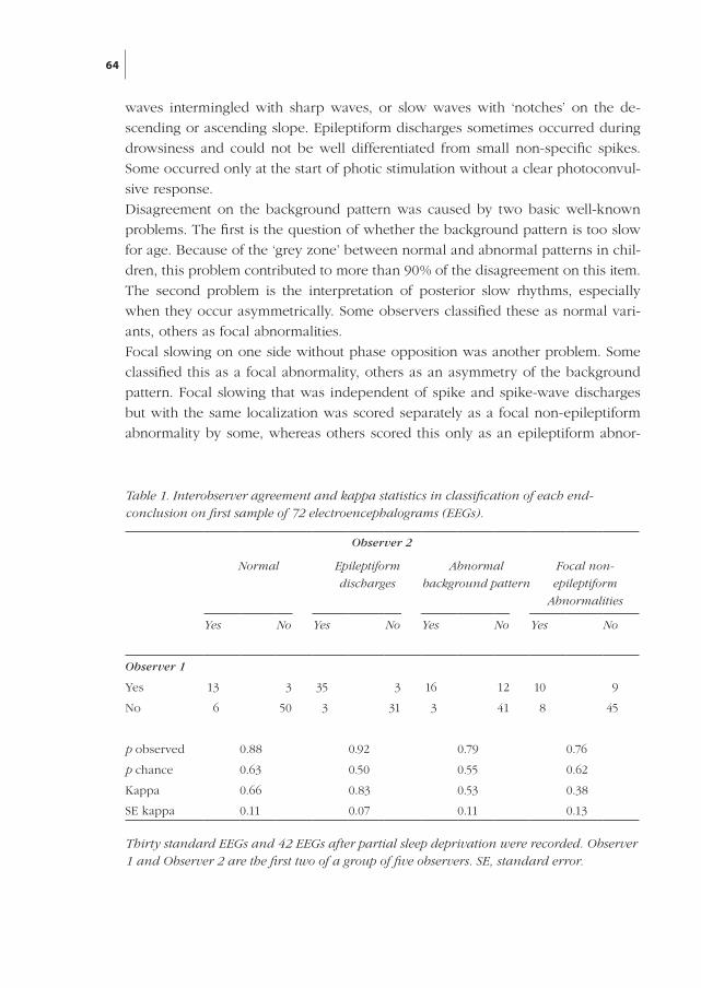
64
waves intermingled with sharp waves, or slow waves with ‘notches’ on the de-
scending or ascending slope. Epileptiform discharges sometimes occurred during
drowsiness and could not be well differentiated from small non-specific spikes.
Some occurred only at the start of photic stimulation without a clear photoconvul-
sive response.
Disagreement on the background pattern was caused by two basic well-known
problems. The first is the question of whether the background pattern is too slow
for age. Because of the ‘grey zone’ between normal and abnormal patterns in chil-
dren, this problem contributed to more than 90% of the disagreement on this item.
The second problem is the interpretation of posterior slow rhythms, especially
when they occur asymmetrically. Some observers classified these as normal vari-
ants, others as focal abnormalities.
Focal slowing on one side without phase opposition was another problem. Some
classified this as a focal abnormality, others as an asymmetry of the background
pattern. Focal slowing that was independent of spike and spike-wave discharges
but with the same localization was scored separately as a focal non-epileptiform
abnormality by some, whereas others scored this only as an epileptiform abnor-
Observer 2
Normal Epileptiform
discharges
Abnormal
background pattern
Focal non-
epileptiform
Abnormalities
Yes No Yes No Yes No Yes No
Observer 1
Yes 13 3 35 3 16 12 10 9
No 6 50 3 31 3 41 8 45
p observed 0.88 0.92 0.79 0.76
p chance 0.63 0.50 0.55 0.62
Kappa 0.66 0.83 0.53 0.38
SE kappa 0.11 0.07 0.11 0.13
Table 1. Interobserver agreement and kappa statistics in classification of each end-conclusion on first sample of 72 electroencephalograms (EEGs).
Thirty standard EEGs and 42 EEGs after partial sleep deprivation were recorded. Observer 1 and Observer 2 are the first two of a group of five observers. SE, standard error.
290158_Stroink_BW.indd 64 11-04-2008 10:53:46
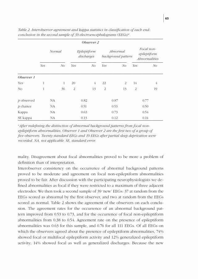
65
mality. Disagreement about focal abnormalities proved to be more a problem of
definition than of interpretation.
Interobserver consistency on the occurrence of abnormal background patterns
proved to be moderate and agreement on focal non-epileptiform abnormalities
proved to be fair. After discussion with the participating neurophysiologists we de-
fined abnormalities as focal if they were restricted to a maximum of three adjacent
electrodes. We then took a second sample of 39 ‘new’ EEGs: 37 at random from the
EEGs scored as abnormal by the first observer, and two at random from the EEGs
scored as normal. Table 2 shows the agreement of the observers on each conclu-
sion. The agreement rates for the occurrence of an abnormal background pat-
tern improved from 0.53 to 0.73, and for the occurrence of focal non-epileptiform
abnormalities from 0.38 to 0.54. Agreement rate on the presence of epileptiform
abnormalities was 0.63 for this sample, and 0.76 for all 111 EEGs. Of all EEGs on
which the observers agreed about the presence of epileptiform abnormalities, 74%
showed focal or multifocal epileptiform activity and 12% generalized epileptiform
activity; 14% showed focal as well as generalized discharges. Because the new
Observer 2
Normal Epileptiform
discharges
Abnormal
background pattern
Focal non-
epileptiform
Abnormalities
Yes No Yes No Yes No Yes No
Observer 1
Yes 1 1 20 4 22 2 14 4
No 1 36 2 13 2 13 2 19
p observed NA 0.82 0.87 0.77
p chance NA 0.51 0.53 0.50
Kappa NA 0.63 0.73 0.54
SE kappa NA 0.13 0.12 0.14
Table 2. Interobserver agreement and kappa statistics in classification of each end-conclusion in the second sample of 39 electroencephalograms (EEGs)a.
aAfter redefining the distinction of abnormal background patterns from focal non-epileptiform abnormalities. Observer 1 and Observer 2 are the first two of a group of five observers. Twenty standard EEGs and 19 EEGs after partial sleep deprivation were recorded. NA, not applicable. SE, standard error.
290158_Stroink_BW.indd 65 11-04-2008 10:53:46
66
sample contained only two normal EEGs, it is not useful to assess interobserver
agreement on the question of whether the EEG was normal or abnormal.
Agreement rates for the interpretation of the standard EEG proved to be better than
those for the partial sleep deprivation EEG for all categories.
Discussion
Absolute criteria for the clinical significance of a given kappa value are lacking
and the prevalence of a positive test result may influence the kappa.4 15 The values
found in our study may be interpreted as substantial for the interpretation of the
EEG as normal or abnormal, and almost perfect for the presence of epileptiform
abnormalities. Agreement was substantial for the occurrence of abnormal back-
ground patterns, and moderate for focal non-epileptiform abnormalities after ad-
justment of the definition used to delineate these two aspects.
These agreement rates for epileptiform abnormalities and background pattern are
clearly above the usual level for clinical agreement. For most clinicians the pres-
ence of epileptiform discharges will be the most important question. For focal
non-epileptiform abnormalities, after adjustment of the definition, kappa reaches a
usual level for clinical agreement4. Moreover, nowadays magnetic resonance imag-
ing is the investigation of choice if focal structural lesions are suspected.
The examiner and the examined4 may cause interobserver variation. Different ex-
aminers may have different opinions on the interpretation of certain graphical ele-
ments and some phenomena may be difficult to interpret. We confined ourselves
to ‘coarse’ conclusions because these might be used to guide the clinical manage-
ment of the children.
Differences in interpretation could not be resolved into one item on which opinions
differed repeatedly. Interpretation of EEGs after partial sleep deprivation proved
to be more difficult as a result of ambiguity of sleep or drowsiness phenomena.
Other differences in opinion were caused by well-known problems such as the
question of whether the background pattern is normal for age. The definition of
focal non-epileptiform abnormalities proved to be particularly difficult. In a previ-
ous comparable study in adults with first epileptic seizures, agreement rates were
moderate.11 This might be explained by differences in the population studied and
hence by the nature of the EEG abnormalities. If epileptiform discharges occur,
they are more frequent in children than in adults.
The classification of the background pattern in children is more difficult because
of the question of whether the background pattern is too slow for age. Compari-
son with other studies on the reliability of visual interpretation of EEGs is difficult
290158_Stroink_BW.indd 66 11-04-2008 10:53:46
67
because different categories were used, the populations concerned were not com-
parable to ours, or the observed agreement rates were not corrected for agreement
due to chance.6-10
Conclusion
The reliability of the visual interpretation of the EEG in accordance with the crite-
ria used in our study in children with newly diagnosed unprovoked seizure(s) is
almost perfect for the presence of epileptiform discharges, substantial for abnormal
background patterns, and moderate for focal non-epileptiform abnormalities. The
agreement rates for non-epileptiform abnormalities can be improved by the use of
well-defined descriptive criteria.
References
1. Berg AT, Shinnar S. The risk of seizure recurrence following a first unprovoked seizure: A
quantitative review. Neurology 1991;41:965-972.
2. van Donselaar CA, Geerts AT, Schimsheimer RJ. Idiopathic first seizure in adult life: who
should be treated? BMJ 1991;302:620-623.
3. Stroink H, Brouwer OF, Arts WF, Geerts AT, Peters AC, van Donselaar CA. The first
unprovoked, untreated seizure in childhood: a hospital based study of the accuracy of
the diagnosis, rate of recurrence, and long term outcome after recurrence. Dutch Study of
Epilepsy in Childhood. J Neurol Neurosurg Psychiatry 1998;64:595-600.
4. Sackett DL, Haynes RB, Guyatt GH, Tugwell P. Clinical epidemiology: a basic science for
clinical medicine. 2nd edition. Boston, Toronto, London: Little, Brown and Company 1991.
5. Blum RH. A note on the reliability of electroencephalographic judgments. Neurology
1954;4:143-146.
6. Houfek EE, Ellingson RJ. On the reliability of clinical EEG interpretation. J Nerv Ment Dis
1959;128:425-437.
7. Woody RH. Inter-judge reliability in clinical electroencephalography. J Clin Psychol
1968;24:251-256.
8. Rose SW, Penry JK, White BG, Sato S. Reliability and validity of visual EEG assessment in
third grade children. Clin Electroencephalogr 1973;4:197–205.
9. Struve FA, Becka DR, Green MA, Howard A. Reliability of clinical interpretation of
electroencephalogram. Clinic Electroencephalogr 1975;6:54-60.
10. Walczak TS, Radtke RA, Lewis DV. Accuracy and interobserver reliability of scalp ictal EEG.
Neurology 1992;42:2279-2285.
11. van Donselaar CA, Schimsheimer RJ, Geerts AT, Declerck AC. Value of the
electroencephalogram in adult patients with untreated idiopathic first seizures. Arch Neurol
1992;49:231-237.
12. Arts WF, Geerts AT, Brouwer OF, Peters AC, Stroink H, van Donselaar CA. The early
290158_Stroink_BW.indd 67 11-04-2008 10:53:46

68
prognosis of epilepsy in childhood: the prediction of a poor outcome. The Dutch study of
epilepsy in childhood. Epilepsia 1999;40:726-734.
13. Cohen J. A coefficient of agreement for nominal scales. Educ Psychol Measurement
1960;20:37-46.
14. Schouten H. Measuring pairwise inter-observer agreement when all subjects are judged by
the same observers. Stat Neerl 1982;36:45-61.
15. Longstreth WT, Jr., Koepsell TD, van Belle G. Clinical neuroepidemiology. I. Diagnosis.
Arch Neurol 1987;44:1091-1099.
290158_Stroink_BW.indd 68 11-04-2008 10:53:47
69
Part II Prognosis
290158_Stroink_BW.indd 69 11-04-2008 10:53:47

290158_Stroink_BW.indd 70 11-04-2008 10:53:47
71
Chapter 6
The first unprovoked, untreated seizure in
childhood: A hospital based study of the
accuracy of the diagnosis, rate of recurrence,
and long term outcome after recurrence
Stroink H, Brouwer OF, Arts WF, Geerts AT, Peters AC, van Donselaar CAJ Neurol Neurosurg Psychiatry 1998;64:595-600
290158_Stroink_BW.indd 71 11-04-2008 10:53:47

290158_Stroink_BW.indd 72 11-04-2008 10:53:47
73
Abstract
Objective: To assess the accuracy of the diagnosis of a first unprovoked seizure
in childhood, the recurrence rate within two years, the risk factors for recurrence,
and the long term outcome two years after recurrence.
Methods: One hundred and fifty six children aged one month to 16 years after a
first seizure, and 51 children with a single disputable event were followed up. The
diagnosis of a seizure was confirmed by a panel of three child neurologists on the
basis of predescribed diagnostic criteria. None of the children was treated after the
first episode.
Results: Five children with a disputable event developed epileptic seizures during
follow up. The diagnosis did not have to be revised in any of the 156 children with
a first seizure. The overall recurrence rate after two years was 54%. Significant risk
factors were an epileptiform EEG (recurrence rate 71%) and remote symptomatic
aetiology and/or mental retardation (recurrence rate 74%). For the 85 children with
one or more recurrences, terminal remission irrespective of treatment two years
after the first recurrence was >12 months in 50 (59%), <six months in 22 (26%), and
six to 12 months in 11 (13%) and unknown in two (2%). Taking the no recurrence
and recurrence groups together, a terminal remission of at least 12 months was
present in 121 out of the 156 children (78%).
Conclusions: The diagnosis of a first seizure can be made accurately with the
help of strict diagnostic criteria. The use of these criteria may have contributed to
the rather high risk of recurrence in this series. However, the overall prognosis for
a child presenting with a single seizure is excellent, even if treatment with antiepi-
leptic drugs is not immediately instituted.
Introduction
Despite several studies,1-12 there is still no definite answer concerning the manage-
ment strategy of children with a first unprovoked epileptic seizure. Besides know-
ledge of the risk of recurrence and the predictive factors, knowledge of the long
term prognosis after a recurrence is a prerequisite for the formulation of adequate
treatment guidelines.
Reported estimates of the recurrence risk after a first unprovoked seizure in child-
hood range from 23% to 71% after three years.2 4 The main factors associated with
a higher risk of recurrence are an EEG showing epileptiform abnormalities and
remote symptomatic aetiology.12
Possible causes for the widely diverging recurrence rates are differences of study
290158_Stroink_BW.indd 73 11-04-2008 10:53:47
74
design, case definitions used for ascertainment, referral patterns within the popu-
lation studied, delay after the seizure before inclusion in the study, and the preva-
lence of various potential risk factors within the population studied.12 13 A surprising
factor is the absence of discussion about diagnostic uncertainty. In none of the stu-
dies mentioned above have diagnostic criteria been used to differentiate between
epileptic and non-epileptic first fits. In particular in young children and infants the
differential diagnosis of a seizure is extensive, and confirming or refuting the epi-
leptic origin of such an event may be quite difficult.
It is still a matter of discussion whether or not children should be treated after a
first unprovoked seizure. Treatment after a first fit may lead to a significant decre-
ase in the risk of relapse.11 Whether early suppression of seizures contributes to a
better long term outcome after recurrence, however, has not yet been defined.
This study was designed to assess prospectively the risk of recurrence in an ac-
curately diagnosed cohort of children with an untreated first unprovoked seizure,
to identify predictive factors for such a recurrence, and to estimate the long term
outcome of those children who had a relapse. To improve diagnostic accuracy, we
used predefined diagnostic criteria formulated in simple descriptive terms, as well
as the expert opinion of a panel of paediatric neurologists.
Methods
PatientsMost patients in this prospective study were derived from a consecutive series of
881 children, aged between one month and 16 years, who were referred with one
or more possible unprovoked seizures, or at least one episode of status epilepticus,
to one of the four participating hospitals: two university hospitals, one children’s
hospital, and one general hospital in the southwest region of The Netherlands. This
cohort forms the basis of the Dutch Study of Epilepsy in Childhood (DSEC), which
tries to answer several clinical-epidemiological questions about newly diagnosed
childhood epilepsy.14 Inclusion for the first seizure part of the DSEC started 1 Ja-
nuary 1988 and ended 1 August 1992. Children were mainly referred by general
practitioners, by paediatricians of the participating hospitals, or were first seen
in the emergency room of the participating hospitals. All children with possible
seizures were recruited, but to be eligible for entry into the study, a committee of
paediatric neurologists (HS, WFA, OFB, and ACBP, excluding the attending neuro-
logist) had to agree that the description of the single episode, as described by the
child, or an eye witness, or both concurred with predefined descriptive diagnostic
criteria of an epileptic seizure, adapted from Van Donselaar et al,15 without having
290158_Stroink_BW.indd 74 11-04-2008 10:53:47
75
any knowledge of the results of the EEG. The committee excluded children with a
clear non-epileptic event such as a reflex anoxic seizure or syncope. Children with
an event classified by the committee as “disputable” were followed up separately
for one year to test our diagnostic procedure.
Children with a seizure due to an acute neurological insult (meningitis, trauma),
metabolic disturbances, or fever (temperature over 38.00C) were excluded. Child-
ren with a history of earlier seizures other than neonatal or febrile seizures; with a
single episode of status epilepticus; with a recurrence within 24 hours; or with an
interval between the seizure and the first visit to the hospital of more than three
months, were not included in the first seizure part of the DSEC, but in the study
part on the general prognosis of newly diagnosed epilepsy in childhood (publis-
hed later on).16 17 Of the remaining 170, we excluded 10 children because of pos-
sible fever reported by the parents (precise temperature not known). Four other
children were excluded because they had been treated with antiepileptic drugs.
One child was treated mistakenly after only one seizure; three other children were
treated because of multiple recurrences associated with fever, but they had not had
unprovoked recurrences at that time. Finally 156 children remained for inclusion.
ClassificationThe committee classified seizures according to the revised classification of the
International League Against Epilepsy (ILAE).18 The aetiology was classified as re-
mote symptomatic if the child was known to have a static encephalopathy caused
by a prenatal or perinatal encephalopathy or a prior neurological insult such as
infection, stroke, or cerebral trauma. Children with mental retardation (estimated
IQ below 70) were included in this group. According to the recent guidelines on
epidemiological research of the ILAE,19 patients with a genetically determined type
of epilepsy manifesting through a single seizure were called idiopathic. All other
children were considered cryptogenic. In this analysis idiopathic and cryptogenic
cases were grouped together. This seems to be justifiable, because it is usually not
possible to distinguish between them after only one seizure.20
Additional investigationsA standard EEG (EEG1) was obtained in all patients. If it did not disclose epi-
leptiform abnormalities, a second EEG (EEG2) was performed after partial sleep
deprivation, or during the daytime sleep in very young children. All EEGs were
classified as normal or abnormal and scored for the presence of epileptiform ab-
normalities (focal and generalised spikes or spike and wave complexes), and other
abnormalities (abnormal background pattern or focal non-epileptiform abnorma-
290158_Stroink_BW.indd 75 11-04-2008 10:53:47
76
lities) by clinical neurophysiologists who were unaware of the clinical data. Brain
CT was scheduled in all children if possible without anaesthesia. The decision to
perform CT in the remaining children was up to the child neurologist.
Follow upWe followed up all children with a single seizure on a regular basis for 24 months
by hospital visits and by telephone interviews. After a recurrence, defined as any
unprovoked seizure after inclusion into the study, the children were seen again and
a detailed history was taken. We followed up all these children after their recur-
rence for 24-72 months (mean 42, median 44, 25, and 75 percentile: 30; 54 months)
until 1 August 1994, with the exception of two children who were lost 0 and
10 months after the recurrence.
No antiepileptic drugs were prescribed after the first seizure. The decision whether
or not to start treatment after one or more recurrences was left to the attending
paediatric neurologist.
The outcome was measured by two methods. Firstly, the duration of the seizure
free period irrespective of treatment existing at two years after the first recurrence
(terminal remission, TR) according to the following definition: excellent, no recur-
rence at all; good, TR at least 12 months; moderate, TR six to 12 months; poor,
TR less than six months. Secondly, the maximum period of seizure freedom after
recurrence irrespective of treatment (longest remission ever, LRE) according to
a slight modification of the definition of Arts et al:21excellent, no seizures at all;
good, LRE at least 12 months; moderate, LRE six to 12 months; poor, LRE less than
six months.
Fifty one children with a single ictal event in whom no clear diagnosis could be
made were followed up for one year to assess the accuracy of our diagnostic pro-
cedure.
AnalysisKaplan-Meier survival analysis was used for calculation of the recurrence rates.22
Univariate and multivariate analyses were performed using Cox’s proportional ha-
zards model.23 The multivariate analysis was done with a full model and with step-
wise backward elimination of variables. In the second, we used simple parameter
coding, and a probability of removal of 0.10.
Informed consentThe study was approved by the ethics committees of all involved hospitals, and
informed consent was obtained in all cases before enrolment.
290158_Stroink_BW.indd 76 11-04-2008 10:53:47
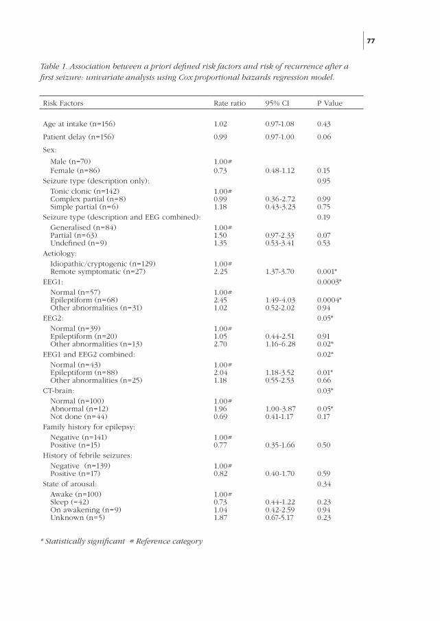
77
Risk Factors Rate ratio 95% CI P Value
Age at intake (n=156) 1.02 0.97-1.08 0.43
Patient delay (n=156) 0.99 0.97-1.00 0.06
Sex:
Male (n=70) 1.00# Female (n=86) 0.73 0.48-1.12 0.15Seizure type (description only): 0.95 Tonic clonic (n=142) 1.00# Complex partial (n=8) 0.99 0.36-2.72 0.99 Simple partial (n=6) 1.18 0.43-3.23 0.75Seizure type (description and EEG combined): 0.19 Generalised (n=84) 1.00# Partial (n=63) 1.50 0.97-2.33 0.07 Undefined (n=9) 1.35 0.53-3.41 0.53Aetiology: Idiopathic/cryptogenic (n=129) 1.00# Remote symptomatic (n=27) 2.25 1.37-3.70 0.001*EEG1: 0.0003* Normal (n=57) 1.00# Epileptiform (n=68) 2.45 1.49-4.03 0.0004* Other abnormalities (n=31) 1.02 0.52-2.02 0.94EEG2: 0.05* Normal (n=39) 1.00# Epileptiform (n=20) 1.05 0.44-2.51 0.91 Other abnormalities (n=13) 2.70 1.16-6.28 0.02*EEG1 and EEG2 combined: 0.02* Normal (n=43) 1.00# Epileptiform (n=88) 2.04 1.18-3.52 0.01* Other abnormalities (n=25) 1.18 0.55-2.53 0.66CT-brain: 0.03* Normal (n=100) 1.00# Abnormal (n=12) 1.96 1.00-3.87 0.05* Not done (n=44) 0.69 0.41-1.17 0.17Family history for epilepsy: Negative (n=141) 1.00# Positive (n=15) 0.77 0.35-1.66 0.50History of febrile seizures: Negative (n=139) 1.00# Positive (n=17) 0.82 0.40-1.70 0.59State of arousal: 0.34 Awake (n=100) 1.00# Sleep (=42) 0.73 0.44-1.22 0.23 On awakening (n=9) 1.04 0.42-2.59 0.94 Unknown (n=5) 1.87 0.67-5.17 0.23
* Statistically significant # Reference category
Table 1. Association between a priori defined risk factors and risk of recurrence after a first seizure: univariate analysis using Cox proportional hazards regression model.
290158_Stroink_BW.indd 77 11-04-2008 10:53:47
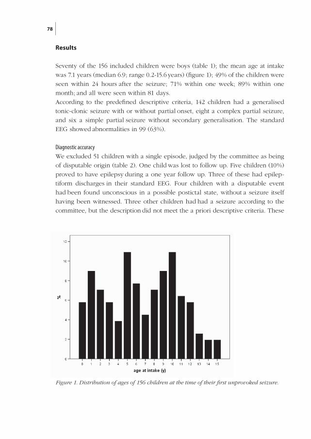
78
Results
Seventy of the 156 included children were boys (table 1); the mean age at intake
was 7.1 years (median 6.9; range 0.2-15.6 years) (figure 1); 49% of the children were
seen within 24 hours after the seizure; 71% within one week; 89% within one
month; and all were seen within 81 days.
According to the predefined descriptive criteria, 142 children had a generalised
tonic-clonic seizure with or without partial onset, eight a complex partial seizure,
and six a simple partial seizure without secondary generalisation. The standard
EEG showed abnormalities in 99 (63%).
Diagnostic accuracyWe excluded 51 children with a single episode, judged by the committee as being
of disputable origin (table 2). One child was lost to follow up. Five children (10%)
proved to have epilepsy during a one year follow up. Three of these had epilep-
tiform discharges in their standard EEG. Four children with a disputable event
had been found unconscious in a possible postictal state, without a seizure itself
having been witnessed. Three other children had had a seizure according to the
committee, but the description did not meet the a priori descriptive criteria. These
Figure 1. Distribution of ages of 156 children at the time of their first unprovoked seizure.
290158_Stroink_BW.indd 78 11-04-2008 10:53:47
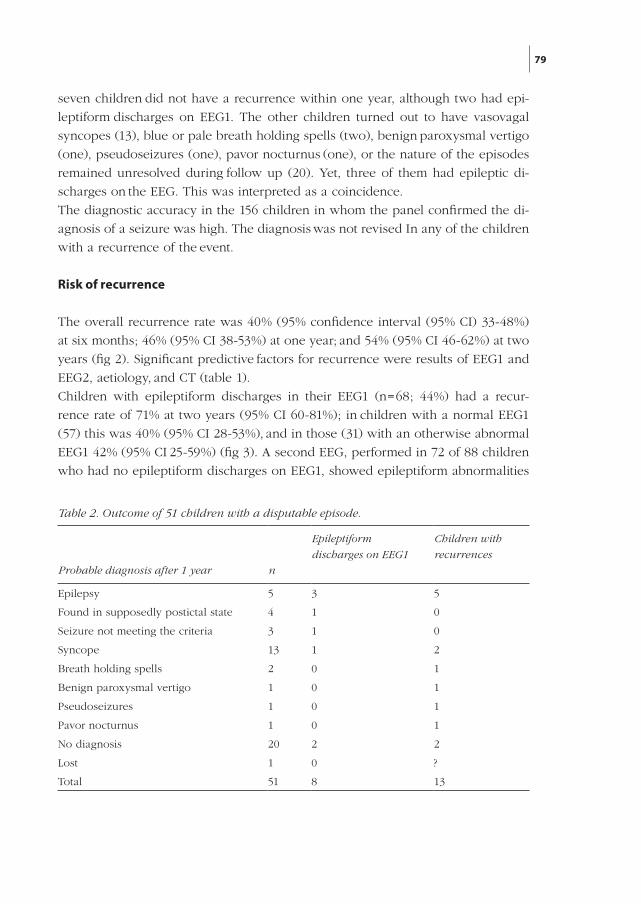
79
seven children did not have a recurrence within one year, although two had epi-
leptiform discharges on EEG1. The other children turned out to have vasovagal
syncopes (13), blue or pale breath holding spells (two), benign paroxysmal vertigo
(one), pseudoseizures (one), pavor nocturnus (one), or the nature of the episodes
remained unresolved during follow up (20). Yet, three of them had epileptic di-
scharges on the EEG. This was interpreted as a coincidence.
The diagnostic accuracy in the 156 children in whom the panel confirmed the di-
agnosis of a seizure was high. The diagnosis was not revised In any of the children
with a recurrence of the event.
Risk of recurrence
The overall recurrence rate was 40% (95% confidence interval (95% CI) 33-48%)
at six months; 46% (95% CI 38-53%) at one year; and 54% (95% CI 46-62%) at two
years (fig 2). Significant predictive factors for recurrence were results of EEG1 and
EEG2, aetiology, and CT (table 1).
Children with epileptiform discharges in their EEG1 (n=68; 44%) had a recur-
rence rate of 71% at two years (95% CI 60-81%); in children with a normal EEG1
(57) this was 40% (95% CI 28-53%), and in those (31) with an otherwise abnormal
EEG1 42% (95% CI 25-59%) (fig 3). A second EEG, performed in 72 of 88 children
who had no epileptiform discharges on EEG1, showed epileptiform abnormalities
Table 2. Outcome of 51 children with a disputable episode.
Epileptiform
discharges on EEG1
Children with
recurrences
Probable diagnosis after 1 year n
Epilepsy 5 3 5
Found in supposedly postictal state 4 1 0
Seizure not meeting the criteria 3 1 0
Syncope 13 1 2
Breath holding spells 2 0 1
Benign paroxysmal vertigo 1 0 1
Pseudoseizures 1 0 1
Pavor nocturnus 1 0 1
No diagnosis 20 2 2
Lost 1 0 ?
Total 51 8 13
290158_Stroink_BW.indd 79 11-04-2008 10:53:47
80
in another 20 children (28%). Aetiology also proved to be a significant predictive
factor for recurrence (fig 4). Recurrence rate at two years was 50% (95% CI 41-59%)
in 129 children with a cryptogenic or idiopathic seizure, and 74% (95% CI 57-91%)
in 27 children with remote symptomatic aetiology or mental retardation without
known cause (table 1).
Brain CT was obtained in 112 children. The abnormalities (mostly atrophy) found
in 12 children were without therapeutic consequences. Recurrence rate at two
years was 75% (95% CI 51-100%) in those with abnormal CT findings, 56% (95% CI
46-66%) in the children with normal CT and 43% (95% CI: 29-58%) in the children
in whom no CT was performed.
No significant influence on the recurrence rate was found for the other variables
investigated (table 1).
Full model multivariate analysis was carried out with 11 variables. An epilepti-
form EEG1 was the most important predictive factor for seizure recurrence. Remote
symptomatic aetiology was also significantly associated with risk of recurrence.
After stepwise backward elimination of variables not contributing to the model,
aetiology, EEG1, and the sleep state remained. EEG1 was the most significant vari-
able.
Long term outcome after recurrenceAt the end of the follow up of all children, 71 children (46%) had an excellent
outcome without any recurrent seizure. Sixty nine of the 85 children with a recur-
rence were treated with antiepileptic drugs. Of the children with a recurrence,
27 (32%) had an excellent outcome, 23 (27%) a good, 11 (13%) a moderate, and
22 (26%) a poor outcome defined by terminal remission. Two children were lost
0 and 10 months after recurrence. This means that of all admitted children with a
single seizure 121 (78%) had an excellent or good outcome. According to the defi-
nition of Arts et al19 (LRE), the outcome was excellent in 27 children (32%) with a
recurrence, good in 32 (38%) moderate in 17 (20%) and poor in only seven (8%).
The outcome was excellent or good in 130 (83%) of all admitted children.
Discussion
When the clinician is confronted with the problem of a child who has experienced
a single episode that seems to be of epileptic origin, some questions have to be
considered. Was the event really epileptic? If so, what is the risk of more seizures
occurring? Should anticonvulsant treatment be offered and with what goal? What is
the long term outcome with or without treatment?
290158_Stroink_BW.indd 80 11-04-2008 10:53:47
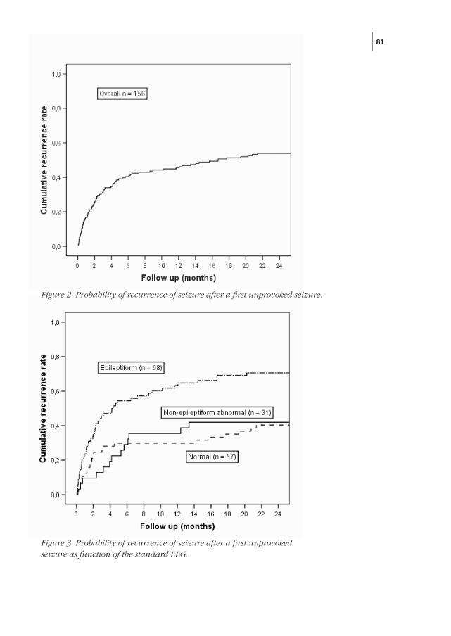
81
Figure 3 Probability of recurrence of seizure after a first unprovoked seizure as function of the standard EEG.
Figure 3. Probability of recurrence of seizure after a first unprovoked seizure as function of the standard EEG.
Figure 2. Probability of recurrence of seizure after a first unprovoked seizure.
290158_Stroink_BW.indd 81 11-04-2008 10:53:47
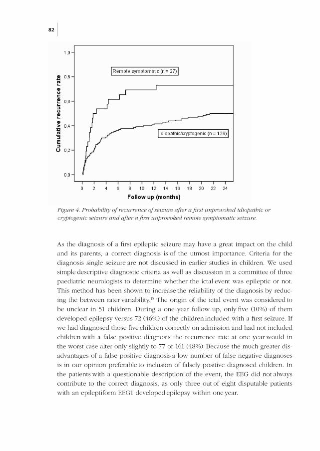
82
As the diagnosis of a first epileptic seizure may have a great impact on the child
and its parents, a correct diagnosis is of the utmost importance. Criteria for the
diagnosis single seizure are not discussed in earlier studies in children. We used
simple descriptive diagnostic criteria as well as discussion in a committee of three
paediatric neurologists to determine whether the ictal event was epileptic or not.
This method has been shown to increase the reliability of the diagnosis by reduc-
ing the between rater variability.15 The origin of the ictal event was considered to
be unclear in 51 children. During a one year follow up, only five (10%) of them
developed epilepsy versus 72 (46%) of the children included with a first seizure. If
we had diagnosed those five children correctly on admission and had not included
children with a false positive diagnosis the recurrence rate at one year would in
the worst case alter only slightly to 77 of 161 (48%). Because the much greater dis-
advantages of a false positive diagnosis a low number of false negative diagnoses
is in our opinion preferable to inclusion of falsely positive diagnosed children. In
the patients with a questionable description of the event, the EEG did not always
contribute to the correct diagnosis, as only three out of eight disputable patients
with an epileptiform EEG1 developed epilepsy within one year.
Figure 4. Probability of recurrence of seizure after a first unprovoked idiopathic or
cryptogenic seizure and after a first unprovoked remote symptomatic seizure.
290158_Stroink_BW.indd 82 11-04-2008 10:53:47
83
The overall recurrence rate of 54% at two years after a first unprovoked seizure
found in this study is higher than in other recent prospective studies3 8 9 and in a
recent meta-analysis.12 Factors that may have increased the recurrence rate in our
study are the withholding of antiepileptic drug treatment after the first fit, and the
high diagnostic accuracy by the use of strict diagnostic criteria. Furthermore, 49%
of the children were seen within 24 hours and 71% within one week. Therefore,
the number of children who were not included because of an early recurrence, was
probably small.
The risk factors for recurrence identified in this study were similar to those re-
ported by others.12 An EEG with epileptiform abnormalities proved to be the main
risk factor for recurrence. Other factors associated with a higher risk of recurrence
found in this study were remote symptomatic aetiology, or mental retardation, or
both and abnormal CT. The children in whom no CT was carried out, had the low-
est recurrence rate. Obviously, the neurologists selected children for CT on clinical
grounds. In our opinion CT is not routinely indicated for children presenting with
a first seizure.
Remarkably, whereas others have found that the occurrence of a first seizure dur-
ing sleep is associated with an increased risk of recurrence,24 we found the op-
posite. We think that the use of strict criteria and discussion of each child by the
expert committee lowered the number of children with non-epileptic events. Non-
epileptic events are less likely to occur during sleep. This may have caused a bias
in other studies.
Despite the relatively high recurrence rate, most children did well in the end. Our
strategy to delay treatment after the first unprovoked seizure in our study group of
156 children led to a rather high recurrence rate of 54%. The long term outcome
was poor in only 22 out of 156 children (14%) using terminal remission as the crite-
rion. Many of the children with a poor outcome had infrequent generalised tonic-
clonic or rolandic seizures, of which one or two coincidentally occurred during the
final half year of the two year follow up. This explains the better outcome in terms
of longest remission ever.21
There is controversy over whether treatment should be offered after a single sei-
zure. In Europe, children with single unprovoked epileptic seizures are usually
not treated. The current clinical practice is to defer treatment until two or more
seizures have occurred, although children perceived to be at high risk for recur-
rence may be treated after a single fit. The Italian First Seizure Trial Group carried
out a controlled randomised trial of anticonvulsant therapy after a first generalised
tonic-clonic seizure that occurred within the preceding seven days, in a large co-
hort including 113 children.11 After two years, the recurrence rate for these children
290158_Stroink_BW.indd 83 11-04-2008 10:53:48
84
was significantly lower in the treated group (25% v 51%). Treatment after the first
seizure did not, however, improve long term prognosis.25
For an answer to the question whether treatment should be started in a child pre-
senting with a first epileptic seizure, more knowledge of the long term outcome
after recurrence is urgently needed. From our study, it may be concluded that
the indication for starting long term treatment with AEDs after a single seizure in
childhood is weak, because the risk of developing intractable epilepsy is already
low. Immediate treatment will probably not further improve long term prognosis.25
Treatment of all children after a single seizure therefore means treatment of many
who will never have a second seizure; treatment of many whose epilepsy will have
a benign course irrespective of treatment; and treatment of only a small minority in
the hope of preventing them becoming intractable. Unfortunately, we are not able
to predict which children will do badly after a single seizure. This would allow us
to restrict treatment to these children. At this time, it seems to be advisable to delay
long term anticonvulsant treatment until recurrent seizures are adversely affecting
the child’s life without signs of spontaneous remission. In so doing many children
will never have to start long term treatment.
References
1. Thomas MH. The single seizures: its study and management. J Am Med Assoc
1959;169:457-459.
2. Pearce JL, Mackintosh HT. Prospective study of convulsions in childhood. N Z Med J
1979;89:1-3.
3. Camfield PR, Camfield CS, Dooley JM, Tibbles JAR, Fung T, Garner B. Epilepsy after a first
unprovoked seizure in childhood. Neurology 1985;35:1657-1660.
4 Elwes RD, Chesterman P, Reynolds EH. Prognosis after a first untreated tonic-clonic
seizure. Lancet 1985;2:752-753.
5. Annegers JF, Shirts SB, Hauser WA, Kurland LT. Risk of recurrence after an initial
unprovoked seizure. Epilepsia 1986;27:43-50.
6. Boulloche J, Leloup P, Mallet E, Parain D, Tron P. Risk of recurrence after a single,
unprovoked, generalized tonic-clonic seizure. Dev Med Child Neurol 1989;31:626-632.
7. Camfield P, Camfield C, Dooley J, Smith E, Garner B. A randomized study of
carbamazepine versus no medication after a first unprovoked seizure in childhood.
Neurology 1989;39:851-852.
8. Hauser WA, Rich SS, Annegers JF, Anderson VE. Seizure recurrence after a 1st unprovoked
seizure: An extended follow-up. Neurology 1990;40:1163-1170.
9. Shinnar S, Berg AT, Moshe SL, Petix M, Maytal J, Kang H, et al. Risk of Seizure Recurrence
Following a First Unprovoked Seizure in Childhood: A Prospective Study. Pediatrics
1990;85:1076-1085.
290158_Stroink_BW.indd 84 11-04-2008 10:53:48
85
10. Hart YM, Sander JW, Johnson AL, Shorvon SD. National General Practice Study of Epilepsy:
recurrence after a first seizure. Lancet 1990;336:1271-1274.
11. FIR.S.T. GROUP. Randomized clinical trial on the efficacy of antiepileptic drugs in reducing
the risk of relapse after a first unprovoked tonic-clonic seizure. First Seizure Trial Group.
Neurology 1993;43:478-483.
12. Berg AT, Shinnar S. The risk of seizure recurrence following a first unprovoked seizure: A
quantitative review. Neurology 1991;41:965-972.
13. Chadwick D. Epilepsy after first seizures: risks and implications. J Neurol Neurosurg
Psychiatry 1991;54:385-387.
14. Brouwer OF, van Donselaar CA, Stroink H, Arts WF, Geerts AT, Peters AC. The Dutch
study of epilepsy in childhood: design of the study. Epilepsia 1995;36:S28.
15. van Donselaar CA, Geerts AT, Meulstee J, Habbema JD, Staal A. Reliability of the diagnosis
of a first seizure. Neurology 1989;39:267-271.
16. Arts WF, Geerts AT, Brouwer OF, Peters AC, Stroink H, van Donselaar CA. The early
prognosis of epilepsy in childhood: the prediction of a poor outcome. The Dutch study of
epilepsy in childhood. Epilepsia 1999;40:726-734.
17. Arts WF, Brouwer OF, Peters AC, Stroink H, Peeters EA, Schmitz PI, et al. Course and
prognosis of childhood epilepsy: 5-year follow-up of the Dutch Study of Epilepsy in
Childhood. Brain 2004;127:1774-1784.
18. ILAE C. Proposal for revised clinical and electroencephalographic classification of epileptic
seizures. From the commission on Classification and Terminology of the International
League Against Epilepsy. Epilepsia 1981;22:489-501.
19. Commission on Epidemiology and Prognosis ILAE. Guidelines for epidemiological studies
on epilepsy. Epilepsia 1993;34:592-596.
20. Shinnar S, Berg AT, Moshe SL, O’Dell C, Alemany M, Newstein D, et al. The Risk of Seizure
Recurrence After a First Unprovoked Afebrile Seizure in Childhood: An Extended Follow-
up. Pediatrics 1996;98:216-225.
21. Arts WF, van Donselaar CA, Stroink H, Peters AC, Brouwer OF. Follow-up of intractable
seizures in childhood. Ann Neurol 1991;30:115.
22. SPSS for Windows. Advanced statistics, release 5. Chicago, Illinois: SPSS Inc, 1992.
23. Cox DR. Regression models and life-tables. Journal of the Royal Statistical Society (B)
1972:187-220.
24. Shinnar S, Berg AT, Ptachewich Y, Alemany M. Sleep state and the risk of seizure
recurrence following a first unprovoked seizure in childhood. Neurology 1993;43:701-706.
25. Musicco M, Beghi E, Solari A, Viani F. Treatment of first tonic-clonic seizure does not
improve the prognosis of epilepsy. First Seizure Trial Group (FIRST Group). Neurology
1997;49:991-998.
290158_Stroink_BW.indd 85 11-04-2008 10:53:48

290158_Stroink_BW.indd 86 11-04-2008 10:53:48
87
Chapter 7
Status epilepticus in children with epilepsy
Stroink H, Geerts AT, van Donselaar CA, Peters ACB, Brouwer OF, Peeters EA, Arts WF Epilepsia 2007;48:1708-1715.
290158_Stroink_BW.indd 87 11-04-2008 10:53:48

290158_Stroink_BW.indd 88 11-04-2008 10:53:48
89
Abstract
Purpose: To study course and outcome of epilepsy in children having had a status
epilepticus (SE) as the presenting sign or after the diagnosis.
Methods: A total of 494 children with newly diagnosed epilepsy, aged one month
through 15 years, were followed prospectively for five years.
Results: A total of 47 Children had SE. Forty-one of them had SE when epilepsy
was diagnosed. For 32 (78%), SE was the first seizure. SE recurred in 13 out of 41
(32%). Terminal remission at five years (TR5) was not significantly worse for these
41 children: 31.7% had a TR5 <1 year versus 21.2% of 447 children without SE.
They were not more often intractable. Five out of six children with first SE after
diagnosis had a TR5 <1 year. Mortality was not significantly increased for children
with SE. Independent factors associated with SE at presentation were remote symp-
tomatic and cryptogenic aetiology, and a history of febrile seizures. Children with
first SE after inclusion more often had symptomatic aetiology.
Conclusions: Although we find a trend for shorter TR5 in children with SE at
presentation, outcome and mortality are not significantly worse. Aetiology is an
important factor for prognosis. Children with SE during the course of their epilepsy
have a worse prognosis and a high recurrence rate of SE. This outcome is not due
to the SE itself, but related to the aetiology and type of epilepsy. The occurrence
of SE is just an indicator of the severity of the disease.
Introduction
Although the mortality in recent years has declined considerably,1-5 status epilepti-
cus (SE) in children remains a medical emergency. Analyzing causes, risk factors,
recurrence rate and outcome of SE in children with epilepsy may help to improve
the immediate and long-term care for these children. Only a few studies on this
subject have been published.2 6 7 The Dutch Study of Epilepsy in Children (DSEC)
is well suited to perform this type of analysis. We studied the occurrence of SE
and the course after SE in our cohort of children with epilepsy. A comparison was
made between children with and without SE.
Patients and methods
The prospective multicenter hospital-based DSEC started in 1988. After informed
consent, we enrolled consecutively all children aged one month through 15 years
who were referred between August 1988 and August 1992 because of a possible
290158_Stroink_BW.indd 89 11-04-2008 10:53:48
90
unprovoked single seizure or epilepsy to one of the participating hospitals. At in-
take, 494 children suffered at least two or more unprovoked seizures or one unpro-
voked SE. SE was defined as a seizure with duration of at least 30 min or recurrent
seizures lasting a total of more than 30 min without regaining consciousness in-
between. We knew at that time that most children after a SE would be treated with
antiepileptic drugs (AEDs) for at least some weeks to months. However, children
who had ever used AEDs or started to use AEDs after the first seizure, had to be
excluded from our first-seizure study according to its protocol.8 This explains why
children presenting with a solitary SE were not followed in the first seizure cohort,
but in the cohort of children with new onset epilepsy, although they did not fully
comply with the ILAE definition of epilepsy. Most children were referred directly
by their general practitioner (51%) or by the paediatricians of the participating hos-
pitals (25%). These paediatricians routinely referred all children with possible sei-
zures to the departments of paediatric neurology. Some (16%), especially children
with SE, were first seen in the emergency department. We excluded children with
only febrile or acute symptomatic seizures, as they did not fulfill the requirements
for the diagnosis of epilepsy, as well as children who were referred from other hos-
pitals for a second opinion. The children were followed for five years. The mean
age at intake of the total cohort (494) was 5.9 years (standard error (se) 0.2 years;
median 5.5 years). Details have been described elsewhere.9-11 We defined outcome
in terms of terminal remission (TR): the interval from the last seizure (not the last
SE!) to the end of follow-up at five years (TR5). TR5 was dichotomized (TR5<1
year, TR5>1 year). Besides, intractability was defined as a longest remission of less
than three months during the last year of follow-up, despite adequate treatment.11
Adequate treatment was defined as the optimal use of at least two AEDs.12-14
We studied the following factors for possible association with SE: aetiology, age,
prior febrile seizures, classification of seizure type and epilepsy syndrome, and the
results of prophylactic treatment with AEDs after the first SE. Results were compa-
red with the few earlier studies on this subject. We used survival analysis to inves-
tigate the risk of a first SE and the risk of a first recurrence of SE during follow-up.
These cumulative risks from the onset of epilepsy until the end of follow-up were
displayed by means of Kaplan–Meier curves. Univariate logistic regression analysis
was used to investigate which variables were associated with a first SE. To inves-
tigate which combination of independent variables was associated with SE before
or at diagnosis, we performed a multivariate logistic regression analysis with a
stepwise backward method (conditional). A variable was eliminated if its removal
statistic had a probability greater than or equal to 0.10. We used simple parameter
coding (each category of any particular variable was compared to the reference ca-
290158_Stroink_BW.indd 90 11-04-2008 10:53:48
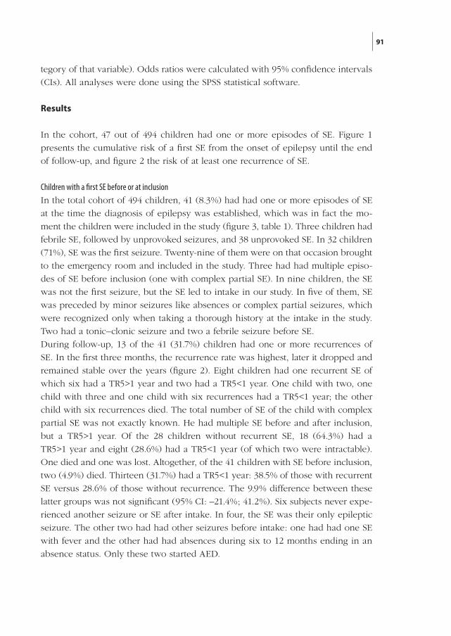
91
tegory of that variable). Odds ratios were calculated with 95% confidence intervals
(CIs). All analyses were done using the SPSS statistical software.
Results
In the cohort, 47 out of 494 children had one or more episodes of SE. Figure 1
presents the cumulative risk of a first SE from the onset of epilepsy until the end
of follow-up, and figure 2 the risk of at least one recurrence of SE.
Children with a first SE before or at inclusionIn the total cohort of 494 children, 41 (8.3%) had had one or more episodes of SE
at the time the diagnosis of epilepsy was established, which was in fact the mo-
ment the children were included in the study (figure 3, table 1). Three children had
febrile SE, followed by unprovoked seizures, and 38 unprovoked SE. In 32 children
(71%), SE was the first seizure. Twenty-nine of them were on that occasion brought
to the emergency room and included in the study. Three had had multiple episo-
des of SE before inclusion (one with complex partial SE). In nine children, the SE
was not the first seizure, but the SE led to intake in our study. In five of them, SE
was preceded by minor seizures like absences or complex partial seizures, which
were recognized only when taking a thorough history at the intake in the study.
Two had a tonic–clonic seizure and two a febrile seizure before SE.
During follow-up, 13 of the 41 (31.7%) children had one or more recurrences of
SE. In the first three months, the recurrence rate was highest, later it dropped and
remained stable over the years (figure 2). Eight children had one recurrent SE of
which six had a TR5>1 year and two had a TR5<1 year. One child with two, one
child with three and one child with six recurrences had a TR5<1 year; the other
child with six recurrences died. The total number of SE of the child with complex
partial SE was not exactly known. He had multiple SE before and after inclusion,
but a TR5>1 year. Of the 28 children without recurrent SE, 18 (64.3%) had a
TR5>1 year and eight (28.6%) had a TR5<1 year (of which two were intractable).
One died and one was lost. Altogether, of the 41 children with SE before inclusion,
two (4.9%) died. Thirteen (31.7%) had a TR5<1 year: 38.5% of those with recurrent
SE versus 28.6% of those without recurrence. The 9.9% difference between these
latter groups was not significant (95% CI: –21.4%; 41.2%). Six subjects never expe-
rienced another seizure or SE after intake. In four, the SE was their only epileptic
seizure. The other two had had other seizures before intake: one had had one SE
with fever and the other had had absences during six to 12 months ending in an
absence status. Only these two started AED.
290158_Stroink_BW.indd 91 11-04-2008 10:53:48
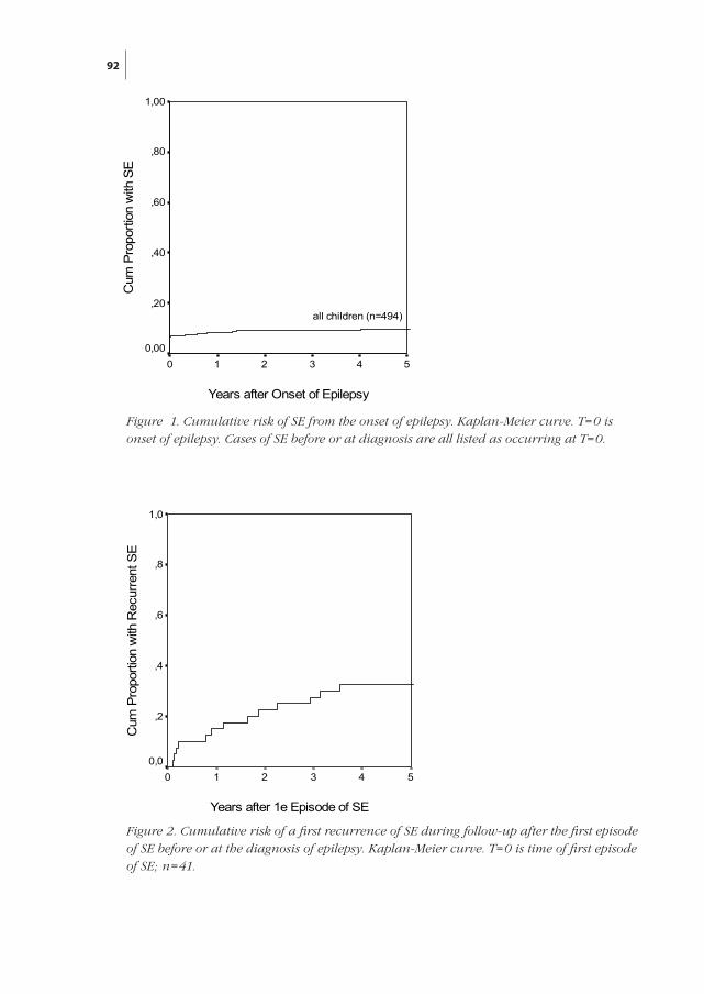
92
Years after 1e Episode of SE
543210
Cum
Pro
porti
on w
ith R
ecur
rent
SE
1,0
,8
,6
,4
,2
0,0
Years after Onset of Epilepsy
543210
Cum
Pro
porti
on w
ith S
E
1,00
,80
,60
,40
,20
0,00
all children (n=494)
Figure 2. Cumulative risk of a first recurrence of SE during follow-up after the first episode of SE before or at the diagnosis of epilepsy. Kaplan-Meier curve. T=0 is time of first episode of SE; n=41.
Figure 1. Cumulative risk of SE from the onset of epilepsy. Kaplan-Meier curve. T=0 is onset of epilepsy. Cases of SE before or at diagnosis are all listed as occurring at T=0.
290158_Stroink_BW.indd 92 11-04-2008 10:53:48
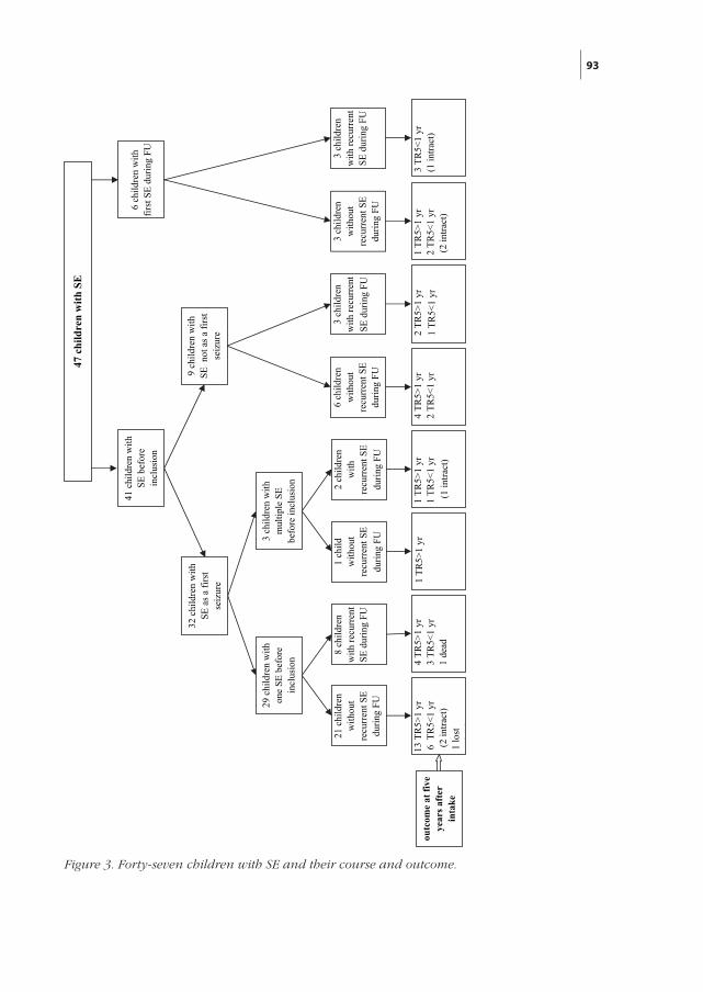
93
3 ch
ildre
n w
ithou
t re
curr
ent S
E du
ring
FU
21 c
hild
ren
with
out
recu
rren
t SE
durin
g FU
8 ch
ildre
n w
ith re
curr
ent
SE d
urin
g FU
1 ch
ild
with
out
recu
rren
t SE
durin
g FU
2 ch
ildre
n w
ith
recu
rren
t SE
durin
g FU
6 ch
ildre
n w
ithou
t re
curr
ent S
E du
ring
FU
3 ch
ildre
n w
ith re
curr
ent
SE d
urin
g FU
47 c
hild
ren
with
SE
9 ch
ildre
n w
ith
SE n
ot a
s a fi
rst
seiz
ure
32 c
hild
ren
with
SE
as a
firs
t se
izur
e
3 ch
ildre
n w
ith
mul
tiple
SE
befo
re in
clus
ion
29 c
hild
ren
with
on
e SE
bef
ore
incl
usio
n
41 c
hild
ren
with
SE
bef
ore
incl
usio
n
1 T
R5>
1 yr
2 T
R5<
1 yr
(2
intra
ct)
13 T
R5>
1 yr
6 T
R5<
1 yr
(2
intra
ct)
1 lo
st1
dd
4 T
R5>
1 yr
3 T
R5<
1 yr
1 d
ead
1 T
R5>
1 yr
1 T
R5>
1 yr
1 T
R5<
1 yr
(1
intra
ct)
4 T
R5>
1 yr
2 T
R5<
1 yr
2 T
R5>
1 yr
1 T
R5<
1 yr
outc
ome
at fi
ve
year
s afte
r in
take
6 ch
ildre
n w
ith
first
SE
durin
g FU 3
child
ren
with
recu
rren
t SE
dur
ing
FU
3 T
R5<
1 yr
(1 in
tract
)
Figure 3. Forty-seven children with SE and their course and outcome.
290158_Stroink_BW.indd 93 11-04-2008 10:53:48
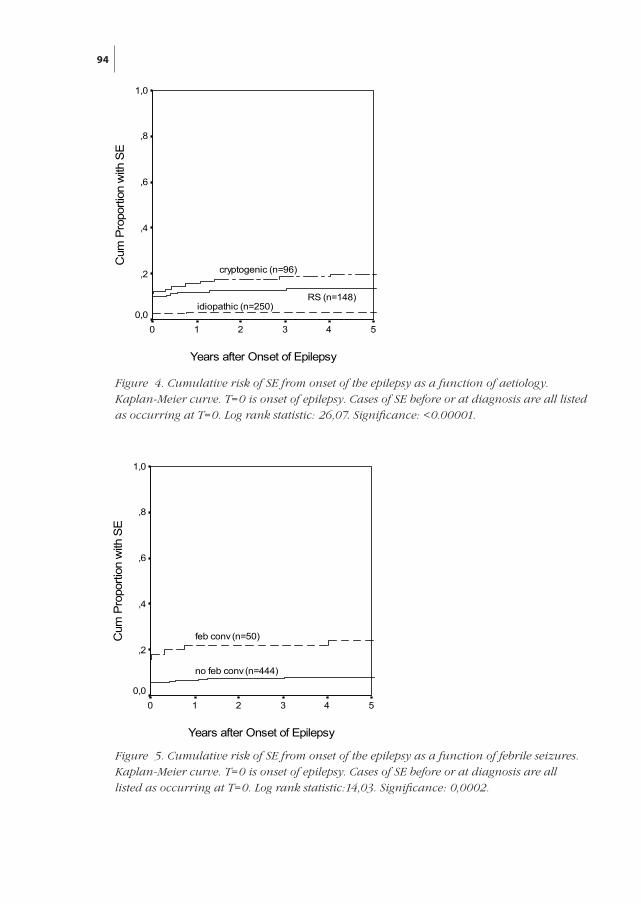
94
Years after Onset of Epilepsy
543210
Cum
Pro
porti
on w
ith S
E
1,0
,8
,6
,4
,2
0,0
cryptogenic (n=96)
RS (n=148)idiopathic (n=250)
Years after Onset of Epilepsy
543210
Cum
Pro
porti
on w
ith S
E
1,0
,8
,6
,4
,2
0,0
feb conv (n=50)
no feb conv (n=444)
Figure 5. Cumulative risk of SE from onset of the epilepsy as a function of febrile seizures. Kaplan-Meier curve. T=0 is onset of epilepsy. Cases of SE before or at diagnosis are all listed as occurring at T=0. Log rank statistic:14,03. Significance: 0,0002.
Figure 4. Cumulative risk of SE from onset of the epilepsy as a function of aetiology. Kaplan-Meier curve. T=0 is onset of epilepsy. Cases of SE before or at diagnosis are all listed as occurring at T=0. Log rank statistic: 26,07. Significance: <0.00001.
290158_Stroink_BW.indd 94 11-04-2008 10:53:48
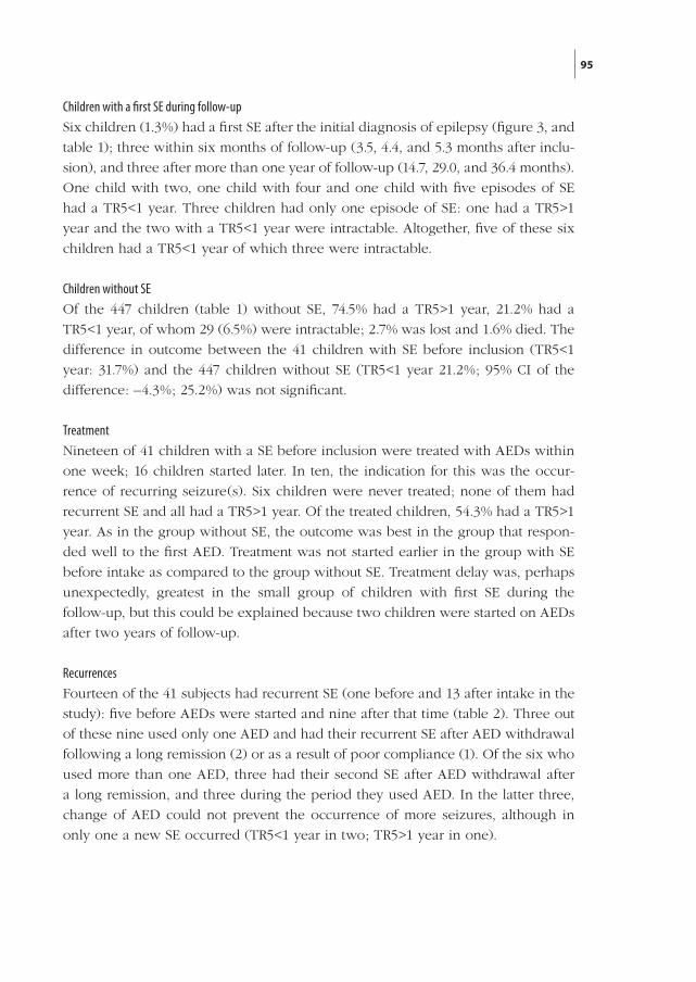
95
Children with a first SE during follow-upSix children (1.3%) had a first SE after the initial diagnosis of epilepsy (figure 3, and
table 1); three within six months of follow-up (3.5, 4.4, and 5.3 months after inclu-
sion), and three after more than one year of follow-up (14.7, 29.0, and 36.4 months).
One child with two, one child with four and one child with five episodes of SE
had a TR5<1 year. Three children had only one episode of SE: one had a TR5>1
year and the two with a TR5<1 year were intractable. Altogether, five of these six
children had a TR5<1 year of which three were intractable.
Children without SEOf the 447 children (table 1) without SE, 74.5% had a TR5>1 year, 21.2% had a
TR5<1 year, of whom 29 (6.5%) were intractable; 2.7% was lost and 1.6% died. The
difference in outcome between the 41 children with SE before inclusion (TR5<1
year: 31.7%) and the 447 children without SE (TR5<1 year 21.2%; 95% CI of the
difference: –4.3%; 25.2%) was not significant.
TreatmentNineteen of 41 children with a SE before inclusion were treated with AEDs within
one week; 16 children started later. In ten, the indication for this was the occur-
rence of recurring seizure(s). Six children were never treated; none of them had
recurrent SE and all had a TR5>1 year. Of the treated children, 54.3% had a TR5>1
year. As in the group without SE, the outcome was best in the group that respon-
ded well to the first AED. Treatment was not started earlier in the group with SE
before intake as compared to the group without SE. Treatment delay was, perhaps
unexpectedly, greatest in the small group of children with first SE during the
follow-up, but this could be explained because two children were started on AEDs
after two years of follow-up.
RecurrencesFourteen of the 41 subjects had recurrent SE (one before and 13 after intake in the
study): five before AEDs were started and nine after that time (table 2). Three out
of these nine used only one AED and had their recurrent SE after AED withdrawal
following a long remission (2) or as a result of poor compliance (1). Of the six who
used more than one AED, three had their second SE after AED withdrawal after
a long remission, and three during the period they used AED. In the latter three,
change of AED could not prevent the occurrence of more seizures, although in
only one a new SE occurred (TR5<1 year in two; TR5>1 year in one).
290158_Stroink_BW.indd 95 11-04-2008 10:53:48
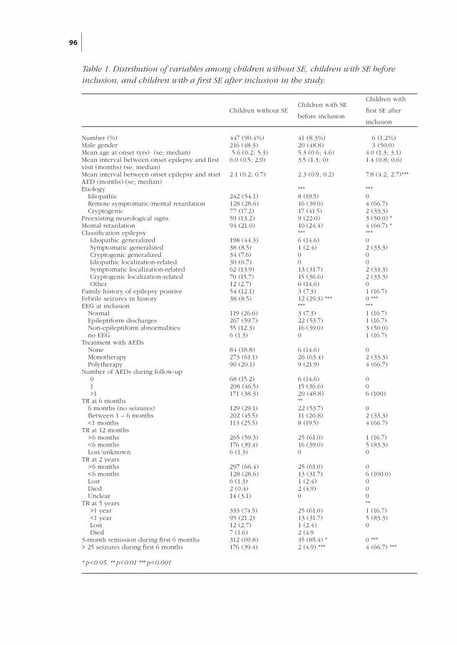
96
Table 1. Distribution of variables among children without SE, children with SE before inclusion, and children with a first SE after inclusion in the study.
* p<0.05, ** p<0.01 *** p<0.001
Number (%) Male genderMean age at onset (yrs) (se; median)Mean interval between onset epilepsy and first visit (months) (se; median)Mean interval between onset epilepsy and start AED (months) (se; median)Etiology Idiopathic Remote symptomatic/mental retardation CryptogenicPreexisting neurological signsMental retardationClassification epilepsy Idiopathic generalized Symptomatic generalized Cryptogenic generalized Idiopathic localization-related Symptomatic localization-related Cryptogenic localization-related OtherFamily history of epilepsy positiveFebrile seizures in historyEEG at inclusion Normal Epileptiform discharges Non-epileptiform abnormalities no EEGTreatment with AEDs None Monotherapy PolytherapyNumber of AEDs during follow-up 0 1 >1TR at 6 months 6 months (no seizures) Between 1 – 6 months <1 monthsTR at 12 months >6 months <6 months Lost/unknownTR at 2 years >6 months <6 months Lost Died UnclearTR at 5 years >1 year <1 year Lost Died3-month remission during first 6 months> 25 seizures during first 6 months
447 (90.4%)216 (48.3) 5.6 (0.2; 5.3)6.0 (0.5; 2.9)
2.1 (0.2; 0.7)
242 (54.1)128 (28.6)77 (17.2)59 (13.2)94 (21.0)
198 (44.3)38 (8.5)34 (7.6)30 (6.7)62 (13.9)70 (15.7)12 (2.7)54 (12.1)38 (8.5)
119 (26.6)267 (59.7)55 (12.3)6 (1.3)
84 (18.8)273 (61.1)90 (20.1)
68 (15.2)208 (46.5)171 (38.3)
129 (29.1)202 (45.5)113 (25.5)
265 (59.3)176 (39.4)6 (1.3)
297 (66.4)128 (28.6)6 (1.3)2 (0.4)14 (3.1)
333 (74.5)95 (21.2)12 (2.7)7 (1.6)312 (69.8)176 (39.4)
41 (8.3%)20 (48.8)5.3 (0.6; 4.6)3.5 (1.3; 0)
2.3 (0.9; 0.2)
***8 (19.5)16 (39.0)17 (41.5)9 (22.0) 10 (24.4)***6 (14.6)1 (2.4)0013 (31.7)15 (36.6)6 (14.6)3 (7.3)12 (29.3) ******3 (7.3)22 (53.7)16 (39.0)0
6 (14.6)26 (63.4)9 (21.9)
6 (14.6)15 (36.6)20 (48.8)**22 (53.7)11 (26.8)8 (19.5)
25 (61.0)16 (39.0)0
25 (61.0)13 (31.7)1 (2.4)2 (4.9)0
25 (61.0)13 (31.7)1 (2.4)2 (4.935 (85.4) *2 (4.9) ***
6 (1.2%) 3 (50.0)4.0 (1.3; 3.1)1.4 (0.8; 0.6)
7.8 (4.2; 2.7)***
***0 4 (66.7)2 (33.3)3 (50.0) *4 (66.7) ****02 (33.3)002 (33.3)2 (33.3)01 (16.7)0 ******1 (16.7)1 (16.7)3 (50.0)1 (16.7)
02 (33.3)4 (66.7)
006 (100)
02 (33.3)4 (66.7)
1 (16.7)5 (83.3)0
06 (100.0)000**1 (16.7)5 (83.3)0
0 ***4 (66.7) ***
Children without SE Children with SE
before inclusion
Children with
first SE after
inclusion
290158_Stroink_BW.indd 96 11-04-2008 10:53:48
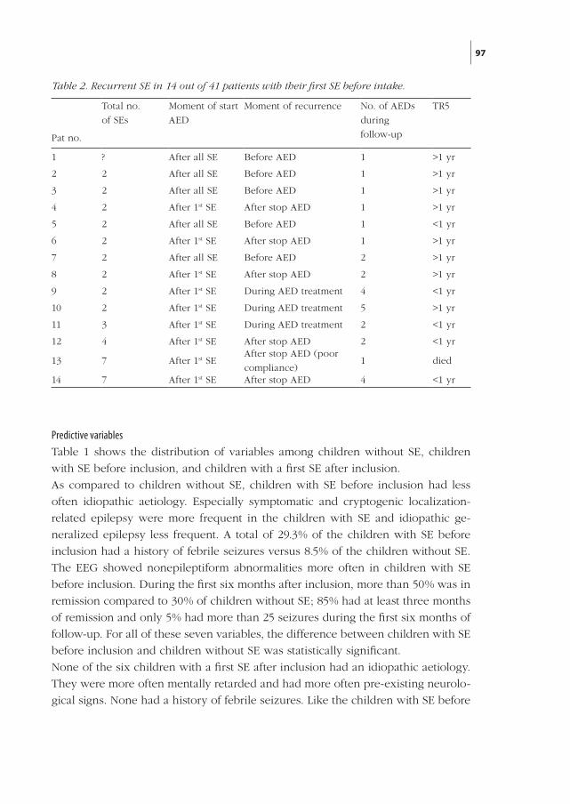
97
Predictive variablesTable 1 shows the distribution of variables among children without SE, children
with SE before inclusion, and children with a first SE after inclusion.
As compared to children without SE, children with SE before inclusion had less
often idiopathic aetiology. Especially symptomatic and cryptogenic localization-
related epilepsy were more frequent in the children with SE and idiopathic ge-
neralized epilepsy less frequent. A total of 29.3% of the children with SE before
inclusion had a history of febrile seizures versus 8.5% of the children without SE.
The EEG showed nonepileptiform abnormalities more often in children with SE
before inclusion. During the first six months after inclusion, more than 50% was in
remission compared to 30% of children without SE; 85% had at least three months
of remission and only 5% had more than 25 seizures during the first six months of
follow-up. For all of these seven variables, the difference between children with SE
before inclusion and children without SE was statistically significant.
None of the six children with a first SE after inclusion had an idiopathic aetiology.
They were more often mentally retarded and had more often pre-existing neurolo-
gical signs. None had a history of febrile seizures. Like the children with SE before
Pat no.
Total no.
of SEs
Moment of start
AED
Moment of recurrence No. of AEDs
during
follow-up
TR5
1 ? After all SE Before AED 1 >1 yr
2 2 After all SE Before AED 1 >1 yr
3 2 After all SE Before AED 1 >1 yr
4 2 After 1st SE After stop AED 1 >1 yr
5 2 After all SE Before AED 1 <1 yr
6 2 After 1st SE After stop AED 1 >1 yr
7 2 After all SE Before AED 2 >1 yr
8 2 After 1st SE After stop AED 2 >1 yr
9 2 After 1st SE During AED treatment 4 <1 yr
10 2 After 1st SE During AED treatment 5 >1 yr
11 3 After 1st SE During AED treatment 2 <1 yr
12 4 After 1st SE After stop AED 2 <1 yr
13 7 After 1st SEAfter stop AED (poor
compliance)1 died
14 7 After 1st SE After stop AED 4 <1 yr
Table 2. Recurrent SE in 14 out of 41 patients with their first SE before intake.
Number (%) Male genderMean age at onset (yrs) (se; median)Mean interval between onset epilepsy and first visit (months) (se; median)Mean interval between onset epilepsy and start AED (months) (se; median)Etiology Idiopathic Remote symptomatic/mental retardation CryptogenicPreexisting neurological signsMental retardationClassification epilepsy Idiopathic generalized Symptomatic generalized Cryptogenic generalized Idiopathic localization-related Symptomatic localization-related Cryptogenic localization-related OtherFamily history of epilepsy positiveFebrile seizures in historyEEG at inclusion Normal Epileptiform discharges Non-epileptiform abnormalities no EEGTreatment with AEDs None Monotherapy PolytherapyNumber of AEDs during follow-up 0 1 >1TR at 6 months 6 months (no seizures) Between 1 – 6 months <1 monthsTR at 12 months >6 months <6 months Lost/unknownTR at 2 years >6 months <6 months Lost Died UnclearTR at 5 years >1 year <1 year Lost Died3-month remission during first 6 months> 25 seizures during first 6 months
447 (90.4%)216 (48.3) 5.6 (0.2; 5.3)6.0 (0.5; 2.9)
2.1 (0.2; 0.7)
242 (54.1)128 (28.6)77 (17.2)59 (13.2)94 (21.0)
198 (44.3)38 (8.5)34 (7.6)30 (6.7)62 (13.9)70 (15.7)12 (2.7)54 (12.1)38 (8.5)
119 (26.6)267 (59.7)55 (12.3)6 (1.3)
84 (18.8)273 (61.1)90 (20.1)
68 (15.2)208 (46.5)171 (38.3)
129 (29.1)202 (45.5)113 (25.5)
265 (59.3)176 (39.4)6 (1.3)
297 (66.4)128 (28.6)6 (1.3)2 (0.4)14 (3.1)
333 (74.5)95 (21.2)12 (2.7)7 (1.6)312 (69.8)176 (39.4)
41 (8.3%)20 (48.8)5.3 (0.6; 4.6)3.5 (1.3; 0)
2.3 (0.9; 0.2)
***8 (19.5)16 (39.0)17 (41.5)9 (22.0) 10 (24.4)***6 (14.6)1 (2.4)0013 (31.7)15 (36.6)6 (14.6)3 (7.3)12 (29.3) ******3 (7.3)22 (53.7)16 (39.0)0
6 (14.6)26 (63.4)9 (21.9)
6 (14.6)15 (36.6)20 (48.8)**22 (53.7)11 (26.8)8 (19.5)
25 (61.0)16 (39.0)0
25 (61.0)13 (31.7)1 (2.4)2 (4.9)0
25 (61.0)13 (31.7)1 (2.4)2 (4.935 (85.4) *2 (4.9) ***
6 (1.2%) 3 (50.0)4.0 (1.3; 3.1)1.4 (0.8; 0.6)
7.8 (4.2; 2.7)***
***0 4 (66.7)2 (33.3)3 (50.0) *4 (66.7) ****02 (33.3)002 (33.3)2 (33.3)01 (16.7)0 ******1 (16.7)1 (16.7)3 (50.0)1 (16.7)
02 (33.3)4 (66.7)
006 (100)
02 (33.3)4 (66.7)
1 (16.7)5 (83.3)0
06 (100.0)000**1 (16.7)5 (83.3)0
0 ***4 (66.7) ***
290158_Stroink_BW.indd 97 11-04-2008 10:53:49
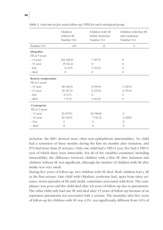
98
inclusion, the EEG showed more often non-epileptiform abnormalities. No child
had a remission of three months during the first six months after inclusion, and
67% had more than 25 seizures. Only one child had a TR5>1 year, five had a TR5<1
year of which three were intractable. For all of the variables examined, including
intractability, the difference between children with a first SE after inclusion and
children without SE was significant, although the number of children with SE after
intake was very small.
During five years of follow-up, two children with SE died. Both children had a SE
as the first seizure. One child with Ohtahara syndrome had, apart from other sei-
zures, seven episodes of SE until death, sometimes associated with fever. The com-
pliance was poor and the child died after 4.8 years of follow-up due to pneumonia.
The other child only had one SE and died after 1.5 years of follow-up because of an
aspiration pneumonia not associated with a seizure. The mortality after five years
of follow-up for children with SE was 4.3%, not significantly different from 1.6% of
Children
without SE
Number (%)
Children with SE
before inclusion
Number (%)
Children with first SE
after inclusion
Number (%)
Number (%) 447 41 6
Idiopathic
TR at 5 years
- >1 year
- <1 year
- lost
- died
201 (83.0)
35 (14.4)
6 (2.5)
0
7 (87.5)
0
1 (12.5)
0
0
0
0
0
Remote symptomatic
TR at 5 years
- >1 year
- <1 year
- lost
- died
80 (62.5)
35 (27.4)
6 (4.7)
7 (5.5)
8 (50.0)
6 (37.6)
0
2 (12.5)
1 (25.0)
3 (75.0)
0
0
Cryptogenic
TR at 5 years
- >1 year
- <1 year
- lost
- died
52 (67.5)
25 (32.5)
0
0
10 (58.8)
7 (41.2)
0
0
0
2 (100)
0
0
Table 3. Outcome at five years follow-up (TR5) for each etiological group.
290158_Stroink_BW.indd 98 11-04-2008 10:53:49
99
children without SE (95% CI of the difference (2.7%): –3.4%; 10.0%).
We performed a multivariate analysis to investigate which combination of determi-
nants was associated with SE at the time the diagnosis of epilepsy was established.
Stepwise backward logistic regression analysis resulted in the selection of aetio-
logy and febrile convulsions as statistically significant variables associated with SE
before or at inclusion (figure 4 and 5). Odds ratios were 4.1 (95% CI 1.7; 10.1) for
remote symptomatic versus idiopathic aetiology; 7.6 (95% CI 3.1; 18.7) for cryptoge-
nic versus idiopathic aetiology; and 5.3 (95% CI 2.4; 11.9) for febrile seizures. The
combination of history of febrile seizures with cryptogenic aetiology correlated
best with SE. The poorest correlation with the occurrence of SE was found for the
combination of idiopathic aetiology with the absence of febrile seizures.
None of the variables in table 1 was predictive of a recurrent SE in the univariate
and multivariate analysis.
Discussion
In this hospital-based cohort study of 494 children with epilepsy, 47 children
(9.5%) had one or more episodes of SE: 8.3% had had the first SE at the time the
diagnosis of epilepsy was established. Other studies reported higher numbers of
children with SE at the onset of epilepsy (Berg et al: 9.1%; Sillanpää: 20%).2 7 15 The
lower number at onset in our study can be explained partially by differences in the
inclusion criteria. In the DSEC, we included only children with newly diagnosed
untreated epilepsy to avoid a bias towards more severely affected epilepsy patients
referred from other hospitals. Moreover, children with febrile seizures were inclu-
ded only if they also had had at least two unprovoked seizures or one unprovoked
SE before the start of AED treatment. Consequently, children treated with AEDs
after an acute symptomatic or febrile SE before the onset of epilepsy were not in-
cluded in the DSEC. Sillanpää’s study2 counted 12 out of 30 children with provoked
SE before the onset of epilepsy, Berg’s15 11 out of 56 children and our study only
three out of 41 children. Excluding these children, 7.7% in our study, 7.0% in Berg’s
study and 12% in Sillanpää’s study had unprovoked SE before inclusion. The rate
of SE during five years follow-up was also higher (8.2%) in the study of Berg7 and
Sillanpää2 as compared to our study (3.8%). We found a strongly increased risk for
SE during follow-up once a child had had SE before inclusion (31.7% vs. 1.2% for
children without). In the study of Berg7 these rates were 31.0% versus 5.9% at five
years follow-up, and in the study of Sillanpää 51% versus 9.2%. Besides the already
mentioned reasons, possible explanations for the still somewhat higher number of
children with SE before and the markedly higher number after inclusion in the Fin-
290158_Stroink_BW.indd 99 11-04-2008 10:53:49
100
nish study may be the different populations studied and use of different definitions
of SE. Our study and the study by Berg concern incidence cohorts. The Finnish
cohort consisted of children who developed epilepsy in the period 1961–1964, but
these children were recruited retrospectively in 1972 and followed prospectively
since then.16 This may have caused a bias toward more severe patients. In the
1960s, treatment options with AEDs were limited and benzodiazepines for emer-
gency treatment of seizures by the parents were not available. In the U.S.A., the
FDA approved rectal diazepam gel in July 1997, the last year of inclusion in the
study of Berg. However, these factors may not completely explain the variation
between the studies.7
SE occurred most often at presentation of the epilepsy (figure 1). In 32 of 47
children (68%) with SE in our study, the SE was the first seizure (6.5% of all 494
children; 6.6% in Sillanpää’s study and 4.1% for the children with unprovoked SE in
Berg’s study). Only 15 of 494 children (3.0%) had SE after a history of earlier short
lasting seizures. Presentation of SE early in the course of epilepsy is in accordance
with Sillanpää2 and Berg.15 In the follow-up study of Berg,7 however, SE also oc-
curred later during follow-up with a median time to first SE after initial diagnosis
of epilepsy of 2.5 years.
Factors associated with the occurrence of SE were earlier SE, remote symptomatic
(as in the studies of Berg15 and Sillanpää2) or cryptogenic aetiology (as in the study
of Berg7), and earlier febrile seizures (as in Sillanpää’s study). In our study, after
adjusting for epilepsy type or aetiology, there was no statistically significant dif-
ference in outcome between those with SE prior to intake and those without SE.
This means that SE before intake per se has little impact on long-term outcome.
The frequent remote symptomatic and cryptogenic aetiology in children with SE
probably also explains the more frequent non-epileptiform abnormalities on EEG.
The mean age of the children with SE in our study (5.1 years), especially of those
with their first SE during follow-up, tended to be lower than that of children who
presented without SE (5.6 years), but this difference was not significant. This was
comparable to the findings of Berg.7 15 They found at the beginning of their study,
a younger age of onset of epilepsy in children with provoked and febrile SE (2.2
years), but not in children with unprovoked SE (5.4 years for children with unpro-
voked SE versus 5.9 years for children with epilepsy without SE). As in our study,
children with SE during follow-up tended to be younger.7 In the Finnish study,
children with SE were significantly younger (2.8 years) than children with epilepsy
without SE (5.2 years).2 In this study many children had provoked SE, which may
explain the younger age. Children with provoked SE have a younger age than
children with unprovoked SE, and epilepsy also has an earlier onset after provoked
290158_Stroink_BW.indd 100 11-04-2008 10:53:49
101
SE (Shinnar, Berg).15 17
Mortality was not significantly different between children with SE and without SE
in our and Sillanpää’s study. Both fatalities in our study were related to the under-
lying cause of the epilepsy. Berg found a higher mortality rate, but this was also
entirely caused by the underlying neurological diseases and not by SE itself.7
The outcome of the epilepsy in our study, measured as TR5, was slightly, but not
significantly, worse for children with SE before intake compared to children wit-
hout SE. In the other studies, TR for children with SE also tended to be worse. In
the Finnish study18, after a follow-up of more than 30 years, fewer children with
SE reached a TR of five years as compared to those without SE (RR 0.58; CI 0.34;
0.99; p=0.044). After adjusting for known significant variables like aetiology the
difference was marginally significant (p=0.052) in multivariate analysis. However,
the difference was statistically significant for children in remission without taking
AEDs (risk ratio 0.50; 95% 0.26; 0.94; p=0.023). In the Finnish study, many children
were included with SE after the moment of the diagnosis. In Berg’s study,7 children
with initial SE were somewhat less likely to come into a terminal 3-year remission
(48.1 vs. 62.2%; p=0.05), but not more frequently intractable. However, children
with SE during follow-up had a lower remission rate and were more likely to be
intractable. We agree with Berg that SE during follow-up simply means that the
child still has severe and ongoing seizures, most often despite adequate therapy,
and will therefore be less likely to go into remission.
Conclusions
All studies agree that SE is a common and serious presentation of epilepsy in
children. Mostly SE occur at or prior to the diagnosis of epilepsy. It considerably
enhances the risk for future SE. A trend exists for a shorter TR for children with SE.
However, differences in prognosis in the three studies are not or only borderline
significant. Symptomatic and cryptogenic localization-related epilepsy are much
more frequent in children with SE, and are important negative factors determi-
ning the outcome of epilepsy. After adjusting for type of epilepsy and aetiology,
SE is not significantly associated with a worse outcome any more. In our study,
intractability is more common in children with the first SE after intake. However,
SE is probably not the cause of intractability, but an indicator of the severity of the
epilepsy in these children. No complete agreement exists on the importance of a
history of febrile convulsions for the outcome and for the magnitude of the risk of
SE in children without SE at presentation of epilepsy. Because of the high recur-
rence risk, parents of children who have had a SE should be instructed on how to
290158_Stroink_BW.indd 101 11-04-2008 10:53:49
102
deal with a prolonged seizure and on the use of abortive medication.
References
1. Maytal J, Shinnar S, Moshe SL, Alvarez LA. Low morbidity and mortality of status
epilepticus in children. Pediatrics 1989;83:323-331.
2. Sillanpaa M, Shinnar S. Status epilepticus in a population-based cohort with childhood-
onset epilepsy in Finland. Ann Neurol 2002;52 303-310.
3. Chin RF, Neville BG, Peckham C, Bedford H, Wade A, Scott RC, et al. Incidence, cause, and
short-term outcome of convulsive status epilepticus in childhood: prospective population-
based study. Lancet 2006;368:222-229.
4. Raspall-Chaure M, Chin RF, Neville BG, Scott RC. Outcome of paediatric convulsive status
epilepticus: a systematic review. Lancet Neurol 2006;5:769-779.
5. Gilbert DL, Gartside PS, Glauser TA. Efficacy and mortality in treatment of refractory
generalized convulsive status epilepticus in children: a meta-analysis. J Child Neurol
1999;14:602-629.
6. Berg AT, Levy SR, Testa FM, Shinnar S. Classification of childhood epilepsy syndromes in
newly diagnosed epilepsy: interrater agreement and reasons for disagreement. Epilepsia
1999;40:439-444.
7. Berg AT, Shinnar S, Testa FM, Levy SR, Frobish D, Smith SN, et al. Status epilepticus after
the initial diagnosis of epilepsy in children. Neurology 2004;63:1027-1034.
8. Stroink H, Brouwer OF, Arts WF, Geerts AT, Peters AC, van Donselaar CA. The first
unprovoked, untreated seizure in childhood: a hospital based study of the accuracy of
the diagnosis, rate of recurrence, and long term outcome after recurrence. Dutch Study of
Epilepsy in Childhood. J Neurol Neurosurg Psychiatry 1998;64:595-600.
9. Arts WF, Geerts AT, Brouwer OF, Peters AC, Stroink H, van Donselaar CA. The early
prognosis of epilepsy in childhood: the prediction of a poor outcome. The Dutch study of
epilepsy in childhood. Epilepsia 1999;40:726-734.
10. Stroink H, van Donselaar CA, Geerts AT, Peters AC, Brouwer OF, Arts WF. The accuracy of
the diagnosis of paroxysmal events in children. Neurology 2003;60:979-982.
11. Arts WF, Brouwer OF, Peters AC, Stroink H, Peeters EA, Schmitz PI, et al. Course and
prognosis of childhood epilepsy: 5-year follow-up of the Dutch Study of Epilepsy in
Childhood. Brain 2004;127:1774-1784.
12. Huttenlocher PR, Hapke RJ. A follow-up study of intractable seizures in childhood. Ann
Neurol 1990;28:699-705.
13. Berg AT, Shinnar S, Levy SR, Testa FM, Smith-Rapaport S, Beckerman B. Early development
of intractable epilepsy in children: a prospective study. Neurology 2001;56:1445-1452.
14. Berg AT, Kelly MM. Defining intractability: comparisons among published definitions.
Epilepsia 2006;47:431-436.
15. Berg AT, Shinnar S, Levy SR, Testa FM. Status epilepticus in children with newly diagnosed
epilepsy. Ann Neurol 1999;45:618-623.
16. Sillanpaa M, Jalava M, Kaleva O, Shinnar S. Long-Term Prognosis of Seizures with Onset in
Childhood. N Engl J Med 1998;338:1715-1722.
290158_Stroink_BW.indd 102 11-04-2008 10:53:49
103
17. Shinnar S, Pellock JM, Moshe SL, Maytal J, O’Dell C, Driscoll SM, et al. In whom does
status epilepticus occur: age-related differences in children. Epilepsia 1997;38:907-914.
18. Shinnar S, Pellock JM. Update on the Epidemiology and Prognosis of Pediatric Epilepsy. J
Child Neurol 2002;17:S4-17.
290158_Stroink_BW.indd 103 11-04-2008 10:53:49

290158_Stroink_BW.indd 104 11-04-2008 10:53:49
105
Chapter 8
General discussion
290158_Stroink_BW.indd 105 11-04-2008 10:53:49

290158_Stroink_BW.indd 106 11-04-2008 10:53:49

107
Diagnosis
The diagnoses seizure and epilepsy may be difficult in many cases. The diagnosis
depends on the description of the event by an eyewitness and the interpretation
of the description by the physician. A description of an event may be unclear be-
cause of insufficient observation, a lack of an eyewitness, a lack of capacity of the
eyewitness to give a clear description or due to interpretations by the observer. The
physician himself may misinterpret the account of the witness. A misdiagnosis may
also be the consequence when physicians rely more on EEG findings than on cli-
nical signs.1-3 A misdiagnosis of epilepsy has many medical consequences like un-
necessary investigations, prescription of AEDs which may have many side effects
in childhood,4 5 and psychosocial and socioeconomic consequences may occur
also in childhood.6 Unjustified rejection of the diagnosis epilepsy may also have
disadvantages for the patient. It may even be fatal when, for example, a cardiac
arrhythmia is overlooked as the cause for the spells.7 8 In case of doubt however,
a misdiagnosis of epilepsy does more harm for the patient than delaying the diag-
nosis for some time.2 3 This is even more so in childhood. Children may be more
susceptible for the adverse effects of AEDs.4 5 Children do not drive cars, and they
cannot loose their job. So for those children, who do not have frequent seizures,
the need for treatment with AEDs may be less urgent than for adults.
In clinical and epidemiological studies on epilepsy without doubt patients are in-
cluded with other paroxysmal disorders, and not all patients who do have epilepsy
will be included. In most studies on prognosis and treatment of epilepsy the pro-
blem of diagnosing epilepsy (reliability and accuracy) and the inclusion of the cor-
rect patients is not mentioned. In this thesis the reliability and accuracy of several
aspects of the diagnostic process are studied. The extent of this problem in daily
practice and in studies, pitfalls and methods to reduce the amount of misdiagnoses
are discussed in chapter 2.3
In chapter 3 we describe our study in which several paediatric neurologists had to
diagnose one paroxysmal event.9 The written description of the event was presen-
ted to them. In case they concluded the child suffered a seizure they had to classify
the type of the seizure according the ILAE classification.10 Using their clinical jud-
gement, the individual observers only reached fair to moderate agreement on the
diagnosis of a first seizure. The mean (SE) kappa was 0.41 (0.03). With use of de-
fined descriptive criteria the mean kappa was 0.45 (0.03). After discussion between
all participating paediatric neurologists the mean kappa for agreement increased to
0.60 (0.06), which is on the border of moderate to substantial for making a clinical
290158_Stroink_BW.indd 107 11-04-2008 10:53:49
108
diagnosis. The agreement for the seizure classification by individual observers was
only moderate (mean kappa 0.46) for clinical judgment, also when the criteria for
diagnosis were used (kappa 0.57). After discussion within each panel the kappa
between the two panels was substantial (0.69). Finally the results of all additional
investigations were given. The panels succeeded only in a small minority of the
children with a single seizure to make a specific syndrome diagnosis.
The paediatric neurologists in this study were experienced, and the criteria used
for the diagnosis seizure increased the interrater agreement. In daily practice one
would expect a lower interrater agreement.11-15
In chapter 4 the results are presented of our study on the accuracy of the diagnoses
single seizure and epilepsy.16 17 Children with the diagnosis epilepsy were followed
for five years, children with a single seizure for at least two years. After two years
follow-up the diagnosis of all these children was reassessed. Children with an
unclear event were followed for one year to reassess whether new episodes might
yield firm evidence for a definite diagnosis. After each new event the patient was
re-evaluated and the diagnosis reconsidered. The definitions used in this section
are explained in table 1a and 1b.
In none of the 170 children with a single seizure during the follow-up the diagnosis
did prove to be wrong (false negative diagnosis in 0%, positive predictive value
100%). In four of the 54 children with a single unclear event, recurrent episodes
enabled a definite diagnosis of epilepsy (false negative diagnosis in 7.4%; negative
predictive value 92.6%). 170 out of 174 children with a single seizure were recog-
nised at inclusion: sensitivity 97.7%.
In 412 of the 536 children seen with multiple events, an initial diagnosis of epi-
lepsy was made. In 367 children the diagnosis was made on the history alone,
in 45 children on the combination of the history and the results of the EEG. Af-
ter follow-up, the initial diagnosis epilepsy was probably incorrect in 19 children
(false positive diagnosis 4.6%; positive predictive value 95.4%). Seven of 124 child-
ren with multiple unclear episodes at intake later turned out to have epilepsy (false
negative diagnosis 5.6%; negative predictive value 94.4%). 393 out of 400 children
with epilepsy were diagnosed so at inclusion: sensitivity 98.3%. 117 of the 136
children without epilepsy were recognised at inclusion: specificity 86%.
For all 760 included children with one or more events the sensitivity was 98.1%
and the specificity 89.8%.
In all children with uncertain events one or two EEG registrations were done.
Several of these children had epileptiform discharges on the EEG. Three of the
eight children with one unclear event and one of 27 children with multiple unclear
290158_Stroink_BW.indd 108 11-04-2008 10:53:49
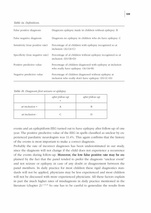
109
events and an epileptiform EEG turned out to have epilepsy after follow-up of one
year. The positive predictive value of the EEG in spells classified as unclear by ex-
perienced paediatric neurologists was 11.4%. This again confirms that the history
of the events is most important to make a correct diagnosis.
Probably the rate of incorrect diagnoses has been underestimated in our study,
since the diagnosis will not change if the child does not experience a recurrence
of the events during follow-up. Moreover, the low false positive rate may be ex- Moreover, the low false positive rate may be ex-Moreover, the low false positive rate may be ex-
plained by the fact that the panel tended to prefer the diagnosis “unclear event”
and not seizure or epilepsy in case of any doubt or disagreement between the
panel members. In daily practice for most children these rigid diagnostics stan-
dards will not be applied, physicians may be less experienced and most children
will not be discussed with more experienced physicians. All these factors explain
in part the much higher rates of misdiagnosis in daily practice mentioned in the
literature (chapter 2).3 11-15 So one has to be careful to generalize the results from
False positive diagnosis Diagnosis epilepsy made in children without epilepsy: B
False negative diagnosis Diagnosis no epilepsy in children who do have epilepsy: C
Sensitivity (true positive rate) Percentage of al children with epilepsy recognized so at
inclusion: (A)/(A+C)
Specificity (true negative rate) Percentage of al children without epilepsy recognized so at
inclusion: (D)/(B+D)
Positive predictive value Percentage of children diagnosed with epilepsy at inclusion
who really have epilepsy: (A)/(A+B)
Negative predictive value Percentage of children diagnosed without epilepsy at
inclusion who really don’t have epilepsy: (D)/(C+D)
Table 1a. Definitions.
after follow-up
+
after follow-up
-
at inclusion + A B
at inclusion - C D
Table 1b. Diagnosis first seizure or epilepsy.
290158_Stroink_BW.indd 109 11-04-2008 10:53:49
110
this study to daily practice. On the other hand we can conclude that the results of
the many studies done in the DSEC cohort are indeed very specific for children
with epilepsy.
The electroencephalogram (EEG) is an important tool in the diagnosis of children
with epilepsy. EEG findings are used for the classification of epileptic syn dromes
and may aid to determine the choice of the proper AEDs. The presence of epi-
leptiform discharges is a strong pre dictor for the risk of recur ren ce after a first
seizure.16 18
The reliability and accuracy determine the value of a diagnostic or prognostic
tool. Data on the reliability of the visual interpretation of EEG-findings are scarce,
however.19-24
In chapter 5 the interobserver reliability of the visual interpretation of the EEG in
children with new-onset epilepsy is presented.25 The interrater agreement was sub-
stantial for the interpretation of the EEG as normal or abnormal: mean (SE) kappa
0.66 (0.11); almost perfect for the presence of epileptiform discharges: mean kappa
0.83 (0.07); but moderate for abnormalities of the background pattern: mean kappa
0.53 (0.11); and only slight for the presence of focal non-epileptiform discharges:
mean kappa 0.38 (0.13). After dis cussion with the participating neurophysiologists
we defined non-epileptiform abnor mal ities as focal if they were restricted to a
maximum of three adja cent electrodes. Using these definitions the agreement rates
for the occurrence of an abnormal background-pattern improved from 0.53 to 0.73
(0.12), and for the occurrence of focal non-epileptiform abnormalities from 0.38 to
0.54 (0.14).
As the EEGs were described by experienced clinical neurophysiologists participa-
ting in studies on epilepsy, the reliability in this study will be expected to be better
than in daily practice.26 27
Prognosis
The DSEC started the inclusion of children with a single unprovoked seizure se-
veral months earlier than the inclusion of the children with epilepsy.16 In our study
we tried to avoid systematic errors made in several earlier studies on first seizure.18
28-38 In the earlier studies adult patients or children were included, but often also
adults and children together. The latter does not make much sense as explained in
the general introduction, unless children and adults are followed and analysed as
separate groups. In our single seizure study the diagnosis was made unanimously
by the panel of paediatric neurologists. The diagnosis was based on the history of
290158_Stroink_BW.indd 110 11-04-2008 10:53:49
111
the event, interpreted with predefined descriptive criteria and it did not depend on
EEG findings. Most children were included in the study soon after the seizure. The
interval seizure to inclusion is of great importance because if any recurrences will
occur they mostly do so soon after the first seizure. Every child had an EEG, which
was performed as soon as possible after the seizure.39 No child was treated after
a single unprovoked seizure. Children with an uncertain diagnosis were also fol-
lowed to test the accuracy of our inclusion criteria. Children with a single seizure
were followed for two years. In most children with epilepsy treated with AEDs the
drugs can be tapered after two years seizure freedom, and in some epilepsy syn-
dromes even earlier. So, recurrences occurring more than two years after a single
unprovoked seizure will not be of importance for the decision to start treatment.
In chapter 6 the results are presented of our single seizure study.16 After our study
a few other studies on single seizures in childhood have been published.40-44 Some
of these publications concerned additional data of studies published before.40 43
In comparison with the other studies the recurrence rate was in ours at the upper
end of the range found in all studies.45 46 In part this relatively high recurrence rate
can be explained by the methodical differences.16 Also in other studies no attention
was paid to the accuracy of the diagnosis. We found that even for experienced
paediatric neurologists diagnosing seizure(s) may be subject to error.9 17 Only in
our study no child was treated after a single seizure. Since our publications the
diagnosis “uncertain event” or “unclassified paroxysmal events” has become more
common in the literature.2 9 15-17 44 47
The first question after a single paroxysmal event has to be: how certain is the
diagnosis? If the first question was answered “with a high probability it was an
epileptic seizure”, the second question will be: what is the chance of new seizures?
Then the third question will follow: can new seizures be harmful for the child?
And the last question is: will immediate treatment improve the prognosis for this
child? When these facts are known, one has to balance the advantages and disad-
vantages of treatment with AED(s) for this child. We now know the probability of
new seizures as well as the risk factors: remote symptomatic aetiology and epilep-
tiform discharges on the EEG.16 18 In the DSEC no child died due to the epilepsy
itself.16 48 In the study of Shinnar of 407 children with a mean follow-up of 14.2
years no children died due to the epilepsy as well.49 In our own study, in which
all children remained untreated after a single seizure, the long-term prognosis was
very good for the majority of children.16 17 In a study by Camfield, 31 children were
randomised for treatment or placebo after a single seizure.34 After one year, seven
out of 17 (41%) untreated children were seizure free, and 10 out of 14 (71%) treated
children. In four of the treated children the AED had to be stopped because of side
290158_Stroink_BW.indd 111 11-04-2008 10:53:49
112
effects. So six children (42%) remained in the treated group without unacceptable
side effects and without seizures. The advantage of lowering the number of child-
ren with continuing seizures was in this small study completely undone by the
side effects of the AEDs. Fifteen years later the number of seizures was the same
in both groups. Terminal remission (TR) rates of two years were achieved in 80%
of the treated children and in 88% of the controls.50 51 No other randomised studies
have been done in children after a single seizure. Two randomised clinical trials
including adults and children have been done without analysing the results sepa-
rately.37 41-43 52 Because of the many differences in epilepsy syndromes and of the
side effects between adults and children this is not ideal. In the FIRST study, 27%
of 419 patients was 2-16 years. In this study treatment after a single seizure reduced
the recurrence rate from 42% to 24% after two years. However, starting treatment
after the second seizure did not lower the TR rates of two years after three years
follow-up (84% and 79%), nor the TR rates of five years after 10 years follow-up
(64% in both groups).37 43 52 No data on side effects were given. In the MESS study
children and adults with a single seizure or a few infrequent seizures were rando-
mised for treatment or placebo.41 The age of 3.5% of patients was <5 years, 7,4%
was 5-9 years, and 27% was 10-19 years. Of the patients with a single seizure 32%
of the immediately treated patients had a recurrence and 39% of the patients with
deferred treatment. Using these figures it would be necessary to treat 14 patients
in order to prevent one single seizure recurrence within the first two years. For
the high risk group (patients with an epileptiform EEG and remote symptomatic
aetiology) the number to treat is five patients to prevent one seizure within the first
year. At four, five and eight years follow-up no difference existed for the patients
with a single seizure between immediate or delayed treatment: the TR rates of
two years at five years follow-up were for both groups 92%, at eight years follow-
up for immediate treatment 95% and for deferred treatment 96%. Of all included
patients at five years follow-up 60% of the immediate treatment group and 41% of
the deferred treatment group still used AEDs.41 In the MESS study the two policies
did not differ with respect to quality of life outcomes or serious complications. In a
second paper the authors state that depending on social and psychological factors
medication can be considered if the recurrence risk is relatively high (patients with
an abnormal EEG or neurological deficits).42 Treatment may however contribute to
stigmatize the patient.6
We conclude that early treatment does not improve the long-term outcome of the
epilepsy in terms of recurrence risk and development of intractable epilepsy. Few
arguments exist to start treatment after a single unprovoked seizure in childhood.
For most children time will learn if recurrences occur and with which frequency. In
290158_Stroink_BW.indd 112 11-04-2008 10:53:49
113
case of doubt on the nature of the spells time may also clarify their nature. More-
over, part of the children with recurrences will turn out to have a benign syndrome
like epilepsy with centrotemporal spikes. Although probably a significant number
of children with benign epilepsy with centrotemporal spikes can be recognised
after a single seizure due to the clinical signs of the seizure and the characteristic
EEG, this will not be the case for all syndromes.9 51 Even after a second seizure a
specific syndrome will not be clear in a considerable number of children, or the
syndrome can only be very broadly defined, or has to be revised during follow-up.9
53-55 Even when neurologists in an epilepsy centre have to classify the seizure by
videotape, a substantial variation in the classification exists. In contradiction with
all other studies one claimed to be able to classify an epilepsy syndrome in 77%
of patients presenting with a first seizure.39 This is however in many aspects a curi-
ous study. Although the title suggests a study on first seizure, 45% of the included
patients had earlier seizures. Seizures occurring in the setting of fever or sleep-
deprivation were considered as unprovoked. Patients with a seizure due to earlier
stroke, head injury, encephalitis, cancer and cerebral palsy, were excluded. Patients
with an uncertain diagnosis were not mentioned. Also the inclusion criteria for age
were curious: children were included, but only if aged five years or older (20% of
included patients). No difference was made in analysis between the children and
the adults. Seizures starting at the adult age are almost always of partial onset and
of cryptogenic/symptomatic origin. Most syndrome diagnoses in this study were
no specific diagnoses, but broadly defined diagnoses like idiopathic generalised
epilepsy not further specified, or cryptogenic partial epilepsy.
Recommendations for evaluation and treatment have been published by the Ame-
rican Academy of Neurology, the Child Neurology Society, and the American Epi-
lepsy Society.45 46 These Practice Parameters were based on the results of our and
several other studies on single seizure in childhood. Routine EEG is recommended
in these Practice Parameters, but other studies like neuroimaging and laboratory
depend on specific clinical circumstances. Treatment with AEDs after a single
seizure in childhood is not indicated to prevent epilepsy and will not improve
prognosis. Treatment with AEDs may be considered in circumstances where the
benefits of reducing the risk of a second seizure outweigh the risk of pharmacolo-
gical and psychosocial effects.45 46 Studies published after the Practice Parameters
didn’t have any data that contradict these recommendations; on the contrary, they
reinforced these recommendations.6 15 41 42 44 47 49
290158_Stroink_BW.indd 113 11-04-2008 10:53:49
114
Status epilepticus (SE) is defined in most studies as seizures lasting for 30 minutes
or longer. Nowadays there is much discussion about this definition. Once a sei-
zure lasts for more than 5–10 minutes, it is unlikely to stop spontaneously within
the next few minutes. Mechanisms that start seizures are different from inhibitory
mechanisms. So there are probably two subcategories of children with epilepsy,
one with short lasting seizures and the second one with a tendency to long lasting
seizures.56 Several studies have reported that, the longer a seizure lasts, the less
likely it is to respond to abortive medication.57-59 So it is nowadays recommended
to administer medication after five minutes in an attempt to stop the seizure in
an early stage. Several investigators propose to modify the definition of SE into a
seizure lasting longer than five or 10 minutes.57
In the DSEC we investigated the incidence, causes and prognosis of SE in our co-
hort of children. We used the classical definition of 30 minutes. When the DSEC
started it was the only existing definition. Moreover, other studies on incidence
and prognosis of SE also used this definition. The results are presented in chapter
7.60 Our results confirmed findings of the few other studies on this subject that SE is
most frequent early in the course of epilepsy.56 61-63 In our study in 71% of children
with SE, an unprovoked SE was the first seizure they had. Children who suffered
a SE had a much higher risk (32%) for future SE than children with short seizures
as in the two other studies on this subject.56 61 Children with SE had a trend to a
slightly lower terminal remission rate. The difference was however not significant,
in agreement with the other two studies. Intractability and mortality were not sig-
nificantly higher either. Mortality was due to the underlying disease and not to the
epilepsy in all studies. Most important for prognosis was not the duration of the
seizure, but the aetiology.60-63 An exception was the worse prognosis for children
with SE after inclusion. However this is not due to SE: continuing seizures mean,
by definition, a poor prognosis. As before, fast and adequate treatment for SE is
of utmost importance.58 59 Good instructions should be given verbally and in wri-
ting to the caregivers. They have to know how to act in case of recurrence and
when to administer abortive medication by the rectal, buccal or nasal route. If the
seizure continues for 10 minutes after the administration an ambulance has to be
called. The occurrence of unprovoked SE itself does not influence the decision to
prescribe AEDs. Again the benefits of reducing the risk of future seizures have to
be balanced against the risk of pharmacological and psychosocial effects just like
in all children with seizure(s). However, emotional factors may influence the deci-
sion, because SE is a very anxious and impressive experience for the parents.
290158_Stroink_BW.indd 114 11-04-2008 10:53:49
115
In conclusion there is diagnostic uncertainty in a significant number of children
with paroxysmal events. Moreover the prognosis of epilepsy is in most children
not improved by early treatment. Treatment may have disadvantages, even if the
diagnosis is certain. So the diagnostic process has to be done very carefully. AEDs
should be prescribed only if the diagnosis is certain, and the advantages outweigh
the disadvantages in the particular child.
References
1. Fowle AJ, Binnie CD. Uses and abuses of the EEG in epilepsy. Epilepsia 2000;41 Suppl
3:S10-18.
2. Stephenson JBP. Clinical Diagnosis of Syncopes (Including So-called Breath-Holding Spells)
Without Electroencephalography or Ocular Compression. J Child Neurol 2007;22:502-508.
3. van Donselaar CA, Stroink H, Arts WF for the Dutch Study group of Epilepsy in Childhood.
How confident are we of the diagnosis of epilepsy? Epilepsia 2006;47 Suppl 1:9-13.
4. Committee on Drugs. Behavioral and Cognitive Effects of Anticonvulsant Therapy.
Pediatrics 1995;96:538-540.
5. Loring DW, Meador KJ. Cognitive side effects of antiepileptic drugs in children. Neurology
2004;62:872-877.
6. Jacoby A, Snape D, Baker GA. Epilepsy and social identity: the stigma of a chronic
neurological disorder. Lancet Neurol 2005;4:171-178.
7. McKeon A, Vaughan C, Delanty N. Seizure versus syncope. Lancet Neurol 2006;5:171-180.
8. Zaidi A, Clough P, Cooper P, Scheepers B, Fitzpatrick AP. Misdiagnosis of epilepsy: many
seizure-like attacks have a cardiovascular cause. J Am Coll Cardiol 2000;36:181-184.
9. Stroink H, van Donselaar CA, Geerts AT, Peters AC, Brouwer OF, van Nieuwenhuizen O,
de Coo RF, Geesink H, Arts WF. Interrater agreement of the diagnosis and classification of
a first seizure in childhood. The Dutch Study of Epilepsy in Childhood. J Neurol Neurosurg
Psychiatry 2004;75:241-245.
10. Commission ILAE. Proposal for revised clinical and electroencephalographic classification
of epileptic seizures. From the commission on Classification and Terminology of the
International League Against Epilepsy. Epilepsia 1981;22:489-501.
11. Scheepers B, Clough P, Pickles C. The misdiagnosis of epilepsy: findings of a population
study. Seizure 1998;7:403-406.
12. Smith D, Defalla BA, Chadwick DW. The misdiagnosis of epilepsy and the management of
refractory epilepsy in a specialist clinic. QJM 1999;92:15-23.
13. Chadwick D, Smith D. The misdiagnosis of epilepsy. BMJ 2002;324:495-496.
14. Leach JP, Lauder R, Nicolson A, Smith DF. Epilepsy in the UK: misdiagnosis, mistreatment,
and undertreatment? The Wrexham area epilepsy project. Seizure 2005;14:514-520.
15. Uldall P, Alving J, Hansen LK, Kibaek M, Buchholt J. The misdiagnosis of epilepsy in
children admitted to a tertiary epilepsy centre with paroxysmal events. Arch Dis Child
2006;91:219-221.
290158_Stroink_BW.indd 115 11-04-2008 10:53:50

116
16. Stroink H, Brouwer OF, Arts WF, Geerts AT, Peters AC, van Donselaar CA. The first
unprovoked, untreated seizure in childhood: a hospital based study of the accuracy of
the diagnosis, rate of recurrence, and long term outcome after recurrence. Dutch Study of
Epilepsy in Childhood. J Neurol Neurosurg Psychiatry 1998;64:595-600.
17. Stroink H, van Donselaar CA, Geerts AT, Peters AC, Brouwer OF, Arts WF. The accuracy of
the diagnosis of paroxysmal events in children. Neurology 2003;60:979-982.
18. Shinnar S, Berg AT, Moshe SL, O’Dell C, Alemany M, Newstein D, Kang H, Goldensohn ES,
Hauser WA. The Risk of Seizure Recurrence After a First Unprovoked Afebrile Seizure in
Childhood: An Extended Follow-up. Pediatrics 1996;98:216-225.
19. Blum RH. A note on the reliability of electroencephalographic judgments. Neurology
1954;4:143-146.
20. Houfek EE, Ellingson RJ. On the reliability of clinical EEG interpretation. J Nerv Ment Dis
1959;128:425-437.
21. Rose SW, Penry JK, White BG, Sato S. Reliability and validity of visual EEG assessment in
third grade children. Clin Electroencephalogr 1973;4:197–205.
22. Struve FA, Becka DR, Green MA, Howard A. Reliability of clinical interpretation of
electroencephalogram. Clinical Electroencephalography 1975;6:54-60.
23. Walczak TS, Radtke RA, Lewis DV. Accuracy and interobserver reliability of scalp ictal EEG.
Neurology 1992;42:2279-2285.
24. Woody RH. Inter-judge reliability in clinical electroencephalography. J Clin Psychol
1968;24:251-256.
25. Stroink H, Schimsheimer RJ, de Weerd AW, Geerts AT, Arts WF, Peeters EA, Brouwer
OF, Peters AC, van Donselaar CA. Interobserver reliability of visual interpretation of
electroencephalograms in children with newly diagnosed seizures. Dev Med Child Neurol
2006;48:374-377.
26. Gilbert DL. Interobserver reliability of visual interpretation of electroencephalograms in
children with newly diagnosed seizures. Dev Med Child Neurol 2006;48:1009-1010.
27. Stroink H, Schimsheimer R-J, de AW, Geerts AT, Arts WF, Peeters EA, Brouwer OF,
Boudewijn C, van CA. ‘Stroink et al. reply’. Dev Med Child Neurol 2006;48:1010-1011.
28. Thomas MH. The single seizures: its study and management. J Am Med Assoc
1959;169:457-459.
29. Blom S, Heijbel J, Bergfors PG. Incidence of epilepsy in children: a follow-up study three
years after the first seizure. Epilepsia 1978;19:343-350.
30. Camfield PR, Camfield CS, Dooley JM, Tibbles JAR, Fung T, Garner B. Epilepsy after a first
unprovoked seizure in childhood. Neurology 1985;35:1657-1660.
31. Elwes RD, Chesterman P, Reynolds EH. Prognosis after a first untreated tonic-clonic
seizure. Lancet 1985;2:752-753.
32. Annegers JF, Shirts SB, Hauser WA, Kurland LT. Risk of recurrence after an initial
unprovoked seizure. Epilepsia 1986;27:43-50.
33. Boulloche J, Leloup P, Mallet E, Parain D, Tron P. Risk of recurrence after a single,
unprovoked, generalized tonic-clonic seizure. Dev Med Child Neurol 1989;31:626-632.
34. Camfield P, Camfield C, Dooley J, Smith E, Garner B. A randomized study of
carbamazepine versus no medication after a first unprovoked seizure in childhood.
Neurology 1989;39:851-852.
290158_Stroink_BW.indd 116 11-04-2008 10:53:50
117
35. Hauser WA, Rich SS, Annegers JF, Anderson VE. Seizure recurrence after a 1st unprovoked
seizure: An extended follow-up. Neurology 1990;40:1163-1170.
36. Hart YM, Sander JW, Johnson AL, Shorvon SD. National General Practice Study of Epilepsy:
recurrence after a first seizure. Lancet 1990;336:1271-1274.
37. FIR.S.T. GROUP. Randomized clinical trial on the efficacy of antiepileptic drugs in reducing
the risk of relapse after a first unprovoked tonic-clonic seizure. First Seizure Trial Group.
Neurology 1993;43:478-483.
38. Martinovic Z, Jovic N. Seizure recurrence after a first generalized tonic-clonic seizure, in
children, adolescents and young adults. Seizure 1997;6:461-465.
39. King MA, Newton MR, Jackson GD, Fitt GJ, Mitchell LA, Silvapulle MJ, Berkovic SF.
Epileptology of the first-seizure presentation: a clinical, electroencephalographic, and
magnetic resonance imaging study of 300 consecutive patients. Lancet 1998;352:1007-1011.
40. Shinnar S, Berg AT, O’Dell C, Newstein D, Moshe SL, Hauser WA. Predictors of multiple
seizures in a cohort of children prospectively followed from the time of their first
unprovoked seizure. Ann Neurol 2000;48:140-147.
41. Marson A, Jacoby A, Johnson A, Kim L, Gamble C, Chadwick D, Medical Research Council
MSG. Immediate versus deferred antiepileptic drug treatment for early epilepsy and single
seizures: a randomised controlled trial. Lancet 2005;365:2007-2013.
42. Kim LG, Johnson TL, Marson AG, Chadwick DW, on behalf of the MRC MESS Study group.
Prediction of risk of seizure recurrence after a single seizure and early epilepsy: further
results from the MESS trial. Lancet Neurol 2006;5:317-322.
43. Leone MA, Solari A, Beghi E; for the FIRST Group. Treatment of the first tonic-clonic
seizure does not affect long-term remission of epilepsy. Neurology 2006;67:2227-2229.
44. Hamiwka LD, Singh N, Niosi J, Wirrell EC. Diagnostic inaccuracy in children referred with
“first seizure”: role for a first seizure clinic. Epilepsia 2007;48:1062-1066.
45. Hirtz D, Ashwal S, Berg A, Bettis D, Camfield C, Camfield P, Crumrine P, Elterman R,
Schneider S, Shinnar S. Practice parameter: Evaluating a first nonfebrile seizure in children:
Report of the Quality Standards Subcommittee of the American Academy of Neurology, the
Child Neurology Society, and the American Epilepsy Society. Neurology 2000;55:616-623.
46. Hirtz D, Berg A, Bettis D, Camfield C, Camfield P, Crumrine P, Gaillard WD, Schneider
S, Shinnar S. Practice parameter: Treatment of the child with a first unprovoked seizure:
Report of the Quality Standards Subcommittee of the American Academy of Neurology and
the Practice Committee of the Child Neurology Society. Neurology 2003;60:166-175.
47. Beach R, Reading R. The importance of acknowledging clinical uncertainty in the
diagnosis of epilepsy and non-epileptic events. Arch Dis Child 2005;90:1219-1222.
48. Callenbach PM, Westendorp RG, Geerts AT, Arts WF, Peeters EA, van Donselaar CA, Peters
AC, Stroink H, Brouwer OF. Mortality risk in children with epilepsy: the Dutch Study of
Epilepsy in Childhood. Pediatrics 2001;107:1259-1263.
49. Shinnar S, O’Dell C, Berg AT. Mortality following a first unprovoked seizure in children: a
prospective study. Neurology 2005;64:880-882.
50. Camfield P, Camfield C, Smith S, Dooley J, Smith E. Long-term outcome is unchanged by
antiepileptic drug treatment after a first seizure: a 15-year follow-up from a randomized
trial in childhood. Epilepsia 2002;43:662-663.
290158_Stroink_BW.indd 117 11-04-2008 10:53:50
118
51. Camfield P, Camfield C. Childhood epilepsy: what is the evidence for what we think and
what we do? J Child Neurol 2003;18:272-287.
52. Musicco M, Beghi E, Solari A, Viani F. Treatment of first tonic-clonic seizure does not
improve the prognosis of epilepsy. First Seizure Trial Group (FIRST Group). Neurology
1997;49:991-998.
53. Shinnar S, O’Dell C, Berg AT. Distribution of Epilepsy Syndromes in a Cohort of Children
Prospectively Monitored from the Time of Their First Unprovoked Seizure. Epilepsia
1999;40:1378-1383.
54. Jallon P, Loiseau P, Loiseau J. Newly Diagnosed Unprovoked Epileptic Seizures:
Presentation at Diagnosis in CAROLE Study. Epilepsia 2001;42:464-475.
55. Middeldorp CM, Geerts AT, Brouwer OF, Peters AC, Stroink H, van Donselaar CA, Arts
WF. Nonsymptomatic generalized epilepsy in children younger than six years: excellent
prognosis, but classification should be reconsidered after follow-up: the Dutch Study of
Epilepsy in Childhood. Epilepsia 2002;43:734-739.
56. Shinnar S, Berg AT, Moshe SL, Shinnar R. How long do new-onset seizures in children last?
Ann Neurol 2001;49:659-664.
57. Lowenstein DH, Bleck T, Macdonald RL. It’s Time to Revise the Definition of Status
Epilepticus. Epilepsia 1999;40:120-122.
58. Alldredge BK, Wall DB, Ferriero DM. Effect of prehospital treatment on the outcome of
status epilepticus in children. Pediatr Neurol 1995;12:213-216.
59. Alldredge BK, Gelb AM, Isaacs SM, Corry MD, Allen F, Ulrich S, Gottwald MD, O’Neil
N, Neuhaus JM, Segal MR, Lowenstein DH. A comparison of lorazepam, diazepam,
and placebo for the treatment of out-of-hospital status epilepticus. N Engl J Med
2001;345:631-637.
60. Stroink H, Geerts AT, van Donselaar CA, Peters ACB, Brouwer OF, Peeters EA, Arts WF.
Status Epilepticus in Children with Epilepsy: Dutch Study of Epilepsy in Childhood.
Epilepsia 2007;48:1708-1715.
61. Sillanpaa M, Shinnar S. Status epilepticus in a population-based cohort with childhood-
onset epilepsy in Finland. Ann Neurol 2002;52:303-310.
62. Berg AT, Shinnar S, Levy SR, Testa FM. Status epilepticus in children with newly diagnosed
epilepsy. Ann Neurol 1999;45:618-623.
63. Berg AT, Shinnar S, Testa FM, Levy SR, Frobish D, Smith SN, Beckerman B. Status
epilepticus after the initial diagnosis of epilepsy in children. Neurology 2004;63:1027-1034.
290158_Stroink_BW.indd 118 11-04-2008 10:53:50

119
Summary:
Chapter 2 summarizes the problems in correctly making or rejecting the diagno-
sis of (a) seizure(s) and epilepsy. The diagnosis of a first seizure or epilepsy may
be subject to interobserver variation and inaccuracy. This may have far-reaching
consequences for the patients involved. We reviewed in this chapter the current
literature. Studies on the interobserver variation of the diagnosis of a first seizure
show that such a diagnosis is subject to considerable interobserver disagreement.
Interpretation of the electroencephalogram (EEG) findings is also subject to inter-
observer disagreement and is influenced by the threshold of the reader to classify
EEG findings as epileptiform. The accuracy of the diagnosis of epilepsy varies
from a misdiagnosis rate of 5% in the prospective DSEC, in which the diagnosis
was made by a panel of three experienced paediatric neurologists, to at least 23%
in a British population-based study, and may be even higher in everyday practice.
The level of experience of the treating physician plays an important role. The EEG
may be helpful but one should be reluctant to make a diagnosis of epilepsy mainly
on the EEG findings without a reasonable clinical suspicion based on the history.
Being aware of the possible interobserver variation and inaccuracy, adopting a
systematic approach to the diagnostic process, and timely referral to specialized
care may be helpful to prevent the misdiagnosis of epilepsy.
In chapter 3 we assessed the interrater agreement of the diagnosis and the clas-
sification of a first paroxysmal event in childhood. The descriptions of 100 first par-
oxysmal events were submitted to two panels each consisting of three experienced
paediatric neurologists. Each observer independently made a diagnosis based on
clinical judgment and thereafter a diagnosis based on predefined descriptive crite-
ria. Then, the observers discussed all patients within their panel. The agreement
between the six individual observers was assessed before discussion within each
panel and after that, between the two panels.
Using their clinical judgement, the individual observers reached only fair to moder-
ate agreement on the diagnosis of a first seizure: mean (SE) kappa 0.41 (0.03). With
use of defined descriptive criteria the mean SE kappa was 0.45 (0.03). The kappa
for agreement between both panels after intra-panel discussion increased to 0.60
(0.06). The mean kappa for the seizure classification by individual observers was
0.46 (0.02) for clinical judgment and 0.57 (0.03) with use of criteria. After discus-
sion within each panel the kappa between the panels was 0.69 (0.06). In 24 out of
51 children considered to have had a seizure, agreement was reached between the
290158_Stroink_BW.indd 119 11-04-2008 10:53:50
120
panels on a syndrome diagnosis. However, the epileptic syndromes were in most
cases only broadly defined.
In conclusion the interrater agreement on the diagnosis of a first seizure in child-
hood is only moderate. This phenomenon hampers the interpretation of studies
on first seizures in which the diagnosis is only made by one observer. The use of
a panel increased the interrater agreement considerably. This approach is recom-
mended at least for research purposes. Classification into clinically relevant syn-
dromes is possible only in a minority of children with a single seizure.
In chapter 4 we assessed the accuracy of the diagnosis of epileptic seizures in
children. 881 children were referred to the Dutch Study of Epilepsy in Childhood
because of possible seizures. We based the diagnosis in these children on prede-
fined descriptive criteria, as applied by a panel of three paediatric neurologists.
Children with a definite other diagnosis were excluded in the DSEC. All children
with unclear events were followed up for one year and children with seizures were
followed up for two years to assess the accuracy of the diagnosis.
In 170 of 224 children seen after a single event, the incident was classified initially
as an epileptic seizure, and in 54 as an unclear event. In none of the 170 children
did during the follow-up the diagnosis proved to be wrong. In four of the 54 chil-
dren, recurrent episodes enabled a definite diagnosis of epilepsy. In 412 of the 536
children seen with multiple events, an initial diagnosis of epilepsy was made. After
follow-up, this initial diagnosis was probably incorrect in 19. In contrast, seven of
124 children with multiple unclear episodes at intake later received the diagnosis
epilepsy. So the rate of a false positive diagnosis of an epileptic seizure was 0%,
the positive predicting value 100%, the sensitivity 97.7% and the negative predict-
ing value 92.6%.
In this cohort a false positive diagnosis of epilepsy was made in 4.6% of the chil-
dren initially diagnosed with epilepsy. A definite diagnosis of epilepsy or seizure
was delayed in 5.6% of children initially diagnosed with multiple unclear events
and in 7.4% of children with one unclear event.
In the 35 children who according to the judgement of the panel had an unclear
event, but an epileptiform EEG, only a small minority of four children turned out
to have epilepsy during follow-up.
Probably the rate of incorrect diagnoses will be underestimated in this study, since
the diagnosis will not change if the child does not experience a recurrence of the
events during follow-up. Moreover, the low false positive rate may be explained by
the fact that the panel tended to prefer the diagnosis unclear event and not seizure
or epilepsy in case of any doubt or disagreement between the panel members.
290158_Stroink_BW.indd 120 11-04-2008 10:53:50
121
In daily practice for most children these rigid diagnostic standards will not be ap-
plied, many children will not be discussed with others, and physicians may be less
experienced. All these factors explain in part the much higher rates of misdiagno-
sis in daily practice mentioned in the literature (chapter 2).
In chapter 5 the role of the EEG in the diagnostic process is discussed and the re-
liability of visual interpretation of electroencephalograms (EEG) investigated. The
reliability is of great importance in assessing the value of a diagnostic tool. We pro-
spectively obtained 50 standard EEGs and 61 EEGs after partial sleep deprivation
from 93 children (56 males, 37 females) with a mean age of six years 10 months
(SE 5mo; range 4mo–15y 7mo) with one or more newly diagnosed, unprovoked
seizures. Two clinical neurophysiologists independently classified the background
pattern and the presence of epileptiform discharges or focal non-epileptiform ab-
normalities of each EEG. The agreement was substantial for the interpretation of
the EEG as normal or abnormal (kappa 0.66), and almost perfect for the presence
of epileptiform discharges (kappa 0.83). The agreement for the occurrence of an
abnormal background pattern and focal non-epileptiform discharges was initially
only fair to moderate. After defining these disturbances the agreement was im-
proved to kappa’s of 0.73 and 0.54 (moderate to substantial). In conclusion, the
reliability of the visual interpretation of EEGs in children is for the most important
finding, the presence of epileptiform abnormalities, almost perfect.
In chapter 6 we assessed the accuracy of the diagnosis of a first unprovoked sei-
zure in childhood, the recurrence rate within two years, the risk factors for recur-
rence, and the outcome at two years after recurrence. One hundred and fifty six
children aged one month to 16 years after a first seizure, and 51 children with a
single unclear event were followed up. The diagnosis of a seizure was confirmed
by a panel of three child neurologists on the basis of predefined diagnostic criteria.
None of the children was treated after the first episode.
Five out of 51 children with an unclear event developed epileptic seizures during
one year follow up. The diagnosis did not have to be revised in any of the 156
children with a first seizure during two years follow-up. The overall recurrence
rate after two years was 54%. Significant risk factors were an epileptiform EEG
(recurrence rate 71%) and remote symptomatic aetiology and/or mental retardation
(recurrence rate 74%). For the 85 children with one or more recurrences, terminal
remission irrespective of treatment two years after the first recurrence was >12
months in 50 (59%), <six months in 22 (26%), six to 12 months in 11 (13%) and
unknown in two (2%). Taking the no recurrence and recurrence groups together,
290158_Stroink_BW.indd 121 11-04-2008 10:53:50
122
a terminal remission of at least 12 months was present in 121 out of the 156 chil-
dren (78%).
The diagnosis of a first seizure can be made accurately with the help of strict diag-
nostic criteria. The use of these criteria may have contributed to the rather high risk
of recurrence in this series. However, the overall prognosis for a child presenting
with a single seizure is very good, even if treatment with antiepileptic drugs is not
immediately instituted.
In chapter 7 we studied the course and outcome of epilepsy in children having
had a status epilepticus (SE) as the presenting sign or after the diagnosis.
A total of 494 children with newly diagnosed epilepsy, aged one month through
15 years, were followed prospectively for five years. A total of 47 out of these 494
children had SE. Forty-one of them had SE when epilepsy was diagnosed. For 32
(78%), SE was the first seizure. SE recurred in 13 out of 41 (32%). Terminal remis-
sion at five years (TR5) was not significantly worse for these 41 children: 31.7% had
a TR5 <1 year versus 21.2% of 447 children without SE. They were not more often
intractable. Five out of six children with first SE after diagnosis had a TR5 <1 year.
Mortality was not significantly increased for children with SE. Independent fac-
tors associated with SE at presentation were remote symptomatic and cryptogenic
aetiology, and a history of febrile seizures. Children with first SE after inclusion
more often had symptomatic aetiology. Although we found a trend for shorter TR5
in children with SE at presentation, outcome and mortality were not significantly
worse. Aetiology is an important factor for the prognosis. Children with SE during
the course of their epilepsy have a worse prognosis and a high recurrence rate of
SE. This outcome is not due to the SE itself, but related to the aetiology and type of
epilepsy. The occurrence of SE is just an indicator of the severity of the disease.
290158_Stroink_BW.indd 122 11-04-2008 10:53:50
123
Samenvatting en discussie
Inleiding
Epilepsie is een aandoening waarbij patiënten gedurende een deel van hun leven
bij herhaling epileptische aanvallen doormaken. De epileptische aanvallen zijn het
symptoom van de onderliggende ziekte, de epilepsie. De aanvallen moeten zonder
directe aanleiding optreden. Indien epileptische aanvallen wel een directe (acute)
oorzaak hebben, zoals aanvallen tijdens koorts bij jonge kinderen (koortsstuipen),
aanvallen door een te laag bloedsuiker of aanvallen tijdens een hersenvliesontste-
king, spreekt men niet van epilepsie. Dergelijke aanvallen worden acuut sympto-
matische insulten genoemd. Reden van onderscheid is dat acuut symptomatische
aanvallen optreden onder bijzondere omstandigheden, en er geen sprake is van
een chronische aandoening. Overigens kan een dergelijke omstandigheid, zoals
een hersenvliesontsteking of ernstige hersenschudding, bij een deel van deze pati-
enten blijvend hersenletsel tot gevolg hebben. Sommige van deze patiënten blijken
daardoor later alsnog epilepsie te ontwikkelen. Deze wordt dan “laat symptoma-
tische epilepsie” genoemd. Tijdens een epileptische aanval is er een overmatige
elektrische activiteit in de hersenen, wat het makkelijkste is te omschrijven als
een soort kortsluiting of onweersbui. De verschijnselen tijdens een aanval kunnen
van patiënt tot patiënt sterk verschillen. Dit wordt bepaald door de locatie waar
de overmatige activiteit in de hersenen plaats vindt en de leeftijd van de patiënt.
Bij partiele aanvallen beperkt de ontlading zich tot een gedeelte in de hersenen,
bij gegeneraliseerde aanvallen vindt de ontlading overal in de hersenen plaats. In
tabel 1 staat de grove indeling van de aanvalstypen. Zowel binnen de groep gege-
neraliseerde als binnen de groep partiele aanvallen kan een groot aantal subtypen
worden onderscheiden. Het streven is de aanvallen zo nauwkeurig mogelijk te
classificeren.
In de westerse wereld ontstaat ongeveer bij acht per 10.000 (0.08%) mensen per
jaar epilepsie (incidentie). 0.6% van de westerse bevolking heeft op dit moment
epilepsie (prevalentie). De kans epilepsie te krijgen is verreweg het grootste op
de kinderleeftijd. Bij 75% van alle patiënten begint de epilepsie voor het twintigste
jaar, en meestal al op zeer jonge leeftijd. Bij bejaarden neemt de kans op epilepsie
echter weer toe. De epilepsie is dan een gevolg van een verworven hersenaandoe-
ning (laat symptomatische epilepsie) zoals een doorgemaakte beroerte (CVA).
Op de kinderleeftijd vindt nog een enorme rijping plaats van de hersenen. De
functie van de hersenen verandert met de leeftijd. Epileptische aanvallen heb-
290158_Stroink_BW.indd 123 11-04-2008 10:53:50
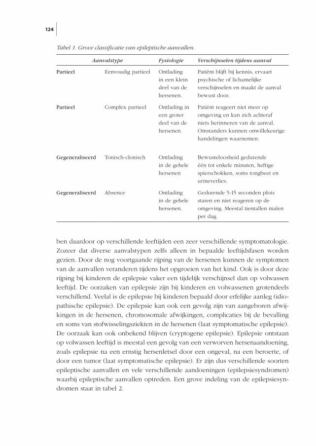
124
ben daardoor op verschillende leeftijden een zeer verschillende symptomatologie.
Zozeer dat diverse aanvalstypen zelfs alleen in bepaalde leeftijdsfasen worden
gezien. Door de nog voortgaande rijping van de hersenen kunnen de symptomen
van de aanvallen veranderen tijdens het opgroeien van het kind. Ook is door deze
rijping bij kinderen de epilepsie vaker een tijdelijk verschijnsel dan op volwassen
leeftijd. De oorzaken van epilepsie zijn bij kinderen en volwassenen grotendeels
verschillend. Veelal is de epilepsie bij kinderen bepaald door erfelijke aanleg (idio-
pathische epilepsie). De epilepsie kan ook een gevolg zijn van aangeboren afwij-
kingen in de hersenen, chromosomale afwijkingen, complicaties bij de bevalling
en soms van stofwisselingsziekten in de hersenen (laat symptomatische epilepsie).
De oorzaak kan ook onbekend blijven (cryptogene epilepsie). Epilepsie ontstaan
op volwassen leeftijd is meestal een gevolg van een verworven hersenaandoening,
zoals epilepsie na een ernstig hersenletsel door een ongeval, na een beroerte, of
door een tumor (laat symptomatische epilepsie). Er zijn dus verschillende soorten
epileptische aanvallen en vele verschillende aandoeningen (epilepsiesyndromen)
waarbij epileptische aanvallen optreden. Een grove indeling van de epilepsiesyn-
dromen staat in tabel 2.
Aanvalstype Fysiologie Verschijnselen tijdens aanval
Partieel Eenvoudig partieel Ontlading
in een klein
deel van de
hersenen.
Patiënt blijft bij kennis, ervaart
psychische of lichamelijke
verschijnselen en maakt de aanval
bewust door.
Partieel Complex partieel Ontlading in
een groter
deel van de
hersenen.
Patiënt reageert niet meer op
omgeving en kan zich achteraf
niets herinneren van de aanval.
Omstanders kunnen onwillekeurige
handelingen waarnemen.
Gegeneraliseerd Tonisch-clonisch Ontlading
in de gehele
hersenen
Bewusteloosheid gedurende
één tot enkele minuten, heftige
spierschokken, soms tongbeet en
urineverlies.
Gegeneraliseerd Absence Ontlading
in de gehele
hersenen.
Gedurende 5-15 seconden plots
staren en niet reageren op de
omgeving. Meestal tientallen malen
per dag.
Tabel 1. Grove classificatie van epileptische aanvallen.
290158_Stroink_BW.indd 124 11-04-2008 10:53:50
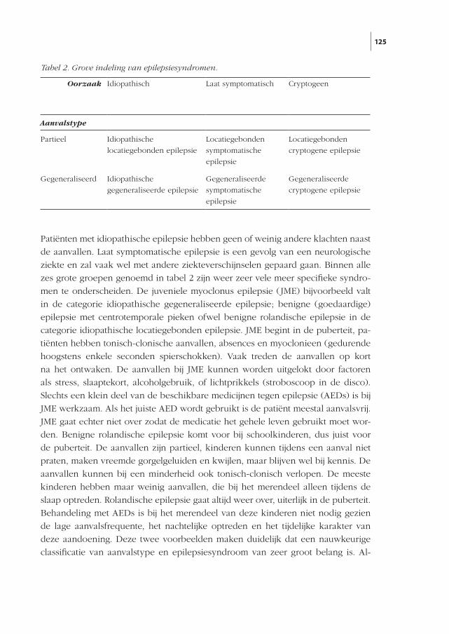
125
Patiënten met idiopathische epilepsie hebben geen of weinig andere klachten naast
de aanvallen. Laat symptomatische epilepsie is een gevolg van een neurologische
ziekte en zal vaak wel met andere ziekteverschijnselen gepaard gaan. Binnen alle
zes grote groepen genoemd in tabel 2 zijn weer zeer vele meer specifieke syndro-
men te onderscheiden. De juveniele myoclonus epilepsie (JME) bijvoorbeeld valt
in de categorie idiopathische gegeneraliseerde epilepsie; benigne (goedaardige)
epilepsie met centrotemporale pieken ofwel benigne rolandische epilepsie in de
categorie idiopathische locatiegebonden epilepsie. JME begint in de puberteit, pa-
tiënten hebben tonisch-clonische aanvallen, absences en myoclonieen (gedurende
hoogstens enkele seconden spierschokken). Vaak treden de aanvallen op kort
na het ontwaken. De aanvallen bij JME kunnen worden uitgelokt door factoren
als stress, slaaptekort, alcoholgebruik, of lichtprikkels (stroboscoop in de disco).
Slechts een klein deel van de beschikbare medicijnen tegen epilepsie (AEDs) is bij
JME werkzaam. Als het juiste AED wordt gebruikt is de patiënt meestal aanvalsvrij.
JME gaat echter niet over zodat de medicatie het gehele leven gebruikt moet wor-
den. Benigne rolandische epilepsie komt voor bij schoolkinderen, dus juist voor
de puberteit. De aanvallen zijn partieel, kinderen kunnen tijdens een aanval niet
praten, maken vreemde gorgelgeluiden en kwijlen, maar blijven wel bij kennis. De
aanvallen kunnen bij een minderheid ook tonisch-clonisch verlopen. De meeste
kinderen hebben maar weinig aanvallen, die bij het merendeel alleen tijdens de
slaap optreden. Rolandische epilepsie gaat altijd weer over, uiterlijk in de puberteit.
Behandeling met AEDs is bij het merendeel van deze kinderen niet nodig gezien
de lage aanvalsfrequente, het nachtelijke optreden en het tijdelijke karakter van
deze aandoening. Deze twee voorbeelden maken duidelijk dat een nauwkeurige
classificatie van aanvalstype en epilepsiesyndroom van zeer groot belang is. Al-
Oorzaak Idiopathisch Laat symptomatisch Cryptogeen
Aanvalstyp e
Partieel Idiopathische
locatiegebonden epilepsie
Locatiegebonden
symptomatische
epilepsie
Locatiegebonden
cryptogene epilepsie
Gegeneraliseerd Idiopathische
gegeneraliseerde epilepsie
Gegeneraliseerde
symptomatische
epilepsie
Gegeneraliseerde
cryptogene epilepsie
Tabel 2. Grove indeling van epilepsiesyndromen.
290158_Stroink_BW.indd 125 11-04-2008 10:53:50
126
leen dan kan een prognose worden gegeven van het beloop, kan beter worden
afgewogen of behandeling met AEDs is gewenst, en zo ja welke van de vele AEDs
geschikt zijn voor deze patiënt, hoe lang moet worden behandeld en of verder on-
derzoek noodzakelijk is. In de praktijk is het helaas niet bij alle patiënten mogelijk
het epilepsiesyndroom nauwkeurig te classificeren.
De consequenties van epilepsie kunnen voor kinderen en volwassenen verschil-
lend zijn. Volwassenen worden beperkt in de verkeersdeelname (rijbewijs), hun
werk, sociale activiteiten, enzovoort. Daarnaast zijn de bijwerkingen van de medi-
cijnen verschillend afhankelijk van de leeftijd. Denk bijvoorbeeld aan de schade-
lijke effecten die kunnen optreden bij het gebruik tijdens zwangerschap voor het
ongeboren kind. Kinderen zijn vaak weer meer gevoelig voor bijwerkingen op het
gedrag en de cognitie. De noodzaak van behandeling met AEDs kan bij kinderen
minder groot zijn.
Gezien de andere symptomatologie, het verschillend beloop, de verschillende oor-
zaken en de gevolgen voor de patiënt is het wenselijk bij behandeling en in weten-
schappelijke onderzoeken een onderscheid te maken tussen epilepsie bij kinderen
en volwassenen. Merkwaardig genoeg wordt nog steeds in veel onderzoeken dit
onderscheid niet gemaakt.
Over het natuurlijke beloop van epilepsie zijn slechts beperkte gegevens bekend
doordat bijna alle patiënten worden behandeld met AEDs, vaak al na één aan-
val. Daarnaast wordt veel onderzoek verricht in gespecialiseerde ziekenhuizen.
Daar worden echter vooral patiënten met de ernstigere vormen van epilepsie naar
verwezen. Hierdoor kan in onderzoeken een vertekend beeld optreden van het
beloop van epilepsie. Ruwweg lijkt het zo dat van de patiënten die worden behan-
deld met medicijnen ongeveer 75% geen aanvallen heeft. Een kleine minderheid
van de kinderen heeft frequent aanvallen die niet met medicijnen zijn te behan-
delen. Hierdoor kan de kwaliteit van leven zeer nadelig worden beïnvloed en de
cognitieve ontwikkeling worden geschaad.
Dutch Study of Epilepsy in Childhood (DSEC)
In 1988 besloten de kinderneurologen Willem Frans Arts, Oebo Brouwer, Boude-
wijn Peters en Hans Stroink, de neuroloog Cees van Donselaar en de epidemioloog
Ada Geerts een onderzoeksgroep op te richten. Aanvankelijk droeg het onderzoek
de naam Zuid-Hollands Kinderepilepsie Onderzoek (ZHKO), later omgedoopt tot
Dutch Study of Epilepsy in Childhood (DSEC). Om de eerder genoemde bezwaren
te ondervangen werden in dit onderzoek alleen kinderen met de leeftijd van één
maand tot 16 jaar geïncludeerd met nieuw ontstane epilepsie of een eenmalige
290158_Stroink_BW.indd 126 11-04-2008 10:53:50
127
epileptische aanval. Aanvallen in de eerste levensmaand zijn veelal acuut sympto-
matisch en worden in onderzoeken naar epilepsie niet betrokken. In de periode
augustus 1988 tot augustus 1992 werden alle kinderen aangemeld met niet acuut
symptomatische epileptische aanvallen verwezen naar de deelnemende zieken-
huizen, alsmede kinderen waarbij de diagnose werd overwogen maar niet zeker
was. Kinderen eerder behandeld met AEDs, alsmede kinderen verwezen vanuit
andere ziekenhuizen werden niet geïncludeerd. De deelnemende ziekenhuizen
waren het Westeinde Ziekenhuis en het Juliana Kinderziekenhuis in Den Haag,
het Academisch Ziekenhuis Leiden en het Academisch Ziekenhuis Rotterdam. Dit
laatste ziekenhuis had twee locaties: het Dijkzigt Ziekenhuis en het Sophia Kin-
derziekenhuis. Van alle kinderen werden de gegevens zorgvuldig vastgelegd zoals
de symptomen van de aanvallen, de medische voorgeschiedenis, het al dan niet
voorkomen van epilepsie in de familie en het lichamelijk neurologische onder-
zoek. Bij alle kinderen werd een EEG (hersenfilmpje) gemaakt, en wanneer dit
geen afwijkingen liet zien volgde een tweede EEG zo mogelijk tijdens slaap. Bij de
meeste kinderen werd een scan van de hersenen gemaakt, en ander onderzoek als
de behandelend kinderneuroloog dit noodzakelijk achtte. Alle aangemelde kinde-
ren werden besproken in een panel bestaande uit drie van de vier deelnemende
kinderneurologen; de behandelend kinderneuroloog nam niet deel aan het be-
spreken van zijn eigen patiënten. In eerste instantie werd de diagnose gesteld op
grond van de aanvalsbeschrijving, zonder kennis van het aanvullend onderzoek,
waaronder het EEG. Alleen bij kinderen met meerdere aanvallen mocht het EEG in
geval van twijfel worden meegewogen in de diagnose wel of niet epilepsie. Door
deze besprekingen in een panel hoopten we de diagnose zo nauwkeurig mogelijk
te stellen, zodat we zo weinig mogelijk kinderen zouden missen met epileptische
aanvallen en zo weinig mogelijk kinderen te includeren met niet epileptische aan-
vallen. Er werden in de DSEC 760 kinderen geïncludeerd: 170 met één epileptische
aanval, 412 met epilepsie, 54 met één en 124 met meerdere aanvallen zonder
diagnose (onduidelijke aanvallen). Dit betekent dat ongeveer 75% van alle kinde-
ren met epilepsie in de regio van de deelnemende ziekenhuizen is geïncludeerd.
Kinderen met aanvallen waarvan het panel meende dat ze niet epileptisch waren,
maar waarvoor ook geen andere verklaring voorhanden was, werden één jaar ver-
volgd. Kinderen met één epileptische aanval kregen geen AEDs voorgeschreven
en werden twee jaar vervolgd. Als er echter nieuwe aanvallen optraden werden
zij vervolgd in het onderzoek van de kinderen met epilepsie. De kinderen met
epilepsie (meerdere epileptische aanvallen) werden aanvankelijk vijf jaar vervolgd.
Later werd besloten de studieduur te verlengen en deze kinderen 15 jaar te vervol-
gen. Het al dan voorschrijven van AEDs was ter beoordeling van de behandelend
290158_Stroink_BW.indd 127 11-04-2008 10:53:50
128
kinderneuroloog in overleg met de ouders en het kind. Tijdens het vervolgen van
de kinderen werd niet alleen gekeken hoe het ziektebeloop was, maar ook of de
diagnose gesteld op moment van inclusie juist bleef.
In de DSEC was zo een bijzondere en grote groep kinderen bijeen gebracht met
niet eerder behandelde epileptische aanvallen die langdurig kon worden vervolgd.
Er waren diverse vraagstellingen in de DSEC. Een groot deel van de onderzoeks-
resultaten is elders gepubliceerd: cognitie en gedrag bij kinderen met epilepsie;
“quality of life” (invloed van de aandoening en de behandeling op het algehele
welbevinden en functioneren); sterfte bij kinderen met epilepsie; de aanvullende
waarde van een tweede EEG tijdens slaap; relatie tussen de behandelduur met
AEDs en de kans op succesvol staken van deze medicatie; de mate waarin de epi-
lepsie familiair voorkomt; het al dan niet bestaan van immunologische afwijkingen
(stoornissen in het afweersysteem) als oorzaak van de epilepsie of juist als gevolg
van de behandeling; het voorspellen van het beloop van de aandoening vroeg na
het stellen van de diagnose: is de epilepsie makkelijk te behandelen en/of tijdelijk,
of juist zeer slecht behandelbaar en/of chronisch. Deze publicaties staan vermeld
in de publicatielijst DSEC.
In dit proefschrift komen de volgende onderwerpen aan de orde: de nauwkeurig-
heid en betrouwbaarheid van de diagnose één epileptische aanval; het beloop na
één epileptische aanval (hoeveel kinderen ontwikkelen epilepsie, is dit te voor-
spellen en is de eventuele epilepsie al dan niet ernstig); de nauwkeurigheid en
betrouwbaarheid van de diagnose epilepsie; de nauwkeurigheid en betrouwbaar-
heid van de beoordeling van het EEG; hoe vaak komt een zeer langdurige epilep-
sieaanval (status epilepticus) voor en wat betekent dit voor het verdere beloop van
de aandoening; praktische adviezen voor de diagnostiek en het al dan niet starten
van een behandeling met AEDs.
Hoofdstuk 2
In hoofdstuk 2 wordt de problematiek rond het stellen van de diagnose besproken.
Op de kinderleeftijd komt een zeer groot en divers scala voor van aanvalsgewijs
verlopende aandoeningen. De diagnose berust op de omschrijving van de aanval-
len door getuigen. Deze beschrijving kan ontbreken doordat tijdens de aanval nie-
mand aanwezig was, onduidelijk zijn doordat de omstander(s) in paniek raakten,
zij de gebeurtenis niet goed kunnen verwoorden of zij zelf de aanval interpreteren
in plaats van te beschrijven. Ook de arts kan de gegevens onjuist interpreteren. Bij
het stellen van de diagnose spelen derhalve vele subjectieve factoren een rol. Bij
het stellen van de diagnose zal daarom een zekere mate van onzekerheid bestaan.
290158_Stroink_BW.indd 128 11-04-2008 10:53:50
129
Ook wordt de diagnose nogal eens op grond van aanvullend onderzoek gesteld,
en dan met name op EEG uitslag, in plaats van op de beschrijving van de aanvallen
(zie hoofdstuk 5). In Groot-Brittannië wordt naar aanleiding van een rechtszaak,
aanhangig gemaakt door ouders bij wie een kind ten onrechte werd behandeld
voor epilepsie, verondersteld dat bij ruim 30% van de kinderen met epilepsie de
diagnose onjuist is. Ook wordt in Groot-Brittannië een groot deel van de kinderen
die wel epilepsie hebben “overbehandeld”, dat wil zeggen met te veel medicijnen
of in te hoge doseringen. Het betreft daarom een groot praktisch probleem. In
dit hoofdstuk worden een aantal adviezen gegeven om onjuiste diagnoses zoveel
mogelijk te voorkomen. Wij menen dat het voor kinderen en volwassenen minder
schadelijk is de diagnose epilepsie in een wat later stadium te stellen dan vroeg-
tijdig deze diagnose (onjuist) te stellen op grond van onvoldoende gegevens. Het
gevolg bij een verkeerde diagnose is onnodig behandelen, vaak met veel AEDs
in hoge doseringen omdat de AEDs niet zullen helpen, onnodig onderzoek, soci-
ale beperkingen en stigmatisering van patiënten die geen epilepsie hebben. Bo-
vendien verbetert vroege behandeling de prognose van epilepsie niet. Dit laatste
komt later nog uitgebreid aan de orde. Bij het stellen van diagnoses bij patiënten
met onduidelijke aanvallen heeft een conservatieve benadering sterk de voorkeur.
Men dient wel op zijn hoede te zijn dat aanvallen ook door een hartritmestoornis
kunnen worden veroorzaakt. Dit is een van de weinige alternatieve diagnoses die
ernstige consequenties heeft.
Hoofdstuk 3
In hoofdstuk 3 wordt beschreven in hoeverre kinderneurologen het onderling eens
zijn of er al dan niet sprake is geweest van een epileptische aanval (reliability). Wij
onderzochten bij 100 kinderen met één doorgemaakte aanval of zes kinderneuro-
logen het al dan niet met elkaar eens waren over de aard van de aanval: wel of niet
een epileptische aanval. In de geneeskunde spelen bij alle oordelen subjectieve
factoren mede een rol en daarom zal er nooit een volledige 100% overeenstem-
ming bestaan tussen het oordeel van verschillende artsen. De mate van overeen-
stemming kan met hulp van een formule in getal worden uitgedrukt. Deze maat is
de kappa. Een kappa van nul betekent dat de mate van overeenstemming tussen
verschillende beoordelaars op puur toeval berust. Met het gooien van dobbelste-
nen of door domweg zonder enige kennis te raden zal men hetzelfde resultaat
bereiken. Een kappa van -1.0 betekent volledige onenigheid; een kappa van 1.0
juist wel volledige overeenstemming tussen de beoordelaars. In de geneeskunde
wordt een kappa van 0.6 tot 0.8 als goed beschouwd en een kappa van 0.8 tot 1.0
290158_Stroink_BW.indd 129 11-04-2008 10:53:50
130
als bijna perfect. Drie deelnemende kinderneurologen (groep 1) waren leden van
de DSEC groep. Drie anderen (groep 2) waren ervaren kinderneurologen: één
werkzaam in een epilepsiecentrum, één in een centrum voor epilepsiechirurgie en
één in een universitair kinderziekenhuis. De deelnemers kregen een uitgebreide
beschrijving van de aanvallen op papier uitgereikt. Wanneer deze kinderneurolo-
gen de diagnose stelden op basis van hun persoonlijke ervaring en kennis bleek er
slechts een zeer matige overeenstemming te bestaan over de aard van de aanvallen
(kappa 0.19 tot 0.60, gemiddeld 0.41). Wanneer criteria werden uitgereikt waaraan
een epileptische aanval moet voldoen trad een lichte verbetering op tot kappa’s
van 0.23 tot 0.68, gemiddeld 0.45. De kinderneurologen uit de DSEC, die gewend
waren aan deze wijze van diagnosticeren, scoorden consequent hoger dan de an-
dere drie kinderneurologen. Vervolgens werd binnen de twee groepen van drie
kinderneurologen over ieder kind gediscussieerd totdat men binnen elk van de
twee groepen tot één gezamenlijke diagnose kwam. De kappa tussen de diagnose
van groep 1 en 2 bedroeg 0.6. Dit is beter dan de aanvankelijke onderlinge indi-
viduele overeenstemming zonder criteria voor de aanval, maar nog steeds matig.
Hierbij dient beseft te worden dat in de dagelijkse praktijk geen aanvalscriteria
worden gebruikt en ook geen uitgebreide bespreking plaats vindt met ervaren kin-
derneurologen over de diagnose. In het dagelijkse leven zal dus een vrij grote mate
van verschil van mening bestaan over de aard van een eenmalige aanval. Als een
diagnose betrouwbaar is te stellen zal de kappa hoog zijn. Omgekeerd betekent
een hoge kappa niet automatisch een juiste diagnose. Een valkuil kan een ieder
op hetzelfde verkeerde spoor zetten waardoor eensluidend een verkeerd oordeel
volgt. Tot slot werden in dit onderzoek alle gegevens aan de deelnemers verstrekt
zoals EEG, scan, enzovoort. Ook met deze gegevens lukte het de deelnemers
slechts bij een klein aantal kinderen een specifieke syndroomdiagnose te stellen.
Hoofdstuk 4
Na het onderzoek van de onderlinge overeenstemming tussen kinderneurologen
over de diagnose volgt in hoofdstuk 4 het onderzoek naar de juistheid (accuracy)
van de diagnose eenmalig insult en epilepsie. Zoals in hoofdstuk 2 en 3 beschre-
ven bestaat er geen gouden standaard voor de diagnose insult of epilepsie. De enig
mogelijke wijze van controle op de diagnose is het vervolgen van patiënten. Indien
uit het beloop nieuwe gegevens naar voren komen kan dit leiden tot een andere
diagnose. Indien geen nieuwe aanvallen meer optreden is de kans dat de diagnose
achteraf veranderd zal worden laag. Er zijn veel methoden om de mate van juist-
heid van de diagnose in getal uit te drukken. Dit leidt nogal eens tot verwarring.
290158_Stroink_BW.indd 130 11-04-2008 10:53:50
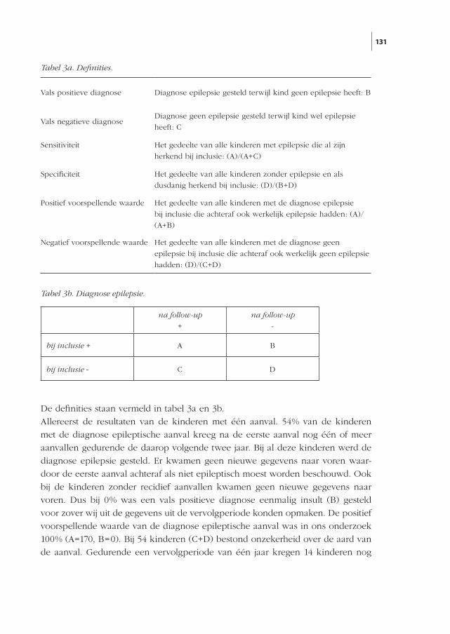
131
De definities staan vermeld in tabel 3a en 3b.
Allereerst de resultaten van de kinderen met één aanval. 54% van de kinderen
met de diagnose epileptische aanval kreeg na de eerste aanval nog één of meer
aanvallen gedurende de daarop volgende twee jaar. Bij al deze kinderen werd de
diagnose epilepsie gesteld. Er kwamen geen nieuwe gegevens naar voren waar-
door de eerste aanval achteraf als niet epileptisch moest worden beschouwd. Ook
bij de kinderen zonder recidief aanvallen kwamen geen nieuwe gegevens naar
voren. Dus bij 0% was een vals positieve diagnose eenmalig insult (B) gesteld
voor zover wij uit de gegevens uit de vervolgperiode konden opmaken. De positief
voorspellende waarde van de diagnose epileptische aanval was in ons onderzoek
100% (A=170, B=0). Bij 54 kinderen (C+D) bestond onzekerheid over de aard van
de aanval. Gedurende een vervolgperiode van één jaar kregen 14 kinderen nog
Vals positieve diagnose Diagnose epilepsie gesteld terwijl kind geen epilepsie heeft: B
Vals negatieve diagnoseDiagnose geen epilepsie gesteld terwijl kind wel epilepsie
heeft: C
Sensitiviteit Het gedeelte van alle kinderen met epilepsie die al zijn
herkend bij inclusie: (A)/(A+C)
Specificiteit Het gedeelte van alle kinderen zonder epilepsie en als
dusdanig herkend bij inclusie: (D)/(B+D)
Positief voorspellende waarde Het gedeelte van alle kinderen met de diagnose epilepsie
bij inclusie die achteraf ook werkelijk epilepsie hadden: (A)/
(A+B)
Negatief voorspellende waarde Het gedeelte van alle kinderen met de diagnose geen
epilepsie bij inclusie die achteraf ook werkelijk geen epilepsie
hadden: (D)/(C+D)
na follow-up
+
na follow-up
-
bij inclusie + A B
bij inclusie - C D
Tabel 3a. Definities.
Tabel 3b. Diagnose epilepsie.
290158_Stroink_BW.indd 131 11-04-2008 10:53:51
132
één of meer aanvallen. Bij vier (C) van deze 14 kinderen waren het epileptische
aanvallen en moest de eerste aanval achteraf waarschijnlijk ook als epileptische
aanval worden beschouwd. Bij 40 kinderen gebeurde gedurende een jaar niets
meer en kwam geen nieuwe informatie beschikbaar. Bij vier (C) van de 54 (C+D)
kinderen bleek dus achteraf een vals negatieve diagnose te zijn gesteld (7.4%). Bij
50 (D) van de 54 kinderen (C+D) bij wie werd gesteld dat ze geen epileptische
aanval hadden doorgemaakt was dit werkelijk zo: negatief voorspellende waarde
92.6%. Bij 170 (A) van de 174 (A+C) kinderen met één epileptisch insult was dit
bij inclusie herkend (sensitiviteit 97.7%). Het beloop na één aanval wordt uitvoerig
besproken in hoofdstuk 6.
Bij inclusie werd bij 412 kinderen (A+B) de diagnose epilepsie gesteld (meerdere
niet geprovoceerde epileptische aanvallen). Bij 367 kinderen was dit op basis van
de aanvalsbeschrijving, bij 45 kinderen op basis van de aanvalsbeschrijving in
combinatie met de EEG bevindingen. Gedurende twee jaar vervolg rees bij 19 (B)
van deze 412 kinderen (A+B) twijfel over de diagnose epilepsie: vals positieve
diagnose epilepsie 4.6%. De positief voorspellende waarde was 95.4%. Bij 124
kinderen (C+D) bestond bij inclusie onzekerheid over de aard van de aanvallen.
75 van deze kinderen kregen gedurende een vervolg van één jaar nog één of meer
aanvallen. Bij 36 van deze kinderen werd een diagnose anders dan epilepsie ge-
steld. Bij zeven (C) werd wel alsnog de diagnose epilepsie gesteld. Bij 5.6% was dus
bij inclusie een vals negatieve diagnose gesteld. De negatief voorspellende waarde
was 94.4%. Uiteindelijk waren er (412-19)+7=400 kinderen met epilepsie (A+C)
waarvan er 412-19 (A) waren herkend bij inclusie: sensitiviteit 393:400=98.3%. Bij
136 kinderen (B+D) werd na het vervolgen geconcludeerd dat zij geen epilep-
sie hadden: 124 kinderen met onduidelijke aanvallen bij inclusie, hiervan vallen
er zeven af, maar komen er 19 bij die aanvankelijk de diagnose epilepsie had-
den gekregen. Bij inclusie waren er hiervan 124-7=117 herkend (D): specificiteit
117:136=86%. Over de gehele groep van 760 kinderen met één of meer aanvallen
was in ons onderzoek de sensitiviteit 98.1% en de specificiteit 89.8%.
In deze studie werden hoge scores bereikt wat betreft nauwkeurigheid van de
diagnose op het moment van inclusie in de studie. Dit is ook zeer wenselijk bij
een wetenschappelijk onderzoek. In vergelijkbare onderzoeken ontbreken deze
gegevens. Een onderzoek is echter een andere situatie dan de dagelijkse praktijk.
In hoofdstuk 2 wordt besproken dat in de praktijk nogal eens onjuiste diagnoses
worden gesteld. In ons onderzoek werd de diagnose gesteld door ervaren kin-
derneurologen. Ook werd gebruik gemaakt van schriftelijk vastgelegde criteria en
werden alle kinderen onderling uitvoerig besproken. Dit staat ver van de dagelijkse
praktijk. Zowel bij kinderen met één als met meerdere aanvallen zonder duidelijke
290158_Stroink_BW.indd 132 11-04-2008 10:53:51
133
diagnose zijn één of twee EEG’s gemaakt. Het betrof de kinderen waar het panel
van meende dat zij geen epileptische aanvallen hadden of er onvoldoende zeker-
heid aanwezig was om dit te concluderen. Een andere diagnose was echter niet
voorhanden. Een deel van deze kinderen had wel epileptiforme stoornissen op het
EEG. Drie van de acht kinderen met één onduidelijke aanval (hoofdstuk 6) en één
van 27 kinderen met meerdere onduidelijke aanvallen met een epileptiform EEG
bleken na één jaar follow-up toch epilepsie te hebben. De positief voorspellende
waarde van het EEG bij de door ervaren kinderneurologen als onduidelijk bestem-
pelde aanvallen was 11.4%. Hieruit blijkt opnieuw dat een goede aanvalsbeschrij-
ving en interpretatie tot de diagnose leiden, en men bij een onduidelijk verhaal niet
een diagnose op grond van EEG bevindingen kan stellen.
Hoofdstuk 5
Bij kinderen met aanvallen en verdenking op epilepsie wordt meestal een EEG
gemaakt. Zoals al in hoofdstuk 2 en 4 vermeld wordt de diagnose epilepsie niet
gesteld op grond van EEG afwijkingen alleen, maar voornamelijk op de aanvals-
beschrijving. Bij een groot gedeelte van de patiënten met epilepsie worden tijdens
een EEG registratie ontladingen gezien met een specifieke configuratie zoals pie-
ken en piekgolfcomplexen. Dergelijke bevindingen worden epileptiforme afwijkin-
gen genoemd. Een aanzienlijk deel van de patiënten met epilepsie heeft dergelijke
epileptiforme afwijkingen echter niet tijdens een EEG registratie. Hiervoor zijn vele
oorzaken aan te geven. Een registratie duurt 30 minuten. Tijdens een EEG registra-
tie is de kans op het doormaken van een aanval gering. Echter ook zonder aanval
kunnen abnormale ontladingen optreden tijdens een EEG registratie. Bij een EEG
registratie worden elektroden op de schedel aangebracht. Hiermee wordt echter
alleen activiteit gemeten van de oppervlakkig gelegen hersendelen en niet van
de dieper gelegen structuren. Een aanzienlijk deel van gezonde kinderen zonder
epilepsie, ongeveer 3.5 tot 5%, heeft juist wel epileptiforme ontladingen tijdens
een EEG registratie zonder ooit epilepsie te krijgen. Het percentage epileptiforme
afwijkingen ligt nog hoger bij kinderen met neurologische aandoeningen als au-
tisme, ADHD, mentale retardatie, ook als deze kinderen geen epilepsie hebben.
Wanneer een kind aanvallen heeft die weliswaar bij epilepsie zouden kunnen
passen, maar er geen volledige zekerheid bestaat kan het EEG wel de doorslag ge-
ven. De EEG bevindingen moeten passen bij de aanvalsbeschrijving. Wanneer het
EEG epileptiforme afwijkingen vertoont, zullen de aard en de locatie verschillen
afhankelijk van het aanvalstype en het epilepsiesyndroom. Het EEG heeft daarom
in combinatie met de klinische gegevens vooral waarde bij het classificeren van
290158_Stroink_BW.indd 133 11-04-2008 10:53:51
134
het aanvalstype en het epilepsiesyndroom. In deze samenvatting zijn als voorbeeld
van epilepsiesyndromen JME en benigne rolandische epilepsie genoemd. Bij deze
syndromen zijn op het EEG meestal epileptiforme afwijkingen aanwezig, die echter
zeer verschillen van elkaar en kenmerkend zijn voor het betreffende syndroom.
Ook wat betreft het EEG kan de vraag worden gesteld of verschillende neurologen
een EEG gelijk beoordelen. In hoofdstuk 5 staan de resultaten van ons onder-
zoek naar de beoordeling van het EEG door zes ervaren klinisch neurofysiologen.
Steeds werd de mening van twee willekeurige neurofysiologen uit deze groep met
elkaar vergeleken. Nadat zij 72 EEGs hadden beoordeeld bleek hun mening wat
betreft het voorkomen van epileptiforme afwijkingen heel goed met elkaar over-
een te stemmen: kappa 0.83. Het EEG kan echter ook andere minder specifieke
stoornissen vertonen. Deze kunnen plaatselijk optreden (focaal) of overal in de
hersenen (diffuus). Over deze stoornissen bleken de neurofysiologen het onderling
niet erg eens te zijn: kappa voor diffuse stoornissen 0.53 en voor focale stoornis-
sen 0.38. Voor een deel kan deze onenigheid worden verklaard doordat het EEG
op de kinderleeftijd voortdurend verandert bij het opgroeien. Vaak is het moeilijk
te beoordelen of een beeld nog wel of niet bij de leeftijd past, wat discussie geeft
over het al dan niet aanwezig zijn van diffuse stoornissen. Bij kinderen komen
veel variaties voor in het EEG patroon waarvan het moeilijk is te beoordelen of
het EEG al dan niet normaal is. De klinisch neurofysiologen waren het in dit on-
derzoek vaak wel met elkaar eens dat het EEG afwijkend was, maar de één be-
oordeelde een stoornis als focaal en de ander dezelfde stoornis als diffuus. Nadat
de klinisch neurofysiologen dit probleem onderling hadden besproken stelden zij
criteria op wanneer een afwijking diffuus of focaal genoemd moest worden. In
een nieuwe serie van 39 EEGs die zij beoordeelden steeg de kappa voor diffuse
stoornissen naar 0.73 en voor focale stoornissen naar 0.54. Voor de praktijk zijn
echter de epileptiforme stoornissen het belangrijkste. Hierover waren de klinische
neurofysiologen het in hoge mate onderling eens. De betekenis van niet specifieke
focale afwijkingen is in de huidige tijd afgenomen. Voorheen werd dit gebruikt om
onderliggende structurele afwijkingen in de hersenen op te sporen. Tegenwoordig
kan dit met kwalitatief zeer hoogwaardige afbeeldingapparatuur (MRI scan).
Ook over dit onderzoek zijn een aantal eerdere opmerkingen van toepassing.
Onze klinisch neurofysiologen waren zeer goed in staat onderling reproduceer-
baar epileptiforme functiestoornissen te beoordelen. Het betreft echter wederom
zeer gespecialiseerde medici met onderzoekservaring. Een EEG op de kinder-
leeftijd is moeilijk te beoordelen. Het EEG verandert voortdurend met de leeftijd.
Kinderen bewegen vaak tijdens het onderzoek wat storing (artefacten) op het EEG
geeft. Ook andere factoren kunnen storing veroorzaken. Artefacten kunnen veel
290158_Stroink_BW.indd 134 11-04-2008 10:53:51
135
gelijkenis vertonen met epileptiforme stoornissen. In de dagelijkse praktijk heeft
niet ieder ziekenhuis klinisch neurofysiologen, of kan de ervaring met kinderen
beperkt zijn. Hierdoor kunnen normale verschijnselen of artefacten voor epilepti-
forme stoornissen worden aangezien. In hoofdstuk 2 is besproken dat EEG bevin-
dingen een grote bron van misverstand kunnen zijn en tot een verkeerde conclusie
over het al dan niet bestaan van epilepsie kunnen leiden. Zolang een kind geen
aanvallen heeft, heeft het kind geen epilepsie, ook niet als er epileptiforme afwij-
kingen op het EEG zijn. Bij bijvoorbeeld hoofdpijn en gedragsproblemen zal een
EEG alleen maar verwarring veroorzaken. Een EEG dient daarom bij kinderen te
worden gemaakt als er op grond van de aanvalsbeschrijving verdenking is op epi-
leptische aanvallen. Het EEG dient te worden beoordeeld door een klinisch neu-
rofysioloog met ervaring met kinder-EEGs. Degene die het EEG aanvraagt dient
op de hoogte te zijn van alle valkuilen en het EEG zonodig te bespreken met de
klinisch neurofysioloog. Indien het EEG op de juiste indicatie wordt aangevraagd,
door een deskundige wordt beoordeeld, en overleg plaats vindt tussen aanvrager
en beoordelaar is het EEG een zeer waardevol gegeven bij de diagnostiek en clas-
sificatie van epilepsie. Indien dit niet gebeurt, kan een EEG zeer nadelige gevolgen
hebben voor de patiënt.
Hoofdstuk 6
In hoofdstuk 6 wordt het onderzoek bij kinderen met een eenmalige (niet acuut
symptomatische) epileptische aanval beschreven. De onzekerheid van de diagnose
is in hoofdstuk 2 t/m 4 besproken. Andere vragen zijn: hoe groot is de kans op
herhaling, ofwel hoe groot is de kans op epilepsie? Welke kinderen krijgen meer
aanvallen en hoe vergaat het deze kinderen vervolgens? Er waren voor ons onder-
zoek eerdere onderzoeken gedaan. Deels betrof het onderzoeken van patiënten
van alle leeftijden zonder onderscheid te maken tussen kinderen en volwassenen.
Er waren echter ook diverse onderzoeken specifiek bij kinderen uitgevoerd. In
deze onderzoeken was het risico op herhaling van de aanvallen zeer uiteenlopend.
Hiervoor zijn meerdere oorzaken aan te wijzen. In geen enkel onderzoek werd ver-
meld hoe de diagnose werd gesteld en of deze achteraf moest worden bijgesteld. In
alle onderzoeken kreeg een deel, en vaak zelfs een groot deel van de kinderen di-
rect AEDs voorgeschreven na één aanval. Aannemelijk is dat dit de herhalingskans
van aanvallen verlaagt. Het interval tussen aanval en inclusie in het onderzoek is
vaak niet genoemd. Duidelijk is dat recidiefaanvallen vaak heel snel na de eerste
aanval volgen. Als er een lange wachttijd is voordat kinderen worden gezien in een
ziekenhuis zullen er minder kinderen met een eenmalige aanval overblijven. Velen
290158_Stroink_BW.indd 135 11-04-2008 10:53:51
136
hebben in de wachttijd al een recidief gehad. De kinderen die na de wachttijd nog
geen nieuwe aanval hebben doorgemaakt hebben een lager risico op herhaling
omdat dit risico met het verstrijken van de tijd steeds lager wordt.
In ons onderzoek werden kinderen relatief snel gezien en geïncludeerd (49% <24
uur en 71% <zeven dagen); de diagnose werd gesteld op grond van de aanvalsbe-
schrijving met behulp van criteria; het EEG werd bij de diagnose eenmalig insult
buiten beschouwing gelaten. Wel werd bij alle kinderen een EEG gemaakt en in-
dien niet afwijkend een tweede EEG na partiële slaapdeprivatie (een korte nacht-
rust). Verreweg de meeste kinderen hadden een tonisch-clonisch insult (“grote
aanval”) doorgemaakt al dan niet met een partiëel begin. Geen enkel kind kreeg
AEDs na de eerste aanval. In vergelijking met andere onderzoeken kreeg een
relatief groot deel van de kinderen, 54%, meer aanvallen binnen twee jaar. Naar
alle waarschijnlijkheid is dit toe te schrijven aan onze nauwkeurige methodiek, het
niet direct behandelen en de snelle inclusie. Ook bij ons was de kans op nieuwe
aanvallen het grootste in de eerste zes maanden (figuur 2 in hoofdstuk 6). De
risicofactoren voor herhaling waren epileptiforme stoornissen op het EEG en/of
het aanwezig zijn van neurologische afwijkingen of een verstandelijke handicap
(dus een laat symptomatische aanval). Bij afwezigheid van deze factoren was de
herhalingskans 40% en bij aanwezigheid van deze factoren 71-74% (figuur 3 en
4 in hoofdstuk 6). De 85 kinderen die een recidief kregen zijn vervolgens 24-72
maanden vervolgd (gemiddeld 42 maanden). Op het einde van de follow-up wa-
ren 50 van deze kinderen minstens 12 maanden aanvalsvrij, en hadden er 22 nog
aanvallen in de laatste zes maanden. Gerekend voor alle kinderen geïncludeerd
met één epileptische aanval was dus op het einde van de follow-up 78% minstens
12 maanden aanvalsvrij, terwijl 14% nog aanvallen had de laatste zes maanden. In
deze laatste groep waren echter ook kinderen die met een lage frequentie tonisch-
clonische insulten of rolandische aanvallen hadden, waarvan er één of een enkele
in het laatste half jaar van de follow-up plaats vond. De kans dat een kind zich pre-
senteert met één epileptische aanval en daarna een langdurige vervelende epilep-
sie ontwikkelt is laag. Bij ernstige epilepsiesyndromen op de kinderleeftijd komen
vaak kleinere aanvallen voor in een hoge frequentie. Die kinderen zullen niet na
één aanval worden gezien, enerzijds omdat de eerste aanval vaak niet als iets ab-
normaals wordt herkend, anderzijds zullen door de hoge aanvalsfrequentie al vele
nieuwe aanvallen zijn gevolgd op het moment dat een arts wordt geraadpleegd.
Wat zijn de conclusies voor de praktijk? De eerste vraag was: hoe zeker is de di-
agnose epileptische aanval? Uit het voorgaande is duidelijk dat er vaak een forse
mate van onzekerheid zal bestaan. De volgende vraag betrof de herhalingskans
als een epileptische aanval erg waarschijnlijk lijkt? Deze loopt uiteen van 40% tot
290158_Stroink_BW.indd 136 11-04-2008 10:53:51
137
74% De door ons gevonden risicofactoren voor het ontstaan van epilepsie stem-
men overeen met die van enkele andere grote onderzoeken: de resultaten van het
EEG en de oorzaak van de aanval (laat symptomatisch). Niettemin krijgt 26-29%
van de kinderen met deze risicofactoren geen epilepsie. De laatste vraag is: wat
is de winst van het direct na één epileptische aanval voorschrijven van AEDs aan
kinderen? In ons onderzoek is de kans op een moeilijk behandelbare chronische
epilepsie vrij klein voor kinderen die zich presenteren met één aanval. Vóór onze
studie is in Canada een onderzoek gedaan bij een kleine groep van 31 kinderen.
De ene helft kreeg een AED, de andere helft niet. Van de behandelde kinderen
kregen er minder recidiefaanvallen dan van de onbehandelde kinderen. Een deel
van deze kinderen kreeg echter vervelende bijwerkingen van de medicatie. Uitein-
delijk was het aantal kinderen dat klachtenvrij was even groot in beide groepen:
de winst door minder aanvallen was volledig verloren gegaan door het optreden
van bijwerkingen bij de behandelde kinderen. Vijftien jaar later is opnieuw geke-
ken hoe het met deze kinderen ging. Er was geen verschil in het aantal doorge-
maakte epileptische aanvallen tussen de direct behandelde kinderen en de con-
trole groep. Van de direct behandelde kinderen was na 15 jaar 80% minstens twee
jaar aanvalsvrij en van de controlegroep 88%. Hierna zijn geen nieuwe (grotere)
onderzoeken gedaan naar het effect van behandeling specifiek bij kinderen. Wel
zijn twee onderzoeken gedaan waaraan zowel kinderen als volwassenen mee-
deden zonder dat bij de analyse onderscheid werd gemaakt. In een Italiaans on-
derzoek betrof het patiënten met uitsluitend één aanval, in een Engels onderzoek
patiënten met één of enkele aanvallen. In beide onderzoeken kwam naar voren
dat behandelde patiënten in de eerste periode minder aanvallen doormaakten. Na
langere tijd verdween dit verschil en was het aantal aanvalsvrije patiënten precies
even groot geworden ongeacht of er wel of niet direct na de eerste aanval met
AEDs werd gestart. Naar de nadelige effecten van medicatie is niet gekeken in het
Italiaanse onderzoek. In het Engelse onderzoek was er geen verschil in “quality of
life” of complicaties tussen beide groepen. Na vijf jaar gebruikten echter wel veel
meer patiënten (60%) AEDs in de groep die direct werd behandeld in vergelijking
met de groep die niet of later werd behandeld (41%). Een vierde vraag, deels al
beantwoord, is of er gevaren zijn verbonden aan het niet direct behandelen. De
kans op een onbehandelbare epilepsie neemt niet toe. In ons onderzoek zijn
geen kinderen overleden door een aanval, ook niet in de groep kinderen die zich
presenteerde met meerdere aanvallen. Ook in een groot Amerikaans onderzoek
werd na een eerste aanval geen sterfte gemeld door nieuwe aanvallen. Wel is de
sterfte onder kinderen met epilepsie hoger dan onder kinderen zonder epilepsie.
Dit is echter een gevolg van de ziekte die de epilepsie veroorzaakt, bijvoorbeeld
290158_Stroink_BW.indd 137 11-04-2008 10:53:51
138
een progressieve onbehandelbare stofwisselingsziekte. Al met al zijn er geen harde
argumenten om direct na een eerste aanval met medicatie te starten. In geval van
twijfel zal de tijd bij een deel van de kinderen de aard van de aanvallen alsnog
duidelijk maken, of de aanvallen verdwijnen spontaan. In de groep kinderen die
wel nieuwe aanvallen krijgt bevinden zich ook kinderen met zogenaamde benigne
(goedaardige) epilepsie. Deze kinderen hebben meestal maar weinig aanvallen,
vaak alleen in de slaap, en na enige tijd verdwijnen de aanvallen spontaan. Op
grond van ons onderzoek en enkele andere onderzoeken hebben the American
Academy of Neurology, the Child Neurology Society, and the American Epilepsy
Society richtlijnen uitgebracht over eventueel onderzoek (2000) en behandeling
(2003) bij een kind met één epileptische aanval: “een EEG wordt aanbevolen, maar
ander onderzoek wordt alleen nodig geacht als daar aanleiding toe is op grond van
specifieke omstandigheden. Behandeling is niet nodig om epilepsie te voorkomen
en verbetert niet de prognose. Behandeling kan worden overwogen als de voor-
delen van het verlagen van de kans op nieuwe aanvallen opwegen tegen het risico
van de bijwerkingen en de psychosociale effecten.”
Hoofdstuk 7
In hoofdstuk 7 staat het beloop beschreven van kinderen met epilepsie die een-
maal of vaker een status epilepticus (SE) doormaakten. Toen de DSEC startte wa-
ren hierover geen gegevens bekend. Zeer langdurige aanvallen noodzaken vaak
tot opname op een intensive care afdeling. Door de lange duur van de aanval
en door de behandeling kunnen allerlei complicaties optreden. Met SE worden
meestal epileptische aanvallen aangeduid die 30 minuten of langer duren. Deze
tijdsduur in de definitie was gebaseerd op gegevens uit proefdierenonderzoek. Uit
dergelijk onderzoek kwamen aanwijzingen dat na een dergelijke duur schade kon
optreden aan de hersenen. De afgelopen jaren is over deze tijdslimiet om meer-
dere redenen discussie ontstaan. Het is gebleken dat als een aanval eenmaal vijf
tot 10 minuten duurt de kans dat deze aanval stopt zonder medisch ingrijpen sterk
afneemt. Tevens is uit diverse onderzoeken gebleken dat naarmate sneller wordt
ingegrepen de kans groter is dat de aanval makkelijker met noodmedicatie kan
worden gestopt. Anderzijds lijkt ondertussen het ontstaan van schade aan de her-
senen door een aanval van 30 minuten veel minder waarschijnlijk. Ook blijkt dat
de aanvalsfrequentie en de aanvalsduur onafhankelijke variabelen zijn. De meeste
patiënten hebben alleen korte aanvallen, een minderheid heeft lange aanvallen.
Omdat de meeste aanvallen spontaan stoppen binnen vijf minuten, en de kans
dat dit alsnog gebeurt na vijf minuten sterk afneemt is het huidige advies na vijf
290158_Stroink_BW.indd 138 11-04-2008 10:53:51
139
minuten noodmedicatie toe te dienen. Indien dit advies wordt uitgevoerd treden
aanzienlijk minder complicaties op. Gezien deze gegevens pleit een aantal onder-
zoekers ervoor de tijdsduur in de definitie van SE te wijzigen in vijf of tien minuten.
Toen de DSEC startte was de enige definitie van SE die van 30 minuten. Deze de-
finitie is ook gebruikt in twee andere onderzoeken die recent zijn uitgevoerd.
Bijna 10% van de kinderen met epilepsie in de DSEC maakte een SE door. Voor de
meeste kinderen (68%; 32 van de 47) was het hun eerste epilepsieaanval. 41 van
deze 47 kinderen (87%) had al een SE op het moment dat ze werden verwezen
naar het ziekenhuis. Slechts zes kinderen kregen voor het eerst een SE nadat ze
al bekend waren met epilepsie. In ons onderzoek werden geen kinderen geïn-
cludeerd met alleen acuut symptomatische aanvallen. Koortsstuipen zijn bij jonge
kinderen de belangrijkste oorzaak van SE. Ondertussen zijn ook resultaten gepu-
bliceerd van een Amerikaans en een Fins onderzoek naar het beloop van epilepsie
bij kinderen die eenmaal of vaker een SE doormaakten. In deze twee onderzoeken
zijn kinderen met een SE door koorts wel geïncludeerd. Als hiervoor wordt gecor-
rigeerd komt SE in onze groep ongeveer evenveel voor als in het Amerikaanse
onderzoek. In de Finse studie was SE iets frequenter. In die studie werden echter
andere onderzoeksmethoden gebruikt en waren de kinderen al in de zestiger jaren
geincludeerd. Dit maakt vergelijking moeilijker. In alle onderzoeken komt een SE
het vaakste voor aan het begin van de aandoening. Als in onze studie een kind
eenmaal een SE had doorgemaakt was de kans op herhaling van een SE ruim 30%.
De Amerikanen vonden eenzelfde herhalingskans. Van de kinderen zonder SE
bij inclusie maakte in ons onderzoek slechts ruim 1% alsnog een SE door. Opval-
lend is dat in ons en in het Finse onderzoek kinderen met een SE voorheen vaak
koortsstuipen hadden doorgemaakt, maar in het Amerikaanse onderzoek niet. In
alle onderzoeken bestaat een trend dat kinderen die een SE hebben doorgemaakt
een iets lagere kans hebben op den duur aanvalsvrij te worden. Significant (zeker)
is dit verschil echter niet. Bovendien blijkt dat vooral kinderen met laat sympto-
matische epilepsie en cryptogene epilepsie een SE doormaken. Ook zonder SE is
de prognose bij laat symptomatische en cryptogene epilepsie minder goed. Een
uitzondering in ons en het Amerikaanse onderzoek waren de kinderen die na
inclusie SE doormaakten. Echter ook kinderen met korte aanvallen die aanvallen
bleven houden deden het slechter. Het gegeven dat aanvallen door blijven gaan
betekent per definitie dat de epilepsie niet goed behandelbaar is. De sterfte was
in geen van de drie onderzoeken hoger bij kinderen met SE. De oorzaak van de
epilepsie lijkt derhalve belangrijker voor de prognose dan de aanvalsduur. Hier-
bij dient aangetekend te worden dat kinderen met een SE in de westerse wereld
redelijk snel en adequaat kunnen worden behandeld. Uit andere onderzoeken is
290158_Stroink_BW.indd 139 11-04-2008 10:53:51
140
bekend dat er ook kinderen en volwassenen zijn met een SE bij wie het aanzienlijk
slechter kan aflopen. Er kan een aanzienlijke sterfte optreden en patiënten kun-
nen ernstige schade overhouden aan de hersenen. Dit zijn echter geen patiënten
met epilepsie, maar patiënten met een acuut symptomatische SE door bijvoorbeeld
ernstig zuurstoftekort na een hartstilstand of hersenletsel door een ongeval. Ook
bij deze patiënten blijkt niet de SE zelf verantwoordelijk voor de prognose, maar
de ernst van de ziekte die de SE tot gevolg had. In de DSEC onderzochten wij SE
bij kinderen met epilepsie en geen acuut symptomatische aanvallen.
Aangezien er wel een hoge kans is op herhaling bij kinderen met een doorge-
maakte SE dienen ouders en/of verzorgers goed geïnstrueerd te zijn hoe te han-
delen bij een eventueel recidief en noodmedicatie te worden voorgeschreven.
Deze instructies dienen ook schriftelijk te worden meegegeven. Noodmedicatie
kan ook buiten het ziekenhuis rectaal, via het wangslijmvlies of via het neusslijm-
vlies worden toegediend door ouders of verzorgers. Als een aanval 10 minuten na
toediening niet stopt moet een ambulance worden gebeld.
Al met al verschilt de prognose dus niet veel voor kinderen met lange aanvallen
(SE) van kinderen met korte aanvallen. De oorzaak van de epilepsie is een belang-
rijkere factor voor de prognose. De afwegingen voor het al dan niet voorschrij-
ven van AEDs kunnen dus hetzelfde zijn voor kinderen met of zonder SE. Lange
aanvallen zullen ouders echter nog meer angst inboezemen dan korte aanvallen.
Hierdoor zullen ouders en artsen waarschijnlijk eerder geneigd zijn tot het gebruik
van AEDs bij kinderen die een SE doormaakten.
Slot
Samengevat bestaat er op de kinderleeftijd een groot scala aan paroxysmale (met
aanvalsgewijs optredende symptomen) aandoeningen. De diagnose wordt voorna-
melijk gesteld op de beschrijving van deze aanvallen door getuigen. Vooral in het
begin van de aandoening kan een diagnose moeilijk te stellen zijn en bestaat er
vaak onzekerheid over de aard van de aanvallen. Er zijn momenteel geen aanwij-
zingen dat in een vroeg stadium behandelen van epilepsie de prognose verbetert.
De diagnose dient daarom zorgvuldig gesteld te worden, AEDs dienen alleen te
worden voor geschreven aan die kinderen waar de diagnose zeker is, en nadat
voor dit kind alle voor- en nadelen van behandeling goed zijn overwogen, en dit
alles in overleg met ouders en kind.
290158_Stroink_BW.indd 140 11-04-2008 10:53:51
141
Abbreviations
AED(s) anti-epileptic drug(s)
CI confidence interval
CVA cerebrovasculair accident
DSEC Dutch Study of Epilepsy in Childhood
EEG(s) electroencephalogram(s)
ILAE International League Against Epilepsy
JME Juveniele myoclonus epilepsie
LR longest remission ever
TR terminal remission
TR5 terminal remission at five years of follow-up
SE status epilepticus
se standard error
ZHKO Zuid-Hollands Kinderepilepsie Onderzoek
290158_Stroink_BW.indd 141 15-04-2008 08:39:31
142
Acknowledgements
This study was financially supported by the Dutch National Epilepsy Fund (grants
no. A 72 and A 85) and by the Prinses Irene Fund, Arnhem, The Netherlands.
290158_Stroink_BW.indd 142 11-04-2008 10:53:51
143
Dankwoord
Mijn dank gaat uit naar alle leden van de DSEC groep. In de eerste plaats naar
mijn promotor Willem Frans Arts, die enorme hoeveelheden werk heeft verzet
binnen de DSEC. Hij is één van de drijvende krachten waardoor het onderzoek
verder gaat en ook frequent wordt gepubliceerd. Cees van Donselaar, die bij de
oprichting van de DSEC veel ervaring inbracht vanuit zijn eerder onderzoeken naar
epilepsie bij volwassenen. Ook leverde hij veel nieuwe ideeën voor onderzoek,
stimuleerde de groep en bleef altijd kritisch. Ada Geerts die alle data onder haar
hoede had, heel veel werk heeft verricht bij alle deelonderzoeken, vaak op lastige
momenten waarbij de andere leden ook vaak erg veel haast hadden. Boudewijn
Peters, die met veel creatieve ideeën kwam, kritisch was en voor veel gezelligheid
zorgde. Oebo Brouwer als nuchtere noorderling zorgde voor de degelijkheid van
de onderzoeken. Tevens gaat mijn dank uit naar Onno van Nieuwenhuizen, Rene
de Coo en Huibert Geesink, die met enthousiasme schaarse vrije tijd offerden voor
het interrater onderzoek bij een eenmalige aanval. Tevens gaat mijn dank uit naar
andere betrokkenen die op uiteenlopende wijze hebben geholpen bij de onder-
zoeken. Met name naar mijn toenmalige collegae in Rotterdam Christa Loonen en
Coriene Catsman. Zij boden mij de mogelijkheid al de kinderen te onderzoeken en
te vervolgen. Ik wil alle kinderen en hun ouders bedanken, die trouw alle bezoe-
ken aflegden, de telefoontjes beantwoorden en onder de vele vragenlijsten steeds
opnieuw invulden. Tot slot wil ik Hans Innemée bedanken. Een heel goede vader
van een zoon met epilepsie en een zeer gedreven schilder. Hij heeft met enthousi-
asme voor deze gelegenheid het prachtige schilderij “In the shade of awareness...”
gemaakt. Hierin verbeeldt hij een aanval bij zijn zoon Martijn (zie toelichting).
Peter de Jong heeft van dit schilderij deze mooie omslag gemaakt. Hiermee heeft
dit boek voor mij een extra betekenis gekregen.
290158_Stroink_BW.indd 143 11-04-2008 10:53:51
144
Curiculum vitae
Hans Stroink is geboren op 20 december 1953 in Zwijndecht. Hij kon nog de ou-
derwetse middelbare schoolopleiding HBS-B volgen aan de Openbare Scholenge-
meenschap Prof. Casimir in Vlaardingen. Van 1971 tot 1978 volgde hij de studie ge-
neeskunde aan de Medische Faculteit Rotterdam (vanaf 1973 Erasmus Universiteit
Rotterdam, ontstaan uit de fusie met de Nederlandse Economische Hogeschool).
De opleiding tot neuroloog vond plaats van 1978 tot 1983 in het Academisch Zie-
kenhuis Rotterdam-Dijkzigt/Sophia Kinderziekenhuis. Gedurende deze opleiding
volgde hij één extra jaar opleiding kinderneurologie.
Vervolgens was hij van 1983 tot 1999 als kinderneuroloog staflid van de afdeling
neurologie Academisch Ziekenhuis Rotterdam Dijkzigt/Sophia Kinderziekenhuis
(sinds 2001 Erasmus MC).
Sinds 1999 is hij werkzaam als kinderneuroloog in Tilburg, aanvankelijk in het St.
Elisabeth Ziekenhuis en Tweesteden Ziekenhuis, sinds juli 2007 in het Tweesteden
Ziekenhuis. Per 1 augustus 2008 treedt hij als kinderneuroloog in dienst van het
Canisius-Wilhelmina Ziekenhuis in Nijmegen.
Hans Stroink is gehuwd en heeft twee zoons.
290158_Stroink_BW.indd 144 11-04-2008 10:53:51
145
Publications H. Stroink
1. Stroink H, Geerts AT, van Donselaar CA, Peters ACB, Brouwer OF, Peeters EA, Arts WF.
Status Epilepticus in Children with Epilepsy: Dutch Study of Epilepsy in Childhood.
Epilepsia 2007;48:1708-1715.
2. Parra J, Lopes da Silva FH, Stroink H, Kalitzin S. Is colour modulation an independent
factor in human visual photosensitivity? Brain 2007;130:1679-1689.
3. Callenbach PM, Arts WF, Ten Houten R, Augustijn P, Gunning WB, Peeters EA, Weber
AM, Stroink H, Geerts Y, Geerts AT, Brouwer OF. Add-on levetiracetam in children and
adolescents with refractory epilepsy: Results of an open-label multi-centre study. Eur J
Paediatr Neurol 2008.
5. van Donselaar CA, Stroink H, Arts WF for the Dutch Study Group of Epilepsy in
Childhood. How confident are we of the diagnosis of epilepsy? Epilepsia 2006;47 Suppl
1:9-13.
6. Stroink H, Schimsheimer RJ, de Weerd AW, Geerts AT, Arts WF, Peeters EA, Brouwer OF,
Boudewijn Peters A, van Donselaar CA. Interobserver reliability of visual interpretation of
electroencephalograms in children with newly diagnosed seizures. Dev Med Child Neurol
2006;48:374-377.
7. Koolen DA, Herbergs J, Veltman JA, Pfundt R, van Bokhoven H, Stroink H, Sistermans
EA, Brunner HG, Geurts van Kessel A, de Vries BB. Holoprosencephaly and preaxial
polydactyly associated with a 1.24 Mb duplication encompassing FBXW11 at 5q35.1. J Hum
Genet 2006;51:721-726.
8. Geerts AT, Arts WF, Brouwer OF, Peters AC, Peeters EA, Stroink H, van Donselaar CA.
Validation of two prognostic models predicting outcome at two years after diagnosis in a
new cohort of children with epilepsy: the Dutch Study of Epilepsy in Childhood. Epilepsia
2006;47:960-965.
9. de Vries B, Haan J, Stam AH, Vanmolkot KR, Stroink H, Laan LA, Gill DS, Pascual J, Frants
RR, van den Maagdenberg AM, Ferrari MD. Alternating hemiplegia of childhood: no
mutations in the glutamate transporter EAAT1. Neuropediatrics 2006;37:302-304.
10. Parra J, Kalitzin SN, Stroink H, Dekker E, de Wit C, Lopes da Silva FH. Removal of
epileptogenic sequences from video material: the role of color. Neurology 2005;64:787-791.
11. Kleefstra T, Rosenberg EH, Salomons GS, Stroink H, van Bokhoven H, Hamel BC, de Vries
BB. Progressive intestinal, neurological and psychiatric problems in two adult males with
cerebral creatine deficiency caused by an SLC6A8 mutation. Clin Genet 2005;68:379-381.
12. Houben ML, Wilting I, Stroink H, van Dijken PJ. Pancreatitis, complicated by a pancreatic
pseudocyst associated with the use of valproic acid. Eur J Paediatr Neurol 2005;9:77-80.
13. Geerts AT, Niermeijer JM, Peters AC, Arts WF, Brouwer OF, Stroink H, Peeters EA,
van Donselaar CA. Four-year outcome after early withdrawal of antiepileptic drugs in
childhood epilepsy. Neurology 2005;64:2136-2138.
14. de Haan GJ, Trenite DK, Stroink H, Parra J, Voskuyl R, van Kempen M, Lindhout D,
Bertram E. Monozygous twin brothers discordant for photosensitive epilepsy: first report of
possible visual priming in humans. Epilepsia 2005;46:1545-1549.
15. Wessels MW, Catsman-Berrevoets CE, Mancini GM, Breuning MH, Hoogeboom JJ, Stroink
290158_Stroink_BW.indd 145 11-04-2008 10:53:51
146
H, Frohn-Mulder I, Coucke PJ, Paepe AD, Niermeijer MF, Willems PJ. Three new families
with arterial tortuosity syndrome. Am J Med Genet A 2004;131:134-143.
16. Stroink H, van Donselaar CA, Geerts AT, Peters AC, Brouwer OF, van Nieuwenhuizen
O, de Coo RF, Geesink H, Arts WF. Dutch Study of Epilepsy in Childhood. Interrater
agreement of the diagnosis and classification of a first seizure in childhood. The Dutch
Study of Epilepsy in Childhood. J Neurol Neurosurg Psychiatry 2004;75:241-245.
17. Smidt MH, Stroink H, Bruinenberg JF, Peeters M. Encephalopathy associated with influenza
A. Eur J Paediatr Neurol 2004;8:257-260.
18. Kors EE, Vanmolkot KR, Haan J, Kheradmand Kia S, Stroink H, Laan LA, Gill DS, Pascual
J, van den Maagdenberg AM, Frants RR, Ferrari MD. Alternating hemiplegia of childhood:
no mutations in the second familial hemiplegic migraine gene ATP1A2. Neuropediatrics
2004;35:293-296.
19. Arts WF, Brouwer OF, Peters AC, Stroink H, Peeters EA, Schmitz PI, van Donselaar CA,
Geerts AT. Course and prognosis of childhood epilepsy: 5-year follow-up of the Dutch
Study of Epilepsy in Childhood. Brain 2004;127:1774-1784.
20. Stroink H, van Donselaar CA, Geerts AT, Peters AC, Brouwer OF, Arts WF. The accuracy of
the diagnosis of paroxysmal events in children. Neurology 2003;60:979-982.
21. Callenbach PM, Jol-Van Der Zijde CM, Geerts AT, Arts WF, Van Donselaar CA, Peters AC,
Stroink H, Brouwer OF, Van Tol MJ. Immunoglobulins in children with epilepsy: the Dutch
Study of Epilepsy in Childhood. Clin Exp Immunol 2003;132:144-151.
22. Stroink H, Dekker E, Trenite DG. Televisie, jeugd en epilepsie. Ned Tijdschr Geneeskd
2002;146:1065-1068.
23. Middeldorp CM, Geerts AT, Brouwer OF, Peters AC, Stroink H, van Donselaar CA, Arts
WF. Nonsymptomatic generalized epilepsy in children younger than six years: excellent
prognosis, but classification should be reconsidered after follow-up: the Dutch Study of
Epilepsy in Childhood. Epilepsia 2002;43:734-739.
24. Carpay JA, Vermeulen J, Stroink H, Brouwer OF, Peters AC, Aldenkamp AP, van Donselaar
CA, Arts WF. Parent-reported subjective complaints in children using antiepileptic drugs:
what do they mean? Epilepsy Behav 2002;3:322-329.
25. van der Knaap MS, Naidu S, Breiter SN, Blaser S, Stroink H, Springer S, Begeer JC, van
Coster R, Barth PG, Thomas NH, Valk J, Powers JM. Alexander disease: diagnosis with MR
imaging. AJNR Am J Neuroradiol 2001;22:541-552.
26. Mancini GM, van Diggelen OP, Huijmans JG, Stroink H, de Coo RF. Pitfalls in the diagnosis
of multiple sulfatase deficiency. Neuropediatrics 2001;32:38-40.
27. Kleijer WJ, van Diggelen OP, Keulemans JL, Losekoot M, Garritsen VH, Stroink H, Majoor-
Krakauer D, Franken PF, Eurlings MC, Taschner PE, Los FJ, Galjaard RJ. First-trimester
diagnosis of late-infantile neuronal ceroid lipofuscinosis (LINCL) by tripeptidyl peptidase I
assay and CLN2 mutation analysis. Prenat Diagn 2001;21:99-101.
28. Callenbach PM, Westendorp RG, Geerts AT, Arts WF, Peeters EA, van Donselaar CA, Peters
AC, Stroink H, Brouwer OF. Mortality risk in children with epilepsy: the Dutch Study of
Epilepsy in Childhood. Pediatrics 2001;107:1259-1263.
29. Grunewald S, Imbach T, Huijben K, Rubio-Gozalbo ME, Verrips A, de Klerk JB, Stroink
H, de Rijk-van Andel JF, Van Hove JL, Wendel U, Matthijs G, Hennet T, Jaeken J, Wevers
RA. Clinical and biochemical characteristics of congenital disorder of glycosylation type
290158_Stroink_BW.indd 146 11-04-2008 10:53:51
147
Ic, the first recognized endoplasmic reticulum defect in N-glycan synthesis. Ann Neurol
2000;47:776-781.
30. Arts WF, Geerts AT, Brouwer OF, Boudewyn Peters AC, Stroink H, van Donselaar CA. The
early prognosis of epilepsy in childhood: the prediction of a poor outcome. The Dutch
Study of Epilepsy in Childhood. Epilepsia 1999;40:726-734.
31. Wang Q, Verhoef S, Tempelaars AM, Bakker PL, Vrtel R, Hesseling-Janssen AL, Nellist M,
Oranje AP, Stroink H, Lindhout D, Halley DJ, van den Ouweland AM. Identification of a
large insertion and two novel point mutations (3671del8 and S1221X) in tuberous sclerosis
complex (TSC) patients. Mutations in brief no. 119. Online. Hum Mutat 1998;11:331-332.
32. Stroink H, Brouwer OF, Arts WF, Geerts AT, Peters AC, van Donselaar CA. The first
unprovoked, untreated seizure in childhood: a hospital based study of the accuracy of
the diagnosis, rate of recurrence, and long term outcome after recurrence. Dutch Study of
Epilepsy in Childhood. J Neurol Neurosurg Psychiatry 1998;64:595-600.
33. Peters AC, Brouwer OF, Geerts AT, Arts WF, Stroink H, van Donselaar CA. Randomized
prospective study of early discontinuation of antiepileptic drugs in children with epilepsy.
Neurology 1998;50:724-730.
34. Cnossen MH, de Goede-Bolder A, van den Broek KM, Waasdorp CM, Oranje AP, Stroink
H, Simonsz HJ, van den Ouweland AM, Halley DJ, Niermeijer MF. A prospective 10 year
follow up study of patients with neurofibromatosis type 1. Arch Dis Child 1998;78:408-412.
35. Carpay HA, Arts WF, Geerts AT, Stroink H, Brouwer OF, Boudewyn Peters AC, van
Donselaar CA. Epilepsy in childhood: an audit of clinical practice. Arch Neurol
1998;55:668-673.
36. Callenbach PM, Geerts AT, Arts WF, van Donselaar CA, Peters AC, Stroink H, Brouwer OF.
Familial occurrence of epilepsy in children with newly diagnosed multiple seizures: Dutch
Study of Epilepsy in Childhood. Epilepsia 1998;39:331-336.
37. van Donselaar CA, Brouwer OF, Geerts AT, Arts WF, Stroink H, Peters AC. Clinical course
of untreated tonic-clonic seizures in childhood: prospective, hospital based study. BMJ
1997;314:401-404.
38. van der Knaap MS, Barth PG, Gabreels FJ, Franzoni E, Begeer JH, Stroink H, Rotteveel
JJ, Valk J. A new leukoencephalopathy with vanishing white matter. Neurology
1997;48:845-855.
39. Stroink H, Van Dongen HR, Meulstee J, Scheltens-de Boer M, Geesink HH. Een bijzonder
geval van ‘doofheid’; het syndroom van Landau-Kleffner. Ned Tijdschr Geneeskd
1997;141:1623-1625.
40. Laan LA, Renier WO, Arts WF, Buntinx IM, vd Burgt IJ, Stroink H, Beuten J, Zwinderman
KH, van Dijk JG, Brouwer OF. Evolution of epilepsy and EEG findings in Angelman
syndrome. Epilepsia 1997;38:195-199.
41. de Klerk JB, Huijmans JG, Stroink H, Robben SG, Jakobs C, Duran M. L-2-hydroxyglutaric
aciduria: clinical heterogeneity versus biochemical homogeneity in a sibship.
Neuropediatrics 1997;28:314-317.
42. Cnossen MH, Stam EN, Cooiman LC, Simonsz HJ, Stroink H, Oranje AP, Halley DJ,
de Goede-Bolder A, Niermeijer MF, de Muinck Keizer-Schrama SM. Endocrinologic
disorders and optic pathway gliomas in children with neurofibromatosis type 1. Pediatrics
1997;100:667-670.
290158_Stroink_BW.indd 147 11-04-2008 10:53:51
148
43. Carpay JA, Vermeulen J, Stroink H, Brouwer OF, Peters AC, Aldenkamp AP, van Donselaar
CA, Arts WF. Seizure severity in children with epilepsy: a parent-completed scale
compared with clinical data. Epilepsia 1997;38:346-352.
44. Carpay JA, de Weerd AW, Schimsheimer RJ, Stroink H, Brouwer OF, Peters AC, van
Donselaar CA, Geerts AT, Arts WF. The diagnostic yield of a second EEG after partial sleep
deprivation: a prospective study in children with newly diagnosed seizures. Epilepsia
1997;38:595-599.
45. Carpay HA, Vermeulen J, Stroink H, Brouwer OF, Peters AC, van Donselaar CA,
Aldenkamp AP, Arts WF. Disability due to restrictions in childhood epilepsy. Dev Med
Child Neurol 1997;39:521-526.
46. Van Gysel D, Oranje AP, Stroink H, Simonsz HJ. Phakomatosis pigmentovascularis. Pediatr
Dermatol 1996;13:33-35.
47. Carpay HA, Arts WF, Vermeulen J, Stroink H, Brouwer OF, Peters AC, van Donselaar CA,
Aldenkamp AP. Parent-completed scales for measuring seizure severity and severity of
side-effects of antiepileptic drugs in childhood epilepsy: development and psychometric
analysis. Epilepsy Res 1996;24:173-181.
48. van der Knaap MS, Barth PG, Stroink H, van Nieuwenhuizen O, Arts WF, Hoogenraad F,
Valk J. Leukoencephalopathy with swelling and a discrepantly mild clinical course in eight
children. Ann Neurol 1995;37:324-334.
49. Buntinx IM, Hennekam RC, Brouwer OF, Stroink H, Beuten J, Mangelschots K, Fryns JP.
Clinical profile of Angelman syndrome at different ages. Am J Med Genet 1995;56:176-183.
50. Arts WF, van Donselaar CA, Stroink H, Peters AC, Brouwer OF. Follow-up of intractable
seizures in childhood. Ann Neurol 1991;30:115.
51. Stroink H, Oranje AP, Hoff M, Lindhout D, Willems MH, Fleury P. Tubereuze sclerose. Ned
Tijdschr Geneeskd 1990;134:1535-1540.
52. Wanders RJ, Barth PG, Schutgens RB, Van den Bosch H, Tager JM, Stroink H, Przyrembel
H, Heymans HS. X-gebonden adrenoleukodystrofie en andere peroxysomale ziekten
veroorzaakt door een falend peroxysomaal beta-oxydatie systeem: klinische expressie,
diagnostiek en behandeling. Tijdschr Kindergeneeskd 1989;57:186-197.
53. Minderaa RB, Stroink H, Blom W, Gunning WB, van Hemel JO. Kinderen met autisme en
verwante contactstoornissen: medische aspecten. Ned Tijdschr Geneeskd 1989;133:225-229.
54. Arts WF, Visser LH, Loonen MC, Tjiam AT, Stroink H, Stuurman PM, Poortvliet DC.
Follow-up of 146 children with epilepsy after withdrawal of antiepileptic therapy. Epilepsia
1988;29:244-250.
55. Plandsoen WC, de Jong DA, Maas AI, Stroink H, Avezaat CJ. Fontanelle pressure
monitoring in infants with the Rotterdam Teletransducer: a reliable technique. Med Prog
Technol 1987;13:21-27.
290158_Stroink_BW.indd 148 11-04-2008 10:53:52
149
Publications DSEC
1. Stroink H, Geerts AT, van Donselaar CA, Peters ACB, Brouwer OF, Peeters EA, Arts WF.
Status Epilepticus in Children with Epilepsy: Dutch Study of Epilepsy in Childhood.
Epilepsia 2007;48:1708-1715.
2. van Donselaar CA, Stroink H, Arts WF for the Dutch Study Group of Epilepsy in
Childhood. How confident are we of the diagnosis of epilepsy? Epilepsia 2006;47 Suppl
1:9-13.
3. Geerts AT, Arts WF, Brouwer OF, Peters AC, Peeters EA, Stroink H, van Donselaar CA.
Validation of two prognostic models predicting outcome at two years after diagnosis in a
new cohort of children with epilepsy: the Dutch Study of Epilepsy in Childhood. Epilepsia
2006;47:960-965.
4. Oostrom KJ, van Teeseling H, Smeets-Schouten A, Peters AC, Jennekens-Schinkel A. Three
to four years after diagnosis: cognition and behaviour in children with ‘epilepsy only’. A
prospective, controlled study. Brain 2005;128:1546-1555.
5. Carpay JA, Aldenkamp AP, van Donselaar CA. Complaints associated with the use of
antiepileptic drugs: results from a community-based study. Seizure 2005;14:198-206.
6. Stroink H, van Donselaar CA, Geerts AT, Peters AC, Brouwer OF, van Nieuwenhuizen O,
de Coo RF, Geesink H, Arts WF. Interrater agreement of the diagnosis and classification of
a first seizure in childhood. The Dutch Study of Epilepsy in Childhood. J Neurol Neurosurg
Psychiatry 2004;75:241-245.
7. Arts WF, Brouwer OF, Peters AC, Stroink H, Peeters EA, Schmitz PI, van Donselaar CA,
Geerts AT. Course and prognosis of childhood epilepsy: 5-year follow-up of the Dutch
Study of Epilepsy in Childhood. Brain 2004;127:1774-1784.
8. Stroink H, van Donselaar CA, Geerts AT, Peters AC, Brouwer OF, Arts WF. The accuracy of
the diagnosis of paroxysmal events in children. Neurology 2003;60:979-982.
9. Oostrom KJ, Smeets-Schouten A, Kruitwagen CL, Peters AC, Jennekens-Schinkel A. Not
only a matter of epilepsy: early problems of cognition and behavior in children with
“epilepsy only”-a prospective, longitudinal, controlled study starting at diagnosis. Pediatrics
2003;112:1338-1344.
10. Oostrom KJ, Schouten A, Kruitwagen CL, Peters AC, Jennekens-Schinkel A. Behavioral
problems in children with newly diagnosed idiopathic or cryptogenic epilepsy attending
normal schools are in majority not persistent. Epilepsia 2003;44:97-106.
11. Callenbach PM, Jol-Van Der Zijde CM, Geerts AT, Arts WF, Van Donselaar CA, Peters AC,
Stroink H, Brouwer OF, Van Tol MJ. Immunoglobulins in children with epilepsy: the Dutch
Study of Epilepsy in Childhood. Clin Exp Immunol 2003;132:144-151.
12. Schouten A, Oostrom KJ, Pestman WR, Peters AC, Jennekens-Schinkel A. Learning and
memory of school children with epilepsy: a prospective controlled longitudinal study. Dev
Med Child Neurol 2002;44:803-811.
13. Oostrom KJ, Schouten A, Kruitwagen CL, Peters AC, Jennekens-Schinkel A. Attention
deficits are not characteristic of schoolchildren with newly diagnosed idiopathic or
cryptogenic epilepsy. Epilepsia 2002;43:301-310.
14. Middeldorp CM, Geerts AT, Brouwer OF, Peters AC, Stroink H, van Donselaar CA, Arts
290158_Stroink_BW.indd 149 11-04-2008 10:53:52
150
WF. Nonsymptomatic generalized epilepsy in children younger than six years: excellent
prognosis, but classification should be reconsidered after follow-up: the Dutch Study of
Epilepsy in Childhood. Epilepsia 2002;43:734-739.
15. Carpay JA, Vermeulen J, Stroink H, Brouwer OF, Peters AC, Aldenkamp AP, van Donselaar
CA, Arts WF. Parent-reported subjective complaints in children using antiepileptic drugs:
what do they mean? Epilepsy Behav 2002;3:322-329.
16. Schouten A, Oostrom K, Jennekens-Schinkel A, Peters AC. School career of children is at
risk before diagnosis of epilepsy only. Dev Med Child Neurol 2001;43:575-576.
17. Oostrom KJ, Schouten A, Kruitwagen CL, Peters AC, Jennekens-Schinkel A. Epilepsy-
related ambiguity in rating the child behavior checklist and the teacher’s report form.
Epileptic Disord 2001;3:39-45.
18. Oostrom KJ, Schouten A, Kruitwagen CL, Peters AC, Jennekens-Schinkel A. Parents’
perceptions of adversity introduced by upheaval and uncertainty at the onset of childhood
epilepsy. Epilepsia 2001;42:1452-1460.
19. Callenbach PM, Westendorp RG, Geerts AT, Arts WF, Peeters EA, van Donselaar CA, Peters
AC, Stroink H, Brouwer OF. Mortality risk in children with epilepsy: the Dutch study of
epilepsy in childhood. Pediatrics 2001;107:1259-1263.
20. Schouten A, Oostrom KJ, Peters AC, Verloop D, Jennekens-Schinkel A. Set-shifting in
healthy children and in children with idiopathic or cryptogenic epilepsy. Dev Med Child
Neurol 2000;42:392-397.
21. Oostrom KJ, Schouten A, Olthof T, Peters AC, Jennekens-Schinkel A. Negative emotions in
children with newly diagnosed epilepsy. Epilepsia 2000;41:326-331.
22. Arts WF, Geerts AT, Brouwer OF, Boudewyn Peters AC, Stroink H, van Donselaar CA. The
early prognosis of epilepsy in childhood: the prediction of a poor outcome. The Dutch
Study of Epilepsy in Childhood. Epilepsia 1999;40:726-734.
23. Stroink H, Brouwer OF, Arts WF, Geerts AT, Peters AC, van Donselaar CA. The first
unprovoked, untreated seizure in childhood: a hospital based study of the accuracy of
the diagnosis, rate of recurrence, and long term outcome after recurrence. Dutch study of
epilepsy in childhood. J Neurol Neurosurg Psychiatry 1998;64:595-600.
24. Peters AC, Brouwer OF, Geerts AT, Arts WF, Stroink H, van Donselaar CA. Randomized
prospective study of early discontinuation of antiepileptic drugs in children with epilepsy.
Neurology 1998;50:724-730.
25. Carpay HA, Arts WF, Geerts AT, Stroink H, Brouwer OF, Peters AC, van Donselaar CA.
Epilepsy in childhood: an audit of clinical practice. Arch Neurol 1998;55:668-673.
26. Callenbach PM, Geerts AT, Arts WF, van Donselaar CA, Peters AC, Stroink H, Brouwer OF.
Familial occurrence of epilepsy in children with newly diagnosed multiple seizures: Dutch
Study of Epilepsy in Childhood. Epilepsia 1998;39:331-336.
27. van Donselaar CA, Brouwer OF, Geerts AT, Arts WF, Stroink H, Peters AC. Clinical course
of untreated tonic-clonic seizures in childhood: prospective, hospital based study. BMJ
1997;314:401-404.
28. Carpay JA, Vermeulen J, Stroink H, Brouwer OF, Peters ACB, Aldenkamp AP, Donselaar
CA, Arts WFM. Seizure Severity in Children with Epilepsy: A Parent-Completed Scale
Compared with Clinical Data. Epilepsia 1997;38:346-352.
29. Carpay JA, de Weerd AW, Schimsheimer RJ, Stroink H, Brouwer OF, Peters AC, van
290158_Stroink_BW.indd 150 11-04-2008 10:53:52
151
Donselaar CA, Geerts AT, Arts WF. The diagnostic yield of a second EEG after partial sleep
deprivation: a prospective study in children with newly diagnosed seizures. Epilepsia
1997;38:595-599.
30. Carpay HA, Vermeulen J, Stroink H, Brouwer OF, Peters AC, van Donselaar CA,
Aldenkamp AP, Arts WF. Disability due to restrictions in childhood epilepsy. Dev Med
Child Neurol 1997;39:521-526.
31. Carpay HA, Arts WF, Vermeulen J, Stroink H, Brouwer OF, Peters AC, van Donselaar CA,
Aldenkamp AP. Parent-completed scales for measuring seizure severity and severity of
side-effects of antiepileptic drugs in childhood epilepsy: development and psychometric
analysis. Epilepsy Res 1996;24:173-181.
32. Carpay HA, Arts WF. Outcome assessment in epilepsy: available rating scales for adults
and methodological issues pertaining to the development of scales for childhood epilepsy.
Epilepsy Res 1996;24:127-136.
290158_Stroink_BW.indd 151 11-04-2008 10:53:52

290158_Stroink_BW.indd 152 11-04-2008 10:53:52