Diagnosis and monitoring of bone metastases in prostate cancer · Fonager, Randi Fuglsang DOI (link...
100
Aalborg Universitet Diagnosis and Monitoring of Bone Metastases in Prostate Cancer Fonager, Randi Fuglsang DOI (link to publication from Publisher): 10.5278/vbn.phd.med.00087 Publication date: 2017 Document Version Publisher's PDF, also known as Version of record Link to publication from Aalborg University Citation for published version (APA): Fonager, R. F. (2017). Diagnosis and Monitoring of Bone Metastases in Prostate Cancer. Aalborg Universitetsforlag. Ph.d.-serien for Det Sundhedsvidenskabelige Fakultet, Aalborg Universitet https://doi.org/10.5278/vbn.phd.med.00087 General rights Copyright and moral rights for the publications made accessible in the public portal are retained by the authors and/or other copyright owners and it is a condition of accessing publications that users recognise and abide by the legal requirements associated with these rights. ? Users may download and print one copy of any publication from the public portal for the purpose of private study or research. ? You may not further distribute the material or use it for any profit-making activity or commercial gain ? You may freely distribute the URL identifying the publication in the public portal ? Take down policy If you believe that this document breaches copyright please contact us at [email protected] providing details, and we will remove access to the work immediately and investigate your claim. Downloaded from vbn.aau.dk on: July 28, 2020
Transcript of Diagnosis and monitoring of bone metastases in prostate cancer · Fonager, Randi Fuglsang DOI (link...

Aalborg Universitet
Diagnosis and Monitoring of Bone Metastases in Prostate Cancer
Fonager, Randi Fuglsang
DOI (link to publication from Publisher):10.5278/vbn.phd.med.00087
Publication date:2017
Document VersionPublisher's PDF, also known as Version of record
Link to publication from Aalborg University
Citation for published version (APA):Fonager, R. F. (2017). Diagnosis and Monitoring of Bone Metastases in Prostate Cancer. AalborgUniversitetsforlag. Ph.d.-serien for Det Sundhedsvidenskabelige Fakultet, Aalborg Universitethttps://doi.org/10.5278/vbn.phd.med.00087
General rightsCopyright and moral rights for the publications made accessible in the public portal are retained by the authors and/or other copyright ownersand it is a condition of accessing publications that users recognise and abide by the legal requirements associated with these rights.
? Users may download and print one copy of any publication from the public portal for the purpose of private study or research. ? You may not further distribute the material or use it for any profit-making activity or commercial gain ? You may freely distribute the URL identifying the publication in the public portal ?
Take down policyIf you believe that this document breaches copyright please contact us at [email protected] providing details, and we will remove access tothe work immediately and investigate your claim.
Downloaded from vbn.aau.dk on: July 28, 2020


RA
ND
I FUG
LSAN
G FO
NA
GER
DIA
GN
OSIS A
ND
MO
NITO
RIN
G O
F BO
NE M
ETASTA
SES IN PR
OSTATE C
AN
CER
DIAGNOSIS AND MONITORINGOF BONE METASTASES IN
PROSTATE CANCER
BYRANDI FUGLSANG FONAGER
DISSERTATION SUBMITTED 2017


DIAGNOSIS AND MONITORING OF BONE METASTASES IN PROSTATE
CANCER
by
Randi Fuglsang Fonager / Region Nordjylland
Thesis submitted 31 March 2017

Dissertation submitted: 31 March 2017
PhD supervisor: Lars Jelstrup Petersen Professor, MD DMSc CBA Department of Clinical Medicine, Aalborg University
Assistant PhD supervisors: Helle Damgaard Zacho, Associate Professor, MD PhD Department of Clinical Medicine, Aalborg University
Niels Christian Langkilde Associate professor, MD DMSc Department of Clinical Medicine, Aalborg University
PhD committee: Professor Jens Brøndum Frøkjær (chairman) Aalborg University Hospital
Professor and Senior Consultant, Michael Borre Aarhus University Hospital
Professor Frode Willoch Oslo University
PhD Series: Faculty of Medicine, Aalborg University
ISSN (online): 2246-1302ISBN (online): 978-87-7112-933-5
Published by:Aalborg University PressSkjernvej 4A, 2nd floorDK – 9220 Aalborg ØPhone: +45 [email protected]
© Copyright: Randi Fuglsang Fonager
Printed in Denmark by Rosendahls, 2017

DIAGNOSIS AND MONITORING OF BONE METASTASES IN PROSTATE CANCER
I
CV
I was born in Haderup in Central Jutland in 1987, lived in Aasiaat, Greenland from 1989-1993, and then moved to Fjerritslev in Northern Jutland where I grew up and graduated from high school.
In 2008 I began my studies for a bachelor’s degree in Medicine with Industrial Specialisation. In 2013 I graduated from Aalborg University with a master’s degree in Medicine with Industrial Specialisation, with specialisation in Translational Medicine. During the last two years of my master’s education, I took part in a randomised controlled trial of vitamin D for the treatment of migraine. I was instrumental in planning the study, gaining approval from the Regional Research Ethics Committee, patient recruitment, and execution of all study-related procedures. The education in Medicine with Industrial Specialisation and its focus on gaining practical experience with the complexity of performing clinical trials sparked my interest in clinical research. When the opportunity then arose for me to conduct this PhD study, which is focused on bone imaging in prostate cancer I began as a PhD student at the department of Nuclear Medicine, Aalborg University Hospital, in September 2013.


DIAGNOSIS AND MONITORING OF BONE METASTASES IN PROSTATE CANCER
III
PREFACE This PhD thesis is based on clinical studies performed in the department of Nuclear Medicine, Aalborg University Hospital, in collaboration with the Department of Urology, Aalborg University Hospital, and the Department of Nuclear Medicine and the Department of Urology, Regional Hospital West Jutland Herning and Holstebro.
Over the course of this PhD study, some changes had to be implemented due to unforeseen challenges. First, this was intended to be a single, large, prospective, multicentre diagnostic test accuracy study of nuclear medicine bone imaging modalities in prostate cancer, with more than 100 patients included. Within the first year of the study, me and my supervisors realised that it was not possible to accomplish this within a realistic timeframe. Therefore, we planned two additional studies, which focused on observer agreement and monitoring of bone metastases in prostate cancer based on the same imaging modalities. This PhD thesis is thus based on three studies that focused on bone imaging in prostate cancer with different aims, outcomes and perspectives.
Financial support was generously provided by the North Denmark Region Research Foundation, the Danish Medical Research Grant/the Højmosegaard Grant, the Heinrich Kopps Grant, the Obel Family Foundation, and Aalborg University Hospital.


DIAGNOSIS AND MONITORING OF BONE METASTASES IN PROSTATE CANCER
V
ACKNOWLEDGEMENTS First, I would like to thank my principal supervisor, Lars J. Petersen, for including me in this project and for his continued and committed guidance throughout all phases of my PhD study. It has been a great pleasure working with Lars, and it has been truly invaluable to gain from his vast knowledge within the field and experience with academic writing. Likewise, I would like to thank my co-supervisor, Helle D. Zacho, for her exceptional contributions. Her vast experience within the field and her ever positive and constructive assistance has been greatly appreciated. I also thank my co-supervisor, Niels Christian Langkilde, for providing a firm introduction to the complex disease that is prostate cancer, as well as guidance within the realities of everyday clinical situations, which has been highly valuable.
Furthermore, I want to express my sincerest gratitude to the Nuclear Medicine Physicians who assisted me with evaluation of the images: June A. Ejlersen, Joan Fledelius, Ramune Aleksyniene, Mette H. Christensen, Christian Haarmark, Helle W. Hendel and Radiologist, Benedicte Lange. Without their contributions, it would not have been possible to complete this PhD.
I am deeply indebted to my colleagues in the department of Nuclear Medicine, Aalborg University Hospital, for their endless enthusiastic contributions to my research that have been exceptional and invaluable. They have gone above and beyond their daily routine to assist me during the course of my studies. In particular, I would like to express my sincere gratitude to urological project nurse Kirsten Steffensen for her engagement and great efforts in assisting me with my studies. Furthermore, I am grateful to the staff of the Department of Nuclear Medicine at Regional Hospital West Jutland Herning for assisting me with the studies. I thank my fellow PhD student Julie B. Nielsen for our professional discussions and for sharing fun and frustrations throughout this PhD study. Finally, I am forever grateful to my family, my husband Kim, and my daughter Emilie. Their love and unconditional support have kept me balanced, and I would not have been able to complete this PhD without them.
Randi Fuglsang Fonager, March 2017


DIAGNOSIS AND MONITORING OF BONE METASTASES IN PROSTATE CANCER
VII
ENGLISH SUMMARY Prostate cancer is one of the most common cancers in men worldwide. Prostate cancer often presents indolent tumours with little or no lethal potential, and patients may die from other causes. Some patients have aggressive prostate cancer that evolves quickly and is associated with increased morbidity and early death. A major complication in advanced prostate cancer is bone metastases, which can cause pain, pathological fractures, and compression of spinal nerves resulting in severe pain radiating to the extremities and possibly sensory and motor disturbances.
Treatment of prostate cancer can roughly be divided into treatment with curative intent and palliative, life-sustaining treatment. In patients with a high risk of metastases, treatment is limited to palliative, life-sustaining therapies. Therefore, accurate methods for the detection of bone metastases are essential. Clinical guidelines recommend using planar whole-body bone scan (BS) for the diagnosis and monitoring of bone metastases. This method uses a radioactive tracer to visualise the skeleton and possible changes. It is a sensitive, but not particularly specific method, as it also detects benign bone disorders.
Technical advances such as single-photon emission computed tomography/computed tomography (SPECT/CT), which allows for tomographic image acquisition and CT for attenuation correction and anatomical co-localisation have emerged since the introduction of BS. The use of positron emission tomography/CT (PET/CT) scanners has also increased in the past decade, including the use of bone PET/CT with 18F-sodium-fluoride (NaF), which, like the BS, is able to visualise the skeleton and possible changes. NaF PET/CT is associated with higher tracer uptake, increased target-to-background ratio and the higher spatial resolution of PET. However, these newer technologies have not been adopted in clinical guidelines because of the lack of clear-cut evidence that these methods improve the diagnosis of bone metastases.
Study I of this PhD was a prospective study, which was in complete compliance with recommendations for diagnostic test accuracy studies. This study was conducted to compare the diagnostic performance of BS, SPECT/CT and NaF PET/CT in prostate cancer patients at high risk of bone metastases and the results showed that there was no statistically significant difference between the three modalities. SPECT/CT and NaF PET/CT demonstrated an apparent improved sensitivity, while the specificity was comparable across the three modalities. Thus, SPECT/CT and NaF PET/CT can be used for the detection of bone metastases and should be included in clinical guidelines.
Another important aspect of prostate cancer management is the monitoring of treatment responses; for bone metastases, this is also done using BS. Bone response monitoring is not uniform, and several methods and approaches are employed.

VIII
Furthermore, response classification might be associated with inter-observer variations. Therefore, Study II of the PhD investigated inter-observer agreement for evaluation of treatment responses in bone on BS for three different methods for classification of response. Considerable variation in observer agreement was observed depending on the method being used. Stringent criteria that distinguish only progression (appearance of two or more new bone metastases) vs non-progression demonstrated an almost perfect agreement (Prostate Cancer Working Group 2 – PCWG-2 criteria). Only moderate agreement was found when using more subjective methods that take into account the degree of response (MD Anderson criteria and standard clinical assessment). Therefore, the use of strict criteria such as PCWG for the evaluation of treatment responses in bone is recommended.
No studies have investigated the use of NaF PET/CT vs BS to assess treatment responses in bone. In Study III of the PhD, which was a prospective exploratory study, no significant difference between NaF PET/CT and BS was found when using the PCWG criteria for treatment response assessment. However, a trend was observed that with more advanced prostate cancer stages, agreement between these two modalities decreased. With evidence showing that progression on BS is correlated with impaired survival, our results indicate that further studies on the use of NaF PET/CT for treatment response monitoring are necessary before this can be recommended as equal to bone scan.
In conclusion, this PhD study demonstrated that SPECT/CT and NaF PET/CT can be considered as equal to BS for the diagnosis of bone metastases and that these modalities should be adopted in clinical guidelines for this purpose. When monitoring treatment responses in bone, the use of strict criteria, such as the PCWG criteria, in both clinical trials as well as in daily clinical situations, is recommended. NaF PET/CT for treatment response monitoring in bone should be investigated more fully, especially with a focus on whether the use of NaF PET/CT is associated with improved patient outcome.

DIAGNOSIS AND MONITORING OF BONE METASTASES IN PROSTATE CANCER
IX
DANSK RESUMÉ Globalt er prostatakræft er en af de hyppigste kræftformer hos mænd. Prostatakræft er ofte et fredeligt forløb uden dødelig udgang. Dog har nogle patienter en aggressiv prostatakræft som vokser hurtigt, forårsager komplikationer og for tidlig død. Knoglemetastaser, som kan være svært invaliderende, er en af de største komplikationer ved prostatakræft og det kan lede til knoglesmerter og patologiske frakturer. Knoglemetastaser i rygsøjlen kan give rygsmerter og føre til afklemning af nerverne i rygmarven med svære smerter, der stråler ud i arme og ben, føleforstyrrelser og evt. lammelser til følge.
Behandling af prostatakræft kan overordnet inddeles i kurativt intenderet behandling og palliativ, livsforlængende behandling. Hos patienter med høj risiko for spredning af kræft gives i reglen kun palliativ, livsforlængende behandling. Derfor er gode og præcise metoder til at detektere spredning af prostatakræft essentielle. Rutinemæssigt anvendes knogleskintigrafi til påvisning og monitorering af knoglemetastaser. Her visualiseres skelettet og eventuelle forandringer vha. et radioaktivt sporstof. Undersøgelsen har en høj sensitivitet, men er relativt uspecifik da den også viser benigne knoglelidelser, gamle frakturer mm.
I de seneste årtier er der sket en udvikling af scannere, f.eks. SPECT/CT som giver mulighed for tomografiske billeder, og fusionering med CT til attenuationskorrektion og lokalisationsbestemmelse. Brugen af PET/CT scannere er desuden steget betydeligt, herunder også knogle-PET/CT, hvor sporstoffet 18F-natrium fluorid (NaF) bruges til at visualisere forandringer i skelettet som ved knogleskintigrafien. I forhold til knogleskintigrafi har NaF PET/CT en højere billedopløsning, optaget af sporstoffet i knoglerne er større og der er et bedre signal-støj-forhold. Til trods for den teknologiske udvikling er det fortsat knogleskintigrafi der anbefales i kliniske vejledninger. Grundlaget for dette er, at der ikke foreligger klar evidens for, at de nye metoder forbedrer diagnosen af knoglemetastaser.
Studie I i denne PhD var et metodologisk velfunderet, prospektivt studie der fulgte retningslinjerne for diagnostiske akuratesse studier. Heri blev de diagnostiske egenskaber for knogleskintigrafi, SPECT/CT og NaF PET/CT sammenlignet hos en gruppe prostatekræftpatienter med høj risiko for knoglemetastaser. Der blev ikke fundet nogen signifikant forskel mellem de tre metoder. Tilsyneladende var sensitivitet højere for både SPECT/CT og NaF PET/CT end for knogleskintigrafi, mens specificiteten var sammenlignelig metoderne imellem. Disse nyere metoder kan dermed anvendes på lige fod med knogleskintigrafi og bør derfor inkluderes i kliniske vejledninger.
Et andet vigtigt element i behandling af prostatakræft er monitorering af behandlingseffekt. Traditionelt bruges også her knogleskintigrafi. Vurdering af

X
behandlingsrespons er ikke ensartet, og der anvendes forskellige metoder og tilgange. Desuden kan der være variation i klassifikation af respons mellem forskellige observatører. Studie II i denne PhD undersøgte observatørvariationen ved vurdering af behandlingsrespons på knogleskintigrafi for tre forskellige metoder til at klassificere respons. Der sås betydelig forskel i overensstemmelsen mellem observatørerne afhængigt af hvilken metode der blev anvendt til klassificering af respons. Ved brugen af meget stringente kriterier (Prostate Cancer Working Group 2, PCWG-2 kriterier), hvor der kun skelnes mellem progression (minimum to nye knoglemetastaser) eller ej, var overensstemmelsen meget god. Ved mere subjektive metoder hvor graden af respons angives (MD Anderson kriterier og rutinemæssig klinisk vurdering) var overensstemmelsen mellem observatørene kun moderat. Brugen af stringente kriterier, gerne PCWG, til vurdering af behandlingsrespons i knogler anbefales derfor både til klinisk brug og til brug i kliniske studier.
Ud over hvilke metoder der skal bruges til at vurdere behandlingsrespons ved knoglemetastaser, er der ingen studier der direkte har sammenlignet NaF PET/CT og knogleskintigrafi ved monitorering af behandlingseffekt om end begge undersøgelsesmetoder anvendes. I studie III i denne PhD blev overenstemmelsen mellem knogleskintigrafi og NaF PET/CT til klassificering af respons vha. PCWG-2 kriterierne undersøgt. Det var et prospektivt, eskplorativt studie der involverede prostatakræft patienter der var under forskellige behandlinger og sygdomsstadier. Der var ingen signifikante forskelle mellem de to metoder, men der var en tendens til, at man med knogleskintigrafi fandt progression hos flere patienter og/eller på et tidligere tidspunkt end med NaF PET/CT. Denne tendens blev mere udtalt ved sene sygdomsstadier. Man har set, at der ved progression på knogleskintigrafi er korrelation til dårligere overlevelse. Derfor indbyder resultaterne til, at NaF PET/CT undersøges nærmere til vurdering af behandlingsrespons ved prostatekræft patienter med knoglemetastaser, før denne metode kan anbefales på lige fod med knogleskintigrafi.
Afslutningsvist har resultaterne af denne PhD vist, at NaF PET/CT og SPECT/CT kan bruges, og bør anbefales, på lige fod med knogleskintigrafi, til detektering af knoglemetastaser ved prostatakræft. Ved evaluaering af behandlingseffekt bør der som udgangspunkt anvendes stringente kriterier, som PCWG, for at give de mest konsekvente besvarelser både i klinikken og i kliniske studier. Brugen af NaF PET/CT til vurdering af behandlingseffekt bør undersøges nærmere, særligt i forhold til om resultaterne af NaF PET/CT scanning bidrager til forbedret patientforløb.

DIAGNOSIS AND MONITORING OF BONE METASTASES IN PROSTATE CANCER
XI
ABBREVIATIONS ADT Androgen-Deprivation Therapy BS Planar whole-body Bone Scan CI Confidence Interval CRPC Castration-Resistant Prostate Cancer nm-/mCRPC non-metastatic/metastatic CRPC CT Computed Tomography DTA Diagnostic Test Accuracy LH Luteinising Hormone LHRH Luteinising Hormone-releasing Hormone M0 Benign Me Equivocal M1 Malignant MRI Magnetic Resonance Imaging NaF 18F-sodium fluoride NPV Negative Predictive Value OS Overall survival PCa Prostate Cancer PCWG Prostate Cancer Working Group PD Progressive Disease PERCIST Positron Emission Tomography Response Criteria in Solid Tumours PET Positron Emission Tomography PFS Progression-Free Survival rPFS radiographic PFS PPV Positive Predictive Value PSA Prostate-Specific Antigen RECIST Response Evaluation Criteria in Solid Tumours SPECT Single-Photon Emission Computed Tomography STARD Standards for Reporting of Diagnostic Accuracy SUV Standardised Uptake Values


DIAGNOSIS AND MONITORING OF BONE METASTASES IN PROSTATE CANCER
XIII
LIST OF PUBLICATIONS This PhD thesis is based on the following four papers
Paper 1 Fonager RF, Zacho HD, Langkilde NC, Petersen LJ. (18)F-fluoride positron emission tomography/computed tomography and bone scintigraphy for diagnosis of bone metastases in newly diagnosed, high-risk prostate cancer patients: study protocol for a multicentre, diagnostic test accuracy study. BMC Cancer. 2016;16:10.
Paper 2 Unpublished work: Fonager RF, Zacho HD, Langkilde NC, Fledelius J, Ejlersen JA, Haarmark C, Hendel HW, Lange MB, Jochumsen MR, Mortensen JC, Petersen LJ. Diagnostic test accuracy study of 18F-sodium fluoride PET/CT, 99mTc-labelled diphosphonate SPECT/CT, and planar bone scintigraphy for diagnosis of bone metastases in newly diagnosed, high risk prostate cancer
Paper 3 Fonager RF, Zacho HD, Albertsen S, Fledelius J, Ejlersen JA, Christensen MH, Aleksyniene R, Biurrun Manresa JA, Petersen LJ. Observer agreement of treatment responses on planar bone scintigraphy in prostate cancer patients: importance of the lesion assessment method. Nuclear Medicine Communications. 2017 Mar;38(3):215-221.
Paper 4 Unpublished work: Fonager RF, Zacho HD, Langkilde NC, Fledelius J, Ejlersen JA, Hendel HW, Haarmark C, Moe M, Petersen LJ.18F-sodium fluoride PET/CT PET/CT and planar bone scintigraphy for bone response monitoring in prostate cancer.
An oral presentation was given at the European Association of Nuclear Medicine annual meeting in Barcelona 2016:
Fonager RF, Zacho HD, Albertsen S, Fledelius J, Ejlersen JA, Christensen MH, Aleksyniene R, Biurrun Manresa JA, Petersen LJ. Observer agreement of treatment responses on planar bone scintigraphy in prostate cancer patients: importance of the lesion assessment method European Journal of Nuclear Medicine and Molecular Imaging. Conference: 29th Annual Congress of the European Association of Nuclear Medicine, EANM 2016. Spain. 43 (1 Supplement 1) (pp S108), 2016.


DIAGNOSIS AND MONITORING OF BONE METASTASES IN PROSTATE CANCER
XV
TABLE OF CONTENTS
CV ............................................................................................................................... I Preface ..................................................................................................................... III Acknowledgements................................................................................................... V English summary................................................................................................... VII Dansk resumé ......................................................................................................... IX Abbreviations ......................................................................................................... XI List of publications .............................................................................................. XIII Chapter 1. Introduction ............................................................................................ 1
Epidemiology .................................................................................................. 1 Staging ............................................................................................................ 1 Bone metastases .............................................................................................. 2 Systemic treatment of prostate cancer ............................................................. 3
1.4.1. Androgen-deprivation therapy ................................................................. 3 1.4.2. Next-generation hormonal therapy ........................................................... 5 1.4.3. Chemotherapy .......................................................................................... 6 1.4.4. Radium-223 .............................................................................................. 6
Bone imaging in nuclear medicine .................................................................. 6 1.5.1. Planar whole-body bone scan ................................................................... 6 1.5.2. SPECT/CT ............................................................................................... 7 1.5.3. PET/CT .................................................................................................... 7 1.5.4. Comparison of BS, SPECT/CT and NaF PET/CT ................................... 8 1.5.5. Challenges in diagnostic test accuracy studies ......................................... 9
Treatment response assessment ..................................................................... 12 1.6.1. Response assessment methods ............................................................... 12 1.6.2. Treatment response assessment in bone by BS and NaF PET/CT ......... 14
Rationale for PhD studies .............................................................................. 16 Chapter 2. Aims ...................................................................................................... 17 Chapter 3. Materials and Methods ........................................................................ 19
Study population ........................................................................................... 19

XVI
Methods ......................................................................................................... 20 3.2.1. Scans ...................................................................................................... 20 3.2.2. Imaging schedule ................................................................................... 21 3.2.3. Image analysis ........................................................................................ 23 3.2.4. Biochemical measurements (Studies I and III) ...................................... 25 3.2.5. Clinical response assessment (Study III) ................................................ 26 3.2.6. Statistics ................................................................................................. 26
Chapter 4. Results ................................................................................................... 27 Paper 1 .......................................................................................................... 27 Paper 2 .......................................................................................................... 29 Paper 3 .......................................................................................................... 31 Paper 4 .......................................................................................................... 33
Chapter 5. Discussion ............................................................................................. 35 Chapter 6. Conclusions ........................................................................................... 41 References ................................................................................................................ 43 Appendices ............................................................................................................... 55

DIAGNOSIS AND MONITORING OF BONE METASTASES IN PROSTATE CANCER
1
CHAPTER 1. INTRODUCTION
EPIDEMIOLOGY Prostate cancer (PCa) is the second most frequent cancer in men worldwide (1, 2). During the past decade, the incidence of PCa has shifted substantially. An increase in regular testing of prostate-specific antigen (PSA) resulted in a dramatic increase in PCa incidence until approximately 2009, while the mortality remained stable. It was later recognised that regular PSA screening resulted in detection of a large proportion of latent and asymptomatic PCa that would never become clinically significant. Thus, regular PSA screening has since decreased and the incidence of PCa has consequently decreased or stabilised in most countries, including Denmark. This has not affected mortality (1, 3, 4).
STAGING Prostate cancer is diagnosed either by opportunistic PSA testing or by PSA testing in the setting of lower urinary tract symptoms, e.g., urgency, frequency, incomplete bladder emptying. A definitive diagnosis is based on histopathological verification from transrectal ultrasound-guided biopsies from the prostate or from post-operative histopathology (5-7). The standard for grading of PCa is the Gleason grading system in which histopathological patterns of the tumour specimens are assessed and assigned a score. The Gleason score is the sum of the scores of the two most frequent patterns found in the tumour specimens, where a higher score is associated with a more aggressive cancer and worse prognosis (8).
Prostate cancer is classified and staged according to the TNM staging system for malignant tumours (9). T-staging is based on the primary tumour: T1) a non-palpable tumour that can be detected by biopsies; T2) the tumour is palpable, but confined in within the prostate; T3) the tumour has extended through the prostatic capsule; and T4) the tumour has invaded adjacent structures. N-staging determines the involvement of regional lymph nodes, and M-staging identifies the presence of distant metastases, distinguishing between non-regional lymph nodes, bone, and other sites (9).
Prostate cancer patients are risk-stratified according to their risk of biochemical recurrence following treatment with curative intent, i.e., radical prostatectomy or external beam radiation; see Table 1 for risk classifications according to clinical guidelines (5-7). The current PhD thesis studied only high-risk and advanced PCa patients who were not eligible for treatment with curative intent.
2
Table 1 Risk stratification according to clinical guidelines
Low-risk Intermediate-risk High-risk
EAU (5) PSA < 10 ng/mL and GS < 7 and cT1-2a
PSA 10-20 ng/mL or GS 7 or cT2b
PSA > 20 or GS > 7 or cT2c
NCCN (6) PSA < 10 ng/mL GS ≤ 6 T1-T2a
PSA 10-20 ng/mL or GS 7 or T2b-T2c
PSA >20 ng/mL or GS 8-10 or T3a
AUA (7) PSA ≤ 10 ng/mL and GS ≤ 6 and cT1c-T2a
PSA 10-20 ng/mL or GS 7 or cT2b
PSA > 20 ng/mL or GS 8-10 or cT2c
EAU, European Association of Urology; NCCN, National Comprehensive Cancer Network; AUA, American Association of Urology; PSA, prostate specific antigen; GS, Gleason Score
BONE METASTASES In many cases, PCa is associated with clinically indolent tumours that have little or no lethal potential. However, some patients present with more aggressive PCa that grows quickly and spreads to other parts of the body, resulting in increased morbidity and early death. Bone metastases are common in advanced PCa and cause severe morbidity for the affected patients, including bone pain, vertebral collapse, pathological fractures, and spinal cord compression (10-12). A large proportion of patients show bone metastases post-mortem (13). It was previously shown that 7.7% of newly diagnosed PCa patients have or will develop bone metastases within the first year post-diagnosis (14). A recent Danish study showed that in a population of newly diagnosed PCa patients, approximately 13% had bone metastases at the time of diagnosis regardless of stage (15), and unexplained bone pain was found to be a predictor of bone metastases in this patient population (16).
Detection of bone metastases is essential in the management of PCa, as treatment with curative intent is only indicated in patients with local or locally advanced PCa, while patients with advanced PCa are treated with palliative, life-sustaining therapies only. Clinical guidelines recommend bone metastases staging in patients with intermediate- and high-risk PCa by planar whole-body bone scan (BS), and they agree that staging is unnecessary in low-risk PCa patients (Table 1) (5-7). This is supported by studies showing that BS is redundant in the majority of patients with low- to intermediate-risk PCa (15, 17, 18). However, despite the lack of evidence showing improved detection or survival, imaging-based staging of low-risk PCa patients is often routinely performed. For this reason, the American Society of Oncology

DIAGNOSIS AND MONITORING OF BONE METASTASES IN PROSTATE CANCER
3
identified staging of bone metastases in PCa as number two of five major opportunities to reduce costs and improve care within oncology (19).
SYSTEMIC TREATMENT OF PROSTATE CANCER Treatment of PCa depends on the clinical disease state, which can roughly be divided into treatment of localised disease with curative intent, or palliative, life-sustaining therapies. Treatment of incurable PCa depends on the clinical disease state of advanced PCa (Figure 1) i.e., non-metastatic clinically localised PCa, metastatic hormone-sensitive PCa, and nonmetastatic (nm) or metastatic (m) castration-resistant prostate cancer (CRPC). An overview of the most commonly used treatment options in Denmark is shown in Figure 1, and the options are briefly described below.
1.4.1. ANDROGEN-DEPRIVATION THERAPY
Androgens promote the growth of normal prostate cells, and initially, androgens are necessary for the growth of PCa by stimulating the proliferation of PCa cells. Therefore, androgen-deprivation therapy (ADT) remains the primary treatment for incurable patients with androgen-dependent PCa (20). Androgen-deprivation therapy involves a complete elimination or blockage of androgens. The most common therapies include surgical removal of the androgen-producing glands (the testes) by bilateral orchiectomy, inhibition of gonadotropin-secretion by luteinising hormone-releasing hormone (LHRH) agonists or antagonists, or by blocking androgen receptors with steroid or non-steroid anti-androgens. Most of these treatments affect libido.
Surgical castration is a simple but irreversible procedure. It is the fastest and easiest way to achieve castration levels of testosterone (< 50 ng/mL), and it is considered the “gold standard” of ADT (11).
Luteinising hormone-releasing hormone agonists initially interact with LHRH receptors and stimulate the production of luteinising hormone (LH), which causes an initial flare response with increased androgen production lasting for a few weeks. The LHRH receptors are then downregulated as the pituitary gland is desensitised, which leads to a decrease in androgen production in the testes. Blood levels of testosterone decrease to castration levels within 2-4 weeks (21, 22). A meta-analysis found that there is no significant difference in overall survival (OS) (p>0.2) and progression-free survival (PFS) between treatment with LHRH agonists and orchiectomy (23). Androgen blockade by LHRH agonists can be reversible and might therefore be preferred. Because of the initial flare response, treatment with LHRH agonists is often combined with anti-androgen therapy initially for complete androgen blockage (11).

4
Figure 1 Clinical states of non-curable prostate cancer and managem
ent options most used in D
enmark. PCa, prostate cancer; LH
RH
, luteinising hormone-releasing
hormone; PSA
, prostate-specific antigen; CRPC, castration resistant prostate cancer; m
CRPC, m
etastatic CR
PC. Inspired by (24).
DIAGNOSIS AND MONITORING OF BONE METASTASES IN PROSTATE CANCER
5
Luteinising hormone-releasing hormone antagonists block the LHRH receptors, thus blocking the release of LH. This causes an almost instant decrease in testosterone, and patients reach castration levels within a few days, i.e., there is no flare reaction. Studies have found that the most commonly used LHRH antagonist, Degarelix, is non-inferior in relation to OS and PSA PFS compared with LHRH agonists (25).
Steroid anti-androgens act by blocking the androgen receptors and by supressing androgen production. Non-steroidal anti-androgens act solely by blocking the androgen receptors. Most often, anti-androgens are used in combination with LHRH agonists. Anti-androgen monotherapy is associated with reduced OS and PFS and is rarely used. In many cases, the libido is preserved when using anti-androgens as monotherapy, which is the most common rationale behind anti-androgen monotherapy (26, 27).
1.4.2. NEXT-GENERATION HORMONAL THERAPY
Initial treatment with ADT leads to a decrease in PSA and clinical improvement. In the majority of patients, however, the cancer inevitably becomes castration-resistant, and it is estimated that 10-20% of PCa patients develop CRPC within five years (28). The definition of CRPC is serum castration levels of testosterone and confirmed PSA progression or progression of osseous or soft-tissue metastases (11). Treatment of CRPC has evolved significantly in recent years with the introduction of highly effective novel therapies.
Abiraterone acetate is an androgen synthesis inhibitor. It inhibits both testicular androgen synthesis and extra-gonadal androgen synthesis. Abiraterone has been shown to have a significant effect on OS in chemotherapy naïve men with mCRPC (34.7 vs 30.2 months for placebo, p=0.0033) (29) as well as in mCRPC patients in the post-chemotherapy setting (15.8 vs 11.2 months for placebo, p < 0.0001) (30). In the post-chemotherapy setting, Abiraterone was also found to have a significant effect on radiographic PFS (rPFS) (5.6 vs 3.6 months for placebo, p < 0.0001) (30).
Enzalutamide is a non-steroidal anti-androgen that inhibits androgen receptor signalling pathways. It competitively binds to androgen receptors, inhibits androgen receptor translocation to the cell nucleus, and inhibits binding of androgen receptors to DNA (31). In chemotherapy naïve PCa patients, Enzalutamide demonstrated a significant effect on rPFS and OS. At 12 months, the rate of rPFS was 65% vs 14% for placebo and there was a 29% decrease in the risk of death compared with placebo (p < 0.001) (31). Enzalutamide has also shown significantly improved OS in the post-chemotherapy setting (18.4 vs 13.6 months for placebo, p < 0.001) (32).

6
1.4.3. CHEMOTHERAPY
Docetaxel is an anti-mitotic chemotherapy agent that is used as first-line chemotherapy in PCa patients. Compared with Mitoxantrone, which is another chemotherapeutic agent that relieves pain and improves quality of life but does not improve OS, Docetaxel was found to improve OS (18.9 months vs 16.5 months, p = 0.009 and 17.5 vs 15.6 months, p = 0.002) (33, 34) as well as rPFS (16.5 vs 8.2 months, p < 0.001) (35). A recent meta-analysis demonstrated that Docetaxel in combination with primary ADT for patients with androgen-dependent metastatic and non-metastatic PCa is associated with improved survival (p = 0.003) (36).
Cabazitaxel is a microtubule inhibitor, which is used as a second-line chemotherapy option for patients who have previously received Docetaxel. Compared with Mitoxantrone, Cabazitaxel showed improved OS and PFS in the post-Docetaxel setting with a 30% decrease in the risk of death (37).
1.4.4. RADIUM-223
Radium-223 is an alpha-emitting radiopharmaceutical that is used to treat bone metastases in PCa. It is absorbed by bone, especially in bone metastases that exhibit increased bone turnover. When it has been absorbed, Radium-223 emits highly potent, short-range alpha particles and kills cancer cells. It is highly effective for pain relief in patients with painful bone metastases and CRPC (38, 39). Furthermore, Radium-223 has demonstrated improved OS compared with placebo, 14.9 vs 11.3 months for placebo (p < 0.001) (40). This PhD study did not involve any patients receiving Radium-223.
BONE IMAGING IN NUCLEAR MEDICINE Reliable detection of bone metastases in PCa is an essential component of patient management. Several imaging modalities can be used for this purpose, including nuclear medicine and radiological imaging modalities. The most widely used and recommended modality for the detection and monitoring of bone metastases in PCa is BS (5-7). A brief introduction of nuclear medicine modalities for bone metastases imaging in PCa is provided below. Radiological imaging modalities are omitted as they are beyond the scope of this PhD thesis.
1.5.1. PLANAR WHOLE-BODY BONE SCAN
Bone scintigraphy is a highly sensitive imaging modality that visualises the distribution of active bone formation within the skeleton by use of a radioactive tracer, 99mTc-labelled diphosphonates. It is not a highly specific modality as it not only visualises cancerous bone metastases but also benign conditions including degenerative and inflammatory bone disease, fractures, and infections (41, 42).

DIAGNOSIS AND MONITORING OF BONE METASTASES IN PROSTATE CANCER
7
1.5.2. SPECT/CT
Single photon emission computed tomography (SPECT) is a technical enhancement of the planar BS in which tomographic image acquisition allows for three-dimensional visualisation of tracer uptake. Most often, SPECT is combined with computed tomography (CT), thus allowing the opportunity to correlate SPECT findings with anatomical image data. Computed tomography is mainly used for attenuation correction and anatomical co-localisation (41, 42). Imaging with SPECT/CT is most often performed as an add-on to BS and is thus acquired immediately after the BS with no additional radiation exposure beyond the CT, which is most often a low-dose CT.
1.5.3. PET/CT 18F-sodium fluoride (NaF), which was initially introduced in the 1960s, is a bone-specific tracer that, like 99mTc-diphosphonates, visualises sites of increased bone turnover. Nevertheless, because of the immense technical requirement for using this tracer, the widespread availability of 99mTc-generators, and the low costs associated with BS imaging, BS was preferred (43-46). As a consequence of the increasing availability of positron emission tomography (PET)/CT scanners, interest in the clinical use of NaF for skeletal imaging has spiked in the past decades (47, 48). The uptake of NaF is higher compared to 99mTc-diphosphonates. Furthermore, it has a faster blood clearance and higher target-to-background ratio. Together with the improved spatial resolution of PET scanners, image quality is improved compared with BS imaging (43-46). Like with SPECT/CT, CT is often performed simultaneously with PET, allowing for attenuation correction and anatomical co-localisation.
Several other PET tracers are currently being used and investigated for the detection of bone metastases in PCa. Unlike NaF, these tracers primarily target malignant cancer cells. 18F-labelled fluorodeoxyglucose is the most commonly applied radiopharmaceutical across many other cancer types . Bone metastases from PCa are mostly sclerotic and therefore have lower metabolic activity compared with lytic lesions. Thus, these lesions are not particularly avid for 18F-labelled fluorodeoxyglucose, and the sensitivity when using this tracer for detection of bone metastases in PCa is lower than that of BS (49-51). 18F-acetate, 18F-choline, and 11C-choline likewise target tumour cells, and have shown promising results with high sensitivity and specificity for the detection of bone metastases. However, these modalities have not currently been adopted for routine use (52, 53). In addition, 68Ga-labelled prostate specific membrane antigen is currently under investigation for bone imaging in PCa and is showing promising results (54, 55). These tracers are, however, not within the scope of this PhD study and will not be discussed further.
8
1.5.4. COMPARISON OF BS, SPECT/CT AND NAF PET/CT
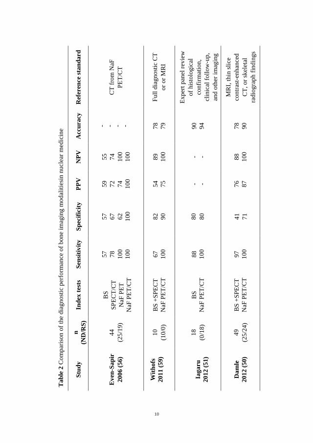
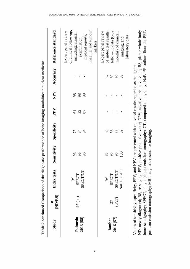
The diagnostic performance of BS versus SPECT and NaF PET (with or without CT) has been investigated in diagnostic test accuracy (DTA) studies on multiple occasions during the past decade. Table 2 shows the results of studies involving at least two of the imaging modalities of interest in this PhD study, i.e., BS, SPECT/CT and NaF PET/CT. The reference standards used in these studies are presented as well. Previous studies have included highly heterogeneous and mixed study populations, and the studies have had some methodological issues, but this will be addressed more thoroughly later.
The sensitivity of BS in previous studies has varied from as low as 57% to as high as 97% (50, 51, 56-59). For studies in which the reference standard was primarily based on other imaging, e.g., full-diagnostic CT or MRI, the sensitivity of BS was lowest (56, 59), while studies that used a combination of imaging, clinical follow-up, etc., found a higher sensitivity of BS (50, 51, 57, 58). The specificity of BS has been even more wide-ranging with reported values from 57-80% (50, 51, 56-59).
Independent of whether SPECT was used alone or in combination with CT, the sensitivity of SPECT ranged from 78-96% (56-58). When it came to the specificity of SPECT, the addition of a CT had a major impact on the results. The specificity for SPECT alone was in the range of 56-64% (57, 58), while the specificity for SPECT/CT ranged from 67-96% (56-58). The improved specificity is probably due to the attenuation correction and anatomical co-localisation gained from CT.
The sensitivity of NaF PET/CT studies has, in most studies, been 100% (50, 51, 56, 57, 59, 60). In a few studies in which NaF PET/CT was compared with 18F-choline PET/CT, the sensitivity of NaF PET/CT was 81% (61, 62). The specificity of NaF PET/CT has shown greater variation with reported specificities ranging from 71-100% (50, 51, 56, 57, 59-62).
When comparing the results for BS, SPECT/CT and NaF PET/CT, the sensitivity of NaF PET/CT was higher than those of SPECT/CT and BS across all studies (50, 51, 56-59). The specificity of NaF PET/CT was outperformed by that of SPECT/CT in the study by Jambor et al. (57). The sensitivity of SPECT/CT was higher than or equal to BS in all studies, and the specificity was higher (56-58). In summary, the diagnostic performance of these three imaging modalities can be ranked with NaF PET/CT demonstrating the best diagnostic performance, followed by SPECT/CT and BS. However, even though studies continue to demonstrate an apparent advantage of NaF PET/CT and SPECT/CT over BS, current guidelines have refrained from including SPECT/CT, NaF PET/CT, or other PET tracers as standard imaging options for the detection of bone metastases in PCa (5-7). The rationale behind this is probably that there is a lack of clear-cut evidence and varying degrees of methodological issues with these studies.

DIAGNOSIS AND MONITORING OF BONE METASTASES IN PROSTATE CANCER
9
1.5.5. CHALLENGES IN DIAGNOSTIC TEST ACCURACY STUDIES
Previous DTA studies within bone imaging in PCa have included highly heterogeneous and limited study populations with various cancer types and have included both newly diagnosed patients and those evaluated for re-staging purposes. In addition, the reference standard has often relied on clinical follow-up, which has not been clearly defined, and sometimes the reference standard was predominantly based on consensus evaluation of the index test, thus resulting in an artificially boosted performance due to circular reasoning (50, 51, 56-62). The methodological issues were summarised in a review by Wondergem et al., who also determined that the level of evidence in these DTA studies was quite low, Oxford Centre for Evidence-Based Medicine level 3b (53, 63).
The persistent presence of methodological issues within DTA studies has long been known, and some common issues include poorly described study populations and sampling procedures, and verification bias (64-67). A major issue is that for most diseases and conditions, it is practically impossible to obtain a single perfect “gold” reference standard, and in previous studies, the reference standard has varied and different solutions have been used to account for imperfect or missing values of the reference standard (68). Within bone imaging, the theoretical “gold” reference standard would be biopsies and histological verification of bone metastases, but this is neither practical nor ethically reasonable; in DTA studies with PCa and bone metastases consensus reviews or expert panels are commonly used (Table 2) (51, 57, 58). In recent years, the use of expert panels has increased, i.e., a consensus reading between a group of experts who determine the final diagnosis on the basis of all available relevant data for each patient (69). While not perfect, this might be one of the more ideal approaches, as it resembles clinical practice. Bertens et al. (69) investigated the use and reporting of expert panels in DTA studies and concluded that expert panels can be used in the absence of a single “gold standard”, and they encouraged the development of formal methodology guidelines.
10
Tab
le 2
Com
paris
on o
f the
dia
gnos
tic p
erfo
rman
ce o
f bon
e im
agin
g m
odal
ities
in n
ucle
ar m
edic
ine
Ref
eren
ce st
anda
rd
CT
from
NaF
PE
T/C
T
Full
diag
nost
ic C
T or
or M
RI
Expe
rt pa
nel r
evie
w
of h
isto
logi
cal
conf
irmat
ion,
cl
inic
al fo
llow
-up,
an
d ot
her i
mag
ing
MR
I, th
in sl
ice
cont
rast
-enh
ance
d C
T, o
r ske
leta
l ra
diog
raph
find
ings
Acc
urac
y
- - - - 78
79
90
94
78
90
NPV
55
74
100
100
89
100 - - 88
100
PPV
59
72
74
100
54
75
- - 76
87
Spec
ifici
ty
57
67
62
100
82
90
80
80
41
71
Sens
itivi
ty
57
78
100
100
67
100
88
100
97
100
Inde
x te
sts
BS
SPEC
T/C
T N
aF P
ET
NaF
PET
/CT
BS
+SPE
CT
NaF
PET
/CT
BS
NaF
PET
/CT
BS
+SPE
CT
NaF
PET
/CT
n (N
D/R
S)
44
(25/
19)
10
(10/
0)
18
(0/1
8)
49
(25/
24)
Stud
y
Eve
n-Sa
pir
2006
(56)
With
ofs
2011
(59)
Ia
garu
20
12 (5
1)
Dam
le
2012
(50)
DIAGNOSIS AND MONITORING OF BONE METASTASES IN PROSTATE CANCER
11
Tab
le 2
con
tinue
d C
ompa
rison
of t
he d
iagn
ostic
per
form
ance
of b
one
imag
ing
mod
aliti
esin
nuc
lear
med
icin
e Ref
eren
ce st
anda
rd
Expe
rt pa
nel r
evie
w
of c
linic
al fo
llow
-up,
in
clud
ing;
clin
ical
ex
amin
atio
n,
med
ical
repo
rts,
imag
ing,
and
tum
our
mar
kers
Expe
rt pa
nel r
evie
w
of i
ndex
test
resu
lts,
follo
w-u
p da
ta (6
-32
mon
ths)
of c
linic
al,
imag
ing,
and
la
bora
tory
dat
a
Val
ues o
f sen
sitiv
ity, s
peci
ficity
, PPV
, and
NPV
are
pre
sent
ed w
ith e
quiv
ocal
resu
lts re
gard
ed a
s mal
igna
nt.
ND
, new
ly d
iagn
osed
; RS,
re-
stag
ing;
PPV
, pos
itive
pre
dict
ive
valu
e; N
PV, n
egat
ive
pred
ictiv
e va
lue;
BS,
pla
nar
who
le-b
ody
bone
sci
ntig
raph
y; S
PEC
T, s
ingl
e-ph
oton
em
issi
on t
omog
raph
y; C
T, c
ompu
ted
tom
ogra
phy;
NaF
, 18F-
sodi
um f
luor
ide;
PET
, po
sitro
n em
issi
on to
mog
raph
y; M
RI,
mag
netic
reso
nanc
e im
agin
g.
Acc
urac
y
- - - 67
69
90
89
NPV
98
98
99
- - - -
PPV
61
52
87
- - - -
Spec
ifici
ty
75
64
94
59
56
88
82
Sens
itivi
ty
96
96
96
85
95
95
100
Inde
x te
sts
BS
SPEC
T SP
ECT/
CT
BS
SPEC
T SP
ECT/
CT
NaF
PET
/CT
n (N
D/R
S)
97 (-
/-)
27
(0/2
7)
Stud
y
Palm
edo
2013
(58)
Jam
bor
2016
(57)

12
Standards for Reporting of Diagnostic Accuracy In an attempt to improve the quality of reporting of all aspects of DTA studies, Bossuyt and colleagues (70) published the Standards for Reporting of Diagnostic Accuracy (STARD). The original STARD checklist contained 25 specific items to be included in the abstract, title, methods, results, and discussion that are needed for complete and accurate reporting in DTA studies. In 2015, Korevaar et al. (66) investigated the reporting of the items included in the STARD checklist and compared this to articles published before STARD and one and 10 years after STARD was first published. They found that reporting in DTA studies has improved since the initiation of STARD. However, there are still some shortcomings, especially regarding patient selection, details on the readers of index tests, and variations in the accuracy of the index test between subgroups of patients, centres, and/or readers. The STARD checklist was updated in 2015 with additional essential items to improve completeness and transparency in DTA studies (71). In summary, some attempts have been made to improve the design and reporting of DTA studies. While some progress has been seen in recent years, there is still room for improvement.
TREATMENT RESPONSE ASSESSMENT 1.6.1. RESPONSE ASSESSMENT METHODS
Disease monitoring is essential for optimal patient management, especially with the dramatic increase in the cost of cancer therapies that has occurred over the past years (72-74). Response assessment is standard when evaluating the efficacy of new therapeutic agents in cancer imaging. In PCa, it remains a challenge to determine response to therapy, as most patients with metastatic PCa have disease limited to bone, and it is well-known that assessment of treatment response in bone metastases is difficult (75).
RECIST and PERCIST The most commonly used set of response criteria in cancer imaging is the Response Evaluation Criteria in Solid Tumours (RECIST) (76), which classifies sclerotic bone lesions as non-measurable. These criteria are therefore of little use when determining response of bone metastases and are hence of limited use in PCa (77). Current guidelines recommend BS for monitoring bone metastases (6, 7, 11). The RECIST criteria were adapted for use in PET in 2009 as the Positron Emission Tomography Response Criteria in Solid Tumours criteria (PERCIST) (78). As with RECIST, PERCIST has not gained footing within PCa as a large proportion of PCa patients remain non-evaluable by PERCIST (79). These criteria are, due to their limited applicability in PCa, beyond the scope of this PhD thesis and will not be described further.

DIAGNOSIS AND MONITORING OF BONE METASTASES IN PROSTATE CANCER
13
MD Anderson criteria Because of the lack of criteria for assessment of tumour response in bone, Hamaoka et al. (80) proposed a set of visual assessment criteria for evaluation of bone metastatic response, known as the MD Anderson criteria. These criteria included similar response categories as those used by RECIST, i.e., complete response, partial response, stable disease or progressive disease, but instead of quantifying response they focused on acknowledging the presence of a response. Besides BS, the MD Anderson criteria included plain radiographs, CT and MRI, but these are beyond the scope of this PhD thesis. It has been shown that the MD Anderson criteria are able to distinguish responders (complete and partial response) from non-responders (stable and progressive disease) with regard to PFS in bone-only metastatic breast cancer patients receiving systemic treatment (23.3 vs 5.5 months, respectively, p = 0.025) (81). Improved OS was likewise demonstrated for responders vs non-responders (61.9 vs 34.4 months, not significant p=0.13) (81). No studies have investigated this relationship in PCa patients.
Prostate Cancer Working Group Criteria It has been a great challenge to determine the efficacy of new therapeutic agents in PCa studies without excluding patients with non-measurable disease. Therefore, the Prostate-Specific Antigen Working Group was established in an attempt to develop consensus criteria for response assessment in PCa studies (82); they were revised in 2008 with the Prostate Cancer Working Group (PCWG) 2 criteria (83), and in 2016 the PCWG-3 criteria were published (24). These response evaluation criteria addressed symptoms, biochemical response, response of measurable disease, and response of non-measurable bone lesions. Since their introduction, the PCWG criteria have been widely adopted in clinical trials for the assessment of treatment response in bone (30-32, 84, 85).
The focus of the PCWG criteria is to rule out or identify the presence of progression on BS. Unlike RECIST and the MD Anderson criteria, the PCWG criteria do not distinguish between stable disease and improvement (partial or complete response) and include only two response categories: 1) progressive disease (PD) and 2) non-progressive disease (non-PD).
Progressive disease according to the PCWG criteria was associated with OS in a large population of men with CRPC, where PD at three months was associated with a median OS of 9.2 months vs 17.8 months in patients showing non-PD (p < 0.0001) (86). At six months, median OS was 10.1 months for PD vs 19.3 months non-PD (p < 0.0001) (86).

14
1.6.2. TREATMENT RESPONSE ASSESSMENT IN BONE BY BS AND NAF PET/CT
Only very few studies have directly investigated the use of BS and other imaging modalities for bone response in PCa.
Bone scan response assessment Sonpavde and colleagues (87) collected data from two large prospective clinical trials and showed that radiographic progression according to the PCWG-2 criteria was associated with poorer OS in patients taken off study due to radiographic progression vs patients who were taken off study for other reasons.
In a large clinical trial comparing abiraterone acetate vs placebo rPFS, according to the PCWG-2 criteria, was positively correlated with OS (Spearman correlation coefficient (R2) = 0.72, 95% CI: 0.65-0.77) (88). The positive correlation was evident in both the treatment group and the placebo group. Furthermore, in the entire group of patients, OS was shorter for those who met the PCWG-2 criteria for confirmed PD on a subsequent scan compared with those who did not have confirmed PD (88).
In patients who had previously failed Docetaxel chemotherapy and were assigned to treatment with Radium-223, response on BS was mixed, showing the appearance of new lesions simultaneously with decreased tracer uptake in bone metastases that demonstrated a high pre-treatment uptake (89).
In recent years, studies have found a correlation between OS and bone response, defined as changes in the Bone Scan Index, a quantitative imaging biomarker developed for assessment of bone tumour load on BS (90-94). Kaboteh et al. (93) compared the correlation between OS and bone response by changes in the Bone Scan Index and response according to the PCWG-2 criteria. Although it was a small and retrospective study, they showed that changes in the Bone Scan Index correlated with OS, while progression by the PCWG-2 criteria did not.
In summary, the results of the above studies indicate that progression on BS, either according to the PCWG criteria or as changes in Bone Scan index, can be correlated with OS and BS is therefore useful in determining response to therapy.
Observer agreement for assessment of BS An important aspect of treatment monitoring is the consistency of response classification among observers. When analysed by Cohen’s kappa, inter-observer agreement for the diagnosis of bone metastases is reportedly moderate to almost perfect depending on the number of categories used for classification of the presence of bone metastases (95-97). Kaboteh et al. (93) assessed BS according to the PCWG-2 criteria and found an agreement of 87% (222/255) among three experienced readers for evaluation of PD vs non-PD. No kappa values were reported. In 173 PCa patients with bone metastases, evaluation of treatment response on BS showed substantial

DIAGNOSIS AND MONITORING OF BONE METASTASES IN PROSTATE CANCER
15
agreement by Cohen’s kappa when using the PCWG-2 criteria (Cohen’s kappa: 0.66) and standard clinical assessment (Cohen’s kappa: 0.70) (98). However, knowledge on observer agreement for assessment of treatment response on BS and has not been investigated at all for NaF PET/CT. A high level of agreement between observers is important as decisions to continue or discontinue treatments and investigational treatments are often based on the detection of progression on BS.
NaF PET/CT response assessment A few minor studies have investigated changes on NaF PET/CT.
Cook et al. (99) showed that changes on NaF PET/CT, as measured by changes in standardised uptake values (SUV), closely followed changes in PSA in five patients after receiving two doses (six weeks apart) of Radium-223.
In a mixed population of bone metastases-positive and -negative PCa patients who were undergoing a wide range of treatments, Apolo and colleagues investigated changes on NaF PET/CT at 6 and 12 months and correlation with clinical assessment of bone response and PSA (100). Clinical assessment of bone response was categorised as regression (regression of existing lesions), stable disease (no new lesions) or progression (new lesions). Changes in SUV correlated with clinical assessment of bone response at 6 and 12 months when analysed by the Kruskal-Wallis rank test (p = 0.0147 and p = 0.0053, repectively). Likewise, there was a correlation between change in PSA and maximal percent change in SUV at 6 and 12 months (R2 = 0.39, p = 0.014 and R2 = 0.58, p = 0.0005, respectively) (100).
Kairemo et al. (101) investigated changes on NaF PET/CT in 10 mCRPC patients receiving Radium-223. They summed the two highest SUV from two skeletal regions and found that after six cycles of Radium-223 all patients responded on NaF PET/CT with ≥ 6% change in SUV compared to baseline. This was accompanied by changes in PSA in some but not all patients.
A survey of the National Oncologic PET Registry in America showed that patient management was changed in up to 53% of PCa patients following a NaF PET/CT (16% when adjusted for patients who already had a pre-treatment plan involving imaging). However, this study did not investigate whether the change in management led to improved patient outcome (47).
Thus, previous studies on response assessment by NAF PET/CT are scarce, has included small study populations, have not compared the results with BS, and furthermore, no studies have investigated response on NaF PET/CT in relation to patient outcome.

16
RATIONALE FOR PHD STUDIES Although there is a growing body of evidence showing an apparent superiority of NaF PET/CT and SPECT/CT over BS, the methodological issues in previous DTA studies, as outlined in section 1.5.5., discouraged clinical guidelines from including these newer modalities as standard for the diagnosis of bone metastases in PCa. Therefore, properly designed studies are needed to establish the improved diagnostic performance of these modalities.
Monitoring of treatment responses in bone is essential for patient management in PCa, both in clinical trials and in clinical practice. Clinical guidelines recommend using BS for monitoring of bone metastases in PCa and the PCWG advocate the use of BS for determination of bone progression in clinical trials. It is essential for patient management that there is a high level of agreement for response classification between readers. Different methods for response classification have been presented; however, observer agreement has not been adequately investigated. Delineation of such agreement is necessary to establish the best method for future use in treatment response monitoring in bone.
Finally, the use of NaF PET/CT in PCa is increasing both for diagnostic purposes as well as for treatment response monitoring. No studies have investigated the use of NaF PET/CT for treatment response assessment in comparison with conventional BS, for which response classification is correlated with OS. Therefore, studies are needed to investigate the concordance between BS and NaF PET/CT for treatment response assessment and how they might differ.

DIAGNOSIS AND MONITORING OF BONE METASTASES IN PROSTATE CANCER
17
CHAPTER 2. AIMS
The overall aim of this PhD thesis was to evaluate BS, SPECT/CT and NaF PET/CT for diagnosis and monitoring of bone metastases in PCa. This was done in three sub-studies, for which the specific aims are listed below.
Study I (Papers 1 & 2) To compare the diagnostic performances of BS, SPECT/CT and NaF PET/CT in newly diagnosed high-risk PCa patients in a fully STARD-compliant DTA study (102).
Study II (Paper 3) To evaluate observer agreement in the assessment of treatment responses in bone on BS in PCa patients undergoing different anti-cancer treatments at varying stages of PCa, using three different methods for treatment response evaluation (103).
Study III (Paper 4) To prospectively explore the concordance between BS and NaF PET/CT when evaluating bone metastases response in PCa patients receiving various anti-cancer treatments and to explore the relationships between imaging, clinical, and biochemical responses.

DIAGNOSIS AND MONITORING OF BONE METASTASES IN PROSTATE CANCER
19
CHAPTER 3. MATERIALS AND METHODS
STUDY POPULATION Study I From February 2014 to December 2015, consecutive patients with newly diagnosed high-risk PCa from the Departments of Urology at Aalborg University Hospital, Regional Hospital West Jutland Holstebro, and Regional Hospital Viborg were invited to participate in this study. Eligibility criteria consisted of the following: 1) biopsy-proven PCa; 2) PSA blood levels ≥ 50 ng/mL; 3) eligible for ADT; 4) no other cancer within five years; 5) ability to comply with study procedures; and 6) have not received any investigational drugs.
The Regional Research Ethics committee (N-20130068) and the Danish Data Protection Agency approved the study. Written informed consent was obtained from 52 patients. Of these, 10 patients either withdrew consent or were excluded for incompatibility reasons. Furthermore, three patients died before completion of the study, resulting in 39 patients remaining. Two of these were excluded from the final analysis because a final diagnosis could not be determined.
Study II In this retrospective study, all patients who, during the period from January 2009 to November 2014, had undergone two or more bone scans within one year at the Department of Nuclear Medicine, Aalborg University Hospital, were identified from the hospital records (103). Patients were then selected according to the following criteria: 1) patients were treated with either ADT, NGH, or chemotherapy; and 2) patients had two BS performed within the same treatment, a baseline BS and a follow-up BS. The baseline BS was performed within three months before initiation of therapy or a maximum 14 days within treatment. The follow-up BS was performed 12-52 weeks within treatment for patients receiving ADT and 12-30 weeks within treatment for patients receiving NGH or chemotherapy. Patients treated successively with different treatments and with more than one BS pair fulfilling the above criteria were allowed to enter the study twice (103).
A total of 105 patients were identified from the hospital records. Of these, 55 patients with 63 evaluable BS pairs fulfilled the above criteria and were included in the final analyses (103).
Study III Patients from Study I who had completed all study-related procedures were included in Study III along with consecutive patients with confirmed bone metastases (by
20
clinical evaluation) scheduled to receive either primary ADT, NGH, or chemotherapy. The latter patients were included in a separate protocol approved by the Regional Research Ethics committee (N-20140057) and the Danish Data Protection Agency. Eligibility criteria for this protocol were as follows: 1) histologically confirmed PCa; 2) bone metastases on BS at inclusion; 3) clinical life expectancy > six months; 4) no other cancer within five years; and 5) ability to comply with study procedures.
A total of 64 patients were included in the final analysis in this study, 23 of which originated from Study I.
METHODS 3.2.1. SCANS
All scans were performed according to current institutional guidelines, which are consistent with European and International guidelines (41-43, 46). Scans were performed at Aalborg University Hospital or at Regional Hospital West Jutland Herning.
3.2.1.1 Planar Whole-Body Bone Scan
Aalborg Bone scans were performed on Symbia dual-head gamma cameras with multi-purpose, low-energy, high-resolution collimators (Symbia T16, Siemens Medical Solutions, Erlangen, Germany). Data acquisition was performed two to three hours after intravenous administration of 750-1000 MBq 99mTc-labelled diphosphonate, with a scan speed of 24 cm/min. The use of an alpha blending technique allowed for an apparent increase in counts by a factor of two, thus accounting for the faster acquisition time in Aalborg.
Herning Bone scans were performed on Symbia dual-head gamma cameras with multi-purpose, low-energy, high-resolution collimators (Symbia T2 and T16, Siemens Medical Solutions, Erlangen, Germany). Data acquisition was performed two to three hours after intravenous administration of 10 MBq/kg 99mTc-labelled diphosphonate, with a scan speed of 10 cm/min.
3.2.1.2 SPECT/CT
Aalborg A three-bed SPECT/CT scan, from vertex to mid-thigh, was performed immediately following the planar imaging using the following parameters: matrix 128 x 128, zoom factor 1, 20 s per view, 32 views, and rotation of the detectors by 180 degrees in a non-circular orbit using step-and-shoot mode. A low-dose CT without intravenous contrast was acquired and used for attenuation correction and anatomical co-registration: 30 mA, 130 kV, slice thickness 3 mm.
DIAGNOSIS AND MONITORING OF BONE METASTASES IN PROSTATE CANCER
21
Herning A three-bed SPECT/CT scan, from vertex to mid-thigh, was performed immediately following the planar imaging using the following parameters: matrix 128 x 128, zoom factor 1, 10 s per view, 64 views, and rotation of the detectors by 180 degrees in a non-circular orbit in a continuous mode. The low-dose CT had the following parameters: reference mA 100 (CARE dose), 130 kV, and a slice thickness of 5 mm.
3.2.1.3 NaF PET/CT
Aalborg Patients were scanned on a dedicated VCT discovery True 64 PET/CT, (GE Healthcare). Scans were performed after intravenous administration of 200 MBq NaF in 3D mode from vertex to mid-thigh, encompassing 7-9 bed position (150 s per bed position). The images were reconstructed by iterative construction, using low-dose CT images for attenuation correction and anatomical co-localisation. The CT parameters were 70-200 mA smart mA, 120 kV. The slice thickness was 0.625 mm.
Herning Patients were scanned on a dedicated Biograph mCT 64, 4R PET/CT (Siemens Medical Solutions). All scans were performed 30 minutes after intravenous administration of approximately 200 MBq NaF in 3D mode from vertex to mid-thigh, encompassing 7-9 bed positions (120-180 s per bed position according to body mass index). Images were reconstructed as in Aalborg, using low-dose CT images for attenuation correction and anatomical co-localisation. The CT parameters were 30 mAs, 120 kV. The slice thickness was 0.625 mm.
3.2.2. IMAGING SCHEDULE
Study I Patients were scanned at baseline and after 26 weeks of therapy. Patients were routinely referred for BS for staging purposes. As an add-on, the three-bed SPECT/CT was performed immediately after the BS. NaF PET/CT was then performed. All scans had to be performed before or a maximum of 14 days within treatment. After 26±4 weeks, all three scans were repeated, and these data were used to assist in the determination of a final diagnosis (102). Figure 2 shows the scan times and the number of patients scanned at each time-point.

22
Figure 2 An overview of scan times in Study I, and the number of scans available for evaluation at each time-point. BS, bone scan; SPECT/CT, single-photon emission computed tomography / computed tomography; NaF PET/CT, 18F-sodium fluoride positron emission tomography /CT.
3.2.2.1 Study II
See Study Population, section 3.1, Study II (103).
Study III Before initiation of either ADT, NGH, or chemotherapy, patients were routinely referred for BS for assessment of bone metastases status. Additionally, a NaF PET/CT scan was performed no later than 14 days within treatment. These constituted the baseline scans. Including the baseline scans, imaging with BS and NaF PET/CT was performed 2-4 times within a 26-week time period for patients receiving ADT or NGH and 2-3 times within a 20-week time period for patients receiving chemotherapy. Figure 3 shows the scan times and the number of patients scanned at each time-point by treatment.
Figure 3 An overview of scan times for Study III, and the number of evaluable scans at each time point according to treatment. This includes patients who had both a bone scan and a 18F-sodium fluoride positron emission tomography/computed tomography at each time point.

DIAGNOSIS AND MONITORING OF BONE METASTASES IN PROSTATE CANCER
23
3.2.3. IMAGE ANALYSIS
For all three studies, specific forms and guides for completion of these were designed. These are presented in Appendices A-C.
Study I Expert readings Two nuclear medicine specialists with more than 10 years of experience with BS evaluated all BS images. Likewise, two nuclear medicine specialists with 5 and 10 years of experience with NaF PET/CT evaluated all NaF PET/CT images. Readers were blinded to any clinical and biochemical information regarding the patients, except for the diagnosis of high-risk PCa. Readers were first asked to rate the images on a three-point scale of benign (M0), equivocal (Me), or malignant (M1) and subsequently on a dichotomous scale of M0 or M1. The readers were then asked to indicate if the patient had more than 10 suspicious bone lesions or a scan compatible with superscan. Finally, the readers were asked to draw lesions on a schematic drawing of the skeleton if the patient had 10 or fewer suspicious bone lesions. The readers used the form included in Appendix A-1 and were guided by A-2. In cases of disagreement, the readers performed a consensus reading.
Reference standard To determine the final diagnosis, a multidisciplinary committee consisting of experienced specialists, a urologist, a radiologist and a nuclear medicine physician reviewed all relevant and available information about each patient. The reference standard was thus a compilation of the following:
1) Results of the expert readings 2) All available baseline and follow-up imaging 3) All available biochemical information about the patient 4) A standard questionnaire filled out routinely with each BS including
information about any bone-related disease or trauma, joint replacements, and any unexplained bone pain.
5) If necessary, any existing routine imaging was also made available if a final diagnosis could not be determined based on points 1-4.
The multidisciplinary committee used the form included in Appendix A-3.
Study II Five experienced nuclear medicine physicians participated in the evaluation of images in Study II (103). Two readers evaluated each baseline and follow-up image individually (Appendix B-1). Images were evaluated for the presence of bone metastases (M0/M1) and the number of suspicious lesions within five skeletal regions (skull, thorax incl. sternum, columna, pelvis, and extremities incl. scapula). Readers were asked to count up to 20 lesions within each region (103), and these counts were subsequently reclassified according to the extent of disease classification by

24
Soloway et al. (104). Each pair of baseline and follow-up images was then evaluated side-by-side for response using three different methods for response evaluation; readers classified patients according to specific response categories as shown in Table 3 and Appendix B-2, B-3, B-4 (103).
The image pairs were evaluated three times by three different methods. Therefore, to account for the bias that the readers could remember an image pair, the image pairs were evaluated by two different readers for each response evaluation method. For instance, for Patient 1, readers 1 and 2 evaluated the images by Standard Clinical Assessment, readers 3 and 4 evaluated them by MD Anderson criteria, and readers 5 and 1 evaluated them by the PCWG-2 criteria (103).
Table 3 Response assessment methods and categories
Response criteria Response categories
Standard clinical assessment
Regressive disease -Regression of lesions/disappearance of hot spots Stable disease -No change Progressive disease -Progression of lesions/new lesions
MD Anderson (80) Complete response -Disappearance of hot spots or tumour signal Partial response -Regression of lesions Stable disease -No new lesions Progressive disease -New lesions or increased activity
Prostate Cancer Working Group 2 (83)
Non-progressive disease -No or maximum one new lesion Progressive disease -Two or more new lesions, confirmed on subsequent scan (if one existed)

DIAGNOSIS AND MONITORING OF BONE METASTASES IN PROSTATE CANCER
25
Study III The same nuclear medicine specialists who performed the expert readings in Study I evaluated all BS and NaF PET/CT images in Study III. Readers received careful instructions on how to evaluate images (Appendix C-1).
As in Study II, readers first determined the presence of bone metastases (M0/M1) and the number of bone metastases in the following intervals: 0, 1, 2-4, 5-9, 10-20, or >20 (modified from Soloway et al (104)) (Appendix C-2). The readers were then asked to evaluate images for response using modified PCWG-2 criteria (Table 3). The baseline scan was evaluated side-by-side with the first follow-up scan (Appendix C-3). In the case of PD, the second follow-up scan (if one existed) was used to confirm the presence of PD (Appendix C-4). In the case of non-PD, the baseline and the second follow-up scan (if one existed) were evaluated side-by-side (Appendix C-5), and then the third follow-up scan (if present) was used to confirm any PD. Additionally, readers were asked to rate the images according to Standard Clinical Assessment as described in Table 3 and Appendix C-1 to C-5.
3.2.4. BIOCHEMICAL MEASUREMENTS (STUDIES I AND III)
Prostate specific antigen In Study I, blood levels of PSA were measured at baseline and at each subsequent set of follow-up scans. The same applied for patients receiving ADT or NGH in Study III. For patients receiving chemotherapy in Study III, PSA was measured at several varying time points during the study according to clinical practice.
Changes in PSA were classified according to the PCWG criteria as non-PD or PD. Non-progressive disease included PSA response (a decrease in PSA of > 50%) and stable disease (decrease in PSA of ≤ 50% or increase < 25%). Progressive disease included drifters (initial PSA response followed by progression, with a ≥ 25% increase in PSA from nadir) and progressive disease (an increase in PSA of ≥ 25% from nadir).
Testosterone In Study I, testosterone levels were measured at baseline and at each subsequent set of follow-up scans. This was done to ensure that levels fell below castration levels during treatment, thus confirming that the treatment was effective in performing a medical castration, even in cases of progressing PSA levels.
In Study III, the same as above applied for patients receiving ADT. For patients receiving NGH, the testosterone levels were measured to ensure that patients were correctly classified as having CRPC. As it had no clinical significance, testosterone levels were not measured in patients receiving chemotherapy.

26
3.2.5. CLINICAL RESPONSE ASSESSMENT (STUDY III)
Assessment of clinical response to therapy was performed retrospectively based on patient charts. The responsible urologist reviewed charts for all patients receiving ADT or NGH and the responsible oncologist reviewed charts for patients receiving chemotherapy. Clinical response was based on changes in performance status, pain and other cancer-related symptoms. Response was assessed on a three-point scale: regression, stable disease, or progressive disease.
3.2.6. STATISTICS
Data are presented only on a patient level, and no lesion-based analyses were performed for this PhD thesis. Statistical analysis was performed using StataIC version 13 (StataCorp LCC, TX, USA) and Microsoft Office Excel 2013.
Study I Sample size was based on recommendations for sample size calculations in DTA studies (102, 105). Sensitivity, specificity, and positive and negative predictive values were calculated for all three imaging modalities, BS, SPECT/CT, and NaF PET/CT, and presented with 95% confidence intervals (CI). The diagnostic performances were compared using McNemar’s test.
Study II Agreement between assessments of the presence of bone metastases and for response evaluations was calculated using Cohen’s kappa (106). The terminology by Landis and Koch was used to evaluate the extent of agreement according to kappa values, where 0.00-0.20: slight; 0.21-0.40: fair; 0.41 – 0.60: moderate; 0.61 – 0.80: substantial; and 0.81 – 1.00: almost perfect agreement (107). These are presented with 95% CIs as well. Bland-Altman plots (108) were constructed to assess the difference between readers when counting the number of bone lesions on BS within five skeletal regions (103).
Study III The proportions of PD vs non-PD for BS and NaF PET/CT, biochemical and clinical response assessments were compared using the McNemar test with p < 0.05 considered statistically significant. Data are presented with 95% CIs. Agreement between BS and NaF PET/CT for response evaluation was evaluated by Cohen’s kappa for the total study population. Crude agreement was presented for the individual treatment groups.

DIAGNOSIS AND MONITORING OF BONE METASTASES IN PROSTATE CANCER
27
CHAPTER 4. RESULTS
A brief summary of the main results of each paper is presented below. Detailed description of the results are found in the individual papers.
PAPER 1 18F-fluoride positron emission tomography/computed tomography and bone scintigraphy for diagnosis of bone metastases in newly diagnosed, high-risk prostate cancer patients: study protocol for a multicentre, diagnostic test accuracy study.
Randi F. Fonager, Helle D. Zacho, Niels C. Langkilde and Lars J. Petersen
BMC Cancer (2016) 16:10. DOI 10.1186/s12885-016-2047-1 (102)
Summary Study I was a fully STARD-compliant DTA study. Paper 1 presents the study protocol, which includes detailed descriptions of the study design, the study population and rationale for selection of patients. Furthermore, it includes descriptions of imaging procedures and a detailed plan for image analysis.


DIAGNOSIS AND MONITORING OF BONE METASTASES IN PROSTATE CANCER
29
PAPER 2 Diagnostic test accuracy study of 18F-sodium fluoride PET/CT, 99mTc-labelled diphosphonate SPECT/CT, and planar bone scintigraphy for diagnosis of bone metastases in newly diagnosed, high risk prostate cancer.
Randi F. Fonager, Helle D. Zacho, Niels C. Langkilde, Joan Fledelius, June A. Ejlersen, Christian Haarmark, Helle W. Hendel, Mine Benedicte Lange, Mads R. Jochumsen, Jesper C. Mortensen, and Lars J. Petersen.
Manuscript under revision.
Summary In a completely STARD-compliant DTA study, the diagnostic performance of NaF PET/CT, SPECT/CT and BS was compared in 37 newly diagnosed high-risk PCa patients (PSA ≥ 50 ng/mL). The prevalence of bone metastases in this study was 73%. More than 10 bone metastases were found by at least one imaging modality in 74% of patients. NaF PET/CT and SPECT/CT numerically outperformed BS with regard to sensitivity, specificity, PPV and NPV. The sensitivities were 78% for BS, 89% for SPECT/CT, and 89% for NaF PET/CT, and the specificities were 90%, 100%, and 90%, respectively. The PPVs for BS, SPECT/CT and NaF PET/CT were 96%, 100%, and 96%, and the NPVs were 60%, 77% and 75%, respectively. No statistically significant difference between the three imaging modalities was observed. In addition to the dichotomous classification of images, the images were evaluated with an equivocal rating option. The proportion of equivocal results was low for all three imaging modalities but lowest for NaF PET/CT (5% vs 11% for SPECT/CT and BS). In conclusion, all three imaging modalities showed high sensitivity and specificity. NaF PET/CT and SPECT/CT showed numerically improved, but not statistically superior, sensitivity compared with BS in this limited and selected patient cohort. In conclusion, SPECT/CT and NaF PET/CT can be used for the diagnosis of bone metastases on an equal level as BS.


DIAGNOSIS AND MONITORING OF BONE METASTASES IN PROSTATE CANCER
31
PAPER 3 Observer agreement of treatment responses on planar bone scintigraphy in prostate cancer patients: importance of the lesion assessment method.
Randi F. Fonager, Helle D. Zacho, Signe Albertsen, Joan Fledelius, June A. Ejlersen, Mette H. Christensen, Ramune Aleksyniene, José A. Biurrun Manresa, and Lars J. Petersen.
Nucl Med Commun. 2017 Mar;38(3):215-221. (103) DOI: 10.1097/MNM.0000000000000643.
Summary The observer agreement for treatment response assessment on BS was investigated in 63 paired BS from 55 patients. Treatment response was assessed by three different methods: 1) Standard clinical assessment; 2) MD Anderson criteria for bone response; and 3) PCWG-2 criteria. Furthermore, observer agreement for the presence of bone metastases and number of bone metastases at baseline was investigated. This study showed that the observer agreement for treatment response assessment was highest when using the PCWG-2 criteria, which showed substantial to almost perfect agreement (Cohen’s kappa 0.84, 95% CI: 0.69–0.99). When using standard clinical assessment, observer agreement was moderate (Cohen’s kappa 0.52, 95% CI: 0.36-0.69). Moderate agreement was also found for the MD Anderson criteria (Cohen’s kappa 0.60, 95% CI: 0.44–0.77). Evaluation of baseline images for the presence of bone metastases showed almost perfect agreement (Cohen’s kappa 0.94, 95% CI: 0.82-1.00). There was a large variation for lesion counting at the patient level. The difference in lesion count from one reader to another could be as high as ±15 lesions depending on the skeletal region. This variation seemed to increase with an increasing number of lesions. In conclusion, response evaluation by individual counting of lesions on baseline and follow-up images cannot be recommended. The use of strict criteria, such as the PCWG-2 criteria, for assessment of treatment response evaluation by BS is therefore recommended (103).


DIAGNOSIS AND MONITORING OF BONE METASTASES IN PROSTATE CANCER
33
PAPER 4 Prospective, comparative study of 18F-sodium fluoride PET/CT and planar bone scintigraphy for treatment response in prostate cancer.
Randi F. Fonager, Helle D. Zacho, Niels C. Langkilde, Joan Fledelius, June A. Ejlersen, Helle W. Hendel, Christian Haarmark, Mette Moe, Jesper C. Mortensen, Mads R. Jochumsen and Lars J. Petersen
Manuscript in preparation
Summary In this study, the agreement between NaF PET/CT and BS for treatment response assessment by the PCWG criteria was investigated in PCa patients undergoing various palliative anti-cancer treatments at different stages of PCa. Furthermore, the concordance between imaging (NaF PET/CT and BS), biochemical (PSA) and clinical response was evaluated. There was no statistically significant difference in the proportion of patients showing PD on BS vs NaF PET/CT (p=0.18). Analysis by Cohen’s kappa showed moderate agreement between BS and NaF PET/CT for classification of PD vs non-PD (Cohen’s kappa: 0.53). Crude agreement between NaF PET/CT and BS for assessment of treatment response in bone from baseline to last follow-up scan was 86%. There was a trend that with more advancing PCa, crude agreement decreased, i.e., crude agreement was 89% for ADT, 88% for NGH, and 80% for chemotherapy. When considering all intermediate scans and hence the time point for detection of PD, crude agreement decreased even further for NGH and chemotherapy: 75% and 70%, respectively. Most often, BS detected PD when NaF PET/CT did not, or BS detected PD on an earlier scan compared with NaF PET/CT. There were no statistically significant differences between the proportions of patients showing PD on BS and NaF PET/CT compared with PSA and clinical progression. Further studies are needed to investigate the use of NaF PET/CT for response monitoring of bone metastases in PCa and validation of NaF PET/CT for response assessment is necessary before this modality can be recommend for this purpose.


DIAGNOSIS AND MONITORING OF BONE METASTASES IN PROSTATE CANCER
35
CHAPTER 5. DISCUSSION
The overall aim of this PhD thesis was to evaluate BS, SPECT/CT and NaF PET/CT for diagnosis and monitoring of bone metastases in PCa. More specifically, the aim was to compare BS, SPECT/CT and NaF PET/CT for diagnosis of bone metastases in PCa (Study I), to evaluate observer agreement for evaluation of treatment responses on by three different methods for classification of response (Study II), and to compare BS and NaF PET/CT for monitoring of bone metastases in PCa (Study III).
Diagnostic performance Diagnosis of bone metastases is crucial for PCa patient management. It is therefore extremely important that diagnostic tests have a sufficiently high sensitivity to detect even small bone metastases and hence a low number of false negative results. Similar to previous DTA studies (Table 2) (31, 32, 37-40), the results of Study I showed a high sensitivity for all three imaging modalities. On a patient level, the sensitivity of SPECT/CT and NaF PET/CT numerically outperformed the sensitivity of BS (89% and 89% vs 78%, respectively). This was expected due to the inherent improved image quality of the latter imaging modalities, i.e., three-dimensional images, CT for attenuation correction and anatomical co-localisation, the improved spatial resolution of PET/CT, and the higher target-to-background ratio gained with NaF PET/CT.
The specificity of a diagnostic test is likewise of great importance. The specificity reflects the proportion of patients who are correctly identified as not having the disease, i.e., a high specificity reflects a low number of false positive results. The results of Study I showed that the specificity of NaF PET/CT was equal to that of BS (90%). This was in contrast to other studies, which have mostly demonstrated improved specificities of NaF PET/CT over BS (Table 2) (50, 51, 56-59). An even higher specificity was demonstrated by SPECT/CT (100%). The high specificity of BS in Study I, compared with previous studies, can in part be ascribed to the low number of patients without bone metastases in this study. However, it can also be explained by the experience of the expert readers who had more than 10 years of experience with BS reading starting before SPECT/CT became available as an add-on to BS. Therefore, these readers are likely to provide confident and unequivocal answers to BS.
The rationale for conducting this DTA study was the lack of properly designed large studies that adequately demonstrate the diagnostic performance of SPECT/CT and NaF PET/CT. Such evidence is needed for clinical guidelines to include these imaging modalities as options for diagnosis of bone metastases in PCa. Even if Study I did not achieve the goal of 114 evaluable patients, this study was large among comparable studies, and it was conducted with stringent procedures in accordance with the STARD guideline for DTA studies. The major challenge with DTA studies in imaging has been the lack of a proper reference standard. It must be realised, though, that

36
without histological verification of all potential bone metastases, a single, unambiguous gold standard is utopian (68, 69). Thus, a reference standard based on the findings on the index test as a guide for determining the final diagnosis and a well-documented stringent procedure, as in Study I, can be considered sufficient to determine the final diagnosis.
The diagnostic performance of BS, SPECT/CT and NaF PET/CT would most likely be different in an unselected population of newly diagnosed PCa patients or patients with recurrent disease following radical prostatectomy. However, a metastasis-enriched population was selected in Study I to optimize the sample size and statistical power (102). Study I was planned to include a high number of patients, which in combination with a methodologically sound design should have been able to provide firm evidence of the equal or superior diagnostic performances of SPECT/CT and NaF PET/CT for the diagnosis of bone metastases. Study I was prematurely ceased due to the realisation that recruitment of 114 patients was unlikely to be achieved within a realistic timeframe. Although Study I was large among similar studies, the power was limited due to the small number of patients in the study (50, 51, 56-58, 60, 91). The impaired recruitment in Study I could in part be ascribed to the decrease in the proportion of newly diagnosed patients with high-risk PCa that has occurred during the past few decades (3, 109, 110). Furthermore, it is known that high-risk patients are likely to show symptoms of morbidity (10, 11, 111), which may make patients reluctant to participate in studies that require additional procedures.
SPECT/CT is generally associated with a long acquisition time, thus causing additional discomfort to the patient. Recently, it was demonstrated that when using SPECT/CT for assessment of inconclusive bone lesions on BS the acquisition time of SPECT/CT can be reduced to as little as three minutes without compromising the diagnostic confidence of the examination (112). Thus, the discomfort of add-on SPECT/CT is notably reduced. This supports the use of SPECT/CT as an add-on to BS to gain an increased specificity of the examination. NaF PET/CT might be more comfortable for patients because the waiting time from injection of the tracer to the scan is shorter compared with BS (30-60 min vs 2-4 h for BS) and additionally NaF PET/CT has a faster acquisition time.
As mentioned above, Study I included the results of the index tests in the determination of a final diagnosis, thus causing a slight risk of bias with circular reasoning in the final decision making. However, as mentioned, it can be argued that without the results of the index test as a guide for final decision making, a firm reference standard is difficult to obtain in imaging studies, even if guidelines for DTA studies, such as STARD, recommend otherwise (71). Aside from this, a major strength of Study I was the strict methodology, which was in complete compliance with the STARD recommendations. Therefore, even in the absence of a single, large DTA study with a high level of evidence that shows the improved diagnostic performance of SPECT/CT and NaF PET/CT over BS, it can be argued that the existing body of

DIAGNOSIS AND MONITORING OF BONE METASTASES IN PROSTATE CANCER
37
evidence and the results of Study I provide sufficient evidence of this. SPECT/CT and NaF PET/CT can be considered as equal to BS for diagnostic purposes in PCa, and clinical guidelines should embed these imaging modalities for bone metastases staging in PCa with equal recommendations as those for BS.
Treatment response assessment Assessment of treatment responses in bone is, as previously mentioned, an important aspect of patient management both in daily clinical practice and in clinical trials. Just as for the diagnosis of bone metastases, clinical guidelines and the PCWG recommend the use of BS for treatment response assessment in PCa. The methods for classifying responses have varied, and different response assessment methods have been presented. In addition, a high level of agreement between readers for response classification is extremely important, since progression on BS often is a determining factor for ceasing or continuing a treatment or investigational drug. However, observer agreement has not been investigated in the assessment of treatment responses in bone metastases from PCa.
The results of Study II showed that inter-observer agreement according to Cohen’s kappa for bone response assessment was substantial/almost perfect when using the PCWG-2 criteria (Cohen’s kappa: 0.84). Agreement was moderate when using standard clinical assessment, as per daily clinical routine (Cohen’s kappa: 0.52), or the MD Anderson criteria (Cohen’s kappa: 0.60) (103). Standard clinical assessment and the MD Anderson criteria both have more response categories than PCWG-2, which by definition is associated with lower kappa values. However, agreement did not change when reclassifying these response classifications into non-PD vs PD. Thus, it is obvious that agreement for classification of response to treatment benefitted from the application of stringent criteria like the PCWG-2 (103). The results of Study II were consistent with the few existing studies on observer agreement for treatment response assessment in bone (93, 98).
There is no general consensus on how to report treatment responses in clinical practice, but the results of Study II suggest that a uniform approach using stringent criteria will be beneficial for patient management by providing the most consistent results. Therefore, strict criteria should be used both in clinical trials and in clinical practice for treatment response assessment of bone metastases in PCa (103). The study population in Study II was mixed and included patients undergoing different anti-cancer treatments and the small study population did not allow for subgroup analysis of variations in inter-observer agreement depending on disease state or treatment (103). This could have been an interesting perspective, as it has the potential to influence the results of future studies and recommendations for the use of response classification methods. Nonetheless, the results of Study II did demonstrate a substantial to almost perfect inter-observer agreement when using the PCWG criteria, and the PCWG criteria are becoming widely accepted for the evaluation of bone metastases treatment response in clinical trials (103, 113). This is well supported by

38
the fact that progression on BS according to the PCWG-2 criteria is associated with impaired survival (87). Overall, this suggests, that the PCWG criteria might be the ideal choice for future application in clinical trials and daily clinical practice.
The results from Study II on observer agreement clearly demonstrated that the PCWG criteria provided the most consistent results for evaluation of treatment response on BS (103). Based on these results, it was decided that the PCWG criteria should be used in Study III and in an exploratory setting, Study III prospectively compared the concordance between BS and NaF PET/CT for treatment response assessment in PCa. As mentioned, NaF PET/CT is not currently included in the clinical guidelines for the diagnosis or monitoring of bone metastases. The 2010 guideline from the Society of Nuclear Medicine stated that the role of NaF PET/CT is to be determined (46), while the 2015 European Association of Nuclear Medicine procedure guideline for NaF PET/CT recommended the use of NaF PET/CT for both diagnoses of bone metastases and for evaluation of treatment responses in bone metastases (43). However, no studies have directly compared the use of BS and NaF PET/CT for treatment response assessment in bone metastases.
The results of Study III showed that there are some non-statistically significant variations in response classification between BS and NaF PET/CT and in the timing of progression occurrence. Agreement between BS and NaF PET/CT was moderate for the classification of PD/non-PD from baseline to last follow-up scan when using the PCWG criteria (Cohen’s kappa 0.53). In most cases where BS and NaF PET/CT showed discordance, BS detected PD when NaF PET/CT did not, or BS detected PD at an earlier time-point than NaF PET/CT, e.g., BS showed PD on the first follow-up scan while NaF PET/CT did not detect PD until the second follow-up scan. There was a trend that crude agreement declined with more advanced stages of PCa, i.e., CRPC for which the PCWG criteria were developed. This trend was even more evident when all individual scans were taken into consideration i.e., first, second and sometimes third follow-up scan. It is commonly known that NaF PET/CT detects a higher number of bone metastases than BS (56, 59). The discordance between BS and NaF PET/CT might therefore, in some cases be due to the appearance of new lesions on follow-up BS that were already visible on baseline NaF PET/CT, thus accounting for the classification of PD on BS but not on NaF PET/CT.
When using standard clinical assessment to determine bone response, overall crude agreement between BS and NaF PET/CT from baseline to last follow-up scan was lower than when using the PCWG criteria, this was expected since standard clinical assessment has more response categories than the PCWG criteria (Table 3). Like for the PCWG criteria, standard clinical assessment also demonstrated decreased agreement with advancing PCa stages.
Previous studies that have investigated treatment response on NaF PET/CT have focused on post-treatment changes in SUV inspired by the PERCIST criteria (99-101).

DIAGNOSIS AND MONITORING OF BONE METASTASES IN PROSTATE CANCER
39
However, there is no universally agreed-upon reference level for changes in SUV that is considered significant, and no studies have investigated the impact of SUV changes on patient management. Other methods for quantification of NaF PET/CT have recently been presented (114, 115). Quantitative methods for evaluation of treatment response on NaF PET/CT are interesting, especially if small and clinically significant changes can be detected early during treatment. However, quantitative evaluation of NaF PET/CT remains under investigation and this was beyond the scope of this PhD thesis.
Study III was limited by the small number of patients, and this precluded formal statistical subgroup analysis of the individual treatment groups. Sub-group analyses would have been interesting in light of the trend that crude agreement between BS and NaF PET/CT decreased with advancing PCa stages, i.e., CRPC patients, especially those receiving chemotherapy. Larger studies are encouraged to show variations for different anti-cancer treatments as this might be critical for the validation of NaF PET/CT for bone response assessment, especially when using the PCWG criteria.
Clinical guidelines (5, 6) and PCWG-3 (24) emphasize that NaF PET/CT should be validated for bone response assessment before it can be recommended along with BS. This is supported by the results of Study III, which showed that there were some variations in the classification of bone response between BS and NaF PET/CT. The results of Study II demonstrated a high level of agreement between readers for evaluation of bone response on BS by the PCWG criteria (103) and it has previously been demonstrated that PD on BS, when assessed by the PCWG criteria, is associated with impaired OS (87). Therefore, recent recommendations of BS for bone response evaluation in PCa by PCWG-3 (24) and clinical guidelines (5, 6) are well substantiated. Combined, this indicates a need for further investigation of bone response assessment by NaF PET/CT. This is relevant both when assessing images on a visual basis, like in Study III, as well as when using quantitative methods for assessment of changes, such as SUV. In addition, even though a change in management was reported in up to 53% of PCa patients following NaF PET/CT (47) , a proper investigation is necessary to determine whether bone response assessment by NaF PET/CT is associated with improved patient outcome.
In conclusion, larger studies on NaF PET/CT for response assessment with a focus on patient outcome are encouraged, and until the results of NaF PET/CT have been validated, BS should be retained as the primary modality for bone response assessment.


DIAGNOSIS AND MONITORING OF BONE METASTASES IN PROSTATE CANCER
41
CHAPTER 6. CONCLUSIONS
SPECT/CT and NaF PET/CT show apparent improved diagnostic performance over BS. If feasible, both SPECT/CT and NaF PET/CT can be used as modalities equal to BS for the detection of bone metastases in PCa, and they should be adopted in future clinical guidelines.
Observer agreement on bone response shows the least variation when using stringent criteria that distinguish only non-PD from PD and the use of stringent criteria should be encouraged for treatment response evaluation in clinical trials as well as in clinical practice. The PCWG criteria will likely become the standard approach in the future.
Treatment response assessment by NaF PET/CT should be investigated further to determine the association with patient outcomes, e.g., OS and changes in patient management. The recommendations for the use of BS for bone response assessment are well substantiated, and until NaF PET/CT has been validated for bone response assessment, NaF PET/CT cannot be recommended for bone metastases monitoring in PCa.

DIAGNOSIS AND MONITORING OF BONE METASTASES IN PROSTATE CANCER
43
REFERENCES
(1) Ferlay J, Soerjomataram I, Dikshit R, Eser S, Mathers C, Rebelo M, et al. Cancer incidence and mortality worldwide: sources, methods and major patterns in GLOBOCAN 2012. Int J Cancer. 2015;136(5):E359-386.
(2) Ferlay J, Steliarova-Foucher E, Lortet-Tieulent J, Rosso S, Coebergh JW, Comber H, et al. Cancer incidence and mortality patterns in Europe: estimates for 40 countries in 2012. European journal of cancer (Oxford, England : 1990). 2013;49(6):1374-1403.
(3) Outzen M, Brasso K, Martinussen N, Christensen J, Tjonneland A, Friis S, et al. Prostate cancer in Denmark 1978-2009--trends in incidence and mortality. Acta Oncol. 2013;52(4):831-836.
(4) Wong MC, Goggins WB, Wang HH, Fung FD, Leung C, Wong SY, et al. Global Incidence and Mortality for Prostate Cancer: Analysis of Temporal Patterns and Trends in 36 Countries. Eur Urol. 2016;70(5):862-874.
(5) Mottet N, Bellmunt J, Bolla M, Briers E, Cumberbatch MG, De Santis M, et al. EAU-ESTRO-SIOG Guidelines on Prostate Cancer. Part 1: Screening, Diagnosis, and Local Treatment with Curative Intent. Eur Urol. 2016.
(6) Mohler JL, Armstrong AJ, Bahnson RR, D'Amico AV, Davis BJ, Eastham JA, et al. Prostate Cancer, Version 1.2016. J Natl Compr Canc Netw. 2016;14(1):19-30.
(7) Thompson I, Thrasher JB, Aus G, Burnett AL, Canby-Hagino ED, Cookson MS, et al. Guideline for the management of clinically localized prostate cancer: 2007 update. J Urol. 2007;177(6):2106-2131.
(8) Gleason DF, Mellinger GT. Prediction of prognosis for prostatic adenocarcinoma by combined histological grading and clinical staging. J Urol. 1974;111(1):58-64.
(9) Sobin LHG, M.; Wittekind, C. (eds). TNM classification of malignant tumors. UICC International Union Against Cancer. 7th ed: Wiley-Blackwell; 2009.
(10) Coleman R, Body JJ, Aapro M, Hadji P, Herrstedt J, Group EGW. Bone health in cancer patients: ESMO Clinical Practice Guidelines. Ann Oncol. 2014;25 Suppl 3:iii124-137.
44
(11) Cornford P, Bellmunt J, Bolla M, Briers E, De Santis M, Gross T, et al. EAU-ESTRO-SIOG Guidelines on Prostate Cancer. Part II: Treatment of Relapsing, Metastatic, and Castration-Resistant Prostate Cancer. Eur Urol. 2016.
(12) Siegel RL, Miller KD, Jemal A. Cancer Statistics, 2017. CA Cancer J Clin. 2017;67(1):7-30.
(13) Bubendorf L, Schopfer A, Wagner U, Sauter G, Moch H, Willi N, et al. Metastatic patterns of prostate cancer: an autopsy study of 1,589 patients. Hum Pathol. 2000;31(5):578-583.
(14) Norgaard M, Jensen AO, Jacobsen JB, Cetin K, Fryzek JP, Sorensen HT. Skeletal related events, bone metastasis and survival of prostate cancer: a population based cohort study in Denmark (1999 to 2007). J Urol. 2010;184(1):162-167.
(15) Zacho HD, Barsi T, Mortensen JC, Mogensen MK, Bertelsen H, Josephsen N, et al. Prospective multicenter study of bone scintigraphy in consecutive patients with newly diagnosed prostate cancer. Clin Nucl Med. 2014;39(1):26-31.
(16) Zacho HD, Morch CD, Barsi T, Mortensen JC, Bertelsen H, Petersen LJ. Unexplained Bone Pain Is an Independent Risk Factor for Bone Metastases in Newly Diagnosed Prostate Cancer: A Prospective Study. Urology. 2016.
(17) Briganti A, Passoni N, Ferrari M, Capitanio U, Suardi N, Gallina A, et al. When to perform bone scan in patients with newly diagnosed prostate cancer: external validation of the currently available guidelines and proposal of a novel risk stratification tool. Eur Urol. 2010;57(4):551-558.
(18) Zacho HD, Barsi T, Mortensen JC, Bertelsen H, Petersen LJ. Validation of contemporary guidelines for bone scintigraphy in prostate cancer staging: A prospective study in patients undergoing radical prostatectomy. Scand J Urol. 2016;50(1):29-32.
(19) Schnipper LE, Smith TJ, Raghavan D, Blayney DW, Ganz PA, Mulvey TM, et al. American Society of Clinical Oncology identifies five key opportunities to improve care and reduce costs: the top five list for oncology. J Clin Oncol. 2012;30(14):1715-1724.
(20) Feldman BJ, Feldman D. The development of androgen-independent prostate cancer. Nat Rev Cancer. 2001;1(1):34-45.

DIAGNOSIS AND MONITORING OF BONE METASTASES IN PROSTATE CANCER
45
(21) Lepor H, Shore ND. LHRH Agonists for the Treatment of Prostate Cancer: 2012. Reviews in urology. 2012;14(1-2):1-12.
(22) Klotz L, Boccon-Gibod L, Shore ND, Andreou C, Persson BE, Cantor P, et al. The efficacy and safety of degarelix: a 12-month, comparative, randomized, open-label, parallel-group phase III study in patients with prostate cancer. BJU Int. 2008;102(11):1531-1538.
(23) Seidenfeld J, Samson DJ, Hasselblad V, Aronson N, Albertsen PC, Bennett CL, et al. Single-therapy androgen suppression in men with advanced prostate cancer: a systematic review and meta-analysis. Annals of internal medicine. 2000;132(7):566-577.
(24) Scher HI, Morris MJ, Stadler WM, Higano C, Basch E, Fizazi K, et al. Trial Design and Objectives for Castration-Resistant Prostate Cancer: Updated Recommendations From the Prostate Cancer Clinical Trials Working Group 3. J Clin Oncol. 2016;34(12):1402-1418.
(25) Klotz L, Miller K, Crawford ED, Shore N, Tombal B, Karup C, et al. Disease control outcomes from analysis of pooled individual patient data from five comparative randomised clinical trials of degarelix versus luteinising hormone-releasing hormone agonists. Eur Urol. 2014;66(6):1101-1108.
(26) Anderson J. The role of antiandrogen monotherapy in the treatment of prostate cancer. BJU Int. 2003;91(5):455-461.
(27) Kunath F, Grobe HR, Rucker G, Motschall E, Antes G, Dahm P, et al. Non-steroidal antiandrogen monotherapy compared with luteinising hormone-releasing hormone agonists or surgical castration monotherapy for advanced prostate cancer. The Cochrane database of systematic reviews. 2014(6):Cd009266.
(28) Kirby M, Hirst C, Crawford ED. Characterising the castration-resistant prostate cancer population: a systematic review. International journal of clinical practice. 2011;65(11):1180-1192.
(29) Ryan CJ, Smith MR, Fizazi K, Saad F, Mulders PF, Sternberg CN, et al. Abiraterone acetate plus prednisone versus placebo plus prednisone in chemotherapy-naive men with metastatic castration-resistant prostate cancer (COU-AA-302): final overall survival analysis of a randomised, double-blind, placebo-controlled phase 3 study. The Lancet Oncology. 2015;16(2):152-160.

46
(30) Fizazi K, Scher HI, Molina A, Logothetis CJ, Chi KN, Jones RJ, et al. Abiraterone acetate for treatment of metastatic castration-resistant prostate cancer: final overall survival analysis of the COU-AA-301 randomised, double-blind, placebo-controlled phase 3 study. The Lancet Oncology. 2012;13(10):983-992.
(31) Beer TM, Armstrong AJ, Rathkopf DE, Loriot Y, Sternberg CN, Higano CS, et al. Enzalutamide in metastatic prostate cancer before chemotherapy. The New England journal of medicine. 2014;371(5):424-433.
(32) Scher HI, Fizazi K, Saad F, Taplin ME, Sternberg CN, Miller K, et al. Increased survival with enzalutamide in prostate cancer after chemotherapy. The New England journal of medicine. 2012;367(13):1187-1197.
(33) Tannock IF, de Wit R, Berry WR, Horti J, Pluzanska A, Chi KN, et al. Docetaxel plus prednisone or mitoxantrone plus prednisone for advanced prostate cancer. The New England journal of medicine. 2004;351(15):1502-1512.
(34) Petrylak DP, Tangen CM, Hussain MH, Lara PN, Jr., Jones JA, Taplin ME, et al. Docetaxel and estramustine compared with mitoxantrone and prednisone for advanced refractory prostate cancer. The New England journal of medicine. 2004;351(15):1513-1520.
(35) Rathkopf DE, Smith MR, de Bono JS, Logothetis CJ, Shore ND, de Souza P, et al. Updated interim efficacy analysis and long-term safety of abiraterone acetate in metastatic castration-resistant prostate cancer patients without prior chemotherapy (COU-AA-302). Eur Urol. 2014;66(5):815-825.
(36) Tucci M, Bertaglia V, Vignani F, Buttigliero C, Fiori C, Porpiglia F, et al. Addition of Docetaxel to Androgen Deprivation Therapy for Patients with Hormone-sensitive Metastatic Prostate Cancer: A Systematic Review and Meta-analysis. Eur Urol. 2016;69(4):563-573.
(37) de Bono JS, Oudard S, Ozguroglu M, Hansen S, Machiels JP, Kocak I, et al. Prednisone plus cabazitaxel or mitoxantrone for metastatic castration-resistant prostate cancer progressing after docetaxel treatment: a randomised open-label trial. Lancet (London, England). 2010;376(9747):1147-1154.
(38) Nilsson S, Strang P, Aksnes AK, Franzen L, Olivier P, Pecking A, et al. A randomized, dose-response, multicenter phase II study of radium-223 chloride for the palliation of painful bone metastases in patients with castration-resistant prostate cancer. European journal of cancer (Oxford, England : 1990). 2012;48(5):678-686.

DIAGNOSIS AND MONITORING OF BONE METASTASES IN PROSTATE CANCER
47
(39) Sartor O, Coleman R, Nilsson S, Heinrich D, Helle SI, O'Sullivan JM, et al. Effect of radium-223 dichloride on symptomatic skeletal events in patients with castration-resistant prostate cancer and bone metastases: results from a phase 3, double-blind, randomised trial. The Lancet Oncology. 2014;15(7):738-746.
(40) Parker C, Nilsson S, Heinrich D, Helle SI, O'Sullivan JM, Fossa SD, et al. Alpha emitter radium-223 and survival in metastatic prostate cancer. The New England journal of medicine. 2013;369(3):213-223.
(41) Van den Wyngaert T, Strobel K, Kampen WU, Kuwert T, van der Bruggen W, Mohan HK, et al. The EANM practice guidelines for bone scintigraphy. Eur J Nucl Med Mol Imaging. 2016;43(9):1723-1738.
(42) Donohoe KJ, Brown ML, Collier BD. Society of nuclear medicine procedure guideline for bone scintigraphy. Bone Scintigr. 2003;205:209.
(43) Beheshti M, Mottaghy FM, Payche F, Behrendt FF, Van den Wyngaert T, Fogelman I, et al. (18)F-NaF PET/CT: EANM procedure guidelines for bone imaging. Eur J Nucl Med Mol Imaging. 2015;42(11):1767-1777.
(44) Grant FD, Fahey FH, Packard AB, Davis RT, Alavi A, Treves ST. Skeletal PET with 18F-fluoride: applying new technology to an old tracer. J Nucl Med. 2008;49(1):68-78.
(45) Wong KK, Piert M. Dynamic bone imaging with 99mTc-labeled diphosphonates and 18F-NaF: mechanisms and applications. J Nucl Med. 2013;54(4):590-599.
(46) Segall G, Delbeke D, Stabin MG, Even-Sapir E, Fair J, Sajdak R, et al. SNM practice guideline for sodium 18F-fluoride PET/CT bone scans 1.0. J Nucl Med. 2010;51(11):1813-1820.
(47) Hillner BE, Siegel BA, Hanna L, Duan F, Shields AF, Coleman RE. Impact of 18F-fluoride PET in patients with known prostate cancer: initial results from the National Oncologic PET Registry. J Nucl Med. 2014;55(4):574-581.
(48) Hillner BE, Siegel BA, Hanna L, Duan F, Shields AF, Quinn B, et al. Impact of (18)F-Fluoride PET on Intended Management of Patients with Cancers Other Than Prostate Cancer: Results from the National Oncologic PET Registry. J Nucl Med. 2014;55(7):1054-1061.
48
(49) Cook GJ, Houston S, Rubens R, Maisey MN, Fogelman I. Detection of bone metastases in breast cancer by 18FDG PET: differing metabolic activity in osteoblastic and osteolytic lesions. J Clin Oncol. 1998;16(10):3375-3379.
(50) Damle NA, Bal C, Bandopadhyaya GP, Kumar L, Kumar P, Malhotra A, et al. The role of 18F-fluoride PET-CT in the detection of bone metastases in patients with breast, lung and prostate carcinoma: a comparison with FDG PET/CT and 99mTc-MDP bone scan. Jpn J Radiol. 2013;31(4):262-269.
(51) Iagaru A, Mittra E, Dick DW, Gambhir SS. Prospective evaluation of (99m)Tc MDP scintigraphy, (18)F NaF PET/CT, and (18)F FDG PET/CT for detection of skeletal metastases. Mol Imaging Biol. 2012;14(2):252-259.
(52) Evangelista L, Bertoldo F, Boccardo F, Conti G, Menchi I, Mungai F, et al. Diagnostic imaging to detect and evaluate response to therapy in bone metastases from prostate cancer: current modalities and new horizons. Eur J Nucl Med Mol Imaging. 2016;43(8):1546-1562.
(53) Wondergem M, van der Zant FM, van der Ploeg T, Knol RJ. A literature review of 18F-fluoride PET/CT and 18F-choline or 11C-choline PET/CT for detection of bone metastases in patients with prostate cancer. Nucl Med Commun. 2013;34(10):935-945.
(54) Freitag MT, Radtke JP, Hadaschik BA, Kopp-Schneider A, Eder M, Kopka K, et al. Comparison of hybrid (68)Ga-PSMA PET/MRI and (68)Ga-PSMA PET/CT in the evaluation of lymph node and bone metastases of prostate cancer. Eur J Nucl Med Mol Imaging. 2016;43(1):70-83.
(55) Pyka T, Okamoto S, Dahlbender M, Tauber R, Retz M, Heck M, et al. Comparison of bone scintigraphy and 68Ga-PSMA PET for skeletal staging in prostate cancer. Eur J Nucl Med Mol Imaging. 2016;43(12):2114-2121.
(56) Even-Sapir E, Metser U, Mishani E, Lievshitz G, Lerman H, Leibovitch I. The detection of bone metastases in patients with high-risk prostate cancer: 99mTc-MDP Planar bone scintigraphy, single- and multi-field-of-view SPECT, 18F-fluoride PET, and 18F-fluoride PET/CT. J Nucl Med. 2006;47(2):287-297.
(57) Jambor I, Kuisma A, Ramadan S, Huovinen R, Sandell M, Kajander S, et al. Prospective evaluation of planar bone scintigraphy, SPECT, SPECT/CT, 18F-NaF PET/CT and whole body 1.5T MRI, including DWI, for the detection of bone metastases in high risk breast and prostate cancer patients: SKELETA clinical trial. Acta Oncol. 2016;55(1):59-67.
DIAGNOSIS AND MONITORING OF BONE METASTASES IN PROSTATE CANCER
49
(58) Palmedo H, Marx C, Ebert A, Kreft B, Ko Y, Turler A, et al. Whole-body SPECT/CT for bone scintigraphy: diagnostic value and effect on patient management in oncological patients. Eur J Nucl Med Mol Imaging. 2014;41(1):59-67.
(59) Withofs N, Grayet B, Tancredi T, Rorive A, Mella C, Giacomelli F, et al. (1)(8)F-fluoride PET/CT for assessing bone involvement in prostate and breast cancers. Nucl Med Commun. 2011;32(3):168-176.
(60) Mosavi F, Johansson S, Sandberg DT, Turesson I, Sorensen J, Ahlstrom H. Whole-body diffusion-weighted MRI compared with (18)F-NaF PET/CT for detection of bone metastases in patients with high-risk prostate carcinoma. AJR Am J Roentgenol. 2012;199(5):1114-1120.
(61) Beheshti M, Vali R, Waldenberger P, Fitz F, Nader M, Loidl W, et al. Detection of bone metastases in patients with prostate cancer by 18F fluorocholine and 18F fluoride PET-CT: a comparative study. Eur J Nucl Med Mol Imaging. 2008;35(10):1766-1774.
(62) Langsteger W, Balogova S, Huchet V, Beheshti M, Paycha F, Egrot C, et al. Fluorocholine (18F) and sodium fluoride (18F) PET/CT in the detection of prostate cancer: prospective comparison of diagnostic performance determined by masked reading. Q J Nucl Med Mol Imaging. 2011;55(4):448-457.
(63) Oxford Centre for Evidence-Based Medicine. OCEBM Levels of Evidence Working Group. “The Oxford Levels of Evidence 2” [8 February 2017]. Available at http://www.cebm.net/index.aspx?o=5653.
(64) Reid MC, Lachs MS, Feinstein AR. Use of methodological standards in diagnostic test research. Getting better but still not good. Jama. 1995;274(8):645-651.
(65) Smidt N, Rutjes AW, van der Windt DA, Ostelo RW, Bossuyt PM, Reitsma JB, et al. The quality of diagnostic accuracy studies since the STARD statement: has it improved? Neurology. 2006;67(5):792-797.
(66) Korevaar DA, Wang J, van Enst WA, Leeflang MM, Hooft L, Smidt N, et al. Reporting diagnostic accuracy studies: some improvements after 10 years of STARD. Radiology. 2015;274(3):781-789.
(67) Naaktgeboren CA, de Groot JA, Rutjes AW, Bossuyt PM, Reitsma JB, Moons KG. Anticipating missing reference standard data when planning diagnostic accuracy studies. Bmj. 2016;352:i402.

50
(68) Reitsma JB, Rutjes AW, Khan KS, Coomarasamy A, Bossuyt PM. A review of solutions for diagnostic accuracy studies with an imperfect or missing reference standard. Journal of clinical epidemiology. 2009;62(8):797-806.
(69) Bertens LC, Broekhuizen BD, Naaktgeboren CA, Rutten FH, Hoes AW, van Mourik Y, et al. Use of expert panels to define the reference standard in diagnostic research: a systematic review of published methods and reporting. PLoS medicine. 2013;10(10):e1001531.
(70) Bossuyt PM, Reitsma JB, Bruns DE, Gatsonis CA, Glasziou PP, Irwig LM, et al. Towards complete and accurate reporting of studies of diagnostic accuracy: the STARD initiative. Clin Radiol. 2003;58(8):575-580.
(71) Bossuyt PM, Reitsma JB, Bruns DE, Gatsonis CA, Glasziou PP, Irwig L, et al. STARD 2015: an updated list of essential items for reporting diagnostic accuracy studies. BMJ. 2015;351:h5527.
(72) Pilon D, Queener M, Lefebvre P, Ellis LA. Cost per median overall survival month associated with abiraterone acetate and enzalutamide for treatment of patients with metastatic castration-resistant prostate cancer. J Med Econ. 2016;19(8):777-784.
(73) Sanyal C, Aprikian AG, Cury FL, Chevalier S, Dragomir A. Management of localized and advanced prostate cancer in Canada: A lifetime cost and quality-adjusted life-year analysis. Cancer. 2016;122(7):1085-1096.
(74) Savage P, Mahmoud S. Development and economic trends in cancer therapeutic drugs: a 5-year update 2010-2014. Br J Cancer. 2015;112(6):1037-1041.
(75) Cook GJ, Azad G, Padhani AR. Bone imaging in prostate cancer: the evolving roles of nuclear medicine and radiology. Clin Transl Imaging. 2016;4(6):439-447.
(76) Eisenhauer EA, Therasse P, Bogaerts J, Schwartz LH, Sargent D, Ford R, et al. New response evaluation criteria in solid tumours: revised RECIST guideline (version 1.1). European journal of cancer (Oxford, England : 1990). 2009;45(2):228-247.
(77) Scher HI, Morris MJ, Kelly WK, Schwartz LH, Heller G. Prostate cancer clinical trial end points: "RECIST"ing a step backwards. Clin Cancer Res. 2005;11(14):5223-5232.

DIAGNOSIS AND MONITORING OF BONE METASTASES IN PROSTATE CANCER
51
(78) Wahl RL, Jacene H, Kasamon Y, Lodge MA. From RECIST to PERCIST: Evolving Considerations for PET response criteria in solid tumors. J Nucl Med. 2009;50 Suppl 1:122s-150s.
(79) Jadvar H, Desai B, Ji L, Groshen S, Mills J, Murray R, et al. Comparison of RECIST 1.0, PERCIST 1.0 and PCWG2 treatment response criteria in metastatic castrate-sensitive prostate cancer. Journal of Nuclear Medicine Conference SNMMI 2015. 2015:56.
(80) Hamaoka T, Madewell JE, Podoloff DA, Hortobagyi GN, Ueno NT. Bone imaging in metastatic breast cancer. J Clin Oncol. 2004;22(14):2942-2953.
(81) Hamaoka T, Costelloe CM, Madewell JE, Liu P, Berry DA, Islam R, et al. Tumour response interpretation with new tumour response criteria vs the World Health Organisation criteria in patients with bone-only metastatic breast cancer. Br J Cancer. 2010;102(4):651-657.
(82) Bubley GJ, Carducci M, Dahut W, Dawson N, Daliani D, Eisenberger M, et al. Eligibility and response guidelines for phase II clinical trials in androgen-independent prostate cancer: recommendations from the Prostate-Specific Antigen Working Group. J Clin Oncol. 1999;17(11):3461-3467.
(83) Scher HI, Halabi S, Tannock I, Morris M, Sternberg CN, Carducci MA, et al. Design and end points of clinical trials for patients with progressive prostate cancer and castrate levels of testosterone: recommendations of the Prostate Cancer Clinical Trials Working Group. J Clin Oncol. 2008;26(7):1148-1159.
(84) Ryan CJ, Smith MR, de Bono JS, Molina A, Logothetis CJ, de Souza P, et al. Abiraterone in metastatic prostate cancer without previous chemotherapy. The New England journal of medicine. 2013;368(2):138-148.
(85) Beer TM, Armstrong AJ, Rathkopf D, Loriot Y, Sternberg CN, Higano CS, et al. Enzalutamide in Men with Chemotherapy-naive Metastatic Castration-resistant Prostate Cancer: Extended Analysis of the Phase 3 PREVAIL Study. Eur Urol. 2017;71(2):151-154.
(86) Halabi S, Vogelzang NJ, Ou SS, Owzar K, Archer L, Small EJ. Progression-free survival as a predictor of overall survival in men with castrate-resistant prostate cancer. J Clin Oncol. 2009;27(17):2766-2771.
(87) Sonpavde G, Pond GR, Armstrong AJ, Galsky MD, Leopold L, Wood BA, et al. Radiographic progression by Prostate Cancer Working Group (PCWG)-2 criteria as an intermediate endpoint for drug development in

52
metastatic castration-resistant prostate cancer. BJU Int. 2014;114(6b):E25-31.
(88) Morris MJ, Molina A, Small EJ, de Bono JS, Logothetis CJ, Fizazi K, et al. Radiographic progression-free survival as a response biomarker in metastatic castration-resistant prostate cancer: COU-AA-302 results. J Clin Oncol. 2015;33(12):1356-1363.
(89) Nome R, Hernes E, Bogsrud TV, Bjoro T, Fossa SD. Changes in prostate-specific antigen, markers of bone metabolism and bone scans after treatment with radium-223. Scand J Urol. 2015;49(3):211-217.
(90) Anand A, Morris MJ, Larson SM, Minarik D, Josefsson A, Helgstrand JT, et al. Automated Bone Scan Index as a quantitative imaging biomarker in metastatic castration-resistant prostate cancer patients being treated with enzalutamide. EJNMMI Res. 2016;6(1):23.
(91) Armstrong AJ, Kaboteh R, Carducci MA, Damber JE, Stadler WM, Hansen M, et al. Assessment of the bone scan index in a randomized placebo-controlled trial of tasquinimod in men with metastatic castration-resistant prostate cancer (mCRPC). Urol Oncol. 2014;32(8):1308-1316.
(92) Dennis ER, Jia X, Mezheritskiy IS, Stephenson RD, Schoder H, Fox JJ, et al. Bone scan index: a quantitative treatment response biomarker for castration-resistant metastatic prostate cancer. J Clin Oncol. 2012;30(5):519-524.
(93) Kaboteh R, Gjertsson P, Leek H, Lomsky M, Ohlsson M, Sjostrand K, et al. Progression of bone metastases in patients with prostate cancer - automated detection of new lesions and calculation of bone scan index. EJNMMI Res. 2013;3(1):64.
(94) Mitsui Y, Shiina H, Yamamoto Y, Haramoto M, Arichi N, Yasumoto H, et al. Prediction of survival benefit using an automated bone scan index in patients with castration-resistant prostate cancer. BJU Int. 2012;110(11 Pt B):E628-634.
(95) Ore L, Hardoff R, Gips S, Tamir A, Epstein L. Observer variation in the interpretation of bone scintigraphy. Journal of clinical epidemiology. 1996;49(1):67-71.
(96) Sadik M, Suurkula M, Hoglund P, Jarund A, Edenbrandt L. Quality of planar whole-body bone scan interpretations--a nationwide survey. Eur J Nucl Med Mol Imaging. 2008;35(8):1464-1472.

DIAGNOSIS AND MONITORING OF BONE METASTASES IN PROSTATE CANCER
53
(97) Zacho HD, Manresa JA, Mortensen JC, Bertelsen H, Petersen LJ. Observer agreement and accuracy in the evaluation of bone scans in newly diagnosed prostate cancer. Nucl Med Commun. 2015;36(5):445-451.
(98) Anand A, Morris MJ, Kaboteh R, Bath L, Sadik M, Gjertsson P, et al. Analytic Validation of the Automated Bone Scan Index as an Imaging Biomarker to Standardize Quantitative Changes in Bone Scans of Patients with Metastatic Prostate Cancer. J Nucl Med. 2016;57(1):41-45.
(99) Cook G, Jr., Parker C, Chua S, Johnson B, Aksnes AK, Lewington VJ. 18F-fluoride PET: changes in uptake as a method to assess response in bone metastases from castrate-resistant prostate cancer patients treated with 223Ra-chloride (Alpharadin). EJNMMI Res. 2011;1(1):4.
(100) Apolo AB, Lindenberg L, Shih JH, Mena E, Kim JW, Park JC, et al. Prospective Study Evaluating Na18F PET/CT in Predicting Clinical Outcomes and Survival in Advanced Prostate Cancer. J Nucl Med. 2016;57(6):886-892.
(101) Kairemo K, Joensuu T. Radium-223-Dichloride in Castration Resistant Metastatic Prostate Cancer-Preliminary Results of the Response Evaluation Using F-18-Fluoride PET/CT. Diagnostics (Basel). 2015;5(4):413-427.
(102) Fonager RF, Zacho HD, Langkilde NC, Petersen LJ. (18)F-fluoride positron emission tomography/computed tomography and bone scintigraphy for diagnosis of bone metastases in newly diagnosed, high-risk prostate cancer patients: study protocol for a multicentre, diagnostic test accuracy study. BMC Cancer. 2016;16:10.
(103) Fonager RF, Zacho HD, Albertsen S, Fledelius J, Ejlersen JA, Christensen MH, et al. Observer agreement of treatment responses on planar bone scintigraphy in prostate cancer patients: importance of the lesion assessment method. Nucl Med Commun. 2017;38(3):215-221.
(104) Soloway MS, Hardeman SW, Hickey D, Raymond J, Todd B, Soloway S, et al. Stratification of patients with metastatic prostate cancer based on extent of disease on initial bone scan. Cancer. 1988;61(1):195-202.
(105) Hayen A, Macaskill P, Irwig L, Bossuyt P. Appropriate statistical methods are required to assess diagnostic tests for replacement, add-on, and triage. Journal of clinical epidemiology. 2010;63(8):883-891.
(106) Kundel HL, Polansky M. Measurement of observer agreement. Radiology. 2003;228(2):303-308.

54
(107) Landis JR, Koch GG. The measurement of observer agreement for categorical data. Biometrics. 1977;33(1):159-174.
(108) Bland JM, Altman DG. Measuring agreement in method comparison studies. Stat Methods Med Res. 1999;8(2):135-160.
(109) Buzzoni C, Auvinen A, Roobol MJ, Carlsson S, Moss SM, Puliti D, et al. Metastatic Prostate Cancer Incidence and Prostate-specific Antigen Testing: New Insights from the European Randomized Study of Screening for Prostate Cancer. Eur Urol. 2015;68(5):885-890.
(110) Hoffman RM, Meisner AL, Arap W, Barry M, Shah SK, Zeliadt SB, et al. Trends in United States Prostate Cancer Incidence Rates by Age and Stage, 1995-2012. Cancer Epidemiol Biomark Prev. 2016;25(2):259-263.
(111) Siegel RL, Miller KD, Jemal A. Cancer statistics, 2016. CA Cancer J Clin. 2016;66(1):7-30.
(112) Zacho HD, Manresa JA, Aleksyniene R, Ejlersen JA, Fledelius J, Bertelsen H, et al. Three-minute SPECT/CT is sufficient for the assessment of bone metastasis as add-on to planar bone scintigraphy: prospective head-to-head comparison to 11-min SPECT/CT. EJNMMI Res. 2017;7(1):1.
(113) Lecouvet FE, Talbot JN, Messiou C, Bourguet P, Liu Y, de Souza NM, et al. Monitoring the response of bone metastases to treatment with Magnetic Resonance Imaging and nuclear medicine techniques: a review and position statement by the European Organisation for Research and Treatment of Cancer imaging group. European journal of cancer (Oxford, England : 1990). 2014;50(15):2519-2531.
(114) Lindgren Belal S, Sadik M, Kaboteh R, Hasani N, Enqvist O, Svarm L, et al. 3D skeletal uptake of 18F sodium fluoride in PET/CT images is associated with overall survival in patients with prostate cancer. EJNMMI Res. 2017;7(1):15.
(115) Rohren EM, Etchebehere EC, Araujo JC, Hobbs BP, Swanston NM, Everding M, et al. Determination of Skeletal Tumor Burden on 18F-Fluoride PET/CT. J Nucl Med. 2015;56(10):1507-1512.

55
APPENDICES


57
TABLE OF CONTENTS
Appendix A. Study I assessment forms ................................................................. 59 A-1. Initial expert readings .................................................................................. 61 A-2. Guide for initial experts readings ................................................................. 62 A-3. Final diagnosis ............................................................................................. 63
Appendix B. Study II assessment forms ................................................................ 65 B-1. Individual assessment of baseline and follow-up images ............................. 67 B-2. Standard Clinical Assessment ...................................................................... 68 B-3. MD Anderson criteria .................................................................................. 69 B-4. Prostate Cancer Working Group 2 criteria ................................................... 70
Appendix C. Study III Assessment forms ............................................................. 71 C-1. Guide for assessment of images in Study III ................................................ 73 C-2. Baseline images ............................................................................................ 74 C-3. Baseline vs first follow-up scan ................................................................... 75 C-4. First follow-up scan vs second follow-up scan ............................................ 76 C-5. Baseline vs second follow-up scan ............................................................... 77


59
Appendix A. Study I assessment forms


APPENDIX A
61
A-1. Initial expert readings
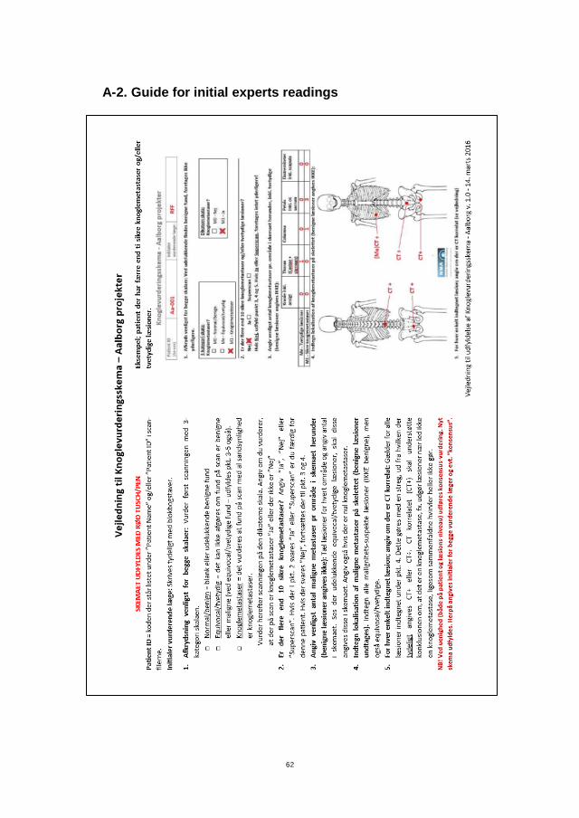
62
A-2. Guide for initial experts readings

APPENDIX A
63
A-3. Final diagnosis


65
Appendix B. Study II assessment forms


APPENDIX B
67
B-1. Individual assessment of baseline and follow-up images

68
B-2. Standard Clinical Assessment

APPENDIX B
69
B-3. MD Anderson criteria

70
B-4. Prostate Cancer Working Group 2 criteria

71
Appendix C. Study III Assessment forms

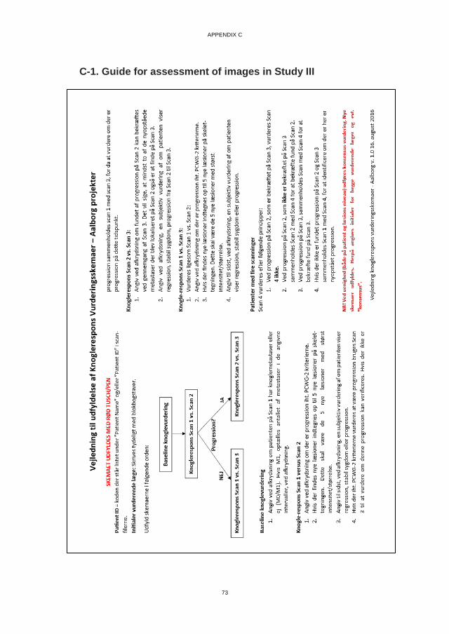
APPENDIX C
73
C-1. Guide for assessment of images in Study III

74
C-2. Baseline images

APPENDIX C
75
C-3. Baseline vs first follow-up scan
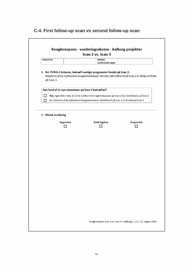
76
C-4. First follow-up scan vs second follow-up scan

APPENDIX C
77
C-5. Baseline vs second follow-up scan

RA
ND
I FUG
LSAN
G FO
NA
GER
DIA
GN
OSIS A
ND
MO
NITO
RIN
G O
F BO
NE M
ETASTA
SES IN PR
OSTATE C
AN
CER
ISSN (online): 2246-1302ISBN (online): 978-87-7112-933-5


![[XLS]sscnr.net.insscnr.net.in/newlook/Downloads/CAPFS_EXAM_201226713.xls · Web viewKAVITA DATTATRYA GUJAR SLC\00087 2201503934 AWADHESH KUMAR SLC\00674 2201503943 SLD\00375 2201503955](https://static.fdocuments.us/doc/165x107/5aeca1377f8b9a36698fd303/xlssscnrnet-viewkavita-dattatrya-gujar-slc00087-2201503934-awadhesh-kumar-slc00674.jpg)