Diagnosis and Management of Barrett’s Esophagus PRACTICAL GASTROENTEROLOGY • JULY 2015 FRONTIERS...
7
38 PRACTICAL GASTROENTEROLOGY • JULY 2015 FRONTIERS IN ENDOSCOPY, SERIES #20 Douglas G. Adler MD, FACG, AGAF, FASGE, Series Editor Diagnosis and Management of Barrett’s Esophagus Marisa Belaidi, Virendra Joshi MD, AGAF Louisiana State University School of Medicine, Ochsner Clinic Foundation, New Orleans, LA Virendra Joshi Marisa Belaidi both time-consuming and inconsistent in detecting dysplasia, this method does not more reliably predict the detection of cancer. 2 This revelation, coupled with increased understanding of the underlying histology and pathogenesis of Barrett’s Esophagus, has led to the creation of several new tools aimed at improving detection rates and yielding better patient prognoses. This review will describe the most current diagnostic techniques and treatment options of Barrett’s Esophagus and early esophageal cancer, their basic risks and benefits, and their clinical applicability. DETECTION Chromoendoscopy In the setting of BE, chromoendoscopy refers to the topical application of dye to esophageal mucosa in order to improve tissue visualization on endoscopy (Figure 1). Based on their staining properties, dyes are divided into one of three categories: contrast, absorptive (vital) or reactive. Methylene Blue (0.5%-1.0%), a vital dye (continued on page 40) F irst described by surgeon Norman Barrett in the mid-twentieth century, Barrett’s Esophagus (BE) refers to the transformation of normal stratified squamous epithelium to simple columnar epithelium in the distal esophagus, secondary to chronic injury and inflammation. Because intestinal metaplasia is a major risk factor for esophageal adenocarcinoma, the incidence of which has been steadily increasing in recent years, early detection and treatment have become a major priority in the gastrointestinal community. Historically, surveillance practice standards for patients with a history of BE followed the Seattle Protocol, a system that required physicians to obtain several random, 4-quadrant biopsies for every 1-2cm of involved esophageal mucosa under the visualization of white-light endoscopy (WLE). 1 However, a growing body of evidence suggests that, in addition to being INTRODUCTION
Transcript of Diagnosis and Management of Barrett’s Esophagus PRACTICAL GASTROENTEROLOGY • JULY 2015 FRONTIERS...
38 PRACTICAL GASTROENTEROLOGY • JULY 2015
FRONTIERS IN ENDOSCOPY, SERIES #20
Douglas G. Adler MD, FACG, AGAF, FASGE, Series Editor
Diagnosis and Management of Barrett’s Esophagus
Marisa Belaidi, Virendra Joshi MD, AGAF Louisiana State University School of Medicine, Ochsner Clinic Foundation, New Orleans, LA
Virendra JoshiMarisa Belaidi
both time-consuming and inconsistent in detecting dysplasia, this method does not more reliably predict the detection of cancer.2 This revelation, coupled with increased understanding of the underlying histology and pathogenesis of Barrett’s Esophagus, has led to the creation of several new tools aimed at improving detection rates and yielding better patient prognoses. This review will describe the most current diagnostic techniques and treatment options of Barrett’s Esophagus and early esophageal cancer, their basic risks and benefits, and their clinical applicability.
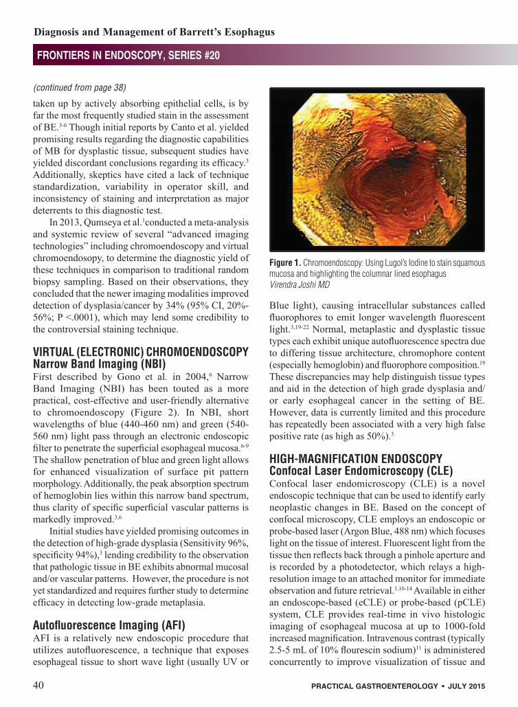
DETECTIONChromoendoscopy In the setting of BE, chromoendoscopy refers to the topical application of dye to esophageal mucosa in order to improve tissue visualization on endoscopy (Figure 1). Based on their staining properties, dyes are divided into one of three categories: contrast, absorptive (vital) or reactive. Methylene Blue (0.5%-1.0%), a vital dye
(continued on page 40)
First described by surgeon Norman Barrett in the mid-twentieth century, Barrett’s Esophagus (BE) refers to the transformation of normal stratified
squamous epithelium to simple columnar epithelium in the distal esophagus, secondary to chronic injury and inflammation. Because intestinal metaplasia is a major risk factor for esophageal adenocarcinoma, the incidence of which has been steadily increasing in recent years, early detection and treatment have become a major priority in the gastrointestinal community. Historically, surveillance practice standards for patients with a history of BE followed the Seattle Protocol, a system that required physicians to obtain several random, 4-quadrant biopsies for every 1-2cm of involved esophageal mucosa under the visualization of white-light endoscopy (WLE).1 However, a growing body of evidence suggests that, in addition to being
INTRODUCTION
40 PRACTICAL GASTROENTEROLOGY • JULY 2015
FRONTIERS IN ENDOSCOPY, SERIES #20
Diagnosis and Management of Barrett’s Esophagus
Blue light), causing intracellular substances called fluorophores to emit longer wavelength fluorescent light.3,19-22 Normal, metaplastic and dysplastic tissue types each exhibit unique autofluorescence spectra due to differing tissue architecture, chromophore content (especially hemoglobin) and fluorophore composition.19 These discrepancies may help distinguish tissue types and aid in the detection of high grade dysplasia and/or early esophageal cancer in the setting of BE. However, data is currently limited and this procedure has repeatedly been associated with a very high false positive rate (as high as 50%).3
HIGH-MAGNIFICATION ENDOSCOPYConfocal Laser Endomicroscopy (CLE) Confocal laser endomicroscopy (CLE) is a novel endoscopic technique that can be used to identify early neoplastic changes in BE. Based on the concept of confocal microscopy, CLE employs an endoscopic or probe-based laser (Argon Blue, 488 nm) which focuses light on the tissue of interest. Fluorescent light from the tissue then reflects back through a pinhole aperture and is recorded by a photodetector, which relays a high-resolution image to an attached monitor for immediate observation and future retrieval.1,10-14 Available in either an endoscope-based (eCLE) or probe-based (pCLE) system, CLE provides real-time in vivo histologic imaging of esophageal mucosa at up to 1000-fold increased magnification. Intravenous contrast (typically 2.5-5 mL of 10% flourescin sodium)11 is administered concurrently to improve visualization of tissue and
taken up by actively absorbing epithelial cells, is by far the most frequently studied stain in the assessment of BE.3-6 Though initial reports by Canto et al. yielded promising results regarding the diagnostic capabilities of MB for dysplastic tissue, subsequent studies have yielded discordant conclusions regarding its efficacy.3 Additionally, skeptics have cited a lack of technique standardization, variability in operator skill, and inconsistency of staining and interpretation as major deterrents to this diagnostic test.
In 2013, Qumseya et al.1conducted a meta-analysis and systemic review of several “advanced imaging technologies” including chromoendoscopy and virtual chromoendosopy, to determine the diagnostic yield of these techniques in comparison to traditional random biopsy sampling. Based on their observations, they concluded that the newer imaging modalities improved detection of dysplasia/cancer by 34% (95% CI, 20%-56%; P <.0001), which may lend some credibility to the controversial staining technique.
VIRTUAL (ELECTRONIC) CHROMOENDOSCOPY Narrow Band Imaging (NBI)First described by Gono et al. in 2004,6 Narrow Band Imaging (NBI) has been touted as a more practical, cost-effective and user-friendly alternative to chromoendoscopy (Figure 2). In NBI, short wavelengths of blue (440-460 nm) and green (540-560 nm) light pass through an electronic endoscopic filter to penetrate the superficial esophageal mucosa.6-9 The shallow penetration of blue and green light allows for enhanced visualization of surface pit pattern morphology. Additionally, the peak absorption spectrum of hemoglobin lies within this narrow band spectrum, thus clarity of specific superficial vascular patterns is markedly improved.3,6
Initial studies have yielded promising outcomes in the detection of high-grade dysplasia (Sensitivity 96%, specificity 94%),3 lending credibility to the observation that pathologic tissue in BE exhibits abnormal mucosal and/or vascular patterns. However, the procedure is not yet standardized and requires further study to determine efficacy in detecting low-grade metaplasia.
Autofluorescence Imaging (AFI) AFI is a relatively new endoscopic procedure that utilizes autofluorescence, a technique that exposes esophageal tissue to short wave light (usually UV or
Figure 1. Chromoendoscopy: Using Lugol’s Iodine to stain squamous mucosa and highlighting the columnar lined esophagusVirendra Joshi MD
(continued from page 38)

FRONTIERS IN ENDOSCOPY, SERIES #20
Diagnosis and Management of Barrett’s Esophagus
PRACTICAL GASTROENTEROLOGY • JULY 2015 41
cell structures. To ensure the highest image quality, the endoscopist must achieve adequate contact between the endoscope/probe and mucosal surface, which can be aided via gentle suction to stabilize the connection.10
In order to classify BE, eCLE utilizes the Mainz Confocal Barrett’s Classification System. Established by Keisslich et al, the Mainz Criteria relies on cell and vessel architecture to distinguish nondysplastic BE (columnar mucosa with goblet cells in a villiform pattern, normal capillaries) from neoplastic BE (irregularly shaped black cells, leaky capillaries),10-11 pCLE relies on the Miami criteria for dysplastic BE, which includes irregular vessels, fusion of villi and crypts and epithelial irregularities (thickness, inhomogeneity, dark border).
Additionally, recent studies indicate that combining eCLE with high-definition white light endoscopy may increase diagnostic yield of neoplasia versus traditional random biopsy sampling, without requiring as many unnecessary biopsies.
Optical Coherence Tomography (OCT)Optical coherence tomography (OCT) is a well-established imaging modality that utilizes low-coherence interferometry (near-infrared light) to generate high-resolution (10 - 15-μm), three dimensional cross-sectional imaging of in vivo tissue pathology.15-17 Initially utilized in ophthalmology, OCT has proved helpful in the diagnosis of various medical conditions, including specialized intestinal metaplasia in BE (sensitivity 81%). It has also been used in the detection of buried Barrett’s epithelium following
Figure 2a. Virtual Chromoendoscopy: Using NBI (Olympus) to highlight Barrett’s mucosaVirendra Joshi MD
radiofrequency ablation.18 However, the scan does not allow simultaneous biopsy of tissue, cannot clearly distinguish low-grade vs. high-grade dysplasia in the context of BE, and would be a tedious and impractical method to survey the full length of the esophagus.16
Volumetric Laser Endomicroscopy (VLE) is a next-generation technology, based upon OCT. While traditional OCT relies on Time Domain Interferometry to measure depth intensity, VLE utilizes Fourier Domain Interferometry (optical frequency domain imaging). This proprietary swept-source laser enables dramatically greater resolution (~7 microns) and faster acquisition times (100x faster) than conventional OCT. Its optical probe enables volumetric (circumferential + longitudinal) measurement of the entire distal esophagus.16
Suter et al.17 recently conducted a small, single-center feasibility study to assess the safety and practicality of VLE-guided biopsy in vivo, from which they determined that VLE-guided esophageal biopsy is a well-tolerated procedure that may have utility as a “first-look” procedure to mark tissue regions of interest for subsequent biopsy and therapeutic guidance.
TREATMENTIn addition to new detection methods, treatment options for Barrett’s Esophagus with high-grade dysplasia (HGD) and early esophageal adenocarcinoma (EAC) are constantly evolving. Once regarded as the gold standard of treatment, esophagectomy has been largely replaced with endoscopic, organ-sparing therapies that
Figure 2b. Virtual Chromoendoscopy: Using FICE (Pentax) to highlight Barrett’s mucosaClinical Gastroenterology and Hepatology

FRONTIERS IN ENDOSCOPY, SERIES #20
Diagnosis and Management of Barrett’s Esophagus
FRONTIERS IN ENDOSCOPY, SERIES #20
42 PRACTICAL GASTROENTEROLOGY • JULY 2015
including Argon Plasma and Nd:YAG laser.RFA refers to a process of controlled thermal injury
(damage is limited to mucosa and lamina propria) that occurs secondary to the induction of an electromagnetic field via an alternating electrical current. The exothermic reaction necessary for RFA can be generated by one of two specific devices: the HALO360 and the HALO90 (BARRX Medical, Inc, Sunnyvale, CA, USA).
Shaheen et al. conducted a multicenter, sham-controlled trial in which they assessed the efficacy of radiofrequency ablation versus a sham procedure in 127 patients with dysplastic BE. The authors concluded that RFA was associated with a significantly higher rate of complete eradication of both dysplasia (81.0%-90.5% vs. 19.0-22.7%, P<0.001) and intestinal metaplasia (77.4% vs. 2.3%, P<0.001) compared to the control25.
Overall, RFA is generally a safe, well-tolerated procedure. Side effects may include mild non-cardiac chest pain, nausea, and bleeding.22
CryotherapyCryotherapy is an ablative noncontact ablative method that delivers a cryogen (most commonly liquid nitrogen, LN2) under low-pressure spray with a decompressive gastric tube to the BE esophageal mucosa. The cryogen is administered in several cycles of rapid freezing (-196°C) and slow thawing. The extreme flash freezes tissue, selectively kills cells while preserving collagen matrix, creates vascular stasis and induces an analgesic effect.26-28 In contrast to burning techniques, spray
are equally as efficacious and incur significantly lower morbidity and mortality rates than traditional surgery.
RESECTIONEndoscopic Mucosal Resection (EMR) and Endoscopic Submucosal Dissection (ESD)EMR and ESD are two types of endoscopic treatment aimed at eradicating both HGD and intramucosal EAC in the setting of Barrett’s Esophagus. Due to their relative non-invasiveness, high post-procedure remission rates (up to 97%) and ability to provide a definitive histologic diagnosis, resection techniques are becoming increasingly popular therapeutic modalities.22,23 While EMR is primarily utilized in the treatment of small (<2cm) superficial tumors, ESD is preferred for larger (>2cm), more extensive lesions whose histologic accuracy and clinical outcome may be jeopardized by piece-meal resection.
Both procedures require a high operator skill level and can be associated with adverse events such as bleeding, stricture formation (more common in ESD) and esophageal stenosis.
Resection techniques may also be combined with ablative therapies for a potentially superior response rate with less post-procedure complications.
TISSUE ABLATIONRadiofrequency Ablation (RFA)Over the past 5 years RFA has become a primary therapy for high-grade dysplasia in Barrett’s epithelium. RFA replaced other previously used ablative therapies (continued on page 45)
Figure 3b. High Magnification Endoscopy: Confocal Laser Endomicroscopy Dysplastic GlandsVirendra Joshi MD
Figure 3a. Confocal Laser Endomicroscopy (CLE):Non-dysplastic Barrett’sVirendra Joshi MD

FRONTIERS IN ENDOSCOPY, SERIES #20
Diagnosis and Management of Barrett’s Esophagus
PRACTICAL GASTROENTEROLOGY • JULY 2015 45
FRONTIERS IN ENDOSCOPY, SERIES #20
just below the NSCJ ( neosquamocolumnar junction). Surveillance endoscopies should include this area to accurately identify patients with disease recurrence.33 In addition, cryotherapy appears to be a promising new strategy for salvage therapy of patients who “fail” thermal therapy with RFA.
Photodynamic Therapy (PDT)PDT is characterized by the activation of a chemical photosensitizer present in neoplastic tissue (most commonly porphimer sodium) by an endoscopic laser. The activated photosensitizer then reacts with oxygen, producing free radicals that induce cell damage and eventual apoptosis.22 PDT has often been used as an adjunct therapy to proton pump inhibitors (PPI) because the combination has shown superior eradication of BE with HGD and prevention of disease progression than either treatment alone. However, remission rates are significantly lower than esophageal resection techniques and side effects may include photosensitivity, fever, dysphagia, recurrence and progression of disease.30 One of the major side effects seen in a multicenter study by Overholt et al.30 was incidence of strictures, which correlated with number of applications and overlap of treatment. Overall, 36% of patients developed strictures, which were managed successfully with dilations. 12% of patients developed strictures after one PDT as opposed to 32% from two treatments and 9% after a third treatment. Additionally, they reported no improvement in the rate of stricture formation when oral steroids were administered after PDT.32 Careful patient
cryotherapy promotes less fibrosis and preserves underlying tissue architecture. It is a safe, well-tolerated, non-toxic therapy that literature indicates may serve as a compliment to other established technologies for BE, including EMR, or may be of use when all other treatments are ill advised.27 28 Notably, cryotherapy should be avoided during pregnancy, in the setting of compromised or damaged tissue and in the event of increased anatomical flow resistance (gas evacuation). Cryoablation can successfully eradicate residual Barrett’s in patients with esophageal cancer post chemo- radiation.28.
In a retrospective review of 32 patients Greenwald et al. found CE-HGD was 100% (32/32), and CE-IM was 84% (27/32) at 2-year follow-up. At last follow-up (range 24-57 months), CE-HGD was 31/32 (97%), and CE-IM was 26/32 (81%). Recurrent HGD was found in 6 (18%), with CE-HGD in 5 after repeat treatment. One patient progressed to adenocarcinoma, downgraded to HGD after repeat cryotherapy. BE segment length ≥3 cm was associated with a higher recurrence of IM (P = .004; odds ratio 22.6) but not HGD. No serious adverse events occurred. Stricture was seen in 3 patients (9%), all successfully dilated.34 A prospective study of patients undergoing cryoablation suggested recurrent disease commonly involves the area
Figure 3c. High Magnification Endoscopy: Optical Coherence Tomography (OCT) Layered Architecture of Normal EsophagusVirendra Joshi MD
Figure 3d. OCT Loss of Layered Architecture non-dysplastic Barrett’s, Normal Appearing GlandVirendra Joshi MD
(continued from page 42)
practicalgastro.com

46 PRACTICAL GASTROENTEROLOGY • JULY 2015
FRONTIERS IN ENDOSCOPY, SERIES #20
Diagnosis and Management of Barrett’s Esophagus
(CLE ) and more recently developed volumetric endomicroscopy (VLE) will continue to impact early detection by serving as “red flag technologies” helping to target biopsies and decrease sampling errors. Dysplastic Barrett’s and early cancer can now be treated by ablative therapies (thermal, non-thermal) and endoscopic mucosal resection (EMR, ESD) with minimal morbidity. These minimally invasive diagnostic and therapeutic strategies should be individualized and tailored to individual patient needs.
education is critical for the management of side effects and to reduce the risk of photosensitivity reactions.31
CONCLUSIONThere has been a paradigm shift in the detection and management of Barrett’s esophagus over the past decade with use of advanced imaging for detection and management dysplastic Barrett’s and early esophageal cancer. Novel advanced imaging modalities as Narrow Band Imaging (NBI), Confocal Laser Endomicroscopy
Figure 5a. Treatment for Barrett’s with Dysplasia:Radiofrequency Ablation (RFA)Virendra Joshi MD
Figure 5c. Treatment for Barrett’s with Dysplasia: PDT Catheter in PlaceH. Wolfsen MD (Mayo Clinic)
Figure 5b. Treatment for Barrett’s with Dysplasia: CryotherapyVirendra Joshi MD
Figure 5d. Treatment for Barrett’s with Dysplasia: PDT TherapyH. Wolfsen MD (Mayo Clinic)

FRONTIERS IN ENDOSCOPY, SERIES #20
Diagnosis and Management of Barrett’s Esophagus
PRACTICAL GASTROENTEROLOGY • JULY 2015 47
References
1. Qumseya BJ, Wang H, Badie N et al. Advanced imaging technologies increase detection of dysplasia and neoplasia in patients with Barrett’s esophagus: a meta-analysis and system-atic review. Clin Gastroenterol Hepatol., 2013;11(12):1562-70.
2. Kariv R, Plesec TP, Goldblum JR et al. The Seattle protocol does not more reliably predict the detection of cancer at the time of esophagectomy than a less intensive surveillance protocol. Clin Gastroenterol Hepatol., 2009 Jun;7(6):653-8.
3. Singh R, Mei SC, Sethi S. Advanced endoscopic imaging in Barrett’s oesophagus: A review on current practice. World J Gastroenterol, 2011;17(38):4271-76.
4. Sharma P, Topalovski M, Mayo MS et al. Methylene blue chro-moendoscopy for detection of short-segment Barrett’s esopha-gus. Gastrointest Endosc., 2001;54:289.
5. Ngamruengphong S, Sharma VK, Das A. Diagnostic yield of methylene blue chromoendoscopy for detecting specialized intestinal metaplasia and dysplasia in Barrett’s esophagus: a meta-analysis. Gastrointest Endos., 2009; 69:1021.
6. Gono K, Obi T, Yamaguchi M, et al. Appearance of enhanced tissue features in narrowband endoscopic imaging. J Biomed Opt, 2004;9:568–577.
7. Yoshida T, Inoue H, Usui S et al. Narrow-band imaging system with magnifying endoscopy for superficial esophageal lesions. Gastrointest Endosc., 2004;59:288.
8. Sharma P. Narrow band imaging in Barrett’s esophagus. Clin Gastroenterol Hepatol., 2005; 3:S21.
9. Sharma P, Hawes RH, Bansal A, et al. Standard endoscopy with random biopsies versus narrow band imaging targeted biopsies in Barrett’s oesophagus: a prospective, international, randomised controlled trial. Gut, 2013;62:15.
10. Leggett CL, Gorospe EC. Application of confocal laser endomi-croscopy in the diagnosis and management of Barrett’s esopha-gus. Ann Gastroenterol., 2014;27(3):193-199.
11. Dunbar KB. Endomicroscopy in Barrett’s Esophagus. Gastrointest Endosc Clin N Am., 2013; 23(3):565-79.
12. Badreddine R, Wang K, Ganapathy A et al. Confocal laser microscopy (CLM) guided endoscopic mucosal resection in Barrett’s esophagus with high grade dysplasia. Gastrointest Endosc., 2008;67:AB179.
13. Jayasekera C, Taylor AC, Desmond PV et al. Added value of narrow band imaging and confocal laser endomicroscopy in detecting Barrett’s esophagus neoplasia. Endoscopy, 2012; 44:1089.
14. Canto MI, Anandasabapathy S, Brugge W et al. In vivo endo-microscopy improves detection of Barrett’s esophagus-related neoplasia: a multicenter international randomized controlled trial. Gastrointest Endosc., 2014;79:211.
15. Li XD, Boppart SA, Van Dam J et al. Optical coherence tomog-raphy: advanced technology for the endoscopic imaging of Barrett’s esophagus. Endoscopy, 2000;32(12):921-30.
16. Carignan CS and Yagi Y. Optical endomicroscopy and the road to real-time, in vivo pathology: present and future. Diagn Pathol., 2012;7: 98.
17. Suter MJ, Gora MJ, Lauwers GY et al. Esophageal-guided biopsy with volumetric laser endomicroscopy and laser cau-tery marking: a pilot clinical study. Gastrointest Endosc., 2014;79(6):886-96.
18. Zhou C, Tsai TH, Lee HC, et al. Characterization of buried glands before and after radiofrequency ablation by using 3-dimensional optical coherence tomography (with videos). Gastrointest Endosc., 2012;76:32.
19. Boerwinkel DF, Holz JA, Kara MA et al. Effects of autofluores-cence imaging on detection and treatment of early neoplasia in patients with Barrett’s esophagus. Clin Gastroenterol Hepatol., 2014;12:774.
20. Boerwinkel DF, Holz JA, Aalders MC, et al. Third-generation autofluorescence endoscopy for the detection of early neoplasia in Barrett’s esophagus: a pilot study. Dis Esophagus, 2014; 27:276.
21. Mannath J, Subramanian V, Telakis E et al. An inter-observer agreement study of autofluorescence endoscopy in Barrett’s esophagus among expert and non-expert endoscopists. Dig Dis Sci., 2013;58:465.
22. Shekhar R, Biyyani S, Chak A. Barrett’s esophagus: review of diagnosis and treatment. Gastroenterology Report, 2013;1:9-18.
23. Lee JK and Enns R. Endoscopic mucosal resection in the setting of Barrett’s esophagus. Can J Gastroenterol., 2007;21(3):151–154.
24. Barret M, Pratico CA, Beuvon F et al. Esophageal circumfer-ential en bloc endoscopic submucosal dissection: assessment of a new technique. Surg Laparosc Endosc Percutan Tech., 2013;23(5):e182-7.
25. Shaheen NJ, Sharma P, Overholt BF et al. Radiofrequency ablation in Barrett’s esophagus with dysplasia. N Engl J Med., 2009;360(22):2277-88.
26. Gage AA, Baust JM, Baust JG. Experimental cryosurgery inves-tigations in vivo. Cryobiology, 2009:59(3):229-43.
27. Greenwald BD, Dumot JA, Abrams JA et al. Endoscopic spray cryotherapy for esophageal cancer: safety and efficacy. Gastrointest Endosc., 2010;71(4):686-93.
28. Barthel JS, Kucera S, Harris C et al. Cryoablation of per-sistent Barrett’s epithelium after definitive chemoradiation therapy for esophageal adenocarcinoma. Gastrointest Endosc, 2011;74(1):51-57.
29. Bronner MP, Overholt BF, Taylor SL, Haggitt RC, et al: Squamous overgrowth is not a safety concern for photo- dynamic therapy for Barrett’s esophagus with high-grade dys-plasia. Gastroenterology 2009;136: 56–64.
30. Overholt BF, Lightdale CJ, Wang KK, Canto MI, et al: Photodynamic therapy with porfimer sodium for ablation of high-grade dysplasia in Barrett’s esophagus: international, par-tially blinded, randomized phase III trial. Gastrointest Endosc 2005;62:488–498.
31. Phan M, Dyke S, Whittaker MA, Simmerman A, Abrams S, Panjehpour M, Overholt BF: An educational tool for photody-namic therapy of Barrett’s esophagus with high- grade dysplasia. Gastroenterol Nurs 2005;28:413–419.
32. Panjehpour M, Overholt BF, Haydek JM, Lee SG: Results of photodynamic therapy for ablation of dysplasia and early cancer in Barrett’s esophagus and effect of oral steroids on stricture formation. Am J Gastroenterol 2000; 95:2177–2184.
33. Halsey KD1, Chang JW, Waldt A, Greenwald BD;Recurrent disease following endoscopic ablation of Barrett’s high-grade dysplasia with spray cryotherapy. Endoscopy. 2011 Oct;43(10):844-8.
34. Gosain S1, Mercer K, Twaddell WS, Uradomo L, Greenwald BD;Liquid nitrogen spray cryotherapy in Barrett’s esopha-gus with high-grade dysplasia: long-term results.Gastrointest Endosc. 2013 Aug;78(2):260-5.
Figure 5e. Treatment for Barrett’s with Dysplasia: Ablation PDT (Photodynamic Therapy) Post PDTH. Wolfsen MD (Mayo Clinic)