Diabetes mellitus in pregnancy: Screening and diagnosis€¦ · Diabetes mellitus in pregnancy:...
57
Diabetes mellitus in pregnancy: Screening and diagnosis Dr lama almehaisen,MD,MRCOG Consultant obstetrics ang gyn /urogyn Associated prof BAU
Transcript of Diabetes mellitus in pregnancy: Screening and diagnosis€¦ · Diabetes mellitus in pregnancy:...
Diabetes mellitus in
pregnancy: Screening
and diagnosis Dr lama almehaisen,MD,MRCOG
Consultant obstetrics ang gyn /urogyn
Associated prof BAU
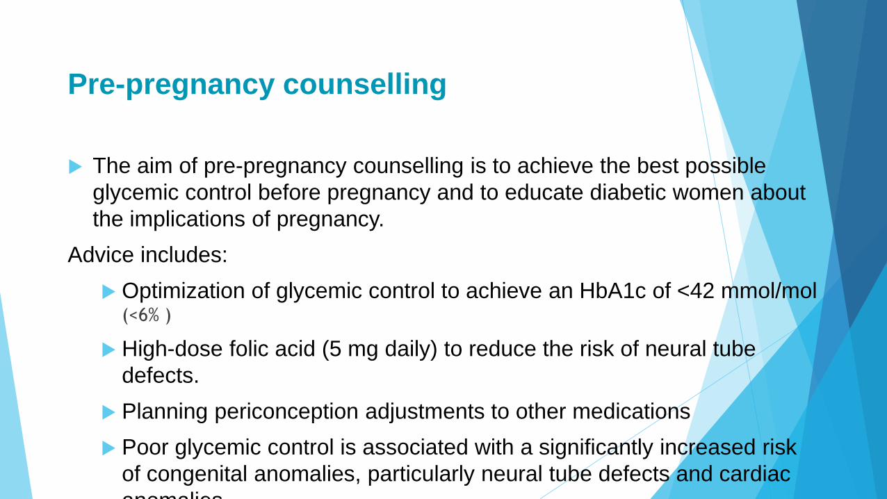
Pre-pregnancy counselling
The aim of pre-pregnancy counselling is to achieve the best possible
glycemic control before pregnancy and to educate diabetic women about
the implications of pregnancy.
Advice includes:
Optimization of glycemic control to achieve an HbA1c of <42 mmol/mol (<6% )
High-dose folic acid (5 mg daily) to reduce the risk of neural tube
defects.
Planning periconception adjustments to other medications
Poor glycemic control is associated with a significantly increased risk
of congenital anomalies, particularly neural tube defects and cardiac
anomalies.
The most critical period for the embryo is therefore the period of organogenesis, which
occurs in the first 42 days of pregnancy,
The level of HbA1c in early pregnancy also correlates with the risk of early fetal loss. An
HbA1c of >85 mmol/mol is associated with a fetal loss during pregnancy of around 30%.
Pre-pregnancy care is associated with reduced rates of congenital malformation.
Targets for therapy pre-pregnancy are premeal glucose levels of 4–6 mmol/l.
Diabetic vascular complications are common in women of reproductive age
It is important that a plan for medication adjustment is made and
women are counselled regarding the additional potential complications
associated with diabetic microvascular disease ie Nephropathy.
There is also a risk that retinopathy can progress in pregnancy and
during the postpartum period.
Maternal and fetal complications of types 1
and 2diabetes mellitus
1. Congenital abnormality is an important cause of mortality and morbidity
in diabetic pregnancies
2. It is seen 2–4 times more often than in pregnancies without diabetes
with a threefold excess of cardiac and neural tube defects.
3. structural malformations, fetal macrosomia is a frequent complication
associated with maternal diabetes and frequently contributes to a
traumatic birth and shoulder dystocia.
4. Accelerated growth patterns are typically seen in the late second and
third trimesters and are attributable to poorly controlled diabetes in the
majority of cases.
5. Stillbirth, particularly in the third trimester, remains too common in
pregnancies complicated by maternal diabetes, being five times higher
than in the general population.
6. increased incidence of infection, severe hyperglycaemia or
hypoglycaemia, diabetic ketoacidosis and the complications that may
arise from the increased operative delivery rate.
7. The risk of preeclampsia is increased 3X in women with diabetes,
and particularly in those with underlying microvascular disease.
frequently prompt early term delivery in women with diabetes, which in
turn increases the likelihood of neonatal unit admission and reduces
breastfeeding rates.
In general, maternal morbidity in diabetic pregnancies is related to the
severity of diabetic-related vascular disease preceding the pregnancy.
All women with diabetes should be offered low-dose aspirin from 12
weeks’ gestation to reduce the risk of preeclampsia.
Management of types 1 and 2 diabetes in
pregnancy
(anti-natal plan )
The primary goal is to optimize glycaemic control.
Blood glucose monitoring is encouraged 7 times a day (before and
2 hour after meals) with targets of 4-6 mmol/l and 2-hour
postprandial levels of <6-8 mmol/l
use of oral hypoglycaemic agents such as metformin and/or insulin
where appropriate,
Insulin resistance increases dramatically over the course of
pregnancy and therefore women with type 1 and type 2 diabetes
are usually required to increase their dose of insulin or metformin
during the second half of pregnancy.
A plan for the pregnancy should be set out in early pregnancy and
should include renal and retinal screening, fetal surveillance and a
plan for delivery.
Women with diabetes should be offered a fetal anomaly scan at 18–
21 weeks with an assessment of the cardiac outflow tracts.
Serial growth scans are also recommended to assess fetal growth
and diagnose macrosomia and polyhydramnios
If antenatal corticosteroids are indicated, additional insulin
therapy is required to maintain normoglycaemia
Timing and mode of delivery should be determined on an
individual basis.
In general, provided the pregnancy has gone well, the aim
would be to achieve a vaginal delivery at between 38 and 39
weeks.
However, the development of macrosomia or maternal
complications such as pre-eclampsia, together with the rate of
failed induction, is such that the caesarean section rate
amongst diabetic women often is as high as 50%.
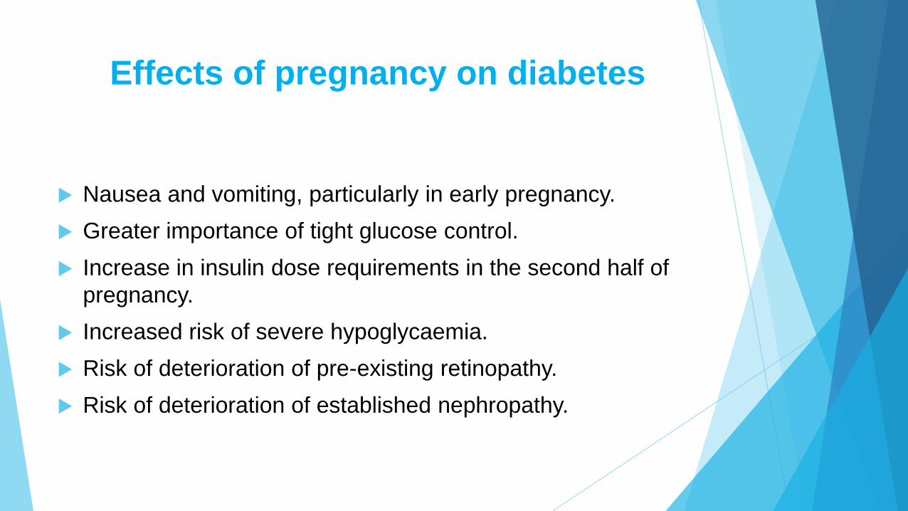
Effects of pregnancy on diabetes
Nausea and vomiting, particularly in early pregnancy.
Greater importance of tight glucose control.
Increase in insulin dose requirements in the second half of
pregnancy.
Increased risk of severe hypoglycaemia.
Risk of deterioration of pre-existing retinopathy.
Risk of deterioration of established nephropathy.
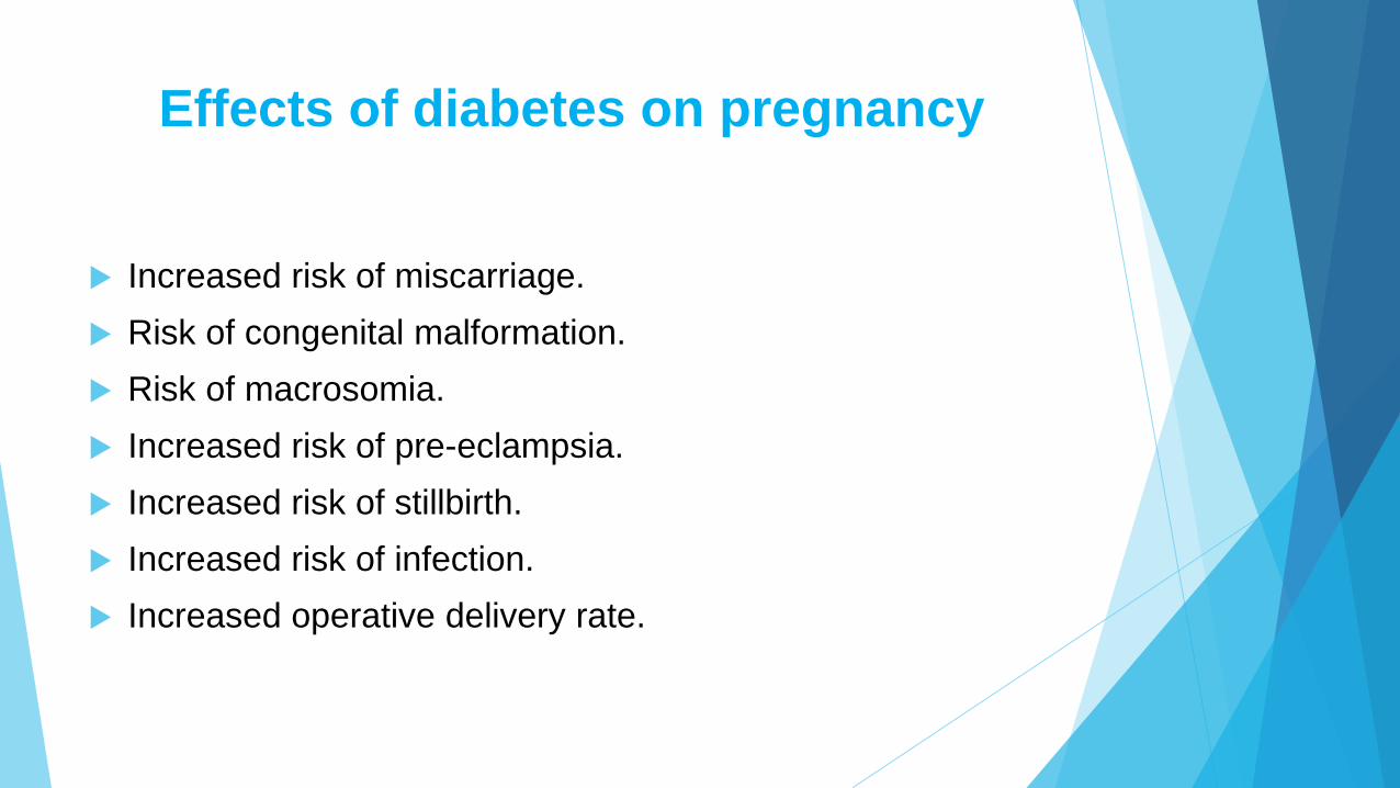
Effects of diabetes on pregnancy
Increased risk of miscarriage.
Risk of congenital malformation.
Risk of macrosomia.
Increased risk of pre-eclampsia.
Increased risk of stillbirth.
Increased risk of infection.
Increased operative delivery rate.
TERMINOLOGY
Historically, the term "gestational diabetes" has been defined as onset or
first recognition of abnormal glucose tolerance during pregnancy
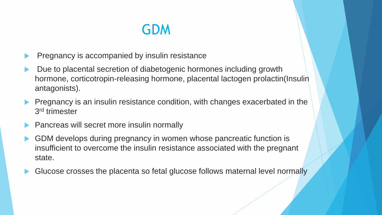
GDM
Pregnancy is accompanied by insulin resistance
Due to placental secretion of diabetogenic hormones including growth
hormone, corticotropin-releasing hormone, placental lactogen prolactin(Insulin
antagonists).
Pregnancy is an insulin resistance condition, with changes exacerbated in the
3rd trimester
Pancreas will secret more insulin normally
GDM develops during pregnancy in women whose pancreatic function is
insufficient to overcome the insulin resistance associated with the pregnant
state.
Glucose crosses the placenta so fetal glucose follows maternal level normally
GDM
GDM complicates 10–15% of pregnancies depending on the diagnostic
criteria used.
Screening for diabetes in pregnancy is designed to detect previously
undiagnosed type 2 diabetes and diabetes developing during pregnancy.
Women who develop GDM are at increased risk of type 2 diabetes in later
life, and education about diet and lifestyle during pregnancy can have
important implications for future health.
No single screening method has been shown to be perfect in terms of
sensitivity and specificity for GDM.
screening is generally targeted at high-risk groups
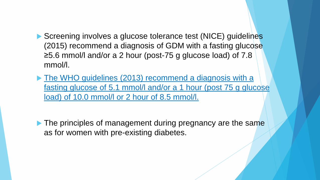
Screening involves a glucose tolerance test (NICE) guidelines
(2015) recommend a diagnosis of GDM with a fasting glucose
≥5.6 mmol/l and/or a 2 hour (post-75 g glucose load) of 7.8
mmol/l.
The WHO guidelines (2013) recommend a diagnosis with a
fasting glucose of 5.1 mmol/l and/or a 1 hour (post 75 g glucose
load) of 10.0 mmol/l or 2 hour of 8.5 mmol/l.
The principles of management during pregnancy are the same
as for women with pre-existing diabetes.
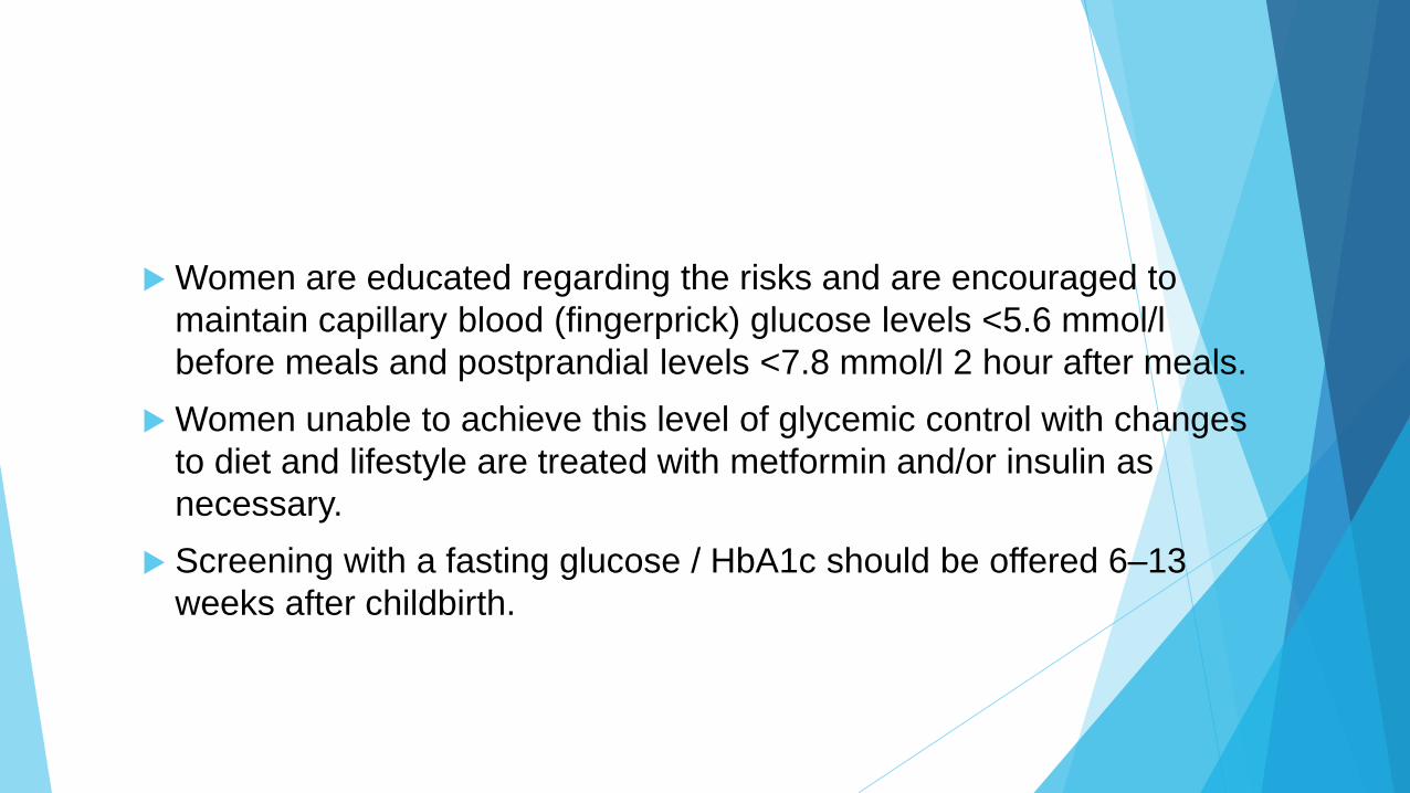
Women are educated regarding the risks and are encouraged to
maintain capillary blood (fingerprick) glucose levels <5.6 mmol/l
before meals and postprandial levels <7.8 mmol/l 2 hour after meals.
Women unable to achieve this level of glycemic control with changes
to diet and lifestyle are treated with metformin and/or insulin as
necessary.
Screening with a fasting glucose / HbA1c should be offered 6–13
weeks after childbirth.
Factors associated
Factors associated with poor pregnancy outcome in diabetes
actors associated with poor Maternal social deprivation.
No folic acid intake pre-pregnancy.
Suboptimal approach of the woman to managing her diabetes.
Suboptimal preconception care.
Suboptimal glycemic control at any stage.
Suboptimal maternity care during pregnancy.
Suboptimal fetal surveillance of big babies.
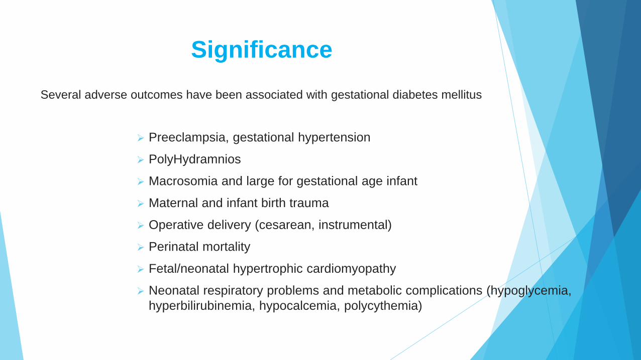
Significance
Several adverse outcomes have been associated with gestational diabetes mellitus
Preeclampsia, gestational hypertension
PolyHydramnios
Macrosomia and large for gestational age infant
Maternal and infant birth trauma
Operative delivery (cesarean, instrumental)
Perinatal mortality
Fetal/neonatal hypertrophic cardiomyopathy
Neonatal respiratory problems and metabolic complications (hypoglycemia,
hyperbilirubinemia, hypocalcemia, polycythemia)
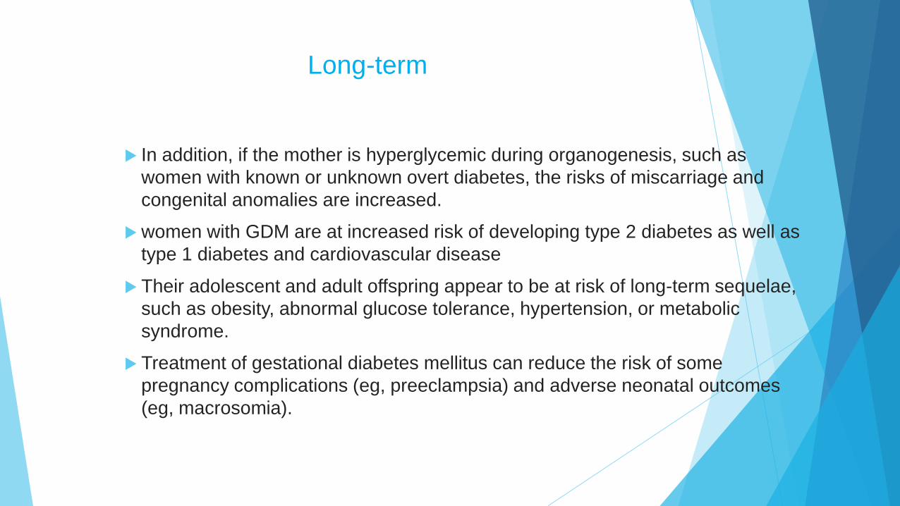
Long-term
In addition, if the mother is hyperglycemic during organogenesis, such as
women with known or unknown overt diabetes, the risks of miscarriage and
congenital anomalies are increased.
women with GDM are at increased risk of developing type 2 diabetes as well as
type 1 diabetes and cardiovascular disease
Their adolescent and adult offspring appear to be at risk of long-term sequelae,
such as obesity, abnormal glucose tolerance, hypertension, or metabolic
syndrome.
Treatment of gestational diabetes mellitus can reduce the risk of some
pregnancy complications (eg, preeclampsia) and adverse neonatal outcomes
(eg, macrosomia).
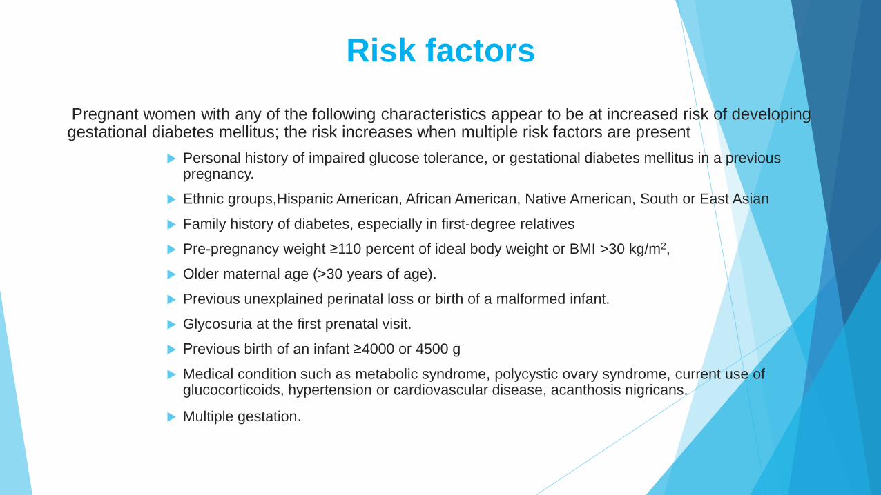
Risk factors
Pregnant women with any of the following characteristics appear to be at increased risk of developing gestational diabetes mellitus; the risk increases when multiple risk factors are present
Personal history of impaired glucose tolerance, or gestational diabetes mellitus in a previous pregnancy.
Ethnic groups,Hispanic American, African American, Native American, South or East Asian
Family history of diabetes, especially in first-degree relatives
Pre-pregnancy weight ≥110 percent of ideal body weight or BMI >30 kg/m2,
Older maternal age (>30 years of age).
Previous unexplained perinatal loss or birth of a malformed infant.
Glycosuria at the first prenatal visit.
Previous birth of an infant ≥4000 or 4500 g
Medical condition such as metabolic syndrome, polycystic ovary syndrome, current use of glucocorticoids, hypertension or cardiovascular disease, acanthosis nigricans.
Multiple gestation.
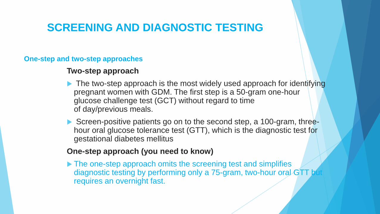
SCREENING AND DIAGNOSTIC TESTING
One-step and two-step approaches
Two-step approach
The two-step approach is the most widely used approach for identifying pregnant women with GDM. The first step is a 50-gram one-hour glucose challenge test (GCT) without regard to time of day/previous meals.
Screen-positive patients go on to the second step, a 100-gram, three-hour oral glucose tolerance test (GTT), which is the diagnostic test for gestational diabetes mellitus
One-step approach (you need to know)
The one-step approach omits the screening test and simplifies diagnostic testing by performing only a 75-gram, two-hour oral GTT but requires an overnight fast.
Timing of screening/testing
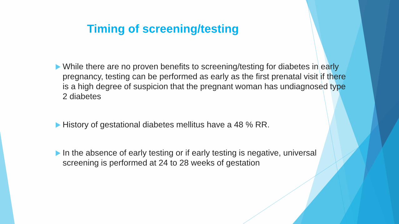
While there are no proven benefits to screening/testing for diabetes in early
pregnancy, testing can be performed as early as the first prenatal visit if there
is a high degree of suspicion that the pregnant woman has undiagnosed type
2 diabetes
History of gestational diabetes mellitus have a 48 % RR.
In the absence of early testing or if early testing is negative, universal
screening is performed at 24 to 28 weeks of gestation
Diagnostic testing methods
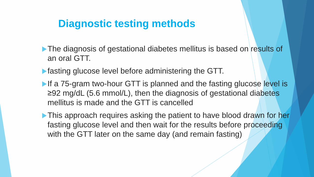
The diagnosis of gestational diabetes mellitus is based on results of
an oral GTT.
fasting glucose level before administering the GTT.
If a 75-gram two-hour GTT is planned and the fasting glucose level is
≥92 mg/dL (5.6 mmol/L), then the diagnosis of gestational diabetes
mellitus is made and the GTT is cancelled
This approach requires asking the patient to have blood drawn for her
fasting glucose level and then wait for the results before proceeding
with the GTT later on the same day (and remain fasting)
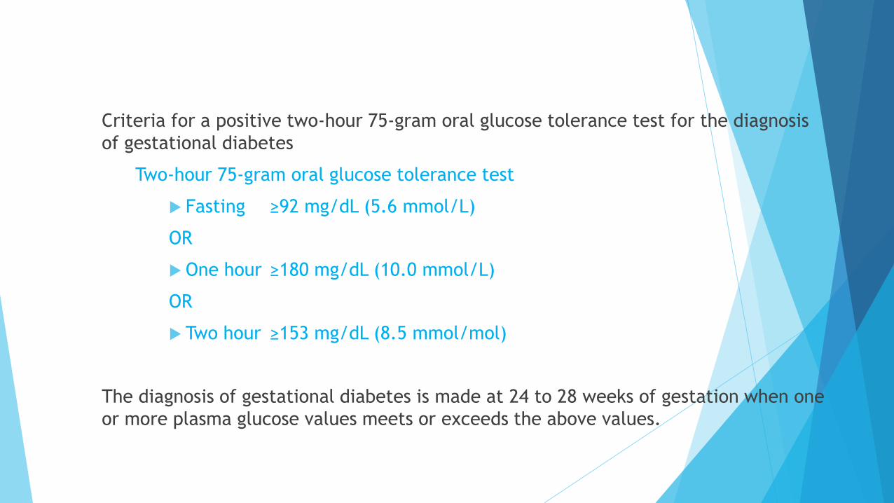
Criteria for a positive two-hour 75-gram oral glucose tolerance test for the diagnosis
of gestational diabetes
Two-hour 75-gram oral glucose tolerance test
Fasting ≥92 mg/dL (5.6 mmol/L)
OR
One hour ≥180 mg/dL (10.0 mmol/L)
OR
Two hour ≥153 mg/dL (8.5 mmol/mol)
The diagnosis of gestational diabetes is made at 24 to 28 weeks of gestation when one
or more plasma glucose values meets or exceeds the above values.
Patients unable to tolerate oral hyperosmolar glucose
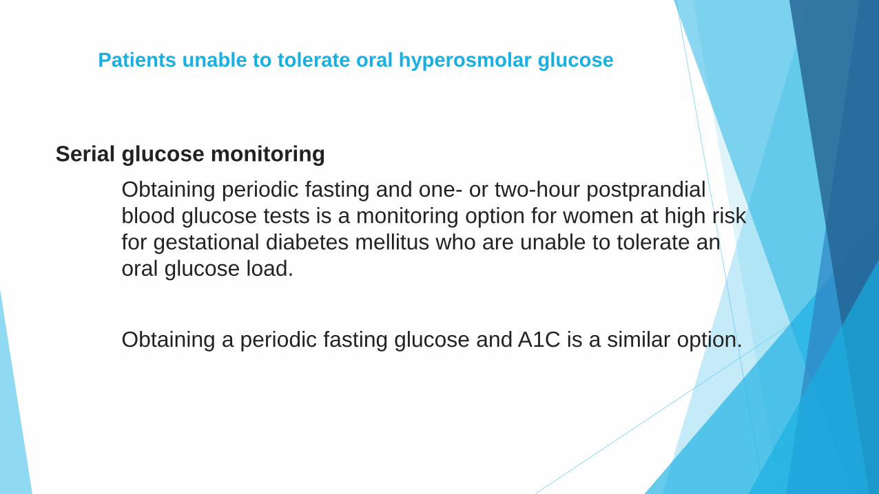
Serial glucose monitoring
Obtaining periodic fasting and one- or two-hour postprandial
blood glucose tests is a monitoring option for women at high risk
for gestational diabetes mellitus who are unable to tolerate an
oral glucose load.
Obtaining a periodic fasting glucose and A1C is a similar option.
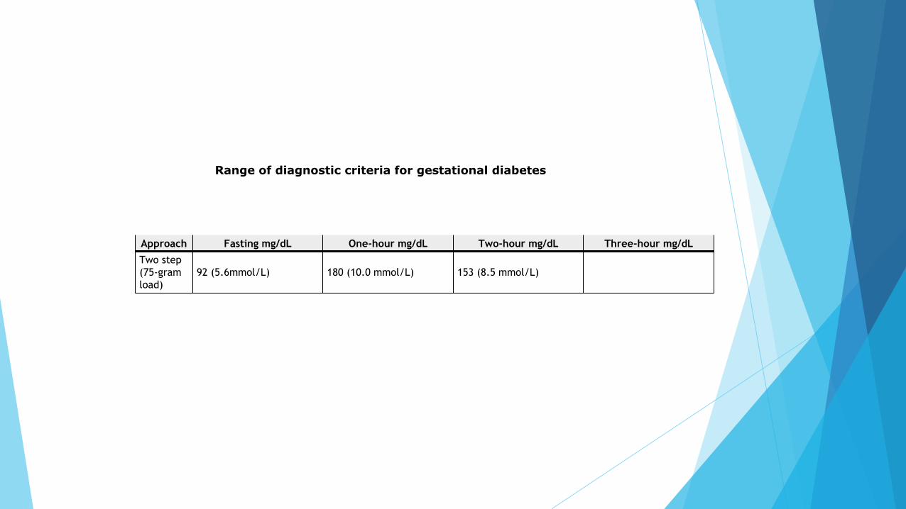
Approach Fasting mg/dL One-hour mg/dL Two-hour mg/dL Three-hour mg/dL
Two step
(75-gram
load)
92 (5.6mmol/L) 180 (10.0 mmol/L) 153 (8.5 mmol/L)
Range of diagnostic criteria for gestational diabetes
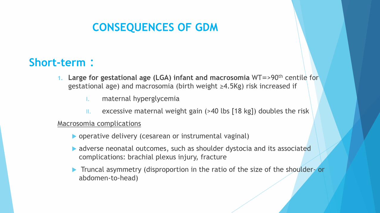
CONSEQUENCES OF GDM
Short-term : 1. Large for gestational age (LGA) infant and macrosomia WT=>90th centile for
gestational age) and macrosomia (birth weight ≥4.5Kg) risk increased if
I. maternal hyperglycemia
II. excessive maternal weight gain (>40 lbs [18 kg]) doubles the risk
Macrosomia complications
operative delivery (cesarean or instrumental vaginal)
adverse neonatal outcomes, such as shoulder dystocia and its associated
complications: brachial plexus injury, fracture
Truncal asymmetry (disproportion in the ratio of the size of the shoulder- or
abdomen-to-head)
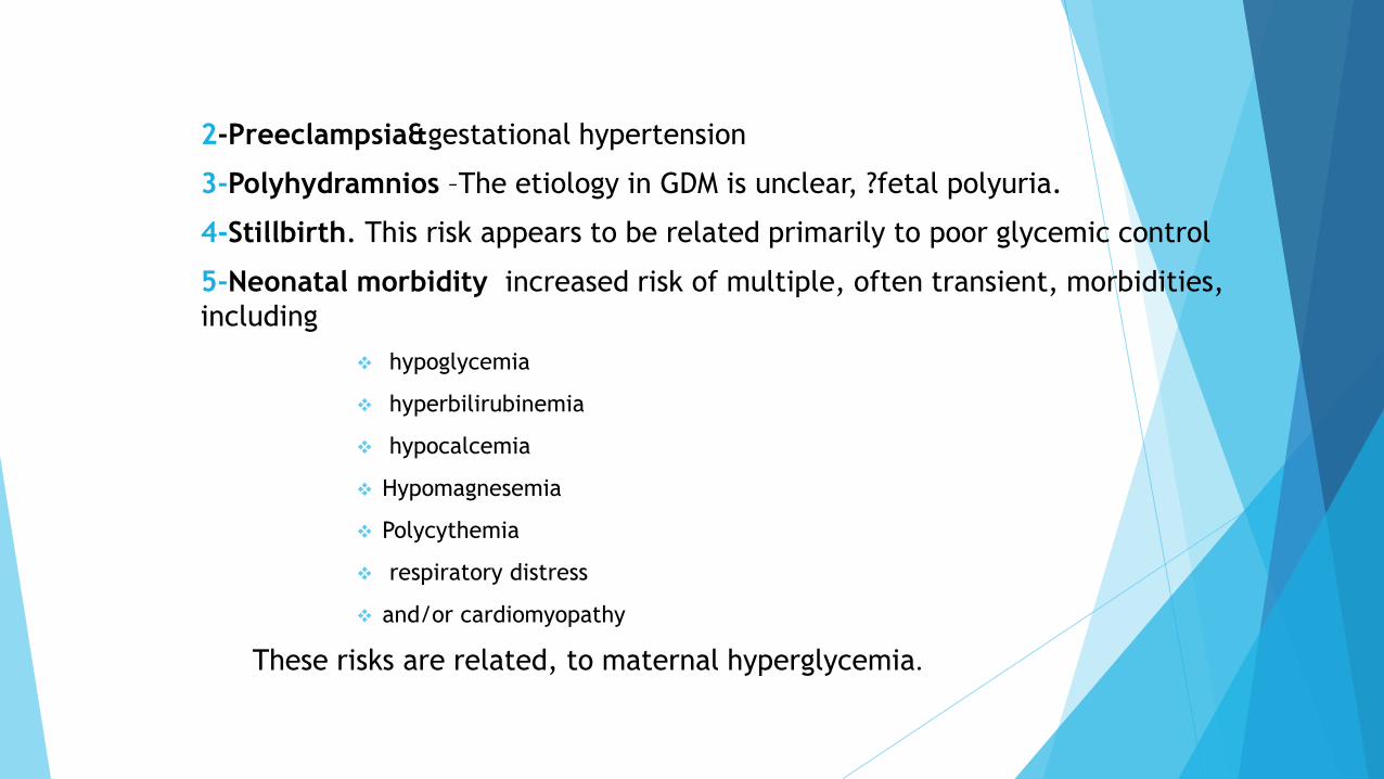
2-Preeclampsia&gestational hypertension
3-Polyhydramnios –The etiology in GDM is unclear, ?fetal polyuria.
4-Stillbirth. This risk appears to be related primarily to poor glycemic control
5-Neonatal morbidity increased risk of multiple, often transient, morbidities,
including
hypoglycemia
hyperbilirubinemia
hypocalcemia
Hypomagnesemia
Polycythemia
respiratory distress
and/or cardiomyopathy
These risks are related, to maternal hyperglycemia.
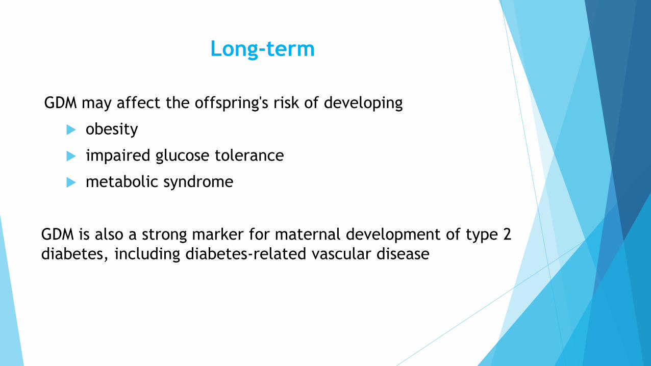
Long-term
GDM may affect the offspring's risk of developing
obesity
impaired glucose tolerance
metabolic syndrome
GDM is also a strong marker for maternal development of type 2
diabetes, including diabetes-related vascular disease

Approach to patients
Glucose monitoring and control —Glucose monitoring, medical nutritional therapy,
exercise, and the use of insulin and anti-hyperglycemic agents
Antenatal fetal testing
Women on insulin or oral anti-hyperglycemic drugs or with poor glycemic control
twice weekly CTG plus an amniotic fluid index beginning at 32 weeks of
gestation in women who need insulin or an oral antihyperglycemic agent
to achieve good glycemic control,
in all women with poor glycemic control ,we generally recommends
that These women typically undergo periodic antenatal testing, usually
initiated at approximately 32 weeks of gestation.
Women euglycemic on nutritional therapy alone
antenatal fetal surveillance (nonstress testing or biophysical profile scoring) is a reasonable
approach for these women
Assessment of fetal growth
Identification of accelerated fetal growth before delivery may be useful to identify
maternal-fetal pairs who may benefit from scheduled cesarean delivery to avoid trauma
from shoulder dystocia
Timing of delivery
main question in GDM is whether to induce labor and, if so, why?
Avoidance of late stillbirth
delivery-related complications of continued fetal growth, such as shoulder
dystocia or cesarean delivery.
The potential disadvantages are the risks of induction
longer labor
increased tendency for intervention
increased neonatal morbidity if induction is before 39 weeks.
Increasing evidence suggests that induction of labor in women with GDM
does not lead to higher cesarean delivery rates than expectant
management
pregnancies of women who remain euglycemic with nutritional
therapy and exercise alone these patients should not be electively
delivered prior to 39 weeks of gestation Timing of induction between
39+0 and 41+0 weeks is more controversial.
women with GDM whose glucose levels are medically managed with
insulin or oral agents we recommend induction of labor at 39 weeks of
gestation .
Scheduled cesarean delivery
to avoid birth trauma is typically offered to women with GDM and estimated fetal weight ≥4500 grams.
Labor and delivery
During labor, periodic assessment of maternal glucose levels
transient hypoglycemia can be caused by intrapartum maternal hyperglycemia, which induces an acute rise in fetal insulin
Insulin requirements usually decrease during labor,
Women with GDM who were euglycemic without use of insulin or oral antihyperglycemic drugs during pregnancy do not normally require insulin during labor and delivery, and thus do not need their blood glucose levels checked hourly.
Women with GDM who used insulin or oral antihyperglycemic drugs to maintain euglycemia occasionally need insulin during labor and delivery to maintain euglycemia. The Endocrine Society suggests target glucose levels of 72 to 126 mg/dL (4.0 to 7.0 mmol/L)
check blood glucose measurements every two hours during labor
For women undergoing scheduled cesarean delivery, insulin or antihyperglycemic drugs are withheld the morning of surgery and the woman is not allowed any oral intake after midnight.
POSTPARTUM MANAGEMENT AND FOLLOW-UP
Women with gestational diabetes mellitus (GDM) should be able to resume a normal diet postpartum.
After delivery, the hyperglycemic effects of placental hormones dissipate rapidly. Thus, most women
revert back to their pre-pregnancy glycemic status almost immediately.
Contraception — While any type of contraception is acceptable, as long as the usual medical
contraindications to use are absent, we recommend long-acting reversible contraception (LARC;
eg, intrauterine device, contraceptive implant) because of the minimal risk of unplanned pregnancy
with these methods
Breastfeeding — Breastfeeding should be encouraged, as it benefits both mother and child
Breastfeeding improves maternal glucose metabolism and thus may reduce the glucose levels
obtained during a postpartum glucose tolerance test
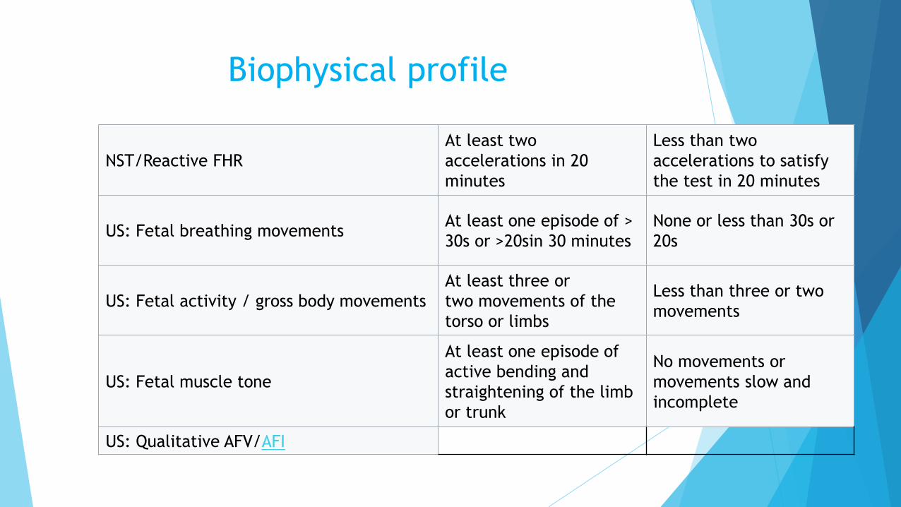
Biophysical profile
NST/Reactive FHR
At least two
accelerations in 20
minutes
Less than two
accelerations to satisfy
the test in 20 minutes
US: Fetal breathing movements At least one episode of >
30s or >20sin 30 minutes
None or less than 30s or
20s
US: Fetal activity / gross body movements
At least three or
two movements of the
torso or limbs
Less than three or two
movements
US: Fetal muscle tone
At least one episode of
active bending and
straightening of the limb
or trunk
No movements or
movements slow and
incomplete
US: Qualitative AFV/AFI

THROMBOCYTOPENIA
Immune thrombocytopenia (ITP)
Drug-induced thrombocytopenia
Preeclampsia
HELLP syndrome
Disseminated intravascular coagulation
Acquired, autoimmune thrombotic thrombocytopenic purpura (TTP)
Hereditary TTP
Complement-mediated thrombotic microangiopathy (C-TMA)
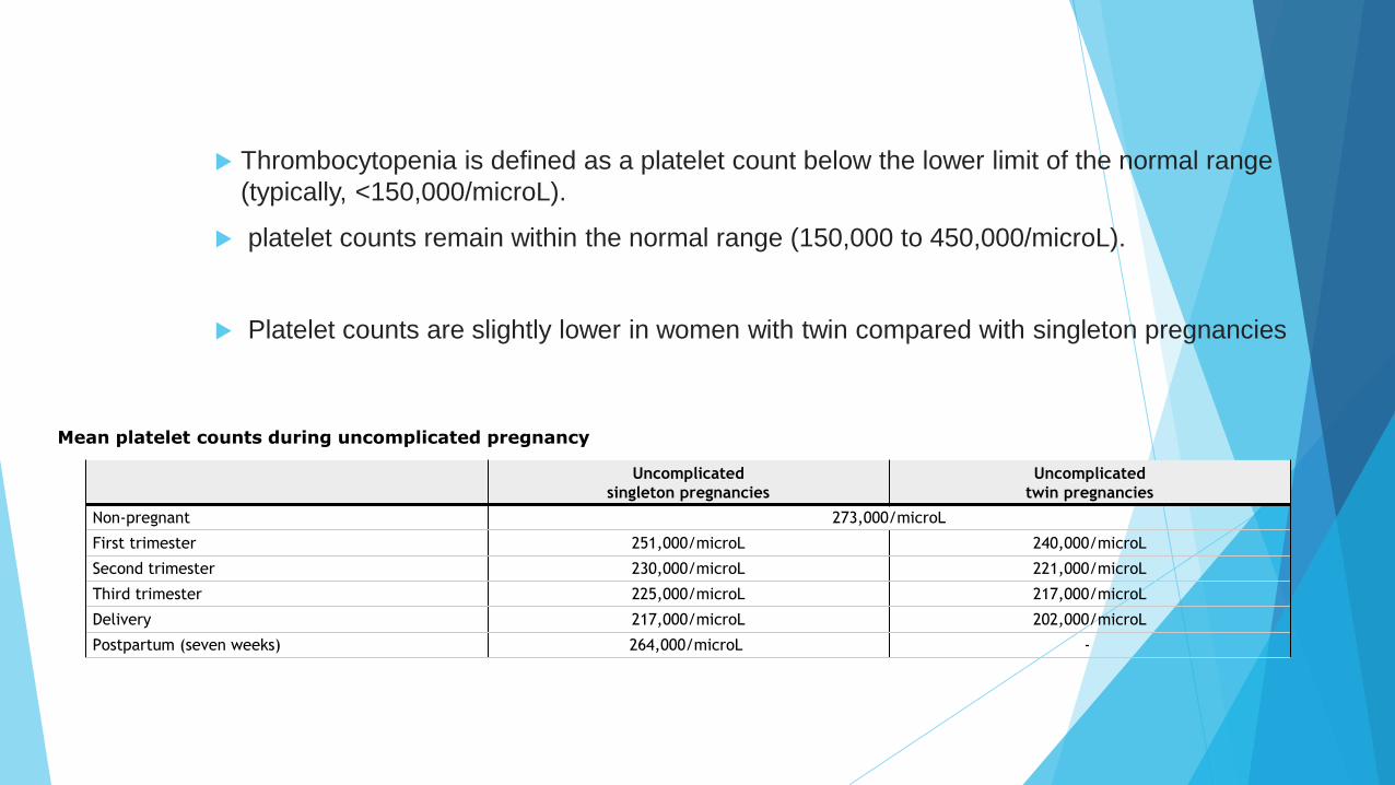
Thrombocytopenia is defined as a platelet count below the lower limit of the normal range
(typically, <150,000/microL).
platelet counts remain within the normal range (150,000 to 450,000/microL).
Platelet counts are slightly lower in women with twin compared with singleton pregnancies
Uncomplicated
singleton pregnancies
Uncomplicated
twin pregnancies
Non-pregnant 273,000/microL
First trimester 251,000/microL 240,000/microL
Second trimester 230,000/microL 221,000/microL
Third trimester 225,000/microL 217,000/microL
Delivery 217,000/microL 202,000/microL
Postpartum (seven weeks) 264,000/microL -
Mean platelet counts during uncomplicated pregnancy
Gestational thrombocytopenia (GT)
Also called incidental thrombocytopenia of pregnancy Benign
self-limited
requires no additional evaluation or treatment
GT accounts for the vast majority of cases of thrombocytopenia discovered during pregnancy
GT may occur during the first trimester, but it becomes more common as gestation progresses
the highest frequency at the time of delivery, when the frequency is 5 to 10 percent
Most common at delivery, but can occur at any time during pregnancy.
Mild thrombocytopenia. (In 99 percent of women, the platelet count is ≥100,000 /microL.)
No increased bleeding or bruising.
No associated abnormalities on complete blood count (CBC).
No fetal or neonatal thrombocytopenia.
The mechanism(s) of GTp has not been documented, but it may be assumed to be
a physiologic adaptation of pregnancy related to the increased plasma volume,
pooling or consumption of platelets in the placenta
The placenta has many vascular characteristics in common with the spleen, a major
site of physiologic platelet sequestration
GT is a diagnosis of exclusion. The diagnosis of GT is accepted if the woman has
mild thrombocytopenia (platelet count 100,000 to 150,000/microL), especially during
late pregnancy and at delivery, with no other associated findings on CBC or physical
examination
GT resolves postpartum, usually
A history of mild thrombocytopenia during a previous pregnancy
supports the diagnosis of GT because the risk of recurrent GT is 14-fold
greater
GT requires no treatment and no change of normal prenatal care and
management of delivery.
No diagnostic testing is necessary because a platelet
count >100,000/microL causes no risk for the mother or the fetus.
Immune thrombocytopenia (ITP)
1 -3 in 10,000 pregnancies
only a minimal number have platelet counts <50,000/microL
This is approximately 10-fold greater than ITP in the general population,
ITP may occur during any trimester or the diagnosis may be known prior to the pregnancy.
The severity of thrombocytopenia is variable and may change during the pregnancy.
Most deliveries were vaginal, and one-fourth of the infants had thrombocytopenia.
ITP is an autoimmune condition in which antiplatelet autoantibodies interfere with platelet production and cause
destruction of circulating platelets.
The diagnosis of ITP is based only on the exclusion of other causes of thrombocytopenia. Therefore, in a
pregnant woman with mild thrombocytopenia (platelet count 100,000 to 150,000/microL), GT and ITP cannot
be distinguished.
The diagnosis of GT is much more likely than ITP in such patients because the frequency of GT is 100-fold
greater than the frequency of ITP during pregnancy.
Preeclampsia with severe
features/HELLP "HELLP syndrome" hemolytic anemia, elevated liver function tests, and low platelet count)
Both terms ("preeclampsia with severe features" and "HELLP") describe a pregnant woman
who is acutely ill with thrombocytopenia and who requires delivery to halt the disease
process.
There is substantial overlap between these syndromes, but it is possible to have HELLP
without hypertension and it is also possible to have preeclampsia with severe features
without all of the manifestations of HELLP.
Preeclampsia –5 % of pregnant women.
Preeclampsia is associated with thrombocytopenia in approximately 15 % , and with severe
thrombocytopenia (platelet count <50,000/microL) in under 5 %, with the likelihood of
thrombocytopenia correlating with the severity of disease.
Disseminated intravascular coagulation (DIC)
Is a systemic process in which coagulation and fibrinolysis become activated within the
vasculature, often massively.
This can lead to depletion of clotting factors and platelets
severe bleeding and/or diffuse oozing
as well as increased risk of thrombosis.
There is always an underlying cause that initiates systemic
activation of the clotting cascade.
Causes of DIC in pregnancy include
abruptio placentae
retained dead fetus
amniotic fluid embolism
septic abortion
others.
Management of DIC involves identifying and treating the underlying cause
Transfusions may be needed while bleeding is being controlled.
Acute fatty liver of pregnancy
Acute fatty liver of pregnancy (AFLP) is a form of liver injury that typically
occurs in the third trimester.
The major clinical findings relate to fatty infiltration of the liver and include
nausea, vomiting, and abdominal pain.
The platelet count may be decreased.
If liver function is severely impaired, the PT and aPTT will be prolonged,
and the fibrinogen may be low.

TTP Thrombotic thrombocytopenic purpura
(TTP)
A significant proportion of patients with hereditary TTP have their first presentation of disease
during pregnancy,
acquired TTP is more common than hereditary TTP and thus more likely in a pregnant patient
without a family history of TTP.
Features suggestive of TTP include
Thrombocytopenia and schistocytes combined with severe neurologic findings (although
half of patients with TTP have no or only minor neurologic abnormalities)
Absence of features of DIC (eg, absence of coagulation abnormalities).
TTP can occur during any trimester or postpartum.
DETERMINING THE LIKELY CAUSE(S)
our approach to the evaluation takes into account the severity of thrombocytopenia, clinical presentation, and trimester
Helpful information includes the following:
Course of the pregnancy so far, including presence or absence of complications
Symptoms of infection such as fever and chills
New daily medications within the past three weeks, or occasional medications taken immediately before symptoms occurred
Personal or family history of excessive bleeding, bruising, pregnancy complications, or known thrombotic microangiopathy (TMA) syndrome
Systemic lupus erythematosus (SLE) or other autoimmune disorder
History of liver disease
Timing of the drop in platelet count (which trimester, how rapidly)
Presence of anemia more severe than expected for the stage of pregnancy
Abnormalities of the peripheral blood smear, such as abnormal white blood cells or nucleated red blood cells
Treatment of bleeding or severe thrombocytopenia
The risk of severe bleeding due to thrombocytopenia only increases substantially with
platelet counts below 50,000/microL.
For women with platelet counts of 50,000 to 100,000/microL, increased bleeding may occur
with invasive procedures, but will not occur spontaneously.
For women with platelet counts <50,000 and severe bleeding (bleeding into a closed space,
bleeding requiring transfusion, bleeding that will not stop) or bleeding that is expected to
become severe, platelet transfusion should be given immediately, regardless of the
underlying cause of thrombocytopenia.
Platelet transfusions are not appropriate for women without active bleeding, unless
surgery and/or delivery is imminent.
The platelet count threshold for a non-bleeding pregnant woman nearing delivery or a
procedure depends on the expected mode of delivery or type of procedure. In the
absence of bleeding, we use the following thresholds:
Vaginal delivery – Transfuse to a platelet count of 30,000/microL
Cesarean delivery – Transfuse to a platelet count of 50,000/microL
Need for urgent/emergency delivery
Conditions treated by delivery — Thrombocytopenic conditions that are
treated by delivery include the following, management of which is discussed in
detail separately:
Preeclampsia with severe features or HELLP syndrome
Disseminated intravascular coagulation (DIC) (when due
to retained dead fetus or intra-amniotic infection)

Conditions not treated by delivery — Conditions that are not treated by
delivery include:
Thrombotic thrombocytopenic purpura (TTP)
Complement-mediated thrombotic microangiopathy (C-TMA)
Drug-induced thrombocytopenia
DIC (when due to a non-obstetric cause such as malignancy or
extrauterine infection)
SUMMARY AND RECOMMENDATIONS
Platelet counts decrease 15 to 20 percent during the course of uncomplicated
pregnancies
Mild thrombocytopenia (platelet count 100,000 to 150,000/microL) is most often due to
gestational thrombocytopenia (GT) and does not require further evaluation
GT is by far the most common cause of thrombocytopenia in pregnancy and is the
presumptive diagnosis in a women with a platelet count between 100,000
and 149,000/microL, provided there are no other abnormal findings. GT is a diagnosis
of exclusion; it is a benign, physiologic condition seen in 5 to 10 percent of pregnant
women that requires no evaluation or treatment.
Platelet counts <100,000/microL occur in only 1 percent of women with GT
Immune thrombocytopenia (ITP) is an autoimmune condition in which
autoantibodies interfere with platelet production and cause destruction of
circulating platelets. ITP can precede pregnancy or can occur at any stage of
the pregnancy or postpartum. ITP is a diagnosis of exclusion.
The majority of pregnant women with relatively mild or incidentally discovered
thrombocytopenia (platelet count between 100,000 and 150,000/microL)
without other cytopenias or major clinical findings will have GT. It is not possible
or necessary to distinguish GT from mild ITP because both are diagnoses of
exclusion and neither requires therapy.
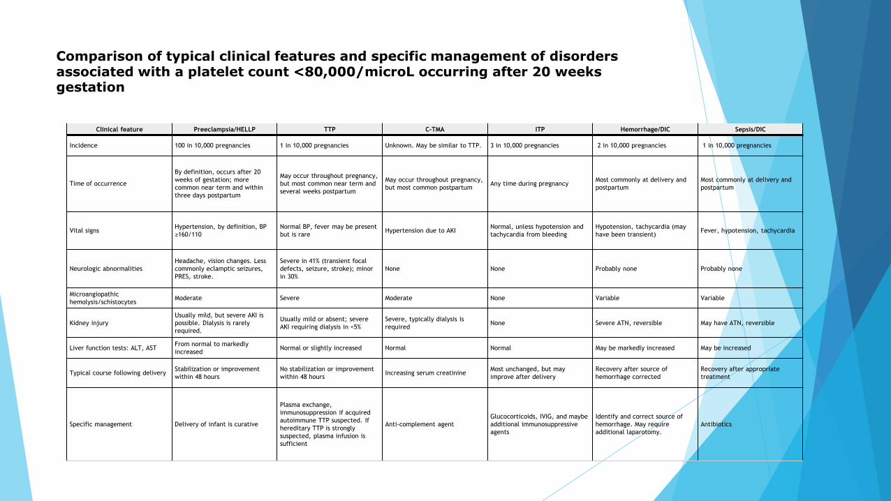
Comparison of typical clinical features and specific management of disorders associated with a platelet count <80,000/microL occurring after 20 weeks gestation
Clinical feature Preeclampsia/HELLP TTP C-TMA ITP Hemorrhage/DIC Sepsis/DIC
Incidence 100 in 10,000 pregnancies 1 in 10,000 pregnancies Unknown. May be similar to TTP. 3 in 10,000 pregnancies 2 in 10,000 pregnancies 1 in 10,000 pregnancies
Time of occurrence
By definition, occurs after 20
weeks of gestation; more
common near term and within
three days postpartum
May occur throughout pregnancy,
but most common near term and
several weeks postpartum
May occur throughout pregnancy,
but most common postpartum Any time during pregnancy
Most commonly at delivery and
postpartum
Most commonly at delivery and
postpartum
Vital signs Hypertension, by definition, BP
≥160/110
Normal BP, fever may be present
but is rare Hypertension due to AKI
Normal, unless hypotension and
tachycardia from bleeding
Hypotension, tachycardia (may
have been transient) Fever, hypotension, tachycardia
Neurologic abnormalities
Headache, vision changes. Less
commonly eclamptic seizures,
PRES, stroke.
Severe in 41% (transient focal
defects, seizure, stroke); minor
in 30%
None None Probably none Probably none
Microangiopathic
hemolysis/schistocytes Moderate Severe Moderate None Variable Variable
Kidney injury
Usually mild, but severe AKI is
possible. Dialysis is rarely
required.
Usually mild or absent; severe
AKI requiring dialysis in <5%
Severe, typically dialysis is
required None Severe ATN, reversible May have ATN, reversible
Liver function tests: ALT, AST From normal to markedly
increased Normal or slightly increased Normal Normal May be markedly increased May be increased
Typical course following delivery Stabilization or improvement
within 48 hours
No stabilization or improvement
within 48 hours Increasing serum creatinine
Most unchanged, but may
improve after delivery
Recovery after source of
hemorrhage corrected
Recovery after appropriate
treatment
Specific management Delivery of infant is curative
Plasma exchange,
immunosuppression if acquired
autoimmune TTP suspected. If
hereditary TTP is strongly
suspected, plasma infusion is
sufficient
Anti-complement agent
Glucocorticoids, IVIG, and maybe
additional immunosuppressive
agents
Identify and correct source of
hemorrhage. May require
additional laparotomy.
Antibiotics