Dhara Gandhi, Susy Albert*, Neeta Pandya - LUeeb.lu.lv/EEB/201112/EEB_9_Gandhi.pdfMorphological and...
9
Morphological and micromorphological characterization of some legume seeds from Gujarat, India Dhara Gandhi, Susy Albert*, Neeta Pandya Department of Botany, Faculty of Science, e Maharaja Sayajirao University of Baroda, Vadodara 390 002, Gujarat, India *Corresponding author, E-mail: drsusyalbert@rediffmail.com, [email protected] Abstract Light and scanning electron microscopical studies on seed morphological and micromorphological features of 17 legume species belonging to three genera (Crotolaria, Alysicarpus and Indigofera), of Faboideae, Fabaceae were examined and described. Seed characteristics, particularly exomorphic features, that are revealed through scanning electron microscopy can be used in resolving problems of systematics of species. Mature seeds of Crotolaria differ from those of Alysicarpus and Indigofera by its prominent kidney shape. Seeds of Alysicarpus and Indigofera are biconvex and shape varies from rectangular spherical oblong to ovoid. However, within the genera, the species differ in size, surface and hilum characteristics. e study showed that the seed coat ornamentation/spermoderm pattern can be helpful in identification of species. Key words: hilum, legume, micromorphology, morphology, scanning electron microscopy, seed surface. Abbreviations: SEM, scanning electron microscopy. Environmental and Experimental Biology (2011) 9: 105–113 Original Paper Introduction Exomorphic features of seeds, in addition to vegetative and reproductive characters, have long been employed as important tools in various scientific studies. However, most of the light microscopic feature used are concerned with general shape and size rather than details of surface ornamentation. Seed morphology has been shown to provide useful characteristics for the analysis of taxonomic relationships in a wide variety of plant families (Esau 1953; Shelter 1986; Takhtajan 1991; Buss et al. 2001, Zhang et al. 2005; Gontchaova et al. 2009). In addition to gross morphology of seeds, sculpturing details of outer seed coat are quite variable between different species and can be of systematic importance. (Chowdhury, Buth 1970; Gohary, Mohammed 2007). Seed characteristics, particularly exomorphic features revealed by means of scanning electron micros- copy (SEM), have been used in resolving problems of systematics of species (Karihaloo, Malik 1994; Koul et al. 2000) and evolutionary relationships (Segarra, Mateu 2002). e importance of ultrastructural pattern analysis of the seed coat observed under the SEM has been well recognised as a reliable approach for assessing phenetic relationship and identification of species or taxa (Barthlott 1981; Tobe et al. 1987; Koul et al. 2000; Yoshizaki 2003; Javadi, Yamaguchi, 2004). e Indian subcontinent is the centre of origin, endemism and diversity of a large number of cultivated legumes. e genus Crotalaria represents the largest legume taxa in India. Crotalaria species are important because of their accumulation of pyrrolizidine alkaloids. Crotalaria juncea, a widely cultivated fibre crop, known as Sunnhemp, Bombay hemp, Madras hemp, Rattle pods, etc. is not known in the wild, and is also used for its food and medicinal values by ethnic communities. Several other species of Crotalaria are economically important for fibre, forage/animal feed, green manure and for medicinal purpose (Wealth of India 1950; Ambasta et al. 1986; Pandey, Gupta 2003). Indigofera species are rich in organic and fatty acids, flavonoids such as carotenoids, and coumarins (Yinusa et al. 2007). Indigofera tinctoria is used to produce indigo dyes. Some other species of Indigofera are used for different purposes, for example seeds of Indigofera articulate are used for treatment of toothache. Indigofera oblongifolia, Indigofera suffruticosa, and Indigofera aspalthoides are used as anti-inflammatories for treatment of insect stings, snake bites and swellings, and Indigofera arrecta extract is used to relieve ulcer pain. Alysicarpus is another potential crop legume rich in protein. Alysicarpus ovalifolius, a protein-rich fodder, is a highly palatable feed for livestock grazing in rangelands. It is a valuable component of vegetation collected and traded as fodder in many regions. Alysicarpus vaginalis is known as soil improver, having a good fodder and forage value, and it is also used in treatment of cough. Alysicarpus rugosus seed containes higher amounts of crude protein and crude lipid when compared with most of the commonly consumed pulses (Siddhuraju et al. 1992). Various seed morphological studies of leguminous taxa have been performed from time to time (Sharma et 105
Transcript of Dhara Gandhi, Susy Albert*, Neeta Pandya - LUeeb.lu.lv/EEB/201112/EEB_9_Gandhi.pdfMorphological and...

Morphological and micromorphological characterization of some legume seeds from Gujarat, India
Dhara Gandhi, Susy Albert*, Neeta Pandya
Department of Botany, Faculty of Science, The Maharaja Sayajirao University of Baroda, Vadodara 390 002, Gujarat, India
*Corresponding author, E-mail: [email protected], [email protected]
Abstract
Light and scanning electron microscopical studies on seed morphological and micromorphological features of 17 legume species belonging to three genera (Crotolaria, Alysicarpus and Indigofera), of Faboideae, Fabaceae were examined and described. Seed characteristics, particularly exomorphic features, that are revealed through scanning electron microscopy can be used in resolving problems of systematics of species. Mature seeds of Crotolaria differ from those of Alysicarpus and Indigofera by its prominent kidney shape. Seeds of Alysicarpus and Indigofera are biconvex and shape varies from rectangular spherical oblong to ovoid. However, within the genera, the species differ in size, surface and hilum characteristics. The study showed that the seed coat ornamentation/spermoderm pattern can be helpful in identification of species.
Key words: hilum, legume, micromorphology, morphology, scanning electron microscopy, seed surface.Abbreviations: SEM, scanning electron microscopy.
Environmental and Experimental Biology (2011) 9: 105–113 Original Paper
Introduction
Exomorphic features of seeds, in addition to vegetative and reproductive characters, have long been employed as important tools in various scientific studies. However, most of the light microscopic feature used are concerned with general shape and size rather than details of surface ornamentation.
Seed morphology has been shown to provide useful characteristics for the analysis of taxonomic relationships in a wide variety of plant families (Esau 1953; Shelter 1986; Takhtajan 1991; Buss et al. 2001, Zhang et al. 2005; Gontchaova et al. 2009). In addition to gross morphology of seeds, sculpturing details of outer seed coat are quite variable between different species and can be of systematic importance. (Chowdhury, Buth 1970; Gohary, Mohammed 2007). Seed characteristics, particularly exomorphic features revealed by means of scanning electron micros-copy (SEM), have been used in resolving problems of systematics of species (Karihaloo, Malik 1994; Koul et al. 2000) and evolutionary relationships (Segarra, Mateu 2002). The importance of ultrastructural pattern analysis of the seed coat observed under the SEM has been well recognised as a reliable approach for assessing phenetic relationship and identification of species or taxa (Barthlott 1981; Tobe et al. 1987; Koul et al. 2000; Yoshizaki 2003; Javadi, Yamaguchi, 2004).
The Indian subcontinent is the centre of origin, endemism and diversity of a large number of cultivated legumes. The genus Crotalaria represents the largest legume
taxa in India. Crotalaria species are important because of their accumulation of pyrrolizidine alkaloids. Crotalaria juncea, a widely cultivated fibre crop, known as Sunnhemp, Bombay hemp, Madras hemp, Rattle pods, etc. is not known in the wild, and is also used for its food and medicinal values by ethnic communities. Several other species of Crotalaria are economically important for fibre, forage/animal feed, green manure and for medicinal purpose (Wealth of India 1950; Ambasta et al. 1986; Pandey, Gupta 2003). Indigofera species are rich in organic and fatty acids, flavonoids such as carotenoids, and coumarins (Yinusa et al. 2007). Indigofera tinctoria is used to produce indigo dyes. Some other species of Indigofera are used for different purposes, for example seeds of Indigofera articulate are used for treatment of toothache. Indigofera oblongifolia, Indigofera suffruticosa, and Indigofera aspalthoides are used as anti-inflammatories for treatment of insect stings, snake bites and swellings, and Indigofera arrecta extract is used to relieve ulcer pain. Alysicarpus is another potential crop legume rich in protein. Alysicarpus ovalifolius, a protein-rich fodder, is a highly palatable feed for livestock grazing in rangelands. It is a valuable component of vegetation collected and traded as fodder in many regions. Alysicarpus vaginalis is known as soil improver, having a good fodder and forage value, and it is also used in treatment of cough. Alysicarpus rugosus seed containes higher amounts of crude protein and crude lipid when compared with most of the commonly consumed pulses (Siddhuraju et al. 1992).
Various seed morphological studies of leguminous taxa have been performed from time to time (Sharma et
105

al. 1977; Agarwal 1984; Buth, Narayan,1986; Sahai 1999; Murthy, Sanjappa 2002; Mallick, Sawhney 2003; Salimpour et al. 2007; Al-Ghamdi, Al-Zahrani, 2010). Seed and seed coat anatomy of some members of the Crotolaria (Buth, Narayan 1986) and Indigofera (Agrawal 1984) have been studied. However, in these studies, only a few species of those in the present study were examined.
The present work has been undertaken to delinate specific variation of micromorphological characteristics in seeds of some legume species growing in association with grasses in the grasslands of the Baria and Godhra forest division in Gujarat.
Materials and methods
Seed characters of 17 species belonging to Fabaceae from three genera (Crotolaria, Alysicarpus and Indigofera) were studied using freshly collected mature seeds (Table 1). Plant specimens with mature pods were collected from different grasslands and forest areas of Baria and Godhra in Gujarat. The plant specimens were authentically identified at ‘The Blatter Herbarium’, St. Xavier’s College, Mumbai. About 10 to 15 mature seeds of each taxon, procured by cleaning and manually separating from the pods, were used for the light and scanning electron microscopic studies. For light microscopic studies, mature, dry seeds were thoroughly cleaned with alcohol to avoid any alteration in the micromorphological features and examined for diagnostic features of shape, size, colour and size. About 10 seeds were examined for dimensional details.
Micromorphological features and hilum characteristics were examined under SEM at the Metallurgy Department, Faculty of Technology and Engineering, The Maharaja Sayajirao University of Baroda, and photographed at different magnifications. Seed samples were washed with absolute alcohol or acetone for 1 to 2 min to remove any debris present. They were further subjected to ultrasonic cleaning by changing absolute alcohol repeatedly and then directly mounting over carbon conducting tape mounted on brass stubs. For evaluation of uniformity, seeds were placed on the stub with their dorsal, ventral and lateral side upwards so that characteristic features of all the different sides could be scanned and photographed using JEOL JEM - 5610 SEM. To achieve better resolution the accelerating voltage varied up to 15 kV.
Results and discussion
The study of epidermal surfaces revealed a number of important micro morphological characters, which exhibited interesting interspecific variation that was of significance for identification (Fig. 1 to 4). In the present work both light microscopic and scanning electron microscopic studies were used which complemented each other in obtaining a perfect differentiation between species.
Morphology of the seeds varied significantly in size, shape, colour, surface and hilum colour. Seeds of Crotolaria were characteristically kidney or bean shaped, compared to oblong to rectangular seeds of Indigofera.
Seed colour appeared to be of less diagnostic and system value. Presence of a cracked surface was a common feature noted in many of the legume seeds (Table 2). Seed coat pattern could be categorized into smooth, cracked and papillate. Except Crotolaria spectabilis and Corotolaria albida, all other species of Corotolaria had a smooth surface. Among the four different species of Indigofera, Indigofera tinctoria seeds were rectangular while all of the other species were ovoid. The hilar region is characteristic, as in all Papilionaceous seeds, with a very specialized organisation. Seeds of Crotolaria are characterised by a lateral notch formed between the radical tip and the cotyledon, which is the seat of hilum.
In all of the species of Indigofera, hilum was present in the center of the seed and was spherical in shape. A common feature observed by SEM was a pitted structure present on the surface. In Alysicarpus, the hilum was located slightly away from the center, towards the distal part of seed, which gave a dumbbell shaped appearance to the seed when viewed laterally. Indigofera tinctoria differed from Indigofera linifolia by having a smooth surface. Indigofera cordifolia and Indigofera echinata had a pitted surface.
According to Skvortsov and Rusanovitch (1974) the spermaoderm characteristics are genetically determined and are the main source of intra- or interspecific variation. Lersten (1981) stated that the spermaoderm pattern reflects
D. Gandhi, S. Albert, N. Pandya
106
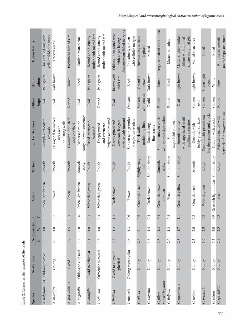
Table 1. List of legume species studied
No. Botanical name Blatter Herbarium No.1 Alysicarpus bulgaumensis Wt. 138802 Alysicarpus monilifer (L.) DC. 143513 Alysicarpus procumbens (Roxb.) Schindl 138694 Alysicarpus vaginalis (L.) DC. –5 Indigofera cordifolia B.Heyne ex Roth. –6 Indigofera echinata Willd. 198657 Indigofera linifolia (L.f.) Retz 199998 Indigofera tinctoria L. 202129 Crotalaria albida Roth. 1639210 Crotolaria calycina Schrank 1643411 Crotolaria filipes var. trichophora 16463 (Bth. ex. Baker) Cooke 12 Crotalaria linifolia L. f. 1420113 Crotolaria mysorensis Roth. 1659614 Crotolaria notonii W. & A. Prodr. 1704015 Crotolaria orixensis Rottler ex Willd. 1710116 Crotolaria retusa L. 1726117 Crotolaria spectabilis Roth. 17264
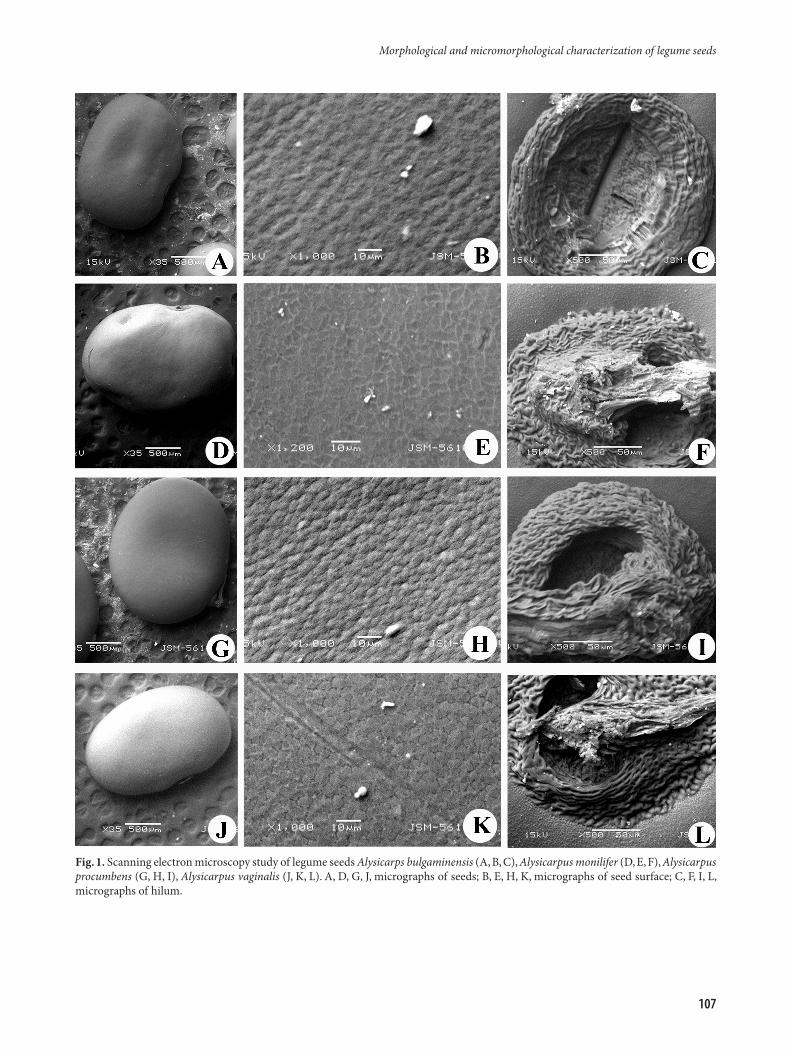
Fig. 1. Scanning electron microscopy study of legume seeds Alysicarps bulgaminensis (A, B, C), Alysicarpus monilifer (D, E, F), Alysicarpus procumbens (G, H, I), Alysicarpus vaginalis (J, K, L). A, D, G, J, micrographs of seeds; B, E, H, K, micrographs of seed surface; C, F, I, L, micrographs of hilum.
Morphological and micromorphological characterization of legume seeds
107

Fig. 2. Scanning electron microscopy study of legume seeds Indigofera cordifolia (A, B, C), Indigofera echinata (D, E, F), Indigofera linifolia (G, H, I), Indigofera tinctoria (J, K, L). A, D, G, J, micrographs of seeds; B, E, H, K, micrographs of seed surface; C, F, I, L, micrographs of hilum.
D. Gandhi, S. Albert, N. Pandya
108
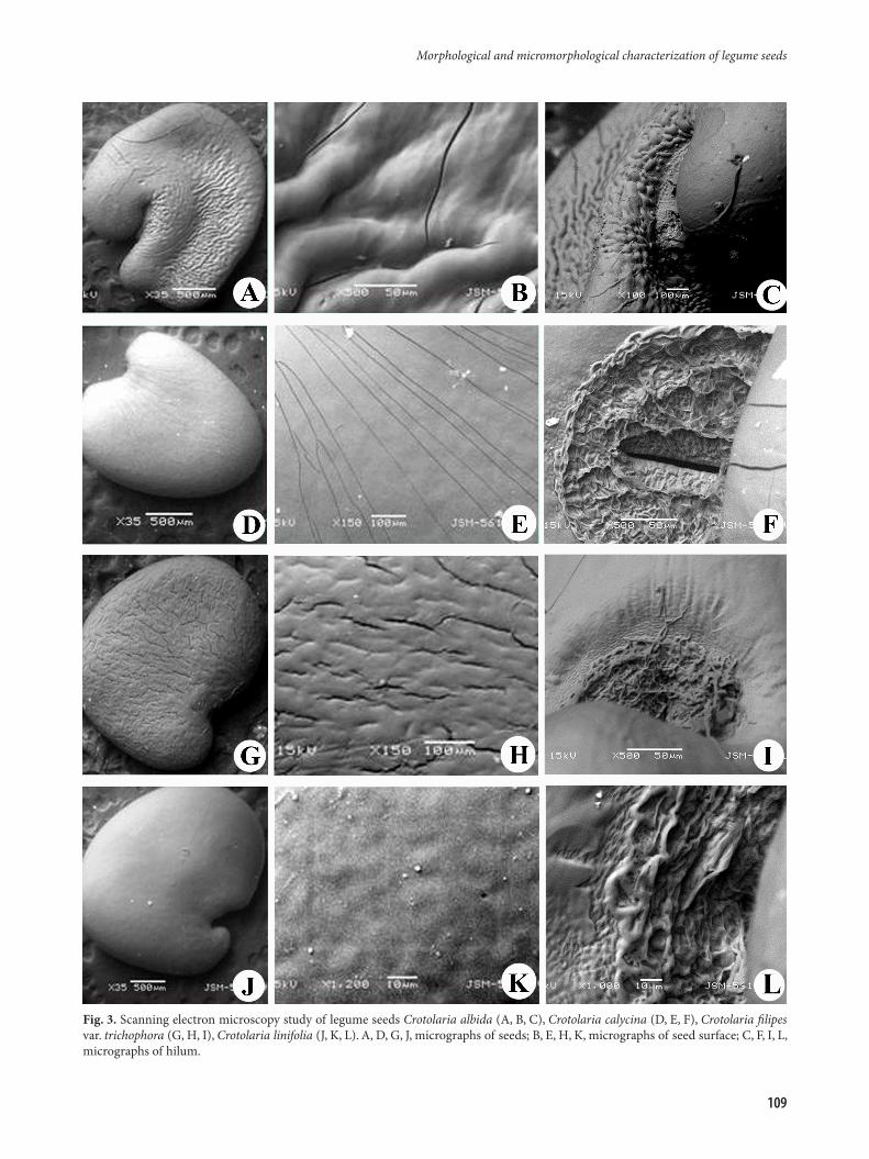
Fig. 3. Scanning electron microscopy study of legume seeds Crotolaria albida (A, B, C), Crotolaria calycina (D, E, F), Crotolaria filipes var. trichophora (G, H, I), Crotolaria linifolia (J, K, L). A, D, G, J, micrographs of seeds; B, E, H, K, micrographs of seed surface; C, F, I, L, micrographs of hilum.
Morphological and micromorphological characterization of legume seeds
109

Fig. 4. Scanning electron microscopy study of legume seeds Crotolaria mysorensis (A, B, C), Crotolaria notonii (D, E, F), Crotolaria orixensis (G, H, I), Crotolaria retusa (J, K, L), Crotolaria spectabilis (M, N, O). A, D, G, J, M, micrographs of seeds; B, E, H, K, N, micrographs of seed surface; C, F, I, L, O, micrographs of hilum.
D. Gandhi, S. Albert, N. Pandya
110
Morphological and micromorphological characterization of legume seeds
111
Tabl
e 2.
Cha
ract
erist
ic fe
atur
es o
f the
seed
s
Spec
ies
Seed
shap
e Se
ed si
ze (m
m)
Col
our
Text
ure
Surf
ace
feat
ures
H
ilum
H
ilum
H
ilum
feat
ure
L W
T
shap
e co
lour
A. b
ulga
umen
sis
Obl
ong
to o
void
1.
3 1.
0 0.
7 G
reen
ligh
t bro
wn
Smoo
th
Com
plex
retic
ulat
e O
val
Pale
gre
en
Thic
k w
alle
d ri
m, w
ith
netw
ork
cells
in fo
lded
man
ner
A. m
onili
fer
Obl
ong
2.0
1.0
0.7
Brow
n Sm
ooth
H
exag
onal
to u
neve
n O
val
Dar
k br
own
Une
ven
area
s
area
s with
undu
latin
g w
alls
A. p
rocu
mbe
ns
Ovo
id
1.9
1.5
0.3
Gre
en
Smoo
th
Gra
nula
r und
ulat
ed
Roun
d Br
own
Prom
inen
t mat
ted
rim
an
d kn
obbe
dA
. vag
inal
is O
blon
g to
elli
psoi
d 1.
2 0.
8 0.
6 G
reen
ligh
t bro
wn
Smoo
th,
Disp
erse
d ro
und
O
val
Blac
k Su
nken
mat
ted
rim
shin
y ro
ugh
unev
en st
ruct
ures
I. co
rdifo
lia
Ovo
id to
orb
icul
ar
1.1
1.0
0.3
Whi
te d
ull g
reen
Ro
ugh
Pitte
d- v
erru
cate
, O
val
Pale
gre
en
Roun
d an
d di
stin
ctly
roun
ded
sunk
en w
ith m
atte
d ri
mI.
echi
nata
O
rbic
ular
to ro
und
1.3
1.0
0.4
Whi
te d
ull g
reen
Ro
ugh
Dee
ply
pitte
d
Roun
d Pa
le g
reen
Ro
und
and
dist
inct
ly
and
verr
ucat
e,
sunk
en w
ith m
atte
d ri
m
levi
gate
with
mou
ndI.
linifo
lia
Ovo
id to
elli
psoi
d,
1.2
1.2
1.2
Dar
k br
own
Roug
h D
eepl
y pi
tted
and
Ova
l Br
own
with
O
blon
g- h
exag
onal
are
as
sp
heri
cal
ve
rruc
ate,
levi
gate
thic
k ri
m
with
edg
es h
avin
g
surf
ace
with
mou
nd
disc
ontin
uous
lifte
d m
argi
nsI.
tinct
oria
O
blon
g-re
ctan
gula
r 1.
6 1.
3 0.
9 Br
own
Roug
h Su
nken
with
gra
nula
r O
bova
te
Blac
k In
dist
inct
ly su
nken
m
argi
n/tu
berc
ulat
e
w
ith c
ellu
lar m
argi
nC.
alb
ida
Kid
ney
2.
6 2.
1 0.
5 G
reen
ish b
lack
Sl
ight
ly ro
ugh,
Th
ick
wal
led
Sunk
en
Gre
enish
Su
rrou
ndin
g su
rfac
e
du
ll un
dula
ting
lines
w
ith c
rack
s br
own
is pi
tted
C. ca
lyci
na
Kid
ney
1.6
1.4
0.4
Dar
k br
own
Smoo
th, s
hiny
Sm
ooth
, lon
g O
void
D
ark
brow
n M
atte
d
line
crac
ksC.
filip
es
Kid
ney
1.8
1.3
0.4
Gre
enish
bro
wn
Smoo
th,
Smoo
th, u
neve
n cr
acks
Ro
und
Brow
n Ir
regu
lar m
atte
d an
d su
nken
var.
trich
opho
ra
to b
row
n sh
iny
with
min
ute
depr
essio
nsC.
lini
folia
K
idne
y 2.
0 1.
3 0.
4 Bl
ack
Smoo
th, s
hiny
Sm
ooth
, Ro
und
Blac
k M
atte
d re
ticul
ate
su
perfi
cial
pits
seen
C. m
ysor
ensis
K
idne
y 2.
8 1.
7 1.
1 G
reen
ish y
ello
w
Smoo
th, s
hiny
Sm
ooth
surf
ace
O
val
Ligh
t bro
wn
Mat
ted
sligh
tly su
nken
w
ith su
perfi
cial
smal
l
hi
lum
with
upl
ifted
pa
pilla
te st
ruct
ures
sq
uare
mar
gine
d pi
tsC.
not
onii
Kid
ney
2.3
2.0
0.3
Gra
yish
bla
ck
Smoo
th
Smoo
th, w
ith
Sunk
en
Ligh
t bro
wn
Mat
ted
stru
ctur
es
feeb
ly p
itted
surf
ace
oval
C. o
rixe
nsis
Kid
ney
3.0
2.5
0.8
Whi
tish
gree
n Ro
ugh
Smoo
th w
ith p
rom
inen
t Su
nken
G
reen
-ligh
t Pi
tted
lin
e de
pres
sions
and
cra
cks
br
own
C. re
tusa
K
idne
y 1.
5 3.
0 0.
9 G
reen
ish li
ght b
row
n Sm
ooth
, shi
ny
Smoo
th, w
ith c
rack
s Su
nken
W
hite
Pi
tted
C. sp
ecta
bilis
K
idne
y 2.
9 2.
3 0.
3 Bl
ack
Roug
h Re
ticul
ate
mat
ted
with
Ro
und
Brow
n Ri
m p
osse
s sm
ooth
ne
twor
k of
inte
rwov
en ru
gae
mat
ted
type
stru
ctur
es

epidermal configuration and cuticular deposition as influenced by seed expansion. Gutterman and Heydecker (1973) demonstrated that day length affects seed coat structure while Sharma et al. (1977) concluded that edaphic factors are responsible for the difference. In studies on Indigofera pseudo-tinctoria, Agrawal (1984) concluded the spermoderm pattern was similar but that the density of the ornamentation varied. Such a difference appears to be due to varying amounts of surface deposition. Our observations of seed surface patterns in Indigofera linifolia and Indigofera tinctoria confirmed the findings of Agrawal (1984) and Murthy (2002).
The present study supports the use of seed coat patterns as features for species identification. The seeds display diversity in shape, dimensions and seed coat surface. The SEM study revealed seed coat remarkable topographic diversity among different species, to be characteristic of each species. This kind of study with more species may help to open a frame work of our knowledge about interspecific relationships in the genus. The present study provided some useful characters of seed for infrageneric classification and also for delimiting species. Light microscopic features supplemented with SEM proved to be a great tool to achieve more accurate seed identification, as previously suggested by Brisson and Peterson (1976). This method can be used as a routine technique in the study of spermoderm morphology (Heywood 1971; Barthlott 1984).
Acknowledgements
We thank Dr. M.N. Patel, Head of Faculty of Technology and Engineering, The Maharaja Sayajirao University of Baroda, Vadodara, Gujarat for providing facilities and support in the SEM studies; The Conservator of Forests, Working Plan Circle, Forest Department, Vadodara, Gujarat state for providing financial support; Dr. (Mrs.) U.C. Bapat for providing access to herbarium specimens to confirm the identity of the specimen.
References
Agarwal S. 1984. Seed structure in some Indigofera species. J. Indian Bot. Soc. 63: 11–19.
Al-Ghamdi F.A., Al-Zahrani R.M. 2010 Seed morphology of some species of Tephrosia Pers. (Fabaceae) from Saudi Arabia. Identification of species and systematic significance. Feddes Repertorium 121: 59–65.
Ambasta S.P., Ramachandran K., Kashyapa K., Chand R. (eds). 1986. Useful plants of India. Publications and Information Directorate. Council Scientific and Industrial Research, New Delhi, pp. 146–147.
Barthlott W. 1981. Epidermal and seed surface characters of plants: systematic applicability and some evolutionary aspects. Nordic J. Bot. 1: 345–355.
Barthlott W. 1984. Microstructural features of seed surfaces. In Heywood V.H., Moore D.M. (eds.), Current Concepts in Plant Taxonomy. Academic Press, London. 488 p.
Buss C.C., Lammers T.G., Wise R.R. 2001. Seed coat morphology and its systematic implications in Cyanea and other genera of
Lobelioideae (Campanulaceae). Amer. J. Bot. 88: 1301–1308.Buth G.M., Narayan A. 1986. Seed and seed coat anatomy of some
members of Crotolaria (Papilionaceae). J. Indian Bot. Soc. 66: 317–324.
Chowdhury K.A., Buth G.M. 1970 Seed coat structure and anatomy of Indian Pulses. J. Linn. Soc. Bot. 63: 169–179.
Esau K. 1953. Anatomy of Seed Plants. John Wiley and Sons, New York.
Gohary I., Mohammed A.H. 2007. Seed morphology of Acacia in Egypt and its taxonomic significance. Int. J Agric. Biol. 9: 435–438.
Gontcharova S.B., Gontcharova A.A., Yakubov V.V., Kondo K. 2009. Seed surface morphology in some representatives of the genus Rhodiola sect. Rhodiola (Crassulacea) in Russian Far East. Flora 204: 17–24.
Heydecker W. 1973. Germination of an idea the priming of seeds. University of Nottingham School of Agriculture Report 1973/1974. 50-67.
Heywood V.H. 1971. The characteristics of the scanning electron microcopes and their importance in biological studies. In Heywood V.H. (ed) Scanning Electron Microscopy: Systematic and Evolutionary Applications. Academic Press, London.
Javadi F., Yamaguchi H. 2004. A note on seed coat and plumule morphological variation in the genus Cicer (Fabaceae). Sci. Rep. Grad. Sch. Agric. Biol. Sci. 56: 7–16.
Karihaloo J.L., Malik, S.K. 1994. Systematic relationships among some Solanum L. sect. melongana L. Evidence from seed characters. Indian J. Plant Genet. Resour. 7: 13–21.
Koul K.K., Ranjna N., Raina S. N. 2000. Seed coat microsculpturing in Brassica and allied genera (Subtribe Brassicinae, Raphanine, Moricandiinae). Ann. Bot. 86: 385–397.
Lersten N.R. 1981. Testa topography in Leguminosae, subfamily Papilionoideae. Proc. Iowa Acad. Sci. 88: 180–191.
Mallick D.K., Sawhney S. 2003. Seed coat ornamentation in wild and cultivated lentil taxa. Phytomorphology 53: 187–195.
Murthy G.V.S., Sanjappa M. 2002. SEM studies on seed morphology of Indigofera L. (Fabaceae) and its taxonomic utility. Rheedea 12: 21–51.
Pandey A., Gupta R. 2003. Fibre yielding plants of India: genetic resources, perspective for collection and utilization. Nat. Prod. Radiance 2:194–204.
Sahai K. 1999. Structural diversity in the lens of the seeds of some Cassia L. (Caesalpinioideae) species and its taxonomic significance. Phytomorphology. 49: 203–208.
Salimpour F., Mostafavi G., Sharifnia F. 2007. Micromorphologic study of the seed of the genus Trifolium, section Lotoidea, in Iran. Pak. J. Biol. Sci. 10: 378–382.
Segarra J.G., Mateu I. 2002. Seed morphology of Linaria species from Eastern Spain: identification of species and taxonomic implications. Bot. J. Linn. Soc. 135: 375–389.
Sharma S.K., Babu C.R., Johri B.M., Hepworth A. 1977. SEM studies on seed coat pattern in Phaseolous mungo, P. radiatus-sublobatus. Phytomorphology 27:106–111.
Shelter S.J. 1986. Seed morphology in North American Campanulaceae. Ann. Missouri. Bot. Gardens 73: 653–688.
Siddhuraju P., Viajayakumari K., Janardhanan K. 1992. The biochemical composition and nutritional potential of the tribal pulse, Alysicarpus rugosus (Wild.) DC. Food Chem. 45: 251–255.
Skvortsov A.K., Rusanovitch I.I. 1974. Scanning electron microscopy of the seed-coat surface in Epilobium species. Bot. Notes 127: 392–401.
D. Gandhi, S. Albert, N. Pandya
112

Received 20 September 2011; received in revised form 24 October 2011; accepted 10 November 2011
Takhtajan A. 1991. Evolutionary Trends in Flowering Plants. Columbia Univ. Press, New York.
Tobe H., Wagner W.L., Chin H.C. 1987. A systematic and evolutionary study of Oenothera (Onagraceae): Seed coat anatomy. Bot. Gaz. 148: 235–257.
Wealth of India. 1950. The Wealth of India – Raw Materials. Vol. II. Publications and Information Directorate, Council of Scientific and Industrial Research, New Delhi, pp. 372–383.
Yoshizaki M. 2003. Millets in prehistoric remain: Paleobotany
on barnyard millets and azuki beans in Japan. In Yamaguchi H., Kawase M. (eds) Natural History of Millets. Hokaido University Press, Sapporo.
Yinusa I., Ndukwe I.G., Amupitan J.O. 2007. Phytochemical and antimicrobial screening of aerial part of Indigofera pulchra. Chem. Class Journal, CSN Zaria. pp. 162.
Zhang Z.-Y., Yang D.-Z., Lu A.-M., Knapp S. 2005. Seed morphology of the tribe Hyoscyameae (Soloanaceae). Taxon 54: 71–83.
Morphological and micromorphological characterization of legume seeds
113